Article

Micropropagation and Acclimatization of Gymnocalycium cv. Fancy (Cactaceae): Developmental Responses to Different Explant Types and Hormone Conditions

by
Carles Cortés-Olmos
,
Vladimir Marín Guerra-Sandoval
,
Vicente Blanca-Giménez
and
Adrián Rodríguez-Burruezo
*
Instituto Universitario de Conservación y Mejora de la Agrodiversidad Valenciana (COMAV), Universitat Politècnica de València (UPV), Camino de Vera s/n, 46022 Valencia, Spain
*
Author to whom correspondence should be addressed.
Plants 2023, 12(23), 3932; https://doi.org/10.3390/plants12233932
Submission received: 11 October 2023 / Revised: 18 November 2023 / Accepted: 20 November 2023 / Published: 22 November 2023
(This article belongs to the Section Horticultural Science and Ornamental Plants)

Abstract

:
The Gymnocalycium genus includes numerous highly valued species in the ornamental plant market and their propagation is usually carried out using traditional methods. However, there is a lack of information regarding the efficiency of micropropagation through in vitro tissue culture techniques on these species. So, with the objective of establishing an efficient micropropagation protocol that allows for optimizing the plant obtaining processes, the morphogenic potential of Gymnocalycium cv. Fancy has been studied in this work. For this purpose, plants of two different sizes (medium and large) were used as the starting material, from which three types of explants were obtained (apex, central discs and bases). The effect of three plant growth regulators (6-Benzylaminopurine, BAP; Kinetin, KIN; and Thidiazuron, TDZ) at three different concentrations each were studied, and the number of generated shoots, the frequency of appearance of callogenesis and rhizogenesis by explant and condition were assessed. An efficient protocol based on the use of KIN at 4 µM and central discs as a starting explant was developed. Moreover, the obtained sprouts rooted successfully (especially using BAP at 2 µM), and their subsequent acclimatization was very effective. Furthermore, emergence of a new morphotype is presented, that has not previously been described.

1. Introduction

The genus Gymnocalycium (Cactaceae) encompasses a plethora of cactus species that generally grow in arid and semi-arid environments of South America, especially in Argentina, Bolivia, Brazil, Paraguay and Uruguay [1,2,3,4,5]. Their wide distribution area contributes to their ecological range and genetic diversity, a reason for which Gymnocalycium spp. exhibits a diverse array of botanical features and specific ecological adaptations, found in 50 and 80 different species, in addition to a range of varieties and different forms that can be found intra-specifically [6]. Gymnocalycium species typically exhibit a low-growing, globular or cylindrical stem morphology. Their stems are often solitary, although some species may form small clusters (Figure 1a). Also, their spines may differ in size, color and arrangement across species (Figure 1b). In fact, some Gymnocalycium species bear long, hair-like spines that provide protection against herbivores and harsh sunlight, while others have shorter and stouter spines. The differentiation in spines can often be used as a means of identifying different species. Gymnocalycium spp. are renowned for their showy, often brightly colored flowers. These flowers can be solitary or produced in clusters at the apex of the stem (Figure 1c). The flower shapes, sizes and colors vary widely among species, making them particularly appealing to pollinators [1,7,8].
Moreover, this genus garnered substantial recognition and economic importance in the ornamental plant market due to a combination of aesthetic appeal, adaptability, resilience and cultural value [9,10]. Their diverse stem shapes, striking spinations and showy flowers make them desirable choices for both novice plant enthusiasts and experienced cactus hobbyists [10]. Furthermore, they require minimal care, can adapt to different climates and soil types and have the potential for long lifespans, making them interesting for a broad spectrum of gardening and landscaping projects too [9]. In fact, over the past few decades, the demand for ornamental cacti has increased [11].
In this regard, within the global cactus market, it is estimated that around 1.5 million Gymnocalycium plants were sold internationally during 2021 [12], with Gymnocalycium mihanovichii (Frič and Gürke) Britton and Rose (with more than 1.2 million plants) being the most demanded species. Traditional propagation methods are commonly used in cactus production, including sexual propagation through seeds, asexual propagation through cuttings and clonal propagation through grafting [11,13].These techniques are specially used for cacti produced in mass, aimed at maximizing production under specific conditions and controlled environments.
Nevertheless, within the realm of ornamental plants, Gymnocalycium are not just mass-produced products, they are also prized by collectors. In fact, cactus collectors are drawn to special Gymnocalycium plants and are willing to invest in acquiring and cultivating these different, particular or unique cacti. The pursuit of rare and distinctive cactus varieties by customers has created economic opportunities for nurseries and growers specializing in ornamental cacti, creating a lucrative niche market. However, it is important to highlight that most of these species of cactus have limited adaptability to mass production conditions. Thus, finding alternative propagation methods, such as in vitro propagation, can favor the inclusion of other valuable cactus species in the market. In this context, Gymnocalycium spp. have become important assets to cactus collectors, and refining the methods of mass production can improve the overall ornamental plant market.
For the efficient and rapid production of plants in large quantities, in vitro cultivation can play a fundamental role. Depending on the regenerative capacity of the species, direct organogenesis, indirect organogenesis [14], or axillary bud sprouting [15,16] can be employed, with the latter being the most commonly used regeneration method among cacti [11]. Axillary buds are located in the areoles, which are typical structures of the cactus family. Areolas are meristematic areas that appear as small protuberances from which spines emerge, develop hair-like or woolly structures that protect the meristem, form floral buds and produce new shoots (Figure 1d) [17]. Therefore, propagation from axillary buds involves isolating a portion of tissue containing at least one areola, from which new shoots can be induced by providing the plant with the appropriate exogenous growth regulator [16].
This is particularly valuable for species of horticultural interest, primarily grown for their edible value and/or utility in the industry. Two prominent examples are the prickly pear (Opuntia spp.) [16,18,19,20,21,22], known for its edible pads (nopales) and fruit (prickly pears), and the dragon fruit (Hylocereus spp.) [23,24], cultivation of which has extensively been researched [16,19,21,22,25,26,27,28,29,30,31,32,33,34]. On the other hand, it is worth noting that in vitro cultivation provides the opportunity to detect and select infrequent phenotypes [35] that would be difficult to identify under high-density planting conditions. Furthermore, the cultivation conditions themselves can lead to somaclonal variation phenomena [35], resulting in the emergence of new morphotypes with recognizable phenotypic alterations. Naturally, the detection of these infrequent variants is of significant commercial interest.
However, there is a lack of information regarding in vitro micropropagation in ornamental cacti. Therefore, despite its advantages, in vitro propagation of ornamental cacti requires the development of efficient and specific protocols, sometimes even at the genotype level [35,36,37,38]. Consequently, it is crucial to determine which tissues and under what cultivation conditions it is possible to induce responses and overcome challenges related to recalcitrance, hyperhydration and oxidation in the culture medium [39,40,41]. The available information on in vitro propagation of Gymnocalycium is scarce and limited. Therefore, although relevant information exists for various species [42,43,44,45,46,47,48,49,50], more comprehensive research is needed to develop efficient and reproducible protocols for in vitro propagation. Additionally, these protocols should be feasible to implement and enhance performance in the ornamental plant industry.
Given that G. mihanovichii is the most commercially traded species, this study used Gymnocalycium cv. Fancy as the starting plant material. This is a commercial hybrid developed by CACTUSLOFT OE (Cullera, Valencia, Spain), obtained through crosses between Gymnocalycium mihanovichii and Gymnocalycium fiedrichi (Werdermann) Pažout, followed by selections to obtain color variants. Due to its genetic structure, the progeny of this cultivar is heterogeneous, resulting in plants with diverse morphology and coloration, which is particularly intriguing when selecting individuals with distinctive characteristics.
In this experiment, medium (M) and large (L) seedlings were used as explant sources and sectioned into different explant types, including apical (A), basal (B) and central discs (CD) that were assessed. Additionally, plant growth regulators (PGRs) 6-Benzilaminopurine (BAP), Kinetin (KIN) and Thidiazuron (TDZ) were also assessed. PGRs were applied at low (BAP2 [2 µM], KIN2 [2 µM] and TDZ1 [1 µM]), medium (BAP4 [4 µM], KIN4 [4 µM] and TDZ2 [2 µM]) and high (BAP8 [8 µM], KIN8 [8 µM] and TDZ4 [4 µM]) concentrations. In this context, the objective of the current study is to establish an efficient in vitro propagation protocol for Gymnocalycium cv. Fancy, aiming to (I) increase plant production in facilities dedicated to intensive cultivation and (II) facilitate the detection and propagation of special or atypical plants that could be directed towards the collector’s market. Through this, we aim to assess the effect of growth regulators on the development of Gymnocalycium cv. Fancy explants, information that may even be extrapolated to other Gymnocalycium species or other cacti whose populations are threatened or in danger of extinction due to human activity.

2. Results and Discussion

2.1. Callus

A variation in the number of calluses was observed during the assay, reaching in most cases the maximum number of calluses at the end of the induction period (third month) and decreasing from then on (Supplementary Table S1). This decrease in the total number of calluses could be partly related to the subculture of the explants to a basal culture medium in absence of PGRs. In fact, morphogenetic responses sometimes do not occur in the presence of PGRs and adventitious shoots only appear upon a transfer onto MS media [13,51]. In the current case, approximately 25% of calluses accounted for in the third month led to defined shoots at the end of the sixth month. The t test verified that the frequency of appearance of calluses was statistically significant only in those conditions in which the TDZ was present. The rest of the PGRs were not able to induce callus formation in a significant proportion (Supplementary Table S1). Thus, for callus formation, only the results of TDZ treatments will be analyzed in detail.
From the first month, the apical activation could be detected in all three different TDZ concentrations, but also, bases and central discs were induced during the culture, reaching the highest values at the third month. As apices, bases and central discs presented different average of numbers of areoles (Table 1), and their expected response capacity should be different. Thus, the results could be explained from the perspective of productivity (number of calluses obtained per explant) and efficiency (ratio of activated calluses depending on the number of areoles of each explant).

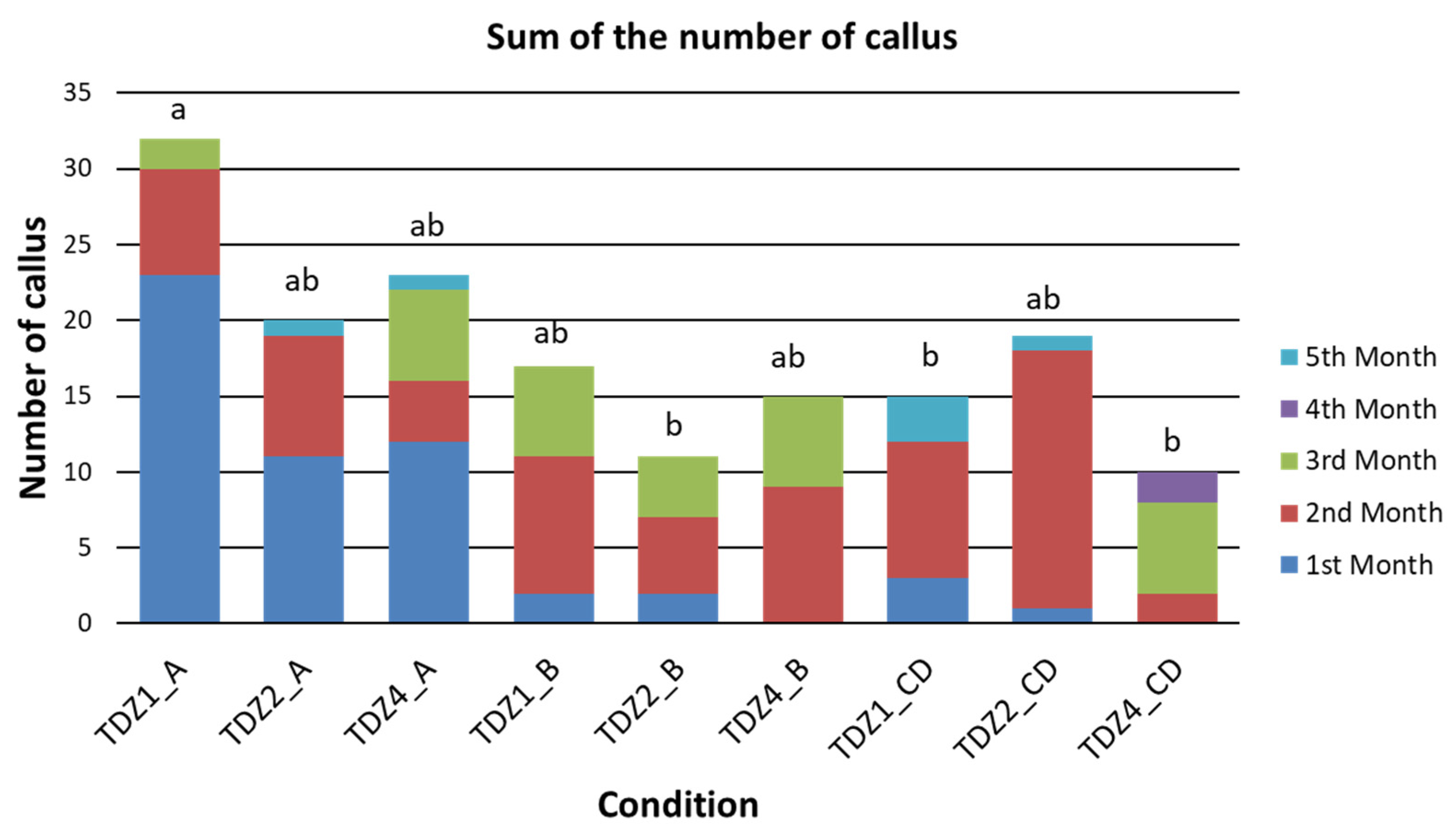
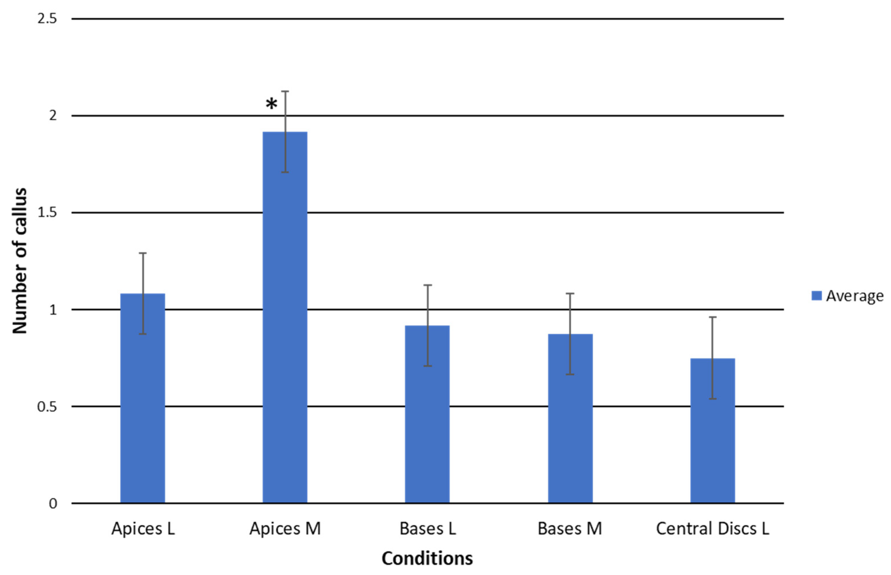
2.1.1. Productivity of TDZ Treatments Related to Callus Production

Considering the three different TDZ concentrations evaluated, apical explants always offered a better response than bases or central discs when they were cultivated under the same conditions (Figure 2). This indicates that apices were the most productive explants during the study regarding callus production. The response obtained in the apices exposed to TDZ1 stood out since the start of the trial. These apices showed the maximum number of calluses of all the experiments at the third month (Figure 2). From then on, its number of calluses decreased until the end of the assay.
On the other hand, the effect of the initial size of explants was also evaluated. To avoid bias in the results, the response to the different treatments was studied in detail by the type of explant. As central disk explants were only derived from large plants, they were not included in this comparison. The values used for this comparison corresponded to the number of calluses detected at the end of the induction period (third month of the trial), which coincides with the moment of the culture with the highest number of calluses observed.
Our findings showed a higher response of smaller apices than the bigger ones, while no differences between different sizes were observed in basal explants (Figure 3). Both results suggest that younger areoles are probably more responsive than older ones as it has been reported by other researchers [52], a reason for which most protocols for prickly pear micropropagation are usually based on young cladodes [18,19,20,29]. Moreover, larger explants were obtained from plants that germinated earlier and, therefore, also contain older areoles than those present in explants of a smaller initial size. Probably, this also explains why the bases, despite having a greater number of areoles but the oldest ones in the plant, showed lower productivity than the apices and similar to the central discs (Figure 3).
In any case, the relationship between callus production and shoot production may depend on various factors, such as the type of explant, the type and concentration of the PGRs used and other factors related with the in vitro culture conditions [16]. In the present work, the reduction in the number of calluses during the growth period seems to be related to an increase in the number of shoots. However, although this relationship may exist, it does not always occur in other species of cacti [53,54].

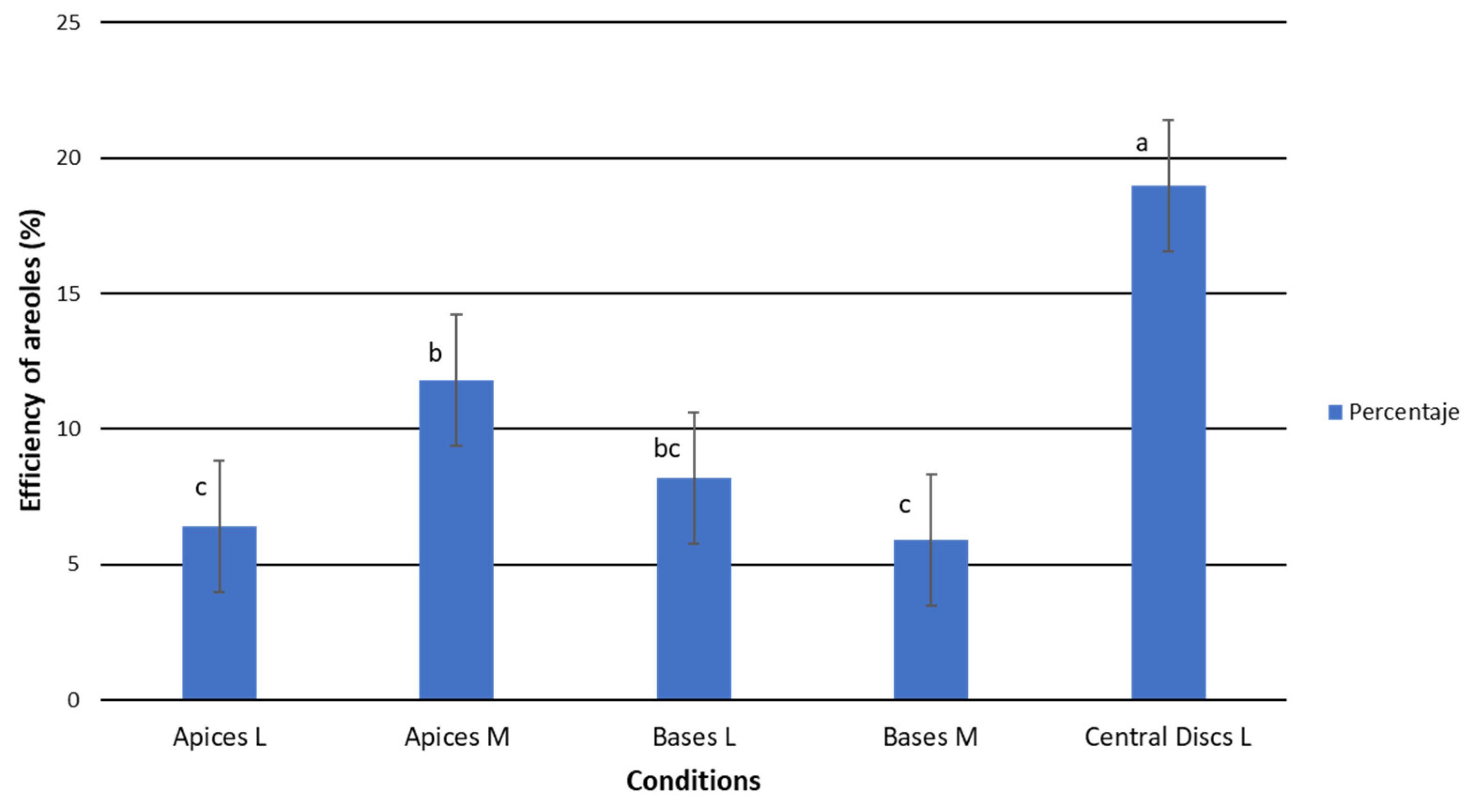
2.1.2. Efficiency of TDZ Treatments in Relation to Callus/Areole Production

The central disc efficiency was significantly higher than those observed for apices and bases, with percentages of activated areoles that produced calluses higher than 30% (Table 2). In this regard, more than 47% of the areoles were activatedin the central discs in the presence of TDZ1. By contrast, areolar activation in apices and bases was significatively lower and the averages ranged between 9.66% and 14.58% (Table 2).
Once again, significant differences were found between the efficiency shown by the medium-sized apices and the large-sized apices (Figure 4). In contrast, no differences were detected between the bases of different sizes. This fact reinforces the discussion proposed above, as it seems that younger areoles within the same explant type usually respond better to the treatments than older ones. Taking into account that the apices have the youngest areolas, these results might seem contradictory. However, the ability to transport endogenous hormones and the metabolic capabilities of the cells are tissue-dependent [55,56], so the CD could be more predisposed to areolar activation. Furthermore, the absence of an apical bud in the central discs could be favoring the activation of a greater proportion of areolas, as has been observed in other studies [9,16,57], hence, the greater efficiency in the central discs may not be solely related to the age of the areolas.

2.2. Shoots

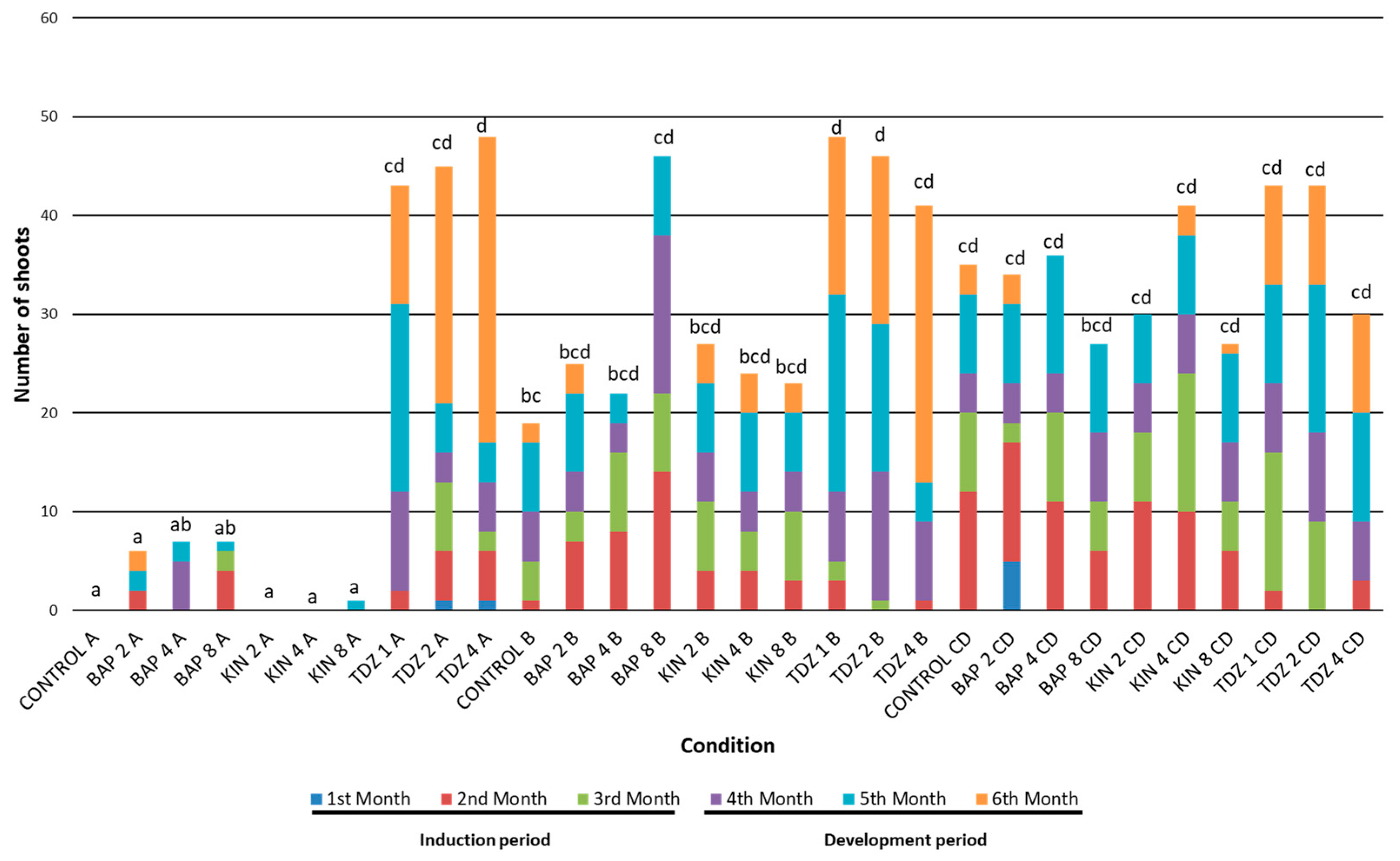
2.2.1. Productivity Related to Shoot Production

The results showed statistically significant effects of the different types of explants from the second month until the end of in vitro culture period (Table 3). Furthermore, the contribution of the different hormones in the medium was higher during the induction period, while the variation due to the different hormonal concentrations was observed to a greater extent during the last three months of culture. In general, with the exception of the seedling size (insignificant throughout), the principal factors displayed higher significance toward the end of the trial (Table 3).
The study of each factor showed that the presence of BAP and KIN increased the production of shoots during the induction period, especially in central disc explants (Supplementary Table S2). Nevertheless, once the induction period ended, a positive response was detected in the explants subjected to the presence of TDZ, leading to the best results at the end of the trial (Supplementary Table S2).
Regarding the hormone concentration, it was observed that the explants established in the control groups (in the absence of PGRs) responded, in general terms, with less efficiency than those subjected to cytokinins. The impact of hormone concentration showed that higher concentrations were less effective at producing shoots, while intermediate concentrations generally allowed for higher rates of shoot production (Supplementary Table S2).
It is known that responses to exogenous hormones differ based on the species and explant source. Higher hormone concentrations can influence the occurrence of calluses while preventing shoot development, which has been observed in purple pitahaya (Hylocereus costaricensis) [34]. Alternatively, higher rates of shoot production in response to high hormone concentrations has been observed in Mammillaria [58].

2.2.2. Efficiency Related to Shoot Production

In terms of efficiency, similar results were observed to those obtained for productivity. Once again, BAP and KIN offered the best values at the end of the induction period, and the response of the central discs was higher than that detected for apices and bases (Supplementary Table S3). As the cultivation progressed, the increase in shoot production by the explants exposed to TDZ became evident. Thus, at the end of the growing period, TDZ was established as the hormone that generated the most efficient responses in the explants studied. Alternatively, the presence of exogenous PGRs, regardless of their concentration, increased the shoot efficiency compared to the control treatment (Supplementary Table S3). However, specific interactions between the hormone type, explant type and hormone concentration might change responses related to the shoot efficiency (Supplementary Tables S2 and S3). For this reason, with the aim of assessing the specific behavior of all possible combinations throughout the trial, the graphical representation of the total production for each combination of factors has also been carried out (Figure 5).

2.2.3. Shoot Production Based on Treatment Factor Combination

Apical explants grown under BAP2, KIN2, KIN4 and KIN8 conditions and the control group showed averages not different from 0, while the rest of the combinations showed significant effects (Table 4). Regarding the total number of shoots, the responses of the explants exposed to the different treatments were highly diverse, especially depending on the type of explant used (Figure 5). But also, the appearance of new shoots during the induction period was considerably different from the response observed during the later developmental period in the absence of regulators.

2.3. General Trends Related to the Induction Period

Throughout the first 3 months (induction period), responses related to shoot productivity and efficiency were generally higher for CD explants and significantly higher by the end of the third and fourth months (Supplementary Tables S2 and S3). The central disc explants cultivated under the KIN4 condition produced a total of 24 shoots, followed by the control and BAP4 groups, with 20 shoots each. BAP2_CD, KIN2_CD and TDZ1_CD established values close to these groups (19, 18 and 16 shoots, respectively), but a lower emergence of new shoots was observed under BAP8 and KIN8 presence (11 shoots each). TDZ2 and TDZ4 concentrations resulted in the lowest values, with nine and three shoots, respectively (Figure 5).
Basal explants showed a lower average production than central discs (Supplementary Table S2). However, when evaluating specific combinations, it was observed that BAP8_B produced a shoot emergence (22) comparable to the best ones observed in the central disc. The rest of the groups showed levels below 16 shoots (Figure 5).
As previously mentioned, apices from BAP8, TDZ1, TDZ2 and TDZ4 were the only ones that showed some kind of response within these explants. Even so, their results were much lower than those found in central discs and lower in a general terms than the ones observed in bases (Figure 5).

2.4. General Trends Related to the Growth Period

During the growing period, in the absence of PGRs, apices showed the lowest values among all studied conditions. Only apical explants cultivated under TDZ displayed high rates of shoot production, particularly TDZ4_A, with a total production of 48 shoots (highest value together with TDZ1_B) (Figure 5).
The apices showed the lowest mean values in the assay when analyzed as a whole, mainly because only those exposed to TDZ responded significantly. KIN, BAP and even the control group either did not provoke a response in the explants (CG_A, KIN2_A and KIN4_A) or gave rise to the worst responses in the assay (BAP2_A, BAP4_A, BAP8_A and KIN8_A). These results evidenced the relevance of the interaction between the hormones and the type of explant (HxTE), so that the presence of TDZ (regardless of its concentration) involved the greatest responses within the overall set of the evaluated apical explants.
The largest monthly increases in the number of shoots for apical explants were observed under TDZ4 and TDZ2 during the sixth month, while shoot formation under TDZ1 was greater during the fifth month (Figure 5). This could indicate that the induction at higher concentrations of TDZ provokes a delay in forming new shoots, both from newly formed or from pre-existing calluses. Similar results were obtained for Rauvolfia serpentina (L.) Benth. ex Kurz plants, in which its subculture to an MS media after induction with different TDZ was related with a positive effect in shoot proliferation [59].
Regarding the response of the bases, the importance of the HxTE interaction is once again highlighted. In fact, the best results were obtained in the presence of TDZ (more than 40 shoots at each concentration), while the rest of the hormones produced fewer shoots on average (KIN: 24.7 shoots; and BAP: 31 shoots). The control group provided the worst result among the combinations studied, with a total of 19 new shoots at the end of the growing period (Figure 5). Furthermore, a delay in shoot production was also observed as the concentration of TDZ increased, establishing the maximum increases in the number of shoots for TDZ1_B in the 5th month and later for TDZ4_B, during the 6th month. In this regard, it has been reported that the highest shoot proliferation after TDZ induction was also found after the fifth month in other species such as Rauvolfia serpentina and Aegle marmelos (L.) Correa [59,60]. Probably, the ability of TDZ to activate endogenous cytokinin biosynthesis or altering cytokinin metabolism could be behind these different effects when variable concentrations of this hormone act over the explants [61,62,63,64,65].
In addition, it was also found that the interaction between the three factors (hormones, H; hormone concentrations, HC; and type of explant, TE) played an essential role in the case of the bases cultivated in the presence of BAP8. In fact, the results obtained for BAP8_B (46 shoots) almost doubled those obtained for BAP2_B and BAP4_B (25 and 22 shoots, respectively) (Figure 5); which demonstrates that the effect of the specific combination of the factors was determinant in the response of the explants of the base type.
On the other hand, the relationship between the results obtained from callus emergence after induction with TDZ and its relative production of shoots at a temporal level was also studied. In this case, it was observed that the general behavior of the explants induced at low concentrations differed with respect to the explants subjected to medium or high TDZ concentrations. Thus, in apical and basal explants induced with TDZ1, a reduction close to 60% in the number of calluses was observed from the third to the fifth month (Supplementary Table S1). This decrease in the total of counted calluses coincides with a greater increase in the number of shoots, which begins to be remarkable from the fourth month (Figure 5).
In contrast, the induction of apices and bases with TDZ2 and TDZ4 led to a later increase in the number of shoots, especially prominent during the last month of the trial. However, it should be noted that the number of calluses in these cases did not undergo significant variation during the last three months (i.e., a reduction of around 35% in the presence of TDZ2 and 5% for TDZ4) compared to the variation detected for the TDZ1 condition (Supplementary Table S1). Thus, the induction with TDZ1 gave rise to the formation of calluses that, during the growing period, underwent a process of organogenesis, causing shoots. However, and given that the variation in the number of calluses was not so remarkable for the TDZ2 and TDZ4 conditions, it appears that the induction at these medium and high concentrations could (i) favor the gradual emission of shoots through the activation of new areolas that had not previously developed callus or, more probably, (ii) suppose an excessive initial hormonal load on the explants, and only with the advance of time and successive subcultures (circumstance that would reduce the endogenous concentration of TDZ in the explants), they were finally able to respond with the issuance of new shoots, as has been observed in other species [59]. These results were comparable to those obtained for Mammillaria pectinifera (Ruempler) F.A.C. Weber and Pelecyphora aselliformis Ehrenberg calluses, where it could be observed that they began to produce new shoots after a subculture in a medium in the absence of PGRs [66,67].
The behavior of the central discs induced with TDZ did not fit to the responses observed for apices and bases. The variation in the number of calluses is much lower than the increase in the number of shoots observed during the growing period. This suggests that the presence of TDZ (regardless of its concentration) could trigger the formation of new shoots from apparently inactive areolas.
For central disc explants, a delay in areola activation was also observed as the TDZ concentration increased during induction. But in this case, TDZ1_CD and TDZ2_CD gave rise to a global production of 43 shoots, while TDZ4_CD finished with 30. This difference could corroborate the previous hypothesis, in which an excessive concentration of the TDZ during induction could be delaying the emergence of new sprouts. Even so, considering the high effect of the interaction between hormones and the explant type, it might be possible that TDZ4 simply did not activate central disk-type explants as efficiently as at lower concentrations.
Finally, and taking into account that the central discs responded in a fairly homogeneous way in all of the evaluated conditions (including the control group), the influence of the hormones in the medium was probably not having as much relevance as for the apices or bases explants. Probably, just the removal of the apex (involving the loss of the apical dominance) could cause the natural activation of the dormant buds of the explants, something usual when working with cacti [66,67]. And consequently, this resulted in outstanding sprout productivity values even in the absence of PGRs.

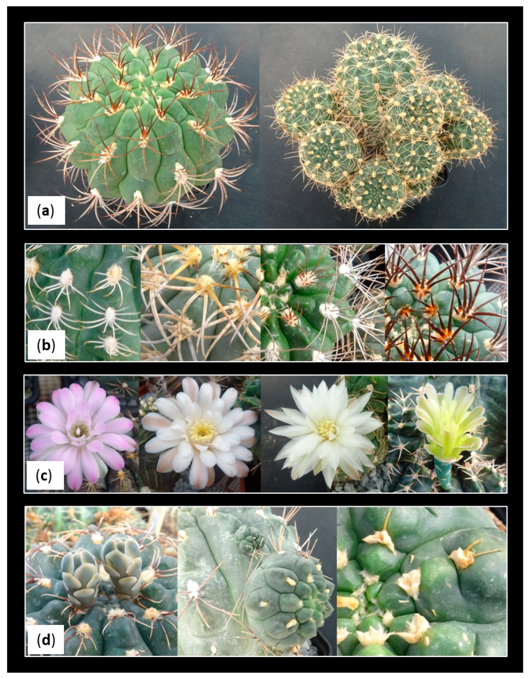
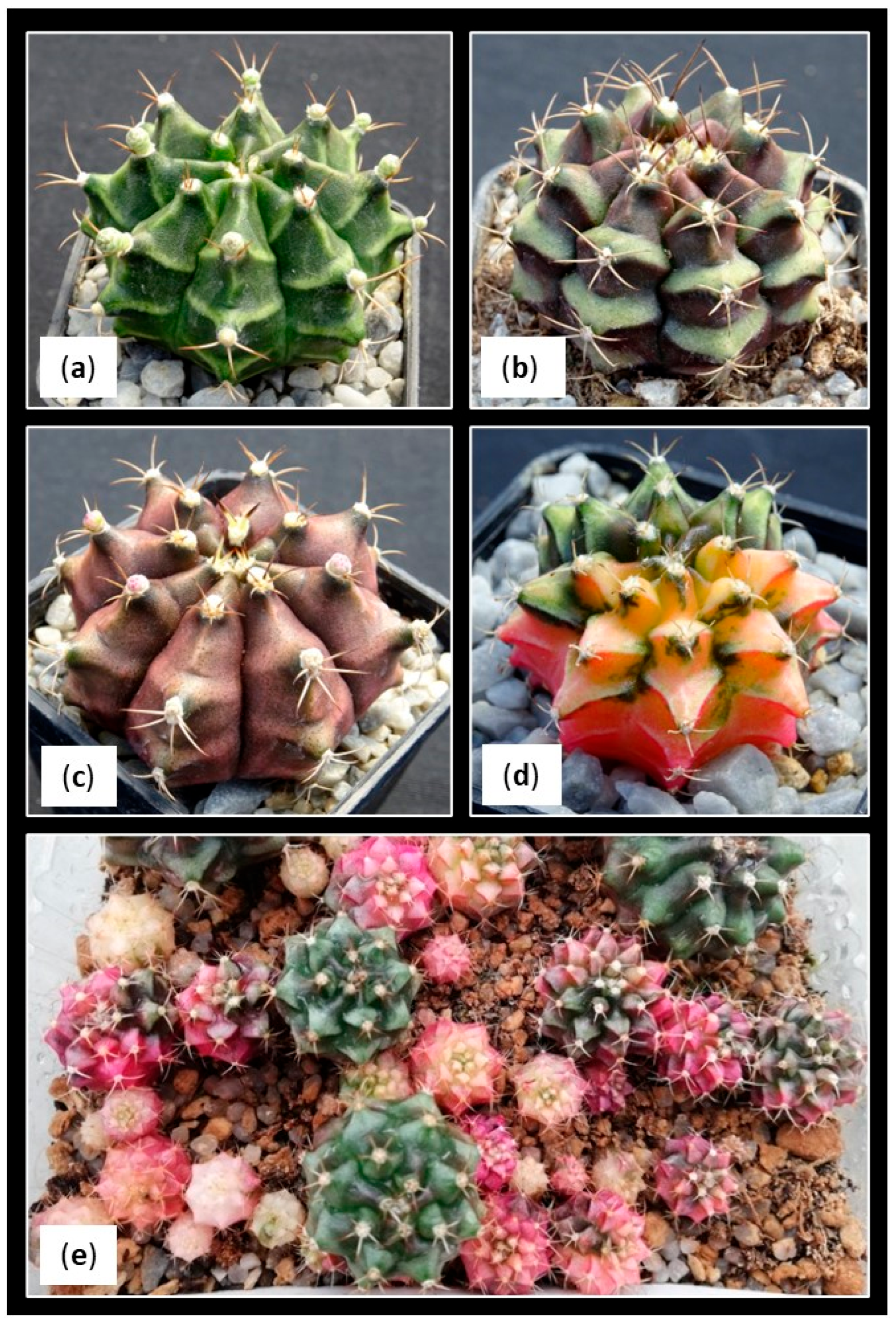
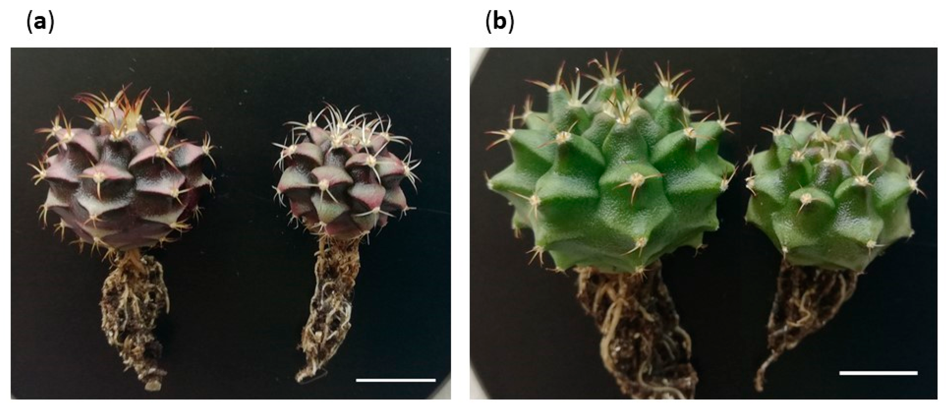
2.5. Descriptions of Morphotypes

As already mentioned, Gymnocalycium cv. Fancy is a commercial hybrid with a large and heterogeneous genetic background. In fact, progenies from sowings are quite heterogeneous, appearing as plants with different morphologies (Figure 6). Even so, most of the Fancy seedlings showed hybrid characteristics with a greater tendency towards the general structure of G. mihanovichii. In this sense, seedlings can be easily grouped based on the prominence of the tubercles, the length of the spines and the general coloration of the plant.
The most expected morphotypes were initially classified as Morphotype 1 (M1) and Morphotype 2 (M2). M1 presented the following characteristics: shoots with the recognizable characteristics of the commercial cultivar, that is, with pronounced tubercles which present a triangular section, elongated spines with a greater angle of curvature and darker coloration, even purplish, especially in the distal area of the tubercle (Figure 7a). M2 presented shoots were characterized by presenting less pronounced and more rounded tubercles, shorter and more erect spines and an intense light-green color with a shiny appearance (Figure 7b). Both morphologies could be representative of the variation usually expected in the progenies of this cultivar. Therefore, they were grouped together for the statistical analyses.
After evaluating the development of the in vitro explants, the appearance of shoots with new unexpected morphotypes that did not correspond to the initial description were observed. These morphotypes showed a particular development, and their presence in greenhouse’s seedlings had not been detected previously. The appearance of different morphological shoot types in cacti grown under in vitro conditions have been also reported by other authors [13,68,69,70], and it is relatively frequent. Although the exact mechanism is still unknown, it seems that the epigenetic factors responsible for regulating gene expression could be behind the appearance of these morphological variants [71].
These new unexpected morphotypes showed the following characteristics: Morphotype 3 (M3) had slightly flattened shoots characterized by showing irregular bumps on their epidermis, a total absence of spines and a light-green coloration. Initially, they do not show any woolly structures typical of the areolas, but as they developed, hairy areas appeared, erratically distributed over the tubercles. In addition, a verrucous-looking tissue also emerged between the shoots´ bumps, which sometimes caused new aberrant offsets, and others continued their irregular development, acquiring a callus-like appearance (Figure 7c). Morphotype 4 (M4) had flattened shoots of a dark-green color with a tendency to accumulate a greater amount of pigments in the basal area of the areoles, with conical tubercles and the presence of large and hairy areoles. In some cases, erect, very short and delicate spines emerged from these areoles. It tended to develop new shoots with similar characteristics, so finally, they evolved to particular caespitose structures (Figure 7d).
The number and specific morphotype of the obtained shoots by condition at the end of the trial are compiled in Table 5. In general terms, more than 80% of the shoots showed the expected morphologies: 75% corresponding to the typical morphology of the Fancy cultivar (M1) and 7–8% to the M2. The remaining 20% were included in the other described morphologies: M3 accounted for 12.38% of the total, while M4 appeared at a frequency of 5.70%. On the other hand, it was observed that the appearance of the different morphotypes was not arbitrary, but it was subject to the effects exerted by the factors studied and their respective interactions (Table 6).

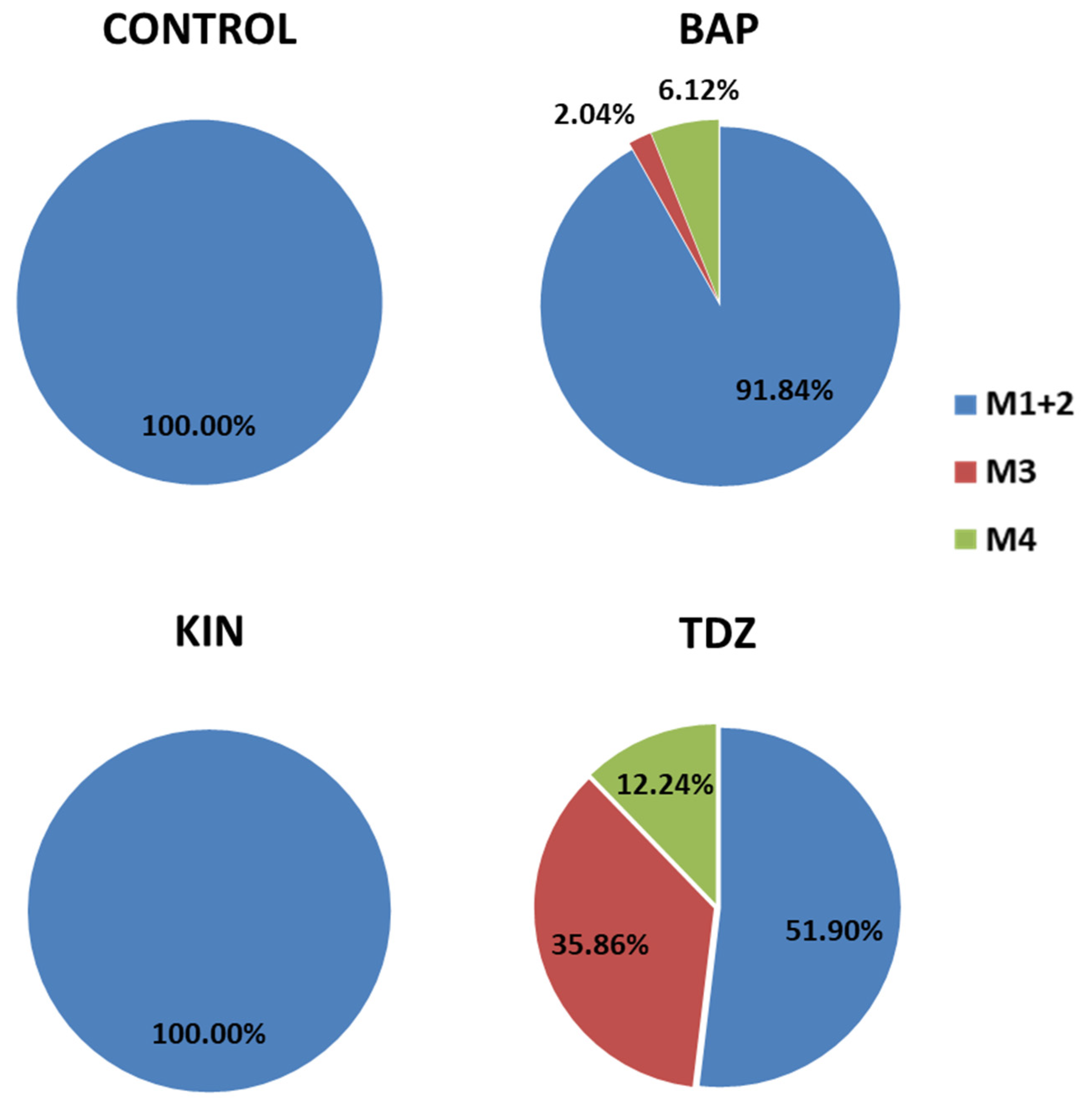
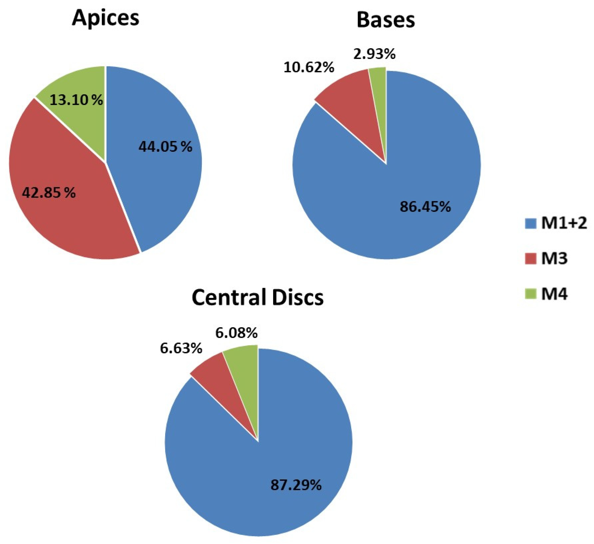
2.5.1. Occurrence of Expected Morphotypes

The expected morphotypes appeared in a higher frequency, exceeding 91% in BAP and reaching a total of 100% in the presence of KIN and in the absence of PGRs, while only 51.9% of the total recorded shoots in TDZ showed the expected morphologies (Figure 8). These results suggest that the presence of TDZ in the medium would be favoring the appearance of new morphotypes; while in the presence of BAP or KIN or absence of PGRs, the explants tend to respond by emitting the expected shoots. Similar results were obtained by Giusti et al. [13], who detected that the use TDZ was associated with callus formation and hyperhydricity of new shoots, while BAP (especially at higher concentrations) and intermediate KIN concentrations showed a high multiplication rate and originated new axillary shoots of quality for Escobaria minima (Baird) D. Hunt, Mammillaria pectinifera and Pelecyphora aselliformis.
Furthermore, the production of M1 and M2 shoots was significantly higher from the central disc and basis explants than that observed for apices (Figure 9). Thus, considering all the results obtained, to maximize production in a shorter time, it can be established that an adequate and efficient method to clonally propagate this commercial cultivar should be based on the use of MS media supplemented with KIN4 and, for their major efficiency, central discs as the type of explant.
From a commercial point of view, these results are very important since establishing the conditions to allow for the maximum production of shoots per explant results in an increase in the efficiency of the procedure and, therefore, in the possibility to optimize the resources and increase the benefits. Plants cultured in vitro usually grow faster and produce shoots, even in plants that grow solitary in the wild, as in the case of Aztekium ritteri Boedeker. In this case, plantlets can produce new shoots after 11 months of being cultivated in vitro [51]. The results show that micropropagation is not only efficient but also reliable. So if the main objective is to propagate a specific commercial material (such as an individual selection of some outstanding specimen), this protocol allows for the production of more homogeneous plants in only a few months.
On the other hand, this micropropagation protocol could be very useful for other Gymnocalycium or related cactus species. The Gymnocalycium genus is included in the second appendix of the CITES agreement (Convention on International Trade in Endangered Species of Wild Fauna and Flora; http://www.cites.org; accessed on 28 September 2023), so its species are not listed as threatened or endangered [72,73]. However, there are always specific populations, made up of few individuals, whose survival is threatened by numerous pressures of anthropogenic origin, such as agricultural and urban expansions, the introduction of exotic grasses [74], the use of herbicides and pesticides in intensive agriculture [75,76,77] or their sale on the black market [78,79,80,81], which leads to a reduction in specific and intraspecific plant biodiversity, especially for those taxonomically and genetically problematic groups [74,82]. Taking into account the efficiency of the methods presented and the little influence of KIN4 on the final phenotype of the shoots, plants available to reintroduce into their habitat could be produced, contributing to the conservation of these vulnerable populations while respecting its genetic integrity. Furthermore, our findings show that it is also possible to propagate Gymnocalycium efficiently from central discs in the absence of PGRs. Considering that the presence of PGRs may alter the morphological and physiological characters and cause somaclonal variation [83], this micropropagation system would make it possible to obtain individuals that did not alter the genetic background of the repopulated populations.

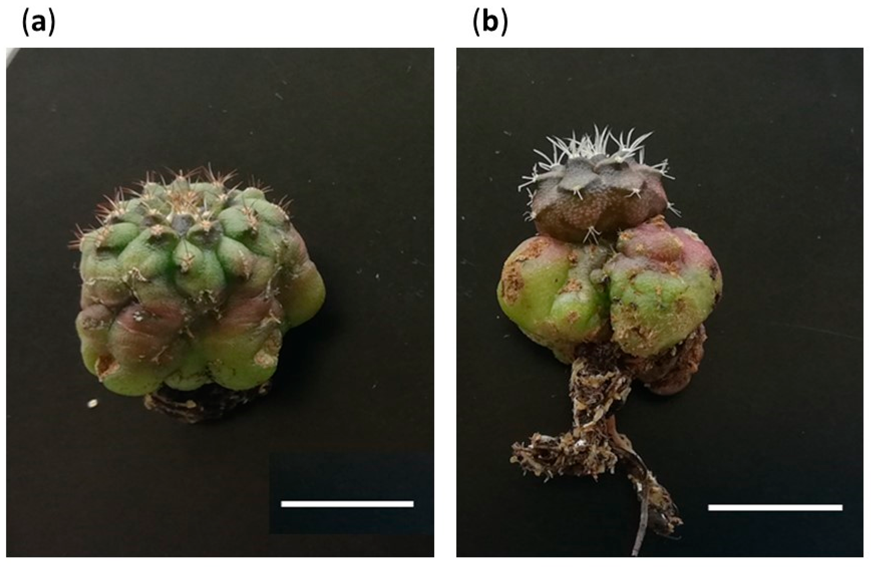
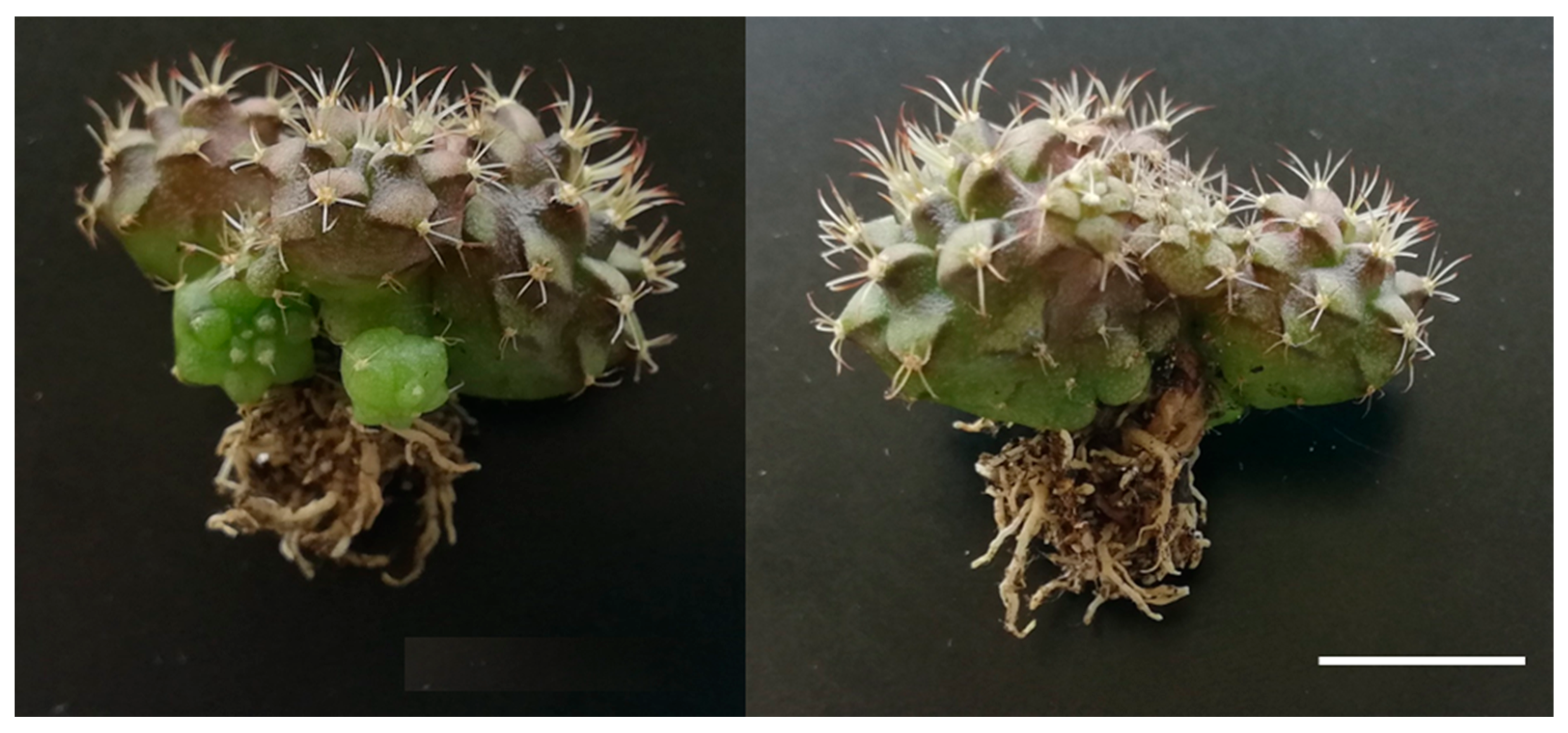
2.5.2. Occurrence of Unexpected Morphotypes

M3 and M4 shoots mainly developed in the presence of TDZ and stood out with relevant proportions in the apical explants (Figure 8 and Figure 9). These results could be related to the previous ones obtained for callus formation and its subsequent evolution in defined shoots; and more specifically, in explants subjected to TDZ. It seems that the presence of TDZ during the induction period activates the development of calluses (especially in apical explants), and these calluses, through processes of indirect organogenesis, are defined as M3 and M4 shoots, so that the vast majority of shoots with M3 and M4 are probably preceded by a callus phase. The generation of callous tissue prior to the organogenesis process is a factor that may favor somaclonal variation [84], and it could be expected that these new morphotypes appear under this condition. These results are in agreement with those observed by Giusti et al. [13] for Escobaria minima (Baird) D. Hunt, Mammillaria pectinifera (Ruempler) F.A.C.Weber and Pelecyphora aselliformis Ehrenberg. In this assay, TDZ media gave them both a profuse callusing and an important shoot proliferation, but a high incidence of hyperhydricity was observed in the final shoots, probably related with their higher concentration in the media. These hyperhydration events have been described in other cactus species in the presence of TDZ [13] but are also frequently observed in numerous plant species [85].
The production of these shoot types was not observed in the control groups or in the presence of KIN, and their respective appearances in BAP were lower than those observed for TDZ, but represented 4.49% of the M3 shoots and 29.27% of the M4 shoots (Table 5). Morphological alterations were also observed in new shoots of Melocactus glaucescens Buining and Brederoo when using BAP as the inductor phytohormone [70], which means that the response in Gymnocalycium cv. Fancy is not an isolated case and specific concentrations of BAP can provoke morphological variations in different species.
Given that M3 and M4 are unexpected morphotypes and would hardly arise under traditional sowing conditions, these results suggest that the presence of TDZ in vitro could be acting as an inducer of somaclonal variation, as has been observed in other works [13,86]. Thus, the indirect organogenesis processes that are observed from the calluses could give rise to new morphotypes that would be of commercial interest. This circumstance can be evaluated from two perspectives: (1) the obtained morphotype presents certain characteristics that make it susceptible to being massively propagated and introduced into the wholesale market, or (2) the obtained morphotype is morphologically strange, slow-growing or requires very special growing conditions, characteristics that limit its cultivation in large quantities. In this case, it could be used for the market aimed at cactus collectors. In any case, new varieties of ornamental plants have been developed through spontaneous mutation in vitro [87,88] and TDZ seems to have an important potential for cactus species in this respect.
There is a very important worldwide market focused on cacti for collectors. This market niche is closely linked to obtaining new hybrids, cultivars or forms that show differential characteristics and that would be likely to be collected by cactus lovers. The protocol described in this assay shows that there is the possibility of forcing the appearance of morphotypes other than the original through the induction of explants with TDZ. Thus, this test shows that the culture of Gymnocalycium cv. Fancy, mainly in TDZ concentrations between 1 µM and 2 µM, is really effective in obtaining phenotypic variants. This occurrence of somaclonal variation during the tissue culture has also been reported in other cactus species such as Mammillaria sp. [89] and Cereus peruvianus [90], but not in such remarkable proportions. Thus, a new alternative to obtain a variation in an efficient way is offered, being a useful breeding tool for those cactus producers and growers whose products are aimed at the collector market.

2.6. Root Emergence

Root emergence was also observed in some specimens. The development of roots could impact the occurrence and/or development of new shoots in vitro in addition to affecting their subsequent acclimatization capacity [91,92,93]. Therefore, the root emergence of the different treatments was also evaluated.
The rooting response was evaluated during 8 weeks, as no more explants emitted roots after this period of time, and the accumulated percentage of rooted explants is represented in Figure 10. Four groups were defined, whose values showed statistically significant differences between them: (i) conditions with a response of > 75%; (ii) conditions with a percentage between 25 and 37.5%; (iii) conditions with a response varying from 6.25 to 12.5%; and (iv) no response (0%).
Results revealed that the rooting percentage (number of rooted explants/total explants per treatment) was closely related with the type of explant used but also with the PGRs present in the media (Table 7). In fact, apical explants gave rise to the best results regardless of the hormone concentration used, with ratios higher than 75%, except for those ones cultivated in th presence on TDZ. However, no bases showed significant responses and only BAP2, KIN4 and KIN8 came to significatively stimulate the root development in central discs (Figure 10).
Rooting in the apical explants could be expected since the removal of the basal zone of the plant should make the explant respond like a cutting taken in the greenhouse for traditional propagation. So usually, the root development is only a response that fits the natural behavior of cacti and can occur spontaneously in PGR-free media, as it has also been observed in other cactus species [94]. This fact has been contrasted in our experiment, where the control group (in absence of plant growth regulators) has provided similar values to those obtained in the presence of KIN and BAP.
By contrast, the presence of TDZ completely blocked the emission of roots. These results could be explained from the point of view of the synthetic origin of this plant growth regulator, which presents both auxin and cytokinin activity [95]. In fact, similar results had been reported by Rubluo et al. [96] when they used the artificial auxin 2,4-dichlorophenoxy-acetic acid (2,4-D). Its presence in the media offered lower rhyzogenesis and even provoked a total inhibition of rooting at highest concentration evaluated (27.14 µM), when compared to those explants cultivated in the presence of natural or synthetic auxins (IAA, 3-indolyl-acetic acid; NAA, 1-naphthalene-acetic acid and IBA, indole-butyric acid). Perhaps, the presence of TDZ inhibits the natural rooting mechanisms in this material, while the increase in the concentration of KIN and BAP (since they are natural hormones) does not seem to have a negative impact on this aspect.
On the contrary, the rooting in the basal zone explants was not foreseen. These explants were cultivated in an unnatural position to maximize the surface in contact with the culture medium, so it was expected that this placement on the medium would reduce the possibility of rooting, which has been confirmed in this experiment.
The central discs showed higher percentages of rooting also in the presence of BAP2, KIN4 and KIN8 groups, but far from those obtained for the apices (Table 7). Given that the central discs were sectioned into two halves, it could be that the rooting response was being conditioned by the fact that the vascular bundle of the explant was fragmented. On the other hand, apices from the control group showed similar results to apices in the presence of KIN and BAP, while central discs rooting in the control group were significantly lower than those reached by BAP2_CD, KIN4_CD and KIN8_CD combinations (Figure 10).
These results could reinforce the hypothesis that explants with an intact vascular bundle and in contact with the culture medium (as the apices) can develop roots naturally and do not require the presence of PGRs to offer high percentages of rooting. In fact, it has been observed that the dissection of the plants usually alters the proportion of the endogenous hormones favoring a high auxin/cytokinin ratio in the apical zone, which generally promotes the root production in this kind of explants [97,98]. However, an incomplete or damaged vascular bundle would not respond easily and would require the presence of certain concentrations of hormones in the medium to stimulate its rooting.
Thus, the presence of BAP2 in the medium could optimize the procedure and offered the highest rooting percentages in the shortest time (4 weeks). This additional information regarding the influence of BAP2 on rooting is of high interest at a productive level, since it allows the speeding up of the cloning processes. Therefore, it is possible to reduce the necessary time to obtain optimal plants capable of overcoming the acclimatization processes.

2.7. Acclimatization

The acclimatization success rate was very remarkable: 96.67% for M1, 100% for M2, 96.67% for M3 and 93.33% for M4. All plants increased their size and showed a healthy appearance. It was also observed that the plants from the expected morphotypes continued their development, resulting in a set of plants representative of the usual variability found within the studied cultivar (Figure 11). However, M3 showed changes in its development.
The acclimatized plants corresponding to M3 began to revert their morphology toward the expected morphotypes. In fact, all the areoles that were activated ex vitro originated from M1 and M2 shoots (Figure 12a) or, in other cases, the apices modified their development and structure giving rise to tissues corresponding to the expected morphotypes (Figure 12b). It seems evident that M3 is linked to the in vitro culture conditions, a fact that could be mainly explained from two points of view: (i) perhaps the presence of PGRs or the high concentrations of other compounds in the media conditions the expected development of some of the new sprouts, as has been observed in other works [99,100,101]; or (ii) alternatively, some shoots suffer a certain degree of vitrification, causing the modification of their natural appearance. In any case, M3 is an artifact generated by the in vitro culture stress, provoked by the presence of phytohormones in the medium or due to problems related to osmotic stress. For this reason, after the acclimatization process, M3 plants showed total instability, reverted their morphology and continued their development with an expected morphology.
On the other hand, M4 shoots kept their particular caespitose development that they showed during the in vitro culture period. In fact, they gave rise to clustered plants with a differential appearance compared to the expected morphotypes (Figure 13). It would be difficult to determine if this variant is the result of a somaclonal variation process, due to a spontaneous mutation, or simply a rare phenotype within the existing variation that cv. Fancy presents. But in any case, it has been possible to identify this new morphology and demonstrate its stability under ex vitro culture conditions. This situation shows that the micropropagation of Gymnocalycium in vitro can also be tremendously interesting when it comes to identifying particular phenotypes within heterogeneous populations and facilitating their subsequent propagation.

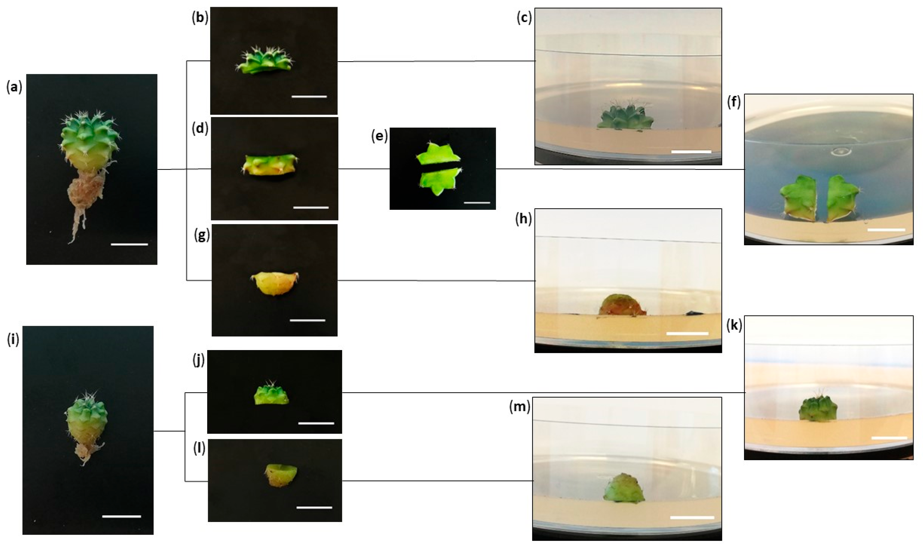
3. Materials and Methods

3.1. Plant Material and Disinfection

Plant material was obtained by in vitro germination of seeds of Gymnocalycium cv. Fancy, kindly donated by Cactusloft OE (Cullera, Valencia, Spain). Seeds were disinfected for 1 min in 70% ethanol (v/v), followed by 25 min in 15% domestic bleach solution (v/v; 4% sodium hypochlorite) supplemented with 0.08% Tween-20 (v/v) and rinsed 3 times in distilled sterilized water under aseptic conditions in a laminar flow cabinet (model AH-100, Telstar, Terrassa, Spain).

3.2. In Vitro Establishment and Culture Conditions

Disinfected seeds were sown in Petri dishes using commercial Murashige and Skoog media [102] at half strength (1/2MS, 2.2 g L−1) supplemented with sucrose (15 g L−1) and agar (7 g L−1), adjusting the pH to 5.7 before autoclaving at 120 °C for 20 min [103]. Seedlings were maintained under in vitro conditions and kept in a growth chamber at 26 ± 2 °C, 16 h day length and photosynthetic photon flux of 50 μmol m−2 s−1 for 8 months. Seedlings were subcultured monthly to a fresh media to promote their development.

3.3. Induction and Tissue Culture Conditions

To assess the morphogenic potential of Gymnocalycium cv. Fancy, the effect of different concentrations of three cytokinins on shoot induction and production was studied: 6-Benzylaminopurine (BAP), Kinetin (KIN) and Thidiazuron (TDZ). Plant growth regulators (PGRs) were acquired from Duchefa Biochemie Company (RV Haarlem, The Netherlands). For this purpose, 176 plants with diameters ranging from 8 mm to 16 mm were selected from the set of 8-month-old seedlings.
The selected plants were divided into two groups based on their initial size. The first group (M) consisted of medium-sized plants (with diameters ranging from 8 to 12 mm) (Figure 14i), while the second group (B) included large-sized plants (with diameters ranging from 12 to 16 mm) (Figure 14a). From the medium-sized plants, two types of explants were obtained: apical and basal (Figure 14j,l). Regarding the large-sized seedlings, additional explants were obtained by dividing a central disc into two halves (Figure 14d,e). In total, three types of explants were obtained for each large-sized plant: apical stem, basal stem and the two halves of the central disc (Figure 14b,e,g).
Explants were placed for their induction on 1/2MS (2.2 g L−1), supplemented with sucrose (15 g L−1), agar (7 g L−1) and different concentrations of three cytokinins, BAP (2, 4 and 8 µM), KIN (2, 4 and 8 µM) and TDZ (1, 2 and 4 µM), adjusting the pH to 5.7. The position of the explants in the culture medium is shown in Figure 14c,f,h,k,m. A medium in absence of PGRs was also included as a control formulation for each type of explant. The induction period lasted for three months, during which time, the explants were grown in the presence of the corresponding hormones. Subsequently, all explants were subcultured to a 1/2MS without PGRs at pH 5.7 to continue evaluating their development.

3.4. Experimental Design

Forty-eight explants were studied per concentration of each hormone, including sixteen of each type of explant. In the case of apices and bases, eight explants from medium and eight explants from large-sized plantlets were included, but only explants from large sized plants (sixteen central disc halves) were evaluated in the case of central discs (Table 8). Six explants per type of explant and size (i.e., medium size apical, large size apical, medium size basal and large size basal explant, as well as two halves from the central disc) were cultured on each Petri dish. So, eight Petri dishes were included per hormone treatment, which makes a total of 80 Petri dishes.
The appearance of calluses and the presence of shoots were evaluated monthly for 6 months. Results were explained in terms of productivity (i.e., the total number of calluses obtained per explant) and efficiency (i.e., ratio of activated callus depending on the number of areoles of each explant only in those explants that responded to the treatments). The morphology of the obtained shoots was also recorded. Rooting was observed in some explants during the first 8 weeks, so the percentage of rooting was studied too.

3.5. Statistical Analysis

In order to identify the conditions that generated responses in the evaluated explants, data were subjected to a Student’s t-test using the Statgraphics 18 X64 statistical software (Statpoint Technologies, The Plains, VA, USA). No sensitive responses were removed from the general analysis to avoid biases in results. A confidence level of 95% was required.
Additionally, a multivariate ANOVA analysis was carried out to assess the impact of the four main factors evaluated in our study. These factors included three different plant growth regulators (KIN, BAP, and TDZ), three different hormone concentrations for each PGR and three different types of explants (apices, bases and central discs), as well as the initial size of the seedlings (medium and large), along with the possible interactions between them. The software used for performing this ANOVA analysis was Statgraphics X64 (Statpoint Technologies, The Plains, VA, USA).
Absolute data (averages, productivity values and general production) and percentage data (percentages and efficiency values) were previously transformed before conducting the analysis using the following formulas:
Absolute   data Y + 1 2
Y = Single data
Percentage   data     p e r c e n t a g e 100 a r c s i n
In addition, comparisons of means were made using the Student–Newman–Keuls test, with a significance level of 5%.

3.6. Acclimatization

One-hundred and twenty of the obtained shoots (30 corresponding to each recorded morphotype) larger than 15 mm were isolated and subcultured in 1/2MS medium supplemented with sucrose (15 g L−1) and agar (7 g L−1), adjusting the pH to 5.7, to promote their root development and initiate the acclimatization process. These shoots were maintained in vitro for 4 weeks, during which time, all of them developed roots.
Shoots were rinsed with distilled water and then transplanted into trays containing a substrate composed of 50% peat and 50% vermiculite. These trays were placed in mini-greenhouses to control environmental humidity. During the first two weeks of acclimatization, humidity was gradually reduced from the initial 100% to ambient humidity, by gradually opening the mini-greenhouse cover. After two weeks of cultivation, the mini-greenhouse covers were completely removed, and the plants continued growing under the growth chamber conditions. These plants were maintained in cultivation for an additional 8 weeks for further study of their development.

4. Conclusions

In the present work, an efficient micropropagation protocol has been developed for Gymnocalycium, based on the use of central discs as explants and the presence of kinetin (KIN4) or 6-benzylaminopurine (BAP8) in the culture medium. Furthermore, the obtained shoots successfully rooted in vitro (especially under the BAP2 condition), and their subsequent acclimatization to potted cultivation was highly effective (96.67%). The reliability and reproducibility of the protocol make it suitable for use in intensive production nurseries. Additionally, since it does not alter the morphology of the resulting shoots, this protocol could also be extrapolated to different Gymnocalycium species (or other cacti), whose populations are threatened. It could also serve as a tool to contribute to their conservation, both for the purpose of reintroducing specimens into their natural habitat and for maintaining certain genotypes ex situ.
Furthermore, it has been observed that the presence of Thidiazuron can induce callus formation, especially in apical explants, and approximately 25% of these calluses eventually developed shoots with unexpected morphologies, which in some cases remained even after acclimatization without PGRs. These results suggest that TDZ, at appropriate concentrations, could be used as an inducer of new stable variation.
Therefore, the optimization of this in vitro micropropagation protocol for Gymnocalycium cv. Fancy can be a valuable tool for both efficiently cloning material and for identifying distinctive individuals within heterogeneous populations. So then, this first approach can be used as a guideline to continue working with variegated and colored Gymnocalycium plants, which nowadays have an increased relevance in the niche of the market aimed at cactus hobbyists.

Supplementary Materials

The following supporting information can be downloaded at: https://www.mdpi.com/article/10.3390/plants12233932/s1, Table S1: Average numbers with their standard errors and total number of calluses obtained per condition; Table S2: Average shoot productivity and the significance of the factors and their interactions per month; Table S3: Average shoot efficiency and the significance of the factors and their interactions per month.

Author Contributions

Conceptualization, C.C.-O. and A.R.-B.; Methodology: C.C.-O.; Validation, V.B.-G. and A.R.-B.; Formal analisys, C.C.-O.; Investigation, C.C.-O. and V.M.G.-S.; Resources, A.R.-B.; Data curation, C.C.-O. and V.M.G.-S.; Writing—original draft preparation, C.C.-O.; Writing—review and editing, V.M.G.-S., V.B.-G. and A.R.-B.; Visualization, C.C.-O., V.B.-G. and A.R.-B.; Supervision, A.R.-B.; Project administration, C.C.-O. and A.R.-B.; Funding acquisition, A.R.-B.. All authors have read and agreed to the published version of the manuscript.

Funding

This research received no external funding.

Data Availability Statement

Data are contained within the article.

Acknowledgments

The authors are grateful to Cactusloft OE (Cullera, Valencia, Spain; www.cactusloft.com), which kindly provided seeds of Gymnocalycium cv. Fancy.

Conflicts of Interest

The authors declare no conflict of interest.

References

  1. Britton, N.L.; Rose, J.N. The Cactaceae: Descriptions and Illustrations of Plants of the Cactus Family; Courier Corporation: Chelmsford, MA, USA, 1963. [Google Scholar]
  2. Demaio, P.H.; Barfuss, M.H.J.; Kiesling, R.; Till, W.; Chiapella, J.O. Molecular phylogeny of Gymnocalycium (Cactaceae): Assessment of alternative infrageneric systems, a new subgenus, and trends in the evolution of the genus. Am. J. Bot. 2011, 98, 1841–1854. [Google Scholar] [CrossRef]
  3. Martino, P.A.; Bauk, K.; Ferrero, M.C.; Gurvich, D.E.; Las Peñas, M.L. Ecological significance of determinate primary root growth: Inter- and intra-specific differences in two species of Gymnocalycium (Cactaceae) along elevation gradients. Flora 2018, 248, 70–75. [Google Scholar] [CrossRef]
  4. Prisa, D. Gigaspora Margarita use to improve flower life in Notocactus and Gymnocalycium plants and roots protection against Fusarium sp. World J. Biol. Pharm. Health Sci. 2020, 2020, 2582–5542. [Google Scholar] [CrossRef]
  5. Repka, R.; Koutecký, P.; Mendel, P.; Frélich, R. Gymnocalycium × applanatum (Cactaceae, Cactoideae)—The first reported nothospecies between the subgenera Gymnocalycium and Trichomosemineum. Folia Geobot. 2021, 56, 255–269. [Google Scholar] [CrossRef]
  6. Perotti, S.B.; Aliscioni, N.L.; Delbón, N.E.; Perea, M.; Hammann, A.; Gurvich, D.E.; Biomass Partitioning, M.; Bao, Y.; Perotti, S.B.; Aliscioni, N.L.; et al. Biomass Partitioning and Morphoanatomical Traits of Six Gymnocalycium (Cactaceae) Species Occurring along a Precipitation Gradient. Diversity 2022, 14, 749. [Google Scholar] [CrossRef]
  7. Putnam, E.W. Gymnocalycium; The National Cactus and Succulent Society: Manchester, UK, 1978. [Google Scholar]
  8. Ortiz, D.G. Four taxa of the genus Gymnocalycium Pfeiffer ex Mittler, marketed in the Iberian Peninsula. Bouteloua 2009, 6, 3–6. [Google Scholar]
  9. Pérez-Molphe-Balch, E.; Santos-Díaz, M.D.S.; Ramírez-Malagón, R.; Ochoa-Alejo, N. Tissue culture of ornamental cacti. Sci. Agric. 2015, 72, 540–561. [Google Scholar] [CrossRef]
  10. Boonyawiwat, N.; Siritrakulsak, T.; Senakun, C.; Boontiang, K. Effect of Colchicine on Morphological and Anatomical Traits of Gymnocalycium mihanovichii (Frič & Gürke) Britton & Rose. Trends Sci. 2023, 20, 6597. [Google Scholar] [CrossRef]
  11. Torres-Silva, G.; Correia, L.N.F.; Koehler, A.D.; Batista, D.S.; Faria, D.V.; Resende, S.V.; Strickler, S.R.; Fouracre, J.; Romanel, E.; Specht, C.D.; et al. Expression of Melocactus glaucescens SERK1 sheds new light on the mechanism of areolar activation in cacti. Plant Cell Tissue Organ Cult. 2021, 147, 437–451. [Google Scholar] [CrossRef]
  12. CITES Appendices Disponible en. Available online: https://cites.org/eng/app/appendices.php (accessed on 13 February 2023).
  13. Giusti, P.; Vitti, D.; Fiocchetti, F.; Colla, G.; Saccardo, F.; Tucci, M. In vitro propagation of three endangered cactus species. Sci. Hortic. 2002, 95, 319–332. [Google Scholar] [CrossRef]
  14. Thorpe, T.A. History of plant tissue culture. Mol. Biotechnol. 2007, 37, 169–180. [Google Scholar] [CrossRef]
  15. Bhatia, S. Plant Tissue Culture. In Modern Applications of Plant Biotechnology in Pharmaceutical Sciences; Bhatia, S., Sharma, K., Dahiya, R., Bera, T., Eds.; Academic Press: Cambridge, MA, USA, 2015; pp. 31–107. [Google Scholar] [CrossRef]
  16. Bouzroud, S.; El Maaiden, E.; Sobeh, M.; Devkota, K.P.; Boukcim, H.; Kouisni, L.; El Kharrassi, Y. Micropropagation of Opuntia and other cacti species through axillary shoot proliferation: A Comprehensive Review. Front. Plant Sci. 2022, 13, 926653. [Google Scholar] [CrossRef]
  17. Ballester-Olmos, J.F. Cactus y Plantas Suculentas; Guillén, R., Ed.; Floraprint; Verdecora: Madrid, Spain, 1997. [Google Scholar]
  18. Mulas, M.; D’Hallewin, G.; Pellizaro, G.; Spano, D. Rooting of Opuntia ficus-indica Mill. young cladodes. Adv. Hortic. Sci. 1992, 6, 44–46. [Google Scholar] [CrossRef]
  19. Estrada-Luna, A.A.; de Jesús Martínez-Hernández, J.; Torres-Torres, M.E.; Chablé-Moreno, F. In vitro micropropagation of the ornamental prickly pear cactus Opuntia lanigera Salm–Dyck and effects of sprayed GA3 after transplantation to ex vitro conditions. Sci. Hortic. 2008, 117, 378–385. [Google Scholar] [CrossRef]
  20. Ghaffari, A.; Hasanloo, T.; Nekouei, M. Micropropagation of tuna (Opuntia ficus—indica) and effect of medium composition on proliferation and rooting. Int. J. Biosci. 2013, 3, 129–139. [Google Scholar] [CrossRef]
  21. Radi, H.; Bouchiha, F.; El Maataoui, S.; Oubassou, E.Z.; Rham, I.; Alfeddy, M.N.; Aissam, S.; Mazri, M.A. Morphological and physio-biochemical responses of cactus pear (Opuntia ficus indica (L.) Mill.) organogenic cultures to salt and drought stresses induced in vitro. Plant Cell Tissue Organ Cult. 2023, 154, 337–350. [Google Scholar] [CrossRef]
  22. Marhri, A.; Tikent, A.; Garros, L.; Merah, O.; Elamrani, A.; Hano, C.; Abid, M.; Addi, M. Rapid and Efficient In vitro propagation protocol of endangered wild prickly pear growing in Eastern Morocco. Horticulturae 2023, 9, 491. [Google Scholar] [CrossRef]
  23. Fan, Q.J.; Zheng, S.C.; Yan, F.X.; Zhang, B.X.; Qiao, G.; Wen, X.P. Efficient regeneration of dragon fruit (Hylocereus undatus) and an assessment of the genetic fidelity of in vitro-derived plants using ISSR markers. J. Hortic. Sci. Biotechnol. 2015, 88, 631–637. [Google Scholar] [CrossRef]
  24. Martínez-Arroyo, M.C.; Mancilla-Álvarez, E.; Spinoso-Castillo, J.L.; Bello-Bello, J.J. Evaluation of the effect of different culture systems on photomixotrophic capacity during in vitro multiplication of pitahaya (Hylocereus undatus). S. Afr. J. Bot. 2023, 159, 396–404. [Google Scholar] [CrossRef]
  25. Cisneros, A.; Garcia, R.B.; Tel-Zur, N. Creation of novel interspecific-interploid Hylocereus hybrids (Cactaceae) via embryo rescue. Euphytica 2013, 189, 433–443. [Google Scholar] [CrossRef]
  26. Qin, J.; Wang, Y.; He, G.; Chen, L.; Cheng, X.; Xu, K.; Zhang, D. High-efficiency micropropagation of dormant buds in spine base of red pitaya (Hylocereus polyrhizus) for industrial breeding. Int. J. Agric. Biol. 2017, 19, 193–198. [Google Scholar] [CrossRef]
  27. Bello-Bello, J.J.; Schettino-Salomón, S.; Ortega-Espinoza, J.; Spinoso-Castillo, J.L. A temporary immersion system for mass micropropagation of pitahaya (Hylocereus undatus). 3 Biotech 2021, 11, 437. [Google Scholar] [CrossRef]
  28. Mállap-Detquizán, G.; Vilca-Valqui, N.C.; Meléndez-Mori, J.B.; Huaman-Huaman, E.; Oliva, M. In vitro multiplication of yellow dragon fruit (Hylocereus megalanthus) from seedlings obtained in vitro. Agron. Mesoam. 2022, 33, 45472. [Google Scholar] [CrossRef]
  29. Mohamed-Yasseen, Y. Micropropagation of pitaya (Hylocereus undatus Britton et Rose). Vitr. Cell. Dev. Biol.—Plant 2002, 38, 427–429. [Google Scholar] [CrossRef]
  30. Silos-Espino, H.; Valdez-Ortiz, A.; Rascón-Cruz, Q.; Rodríguez-Salazar, E.; Paredes-López, O. Genetic transformation of prickly-pear cactus (Opuntia ficus-indica) by Agrobacterium tumefaciens. Plant Cell Tissue Organ Cult. 2006, 86, 397–403. [Google Scholar] [CrossRef]
  31. De Andrade, R.A.; Martins, A.B.G.; Silva, M.T.H. Influence of the material source and the cicatrize time in vegetative propagation of red dragon fruit (Hylocereus undatus Haw). Rev. Bras. Frutic. 2007, 29, 183–186. [Google Scholar] [CrossRef]
  32. Benega-Garcia, R.; Cisneros, A.; Schneider, B.; Tel-Zur, N. Gynogenesis in the vine cacti Hylocereus and Selenicereus (Cactaceae). Plant Cell Rep. 2009, 28, 719–726. [Google Scholar] [CrossRef]
  33. Benega-Garcia, R.; Schneider, B.; Tel-Zur, N. Androgenesis in the vine cacti Selenicereus and Hylocereus (Cactaceae). Plant Cell Tissue Organ Cult. 2009, 96, 191–199. [Google Scholar] [CrossRef]
  34. Viñas, M.; Fernández-Brenes, M.; Azofeifa, A.; Jiménez, V.M. In vitro propagation of purple pitahaya (Hylocereus costaricensis [F.A.C. Weber] Britton & Rose) cv. Cebra. Vitr. Cell. Dev. Biol.-Plant 2012, 48, 469–477. [Google Scholar] [CrossRef]
  35. Barrell, P.J.; Meiyalaghan, S.; Jacobs, J.M.E.; Conner, A.J. Applications of biotechnology and genomics in potato improvement. Plant Biotechnol. J. 2013, 11, 907–920. [Google Scholar] [CrossRef]
  36. Rocha, D.I.; Monte-Bello, C.C.; Dornelas, M.C. Alternative induction of de novo shoot organogenesis or somatic embryogenesis from in vitro cultures of mature zygotic embryos of passion fruit (Passiflora edulis Sims) is modulated by the ratio between auxin and cytokinin in the medium. Plant Cell Tissue Organ Cult. 2015, 120, 1087–1098. [Google Scholar] [CrossRef]
  37. Kulus Dariusz Selected aspects of ornamental plants micropropagation in Poland and worldwide. Life Sci. 2015, 4, 10–25. [CrossRef]
  38. Krishna, H.; Alizadeh, M.; Singh, D.; Singh, U.; Chauhan, N.; Eftekhari, M.; Sadh, R.K. Somaclonal variations and their applications in horticultural crops improvement. 3 Biotech 2016, 6, 1–18. [Google Scholar] [CrossRef]
  39. Balch, E.P.M.; Reyes, M.E.P.; Amador, E.V.; Rangel, E.M.; Ruiz, L.D.R.M.; Viramontes, H.J.L. Micropropagation of 21 species of Mexican cacti by axillary proliferation. Vitr. Cell. Dev. Biol.-Plant 1998, 34, 131–135. [Google Scholar] [CrossRef]
  40. Kevers, C.; Franck, T.; Strasser, R.J.; Dommes, J.; Gaspar, T. Hyperhydricity of micropropagated shoots: A typically stress-induced change of physiological state. Plant Cell Tissue Organ Cult. 2004, 77, 181–191. [Google Scholar] [CrossRef]
  41. Olmos, E.; Piqueras, A.; Ramón Martínez-Solano, J.; Hellín, E. The subcellular localization of peroxidase and the implication of oxidative stress in hyperhydrated leaves of regenerated carnation plants. Plant Sci. 1997, 130, 97–105. [Google Scholar] [CrossRef]
  42. Lin, R.-S. Studies on tissue culture of cactus (Gymnocalycium minansvichii Var.). Agric. Lab. 1982, 31, 220–224. [Google Scholar]
  43. Park, H.-C.; Yoon, Y.-J.; Hahn, E.-J.; Paek, K.-Y. Effect of medium and culture temperature on in vitro tubercle formation of grafted-cacti «Bimoran» (Gymnocalycium mihanovichii Br. & R.). In Proceedings of the Korean Society of Plant Biotechnology Conference, Daejeon, Republic of Korea, 6–11 August 2003; pp. 361–763. [Google Scholar]
  44. Vidican, I.T. Investigation on the of 2,4-dichlorophenoxyacetic acid (2,4-D) on the process of callus from in vitro cultures Echinocactus (Pfiff.) mihanovichii. Analele Univ. Oradea Fasc. Protecția Mediu. 2012, 19, 305–311. [Google Scholar]
  45. Vidican, I.T. Study on regeneration capacity plant Echinocactus (Pfiff.) mihanovichii grown supplemented culture media with a mixture of equal parts of 3-indolilbutiric (AIB), and benzyladenine (BA), added in various concentrations. Analele Univ. Oradea Fasc. Protecția Mediu. 2014, 23, 277–284. [Google Scholar]
  46. Vidican, I.T. Research on the regenerative capacity and organogenous vitro cultures of Echinocactus (Pfiff.) mihanovichii the presence in the culture medium cytokinins benzyladenine (BA). Analele Univ. Oradea Fasc. Protecția Mediu. 2016, 26, 97–104. [Google Scholar]
  47. Vidican, I.T.; Lazăr, A.N.; Stanciu, A.Ș. Study on the in vitro culture increase and development by Echinocactus mihanovichii at the composition of the additional cultural environment with different contains of 3-indolyl butyric acid (IBA). Analele Univ. Oradea Fasc. Protecția Mediu. 2018, 31, 83–90. [Google Scholar]
  48. Rittirong, C.; Kiriphet, S.; Rodduang, P.; Rammasuit, S. Propagation of Gymnocalycium damsii by tissue culture techniques. J. Phys. Gen. Sci. 2019, 3, 9–17. [Google Scholar]
  49. Phakarat, R.; Chattarika, R.; Sujira, K.; Supawadee, R. Effect of plant growth regulators on the production of anthocyanins from the cultivation of Gymnocalycium damsii shoots from the cultivation of the upper parts of the maternal gymnocactus species. Wichcha J. 2020, 39, 116–128. [Google Scholar]
  50. Vidican, I.T.; Lazar, A.; Iancu, C.; Carbunar, M. Comparative study on regenerative and organogenic capacity of Echinocactus mihanovichii and Echinopsis (Zucc.) chamaecereus f. lutea cultivation in vitro in the presence in the cultural medium of 3-indutyl butyric acid (AIB) added in different concentrati. Analele Univ. Oradea Fasc. Protecția Mediu. 2020, 34, 143–152. [Google Scholar]
  51. Rodríguez-Garay, B.; Rubluo, A. In vitro morphogenetic responses of the endangered cactus Aztekium ritteri (Boedeker). Cactus Succul. J. 1992, 64, 116–119. [Google Scholar]
  52. Kaviani, B. Some Useful Information about Micropropagation. J. Ornam. Plants 2015, 5, 29–40. [Google Scholar]
  53. Astello-García, M.G.; Robles-Martínez, M.; Barba-de la Rosa, A.P.; Santos-Díaz, M.d.S. Establishment of callus from Opuntia robusta Wendl., a wild and medicinal cactus, for phenolic compounds production. Afr. J. Biotechnol. 2013, 12, 3204–3207. [Google Scholar]
  54. Cabañas-García, E.; Areche, C.; Gómez-Aguirre, Y.A.; Borquez, J.; Muñoz, R.; Cruz-Sosa, F.; Balch, E.P.M. Biomass production and secondary metabolite identification in callus cultures of Coryphantha macromeris (Engelm.) Britton & Rose (Cactaceae), a traditional medicinal plant. S. Afr. J. Bot. 2021, 137, 1–9. [Google Scholar] [CrossRef]
  55. Iliev, I.; Gajdosova, A.; Libiakova, G.; Jain, S.M. Techniques of micropropagation. In En Plant Cell Culture: Essential Methods; Davey, M.R., Anthony, P., Eds.; John Wiley & Sons, Ltd., Publication: London, UK, 2010; pp. 7–19. ISBN 0470686510. [Google Scholar]
  56. Jain, S.M. Protocols for In Vitro Propagation of Ornamental Plants; Jain, S.M., Ochatt, S.J., Eds.; Methods in Molecular Biology; Humana Press: Totowa, NJ, USA, 2010; Volume 589, ISBN 978-1-60327-390-9. [Google Scholar]
  57. Mauseth, J.D. Structure function relationships in highly modified shoots of Cactaceae. Ann. Bot. 2006, 98, 901. [Google Scholar] [CrossRef]
  58. Lázaro-Castellanos, J.O.; Mata-Rosas, M.; González, D.; Arias, S.; Reverchon, F. In vitro propagation of endangered Mammillaria genus (Cactaceae) species and genetic stability assessment using SSR markers. Vitr. Cell. Dev. Biol.-Plant 2018, 54, 518–529. [Google Scholar] [CrossRef]
  59. Alatar, A.A. Thidiazuron induced efficient in vitro multiplication and ex vitro conservation of Rauvolfia serpentina—A potent antihypertensive drug producing plant. Biotechnol. Biotechnol. Equip. 2015, 29, 489–497. [Google Scholar] [CrossRef]
  60. Ajithkumar, D.; Seeni, S. Rapid clonal multiplication through in vitro axillary shoot proliferation of Aegle marmelos (L.) Corr., a medicinal tree. Plant Cell Rep. 1998, 17, 422–426. [Google Scholar] [CrossRef]
  61. Li, H.; Murch, S.J.; Saxena, P.K. Thidiazuron induced de novo shoot organogenesis on seedlings, etiolated hypocotyls and stem segments of Huang-qin. Plant Cell Tissue Organ Cult. 2000, 62, 169–173. [Google Scholar] [CrossRef]
  62. Ahmad, N.; Siddique, I.; Anis, M. Improved plant regeneration in Capsicum annuum L. from nodal segments. Biol. Plant. 2006, 50, 701–704. [Google Scholar] [CrossRef]
  63. Guo, B.; Abbasi, B.H.; Zeb, A.; Xu, L.L.; Wei, Y.H. Thidiazuron: A multi-dimensional plant growth regulator. Afr. J. Biotechnol. 2011, 10, 8984–9000. [Google Scholar] [CrossRef]
  64. Zhou, J.; Ma, H.; Guo, F.; Luo, X. Effect of thidiazuron on somatic embryogenesis of Cayratia japonica. Plant Cell Tissue Organ Cult. 1994, 36, 73–79. [Google Scholar] [CrossRef]
  65. Sankhla, D.; Davis, T.D.; Sankhla, N. Thidiazuron-induced in vitro shoot formation from roots of intact seedlings of Albizzia julibrissin. Plant Growth Regul. 1994, 14, 267–272. [Google Scholar] [CrossRef]
  66. Vyskot, B.; JáRa, Z. Clonal propagation of cacti through axillary buds in vitro. J. Hortic. Sci. 1984, 59, 449–452. [Google Scholar] [CrossRef]
  67. Martínez-Vázquez, O.; Rubluo, A. In vitro mass propagation of the near-extinct Mammillaria san-angelensis Sánchez-Mejorada. J. Hortic. Sci. 1989, 64, 99–105. [Google Scholar] [CrossRef]
  68. Clayton, P.W.; Hubstenberger, J.F.; Phillips, G.C.; Butler-Nance, S.A. Micropropagation of Members of the Cactaceae Subtribe Cactinae. J. Am. Soc. Hortic. Sci. 1990, 115, 337–343. [Google Scholar] [CrossRef]
  69. Monostori, T.; Tanács, L.; Mile, L. Studies on in vitro propagation methods in cactus species of the genera Melocactus, Cereus and Lobivia. Acta Hortic. 2012, 937, 255–261. [Google Scholar] [CrossRef]
  70. Torres-Silva, G.; Resende, S.V.; Lima-Brito, A.; Bezerra, H.B.; de Santana, J.R.F.; Schnadelbach, A.S. In vitro shoot production, morphological alterations and genetic instability of Melocactus glaucescens (Cactaceae), an endangered species endemic to eastern Brazil. S. Afr. J. Bot. 2018, 115, 100–107. [Google Scholar] [CrossRef]
  71. Radhakrishnan, D.; Kareem, A.; Durgaprasad, K.; Sreeraj, E.; Sugimoto, K.; Prasad, K. Shoot regeneration: A journey from acquisition of competence to completion. Curr. Opin. Plant Biol. 2018, 41, 23–31. [Google Scholar] [CrossRef]
  72. Pilbeam, J. Gymnocalycium a Collector’s Guide; A.A. Balkema Publishers: Rotterdam, The Netherlands, 1995. [Google Scholar]
  73. Demaio, P.H.; Chiapella, J.O. New species in Gymnocalycium: A call for common sense. Cactaceae Syst. Initiat. 2014, 32, 4–6. [Google Scholar]
  74. Weiskopf, S.R.; Rubenstein, M.A.; Crozier, L.G.; Gaichas, S.; Griffis, R.; Halofsky, J.E.; Hyde, K.J.W.; Morelli, T.L.; Morisette, J.T.; Muñoz, R.C.; et al. Climate change effects on biodiversity, ecosystems, ecosystem services, and natural resource management in the United States. Sci. Total Environ. 2020, 733, 137782. [Google Scholar] [CrossRef]
  75. Taylor, N. Rhipsalis Pilocarpa: Cactáceas. Curtis’s Bot. Mag. 1997, 14, 125–129. [Google Scholar] [CrossRef]
  76. Sanchez-Martinez, E.; Hernandez-Martinez, M.M.; Hernandez-Oria, J.G.; Campos, G.R.; Vara, P.M.; Torres-Galeana, L.E. Mammillaria herrerae Werderm., a Mexican species on the brink of extinction. Mitteilungsblatt Arbeitskreises Mammillarienfreunde 2009, 33, 26–37. [Google Scholar]
  77. Perrino, E.V.; Calabrese, G. Endangered segetal species in southern Italy: Distribution, conservation status, trends, actions and ethnobotanical notes. Genet. Resour. Crop. Evol. 2018, 65, 2107–2134. [Google Scholar] [CrossRef]
  78. Anderson, E.F.; Arias Montes, S.; Taylor, N.P.; Cattabriga, A. Threatened Cacti of Mexico; Botanical Gardens Kew: Kew, UK, 1994. [Google Scholar]
  79. Malda, G.; Suzán, H.; Backhaus, R. In vitro culture as a potential method for the conservation of endangered plants possessing crassulacean acid metabolism. Sci. Hortic. 1999, 81, 71–87. [Google Scholar] [CrossRef]
  80. Sajeva, M.; Augugliaro, C.; Smith, M.J.; Oddo, E. Regulating Internet Trade in CITES Species. Conserv. Biol. 2013, 27, 429. [Google Scholar] [CrossRef]
  81. Pisani, D.; Pazienza, P.; Perrino, E.V.; Caporale, D.; De Lucia, C. The economic valuation of ecosystem services of biodiversity components in protected areas: A Review for a Framework of Analysis for the Gargano National Park. Sustainability 2021, 13, 11726. [Google Scholar] [CrossRef]
  82. Wagensommer, R.P.; Perrino, E.V.; Albano, A.; Medagli, P.; Passalacqua, N.G. Article Lectotypification of four Lacaita’s names in the genus Centaurea (Asteraceae). Phytotaxa 2016, 269, 54–058. [Google Scholar] [CrossRef]
  83. Lema-Rumińska, J.; Kulus, D. Micropropagation of Cacti—A Review. Haseltonia 2014, 2014, 46–63. [Google Scholar] [CrossRef]
  84. De Oliveira, S.A.; da Silva-Machado, M.J.P.; Prioli, A.J.; Aparecida-Mangolin, C. In vitro propagation of Cereus peruvianus mill. (cactaceae). Vitr. Cell. Dev. Biol.-Plant 1995, 31, 47–50. [Google Scholar] [CrossRef]
  85. Polivanova, O.B.; Bedarev, V.A. Hyperhydricity in plant tissue culture. Plants 2022, 11, 3313. [Google Scholar] [CrossRef]
  86. Al-Mayahi, A.M.W. In vitro propagation and assessment of genetic stability in date palm as affected by chitosan and thidiazuron combinations. J. Genet. Eng. Biotechnol. 2022, 20, 165. [Google Scholar] [CrossRef]
  87. Hesami, M.; Pepe, M.; Baiton, A.; Salami, S.A.; Jones, A.M.P. New Insight into Ornamental Applications of Cannabis: Perspectives and Challenges. Plants 2022, 11, 2383. [Google Scholar] [CrossRef]
  88. Datta, S.K. Conclusion of Mutation Work on Ornamentals in a Nutshell. In Role of Mutation Breeding In Floriculture Industry; Springer: Singapore, 2023; pp. 355–371. [Google Scholar] [CrossRef]
  89. Corneanu, M.M.; Corneanu, G.C.; Copacescu, S.N. Plant regeneration with somaclonal variability from Mammillaria sp. callus. In Proceedings of the Abstracts of the VIIth International Congress on Plant Tissue and Cell Culture, Amsterdam, The Netherlands, 24–29 June 1990; p. 99. [Google Scholar]
  90. Mangolin, C.A.; Prioli, A.J.; Machado, M.F.P.S. Isozyme patterns in callus cultures and in plants regenerated from calli of Cereus peruvianus (Cactaceae). Biochem. Genet. 1994, 32, 237–247. [Google Scholar] [CrossRef]
  91. McClelland, M.T.; Smith, M.A.L.; Carothers, Z.B. The effects of in vitro and ex vitro root initiation on subsequent microcutting root quality in three woody plants. Plant Cell Tissue Organ Cult. 1990, 23, 115–123. [Google Scholar] [CrossRef]
  92. Amghar, I.; Ibriz, M.; Ibrahimi, M.; Boudra, A.; Gaboun, F.; Meziani, R.; Iraqi, D.; Mazri, M.A.; Diria, G.; Abdelwahd, R. In vitro root induction from Argan (Argania spinosa (L.) Skeels) adventitious shoots: Influence of ammonium nitrate, auxins, silver nitrate and putrescine, and evaluation of plantlet acclimatization. Plants 2021, 10, 1062. [Google Scholar] [CrossRef]
  93. Fenning, T.; O’Donnell, M.; Preedy, K.; Bézanger, A.; Kenyon, D.; Lopez, G. The rooting ability of in vitro shoot cultures established from a UK collection of the common ash (Fraxinus excelsior L.) and their ex vitro survival. Ann. For. Sci. 2022, 79, 1–16. [Google Scholar] [CrossRef]
  94. Retes-Pruneda, J.L.; Valadez-Aguilar, M.d.L.; Pérez-Reyes, M.E.; Pérez-Molphe-Balch, E. In vitro propagation of Echinocereus, Escontria, Mammillaria, Melocactus and Polaskia species (Cactaceae). Bot. Sci. 2007, 81, 9–16. [Google Scholar] [CrossRef]
  95. Pai, S.R.; Desai, N.S. Effect of TDZ on various plant cultures. In Thidiazuron: From Urea Derivative to Plant Growth Regulator; Springer: Berlin/Heidelberg, Germany, 2018; pp. 439–454. [Google Scholar] [CrossRef]
  96. Rubluo, A.; Marín-Hernández, T.; Duval, K.; Vargas, A.; Márquez-Guzmán, J. Auxin induced morphogenetic responses in long-term in vitro subcultured Mammillaria san-angelensis Sánchez-Mejorada (Cactaceae). Sci. Hortic. 2002, 95, 341–349. [Google Scholar] [CrossRef]
  97. Su, Y.H.; Liu, Y.B.; Zhang, X.S. Auxin cytokinin interaction regulates meristem development. Mol. Plant 2011, 4, 616–625. [Google Scholar] [CrossRef]
  98. Otiende, M.A.; Fricke, K.; Nyabundi, J.O.; Ngamau, K.; Hajirezaei, M.R.; Druege, U. Involvement of the auxin-cytokinin homeostasis in adventitious root formation of rose cuttings as affected by their nodal position in the stock plant. Planta 2021, 254, 1–17. [Google Scholar] [CrossRef]
  99. Huetteman, C.A.; Preece, J.E. Thidiazuron: A potent cytokinin for woody plant tissue culture. Plant Cell Tissue Organ Cult. 1993, 33, 105–119. [Google Scholar] [CrossRef]
  100. Vinocur, B.; Carmi, T.; Altman, A.; Ziv, M. Enhanced bud regeneration in aspen (Populus tremula L.) roots cultured in liquid media. Plant Cell Rep. 2000, 19, 1146–1154. [Google Scholar] [CrossRef]
  101. Cortés-Olmos, C.; Gurrea-Ysasi, G.; Prohens, J.; Rodríguez-Burruezo, A.; Fita, A. In vitro germination and growth protocols of the ornamental Lophophora williamsii (Lem.) Coult. as a tool for protecting endangered wild populations. Sci. Hortic. 2018, 237, 120–127. [Google Scholar] [CrossRef]
  102. Murashige, T.; Skoog, F. A revised medium for rapid growth and bio assays with tobacco tissue cultures. Physiol. Plant. 1962, 15, 473–497. [Google Scholar] [CrossRef]
  103. Teixeira, S.L.; Ribeiro, J.M.; Teixeira, M.T. Influence of NaClO on nutrient medium sterilization and on pineapple (Ananas comosus cv Smooth cayenne) behavior. Plant Cell Tissue Organ Cult. 2006, 86, 375–378. [Google Scholar] [CrossRef]
Figure 1. Examples of the variation observed in the genus Gymnocalycium. (a) Solitary (left) and caespitose (right) morphologies; (b) different kind of spination; (c) floral diversity; (d) flower buds emerging from the areoles (left), new shoots developing from the areoles (center) and woolly appearance of some areoles (right).
Plants 12 03932 g001
Figure 2. Total sum of callus production by month based on explant type and TDZ concentration. A = apices; B = bases; CD = central discs. Numbers after TDZ (TDZ1, TDZ2 and TDZ4) refer to TDZ concentrations used (1 µM, 2 µM, 4 µM, respectively). Letters (a, b) above the bars represent significant differences at the fifth month based on sample means for p = 0.05, according to the Student–Newman–Keuls test.
Plants 12 03932 g002
Figure 3. Average number of calluses formed per explant type and initial size of the TDZ treatment at the end of the induction period (third month). Apices L = apical explant from large-size plants; Apices M = apical explant from medium-size plants; Basal L = basal explant from large-size plants; Bases M = basal explant from medium-size plants; Central discs L = central disc explant from large-size plants. The lines in the bars indicate the significant Student–Newman–Keuls difference at p < 0.05. * = indicates the significatively different condition.
Plants 12 03932 g003
Figure 4. Average efficiency (N° activated areoles/N° total areoles)(×100) by explant type as a function of initial explant size in the TDZ treatments. Apices L = apical explant from large plants; Apices M = apical explant from medium plants; Basal L = basal explant from large plants; Bases M = basal explant from medium-sized plants; Central discs L = central disc explant from large plants. Letters (a, b, c) above the bars represent significant differences at the fifth month for p = 0.05, according to Student–Newman–Keuls.
Plants 12 03932 g004
Figure 5. Total shoots produced throughout the experiment based on all factors tested. Conditions: CONTROL (control group), BAP (6-Benzylaminopurine), KIN (Kinetin) and TDZ (Thidiazuron). Numbers following the conditions indicate the hormonal concentration (1, 2, 4 or 8 µM). Capital letters indicate the explants used in each combination: A (apices), B (bases) and CD (central discs). Letters (a, b, c, d) above the bars represent significant differences based on sample means at the sixth month of evaluation, for p = 0.05 according to the Student–Newman–Keuls test.
Plants 12 03932 g005
Figure 6. Morphological heterogeneity observed within Gymnocalycium cv. Fancy. Four-year-old plants with different colors and shapes are shown (ad). One-year-old seedlings are shown (e), illustrating segregation in the progeny.
Plants 12 03932 g006
Figure 7. Different morphotypes observed during the trial: (a) morphotype 1 (M1); (b) morphotype 2 (M2); (c) morphotype 3 (M3); (d) morphotype 4 (M4).
Plants 12 03932 g007
Figure 8. Percent occurrence of each morphotype based on hormone type. BAP = 6-Benzylaminopurine KIN = Kinetin; TDZ= Thidiazuron; M1 + M2 = expected morphotypes (Morphotype 1 plus Morphotype 2); M3 = Morphotype 3 and M4 = Morphotype 4.
Plants 12 03932 g008
Figure 9. Percent occurrence of each morphotype based on explant type. M1 + M2 = Morphotype 1 plus Morphotype 2; M3 = Morphotype 3 and M4 = Morphotype 4.
Plants 12 03932 g009
Figure 10. Percentage of rooting explants per combination at 4 and 8 weeks. Conditions: CONTROL (control group), BAP (6-Benzylaminopurine), KIN (Kinetin) and TDZ (Thidiazuron). The numbers following the conditions indicate the hormonal concentration (1, 2, 4 or 8 µM) to which the explants have been subjected. Capital letters indicate the explants used in each combination: A (apices), B (bases) and CD (central discs).
Plants 12 03932 g010
Figure 11. Plants of Morphotypes 1 (a) and 2 (b) after acclimatization. White bars = 10 mm.
Plants 12 03932 g011
Figure 12. Plants of Morphotype 3 after acclimatization. (a) Plants with areoles that were activated ex vitro and originated shoots with morphologies similar to Morphotypes 1 and 2. (b) Plants that have modified their development and structure, generating tissues corresponding to the expected morphotypes. Bars = 10 mm.
Plants 12 03932 g012
Figure 13. Plants of Morphotype 4 after acclimatization. White bars = 10 mm.
Plants 12 03932 g013
Figure 14. Types of explants of Gymnocalycium cv. Fancy. (a) Large (L) in vitro germinated plant used as explant source; (b) apical explant; (c) position of the apical explant in the medium; (d) sectioned fragment of the central part of the plant (central disc); (e) central disc divided into two halves; (f) position of the central discs explants in the medium; (g) basal explant; (h) position of the basal explants in the medium; (i) in vitro germinated plant of medium size (M) used as explant source; (j) apical explant; (k) position of the apical explant in the medium; (l) basal explant; (m) position of basal explant in the medium. White bars = 10 mm.
Plants 12 03932 g014
Table 1. Average number of areolas per explant type of the entire set of evaluated explants.
ExplantTotal Areoles AverageInitial Size (1)Areoles Average (2)
Apices15.32 ± 0.14L15.58 ± 0.20
ApicesM15.07 ± 0.20
Bases12.70 ± 0.23L12.24 ± 0.32
BasesM13.17 ± 0.31
Central Discs4.12 ± 0.09L4.12 ± 0.09
(1) M = medium initial size; L = large initial size. (2) The values are means ± standard errors.
Table 2. Percentage of activated areolas as a function of explant type and TDZ concentration.
Total Number of ExplantsActivated
Explants
% of Activated Explants% of
Activated Areoles (1)
APICESTDZ1 (2)161381.2514.58 c
TDZ2161168.7510.12 c
TDZ4161275.0011.30 c
BASESTDZ1161168.7513.50 c
TDZ216850.009.66 c
TDZ416956.2512.54 c
CENTRAL DISCSTDZ116743.7547.55 b
TDZ2161062.5032.98 a
TDZ416743.7535.48 a
(1) Values followed by the same letter are not statistically different for p = 0.05 according to the Student–Newman–Keuls; (2) TDZ1 = Thidiazuron at 1 µM; TDZ2 = Thidiazuron at 2 µM; TDZ4 = Thidiazuron at 4 µM.
Table 3. Significance values of the main factors according to each month of evaluation.
Principal Factors Gl (1)Induction PeriodDevelopment
Period
1st 2nd 3rd 4th 5th 6th
Hormones (H)20.3110.001 *0.007 *0.8160.1020.000 *
Type of Explant (TE) 20.2280.000 *0.000 *0.000 *0.000 *0.000 *
Hormone Concentration (HC) 30.3190.8910.4610.008 *0.006 *0.000 *
Seedling Size (SS)10.4360.2380.4970.9070.3170.855
Total Cases 576
(1) Degrees of freedom for the corresponding factor. * Indicates significance at p ≤ 0.05 (test—F of Fisher). Induction period includes values obtained during the first three months (1st, 2nd and 3rd) in presence of PGRs. Development period includes values obtained during the last three months (4th, 5th and 6th) in absence of PGRs.
Table 4. Monthly average number of shoots for each condition.
Condition (2)Induction Period in Presence of PGRs (1)Development Period in Absence of PGRs
1st Month2nd Month3rd Month4th Month5th Month6th Month
Control_A0.00 ± 0.000.00 ± 0.000.00 ± 0.000.00 ± 0.000.00 ± 0.000.00 ± 0.00
Control_B0.00 ± 0.000.09 ± 0.050.34 ± 0.120.64 ± 0.311.06 ± 0.251.22 ± 0.27
Control_CD0.00 ± 0.000.75 ± 0.201.25 ± 0.221.49 ± 0.322.03 ± 0.292.19 ± 0.30
BAP2_A0.00 ± 0.000.13 ± 0.130.13 ± 0.130.13 ± 0.130.19 ± 0.140.25 ± 0.19
BAP4_A0.00 ± 0.000.00 ± 0.000.00 ± 0.000.32± 0.160.44 ± 0.220.44 ± 0.22
BAP8_A0.00 ± 0.000.25 ± 0.140.38 ± 0.180.38 ± 0.180.44 ± 0.200.44 ± 0.20
BAP2_B0.00 ± 0.000.44 ± 0.270.63 ± 0.340.86 ± 0.351.38 ± 0.581.56 ± 0.56
BAP4_B0.00 ± 0.000.50 ± 0.221.00 ± 0.261.18 ± 0.361.38 ± 0.331.38 ± 0.33
BAP8_B0.00 ± 0.000.88 ± 0.311.38 ± 0.412.36 ± 0.282.88 ± 0.822.88 ± 0.82
BAP2_CD0.31 ± 0.221.06 ± 0.321.19 ± 0.341.44 ± 0.291.94 ± 0.352.13 ± 0.40
BAP4_CD0.00 ± 0.000.69 ± 0.181.25 ± 0.231.49 ± 0.302.25 ± 0.272.25 ± 0.27
BAP8_CD0.00 ± 0.000.38 ± 0.150.69 ± 0.271.12 ± 0.341.69 ± 0.441.69 ± 0.44
KIN2_A0.00 ± 0.000.00 ± 0.000.00 ± 0.000.00 ± 0.000.00 ± 0.000.00 ± 0.00
KIN4_A0.00 ± 0.000.00 ± 0.000.00 ± 0.000.00 ± 0.000.00 ± 0.000.00 ± 0.00
KIN8_A0.00 ± 0.000.00 ± 0.000.00 ± 0.000.00 ± 0.000.06 ± 0.060.06 ± 0.06
KIN2_B0.00 ± 0.000.25 ± 0.140.69 ± 0.201.00 ± 0.351.44 ± 0.341.69 ± 0.43
KIN4_B0.00 ± 0.000.25 ± 0.140.50 ± 0.180.74 ± 0.221.25 ± 0.191.50 ± 0.26
KIN8_B0.00 ± 0.000.19 ± 0.100.63 ± 0.240.86 ± 0.281.25 ± 0.251.44 ± 0.27
KIN2_CD0.00 ± 0.000.69 ± 0.251.13 ± 0.271.44 ± 0.291.88 ± 0.291.88 ± 0.29
KIN4_CD0.00 ± 0.000.63 ± 0.181.50 ± 0.271.87 ± 0.252.38 ± 0.182.56 ± 0.26
KIN8_CD0.00 ± 0.000.40 ± 0.160.73 ± 0.211.06 ± 0.241.73 ± 0.271.80 ± 0.30
TDZ1_A0.00 ± 0.000.13 ± 0.130.13 ± 0.130.74 ± 0.231.94 ± 0.352.69 ± 0.33
TDZ2_A0.06 ± 0.060.38 ± 0.260.81 ± 0.391.01 ± 0.381.31 ± 0.482.81 ± 0.54
TDZ4_A0.06 ± 0.060.38 ± 0.260.50 ± 0.260.80 ± 0.411.06 ± 0.353.00 ± 0.43
TDZ1_B0.00 ± 0.000.19 ± 0.140.31 ± 0.200.75 ± 0.342.00 ± 0.453.00 ± 0.50
TDZ2_B0.00 ± 0.000.00 ± 0.000.06 ± 0.060.86 ± 0.341.81 ± 0.312.88 ± 0.36
TDZ4_B0.00 ± 0.000.06 ± 0.060.06 ± 0.060.57 ± 0.270.81 ± 0.262.56 ± 0.62
TDZ1_CD0.00 ± 0.000.13 ± 0.091.00 ± 0.321.44 ± 0.342.06 ± 0.442.69 ± 0.39
TDZ2_CD0.00 ± 0.000.00 ± 0.000.56 ± 0.221.12 ± 0.282.06 ± 0.352.69 ± 0.41
TDZ4_CD0.00 ± 0.000.19 ± 0.190.19 ± 0.190.57 ± 0.251.25 ± 0.321.88 ± 0.29
(1) PGRs (plant growth regulators). Values are means ± standard errors. (2) Conditions: CONTROL (control group), BAP (6-Benzylaminopurine), KIN (Kinetin) and TDZ (Thidiazuron). Numbers following the conditions indicate the hormonal concentration (1, 2, 4 or 8 µM). Capital letters indicate the explants used in each combination: A (apices), B (bases) and CD (central discs)
Table 5. Number of shoots for each morphotype detected in each test condition after sixth month.
Condition (1) N° of ExplantsM1M2M3M4
Control_A 480000
Control_B 4845000
Control_CD 4884000
BAP2_A 161002
BAP4_A 164000
BAP8_A 162030
BAP2_B 1613900
BAP4_B 1620100
BAP8_B 16311401
BAP2_CD1623117
BAP4_CD1634200
BAP8_CD1617802
KIN2_A 160000
KIN4_A 160000
KIN8_A 161000
KIN2_B 1620300
KIN4_B 1620000
KIN8_B 1610900
KIN2_CD1630000
KIN4_CD1636200
KIN8_CD1622400
TDZ1_A 1670188
TDZ2_A 1611091
TDZ4_A 1611060
TDZ1_B 1620084
TDZ2_B 1618073
TDZ4_B 1630140
TDZ1_CD1625125
TDZ2_CD16160164
TDZ4_CD1611054
Total576535548941
Total % 74.417.5112.385.7
(1) Conditions: CONTROL (control group), BAP (6-Benzylaminopurine), KIN (Kinetin) and TDZ (Thidiazuron). Numbers following the conditions indicate the hormonal concentration (1, 2, 4 or 8 µM). Capital letters indicate the explants used in each combination: A (apices), B (bases) and CD (central discs). “Total” indicates the sum of number of shoots of each of the observed morphotypes. “Total %” indicates the percentage of shoots corresponding to each of the observed morphotypes.
Table 6. Significance values of the main factors according to morphotypes.
Expected Morphotypes (1 + 2)Morphotype 3Morphotype 4
Casesp-Value (1)Mean (2)p-Value Mean p-ValueMean
H (3) 0.01 0.00 0.00
BAP192 1.20 ± 0.09 b 0.02 ± 0.03 a 0.06 ± 0.03 a
KIN192 1.04 ± 0.09 ab 0.00 ± 0.03 a 0.00 ± 0.03 a
TDZ192 0.82 ± 0.09 a 0.44 ± 0.03 b 0.15 ± 0.03 b
TE 0.00 0.45 0.14
Apices192 0.19 ± 0.09 a 0.19 ± 0.03 a 0.06 ± 0.03 a
Bases192 1.21 ± 0.09 b 0.15 ± 0.03 a 0.04 ± 0.03 a
Central Discs192 1.66 ± 0.09 c 0.12 ± 0.03 a 0.11 ± 0.03 a
HC 0.06 0.00 0.00
Control144 0.89 ± 0.11 a 0.00 ± 0.04 a 0.00 ± 0.03 a
Low144 1.06 ± 0.11 a 0.20 ± 0.04 b 0.18 ± 0.03 b
Medium144 1.14 ± 0.11 a 0.22 ± 0.04 b 0.06 ± 0.03 a
High144 0.99 ± 0.11 a 0.19 ± 0.04 b 0.05 ± 0.03 a
H × TE 0.00 0.73 0.70
H × HC 0.12 0.00 0.05
TE × HC 0.12 0.01 0.58
H × TE × HC 0.01 0.00 0.48
Total cases576
Total mean 1.021 ± 0.061 0.155 ± 0.023 0.071 ± 0.016
(1) Significant values at p ≤ 0.05 (test—F of Fisher). (2) Values are means ± standard errors. In addition, values followed by the same letter are not statistically different for p = 0.05 according to the Student–Newman–Keuls test; (3) H = hormones; TE = type of explant; HC = hormone concentration; 2 BAP = 6-Benzylaminopurine; KIN = Kinetin; TDZ= Thidiazuron.
Table 7. Percentage of rooting appearance in explants after 4 and 8 weeks.
PGR (1)ConcentrationType of ExplantRooting after 4 Weeks (2)Rooting after 8 Weeks
CONTROL0A84.38 ab90.63 a
0CD--
0B--
BAP2A93.75 a93.75 a
2CD12.50 d37.50 b
2B--
4A56.25 ab75.00 a
4CD--
4B--
8A50.00 bc93.75 a
8CD--
8B--
KIN2A75.00 ab93.75 a
2CD--
2B--
4A87.50 ab93.75 a
4CD18.75 cd25.00 b
4B--
8A81.25 ab87.50 a
8CD18.75 cd31.25 b
8B--
TDZ1A--
1CD--
1B--
2A--
2CD--
2B--
4A--
4CD--
4B--
(1) PGRs =Plant growth regulators, BAP = 6-Benzylaminopurine at 2 µM (2), 4 µM (4), 8 µM (8); KIN = Kinetin at 2 µM (2), 4 µM (4), 8 µM (8); TDZ = Thidiazuron at 1 µM (1), 2 µM (2), 4 µM (4); A = apical explant; B = basal explant and CD = central disc explant. Sign (-) indicates that means are not significantly different from zero, according to Student’s t-test, and therefore, they are not included in the table. (2) Values followed by the same letter are not statistically different for p = 0.05 according to Student–Newman–Keuls.
Table 8. Experimental design of the trial.
PGRs (1)Concentration (µM)Type of
Explant
Initial Size (2)N° of
Explants
BAP2ApexM8
ApexL8
Base M8
Base L8
Central DiscL16
BAP4ApexM8
ApexL8
Base M8
Base L8
Central DiscL16
BAP8ApexM8
ApexL8
Base M8
Base L8
Central DiscL16
KIN2ApexM8
ApexL8
Base M8
Base L8
Central DiscL16
KIN4ApexM8
ApexL8
Base M8
Base L8
Central DiscL16
KIN8ApexM8
ApexL8
Base M8
Base L8
Central DiscL16
TDZ1ApexM8
ApexL8
Base M8
Base L8
Central DiscL16
TDZ2ApexM8
ApexL8
Base M8
Base L8
Central DiscL16
TDZ4ApexM8
ApexL8
Base M8
Base L8
Central DiscL16
CONTROL ApexM16
ApexL16
Base M16
Base L16
Central DiscL32
Total528
(1) PGRs (plant growth regulators); BAP = 6-Benzylaminopurine; KIN = Kinetin; TDZ = Thidiazuron; (2) M = medium initial size; L = large initial size.
Disclaimer/Publisher’s Note: The statements, opinions and data contained in all publications are solely those of the individual author(s) and contributor(s) and not of MDPI and/or the editor(s). MDPI and/or the editor(s) disclaim responsibility for any injury to people or property resulting from any ideas, methods, instructions or products referred to in the content.

