Review

The Past, Present, and Future of Wheat Dwarf Virus Management—A Review

1
Institute for Resistance Research and Stress Tolerance, Julius Kühn Institute (JKI)—Federal Research Centre for Cultivated Plants, 06484 Quedlinburg, Germany
2
Institute of Agricultural and Nutritional Science, Plant Breeding, Martin-Luther-University Halle-Wittenberg, 06108 Halle (Saale), Germany
*
Author to whom correspondence should be addressed.
Plants 2023, 12(20), 3633; https://doi.org/10.3390/plants12203633
Submission received: 4 September 2023 / Revised: 29 September 2023 / Accepted: 4 October 2023 / Published: 20 October 2023
(This article belongs to the Special Issue Genetic Basis of Yield and Yield Stability in Major Crops)

Abstract

:
Wheat dwarf disease (WDD) is an important disease of monocotyledonous species, including economically important cereals. The causative pathogen, wheat dwarf virus (WDV), is persistently transmitted mainly by the leafhopper Psammotettix alienus and can lead to high yield losses. Due to climate change, the periods of vector activity increased, and the vectors have spread to new habitats, leading to an increased importance of WDV in large parts of Europe. In the light of integrated pest management, cultivation practices and the use of resistant/tolerant host plants are currently the only effective methods to control WDV. However, knowledge of the pathosystem and epidemiology of WDD is limited, and the few known sources of genetic tolerance indicate that further research is needed. Considering the economic importance of WDD and its likely increasing relevance in the coming decades, this study provides a comprehensive compilation of knowledge on the most important aspects with information on the causal virus, its vector, symptoms, host range, and control strategies. In addition, the current status of genetic and breeding efforts to control and manage this disease in wheat will be discussed, as this is crucial to effectively manage the disease under changing environmental conditions and minimize impending yield losses.

1. Introduction

As early as the 8th century AD, the Japanese Anthology described the first observations of viroses on Eupatorium chinense L., which, according to current knowledge, were caused by geminiviruses [1]. As a consequence of climate change, insect-transmitted viruses are gaining increased importance because vectors may benefit from a temperature increase in different ways [2,3,4,5]. Damage caused by viruses in agriculture includes not only yield and biomass losses but also the weakening of infected plants, making them more susceptible to abiotic and biotic stressors, so that quality losses may also occur [6]. Currently, there are no approved options for direct chemical control of viruses. So, appropriate measures in accordance with integrated pest management include farm hygiene, quarantine programs for the import and export of plant products, production of virus-free seeds and planting materials, breeding of resistant varieties, and, as a last measure, the control of vector insects by the use of chemical insecticides [7,8].
In Europe, more than 30 different viruses are known to occur in cereals [9]. These include wheat dwarf virus (WDV, family Geminiviridae, genus Mastrevirusas the causal agent of wheat dwarf disease (WDD). The virus is transmitted from plant to plant exclusively by leafhoppers [10,11,12]. The first occurrence was described in the former Czechoslovakia [10], followed by subsequent outbreaks in the 1990s [13,14,15,16]. Outbreaks vary from year to year and differ in the damage they cause, with early infections in the fall leading to drastic yield losses [17,18]. Lindblad and Waern [17] put the average yield losses in winter wheat fields at 35–90% for sites studied in Sweden, while a study in southern Finland found losses of 20–100% [18].
Due to the shift in seasons as a result of climate change and the resulting higher temperatures in late autumn and February/March [19], a longer infection period can be expected due to a higher vector activity [3], possibly leading to increased disease incidences with higher infection rates in fields. The recent increase in the incidence of WDV in European, African, and Asian cereal-growing regions is promoting research activities with regard to plant resistance in wheat and barley. This article provides an overview of the virus, its vector, and ways of control, with a particular emphasis on wheat.

2. Wheat Dwarf Virus (WDV)

2.1. Classification and Genomic Organization of WDV

WDV belongs to a group of viruses originally described as wheat dwarfing viruses within the family Geminiviridae, genus Mastrevirus [20,21,22,23].
Geminiviruses themselves are defined as plant pathogenic circular single-stranded DNA (ssDNA) viruses [24]. Their virion consists of twinned (geminate) icosahedra with a bipartite capsid [25,26] and a genome packaged in 11 subunits [1,26,27]. In addition to nanoviruses (family Nanoviridae), they are the only phytopathogenic representatives with a genome consisting of a circular ssDNA [28]. Actual research on the family Geminiviridae began in the 1980s, although they have been known since the beginning of the 20th century, mainly as causal agents of yield loss in tomato, sugar beet, cassava, maize, and cotton in tropical and subtropical countries [29,30,31,32]. Based on their genome structure, vector, host range, and phylogeny, geminiviruses are classified into 14 genera with 520 species (Figure 1) [21,22,23,33,34,35].
Currently, 45 different mastreviruses are known, which type species is Maize streak virus (MSV) [34,36], and share a common phylogenetic tree [37,38,39]. They predominantly infect monocotyledonous plants, with a few exceptions, such as Tobacco yellow dwarf virus [40], Bean yellow dwarf virus [41], and Chickpea redleaf virus [42,43], which can infect susceptible dicotyledonous host plants. Transmission of these viruses to host plants is mainly persistent and non-propagative through leafhoppers as vectors [42,43]. The Mastrevirus genus has a monopartite circular ssDNA genome with a length of 2.6–2.8 kb [44,45]. The genome of WDV [20], which belongs to this group, is 2.73–2.75 kb in size [14,25,46,47].
The circular genome contains two open reading frames (ORFs) on the sense side and two ORFs on the antisense side, separated by two noncoding regions that encode four viral proteins. On the virion sense strand, ORFs V1 and V2 are responsible for encoding the viral movement protein (MP) and the coat protein (CP). On the complementary sense strand are C1 and C2, which encode the replication-associated proteins (Rep, RepA) and are expressed through script splicing [48,49,50,51,52,53]. The two strands are separated by a large (LIR) and a small (SIR) non-coding intergenic unit, whose sequences are substantially involved in viral replication and regulation of gene expression [54] and control bidirectional transcription based on promoter (transcription initiation step) and terminator (transcription termination step) sequences [55,56]. Between the 5′ ends of the Rep/RepA and MP genes is the LIR sequence [57].
The replication-associated proteins (Rep, RepA) are encoded by a gene and are expressed by a complementary sense transcript. Both forms differ due to an intron in the Rep gene [16,51,58,59,60,61] and are involved in the early stages of infection [27]. Rep is involved in viral replication, while RepA affects the control of the host cell cycle to support viral replication [27]. Translation of RepA occurs directly from the native RNA transcript, whereas production of the Rep protein requires a splice cut of the RNA molecule. Therefore, the proteins have identical N-terminal sequences [62].
MP, as a product of V2, is a 10.9-kDa protein involved in systemic infection of the host by increasing the exclusion limit of plasmodesmata, allowing intercellular spread of viral DNA [63,64]. The functions of the coat protein (CP) have been studied most extensively for mastreviruses [65]. In addition to encapsulating viral DNA with a capsid, it is involved in various functions in the infection cycle, i.e., virus–vector interaction during transmission [62]. Thus, it plays an important role in vector specificity [66], viral nuclear import [67], insect transmission, systemic viral movement, and symptom development [48,65]. For the establishment of systemic infection, both MP and CP (V1 and V2) have been found to be essential, although they do not contribute to virus replication. CP binds ssDNA and dsDNA in vitro in this process, so its presence is essential for the accumulation of viral ssDNA in infected host cells and protoplasts [68].
The geminiviral transcriptional activator protein (TrAP) plays a role in pathogenicity by inhibiting a plant’s transcriptional and post-transcriptional gene silencing [69,70,71,72,73,74,75,76,77]. Enhanced viral replication is initiated by the replication enhancer protein (Ren), which interacts with host factors and Rep [66].
In several wheat isolates, a putative fifth ORF was discovered on the complementary (−) strand, coding for a protein (14.6 kDa) whose function is still unknown [14,46,47,78]. An additional ORF has not yet been detected in barley-adapted WDV isolates [79,80].

2.2. Life Cycle of the Virus

The life cycle of geminiviruses require both host proteins and viral proteins. Infection of the host plant begins as soon as the virus-bearing insect vector secretes saliva into the host plantit. Deposition and unpacking of the viral genome occurs in the phloem companion cells [81,82,83]. Replication of geminiviruses takes place in the nucleus of the companion cells because the sieve elements do not have a nucleus as a consequence of ontogenesis [84]. The entry of viral DNA into the nucleus is supported by the coat protein (CP). This is thought to interact with host-specific transport receptors. Within the intergenic regions, there are signal motifs controlling the two phases of replication. The onset of DNA synthesis is initiated specifically for representatives of the genus by a primer (approximately 80 bp long) located in the SIR, which is complementary to the intergenic region [49,50]. In the first phase, ssDNA is converted into a double-stranded (ds) DNA intermediate [85], which serves as a template for the production of complementary and virus-sense transcripts [55,56]. Replication of the genomic (+) DNA strand is initiated (ori) by cleavage of the virion-sense strand at a specific, highly conserved nona-nucleotide motif (5′ TAATATT ↓ AC 3′) by Rep (replication initiator protein) within the LIR sequence [57,82]. The motif is partially enclosed within the head of a stem-loop structure and contains the initiation point (↓) of the second replication phase to produce the (+) DNA strand using a rolling circle replication process [41,61,85,86,87,88].
For the amplification of viral dsDNA and the production of ssDNA genomes, the dsDNA intermediate is used as a template. Starting from the LIR, passing through the (−) and (+) strands, and continuing to the SIR, bidirectional transcription of the DNA occurs using host DNA polymerase [89]. Geminiviruses do not code for a DNA polymerase in this process, so the production of dsDNA using complementary DNA synthesis depends exclusively on host factors recruited during the early stages of replication [82]. Synthesis of the complementary minus (−) DNA strand begins at the 3′ end of a short complementary primer. This is packaged into viral particles and can hybridize with a sequence in the SIR region [85]. Transcription is bidirectional, with coding regions diverging from the LIR in both strands. For gene expression, geminiviruses use multiple overlapping transcripts [82].
The movement of the virus depends on the outcome of interaction with different parts of the cell (cytoskeleton), the type of plasmodesmata, and the ability of the virus to replicate in different cells [90]. In infected plants, electron microscopy has revealed altered nuclei in the phloem companion and in the parenchyma cells of roots and leaves [91]. In these cells, there is an accumulation of virus particles arranged in groups and rows, filling almost the entire nucleoplasm. High particle concentrations have been detected, especially in plants with wilted leaves in the stem region [92].
To spread the infection, the virus must overcome barriers such as the nuclear envelope and spread between adjacent cells [93]. Viral DNA is transported from the nucleus to the cell membrane as a V2-DNA complex with the help of the transport protein (MP), which binds to host receptors [44]. To spread the infection from one cell to another, the virus must pass through plasmodesmata. This is possible exclusively between the companion cells (CC) and the sieve element (SE) of the CC/SE complex because they are isolated from the surrounding phloem parenchyma cells, as indicated by a very low number of plasmodesmata in barley [94] and their absence in wheat [95]. Depending upon the developmental stage, the size of the protein that can pass through the plasmodesmata varies, as shown forwheat [96]. The authors furthermore demonstrated that a viral movement protein is able to increase the open width of plasmodesmata so that proteins with higher molecular weight can pass through, independent of the leaves’ developmental stage. This would facilitate the systemic movement of a virus such as WDV. WDV is distributed together with photoassimilates and other nutrients along the sieve tube with transport based on turgor-driven mass flow from source to sink [93]. For maize streak virus in maize, it has been shown that younger leaves formed after inoculation are more likely to be infected with the virus than older leaves because the viral antigen is distributed according to the age of the tissue. The virus can, therefore, be detected in the basal meristem of young leaves as it reaches them through the phloem with the metabolites of older leaves. For long-distance transport, probably only the thin-walled SEs that form the above-mentioned CC/SE complexes are relevant, while the thick-walled SEs lack CCs and, thus, the basis for virus replication [97].
Regarding the molecular mechanisms of spread and the associated interaction with host components, many questions remain open in the relationship between geminiviruses and hosts. Cell-to-cell spread is ensured by phosphorilization of the transport protein (MP) by host kinases [98,99,100]. A study of begomoviruses (Geminiviridae) in tomato (Solanum lycopersicum) and soybean (Glycine max [L.] Merr.) identified the cellular interaction partners that support the transport of the viral genome from the nucleus to the cytoplasm. For both plant species, a membrane-associated plant species–specific kinase belonging to the LRR-RLK family of proteins (leucine-rich-repeat receptor-like kinase) was discovered. Within the highly specific interaction, short-term formation of a complex of nuclear shuttle protein (NSP) and NSP-interacting kinase (NIK) occurs, which provides targeted and active recognition of nuclear pores, plasma membrane, and plasmodesmata modes. The complex presumably serves to regulate the biochemical activity of the viral protein in phosphorylating the transport protein. In this case, NSP would regulate the movement of viral DNA through the kinase activity of transmembrane receptors for this purpose. Host kinase as enzyme and viral NSP as substrate are related here [98]. Therefore, the non-host relationship between the wheat and barley strains of WDV could be due to the non-recognition of the viral protein by the plant receptor. In this case, the low incidence of winter barley infected with the wheat strain and winter wheat infected with the barley strain could be attributed to a sequence swap resulting from a mutation [101].

2.3. Phylogenetics

Based on phylogenetic analyses of WDV sequences from isolates of different host species, WDV has been shown to form a clade that is distinctly different from other mastreviruses and consists of multiple strains [102,103]. WDV sequence identity is below the delimitation criterion of <75% for the Mastrevirus species [36,104].
A further Mastrevirus species was later identified in Avena fatua in Germany, based on sequences of isolates collected from plant samples from cereal fields. Oat dwarf virus (ODV) is closely related to the WDV species but is distinct from wheat and barley strains and appears to be one of the causal agents of WDD in oats [104], with symptoms comparable to those of WDD (Figure 1a). Although some relationships exist between WDV and ODV based upon a sequence analysis, the whole genome of ODV has only a nucleotide sequence similarity of approx. 70% compared to the wheat and barley strains of WDV. Based on a phylogenetic analysis, a revision of the classification of the Mastrevirus species into five phylogenetic groups (A–E) was proposed in 2013. In this context, WDV strains that preferentially infect wheat (WDV-W) or barley (WDV-B) should be assigned to groups A and C, respectively [37]. Phylogenetic analysis of 230 isolates identified six strains (A–F) based on sequence similarity. Strains A- and F- were assigned to WDV-B (Figure 1, Clade A1, A1, WDV-Bar), and strains B–E were mainly assigned to WDV-W (Figure 1, Clade WDV-A, WDV-B) [105].
Macdowell et al. [14] and Matzeit [25] sequenced a 2749 bp Swedish isolate (WDV-S), which was isolated from wheat in 1969 [78]. Two other wheat-adapted isolates from the Czech Republic (WDV-C) [46] and France (WDV-F) [47] showed a genome size of 2750 bp. Sequence analyses showed that barley WDV isolates had at least 94% similarity, whereas wheat isolates had at least 98.3 to 98.8% sequence similarity with the respective strains [46,47,78]. LIR and SIR represent the most variable parts of the WDV genome [104]. Within the genomes, nucleotide exchanges in coding regions were observed but did not result in amino acid sequence substitutions, so this had no effect on the gene products [78].
Depending on the WDV isolate, differences in WDV virulence can be observed. Significantly increased symptoms of a WDV infection can be attributed to amino acid substitutions in the CP gene. This was reported in a Ukrainian study in which the Ukrainian isolate Khm-K-Ukr caused a significantly greater reduction in seeds per ear and thousand-grain weight compared to the isolate MIP-12-Ukr, which had fewer mutations in the CP gene than Khm-K-Ukr. The authors of the study suggested that the isolate MIP-K-Ukr has a higher divergence potential so that the CP sequence contains more non-synonymous changes that are subject to selection [106]. This has already been observed for the maize streak virus, where even a few changes in nucleotide sequence have large effects on virus functionality [107].
Within a host, different WDV populations can occur [108], and a lack of antagonism between isolates may favor recombination between viral sequences during host infection. Such a case has already been described for the isolate WDV Bar [TR]. The isolate is a variant of the barley WDV strain described in infected barley in Turkey [109]. Whole genome sequence analysis showed that the barley WDV isolate partially corresponds to a novel WDV-like Mastrevirus species [110]. In addition to the WDV Bar [TR] isolate, sequence alignment analysis of field isolates revealed regions of the viral genome with short, few-nucleotide recombination patterns between wheat and barley strains. This suggests that sequences from barley strains were replaced by functionally homologous sequences from wheat strains [108]. Moreover, intra-specific recombinant genomes were detected with two WDV wheat strains in China [111]. In this context, it should be noted that defective forms of wheat and barley strains containing at least part of the SIR and LIR sequences have also been detected in WDV-infected plants [15,108]. Putative recombinant isolates have also been identified for other members of the Mastrevirus genus, such as the maize streak virus [112].

3. Wheat Dwarf Disease (WDD)

3.1. History

The first dwarfing of wheat in Europe was observed in the early 20th century, with characteristic heavy tillering, dwarfing, and deformation of the plants and subsequent death, while the first similar symptoms were described as early as 1863 in a region that is now part of Poland [113]. In Sweden, the leafhopper species Psammotettix alienus was made responsible for this by Tullgren in 1918 [114] (Table 1). At that time, it was assumed that other insects besides P. alienus were involved in the transmission of the so-called slidsjuka, or sheath disease, due to the partially stuck ears in the leaf sheaths. Overall, there were differing opinions on the cause, but it was consistently observed that the damage occurred particularly in dry and hot years [115]. Field prevalence was relatively low in the 20th century, and thus, there are few descriptions of dwarfing symptoms in the scientific literature, but sometimes in the context of severe outbreaks in wheat [116,117,118,119,120]. Slidsjuka, or WDD, declined in Sweden around 1950 and occurred only sporadically in the following 30–40 years until the 1980s/1990s [121,122,123]. This decline was attributed to changes in agricultural practices. The abandonment of undersowing in winter wheat, which was common in the first half of the century, or even the increased use of combine harvesters, was considered to have had a positive effect on disease control [124].
The direct relationship between virus, vector, and symptoms was first reported in 1961 using samples from wheat fields in western parts of the Czechoslovakia [10,125]. However, there was still confusion about the cause, as no clear virus particles or possible pathogens could be detected [13]. The identification and current taxonomic classification of the virus did not occur until 1980, when, after three decades, there was again an increased incidence of the disease in a number of European countries [20]. In the late 1980s, a new disease (pieds chétifs) occurred in central France, causing severe damage in wheat, with yield losses of more than 50%, and was associated with a high incidence of the leafhopper P. alienus [126]. Initially, only Mycoplasma-like organisms were diagnosed in this context [117]. In collaboration with a Swedish research group, the disease-causing pathogen was identified as WDV [127].
From this time on, the occurrence of vectors and viruses was studied, with WDV occurring mainly in central France and adjacent areas but not in the coastal regions and south of the country [128,129]. The level of knowledge at that time was very low and was mainly based on studies from the Czech Republic [10], Sweden [20], and France [130]. In Germany, the first record probably occurred in 1990 near Dresden by Vacke [92] (Figure 2).
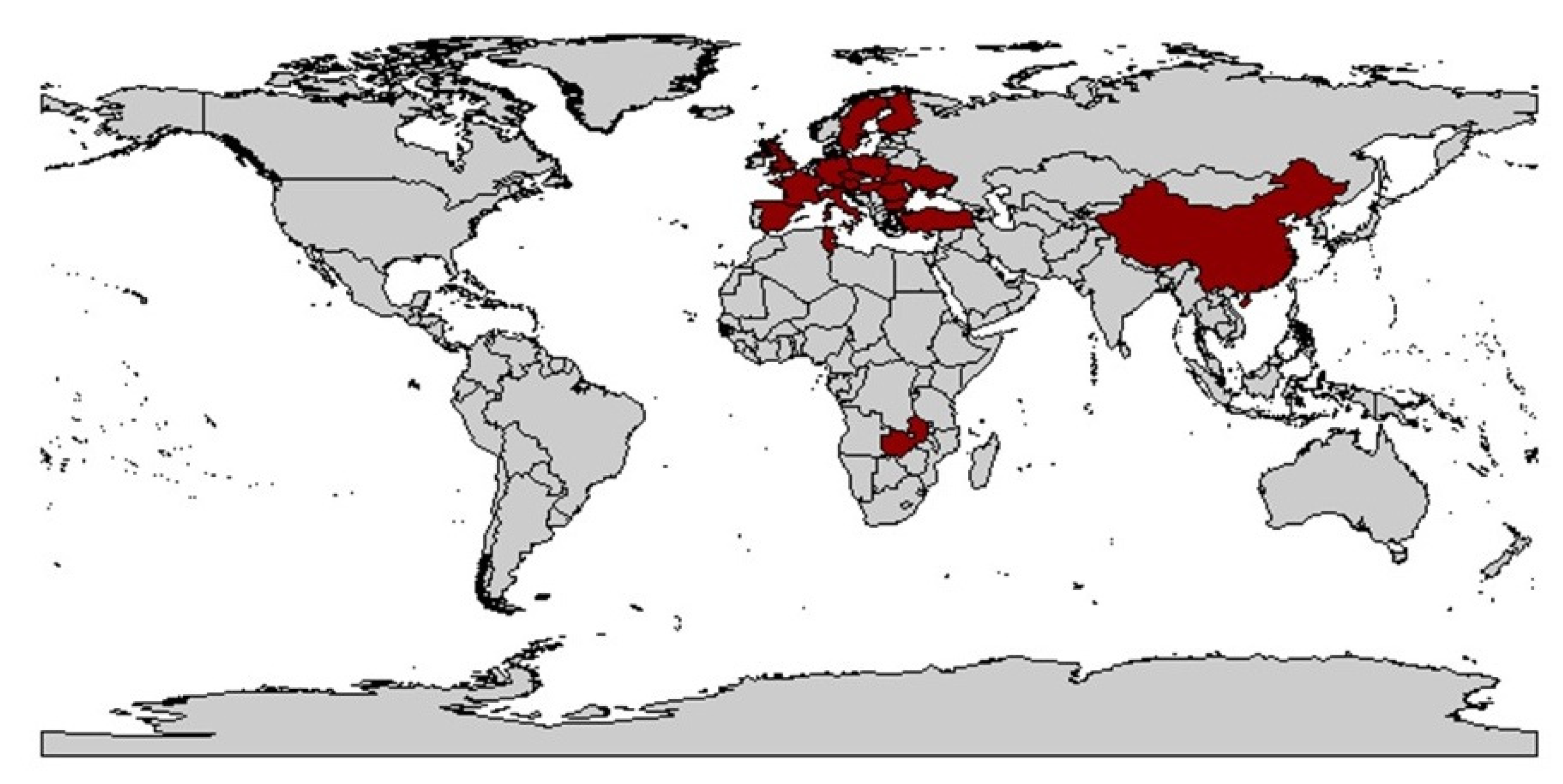
A concrete dispersal route cannot be deduced from the data. However, based on the biology of the animals and their activity, a natural spread over land seems most likely. The virus has been detected in the main Eurasian cereal-growing areas and in its region of origin in the Middle East. This can possibly be attributed to the fact that the climatic requirements for wheat cultivation, for example, match with those of P. alienus. Exceptions like India, as well as Canada and Australia, underline these theories.
The reason for the increasing spread of WDV and the increased occurrence in areas where WDV has been previously reported is not clearly understood but is probably caused by changes in agricultural practices. One of the main causes is assumed to be the increased use of ploughless tillage. Also, the EU regulation on the use of a large part of stubble fields after winter wheat cultivation as set-aside areas was thought to be favorable for P. alienus reproduction and overwintering. Avoiding set-aside areas after the occurrence of WDV-infected wheat and avoiding undersowing crops were therefore considered as possible control measures in Sweden [121]. Furthermore, harvesting with short stubble, early tillage in autumn, and avoiding early sowing had a positive effect on reducing the population of P. alienus [121]. Global climate change may also play a role in promoting the spread of vector-borne diseases. In this context, higher temperatures may favor the colonization of new habitats and hosts. Field monitoring is therefore essential, especially in cereal-growing regions, to identify additional regions where P. alienus may spread together with WDV [116,117,118,119,120] since the spread of WDV results from the migration of virulent vectors from wild or cultivated reservoirs into cereal fields [121,141]. Table 1 provides an overview of the history of WDD.

3.2. Host Range

The host range of WDV includes mainly monocotyledonous plants [37,142]. In addition to a variety of members of the Poaceae family, including important cereals such as wheat (Triticum aestivum L.), barley (Hordeum vulgare L.), rye (Secale cereale L.), oats (Avena sativa), and triticale [11,13,143], WDV also infects various wild and cultivated grasses, including Bromus secalinus L., Lolium multiflorum Lam. [13], Avena fatua L., B. inermis Leyss., B. tectorum L., H. murinum L., L. perenne L., L. temulentum L. [144], A. sterilis L., A. strigosa Schreb., Poa annua L. [103], L. remotum Schrk., Lagurus ovatus L. [145], and Apera spica-venti (L.) P. beauv. [144], which are considered virus reservoirs [13].

3.3. Symptoms of WDD

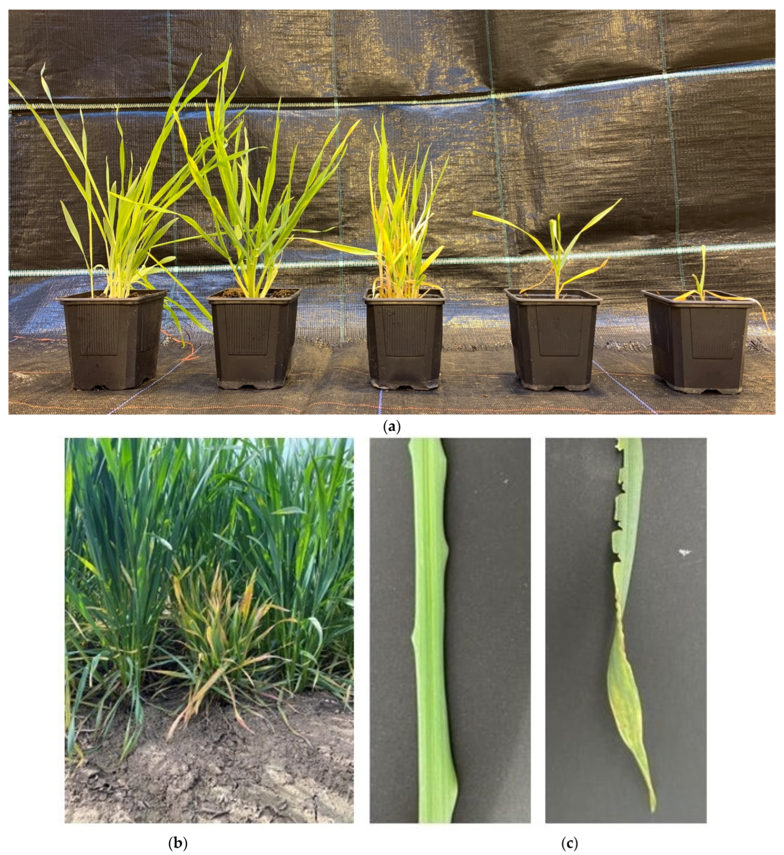
The name of the virus is derived from its main characteristics, the disruption of the shoot growth and the formation of numerous shoots in wheat, resulting in the typical dwarf and bushy growth (Figure 3).
Furthermore, symptoms of WDV infection in wheat also include chlorosis, reduced root size, intense yellow or red discoloration of leaves with or without a mosaic pattern, deformation of leaves, reduced growth hardiness, delayed ear emergence, reduced number of ears as well as sterile flowers, significant yield losses and even complete plant death during early developmental stages of winter wheat and winter barley in winter and spring [13,121,146,147,148,149]. These are partly due to the side effects of infection, such as the effects of expression of viral suppressors of RNA silencing. Symptoms may also affect plant defense responses, leading to plant overreaction in the form of necrosis [150], chlorotic spots, and demarcated streaks on the leaves. The symptoms themselves first appear on the youngest and later on older leaves in association with small cracks and deformations on the youngest leaf, which are characteristic of the infection. This is followed by yellowing of the leaves at the leaf tips and margins with possible partial red coloration [13]. Symptomatic plants usually appear in patches in the field [11,13,148].
In addition to the described symptoms in wheat, the intensity of symptom expression varies among the other infested species. Symptoms in winter barley are similar to those of winter wheat, with no red coloration. Spring barley responds with a lower degree of dwarfing and yellowing of the leaf tips. Similar symptoms occur in winter rye, often associated with anthocyanin formation in leaves and culms. Spring rye shows only minor developmental depression, few leaf spots, and no disruption of generative plants. Oats show minor developmental depression, yellowing, and light red coloration [13]. Triticale shows no increased tillering after WDV infection compared to control plants, but spike-bearing culms shorten by half [151]. In A. spica, growth reductions of 20%, severe tillering, yellowing, and chlorotic spots were observed [13,103]. The wild grass Poa annua shows no symptoms after infection, while Lolium perenne and Lolium multiflorum showed tolerance to WDV in studies with longer plant viability after infection [11].
The extent of damage and the development of symptoms depends on the time of infection. Early infections of winter cereals at the 2–3 leaf stage during fall result in reduced winter hardiness, as well as severe developmental disorders, with pronounced symptoms and negative effects on yield as a result of ear formation that is often partially stuck in the leaf sheaths. The quality of the grains is reduced as they are dried out, shriveled, and partially unable to germinate [13,91]. The root system is also affected by WDV infection. As a result of the infection, there is a reduced formation of secondary roots. The roots appear shorter and thinner overall [91].
Infections in spring result in shortening of internodes and, in some cases, ears. In spring wheat, no severe developmental disorders but shortening of shoots could be observed when infestation occurred from the beginning of shooting to ear swelling (BBCH 31–45). Usually, the first signs of disease in winter wheat appear 18–25 days after infection. In general, symptoms in early-sown wheat are considered to usually appear four to six weeks after infection, while in late-sown wheat, the corresponding symptoms do not become visible until spring, provided the plants are able to overwinter. If infection occurs in spring or early summer, the incubation period lasts three to four weeks. In spring wheat, under greenhouse conditions, the first symptoms are expected 10–15 days after infection, while infections in the field have an incubation period of three weeks [13].
Symptoms caused by infection with Barley yellow dwarf virus (BYDV), which belongs to the Luteoviridae family and is transmitted by aphids, are visually similar to those caused by WDV. When infected in early fall, it causes WDV-like growth depression. The two viruses can only be distinguished from each other by double antibody sandwich enzyme-linked immunosorbent assay (DAS-ELISA) or polymerase chain reaction (PCR), so prior to the discovery of WDV, plants were probably often assigned to BYDV on the basis of dwarfism [151,152].

4. WDV and Its Vector

4.1. Taxonomy and Virus Transmission of P. alienus

WDV is transmitted by the leafhopper species P. alienus, which belongs to the class Insecta order Hemiptera, and uborder Cicadomorpha in the family Cicadellidae. The vector itself is a holarctic species that is common in grasslands and croplands [153]. Occurrence may be particularly high in fallow areas with many self-seeding plants of the Poaceae family. These may serve as reservoirs for WDV [12].
Many species of the Cicadellidae family are vectors of phytopathogenic viruses, including geminiviruses, phytorhabdoviruses, reoviruses, and marafiviruses [154,155,156,157]. In addition to WDV, P. alienus can persistently transmit a rhabdovirus, Wheat Yellow Striate Virus (WYSV, Nucleorhabdovirus genus) [158,159]. Furthermore, P. alienus appears to harbor entomopathogenic viruses that naturally infect insects and can only self-replicate in insect cells. In this context, filovirus-like particles were detected by Lundsgaard [160] in electron microscopic studies, which were confirmed as Taastrup virus (TV) and tentatively assigned to the Mononegavirales [160,161]. Using a next-generation sequencing approach, additional insect-specific viruses were detected, including P. alienus iflavirus1 (PaIV1, genus Iflavirus, family Iflaviridae) [162], Tàiyuán leafhopper virus (TYLeV, genus Mivirus, family Chuviridae) [163], and Hancheng leafhopper Mivirus (HCLeV, genus Mivirus, family Chuviridae) [164]. Transmission electron microscopy (TEM) studies of WYSV-containing sites in salivary glands revealed the presence of reoviruses [165]. Reoviruses include insect-transmitted fijiviruses, which are the most common viral agents of a variety of diseases in gramineae, including Fiji virus (FDV) [166], garlic dwarf virus (GDV) [167], maize dwarf virus (MRDV) [168], Mal de Rio Cuarto virus (MRCV) [169], oat sterility dwarf virus (OSDV) [170], Pangola stunt virus (PaSV) [171], rice black-streaked dwarf virus (RBSDV) [26], and southern rice black-streaked dwarf virus (SRBSDV) [172]. Furthermore, the brown leafhopper Nilaparvata lugens has been found to harbor Nilaparvata lugens reovirus (NLRV), a fijivirus that exclusively infects insects [173]. Most published data suggest that P. alienus is the sole vector of WDV. Some authors have also described a transmission by P. provincialis [137,174]. However, due to the complex taxonomy of species belonging to the genus and the difficulties to distinguish individuals based upon morphological characteristics, the leafhoppers used within the studies are often poorly characterized. This could lead to contradictory results, especially regarding the role of species in WDV transmission [175,176,177].

4.2. Morphology of P. alienus

To easily differentiate adult P. alienus from other leafhoppers, several criteria related to the morphological characteristics of the insects’ head, abdomen, and wings can be used [178,179]. A characteristic of adult P. alienus is their brown coloration with transparent wings, which are longer than the abdomen with a length of 2.7–3.7 mm [180,181]. Accurate species classification requires the morphological description of the male genitalia due to the high variability of the morphological characteristics of the aedeagus. Identification of nymphs and females based on morphological characteristics is currently not possible. This approach often turns out to be unreliable [182,183].
The accuracy of identification of individuals could be improved by using several criteria in parallel, e.g., morphometric parameters in combination with other approaches, such as the emission of species- and sex-specific vibrational signals [184,185,186,187]. Only a few publications have described the vibrational signals emitted by leafhoppers during their sexual communication [182,186,188,189], and a combination of body and aedeagus characteristics combined with the analysis of vibration signals revealed geographic differences between species related to these characteristics. However, this may not only allow the identification of this species but also its origin. Therefore, future studies should include individuals from different countries to improve morphometric data [189]. A more straightforward approach that requires less expert knowledge is the use of DNA barcoding based on sequencing of the mitochondrial cytochrome oxidase I (COI) [190,191]. To date, phylogenetic analysis using DNA barcoding has only been performed for a limited number of species and individuals from Canada, Japan, and Korea [192,193]. Individual specimens of P. confinis and P. helvolus have already been found syntopic to P. alienus using this method [194].

4.3. Life Cycle of P. alienus

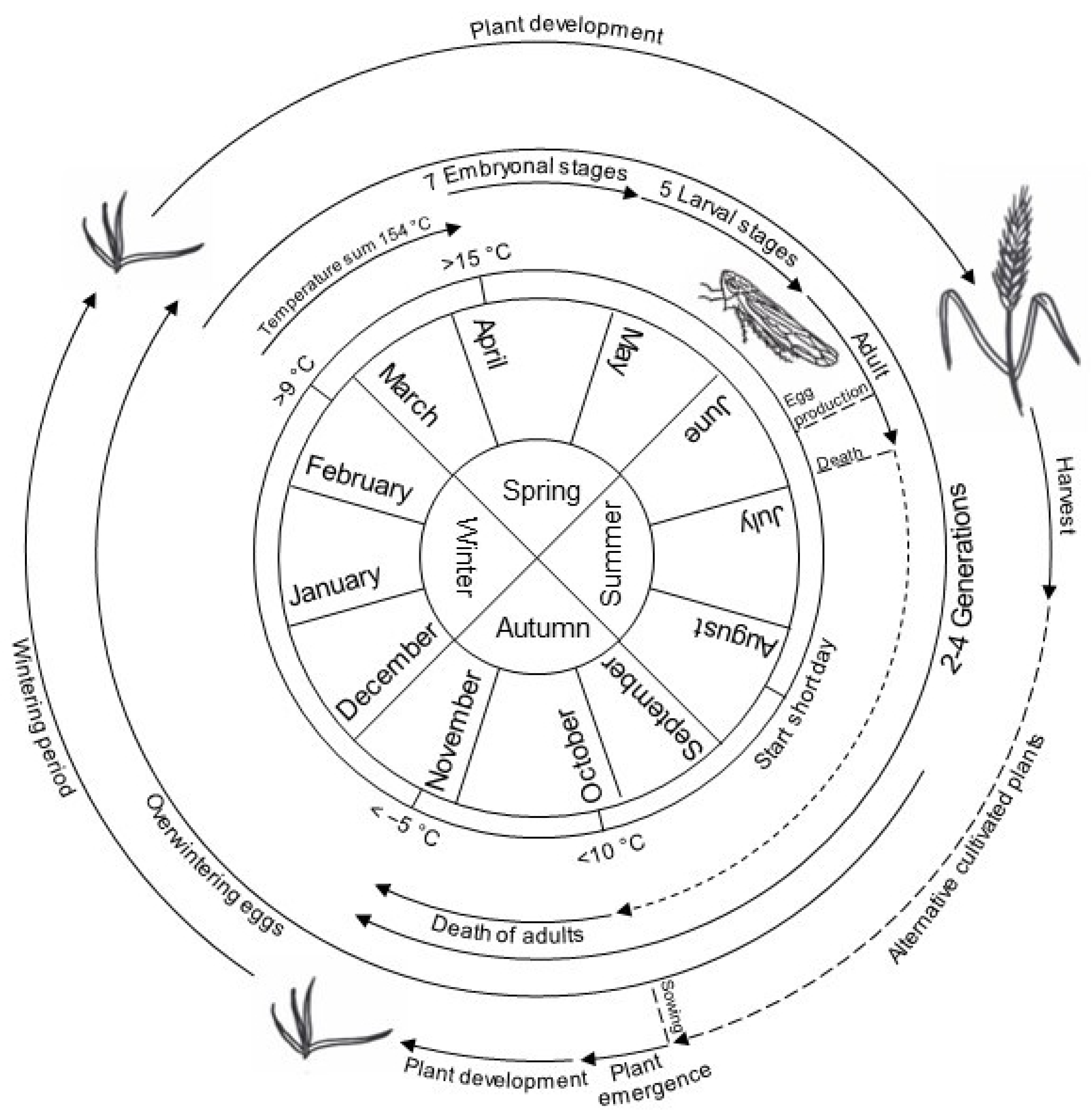
The life cycle of P. alienus has been well studied (Figure 4). High population densities can occur in September, making this the most critical period for WDV infections on young winter cereal plants. Extensive primary infections could be observed until December [12].
Embryonic development is influenced by environmental conditions like temperature and day length. Low temperatures in winter are necessary for the abolition of dormancy (termination) [196]. P. alienus shows seven embryonic stages with a total developmental duration of 16 to 24 days [12]. Depending on temperature, the first larvae hatch in early May. In this context, protandry can be observed, where males hatch earlier than females [196]. The wingless nymphs develop into male and female adult leafhoppers in five stages with a developmental duration of 26–39 days until early summer. Development duration varies, again depending on temperature, but also on host plant species and sex of the leafhoppers. In winter barley, 31 days can be assumed at a temperature of 20 °C [12,195]. After hatching, nymphs move through stocks exclusively by jumping, with older individuals being more mobile than the first two nymphal stages [197]. The newly hatched nymphs acquire the virus from host plants previously infected in the fall, which can lead to secondary infection of plants. It has been observed that the first imagines appear at the end of May, when the temperature sum of all days above 9 °C, measured from the 1st of January of a year, generally reaches 154 °C [12,198]. Fertilization and oviposition occur after the tenth day of the adult stage, so that the first generation begins oviposition in June/July, and the second-generation hatches about 18–20 days later [12,195] and lays its first eggs in early/mid-August. The duration of the entire egg-to-egg life cycle is 58 days [12,195], but higher temperatures may reduce this period, as demonstrated for D. maidis [199]. Dormancy egg laying is induced with the onset of a short day in mid/late August with a rate of 2–20%. From September onwards, up to 100% of eggs are laid as dormancy eggs [196]. Asexual reproduction, as observed in aphids, does not occur in leafhoppers [12,195].
In temperate climate zone, two to four generations per year have been observed so far, depending on environmental conditions [101,177,181], with four complete generations from spring to fall in cereal-growing regions of France, whereas only two P. alienus generations per year occur in northern Europe and northwestern China [200]. Population dynamics studies showed that the density of individuals can reach 43 adults/m2 [12]. The sex ratio in an adult population of P. alienus is close to 1 [200]. The number of adults decreases above a temperature of 10 °C [153]. Freezing temperatures of −5 °C leads to induced death of animals [12,198]. Temperatures above 35 °C have been associated with increased mortality [201]. In contrast, activity and population size of P. alienus increase significantly above a temperature of 15 °C. Thus, a very mild fall therefore leads to very active leafhoppers associated with increased WDV infection rates in the following summer [202].

4.4. Process of Virus Transmission

According to taxonomic affiliation [203] and based on electron microscopic observations [204], P. alienus belongs to the salivary sheath feeders (Auchenorrhyncha), which also includes most of the Sternorrhyncha (aphids, scale insects, psyllids). A salivary sheath is formed in the apoplast by secretions of gel saliva and surrounds the stylet as it moves through plant tissues toward the sieve elements, as shown in aphids [205]. When the stylet reaches the xylem or phloem, the uptake of sap from the vascular cells occurs for the extraction of nutrients [203]. Direct damage by P. alienus caused by sucking activity is considered less important than indirect damage caused by transmission of phloem-restricted WDV [13]. WDV is persistently, circulatively, and non-propagatively transmitted from plant to plant [101,174]. Mechanical, soil- or seed-dependent transmission has not been reported so far [20].
The characteristic of persistent transmission is that a single virus uptake by the vector is sufficient to transmit the virus for months after a short latency period, i.e., the time between the uptake of virus particles and the subsequent release via the salivary glands [20]. A latency period of one to several days is assumed [101,206,207]. Seventeen days after virus acquisition, transmission efficiency was found to be 90%. Transmission efficiency is influenced by environmental conditions, such as temperature, while transmission success depends on the virulence of the virus isolate and the susceptibility of the host [208]. Vector studies on P. alienus are currently focusing on evaluating the transmission of WDV, determining the host plant range, and observing probing behavior on a variety of plants [209].
To date, two pathways of virus movement within the vector and transmission to healthy plants are known. Similar to the persistent virus transmission of other insects, the virus can enter the salivary glands through the anterior midgut and hemocoel [210] or migrate into the lumen of the filtering chamber and on to the midgut lumen after entering the esophagus. Ten minutes after the first feeding, the virus is found throughout the midgut of the insect, and within the next ten minutes, it accumulates throughout the entire filter chamber, midgut, hemocoel, and salivary gland. Four hours after the first feeding, it is no longer detectable in the filter chamber, but it has accumulated in the midgut, hemocoel, and salivary glands, where it remains for the rest of the leafhopper’s life without replicating [207]. The transient direct transfer of particles to the salivary glands occurs within a few minutes, after which the normal circular, non-propagative pathway occurs with the recruitment of the anterior and midgut organs of the leafhopper [211]. Here, the WDV CP not only has an encapsulation function but is also involved in the retention and transmission of WDV in the leafhopper, virus propagation within the plant, and interaction with the Rep protein [105]. Once the vector has acquired the virus by ingestion [10,11,148], the virulent leafhopper can transmit the particles to new hosts each time it sucks. In this process, the virus particles are not lost during molting, so the virus remains in the vector for life. There, it interacts directly with the insect’s organs but does not replicate within the vector [148]. Although the WDV pathosystem is poorly documented in the literature, it has been clearly demonstrated that the virus is not transmitted vertically from virulent females to eggs. Vacke [10] assumed that after the acquisition, all developmental stages are capable of transmitting WDV. This was confirmed by Mehner et al. [11] using transmission tests with larval stages. Larval stages IV and V were more inefficient (22% and 9%, respectively) in terms of virus uptake compared to earlier larval stages and imagines (LI 43%, LII 50%, L3 45%, imago 41%) [11]. Larval stages appear to be more important than adult leafhoppers for WDV dispersal in this regard. Even at low densities, adults and larvae can cause significant yield losses by transmitting the virus to numerous host plants [198]. In the presence of the aphid species Rhopalosiphum padi, a negative effect on larval development, lifespan, and fertility of P. alienus has been observed. Studies of their interaction have ruled out food deprivation as a possible cause. It is hypothesized that the presence of aphids alters leafhopper behavior. This leads to an increase in the number of plants visited by individuals. Thus, this antagonistic interaction between aphids and leafhoppers, commonly found together in cereal fields, indirectly promotes the efficient spread of WDV [198]. Within an experimental approach, the highest infection rates were observed at temperatures of 25 °C. At higher temperatures, leafhoppers tended to settle on the ground, resulting in lower feeding rates and, thus, a decrease in transmission rates [201].

4.5. Host Range and Wild Reservoirs

In particular, the presence of wild grasses in stubble fields as virus reservoirs can lead to an extension of the virus infection period in autumn and promote the occurrence of the disease in spring [12]. The role of wild grasses as WDV virus reservoirs in cropland was demonstrated by Yazdkhasti et al. [212]. The results showed the potential role of ryegrass in the epidemiology of WDV [121] as a symptomless reservoir and underlined the wide host range of WDV [212]. In addition, removal of the overgrowth by plowing immediately after harvest is strongly associated with a reduction in leafhopper [12], probably reducing the spread of WDV from wheat and barley to wild grasses. The host range of P. alienus, as a first-degree oligophagous species [213], is mainly restricted to known host plants of the Poaceae [180,214]. Therefore, in experimental studies, P. alienus has always been reared on grasses such as Hordeum vulgare L. [11,195], Triticum spp. [198], and Festuca gigantea (L.) Vill. [160]. Data from field studies also indicate feeding on other plant species, including alfalfa, carrot [215,216], and ragwort [Ambrosia artemisiifolia L. (Asteraceae)] [217]. This indicates a possible diet of dicotyledonous plants and explains the detection of phytoplasma strains in the body of P. alienus [216,218,219]. These observations contradict the results of a previous study in which P. alienus was not able to survive longer than two days on the two non-grass plants, A. artemisiifolia and Carex tomentosa L. (Cyperaceae). However, in this study, the average survival time of the two species was longer than the starvation control. This is due to the ability of the leafhoppers to possibly take up xylem cell sap from non-host plants [220], where the nutrient and water uptake may contribute to increased survival [209].

4.6. Studies of Insect-Plant Interactions

The behavioral sequence for host plant acceptance of hemipteran insects starts after landing with an exploration of the plant surface, where the plant surface is scanned with the tip of the labium, followed by probing, including cell sap sampling [203,221]. For Cicadellidae, as observed in other hemipteran groups (e.g., aphids), probing seems to be critical to distinguish between host and non-host plants [222]. As a result, not every plant is accepted as a suitable host, and rejection may occur during various stages of probing on the way to the phloem [223,224]. To better understand the behavior of piercing-sucking plant pests and the mechanism of pathogen acquisition and transmission, electrical penetration graph (EPG) technique has been developed to provide real-time observation of the feeding behavior [225,226,227,228]. EPG is probably the most important and widely used technique for studying insect–host–plant interactions, pathogen transmission and acquisition, insecticide effects, and plant resistance [229,230,231,232,233,234]. Within an EPG measurement, insects and plants become integrated into an electrical circuit. The insect closes the electrical circuit by penetrating the plant with its stylet, acting like a switch. Insects and plants act as variable resistors, and different behavior patterns, as well as the stylet’s surrounding environment, affect the electrical resistance, leading to voltage fluctuations that result in different EPG waveforms representing different feeding behavior patterns [228,235,236,237]. The EPG method has been used, for instance, in studies on aphids [226,228,238], leafhoppers [239,240,241,242], mealybugs [243], phylloxerids [244,245], thrips [246,247], and whiteflies [248]. However, data on EPG studies of P. alienus are relatively limited in this regard [221,249,250,251]. Tholt et al. [209] suggested that viruses like WDV are transmitted between insects and plants during the EPG phase Ps4, where the stylet of P. alienus is located in the phloem’s companion cells and sieve cells. In this context, phase Ps4 can be further divided into phase 4a, similar to waveforms E1 shown by aphids, and is associated with the secretion of watery saliva into sieve elements, accompanied by virus transmission. Phase 4b appears to be a homolog to waveform E2 observed in aphids, indicating the ingestion of sieve element sap [209,238,252], probably accompanied by virus acquisition [209]. In addition, Ps4a resembled the X-wave that occurs in other leafhoppers [253,254]. Thus, phase Ps4 is particularly important for WDV transmission [209] and could be used during WDV resistance research.

5. Management of WDD, Its Vector and Virus

Knowledge regarding how to influence the population of P. alienus through appropriate countermeasures is currently insufficient. In field trials, parasitization has been observed very rarely [196]. In Italy, the parasitization of P. alienus larvae and imagines by Gonatopus clavipes Thunberg, G. lunatus Klug (Heminoptera: Dryinidae (cicada wasps)), and representatives of the family Pipunculidae (Diptera: eye flies) native to this country has been observed more frequently [195]. Predominantly in the first generation in May to June, larvae of Gontopus sepoides Westwood have been found on the abdomens of leafhoppers, acting as exoparasites, while Alloneura nigritula Zetterstedt (Pipunculidae) is more commonly found in October to November on P. alienus [255]. In addition, experiments have shown that P. alienus is preyed on by the spider Tibellus oblongus [256].
The actual lack of systematically evaluated, commercially available WDV-resistant and tolerant elite cultivars of wheat and barley means that protection of these cereals against WDV infection relies mainly on agronomic measures and the use of chemically synthesized control agents (insecticides) against P. alienus.
Prevailing cropping practices influence the presence and spread of plant virus diseases, closely correlating with the fluctuating incidence of WDD and the extent of yield losses. The timing of sowing, coordinated with the migration of vectors between fields, is a critical element of an integrated pest management (IPM) strategy [257]. The presence of infected reservoirs, e.g., wild grasses, leads to an increase in the incidence of many viruses, including MSV and WDV [143,258], which in turn involves the field hygiene aspect to reduce WDV infection. Another risk is irregular germination of seedlings [177], as P. alienus is attracted to patchy stands [17]. In addition, feeding behavior, population density, and activity, the latter influenced by weather conditions, affect the intensity and frequency of a WDV infestation [177,259]. A WDV infection is possible at different stages of development (Figure 2), with economic damage decreasing with later infection [17], as has been described for other viruses such as BYDV [260]. Furthermore, it has been shown in wheat that plant resistance can develop after the stage of pseudo stem break (Z30) at the time of the first node (Z31) [202].
Although IPM aims to reduce the application of chemically synthesized insecticides and other pesticides, it does not exclude the possibility of insecticide application. With regard to virus spread, the insecticide-induced reduction of vector insects has been shown to reduce the spread of insect-transmitted viruses [177,258,261]. However, the application of insecticides is associated with negative environmental side effects [262,263], including harmful effects on beneficial insects [264,265,266]. Together, the consideration of these aspects, as well as the broad public request and political will to reduce the use of insecticides, means that the focus for controlling WDV is mainly on agronomic measures and the breeding of resistant/tolerant varieties.

6. Resistance Research and Status Quo in Wheat

Abiotic and biotic factors exert a constant influence on plant populations. Naturally, plants have inherent defense mechanisms that make them resistant to virus invasion [267]. One way is to combat the virus by induced mechanisms, such as RNA silencing with small interfering RNA (siRNA) in response to the virus’s double-stranded RNA (dsRNA), hypersensitive response (HR), or nucleic acid methylation before infection occurs [268]. To date, nothing has been reported on effective and protective defence responses against WDV [269].
In recent decades, various studies have attempted to identify WDV-resistant germplasm among the available wheat and barley accessions. Disease resistance genes in wild relatives of wheat can serve as valuable sources for resistance breeding [270]. Differential resistance to Soil-borne wheat mosaic virus (SBWMV) has been demonstrated in Ae. tauschii and T. monococcum [271,272,273] and in Ae. geniculata to BYDV [274]. Furthermore, Ae. caudata, Ae. ovata and Ae. triuncialis have been shown to respond to WDV infection with milder forms of symptoms compared to spring wheat [13].
Transmission of the virus to the genotypes to be tested has been carried out in previous studies using the natural vector P. alienus or agroinfections. Phenotyping of infected plants is possible under field [3,147,149], and near-field conditions [275,276], or in the greenhouse [275,276,277]. For field inoculation with virus-bearing leafhoppers, both natural and artificial inoculation can be used. In order to protect the crops from natural insect infestation and bird-induced damage, trials can be conducted under semi-field conditions within a gauze house [275,276].
Within phenotyping for resistance, various agronomic parameters may be of interest. Virus infections with WDV affect the performance and yield of infected plants compared to healthy plants. Here, the traits of plant height, number of ears per plant, grains per ear, grain yield per plant, and thousand kernel weight (TKW) per plant can serve as suitable indirect parameters for characterizing resistance [278]. Between tillering and sprouting (BBCH 23–30), as well as after harvest (BBCH 92), a comparative symptom assessment from 1 to 9 can be performed according to Scheurer et al. [279].
Serological and molecular techniques are available for the detection of WDV infection as well as for a precise assignment of isolates to the corresponding strain designations. For the verification of WDV infections in the field, direct virus detection, via ELISA [280] and PCR [101,281,282,283], has proven to be a reliable method [152,284]. Differentiation of the WDV strains in the host plants and vector samples can be made on the basis of the characteristics of viral compounds (capsid proteins, nucleic acids). Due to the high sequence similarity between the CP of the isolates, serological differentiation of these using polyclonal antisera is not possible [147], but the use of monoclonal antibodies has been reported [285]. Several established molecular methods are available for the identification of WDV strain-specific sequences, such as standard PCR [80,102], restriction fragment length polymorphism (RFLP) [286], rolling circle amplification restriction fragment length polymorphism [104], and isothermal recombinase polymerase amplification methods [287]. In addition, molecular-based quantification assays in the form of real-time PCR assays targeting a conserved region of the CP gene sequence and using a Taq-Man probe have been added to the list of detection methods [174].
So far, no highly resistant WDV bread wheat variety is known. However, tendencies to favor different wheat varieties [288] and differences in susceptibility have been found (Table 2).
Based on yield reduction, studies were conducted on winter wheat to identify tolerant groups [149]. These showed only minor quantitative differences between the tested host plants and reference genotypes [3,147]. Most genotypes were susceptible to WDV infection, and only a few genotypes could be classified as moderately resistant. Within screenings, the Czech winter wheat cultivars ‘Banquet’ and ‘Svitava’ showed reduced virus levels, with moderate susceptibility at a yield reduction of 87.3–93.1% [149]. Moderate yield reductions of 82.5–92.6% after WDV inoculation were shown by the Russian cultivars ‘Belocerkovskaya,’ ‘Kharkovskaya,’ ‘Mironovskaya 808’, ‘Yubileynaya’ and ‘Kawvale’ and the Slovak and Czech cultivars ‘Astella,’ ‘Boka,’ ‘Bruneta,’ ‘Bruta,’ ‘Ilona,’ ‘Ina,’ ‘Mona,’ ‘Regina,’ ‘Saskia,’ and ‘Senta’ [147]. The winter wheat varieties ‘Mv Dalma’ and ‘Mv Vekni’ from Martonvásár (Hungary) were described by Benkovics et al. [289] as the first partially resistant varieties. In leafhopper transmission tests, both cultivars were infected (53%) but showed milder symptoms and a 100–10,000 times lower virus titer than the susceptible reference host cultivars ‘Mv Emese’ and ‘Mv Regiment’ (100% infection) four weeks after infection. A difference in the survival rates of the leafhoppers could not be determined. It can, therefore, be assumed that the resistance mechanism of the cultivars is based on the movement or replication of the virus and not on insect feeding [289]. ‘Mv Dalma’ carries a homozygous 1AL.1RS, while ‘Mv Vekni’ carries a homozygous 1BL.1RS rye translocation and contains several stem, leaf, and yellow rust resistance genes derived from Aegilops ventricose (VPM-1, SR38, Lr37, YR17) [289,290,291].
To clarify the genetic basis of partial resistance in ‘MV Vekni,’ in a recent work, F2 populations based on a cross between the susceptible cultivar Regiment were inoculated in greenhouse experiments, and quantitative trait loci (QTL) analysis was performed. Significant QTL were found for the peak markers RFL_Contig6053_2072 and Kukri_rep_c95718_868 on chromosome 6A for virus extinction (LOD = 22.6), which explained a phenotypic variance of 38.4%. The significant deviation from the expected segregation ratio of 3r:1s observed in this work indicated that the resistance is primarily inherited monogenetically due to the action of one major gene eventually accompanied by additional minor QTL that could not be detected within the analysis. The hypothesis of coupling rye introgression with WDV resistance in Vekni could not be confirmed in this work. Within the main QTL interval, among others, a gene encoding protein kinase activity could be identified [292]. These are involved in various defense mechanisms against geminiviruses, leading to attenuation and reduction of infection [293]. Furthermore, genes associated with DNA-directed transcriptional regulation in Triticum aestivum have been found to act as viral defense modulators, influencing the host-dependent DNA replication cycle [51,292].
In a recent study [294], the changes in transcriptome profiles of the resistant wheat genotypes ‘Svitava’ and ‘Fengyou 3’ compared to the susceptible cultivar ‘Akteur’ were investigated after WDV infection. The study provides insights into the specific transcriptome profiles and pathways associated with resistance and susceptibility to WDV in wheat genotypes. RNA-Seq analysis revealed significantly different expressions of transcripts in response to WDV infection in ‘Akteur,’ ‘Fengyou 3’, and ‘Svitava’ genotypes. Gene ontology (GO) analysis showed that different biological processes, cellular components, and molecular functions were activated in the tested genotypes. The resistant genotype showed significant activation of biological processes compared to the susceptible genotype. Certain classes of genes were affected by WDV infection. For example, transport activity was suppressed [294], which could prevent virus movement and accumulation [295]. On the other hand, oxidoreductase and lyase activities were activated [294], which are involved in defense responses and limit virus accumulation [296]. The ‘Svitava’ genotype suppressed reductase protein classes and chaperones. The latter group includes heat shock proteins (HSP), which play a role in viral DNA/protein aggregation and viral reduction [297,298,299]. Suppression of reductase activity is associated with a reduction in reactive oxygen species (ROS) accumulation, which is associated with better adaptation to viral infections [300]. Analyses of GO and KEGG metabolic pathways revealed reprogramming of several transcripts in response to WDV infection, particularly in the carbohydrate, energy, lipid, nucleotide, amino acid, glycan, and vitamin metabolism. Secondary metabolic and photosynthetic pathways were induced in ‘Svitava.’ The susceptible genotype showed down-regulation of photosynthesis-related carbon fixation genes, which, in contrast, were induced in the resistant genotypes. Transcripts for the biosynthesis of other secondary metabolites were upregulated in ‘Svitava’ and downregulated in ‘Fengyou 3’ and ‘Akteur,’ possibly contributing to higher resistance through their antiviral properties [294,301]. Transcription factors (TFs), including AP2/ERF, bHLH, MYB, and WRKY families, were highly enriched under WDV infection [294]. These TFs are known to regulate plant responses to various biotic and abiotic stresses [302,303]. In particular, ERFs have been linked to plant immune responses and resistance to plant viruses [304].
In greenhouse experiments with 13 wild and five domesticated wheat taxa of different ploidy, accessions of the species Aeg. tauschii, Aeg. cylindrical, Aeg. Searsii, and T. spelta showed WDV tolerance. The accessions were initially strongly affected by symptoms 28 days after infection (dpi). Thereafter, there was a decline in symptoms with a relative increase in leaves and shoots at 112 dpi. Within the study, domesticated wheat cultivars did not always show more severe symptoms, but there was a differential impact of infection on growth traits and leaf chlorosis in wild and domesticated wheat cultivars [277]. This could be attributed to a slight RNA silencing suppressor activity of the WDV proteins Rep and RepA [62,305]. Both viral proteins, when expressed in infiltrated transgenic leaves of Nicotiana benthamiana with a green fluorescent protein (GFP) reporter gene, resulted in the inhibition of post-transcriptional gene silencing (PTGS) and RNA silencing of the GFP reporter gene [305].
Within another study, 500 wheat accessions were phenotyped for WDV resistance by artificial inoculation in gauze houses. The majority of accessions showed a strong impact of WDV infection with a wide range of reductions in plant height (3.6–100%), number of ears (0–100%), and yield (2.3–100%) [275]. In contrast to Nygren et al. [277], domesticated wheat varieties within the panel did not have a generally higher infection rate than wild wheat varieties and relatives [275]. The authors concluded that the genetic bottleneck that arose during evolution and domestication did not necessarily lead to higher WDV susceptibility but that these variations created by ancestral hybridization were compensated for. During the study, the partially resistant genotypes ‘MV Dalma’ and ‘MV Vekni’ were confirmed with an average infection rate of 34.5% and 21.5%, respectively, and weaker symptom expression compared to susceptible varieties. In addition, 19 other sources of WDV resistance with lower infection rates than ‘MV Vekni’ were identified, including di-, tetra-, and hexaploid genebank wheat accessions. Ten T. aestivum, two T. vavilovii, two T. sp. (genebank accessions with unknown subspecies), one T. boeoticum, one T. macha, one Ae. geniculata, one Ae. Bicornis, and one Ae. longissima accession had lower infection rates than ‘MV Vekni.’ The cultivar ‘Fisht’ proved to be another resistant cultivar with a low average number of infected plants (5.7%) and less severe virus symptoms (average scoring value 2.3, for symptom scoring see [275]) compared to the reference cultivars ‘Mv Dalma’ (34.5%, 5.9) and ‘Mv Vekni’ (21.5%, 4.6) and the susceptible ‘Mv Regiment’ (64.9%, 6.7) as well as ‘Mv Emese’ (68.1%, 6.9). Overall, the results indicated that there are natural sources of WDV resistance within the wheat gene pool. A subpanel was also used to identify QTL for WDV resistance in hexaploid wheat. The putative 35 QTL (FDR, α < 0.05) for partial WDV resistance for the traits relative plant height (relPH), relative yield (relYield), and relative thousand kernel weight (relTKW) are located on chromosomes 1B, 1D, 2B, 3A, 3B, 4A, 4B, 5A, 6A, 7A, and 7B. Among them, the most significant QTL were detected on chromosome 1B, especially six QTL explaining more than 10% of the phenotypic variance (LOD 5.0–8.7) and two highly significant yield-related QTL explaining 18.3% of the phylogenetic variance (LOD 5.0–8.7), which can be used to develop molecular markers in resistance breeding. The QTL identified here could be associated with genes encoding DNA template regulation of transcription, splicing mRNA by spliceosome, gene silencing by RNA, and protein kinase activity [275]. Genes responsible for the regulation of DNA template transcription may serve as modulators of viral defense, particularly with respect to controlling the host-dependent DNA replication cycle of WDV [51]. Previous research on RNA-mediated gene silencing has also demonstrated the ability of geminiviruses to trigger post-transcriptional gene silencing (PTGS) [306,307], such that viral dsRNA is degraded during the RNA splicing mechanism to small interfering RNAs (siRNAs) that align and degrade silencing complexes to sequence-specific mRNA [308]. Also involved in plant resistance to geminiviruses are protein kinase domains through phosphorylation of viral pathogenesis proteins. The viral protein ßC1 is phosphorylated by SNF1-related kinases, which has negative effects on RNA silencing suppressor function or labeling for degradation in the 26s proteosome. As a result, delayed/reduced viral infection may be observed [309]. Overall, the results suggest that other resistance genes are involved in defense against WDV.
Previous studies have shown that resistance to various viruses is localized to the D chromosome. For example, resistance to Soil-borne Wheat Mosaic Virus (SBWMV) is localized on chromosomes 4D and 5D, and the resistance gene encoding alleles on chromosome 5D is due to Aegilops tauschii [310,311]. Other highly significant marker-trait associations (MTA) were found on chromosome 2D for resistance to Wheat spindle streak mosaic virus (WSSMV) [312]. Of 35 QTL identified, 25 QTL, explaining between 7.4 and 18.3% of the phenotypic variance, were verified in four biparental populations with the cultivar ‘Fisht’ as a parent [275]. Within the segregation analysis, two of the markers showed significant effects on relYield, eleven on relTKW, and ten on relative virus titers. The QTL on chromosome 1B consistently showed highly significant effects in all four populations [275].
A recent QTL study revealed two additional highly significant QTL associated with WDV resistance [313]. The primary QTL, Qwdv.ifa-6A, mapped to the long arm of chromosome 6A between markers Tdurum_contig75700_441 (at 601,412,152 bp) and AX-95197581 (at 605,868,853 bp). Qwdv.ifa-6A originated from the Dutch experimental line SVP-72017 and showed a strong effect in all populations, explaining a significant proportion (up to 73.9%) of the phenotypic variance. The second QTL, Qwdv.ifa-1B, was located on chromosome 1B and derived from the susceptible parental line P1314. The QTL is possibly linked to the 1RS.1BL translocation, which originated from the CIMMYT line CM-82036. Qwdv.ifa-1B was responsible for a substantial portion (up to 15.8%) of the phenotypic variance in WDV resistance [313]. The efficacy of the rye chromatin segment 1RS.1BL against Wheat Streak Mosaic Virus (WSMV) has been reported previously [314], but there is no evidence to date that the same gene confers resistance to both WDV and WSMV. The QTL mapped on the short arm of chromosome 1B in the study by Pfrieme et al. [275] overlaps with the Qwdv.ifa-1B QTL identified within the study by Buerstmayr and Buerstmayr [313]. Although Fisht has the preferable allele on chromosome 1B, the presence of the translocation 1RS.1BL remains unclear. Thus, it remains uncertain whether ‘Fisht’ and P1314 (the resistance donor for Qwdv.ifa-1B) have the same resistance gene. This study has shown that Qwdv.ifa-6A and Qwdv.ifa-1B are clearly additive, suggesting that the pyramidization of resistance QTL could increase both the durability and extent of resistance [313].
Table 2. Overview of the key findings of WDV resistance breeding in historical sequence.
TimeEventReference
1982Report: WDV shows tendencies to prefer different wheat varieties.[288]
2000Screening: Description of five Russian varieties as well as ten Slovakian and Czech varieties with moderate yield reduction after WDV infection.[147]
2005Screening: Description of the Czech winter wheat varieties ‘Banquet’ and ‘Svitava’ with reduced virus titer, moderate susceptibility, and yield reduction.[148]
2010Screening: Description of the Hungarian winter wheat varieties ‘Mv Dalma’ and ‘Mv Vekni’) as partially resistant varieties.[289]
2015Screening: Proof of WDV tolerance of accessions of the species Aeg. Tauschii, Aeg. Cylindrical, Aeg. Searsii, and T. spelta.[277]
2022Screening: Identification of 19 sources of WDV resistance with lower infection rates than ‘MV Vekni,’ including di-, tetra-, and hexaploid genebank wheat varieties as well as the winter wheat variety ‘Fisht.’[275]
2022Genome-wide association study: Detection of 35 putative QTL for partial WDV resistance on chromosomes 1B, 1D, 2B, 3A, 3B, 4A, 4B, 5A, 6A, 7A, and 7B.[275]
2022QTL analysis: Identification of two significant QTL on chromosome 6A in the variety ‘Mv Vekni.’[292]
2023Transcriptome analysis: A study of changes in resistant wheat genotypes ‘Svitava’ and ‘Fengyou 3’ compared to susceptible cultivar ‘Akteur’ after WDV infection.[294]
2023QTL study: Identification of a QTL on chromosome 6A in the Dutch experimental line SVP-75360 and a QTL on chromosome 1B of line P1361.[314]
2024QTL study: Identification of QTL in the winter wheat variety Fisht.
The utility of the discovered QTL for wheat breeding depends on their ability to predict quantitative WDV resistance in a range of genetic backgrounds. For breeding purposes, QTL associated with resistance should explain at least 10% of the phenotypic variance. Their pyramiding is an interesting approach to increase resistance to WDV [275,315,316,317], as already shown for BYDV in barley [278,318]. The use of the identified QTL in marker-assisted selection can be achieved by developing PCR-based markers from verified array-based markers. For example, the use of competitive allele-specific PCR markers (KASP) developed from flanking marker sequences offers an efficient approach in hexaploid wheat [319,320,321]. The introduction of WDV tolerance can be facilitated by the use of molecular markers, avoiding artificial inoculation with virus-bearing leafhoppers, which is difficult to integrate into applied breeding programs.

7. Conclusions

WDV is a worldwide virus disease that affects most cereals and grasses. As a result of climate change, the importance of insect-transmitted viruses will inevitably increase in the coming years. Research conducted within the last decades allows a description of the biology of the putative vector, the virus, and the plant hosts. In this context, the epidemiology of WDV is characterized by the presence of different strains, recombinants, and virus species, as well as a complex taxonomy of vectors and a contradictory host range. Although WDV as a DNA virus is thought to have a lower mutation rate compared to RNA viruses, putative new variants, and recombinants have already been detected in reservoirs and crop species in recent years. Since there are no approved chemical control agents in the European Union, agronomic measures are currently the only way to control WDV. The detection of the first WDV-resistant genotypes and QTL in wheat indicates that resistance is present in the cereal pool. As indicated by this review, further experimental studies on WDV resistance and the epidemiology of the vector are needed and promising, especially given the economic importance of this viral disease. The development of resistant cereal varieties offers the prospect of minimizing the spread and losses due to WDV infections.

Author Contributions

Conceptualization, A.-K.P.; writing—original draft preparation, A.-K.P.; writing—review and editing, A.S., T.W. and K.P.; visualization, A.-K.P. All authors have read and agreed to the published version of the manuscript.

Funding

This research was funded by German Federal Ministry of Food and Agriculture (BMEL) and the German Rentenbank grant number FKZ: 28RZ4IP029.

Data Availability Statement

Not applicable.

Conflicts of Interest

The authors declare no conflict of interest.

References

  1. Buck, K.W. Geminiviruses (Geminiviridae). In Encyclopedia of Virology; Granoff, A., Webster, R.G., Eds.; Academic Press: Cambridge, MA, USA, 1999; pp. 597–606. [Google Scholar] [CrossRef]
  2. Canto, T.; Aranda, M.A.; Fereres, A. Climate change effects on physiology and population processes of hosts and vectors that influence the spread of hemipteran-borne plant viruses. Glob. Chang. Biol. 2009, 15, 1884–1894. [Google Scholar] [CrossRef]
  3. Habekuß, A.; Riedel, C.; Schliephake, E.; Ordon, F. Breeding for resistance to insect-transmitted viruses in barley—An emerging challenge due to global warming. J. Für Kult. 2009, 61, 53–61. [Google Scholar] [CrossRef]
  4. Roos, J.; Hopkins, R.; Kvarnheden, A.; Dixelius, C. The impact of global warming on plant diseases and insect vectors in Sweden. Eur. J. Plant Pathol. 2011, 129, 9–19. [Google Scholar] [CrossRef]
  5. Ziesche, T.M.; Bell, J.; Ordon, F.; Schliephake, E.; Will, T. Long-term monitoring of insects in agricultural landscapes. Mitteilungen Der DGaaE 2020, 22, 101–106. [Google Scholar]
  6. Barnett, O.W.; Main, C.E. Plant Virus Disease—Economic Aspects. In Encyclopedia of Virology; Elsevier: Amsterdam, The Netherlands, 1999; pp. 1318–1326. [Google Scholar] [CrossRef]
  7. Waterworth, H.E.; Hadidi, A. Economic Losses due to Plant Viruses. In Plant Virus Disease control; APS Press: St. Paul, MN, USA, 1998. [Google Scholar]
  8. Fraser, R.S.S. Plant Resistance to Viruses|Natural Resistance. In Encyclopedia of Virology, 2nd ed.; Granoff, A., Webster, R.G., Eds.; Elsevier: Amsterdam, The Netherlands, 1999; pp. 1300–1307. [Google Scholar] [CrossRef]
  9. van Regenmortel, M.H.; Fauquet, C.M.; Bishop, D.H.; Carstens, E.B.; Estes, M.K.; Lemon, S.M.; Maniloff, J.; Mayo, M.A.; McGeoch, D.J.; Pringle, C.R.; et al. Virus Taxonomy: Classification and Nomenclature of Viruses. In Seventh Report of the International Committee on Taxonomy of Viruses; Academic Press: New York, NY, USA, 2000. [Google Scholar]
  10. Vacke, J. Wheat dwarf virus disease. Biol. Plant 1961, 3, 228–233. [Google Scholar] [CrossRef]
  11. Mehner, S.; Manurung, B.; Gruntzig, M.; Habekuss, A.; Witsack, W.; Fuchs, E. Investigations into the ecology of the Wheat dwarf virus (WDV) in Saxony-Anhalt, Germany. J. Plant Dis. Prot. 2003, 110, 313–323. [Google Scholar]
  12. Manurung, B.; Witsack, W.; Mehner, S.; Gruntzig, M.; Fuchs, E. Studies on biology and population dynamics of the leafhopper Psammotettix alienus Dahlb. (Homoptera: Auchenorrhyncha) as vector of Wheat dwarf virus (WDV) in Saxony-Anhalt, Germany. J. Plant Dis. Prot. 2005, 112, 497–507. [Google Scholar]
  13. Vacke, J. Host plants range and symptoms of wheat dwarf virus. Věd Pr Výz Ust. Rostl Výroby Praha-Ruzyně 1972, 17, 151–162. [Google Scholar]
  14. MacDowell, S.W.; Macdonald, H.; Hamilton, W.D.O.; Coutts, R.H.A.; Buck, K.W. The nucleotide sequence of cloned wheat dwarf virus DNA. EMBO J. 1985, 4, 2173–2180. [Google Scholar] [CrossRef] [PubMed]
  15. Macdonald, H.; Coutts, R.H.A.; Buck, K.W. Characterization of a Subgenomic DNA Isolated from Triticum Aestivum Plants Infected with Wheat Dwarf. J. Gen. Virol. 1988, 69, 1339–1344. [Google Scholar] [CrossRef]
  16. Schalk, H.J.; Matzeit, V.; Schiller, B.; Schell, J.; Gronenborn, B. Wheat dwarf virus, a geminivirus of graminaceous plants needs splicing for replication. EMBO J. 1989, 8, 359–364. [Google Scholar] [CrossRef] [PubMed]
  17. Lindblad, M.; Waern, P. Correlation of wheat dwarf incidence to winter wheat cultivation practices. Agric. Ecosyst. Environ. 2002, 92, 115–122. [Google Scholar] [CrossRef]
  18. Lemmetty, A.; Huusela-Veistola, E. First Report of Wheat dwarf virus in Winter Wheat in Finland. Plant Dis. 2005, 89, 912. [Google Scholar] [CrossRef] [PubMed]
  19. Wang, J.; Guan, Y.; Wu, L.; Guan, X.; Cai, W.; Huang, J.; Dong, W.; Zhang, B. Changing Lengths of the Four Seasons by Global Warming. Geophys. Res. Lett. 2021, 48, e2020GL091753. [Google Scholar] [CrossRef]
  20. Lindsten, K.; Lindsten, B.; Abdelmoeti, M.; Junti, N. Purification and some properties of wheat dwarf virus. In Proceedings of the 3rd Conference on Virus Diseases of Gramineae in Europe, Rothamsted, UK, 28–30 May 1980; pp. 27–31. [Google Scholar]
  21. Fauquet, C.M.; Briddon, R.W.; Brown, J.K.; Moriones, E.; Stanley, J.; Zerbini, M.; Zhou, X. Geminivirus strain demarcation and nomenclature. Arch. Virol. 2008, 153, 783–821. [Google Scholar] [CrossRef] [PubMed]
  22. Bernardo, P.; Golden, M.; Akram, M.; Naimuddin, N.N.; Fernandez, E.; Granier, M.; Rebelo, A.G.; Peterschmitt, M.; Martin, D.P.; Roumagnac, P. Identification and characterisation of a highly divergent geminivirus: Evolutionary and taxonomic implications. Virus Res. 2013, 177, 35–45. [Google Scholar] [CrossRef]
  23. Varsani, A.; Navas-Castillo, J.; Moriones, E.; Hernández-Zepeda, C.; Idris, A.; Brown, J.K.; Murilo Zerbini, F.; Martin, D.P. Establishment of three new genera in the family Geminiviridae: Becurtovirus, Eragrovirus and Turncurtovirus. Arch. Virol. 2014, 159, 2193–2203. [Google Scholar] [CrossRef]
  24. Agrios, G.N. Plant Pathology, 3rd ed.; Academic Press: New York, NY, USA, 1988; pp. 3–39. [Google Scholar] [CrossRef]
  25. Matzeit, V. Wheat Dwarf Virus—Ein Geminivirus Monokotyledoner Pflanzen-DNA-Sequenz, Replikation und Einsatz Seines Genoms zur Amplifikation und Expression Fremder Gene. Ph.D. Thesis, Universität zu Köln, Köln, Germany, 1988. [Google Scholar]
  26. Zhang, W.; Olson, N.H.; Baker, T.S.; Faulkner, L.; Agbandje-McKenna, M.; Boulton, M.I.; Davies, J.W.; McKenna, R. Structure of the Maize Streak Virus Geminate Particle. Virology 2001, 279, 471–477. [Google Scholar] [CrossRef]
  27. Boulton, M.I. Functions and interactions of mastrevirus gene products. Physiol. Mol. Plant Pathol. 2002, 60, 243–255. [Google Scholar] [CrossRef]
  28. Drews, G.; Adam, G.; Heinze, C. Molekulare Pflanzenvirologie; Springer: Berlin/Heidelberg, Germany, 2004. [Google Scholar] [CrossRef]
  29. Adejare, G.O.; Coutts, R.H.A. The Isolation and Characterisation of a Virus from Nigerian Cassava Plants Affected by the Cassava Mosaic Disease, and Attempted Transmission of the Disease. J. Phytopathol. 1982, 103, 198–210. [Google Scholar] [CrossRef]
  30. Harrison, B.D. Advances in Geminivirus Research. Annu. Rev. Phytopathol. 1985, 23, 55–82. [Google Scholar] [CrossRef]
  31. Damsteegt, V.D.; Igwegbe, E.C.K. Epidemiology and Control of Maize streak disease. In Plant Virus Disease Control; APS Press: St. Paul, MN, USA, 1998; pp. 484–494. [Google Scholar]
  32. Moffat, A.S. Geminiviruses Emerge as Serious Crop Threat. Science 1999, 286446, 1835. [Google Scholar] [CrossRef]
  33. Lefkowitz, E.J.; Dempsey, D.M.; Hendrickson, R.C.; Orton, R.J.; Siddell, S.G.; Smith, D.B. Virus taxonomy: The database of the International Committee on Taxonomy of Viruses (ICTV). Nucleic Acids Res. 2018, 46, D708–D717. [Google Scholar] [CrossRef] [PubMed]
  34. Fiallo-Olivé, E.; Lett, J.-M.; Martin, D.P.; Roumagnac, P.; Varsani, A.; Zerbini, F.M.; Navas-Castillo, J. ICTV Virus Taxonomy Profile: Geminiviridae 2021. J. Gen. Virol. 2021, 1022, 001696. [Google Scholar] [CrossRef]
  35. Family: Geminiviridae. Available online: https://ictv.global/report/chapter/geminiviridae/geminiviridae (accessed on 12 November 2022).
  36. Fauquet, C.M.; Bisaro, D.M.; Briddon, R.W.; Brown, J.K.; Harrison, B.D.; Rybicki, E.P.; Stenger, D.C.; Stanley, J. Virology division news: Revision of taxonomic criteria for species demarcation in the family Geminiviridae, and an updated list of begomovirus species. Arch. Virol. 2003, 148, 405–420. [Google Scholar] [CrossRef]
  37. Muhire, B.; Martin, D.P.; Brown, J.K.; Navas-Castillo, J.; Moriones, E.; Zerbini, F.M.; Rivera-Bustamante, R.; Malathi, V.G.; Briddon, R.W.; Varsani, A. A Genome-Wide Pairwise-Identity-Based Proposal for the Classification of Viruses in the Genus Mastrevirus (Family Geminiviridae). Arch. Virol. 2013, 158, 1411–1424. [Google Scholar] [CrossRef]
  38. Candresse, T.; Filloux, D.; Muhire, B.; Julian, C.; Galzi, S.; Fort, G.; Bernardo, P.; Daugrois, J.H.; Fernandez, E.; Martin, D.P.; et al. Appearances can be deceptive: Revealing a hidden viral infection with deep sequencing in a plant quarantine context. PLoS ONE 2014, 9, e102945. [Google Scholar] [CrossRef]
  39. NCBI Virus. Available online: https://www.ncbi.nlm.nih.gov/labs/virus/vssi/#/virus?SeqType_s=Nucleotide&VirusLineage_ss=Mastrevirus,%20taxid:11212 (accessed on 22 September 2023).
  40. Morris, B.A.M.; Richardson, K.A.; Haley, A.; Zhan, X.; Thomas, J.E. The nucleotide sequence of the infectious cloned DNA component of tobacco yellow dwarf virus reveals features of geminiviruses infecting monocotyledonous plants. Virology 1992, 187, 633–642. [Google Scholar] [CrossRef]
  41. Gutierrez, C. Geminivirus DNA replication. Mol. Life Sci. CMLS 1999, 56, 313–329. [Google Scholar] [CrossRef]
  42. Thomas, J.E.; Parry, J.N.; Schwinghamer, M.W.; Dann, E.K. Two novel mastreviruses from chickpea (Cicer arietinum) in Australia. Arch. Virol. 2010, 155, 1777–1788. [Google Scholar] [CrossRef]
  43. Zerbini, F.M.; Briddon, R.W.; Idris, A.; Martin, D.P.; Moriones, E.; Navas-Castillo, J.; Rivera-Bustamante, R.; Roumagnac, P.; Varsani, A. ICTV Virus Taxonomy Profile: Geminiviridae. J. Gen. Virol. 2017, 98, 131–133. [Google Scholar] [CrossRef]
  44. Gafni, Y.; Epel, B.L. The role of host and viral proteins in intra- and inter-cellular trafficking of geminiviruses. Physiol. Mol. Plant Pathol. 2002, 60, 231–241. [Google Scholar] [CrossRef]
  45. Ramsell, J.N.E. Genetic Variability of Wheat Dwarf Virus. Ph.D. Thesis, Swedish University of Agricultural Sciences, Uppsala, Sweden, 2007. [Google Scholar]
  46. Woolston, C.J.; Barker, R.; Gunn, H.; Boulton, M.I.; Mullineaux, P.M. Agroinfection and nucleotide sequence of cloned wheat dwarf virus DNA. Plant Mol. Biol. 1988, 11, 35–43. [Google Scholar] [CrossRef] [PubMed]
  47. Bendahmane, M.; Schalk, H.J.; Gronenborn, B. Identification and characterization of wheat dwarf virus from France using a rapid method for geminivirus DNA preparation. Phytopathology 1995, 851, 1449–1455. [Google Scholar] [CrossRef]
  48. Dickinson, V.J.; Halder, J.; Woolston, C.J. The Product of Maize Streak Virus ORF V1 Is Associated with Secondary Plasmodesmata and Is First Detected with the Onset of Viral Lesions. Virology 1996, 220, 51–59. [Google Scholar] [CrossRef] [PubMed]
  49. Gutierrez, C. Geminiviruses and the plant cell cycle. Plant Mol. Biol. 2000, 43, 763–772. [Google Scholar] [CrossRef]
  50. Gutierrez, C. DNA replication and cell cycle in plants: Learning from geminiviruses. EMBO J. 2000, 19, 792–799. [Google Scholar] [CrossRef]
  51. Gutierrez, C.; Ramirez-Parra, E.; Mar Castellano, M.; Sanz-Burgos, A.P.; Luque, A.; Missich, R. Geminivirus DNA replication and cell cycle interactions. Vet. Microbiol. 2004, 98, 111–119. [Google Scholar] [CrossRef]
  52. Rojas, M.R.; Hagen, C.; Lucas, W.J.; Gilbertson, R.L. Exploiting chinks in the plant’s armor: Evolution and emergence of geminiviruses. Annu. Rev. Phytopathol. 2005, 43, 361–394. [Google Scholar] [CrossRef]
  53. Briddon, R.W.; Martin, D.P.; Owor, B.E.; Donaldson, L.; Markham, P.G.; Greber, R.S.; Varsani, A. A novel species of mastrevirus (family Geminiviridae) isolated from Digitaria didactyla grass from Australia. Arch. Virol. 2010, 155, 1529–1534. [Google Scholar] [CrossRef]
  54. Hofer, J.M.I.; Dekker, E.L.; Reynolds, H.V.; Woolston, C.J.; Cox, B.S.; Mullineaux, P.M. Coordinate Regulation of Replication and Virion Sense Gene Expression in Wheat Dwarf Virus. Plant Cell 1992, 4, 213–223. [Google Scholar] [CrossRef]
  55. Morris-Krsinich, B.A.M.; Mullineaux, P.M.; Donson, J.; Boulton, M.I.; Markham, P.G.; Short, M.N.; Davies, J.W. Bidirectional transcription of maize streak virus DNA and identification of the coat protein gene. Nucleic Acids Res. 1985, 130, 7237–7256. [Google Scholar] [CrossRef] [PubMed]
  56. Dekker, E.L.; Woolston, C.J.; Xue, Y.; Cox, B.; Mullineaux, P.M. Transcript mapping reveals different expression strategies for the bicistronic RNAs of the geminivirus wheat dwarf virus. Nucleic Acids Res. 1991, 195, 4075–4081. [Google Scholar] [CrossRef]
  57. Fenoll, C.; Black, D.M.; Howell, S.H. The intergenic region of maize streak virus contains promoter elements involved in rightward transcription of the viral genome. EMBO J. 1988, 7, 1589–1596. [Google Scholar] [CrossRef]
  58. Accotto, G.P.; Donson, J.; Mullineaux, P.M. Mapping of Digitaria streak virus transcripts reveals different RNA species from the same transcription unit. EMBO J. 1989, 8, 1033–1039. [Google Scholar] [CrossRef] [PubMed]
  59. Mullineaux, P.M.; Guerineau, F.; Accotto, G.-P. Processing of complementary sense RNAs of Digitariastreak virus in its host and in transgenic tobacco. Nucleic Acids Res. 1990, 184, 7259–7265. [Google Scholar] [CrossRef]
  60. Wright, E.A.; Heckel, T.; Groenendijk, J.; Davies, J.W.; Boulton, M.I. Splicing features in maize streak virus virion- and complementary-sense gene expression. Plant J. 1997, 12, 1285–1297. [Google Scholar] [CrossRef]
  61. Palmer, K.E.; Rybicki, E.P. The Molecular Biology of Mastreviruses. Adv. Virus Res. 1998, 50, 183–234. [Google Scholar] [CrossRef]
  62. Wang, Y.; Mao, Q.; Liu, W.; Mar, T.; Wei, T.; Liu, Y.; Wang, X. Localization and Distribution of Wheat dwarf virus in Its Vector Leafhopper, Psammotettix alienus. Phytopathology 2014, 104, 897–904. [Google Scholar] [CrossRef]
  63. Noueiry, A.O.; Lucas, W.J.; Gilbertson, R.L. Two proteins of a plant DNA virus coordinate nuclear and plasmodesmal transport. Cell 1994, 76, 925–932. [Google Scholar] [CrossRef] [PubMed]
  64. Liu, H.; Boulton, M.I.; Oparka, K.J.; Davies, J.W. Interaction of the movement and coat proteins of Maize streak virus: Implications for the transport of viral DNA. J. Gen. Virol. 2001, 82, 35–44. [Google Scholar] [CrossRef]
  65. Liu, H.; Andrew, L.P.; Davies, J.W.; Boulton, M.I. A single amino acid change in the coat protein of Maize streak virus abolishes systemic infection, but not interaction with viral DNA or movement protein. Mol. Plant Pathol. 2001, 2, 223–228. [Google Scholar] [CrossRef]
  66. Noris, E.; Vaira, A.M.; Caciagli, P.; Masenga, V.; Gronenborn, B.; Accotto, G.P. Amino Acids in the Capsid Protein of Tomato Yellow Leaf Curl Virus That Are Crucial for Systemic Infection, Particle Formation, and Insect Transmission. J. Virol. 1998, 722, 10050–10057. [Google Scholar] [CrossRef]
  67. Liu, H.; Boulton, M.I.; Thomas, C.L.; Prior, D.A.M.; Oparka, K.J.; Davies, J.W. Maize Streak Virus Coat Protein Is Karyophyllic and Facilitates Nuclear Transport of Viral DNA. Mol. Plant Microb. Interact. 1999, 120, 894–900. [Google Scholar] [CrossRef] [PubMed]
  68. Kotlizky, G.; Boulton, M.I.; Pitaksutheepong, C.; Davies, J.W.; Epel, B.L. Intracellular and Intercellular Movement of Maize Streak Geminivirus V1 and V2 Proteins Transiently Expressed as Green Fluorescent Protein Fusions. Virology 2000, 274, 32–38. [Google Scholar] [CrossRef] [PubMed]
  69. Sunter, G.; Bisaro, D.M. Transactivation of Geminivirus AR1 and BR1 Gene Expression by the Viral AL2 Gene Product Occurs at the Level of Transcription. Plant Cell 1992, 40, 1321–1331. [Google Scholar] [CrossRef]
  70. Hong, Y.; Saunders, K.; Hartley, M.R.; Stanley, J. Resistance to Geminivirus Infection by Virus-Induced Expression of Dianthin in Transgenic Plants. Virology 1996, 220, 119–127. [Google Scholar] [CrossRef] [PubMed]
  71. Voinnet, O.; Pinto, Y.M.; Baulcombe, D.C. Suppression of gene silencing: A general strategy used by diverse DNA and RNA viruses of plants. Proc. Natl. Acad. Sci. USA 1999, 964, 14147–14152. [Google Scholar] [CrossRef]
  72. Shivaprasad, P.V.; Akbergenov, R.; Trinks, D.; Rajeswaran, R.; Veluthambi, K.; Hohn, T.; Pooggin, M.M. Promoters, Transcripts, and Regulatory Proteins of Mungbean Yellow Mosaic Geminivirus. J. Virol. 2005, 793, 8149–8163. [Google Scholar] [CrossRef]
  73. Trinks, D.; Rajeswaran, R.; Shivaprasad, P.V.; Akbergenov, R.; Oakeley, E.J.; Veluthambi, K.; Hohn, T.; Pooggin, M.M. Suppression of RNA Silencing by a Geminivirus Nuclear Protein, AC2, Correlates with Transactivation of Host Genes. J. Virol. 2005, 79, 2517–2527. [Google Scholar] [CrossRef]
  74. Wang, H.; Buckley, K.J.; Yang, X.; Buchmann, R.C.; Bisaro, D.M. Adenosine Kinase Inhibition and Suppression of RNA Silencing by Geminivirus AL2 and L2 Proteins. J. Virol. 2005, 792, 7410–7418. [Google Scholar] [CrossRef] [PubMed]
  75. Chowda-Reddy, R.V.; Dong, W.; Felton, C.; Ryman, D.; Ballard, K.; Fondong, V.N. Characterization of the cassava geminivirus transcription activation protein putative nuclear localization signal. Virus Res. 2009, 145, 270–278. [Google Scholar] [CrossRef] [PubMed]
  76. Castillo-González, C.; Liu, X.; Huang, C.; Zhao, C.; Ma, Z.; Hu, T.; Sun, F.; Zhou, X.; Wang, X.J.; Zhang, X. Geminivirus-Encoded TrAP Suppressor Inhibits the Histone Methyltransferase SUVH4/KYP to Counter Host Defense. eLife 2015, 4, e06671. [Google Scholar] [CrossRef] [PubMed]
  77. Kumar, V.; Mishra, S.K.; Rahman, J.; Taneja, J.; Sundaresan, G.; Mishra, N.S.; Mukherjee, S.K. Mungbean yellow mosaic Indian virus encoded AC2 protein suppresses RNA silencing by inhibiting Arabidopsis RDR6 and AGO1 activities. Virology 2015, 486, 158–172. [Google Scholar] [CrossRef]
  78. Kvarnheden, A.; Lindblad, M.; Lindsten, K.; Valkonen, J.P.T. Genetic diversity of Wheat dwarf virus. Arch. Virol. 2002, 147, 205–216. [Google Scholar] [CrossRef]
  79. Koch, C. Die Bestimmung der DNA-Sequenz des Geminivirus WDV-ER Genoms und Versuche zur Übertragung des Virus auf Gerste mit Agrobacterium tumefaciens. Ph.D. Thesis, Universität Köln, Köln, Germany, 1990. [Google Scholar]
  80. Schubert, J.; Habekuß, A.; Rabenstein, F. Investigation of differences between wheat and barley forms of Wheat dwarf virus and their distribution in host plants. Plant Prot. Sci. Prague 2003, 38, 43–48. [Google Scholar] [CrossRef]
  81. Jeske, H. Geminiviruses. In TT Viruses. Current Topics in Microbiology and Immunology; Springer: Berlin/Heidelberg, Germany, 2009; pp. 185–226. [Google Scholar] [CrossRef]
  82. Hanley-Bowdoin, L.; Bejarano, E.R.; Robertson, D.; Mansoor, S. Geminiviruses: Masters at redirecting and reprogramming plant processes. Nat. Rev. Microbiol. 2013, 111, 777–788. [Google Scholar] [CrossRef]
  83. Wu, B.; Shang, X.; Schubert, J.; Habekuß, A.; Elena, S.F.; Wang, X. Global-scale computational analysis of genomic sequences reveals the recombination pattern and coevolution dynamics of cereal-infecting geminiviruses. Sci. Rep. 2015, 5, 8153. [Google Scholar] [CrossRef]
  84. Van Bel, A.J.E. The phloem, a miracle of ingenuity. Plant Cell Environ. 2003, 26, 125–149. [Google Scholar] [CrossRef]
  85. Kammann, M.; Schalk, H.-J.; Matzeit, V.; Schaefer, S.; Schell, J.; Gronenborn, B. DNA replication of wheat dwarf virus, a geminivirus, requires two cis-acting signals. Virology 1991, 184, 786–790. [Google Scholar] [CrossRef]
  86. Heyraud, F.; Matzeit, V.; Schaefer, S.; Schell, J.; Gronenborn, B. The conserved nonanucleotide motif of the geminivirus stem-loop sequence promotes replicational release of virus molecules from redundant copies. Biochimie 1993, 75, 605–615. [Google Scholar] [CrossRef]
  87. Laufs, J.; Jupin, I.; David, C.; Schumacher, S.; Heyraud-Nitschke, F.; Gronenborn, B. Geminivirus replication: Genetic and biochemical characterization of Rep protein function, a review. Biochimie 1995, 770, 765–773. [Google Scholar] [CrossRef]
  88. Hanley-Bowdoin, L.; Settlage, S.B.; Orozco, B.M.; Nagar, S.; Robertson, D. Geminiviruses: Models for Plant DNA Replication, Transcription, and Cell Cycle Regulation. Crit. Rev. Plant Sci. 1999, 18, 71–106. [Google Scholar] [CrossRef]
  89. Bosque-Pérez, N.A. Eight decades of maize streak virus research. Virus Res. 2000, 71, 107–121. [Google Scholar] [CrossRef] [PubMed]
  90. Astier, S.; Albouy, J.; Maury, Y.; Robaglia, C.; Lecoq, H. Principles of Plant Virology: Genome, Pathogenicity, Virus Ecology; Institut National de la Recherche Agronomique: Paris, France, 2007. [Google Scholar]
  91. Tomenius, K.; Oxelfelt, P. Preliminary Observations of Viruslike Particles in Nuclei in Cells of Wheat Infected with the Wheat Dwarf Disease. J. Phytopathol. 1981, 101, 163–167. [Google Scholar] [CrossRef]
  92. Huth, W.; Lesemann, D.-E. Nachweis des wheat dwarf virus in Deutschland. Nachrichtenblatt Dtsch. Pflanzenschutzdienstes 1994, 46, 105–106. [Google Scholar]
  93. Hehnle, S.; Wege, C.; Jeske, H. Interaction of DNA with the Movement Proteins of Geminiviruses Revisited. J. Virol. 2004, 784, 7698–7706. [Google Scholar] [CrossRef]
  94. Evert, R.F.; Russin, W.A.; Botha, C.E.J. Distribution and frequency of plasmodesmata in relation to photoassimilate pathways and phloem loading in the barley leaf. Planta 1996, 198, 572–579. [Google Scholar] [CrossRef] [PubMed]
  95. Aoki, N.; Scofield, G.N.; Wang, X.-D.; Patrick, J.W.; Offler, C.E.; Furbank, R.T. Expression and localisation analysis of the wheat sucrose transporter TaSUT1 in vegetative tissues. Planta 2004, 219, 176–184. [Google Scholar] [CrossRef] [PubMed]
  96. Crawford, K.M.; Zambryski, P.C. Non-Targeted and Targeted Protein Movement through Plasmodesmata in Leaves in Different Developmental and Physiological States. Plant Physiol. 2001, 125, 1802–1812. [Google Scholar] [CrossRef] [PubMed]
  97. Peterschmitt, M.; Quiot, J.B.; Reynaud, B.; Baudin, P. Detection of maize streak virus antigens over time in different parts of maize plants of a sensitive and a so-called tolerant cultivar by ELISA. Ann. Appl. Biol. 1992, 121, 641–653. [Google Scholar] [CrossRef]
  98. Mariano, A.C.; Andrade, M.O.; Santos, A.A.; Carolino, S.M.B.; Oliveira, M.L.; Baracat-Pereira, M.C.; Brommonshenkel, S.H.; Fontes, E.P.B. Identification of a novel receptor-like protein kinase that interacts with a geminivirus nuclear shuttle protein. Virology 2004, 318, 24–31. [Google Scholar] [CrossRef]
  99. Maule, A.; Leh, V.; Lederer, C. The dialogue between viruses and hosts in compatible interactions. Curr. Opin. Plant Biol. 2002, 5, 279–284. [Google Scholar] [CrossRef] [PubMed]
  100. Plant Resistance to Geminiviruses. Available online: https://biblio.iita.org/documents/S20InbkPatilPlantNothomDev.pdf-c1c85057a36d00c1bca8600d973d2cdc.pdf (accessed on 23 September 2023).
  101. Mehner, S. Zur Ökologie des Wheat Dwarf Virus (WDV) in Sachsen-Anhalt. Ph.D. Thesis, Martin-Luther-Universität Halle-Wittenberg, Halle, Germany, 2005. [Google Scholar]
  102. Commandeur, U.; Huth, W. Differentiation of strains of Wheat dwarf virus in infected wheat and barley plants by means of polymerase chain reaction. J. Plant Dis. Prot. 1999, 106, 550–552. [Google Scholar]
  103. Lindsten, K.; Vacke, J. A possible barley adapted strain of wheat dwarf virus (WDV). Acta Phytopathol. Entomol. Hung. 1991, 26, 175–180. [Google Scholar]
  104. Schubert, J.; Habekuß, A.; Kazmaier, K.; Jeske, H. Surveying cereal-infecting geminiviruses in Germany—Diagnostics and direct sequencing using rolling circle amplification. Virus Res. 2007, 127, 61–70. [Google Scholar] [CrossRef] [PubMed]
  105. Wu, B.; Melcher, U.; Guo, X.; Wang, X.; Fan, L.; Zhou, G. Assessment of codivergence of Mastreviruses with their plant hosts. BMC Evol. Biol. 2008, 8, 335. [Google Scholar] [CrossRef] [PubMed]
  106. Mishchenko, L.T.; Dunich, A.A.; Mishchenko, I.A.; Dashchenko, A.V.; Kozub, N.O.; Kyslykh, T.M.; Molodchenkova, O.O. Wheat dwarf virus in Ukraine: Occurrence, molecular characterization and impact on the yield. J. Plant Dis. Prot. 2022, 129, 107–116. [Google Scholar] [CrossRef]
  107. Shepherd, D.N.; Martin, D.P.; McGivern, D.R.; Boulton, M.I.; Thomson, J.A.; Rybicki, E.P. A three-nucleotide mutation altering the Maize streak virus Rep pRBR-interaction motif reduces symptom severity in maize and partially reverts at high frequency without restoring pRBR–Rep binding. J. Gen. Virol. 2005, 86, 803–813. [Google Scholar] [CrossRef]
  108. Schubert, J.; Habekuß, A.; Wu, B.; Thieme, T.; Wang, X. Analysis of complete genomes of isolates of the Wheat dwarf virus from new geographical locations and descriptions of their defective forms. Virus Genes 2014, 48, 133–139. [Google Scholar] [CrossRef]
  109. Köklü, G.; Ramsell, J.N.E.; Kvarnheden, A. The complete genome sequence for a Turkish isolate of Wheat dwarf virus (WDV) from barley confirms the presence of two distinct WDV strains. Virus Genes 2007, 34, 359–366. [Google Scholar] [CrossRef]
  110. Ramsell, J.N.E.; Boulton, M.I.; Martin, D.P.; Valkonen, J.P.T.; Kvarnheden, A. Studies on the host range of the barley strain of Wheat dwarf virus using an agroinfectious viral clone. Plant Pathol. 2009, 58, 1161–1169. [Google Scholar] [CrossRef]
  111. Wu, X.; Weigel, D.; Wigge, P.A. Signaling in plants by intercellular RNA and protein movement. Genes Dev. 2002, 16, 151–158. [Google Scholar] [CrossRef] [PubMed]
  112. Owor, B.E.; Shepherd, D.N.; Taylor, N.J.; Edema, R.; Monjane, A.L.; Thomson, J.A.; Martin, D.P.; Varsani, A. Successful application of FTA® Classic Card technology and use of bacteriophage ϕ29 DNA polymerase for large-scale field sampling and cloning of complete maize streak virus genomes. J. Virol. Methods 2007, 140, 100–105. [Google Scholar] [CrossRef] [PubMed]
  113. Jungner, J. Die Zwergzikade (Cicadula sexnotata Fall.) und ihre Bekämpfung; Deutsche landwirtschafts-gesellschaft: Berlin, Germany, 1906. [Google Scholar]
  114. Tullgren, A. Zur Morphologie und Systematik der Hemipteren I. Entomol. Tidskr. Entomol. Föreningen I Stockh. 1918, 1918, 113–133. [Google Scholar] [CrossRef]
  115. Lindsten, K.; Vacke, J.; Gerhardson, B. A preliminary report on three cereal virus diseases new to Sweden spread by Macrosteles and Psammotettix leafhoppers. Medd. Fran Statens Vaxtskyddsanst. 1970, 1423, 285–297. [Google Scholar]
  116. Gaborjanyi, R.; Vacke, J.; Bisztray, G. Wheat Dwarf Virus: A New Cereal Pathogen in Hungary; Novenytermeles: Debrecen, Hungary, 1988. [Google Scholar]
  117. Lapierre, H.; Cousin, M.T.; Della Giustina, W.; Moreau, J.P.; Khogali, M.; Roux, J. Nanisme blé: Agent pathogéne et vecteur. Description, biologie, interaction. Phytoma 1991, 432, 26–28. [Google Scholar]
  118. Conti, M. Leafhopper-borne plant viruses in Italy. Mem. Della Soc. Entomol. Ital. 1993, 72, 541–547. [Google Scholar]
  119. Jilaveanu, A.; Vacke, J. Isolation and identification of wheat dwarf virus (WDV) in Romania. Probl. Prot. Plantelor. 1995, 23, 51–62. [Google Scholar]
  120. Najar, A.; Makkouk, K.M.; Boudhir, H.; Kumari, S.G.; Zarouk, R.; Bessai, R.; Othman, F.B. Viral Diseases of Cultivated Legume and Cereal Crops in Tunisia; Firenze University Press: Florence, France, 2000; pp. 1000–1010. [Google Scholar]
  121. Lindsten, K.; Lindsten, B. Wheat dwarf—An old disease with new outbreaks in Sweden. J. Plant Dis. Prot. 1999, 106, 325–332. [Google Scholar]
  122. Sandgren, M.; Lindblad, M. Field studies of Wheat dwarf virus. In Proceedings of the 7th International Congress of Plant Pathology, Edinburgh, UK, 9–16 August 1998. [Google Scholar]
  123. Lindblad, M. What happened to the wheat dwarf disease. Växtskyddsnotiser 2000, 64, 11–13. [Google Scholar]
  124. Lindsten, K. Wheat dwarf—An old disease caused by a unique and earlier unknown virus. Vaextskyddsnotiser 1980. [Google Scholar]
  125. Dlabola, J. Zur Schädlichkeit der Zikaden in Getreidefeldern. Nachrichtenblatt Dtsch. Pflanzenschutzd. 1961, 14, 120–122. [Google Scholar]
  126. Moreau, J.-P.; Lapierre, H.; Navarro, D.; Debray, P.; Fohrer, F.; Lebrun, I. Distinction des effets du nanisme et de la jaunisse sur le blé. Phytoma Défense Végétaux 1992, 443, 21–25. [Google Scholar]
  127. Lindsten, K.; Lindsten, B. Occurrence and transmission of Wheat dwarf virus (WDV) in France. In Proceedings of the Third International Conference on Pest in Agriculture, Montpellier, France, 7–9 December 1993; pp. 7–9. [Google Scholar]
  128. Giustina, W.D.; Lebrun, I.; Lapierre, H.; Lochon, S.; Groupe de Travail „Biologie et Écologie de, P. alienus “. Distribution géographique du vecteur et du virus. Phytoma Défense Végétaux 1991, 432, 30–34. [Google Scholar]
  129. Anonym. New Knowledges about wheat dwarf virus. Phytoma Défense Végétaux 1992, 443, 17–20. [Google Scholar]
  130. Vacher, C.; Felix, I.; Bonnand, E. Lutte contre Psammotettix alienus, Cicadelle vectrice de la maladie des pieds chétifs. Perspect. Agric. 1991, 162, 86–89. [Google Scholar]
  131. Bisztray, G.; Gaborjanyi, R.; Vacke, J. Isolation and characterization of wheat dwarf virus found for the first time in Hungary. J. Plant Dis. Prot. 1989, 96, 449–454. [Google Scholar]
  132. Jezewska, J. First report of Wheat dwarf virus occurring in Poland. Phytopathol. Pol. 2001, 21, 93–100. [Google Scholar]
  133. Achon, M.A.; Serrano, L.; Ratti, C.; Rubies-Autonell, C. First Detection of Wheat dwarf virus in Barley in Spain Associated with an Outbreak of Barley Yellow Dwarf. Plant Dis. 2006, 90, 970. [Google Scholar] [CrossRef]
  134. Viršček Marn, M.; Mavrič Pleško, I. First Report of the Occurrence of Wheat dwarf virus Infecting Wheat in Slovenia. Plant Dis. 2017, 101, 1336. [Google Scholar] [CrossRef]
  135. Behjatnia, S.A.A.; Afsharifar, A.R.; Tahan, V.; Motlagh, M.H.A.; Gandomani, O.E.; Niazi, A.; Izadpanah, K. Widespread occurrence and molecular characterization of Wheat dwarf virus in Iran. Australas. Plant Pathol. 2011, 40, 12–19. [Google Scholar] [CrossRef]
  136. Kapooria, R.G.; Ndunguru, J. Occurrence of viruses in irrigated wheat in Zambia. EPPO Bull. 2004, 34, 413–419. [Google Scholar] [CrossRef]
  137. Ekzayez, A.M.; Kumari, S.G.; Ismail, I. First Report of Wheat dwarf virus and Its Vector (Psammotettix provincialis) Affecting Wheat and Barley Crops in Syria. Plant Dis. 2011, 95, 76. [Google Scholar] [CrossRef]
  138. Xie, J.; Wang, X.; Liu, Y.; Peng, Y.; Zhou, G. First Report of the Occurrence of Wheat dwarf virus in Wheat in China. Plant Dis. 2007, 91, 111. [Google Scholar] [CrossRef]
  139. Wang, X.; Wu, B.; Wang, J.F. First report of Wheat dwarf virus infecting barley in Yunnan, China. J. Plant Pathol. 2008, 90, 400. [Google Scholar]
  140. Bivand, R.; Lewin-Koh, N.; Pebesma, E.; Archer, E.; Baddeley, A.; Bearman, N.; Golicher, D. Package ‘maptools’, Version 1.1–4. 2022.
  141. Felix, I.; Larcher, J.M.; Maraby, J.; Philippeau, G.; Vinatier, K. Risques d’attaques de cicadelles et conditions d’efficacité des insecticides. Perspect. Agric. 1992, 173, 98–106. [Google Scholar]
  142. ICTV. Report Virus Taxonomy: Classification and Nomenclature of Viruses: Ninth Report of the International Committee on Taxonomy of Viruses; King, A.M.Q., Adams, M.J., Carstens, E.B., Lefkowitz, E.J., Eds.; Elsevier: Amsterdam, The Netherlands, 2012. [Google Scholar]
  143. Ramsell, J.N.E.; Lemmetty, A.; Jonasson, J.; Andersson, A.; Sigvald, R.; Kvarnheden, A. Sequence analyses of Wheat dwarf virus isolates from different hosts reveal low genetic diversity within the wheat strain. Plant Pathol. 2008, 57, 834–841. [Google Scholar] [CrossRef]
  144. Vacke, J.; Cibulka, R. Silky bent grass (Apera spica-venti [L.] Beauv.)—A new host and reservoir of wheat dwarf virus. Plant Prot. Sci. 1999, 35, 47–50. [Google Scholar] [CrossRef]
  145. Brunt, A.; Crabtree, K.; Dallwitz, M.; Gibbs, A.; Watson, L. Viruses of Plants; CAB International: Oxfordshire, UK, 1996. [Google Scholar] [CrossRef]
  146. Fohrer, F.; Lebrun, I.; Lapierre, E.H. Acquisitions recéntes sur le virus du nanisme du blé. Phytoma Défense Végétaux 1992, 443, 18–20. [Google Scholar]
  147. Vacke, J.; Cibulka, R. Response of selected winter wheat varieties to wheat dwarf virus infection at an early growth stage. Czech J. Genet. Plant Breed. 2000, 36, 1–4. [Google Scholar]
  148. Manurung, B.; Witsack, W.; Mehner, S.; Gruntzig, M.; Fuchs, E. The epidemiology of Wheat dwarf virus in relation to occurrence of the leafhopper Psammotettix alienus in Middle-Germany. Virus Res. 2004, 100, 109–113. [Google Scholar] [CrossRef] [PubMed]
  149. Širlová, L.; Vacke, J.; Chaloupková, M. Reaction of selected winter wheat varieties to autumnal infection with Wheat dwarf virus. Plant Prot. Sci 2005, 41, 1–7. [Google Scholar] [CrossRef]
  150. Jones, R.A.C. Global Plant Virus Disease Pandemics and Epidemics. Plants 2021, 10, 233. [Google Scholar] [CrossRef] [PubMed]
  151. Huth, W. Weizenverzwergung—Bisher übersehen? Pflanzenschutz Prax. 1994, 4, 37–39. [Google Scholar]
  152. Áy, Z.; Kerényi, Z.; Takács, A.; Papp, M.; Petróczi, I.; Gáborjányi, R.; Silhavy, D.; Pauk, J.; Kertész, Z. Detection of cereal viruses in wheat (Triticum aestivum L.) by serological and molecular methods. Cereal Res. Commun. 2008, 36, 215–224. [Google Scholar] [CrossRef]
  153. Lindblad, M.; Arenö, P. Temporal and spatial population dynamics of Psammotettix alienus, a vector of wheat dwarf virus. Int. J. Pest Manag. 2002, 48, 233–238. [Google Scholar] [CrossRef]
  154. Nault, L.R.; Ammar, E.D. Leafhopper and Planthopper Transmission of Plant Viruses. Annu. Rev. Entomol. 1989, 34, 503–529. [Google Scholar] [CrossRef]
  155. Whitfield, A.E.; Falk, B.W.; Rotenberg, D. Insect vector-mediated transmission of plant viruses. Virology 2015, 479, 278–289. [Google Scholar] [CrossRef]
  156. Dietzgen, R.G.; Kondo, H.; Goodin, M.M.; Kurath, G.; Vasilakis, N. The family Rhabdoviridae: Mono- and bipartite negative-sense RNA viruses with diverse genome organization and common evolutionary origins. Virus Res. 2017, 227, 158–170. [Google Scholar] [CrossRef]
  157. Liu, B.; Yuan, R.; Liang, Z.; Zhang, T.; Zhu, M.; Zhang, X.; Geng, W.; Fang, P.; Jiang, M.; Wang, Z.; et al. Comprehensive analysis of circRNA expression pattern and circRNA–mRNA–miRNA network in Ctenopharyngodon idellus kidney (CIK) cells after grass carp reovirus (GCRV) infection. Aquaculture 2019, 512, 734349. [Google Scholar] [CrossRef]
  158. Du, Z.; Fu, Y.; Liu, Y.; Wang, X. Transmission Characteristics of Wheat Yellow Striate Virus by its Leafhopper Vector Psammotettix alienus. Plant Dis. 2020, 104, 222–226. [Google Scholar] [CrossRef] [PubMed]
  159. Liu, Y.; Du, Z.; Wang, H.; Zhang, S.; Cao, M.; Wang, X. Identification and Characterization of Wheat Yellow Striate Virus, a Novel Leafhopper-Transmitted Nucleorhabdovirus Infecting Wheat. Front. Microbiol. 2018, 9, 468. [Google Scholar] [CrossRef] [PubMed]
  160. Lundsgaard, T. Filovirus-like particles detected in the leafhopper Psammotettix alienus. Virus Res. 1997, 48, 5–40. [Google Scholar] [CrossRef] [PubMed]
  161. Bock, J.O.; Lundsgaard, T.; Pedersen, P.A.; Christensen, L.S. Identification and partial characterization of Taastrup virus: A newly identified member species of the Mononegavirales. Virology 2004, 319, 49–59. [Google Scholar] [CrossRef] [PubMed]
  162. Wang, H.; Liu, Y.; Liu, W.; Cao, M.; Wang, X. Full genome sequence of a novel iflavirus from the leafhopper Psammotettix alienus. Arch. Virol. 2019, 164, 309–311. [Google Scholar] [CrossRef] [PubMed]
  163. Wang, H.; Liu, Y.; Liu, W.; Cao, M.; Wang, X. Sequence analysis and genomic organization of a novel chuvirus, Tàiyuán leafhopper virus. Arch. Virol. 2019, 164, 617–620. [Google Scholar] [CrossRef]
  164. Han, X.; Wang, H.; Wu, N.; Liu, W.; Cao, M.; Wang, X. Leafhopper Psammotettix alienus hosts chuviruses with different genomic structures. Virus Res. 2020, 285, 197–992. [Google Scholar] [CrossRef]
  165. Fu, Y.; Cao, M.; Wang, H.; Du, Z.; Liu, Y.; Wang, X. Discovery and characterization of a novel insect-specific reovirus isolated from Psammotettix alienus. J. Gen. Virol. 2020, 101, 884–892. [Google Scholar] [CrossRef]
  166. Harding, R.M.; Burns, P.; Geijskes, R.J.; McQualter, R.M.; Dale, J.L.; Smith, G.R. Molecular Analysis of Fiji Disease Virus Segments 2, 4 and 7 Completes the Genome Sequence. Virus Genes 2006, 32, 43–47. [Google Scholar] [CrossRef]
  167. Lot, H.; Delecolle, B.; Boccardo, G.; Marzachi, C.; Milne, R.G. Partial characterization of reovirus-like particles associated with garlic dwarf disease. Plant Pathol. 1994, 43, 537–546. [Google Scholar] [CrossRef]
  168. Xie, L.; LV, M.-F.; Yang, J.; Chen, J.-P.; Zhang, H.-M. Genomic and phylogenetic evidence that Maize rough dwarf and Rice black-streaked dwarf fijiviruses should be classified as different geographic strains of a single species. Acta Virol. 2017, 61, 453–462. [Google Scholar] [CrossRef] [PubMed]
  169. Distéfano, A.J.; Conci, L.R.; Muñoz Hidalgo, M.; Guzmán, F.A.; Hopp, H.E.; del Vas, M. Sequence and phylogenetic analysis of genome segments S1, S2, S3 and S6 of Mal de Río Cuarto virus, a newly accepted Fijivirus species. Virus Res. 2003, 92, 113–121. [Google Scholar] [CrossRef]
  170. Isogai, M.; Lindsten, K.; Uyeda, I. Taxonomic characteristics of fijiviruses based on nucleotide sequences of the oat sterile dwarf virus genome. J. Gen. Virol. 1998, 79, 1479–1485. [Google Scholar] [CrossRef]
  171. Teakle, D.; Hicks, S.; Karan, M.; Hacker, J.; Greber, R.; Donaldson, J. Host range and geographic distribution of pangola stunt virus and its planthopper vectors in Australia. Aust. J. Agric. Res. 1991, 42, 819–826. [Google Scholar] [CrossRef]
  172. Zhou, G.; Wen, J.; Cai, D.; Li, P.; Xu, D.; Zhang, S. Southern rice black-streaked dwarf virus: A new proposed Fijivirus species in the family Reoviridae. Chin. Sci. Bull. 2008, 533, 3677–3685. [Google Scholar] [CrossRef]
  173. Nakashima, N.; Koizumi, M.; Watanabe, H.; Noda, H. Complete nucleotide sequence of the Nilaparvata lugens reovirus: A putative member of the genus Fijivirus. J. Gen. Virol. 1996, 77, 139–146. [Google Scholar] [CrossRef] [PubMed]
  174. Zhang, X.; Zhou, G.; Wang, X. Detection of wheat dwarf virus (WDV) in wheat and vector leafhopper (Psammotettix alienus Dahlb.) by real-time PCR. J. Virol. Methods 2010, 169, 416–419. [Google Scholar] [CrossRef]
  175. Le Roux, J.J.; Rubinoff, D. Molecular data reveals California as the potential source of an invasive leafhopper species, Macrosteles sp. nr. severini, transmitting the aster yellows phytoplasma in Hawaii. Ann. Appl. Biol. 2009, 154, 419–427. [Google Scholar] [CrossRef]
  176. Derlink, M.; Pavlovčič, P.; Stewart, A.J.A.; Virant-Doberlet, M. Mate recognition in duetting species: The role of male and female vibrational signals. Anim. Behav. 2014, 90, 181–193. [Google Scholar] [CrossRef]
  177. Abt, I.; Jacquot, E. Wheat Dwarf. In Virus Diseases of Tropical and Subtropical Crops; Tennant, P., Fermin, R., Eds.; Plant Protection Series; CAB International: Boston, MA, USA, 2015; pp. 27–41. [Google Scholar] [CrossRef]
  178. Vilbaste, J. Preliminary key for the identification of the nymphs of North European Homoptera, Cicadinea. II. Cicadelloidea. Ann. Zool. Fenn. 1982, 19, 1–20. [Google Scholar]
  179. Della Giustina, W. Homoptères Cicadellidae 3: Compléments Faune de France 73, INRA ed.; Fédération Française des Sociétés de Sciences Naturelles: Paris, France, 1989. [Google Scholar]
  180. Biedermann, R.; Niedrighaus, R. Die Zikaden Deutschlands. In Bestimmungstafeln für alle Arten; Wissenschaftlich Akademischer Buchvertrieb-Fründ: Scheeßel, Germany, 2004. [Google Scholar]
  181. Kunz, G.; Nickel, H.; Niedrighaus, R. Photographic Atlas of the Planthoppers and Leafhoppers of Germany; Wissenschaftlich Akademischer Buchvertrieb-Fründ: Scheeßel, Germany, 2011. [Google Scholar]
  182. Tishechkin, D.Y. The variability of acoustic signals and some morphological characters in Psammotettix striatus (Homoptera, Cicadellidae) from Russia and adjacent countries. Зooлoгический журнал 1999, 78. [Google Scholar]
  183. Biedermann, R.; Niedringhaus, R. The Plant- and Leafhoppers of Germany: Identification Key to all Species; Wabv Fründ: New York, NY, USA, 2009. [Google Scholar]
  184. Heady, S.E.; Nault, L.R.; Shambaugh, G.F.; Fairchild, L. Acoustic and Mating Behavior of Dalbulus Leaf hoppers (Homoptera: Cicadellidae). Ann. Entomol. Soc. Am. 1986, 79, 727–736. [Google Scholar] [CrossRef]
  185. Gillham, M.C. Variation in acoustic signals within and among leafhopper species of the genus Alebra (Homoptera, Cicadellidae). Biol. J. Linn. Soc. 1992, 45, 1–15. [Google Scholar] [CrossRef]
  186. Tishechkin, D.Y. Vibrational communication in Aphrodinae leafhoppers (Deltocephalinae auct.; Homoptera: Cicadellidae) and related groups with notes on classification of higher taxa. Russ. Entomol. J. 2000, 9, 1–66. [Google Scholar]
  187. Percy, D.M.; Boyd, E.A.; Hoddle, M.S. Observations of acoustic signaling in three sharpshooters: Homalodisca vitripennis, Homalodisca liturata, and Graphocephala atropunctata (Hemiptera: Cicadellidae). Ann. Entomol. Soc. Am. 2008, 101, 253–259. [Google Scholar] [CrossRef]
  188. Tishechkin, D.Y. The use of bioacoustic characters for distinguishing between cryptic species in insects: Potentials, restrictions, and prospects. Entomol. Rev. 2014, 94, 289–309. [Google Scholar] [CrossRef]
  189. Derlink, M.; Abt, I.; Mabon, M.; Julian, C.; Virant-Doberlet, M.; Jacquot, E. Mating behaviour of Psammotettix alienus Dahlbom (Hemiptera: Cicadellidae). Insect Sci. 2018, 25, 148–160. [Google Scholar] [CrossRef]
  190. Schlick-Steiner, B.C.; Steiner, F.M.; Seifert, B.; Stauffer, C.; Christian, E.; Crozier, R.H. Integrative Taxonomy: A Multisource Approach to Exploring Biodiversity. Annu. Rev. Entomol. 2010, 55, 421–438. [Google Scholar] [CrossRef]
  191. Bluemel, J.K.; Derlink, M.; Pavlovcic, P.; Russio, I.-R.M.; Andrew King, R.; Corbett, E.; Sherrard-Smith, E.; Blejec, A.; Wilson, M.R.; Stewart, A.J.A.; et al. Integrating vibrational signals, mitochondrial DNA and morphology for species determination in the genus Aphrodes (Hemiptera: Cicadellidae). Syst. Entomol. 2014, 39, 304–324. [Google Scholar] [CrossRef]
  192. Kamitani, S. DNA Barcodes of Japanese Leafhoppers. ESAKIA 2011, 50, 81–88. [Google Scholar] [CrossRef]
  193. Gwiazdowski, R.A.; Foottit, R.G.; Maw, H.E.L.; Hebert, P.D.N. The Hemiptera (Insecta) of Canada: Constructing a Reference Library of DNA Barcodes. PLoS ONE 2015, 10, e0125635. [Google Scholar] [CrossRef]
  194. Abt, I.; Derlink, M.; Mabon, R.; Virant-Doberlet, M.; Jacquot, E. Integrating multiple criteria for the characterization of Psammotettix populations in European cereal fields. Bull. Entomol. Res. 2018, 108, 185–202. [Google Scholar] [CrossRef] [PubMed]
  195. Guglielmino, A.; Virla, E.G. Postembryonic development and biology of Psammotettix alienus (Dahlbom) (Homoptera, Cicadellidae) under laboratory conditions. Boll. Zool. Agrar. Bachic. 1997, 29, 65–80. [Google Scholar]
  196. Manurung, B. Untersuchungen zur Biologie und Ökologie der Zwergzikade Psammotettix alienus Dahlb. (Auchenorrhyncha) und zu ihrer Bedeutung als Vektor des Wheat Dwarf Virus (Weizenverzwergungs-Virus, WDV). Ph.D. Thesis, Martin–Luther-Universität Halle-Wittenberg, Halle, Germany, 2002. [Google Scholar]
  197. Schiemenz, H.; Emmrich, R.; Witsack, W. Beiträge zur Insektenfauna Ost- deutschlands: Homoptera—Auchenorrhyncha (Cicadina) (Insecta) Teil IV: Unterfamilie Deltocephalinae. Faun. Abh. 1996, 20, 153–258. [Google Scholar]
  198. Alla, S.; Moreau, J.P.; Frerot, B. Effects of the aphid Rhopalosiphum padi on the leafhopper Psammotettix alienus under laboratory conditions. Entomol. Exp. Appl. 2001, 98, 203–209. [Google Scholar] [CrossRef]
  199. Van Nieuwenhove, G.A.; Frías, E.A.; Virla, E.G. Effects of temperature on the development, performance and fitness of the corn leafhopper Dalbulus maidis (DeLong) (Hemiptera: Cicadellidae): Implications on its distribution under climate change. Agric. For. Entomol. 2016, 18, 1–10. [Google Scholar] [CrossRef]
  200. Schiemenz, H. Die Zikadenfauna mitteleuropäischer Trockenrasen (Homoptera, Auchenorrhyncha). Entomol. Abh. Staatl. Mus. Für Tierkd. Dresd. 1969, 36, 201–280. [Google Scholar]
  201. Ghodoum Parizipour, M.H.; Behjatnia, S.A.A.; Afsharifar, A.; Izadpanah, K. Natural hosts and efficiency of leafhopper vector in transmission of Wheat dwarf virus. J. Plant Pathol. 2016, 483–492. [Google Scholar]
  202. Lindblad, M.; Sigvald, R. Temporal spread of wheat dwarf virus and mature plant resistance in winter wheat. Crop Prot. 2004, 23, 229–234. [Google Scholar] [CrossRef]
  203. Backus, E.A. Sensory systems and behaviours which mediate hemipteran plant-feeding: A taxonomic overview. J. Insect Physiol. 1988, 34, 151–165. [Google Scholar] [CrossRef]
  204. Zhao, L.; Dai, W.; Zhang, C.; Zhang, Y. Morphological characterization of the mouthparts of the vector leafhopper Psammotettix striatus (L.) (Hemiptera: Cicadellidae). Micron 2010, 41, 754–759. [Google Scholar] [CrossRef]
  205. Hewer, A.; Becker, A.; van Bel, A.J.E. An aphid’s Odyssey—The cortical quest for the vascular bundle. J. Exp. Biol. 2011, 2142, 3868–3879. [Google Scholar] [CrossRef]
  206. Storey, H.H. Transmission studies of maize streak disease. Ann. Appl. Biol. 1928, 15, 1–25. [Google Scholar] [CrossRef]
  207. Wang, Y.; Dang, M.; Hou, H.; Mei, Y.; Qian, Y.; Zhou, X. Identification of an RNA silencing suppressor encoded by a mastrevirus. J. Gen. Virol. 2014, 95, 2082–2088. [Google Scholar] [CrossRef]
  208. Reynaud, B.; Peterschmitt, M. A study of the mode of transmission of maize streak virus by Cicadulina mbila using an enzyme-linked immunosorbent assay. Ann. Appl. Biol. 1992, 121, 85–94. [Google Scholar] [CrossRef]
  209. Tholt, G.; Samu, F.; Kiss, B. Feeding behaviour of a virus-vector leafhopper on host and non-host plants characterised by electrical penetration graphs. Entomol. Exp. Appl. 2015, 155, 123–136. [Google Scholar] [CrossRef]
  210. Hogenhout, S.A.; Ammar, E.-D.; Whitfield, A.E.; Redinbaugh, M.G. Insect Vector Interactions with Persistently Transmitted Viruses. Annu. Rev. Phytopathol. 2008, 46, 327–359. [Google Scholar] [CrossRef] [PubMed]
  211. Brault, V.; Uzest, M.; Monsion, B.; Jacquot, E.; Blanc, S. Aphids as transport devices for plant viruses. C. R. Biol. 2010, 333, 524–538. [Google Scholar] [CrossRef]
  212. Yazdkhasti, E.; Hopkins, R.J.; Kvarnheden, A. Reservoirs of plant virus disease: Occurrence of wheat dwarf virus and barley/cereal yellow dwarf viruses in Sweden. Plant Pathol. 2021, 70, 1552–1561. [Google Scholar] [CrossRef]
  213. Nickel, H.; Remane, R. Artenliste der Zikaden Deutschands, (Checklist of the planthoppers and leafhoppers of Germany, with notes on food plants, diet width, life cycles, geographic range and conservation status). Beiträge Zikadenkunde 2002, 5, 27–64. [Google Scholar]
  214. Ossiannilsson, F. The Auchenorrhyncha (Homoptera) of Fennoscandia and Denmark. Part 2: The Families Cicadidae, Cercopidae, Membracidae, and Cicadellidae (excl. Deltocephalinae). Scand. Sci. Press 1981, 7, 223–593. [Google Scholar] [CrossRef]
  215. Koppányi, T. A lucernásban kalakuló Cicadinea együttes évszakos és állományok korával járó változásának vizsgálata. A study into the seasonal and crop age related changes of Cicadinea assemblages in alfalfa.). Debreceni Agrártudományi Egy. Tudományos Közleményei 1976, 18, 27–60. [Google Scholar]
  216. Drobnjakovic, T.; Peric, P.; Marcic, D.; Picciau, L.; Alma, A.; Mitrovic, J.; Duduk, B.; Bertaccini, A. Leafhoppers and cixiids in phytoplasma-infected carrot fields: Species composition and potential phytoplasma vectors. Pestic. I Fitomed. 2010, 25, 311–318. [Google Scholar] [CrossRef]
  217. Kiss, B.; Redei, D.; Koczor, S. Occurrence and feeding of hemipterans on common ragweed (Ambrosia artemisiifolia) in Hungary. Bull. Insectol. 2008, 61, 195–196. [Google Scholar]
  218. Riedle-Bauer, M.; Tiefenbrunner, W.; Otreba, J.; Hanak, K.; Schildberger, B.; Regner, F. Epidemiological observations on Bois Noir in Austrian vineyards. Mitteilungen Klosterneubg. 2006, 56, 177–181. [Google Scholar]
  219. Landi, L.; Isidoro, N.; Riolo, P. Natural Phytoplasma Infection of Four Phloem-Feeding Auchenorrhyncha Across Vineyard Agroecosystems in Central-Eastern Italy. J. Econ. Entomol. 2013, 106, 604–613. [Google Scholar] [CrossRef] [PubMed]
  220. Tholt, G.; Kiss, B. Host range of Psammotettix alienus (Dahlbom). Növényvédelem 2011, 47, 229–235. [Google Scholar]
  221. Stafford, C.A.; Walker, G.P. Characterization and correlation of DC electrical penetration graph waveforms with feeding behavior of beet leafhopper, Circulifer tenellus. Entomol. Exp. Appl. 2009, 130, 113–129. [Google Scholar] [CrossRef]
  222. Alvarez, A.E.; Tjallingii, W.F.; Garzo, E.; Vleeshouwers, V.; Dicke, M.; Vosman, B. Location of resistance factors in the leaves of potato and wild tuber-bearing Solanum species to the aphid Myzus persicae. Entomol. Exp. Appl. 2006, 121, 145–157. [Google Scholar] [CrossRef]
  223. Lei, H.; Lenteren, J.C.; Tjallingii, W.F. Analysis of resistance in tomato and sweet pepper against the greenhouse whitefly using electrically monitored and visually observed probing and feeding behaviour. Entomol. Exp. Appl. 1999, 92, 299–309. [Google Scholar] [CrossRef]
  224. Lei, H.; Van Lenteren, J.C.; Xu, R.M. Effects of plant tissue factors on the acceptance of four greenhouse vegetable host plants by the greenhouse whitefly: An Electrical Penetration Graph (EPG) study. Eur. J. Entomol. 2001, 98, 31–36. [Google Scholar] [CrossRef]
  225. McLean, D.L.; Kinsey, M.G. A Technique for Electronically Recording Aphid Feeding and Salivation. Nature 1964, 202939, 1358–1359. [Google Scholar] [CrossRef]
  226. Tjallingii, W.F. Electronic recording of penetration behaviour by aphids. Entomol. Exp. Appl. 1978, 24, 721–730. [Google Scholar] [CrossRef]
  227. Tjallingii, W.F. Electrical nature of recorded signals during stylet penetration by aphids. Entomol. Exp. Appl. 1985, 38, 177–186. [Google Scholar] [CrossRef]
  228. Tjallingii, W.F. Electrical Recording of Stylet Penetration Activities. In Aphids, Their Biology, Natural Enemies and Control; Elsevier: Amsterdam, The Netherlands, 1988; pp. 95–108. [Google Scholar]
  229. Prado, E.; Tjallingii, W.F. Aphid activities during sieve element punctures. Entomol. Exp. Appl. 1994, 72, 157–165. [Google Scholar] [CrossRef]
  230. Harrewijn, P.; Kayser, H. Pymetrozine, a Fast-Acting and Selective Inhibitor of Aphid Feeding. In-situStudies with Electronic Monitoring of Feeding Behaviour. Pestic. Sci. 1997, 49, 130–140. [Google Scholar] [CrossRef]
  231. Mesfin, T.; Bosque-Pérez, N.A. Feeding behavior of Cicadulina storeyi China (Homoptera: Cicadellidae) on maize varieties susceptible or resistant to maize streak virus. Afr. Entomol. 1998, 6, 185–191. [Google Scholar]
  232. Helden, M.; Tjallingii, W.F. Experimental Design and Analysis in EPG Experiments with Emphasis on Plant Resistance Research. In Principles and Applications of Electronic Monitoring and Other Techniques in the Study of Homopteran Feeding Behavior; Entomological Society of America: Annapolis, MD, USA, 2000; Volume 144. [Google Scholar] [CrossRef]
  233. Liu, X.D.; Zhai, B.P.; Zhang, X.X.; Zong, J.M. Impact of transgenic cotton plants on a non-target pest, Aphis gossypii Glover. Ecol. Entomol. 2005, 30, 307–315. [Google Scholar] [CrossRef]
  234. Prado, E.; Fred Tjallingii, W. Behavioral Evidence for Local Reduction of Aphid-Induced Resistance. J. Insect Sci. 2007, 7, 48. [Google Scholar] [CrossRef]
  235. Martin, B.; Fereres, A.; Tjallingii, W.F.; Collar, J.L. Intracellular ingestion and salivation by aphids may cause the acquisition and inoculation of non-persistently transmitted plant viruses. J. Gen. Virol. 1997, 780, 2701–2705. [Google Scholar] [CrossRef]
  236. Sauge, M.H.; Lambert, P.; Pascal, T. Co-localisation of host plant resistance QTLs affecting the performance and feeding behaviour of the aphid Myzus persicae in the peach tree. Heredity 2012, 108, 292–301. [Google Scholar] [CrossRef]
  237. Giordanengo, P. EPG-Calc: A PHP-based script to calculate electrical penetration graph (EPG) parameters. Arthropod Plant Interact. 2014, 8, 163–169. [Google Scholar] [CrossRef]
  238. Tjallingii, W.F.; Esch, T.H.H. Fine structure of aphid stylet routes in plant tissues in correlation with EPG signals. Physiol. Entomol. 1993, 18, 317–328. [Google Scholar] [CrossRef]
  239. Khan, Z.R.; Saxena, R.C. Probing Behavior of Three Biotypes of Nilaparvata lugens (Homoptera: Delphacidae) on Different Resistant and Susceptible Rice Varieties. J. Econ. Entomol. 1988, 81, 1338–1345. [Google Scholar] [CrossRef]
  240. Kimmins, F.M. Electrical penetration graphs from Nilaparvata lugens on resistant and susceptible rice varieties. Entomol. Exp. Appl. 1989, 50, 69–79. [Google Scholar] [CrossRef]
  241. Powell, K.S.; Gatehouse, J.A. Mechanism of Mannose-Binding Snowdrop Lectin for Use against Brown Planthopper in Rice. In Rice Genetics III: (In 2 Parts); World Scientific: Singapore, 1996; pp. 753–758. [Google Scholar] [CrossRef]
  242. Seo, B.Y.; Kwon, Y.-H.; Jung, J.K.; Kim, G.-H. Electrical penetration graphic waveforms in relation to the actual positions of the stylet tips of Nilaparvata lugens in rice tissue. J. Asia Pac. Entomol. 2009, 12, 89–95. [Google Scholar] [CrossRef]
  243. Calatayud, P.-A.; Seligmann, C.D.; Polania, M.A.; Bellotti, A.C. Influence of parasitism by encyrtid parasitoids on the feeding behaviour of the cassava mealybug Phenacoccus herreni. Entomol. Exp. Appl. 2001, 98, 271–278. [Google Scholar] [CrossRef]
  244. Harrewijn, P.; Piron, P.G.M.; Ponsen, M.B. Evolution of vascular feeding in aphids: An electrophysiological study. Entomol. Exp. Appl. 1998, 9, 29–34. [Google Scholar]
  245. Kingston, K.B. Digestive and Feeding Physiology of Grape Phylloxera (Daktulosphaira vitifoliae Fitch). Ph.D. Thesis, Australian National University, Canberra, Australia, 2007. [Google Scholar]
  246. Joost, P.H.; Riley, D.G. Imidacloprid effects on probing and settling behavior of Frankliniella fusca and Frankliniella occidentalis (Thysanoptera: Thripidae) in tomato. J. Econ. Entomol. 2005, 98, 1622–1629. [Google Scholar] [CrossRef]
  247. Kindt, F.; Joosten, N.N.; Tjallingii, W.F. Electrical penetration graphs of thrips revised: Combining DC- and AC-EPG signals. J. Insect Physiol. 2006, 52, 1–10. [Google Scholar] [CrossRef] [PubMed]
  248. Walker, G.P.; Janssen, J.A.M. Electronic Recording of Whitefly (Homoptera: Aleyrodidae) Feeding and Oviposition Behavior. In Principles and Applications of Electronic Monitoring and Other Techniques in the Study of Homopteran Feeding Behavior; Entomological Society of America: Annapolis, MD, USA, 2000; p. 172. [Google Scholar] [CrossRef]
  249. Kimmins, F.M.; Bosque-Perez, N.A. Electrical Penetration Graphs from Cicadulina spp. and the Inoculation of a Persistent Virus into Maize. In Proceedings of the 9th International Symposium on Insect-Plant Relationships; Springer: Berlin/Heidelberg, Germany, 1996; pp. 46–49. [Google Scholar] [CrossRef]
  250. Lett, J.-M.; Granier, M.; Grondin, M.; Turpin, P.; Molinaro, F.; Chiroleu, F.; Peterschmitt, M.; Reynaud, B. Electrical penetration graphs from Cicadulina mbila on maize, the fine structure of its stylet pathways and consequences for virus transmission efficiency. Entomol. Exp. Appl. 2001, 101, 93–109. [Google Scholar] [CrossRef]
  251. Miranda, M.P.; Fereres, A.; Appezzato-da-Gloria, B.; Lopes, J.R.S. Characterization of electrical penetration graphs of Bucephalogonia xanthophis, a vector of Xylella fastidiosain citrus. Entomol. Exp. Appl. 2010, 134, 35–49. [Google Scholar] [CrossRef]
  252. Tjallingii, W.F. Salivary secretions by aphids interacting with proteins of phloem wound responses. J. Exp. Bot. 2006, 57, 739–745. [Google Scholar] [CrossRef] [PubMed]
  253. Wayadande, A.C. Leafhopper Probing Behavior Associated with Maize Chlorotic Dwarf Virus Transmission to Maize. Phytopathology 1993, 83, 522–526. [Google Scholar] [CrossRef]
  254. Backus, E.A.; Holmes, W.J.; Schreiber, F.; Reardon, B.J.; Walker, G.P. Sharpshooter X Wave: Correlation of an Electrical Penetration Graph Waveform with Xylem Penetration Supports a Hypothesized Mechanism for Xylella fastidiosa Inoculation. Ann. Entomol. Soc. Am. 2009, 102, 847–867. [Google Scholar] [CrossRef]
  255. Pillon, O. Importance en Champagne de deux parasitoides de la cicadelle vectrice du nanisme du ble. In Proceedings of the ANPP—3rd International Conference on Pests in Agriculture, Montpellier, France, 7–9 December 1993. [Google Scholar]
  256. Samu, F.; Beleznai, O.; Tholt, G. A potential spider natural enemy against virus vector leafhoppers in agricultural mosaic landscapes—Corroborating ecological and behavioral evidence. Biol. Control 2013, 67, 390–396. [Google Scholar] [CrossRef]
  257. Kogan, M. Integrated Pest Management: Historical Perspectives and Contemporary Developments. Annu. Rev. Entomol. 1998, 43, 243–270. [Google Scholar] [CrossRef]
  258. Shepherd, D.N.; Martin, D.P.; van der Walt, E.; Dent, K.; Varsani, A.; Rybicki, E.P. Maize streak virus: An old and complex ‘emerging’ 62 pathogen. Mol. Plant Pathol. 2010, 11, 1–12. [Google Scholar] [CrossRef]
  259. Yazdkhasti, E. Epidemiology of Wheat Dwarf Virus. Ph.D. Thesis, Acta Universitatis Agriculturae Sueciae, Uppsala, Sweden, 2022. [Google Scholar]
  260. Eweida, M.; Oxelfelt, P.; Tomenius, K. Concentration of virus and ultrastructural changes in oats at various stages of barley yellow dwarf virus infection. Ann. Appl. Biol. 1988, 112, 313–321. [Google Scholar] [CrossRef]
  261. Aranda, M.A.; Freitas-Astúa, J. Ecology and diversity of plant viruses, and epidemiology of plant virus-induced diseases. Ann. Appl. Biol. 2017, 171, 1–4. [Google Scholar] [CrossRef]
  262. Weibull, A.-C.; Östman, Ö. Species composition in agroecosystems: The effect of landscape, habitat, and farm management. Basic Appl. Ecol. 2003, 4, 349–361. [Google Scholar] [CrossRef]
  263. Ansari, M.S.; Moraiet, M.A.; Ahmad, S. Insecticides: Impact on the Environment and Human Health. In Environmental Deterioration and Human Health; Springer: Berlin/Heidelberg, Germany, 2014; pp. 99–123. [Google Scholar] [CrossRef]
  264. Biondi, A.; Desneux, N.; Siscaro, G.; Zappalà, L. Using organic-certified rather than synthetic pesticides may not be safer for biological control agents: Selectivity and side effects of 14 pesticides on the predator Orius laevigatus. Chemosphere 2012, 87, 803–812. [Google Scholar] [CrossRef] [PubMed]
  265. Dudley, N.; Attwood, S.J.; Goulson, D.; Jarvis, D.; Bharucha, Z.P.; Pretty, J. How should conservationists respond to pesticides as a driver of biodiversity loss in agroecosystems? Biol. Conserv. 2017, 209, 449–453. [Google Scholar] [CrossRef]
  266. Guimarães-Cestaro, L.; Martins, M.F.; Martínez, L.C.; Alves, M.L.T.M.F.; Guidugli-Lazzarini, K.R.; Nocelli, R.C.F.; Malaspina, O.; Serrão, J.E.; Teixeira, É.W. Occurrence of virus, microsporidia, and pesticide residues in three species of stingless bees (Apidae: Meliponini) in the field. Sci. Nat. 2020, 107, 1–14. [Google Scholar] [CrossRef]
  267. Staskawicz, B.J. Genetics of Plant-Pathogen Interactions Specifying Plant Disease Resistance. Plant Physiol. 2001, 125, 73–76. [Google Scholar] [CrossRef] [PubMed]
  268. Wang, H.; Hao, L.; Shung, C.-Y.; Sunter, G.; Bisaro, D.M. Adenosine Kinase Is Inactivated by Geminivirus AL2 and L2 Proteins. Plant Cell 2003, 152, 3020–3032. [Google Scholar] [CrossRef]
  269. Leke, W.N. Molecular Epidemiology of Begomoviruses That Infect Vegetable Crops in Southwestern Cameroon. Ph.D. Thesis, Acta Universitatis Agriculturae Sueciae, Uppsala, Sweden, 2010. [Google Scholar]
  270. Wulff, B.B.H.; Moscou, M.J. Strategies for transferring resistance into wheat: From wide crosses to GM cassettes. Front. Plant Sci. 2014, 5, 692. [Google Scholar] [CrossRef]
  271. Kanyuka, K.; Lovell, D.J.; Mitrofanova, O.P.; Hammond-Kosack, K.; Adams, M.J. A controlled environment test for resistance to Soil-borne cereal mosaic virus (SBCMV) and its use to determine the mode of inheritance of resistance in wheat cv. Cadenza and for screening Triticum monococcum genotypes for sources of SBCMV resistance. Plant Pathol. 2004, 53, 154–160. [Google Scholar] [CrossRef]
  272. Ward, E.; Kanyuka, K.; Motteram, J.; Kornyukhin, D.; Adams, M.J. The use of conventional and quantitative real-time PCR assays for Polymyxa graminis to examine host plant resistance, inoculum levels and intraspecific variation. New Phytol. 2005, 875–885. [Google Scholar] [CrossRef]
  273. Hall, M.D.; Brown-Guedira, G.; Klatt, A.; Fritz, A.K. Genetic analysis of resistance to soil-borne wheat mosaic virus derived from Aegilops tauschii. Euphytica 2009, 169, 169–176. [Google Scholar] [CrossRef]
  274. Zaharieva, M.; Monneveux, P.; Henry, M.; Rivoal, R.; Valkoun, J.; Nachit, M.M. Evaluation of a Collection of Wild Wheat Relative Aegilops Geniculata Roth and Identification of Potential Sources for Useful Traits. Dev. Plant Breed. 2001, 739–746. [Google Scholar] [CrossRef]
  275. Pfrieme, A.-K.; Ruckwied, B.; Habekuß, A.; Will, T.; Stahl, A.; Pillen, K.; Ordon, F. Identification and Validation of Quantitative Trait Loci for Wheat Dwarf Virus Resistance in Wheat (Triticum spp.). Front. Plant Sci. 2022, 13, 828639. [Google Scholar] [CrossRef] [PubMed]
  276. Pfrieme, A.K.; Stahl, A.; Pillen, K.; Will, T. Comparison of two different experimental environments for resistance screenings for leafhopper-transmitted wheat dwarf virus in wheat. J. Plant Dis. Prot. 2023; submitted. [Google Scholar]
  277. Nygren, J.; Shad, N.; Kvarnheden, A.; Westerbergh, A. Variation in Susceptibility to Wheat dwarf virus among Wild and Domesticated Wheat. PLoS ONE 2015, 10, e0121580. [Google Scholar] [CrossRef] [PubMed]
  278. Riedel, C.; Habekuß, A.; Schliephake, E.; Niks, R.; Broer, I.; Ordon, F. Pyramiding of Ryd2 and Ryd3 conferring tolerance to a German isolate of Barley yellow dwarf virus-PAV (BYDV-PAV-ASL-1) leads to quantitative resistance against this isolate. Theor. Appl. Genet. 2011, 123, 69–76. [Google Scholar] [CrossRef]
  279. Scheurer, K.S.; Friedt, W.; Huth, W.; Waugh, R.; Ordon, F. QTL analysis of tolerance to a German strain of BYDV-PAV in barley (Hordeum vulgare L.). Theor. Appl. Genet. 2001, 103, 1074–1083. [Google Scholar] [CrossRef]
  280. Clark, M.F.; Adams, A.N. Characteristics of the Microplate Method of Enzyme-Linked Immunosorbent Assay for the Detection of Plant Viruses. J. Gen. Virol. 1977, 34, 475–483. [Google Scholar] [CrossRef] [PubMed]
  281. Hayes, R.J.; Macdonald, H.; Coutts, R.H.A.; Buck, K.W. Agroinfection of Triticum aestivum with Cloned DNA of Wheat Dwarf Virus. J. Gen. Virol. 1988, 69, 891–896. [Google Scholar] [CrossRef]
  282. Boulton, M.I. Agrobacterium-Mediated Transfer of Geminiviruses to Plant Tissues. In Plant Gene Transfer and Expression Protocols; Springer: Berlin/Heidelberg, Germany, 1995; pp. 77–93. [Google Scholar] [CrossRef]
  283. Boulton, M.I. Construction of Infectious Clones for DNA Viruses: Mastreviruses. In Plant Virology Protocols: From Viral Sequence to Protein Function; Springer: Berlin/Heidelberg, Germany, 2008; pp. 503–523. [Google Scholar] [CrossRef]
  284. Bukvayová, N.; Henselová, M.; Vajcíková, V.; Kormanová, T. Occurrence of dwarf virus of winter wheat and barley in several regions of Slovakiaduring the growing seasons 2001–2004. Plant Soil Environ. 2006, 52, 392. [Google Scholar] [CrossRef]
  285. Rabenstein, F.; Sukhacheva, E.; Habekuß, A.; Schubert, J. Differentiation of Wheat dwarf virus isolates from wheat, triticale, and barley by means of a monoclonal antibody. In Proceedings of the X Conference on Viral Diseases of Gramineae in Europe, Louvain-la-Neuve, Belgium, 12–14 September 2005; p. 60. [Google Scholar]
  286. Kundu, J.K.; Gadiou, S.; Červená, G. Discrimination and genetic diversity of Wheat dwarf virus in the Czech Republic. Virus Genes 2009, 38, 468–474. [Google Scholar] [CrossRef] [PubMed]
  287. Glais, L.; Jacquot, E. Detection and Characterization of Viral Species/Subspecies Using Isothermal Recombinase Polymerase Amplification (RPA) Assays. In Plant Pathology: Techniques and Protocols; Springer: Berlin/Heidelberg, Germany, 2015; pp. 207–225. [Google Scholar] [CrossRef]
  288. Thresh, J.M. Cropping Practices and Virus Spread. Annu. Rev. Phytopathol. 1982, 20, 193–216. [Google Scholar] [CrossRef]
  289. Benkovics, A.H.; Vida, G.; Nelson, D.; Veisz, O.; Bedford, I.; Silhavy, D.; Boulton, M.I. Partial resistance to Wheat dwarf virus in winter wheat cultivars. Plant Pathol. 2010, 59, 1144–1151. [Google Scholar] [CrossRef]
  290. Schneider, A.; Molnár-Láng, M. Detection of the 1RS chromosome arm in Martonvásár wheat genotypes containing 1Bl.1Rs or 1Al.1Rs translocations using SSR and STS markers. Acta Agron. Hung. 2009, 57, 409–416. [Google Scholar] [CrossRef]
  291. Schlegel, R. Current List of Wheats with Rye and Alien Introgression. Available online: http://www.ryegene-map.de/rye-introgression/html/entry_m.html. (accessed on 22 May 2020).
  292. Ruckwied, B. Sources of Resistance and Identification of QTLs for Wheat Dwarf Virus (WDV) Resistance in Wheat (Triticum spp.) and Wild Relatives; Julius Kühn-Institut, Bundesforschungsinstitut für Kulturpflanzen: Quedlinburg, Germany, 2022. [Google Scholar]
  293. Kumar, R.V. Plant Antiviral Immunity Against Geminiviruses and Viral Counter-Defense for Survival. Front. Microbiol. 2019, 10, 1460. [Google Scholar] [CrossRef]
  294. Sharaf, A.; Nuc, P.; Ripl, J.; Alquicer, G.; Ibrahim, E.; Wang, X.; Maruthi, M.N.; Kundu, J.K. Transcriptome Dynamics in Triticum aestivum Genotypes Associated with Resistance against the Wheat Dwarf Virus. Viruses 2023, 15, 689. [Google Scholar] [CrossRef] [PubMed]
  295. Schoelz, J.E.; Harries, P.A.; Nelson, R.S. Intracellular Transport of Plant Viruses: Finding the Door out of the Cell. Mol. Plant 2011, 4, 813–831. [Google Scholar] [CrossRef]
  296. Frederickson Matika, D.E.; Loake, G.J. Redox Regulation in Plant Immune Function. Antioxid. Redox Signal. 2014, 21, 1373–1388. [Google Scholar] [CrossRef]
  297. Mayer, M.P. Recruitment of Hsp70 chaperones: A crucial part of viral survival strategies. In Reviews of Physiology, Biochemistry and Pharmacology; Springer: Berlin/Heidelberg, Germany, 2005; pp. 1–46. [Google Scholar] [CrossRef]
  298. Nagy, P.D.; Pogany, J. The dependence of viral RNA replication on co-opted host factors. Nat. Rev. Microbiol. 2012, 10, 137–149. [Google Scholar] [CrossRef]
  299. Yuan, W.; Jiang, T.; Du, K.; Chen, H.; Cao, Y.; Xie, J.; Li, M.; Carr, J.P.; Wu, B.; Fan, Z.; et al. Maize phenylalanine ammonia-lyases contribute to resistance toSugarcane mosaic virusinfection, most likely through positive regulation of salicylic acid accumulation. Mol. Plant Pathol. 2019, 200, 1365–1378. [Google Scholar] [CrossRef]
  300. Zhu, F.; Yuan, S.; Wang, S.-D.; Xi, D.-H.; Lin, H.-H. The higher expression levels of dehydroascorbate reductase and glutathione reductase in salicylic acid-deficient plants may contribute to their alleviated symptom infected with RNA viruses. Plant Signal. Behav. 2011, 6, 1402–1404. [Google Scholar] [CrossRef]
  301. Selway, J.W. Antiviral activity of flavones and flavans. Prog. Clin. Biol. Res. 1986, 213, 521–536. [Google Scholar] [PubMed]
  302. Hrmova, M.; Hussain, S.S. Plant Transcription Factors Involved in Drought and Associated Stresses. Int. J. Mol. Sci. 2021, 221, 5662. [Google Scholar] [CrossRef] [PubMed]
  303. Pandey, A.; Khan, M.K.; Hamurcu, M.; Brestic, M.; Topal, A.; Gezgin, S. Insight into the Root Transcriptome of a Boron-Tolerant Triticum zhukovskyi Genotype Grown under Boron Toxicity. Agronomy 2022, 120, 2421. [Google Scholar] [CrossRef]
  304. Li, Y.; Liu, K.; Tong, G.; Xi, C.; Liu, J.; Zhao, H.; Wang, Y.; Ren, D.; Han, S. MPK3/MPK6-mediated phosphorylation of ERF72 positively regulates resistance to Botrytis cinerea through directly and indirectly activating the transcription of camalexin biosynthesis enzymes. J. Exp. Bot. 2022, 73, 413–428. [Google Scholar] [CrossRef]
  305. Liu, Y.; Jin, W.; Wang, L.; Wang, X. Replication-associated proteins encoded by Wheat dwarf virus act as RNA silencing suppressors. Virus Res. 2014, 190, 34–39. [Google Scholar] [CrossRef]
  306. Chellappan, P.; Vanitharani, R.; Pita, J.; Fauquet, C.M. Short interfering RNA accumulation correlates with host recovery in DNA virus-infected hosts, and gene silencing targets specific viral sequences. J. Virol. 2004, 784, 7465–7477. [Google Scholar] [CrossRef]
  307. Li, F.; Wang, Y.; Zhou, X. SGS3 Cooperates with RDR6 in Triggering Geminivirus-Induced Gene Silencing and in Suppressing Geminivirus Infection in Nicotiana Benthamiana. Viruses 2017, 9, 247. [Google Scholar] [CrossRef]
  308. Ding, S.-W.; Li, H.; Lu, R.; Li, F.; Li, W.-X. RNA silencing: A conserved antiviral immunity of plants and animals. Virus Res. 2004, 102, 109–115. [Google Scholar] [CrossRef]
  309. Shen, Q.; Bao, M.; Zhou, X. A plant kinase plays roles in defense response against geminivirus by phosphorylation of a viral pathogenesis protein. Plant Signal. Behav. 2012, 7, 888–892. [Google Scholar] [CrossRef] [PubMed]
  310. Zhang, D.; Bai, G.; Hunger, R.M.; Bockus, W.W.; Yu, J.; Carver, B.F.; Brown-Guedira, G. Association Study of Resistance to Soilborne wheat mosaic virus in U.S. Winter Wheat. Phytopathology 2011, 1011, 1322–1329. [Google Scholar] [CrossRef] [PubMed]
  311. Liu, S.; Yang, X.; Zhang, D.; Bai, G.; Chao, S.; Bockus, W. Genome-wide association analysis identified SNPs closely linked to a gene resistant to Soil-borne wheat mosaic virus. Theor. Appl. Genet. 2014, 127, 1039–1047. [Google Scholar] [CrossRef] [PubMed]
  312. Hourcade, D.; Bogard, M.; Bonnefoy, M.; Savignard, F.; Mohamadi, F.; Lafarge, S.; Du Cheyron, P.; Mangel, N.; Cohan, J.P. Genome-wide association analysis of resistance to wheat spindle streak mosaic virus in bread wheat. Plant Pathol. 2019, 68, 609–616. [Google Scholar] [CrossRef]
  313. Buerstmayr, M.; Buerstmayr, H. Two major quantitative trait loci control wheat dwarf virus resistance in four related winter wheat populations. Theor. Appl. Genet. 2023, 136, 103. [Google Scholar] [CrossRef]
  314. Li, H.; Conner, R.L.; Liu, Z.; Li, Y.; Chen, Y.; Zhou, Y.; Duan, X.; Shen, T.; Chen, Q.; Graf, R.J.; et al. Characterization of Wheat-Triticale Lines Resistant to Powdery Mildew, Stem Rust, Stripe Rust, Wheat Curl Mite, and Limitation on Spread of WSMV. Plant Dis. 2007, 91, 368–374. [Google Scholar] [CrossRef]
  315. Parlevliet, J.E. Durability of resistance against fungal, bacterial and viral pathogens; present situation. Euphytica 2002, 124, 147–156. [Google Scholar] [CrossRef]
  316. Palloix, A.; Ayme, V.; Moury, B. Durability of plant major resistance genes to pathogens depends on the genetic background, experimental evidence and consequences for breeding strategies. New Phytol. 2009, 183, 190–199. [Google Scholar] [CrossRef]
  317. Brown, J.K.; Idris, A.M.; Ostrow, K.M.; Goldberg, N.; French, R.; Stenger, D.C. Genetic and phenotypic variation of the Pepper golden mosaic virus Complex. Phytopathology 2005, 950, 1217–1224. [Google Scholar] [CrossRef] [PubMed]
  318. Riedel, C. Pyramidisierung von QTL im Hinblick auf eine Verbesserung der Barley Yellow Dwarf Virus Toleranz der Gerste und Genetische Analyse der Toleranz Gegenüber Wheat Dwarf Virus. Ph.D. Thesis, University of Rostock, Rostock, Germany, 2013. [Google Scholar]
  319. Ayalew, H.; Tsang, P.W.; Chu, C.; Wang, J.; Liu, S.; Chen, C.; Ma, X.F. Comparison of TaqMan, KASP and rhAmp SNP genotyping platforms in hexaploid wheat. PLoS ONE 2019, 14, e0217222. [Google Scholar] [CrossRef]
  320. Karlstedt, F. Identification and Mapping of QTL for Resistance against Zymoseptoria tritici in the Winter Wheat Accession HTRI1410 (Triticum aestivum L. subsp. Spelta); Julius Kühn-Institut, Bundesforschungsinstitut für Kulturpflanzen: Quedlinburg, Germany, 2020. [Google Scholar]
  321. Soleimani, B.; Lehnert, H.; Trebing, S.; Habekuß, A.; Ordon, F.; Stahl, A.; Will, T. Identification of Markers Associated with Wheat Dwarf Virus (WDV) Tolerance/Resistance in Barley (Hordeum vulgare ssp. vulgare) Using Genome-Wide Association Studies. Viruses 2023, 15, 1568. [Google Scholar] [CrossRef]
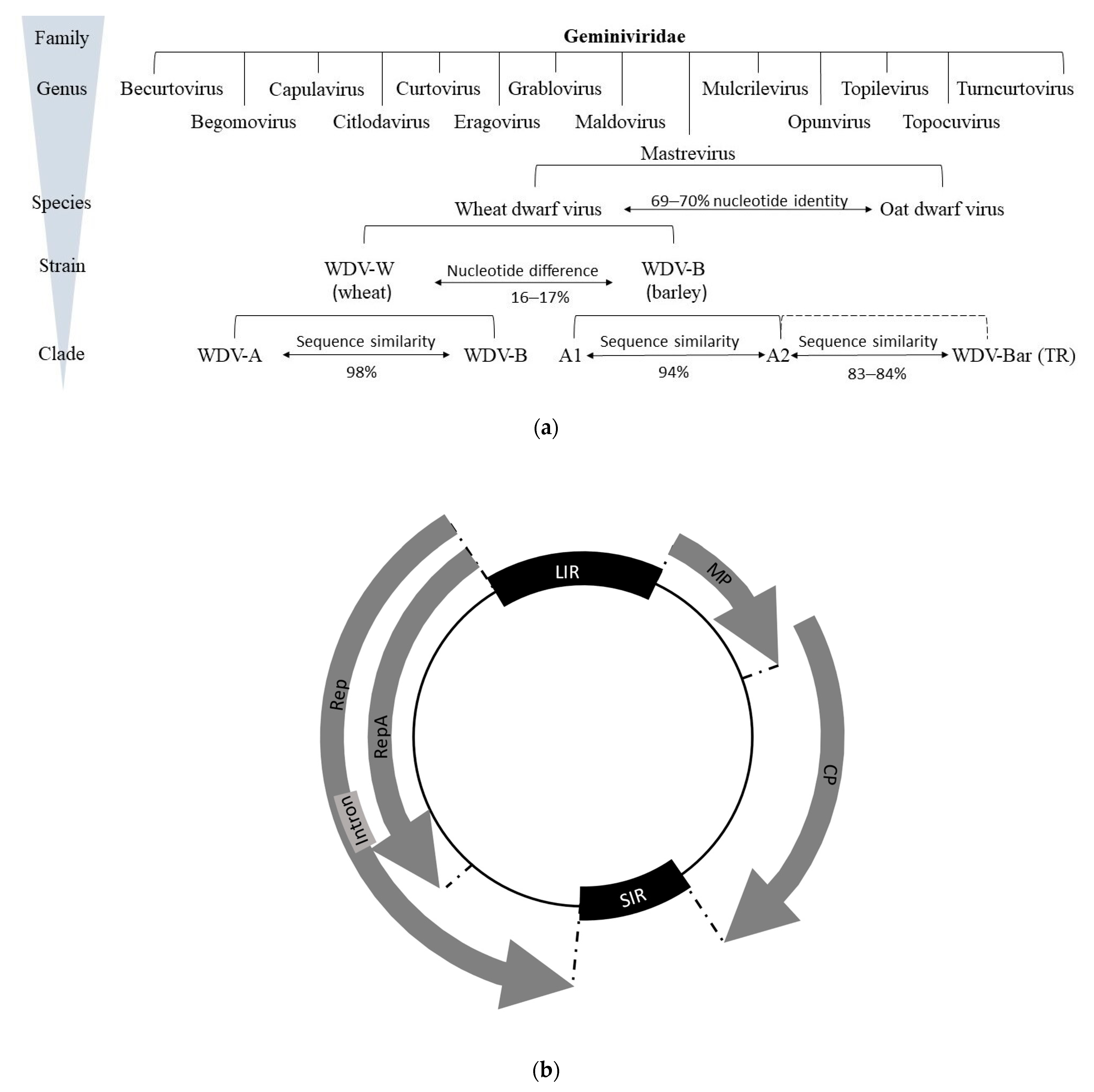
Figure 1. Classification and genomic organization of wheat dwarf virus (WDV): (a) classification of the family Geminiviridae is based on their molecular and biological characteristics. WDV species belong to the mastreviruses and consist of the main strains of wheat and barley, to which the various isolates are subordinated in clades. The percentage of nucleotide similarity is given for the species, strains, and clades. WDV Bar [TR] refers to the recombinant isolate between a barley isolate and a yet unknown member of the mastreviruses. (b) Genomic organization of mastreviruses, which include wheat dwarf virus (WDV). These have a circular ssDNA genome (black circle) and four ORFs. Code of viral proteins: MP—movement protein, CP—capsid protein, RepA—replication-associated protein, Rep—replication initiation protein. Also shown are the non-coding regions of the large intergenic region (LIR) and small intergenic region (SIR).
Plants 12 03633 g001
Figure 2. World map with countries where WDV could be detected (marked in red). WDV was reported in Ukraine [131], Romania [13], Bulgaria [131], Hungary [131], Italy [118], France [47], Sweden [20], Poland [132], Finland [18], Spain [133], the United Kingdom [108], Austria [108] and Slovenia [134], as well as regions in Iran [135], the Middle East (Turkey [109], Africa (Tunisia [120] and Zambia [136]), West Asia (Syria [137], and China [138,139]) [140].
Plants 12 03633 g002
Figure 3. Eight-week-old wheat plants with different degrees (symptom scoring 1, 2, 5, 6, 8) of dwarfing in the greenhouse depending on their genotype (a) and at BBCH stage 30–39 in May 2021 under field conditions (b) after artificial inoculation with symptom-bearing in the middle of the image. (c) Leaves of WDV-infected plants (left) show a stripe-like lightening compared to healthy leaves (right), which later develops into yellowing.
Plants 12 03633 g003
Figure 4. Schematic representation of the life cycle of winter cereals and Psammotettix alienus. The major developmental stages of host cereal plants (from sowing to harvest) are represented by the outer circle. The successive and overlapping biological cycles of P. alienus are represented by arrows in the inner circle. Under optimal conditions (20 °C, 70–95% relative humidity, 18/6 light/dark hours), the life cycle length (from egg to adult death) is 71 days [195]. Eggs produced in the fall overwinter on cereals and hatch in the following growing season (the next spring). According to Manurung et al. [12], the duration of the five larval stages (L1 to L5) is 5.9, 5.1, 5.6, 3, and 9.4 days, respectively. The seven-day-old adults can mate to produce the next generation of insects [12].
Plants 12 03633 g004
Table 1. Overview of the historical development of WDV and its evidence in the individual countries in relation to its reference in the literature. For some events, no direct dates could be derived from the literature, so only a time span could be given.
TimeEventReference
Early 20th centuryThe first observed dwarfing symptoms of wheat, called slidsjuka[114,115]
Early 20th centuryRelatively low field prevalence of WDV; only a few symptoms of dwarfing have been described in scientific literature[116,117,118,119,120]
Early 1950sLess undersowing in wheat; increased use of combine harvesters[124]
Around 1950Decline of slidsjuka due to changes in agricultural practices[121,122,123,124]
1950–1980/1990Slidsjuka occurred sporadically[121,122,123]
1961The first report of a direct relationship between virus, vector, and symptoms; no virus particle detected[10,125]
1980Increased incidence of disease in European countries[124]
1980Identification and taxonomic classification of WDV[124]
1981Leafhopper P. alienus was made responsible for WDV occurence[114]
Late 1980sA new disease (pieds chétifs) occurred in France in association with P. alienus; the disease was identified as WDV[126,127]
Disclaimer/Publisher’s Note: The statements, opinions and data contained in all publications are solely those of the individual author(s) and contributor(s) and not of MDPI and/or the editor(s). MDPI and/or the editor(s) disclaim responsibility for any injury to people or property resulting from any ideas, methods, instructions or products referred to in the content.