Review

The Research Process of PSK Biosynthesis, Signaling Transduction, and Potential Applications in Brassica napus

1
Key Laboratory of Plant Functional Genomics of the Ministry of Education, Yangzhou University, Yangzhou 225009, China
2
Department of Plant Physiology and Biochemistry, Institute of Biology, University of Hohenheim, 70593 Stuttgart, Germany
*
Author to whom correspondence should be addressed.
Plants 2023, 12(17), 3075; https://doi.org/10.3390/plants12173075
Submission received: 30 June 2023 / Revised: 1 August 2023 / Accepted: 23 August 2023 / Published: 28 August 2023
(This article belongs to the Special Issue The Growth and Development of Vegetable Crops)

Abstract

:
Phytosulfokine (PSK) is a disulfated pentapeptide that acts as a growth regulator to control plant growth and development as well as adaptability to biotic and abiotic stress. In the last three decades, PSK has drawn increasing attention due to its various functions. Preproproteins that have been tyrosine sulfonylated and then cleaved by specific enzymes contribute to mature PSK. To transfer a signal from the apoplast to the inner cells, the PSK peptide must bind to the PSK receptors (PSKR1 and PSKR2) at the cell surface. The precise mechanism of PSK signal transduction is still unknown, given that PSKR combines receptor and kinase activity with a capacity to bind calmodulin (CaM). The binding of PSK and PSKR stimulates an abundance of cGMP downstream from PSKR, further activating a cation-translocating unit composed of cyclic nucleotide-gated channel 17 (CNGC17), H+-ATPases AHA1 and AHA2, and BRI-associated receptor kinase 1 (BAK1). Recently, it has been revealed that posttranslational ubiquitination is closely related to the control of PSK and PSKR binding. To date, the majority of studies related to PSK have used Arabidopsis. Given that rapeseed and Arabidopsis share a close genetic relationship, the relevant knowledge obtained from Arabidopsis can be further applied to rapeseed.

1. Introduction

Phytosulfokine (PSK) is a disulfated pentapeptide that has been identified as a growth factor in plants. PSK was initially isolated from the mesophyll cells of Asparagus officinalis in a culture medium [1]. It was hypothesized that PSK was made up of two components, PSK-α and PSK-β, that differed from each other due to a tiny structural variation. More recently, experimental evidence has demonstrated that PSK-β is the degradational byproduct of PSK-α [2]. PSK–γ and PSK-ε, both of which belong to the PSK family, have recently been identified in soybean (Glycine max) and Medicago truncatula, respectively [3,4]. Similarly to most small, post-translationally modified peptides, mature and active PSK that is derived from a precursor preproprotein has to be modified and cleaved. PSK is a precursor protein that must be sulfonated at both tyrosines by tyrosylprotein subfotransferase (TPST), localized in the Golgi apparatus, and further cleaved by subtilisin-like serine proteases (SBTs) in the cytoplasmic matrix. AtSBT1.1 and AtSBT3.8 have been demonstrated to participate in the proteolytic cleavage of AtPSK4 and AtPSK1 propeptides, respectively [5,6,7]. It is still unknown whether other SBT family members or even unidentified proteases are also involved in the modification of PSK preproproteins. After processing, mature PSK binds to the PSK receptor proteins (PSKRs) localized at the plasma membrane. PSKRs have a canonical guanylate cyclase (GC) core and act upstream of cGMP and PSK signaling; therefore, they must undergo sophisticated signal transduction within cells. The regulation of signal transduction is also mediated by the response modules of cyclic nucleotide-gated channel 17 (CNGC17), H+-ATPase AHA1 and AHA2, and BRI-associated receptor kinase 1 (BAK1) on the plasma membrane [8].
Several putative functions of PSK have been reported, including the regulation of plant growth and development, callus formation, sexual reproduction, plant response to biotic and abiotic stress. Research on PSK biosynthesis and signal transduction has advanced dramatically over the last three decades. PSK, emerging as an important small signaling molecule, is receiving increasing global attention. The majority of studies on PSK have so far focused on Arabidopsis and rice (Oryza sativa). Despite being incredibly potent and crucial for plants, the physiological functions of PSK are currently not employed in crops. The PSK precursor genes belong to a rather conservative gene family. Brassica napus (B. napus) and Arabidopsis are genetically closely related. B. napus contains 24 PSK homologous genes based on the sequence alignment of AtPSK1-AtPSK7 in Arabidopsis, and these genes are expressed in different plant tissues. This raises the prospect of enhancing the endogenous PSK content in B. napus to either stimulate yield production or enhance plant tolerance to biotic and abiotic stress.
This review summarizes research on PSK biosynthesis, signal transduction, and the physiological roles of PSK, along with a discussion about the future of PSK-related research. Additionally, potential applications are suggested for genetic transformation, stress tolerance, and cultivation of elite germplasm resources in rapeseed, which might be beneficial for molecular breeding and genetic modification in agriculture.

2. Synthesis and Signal Transduction of PSK Peptide

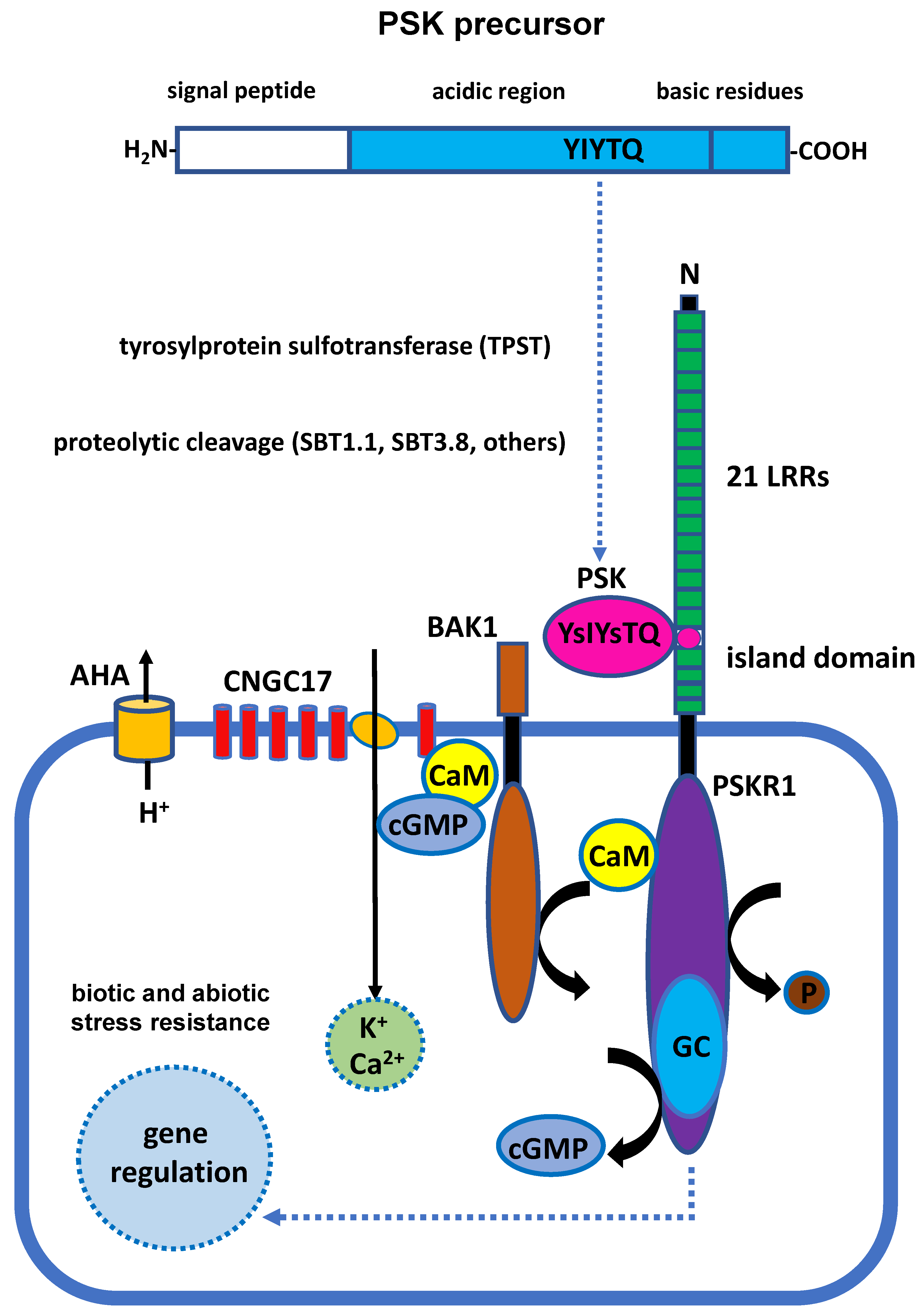
2.1. Biosynthesis of the PSK Peptide

The majority of peptides, which are derived from precursor proteins, go through a series of posttranslational modifications, including tyrosine sulfation, proline hydroxylation, glycosylation, and processing by proteases, before becoming active molecules that ultimately cause physiological responses [9,10]. Seven precursor genes (AtPSK1-AtPSK7) encoding PSK have been identified in Arabidopsis. AtPSK1 shows the highest expression level in the root and seed. The expression of the AtPSK2 and AtPSK3 genes is detectable not only in suspension-cultured cells but also throughout the entire plant, with the highest expression in the root [11]. AtPSK4 is primarily expressed in the root, hypocotyl, seed, leaf, and flower, with the highest expression in the senescent leaf and mature seed. AtPSK5 is mainly expressed in the flower, and the expression of AtPSK6 is comparatively low in most plant tissues. AtPSK7 is specifically expressed in the silique and seed (according to the online database https://www.arabidopsis.org/ (accessed on 20 June 2023)). These PSK precursor genes appear to be differentially expressed in diverse plant tissues and at different plant developmental phases.
PSK precursor proteins contain 80–120 amino acids, which are members of a small protein family [12]. The N-terminus of the precursor protein consists of hydrophobic leader residues for protein secretion, while the PSK signature motif YIYTQ/H is located at the C-terminus of the precursor protein and surrounded by two pairs of basic residues [2,12]. Tyrosylprotein sulfotransferase (TPST), which is localized on the Golgi membrane, is required for the sulfonylation of both tyrosines on the unique PSK motif YIYTQ/H as part of the processing of PSK preproprotein [5]. The sulfonated precursor protein leaves the Golgi apparatus and is further cleaved by subtilisin-like serine proteases in the cytoplasmic matrix (Figure 1). In Arabidopsis, it has been demonstrated that AtSBT1.1 and AtSBT3.8 specifically cleave and modify two different PSK preproproteins, AtPSK4 and AtPSK1. The processing of the AtPSK4 propeptide relies on AtSBT1.1 localized in the apoplast [6]. Recent research has indicated that extracellular AtSBT3.8 cleaves AtPSK1 propeptide in an aspartate-specific manner [13] because the cleavage by AtSBT3.8 relies on aspartic acid residue at the C-terminus [7]. It is still necessary to identify the proteases involved in the modification of the N-terminus of the PSK precursor in Arabidopsis. The SBT family contains 56 members, and it is likely that other SBTs (which require further investigation in the future) are also involved in the cleavage of PSK precursor proteins.

2.2. Characteristics of PSK Receptors

PSK is an autocrine growth regulator produced in the secretory pathway and further released into the extracellular apoplast [1]. The typical phytohormones and regulatory factors in plants are perceived by receptors and conveyed through signaling transduction pathways to elicit a response within plant cells. Earlier studies have described the specific binding sites with PSK in the plasma membrane cells of carrots (Daucus carota), corn (Zea mays), asparagus (Asparagus officinalis), and tomatoes (Solanum lycopersicum) using radioisotope labeling [15,16,17], indicating that this type of receptor is present in almost all higher plants. Soon after, via affinity chromatography, it is shown that carrot cells contain a phytosulfokine receptor (PSKR) with a molecular weight of 120 KD that can specifically bind to PSK [18,19]. The PSKR is localized in the plasma membrane [1]. Extracellular leucine-rich repeats (LRRs), a transmembrane domain, and a cytoplasmic kinase domain are three components that contribute to an integrated and functional PSKR [18,19]. PSKRs are categorized as members of the RLK (receptor-like kinase) protein family that contains a chemical structure with receptor-like kinases [20]. In addition, PSKRs and brassinosteroid receptor BRI1 (brassinosteroid receptor BRASINOSTEROID INSENSITIVE 1) belong to the LRR X RLK subfamily and play a key role in the regulation of plant growth and development [20]. The extracellular LRRs are composed of 21 consecutive LRR domains, and each LRR domain contains 24 amino acid residues. A highly independent island domain with 36 amino acid residues is found between the 17th and 18th LRR domains [18] (Figure 1). The island domain composed of 36 amino acid residues is highly conserved on AtPSKR1 and the brassinosteroid receptor BRI1 [18,21], suggesting that this domain plays a key role in the interplay between PSK and PSKR. In Arabidopsis, two PSKR homologous genes have been discovered [22,23].

2.3. PSKR-Dependent Signal Transduction

Two PSK receptors are present in Arabidopsis: AtPSKR1 and AtPSKR2 [22,23]. Through protein kinase activity assay in vitro and bioinformatic study of these two receptors, it has been revealed that AtPSKR1 has a cyclic guanylate kinase domain and a calmodulin (CaM) binding site [24,25]. Therefore, the interaction between PSK and PSKR promotes the synthesis of cGMP, which then transmits downstream signal transduction and, ultimately, regulates cell growth and development. In contrast, the activity of kinase is mediated by the binding of calcium ions and CaM. In addition, potassium ion is indispensable for the transmission of PSK signals. It is shown that PSK fails to induce protoplast swelling in the absence of exogenous potassium ions [26]. PSK-induced protoplast expansion decreases by approximately 60% at culture medium with 1 mM K+ as compared with the expansion with 10 mM K+ in a dose-dependent manner [26].
Cyclic nucleotide-gated channels (CNGCs) are one of the targets regulated by cGMP in plant cells [27], and CNGC17 specifically promotes protoplast expansion. Moreover, CNGC17 is also co-expressed with PSKR1 to control seedling growth [8]. Therefore, it is likely that PSK activates the cation channel CNGC17 and exerts physiological effects in conjunction with a second messenger, cGMP, which is located downstream of PSKR1. Instead of directly interacting with CNGC17, PSKR1 promotes a physiological response with CNGC17 by forming a protein complex on the plasma membrane with the H+-ATPases AHA1 and AHA2, as well as BAK1 [8] (Figure 1). The relevant studies above indicate that PSK is recognized by PSKR after the precursor protein is processed, matured, and secreted. Afterward, the transmitted PSK signaling further controls plant cell growth and development, as well as plants’ response to environmental stresses through hormone signals, second messengers, ion concentration changes, etc. However, the underlying molecular mechanism is not yet known in detail and requires much more experimental investigation.
Recently, it was discovered that the ubiquitin/proteasome degradation pathway is the mechanism by which the PSKR1 protein activity is mediated. In the absence of PSK, PSKR1 is moderately ubiquitinated as a result of interactions between PSKR1 and plant U-box E3 ligases, PUB12 and PUB13, that regulate PSKR1 abundance in response to pathogen infection. In the presence of PSK, PSKR1 is unable to interact with PUB12 and PUB13, thereby activating PSK-mediated intercellular defense signals [28]. This study highlights how a PSK-PSKR pattern is dynamically regulated by post-translational regulation, laying the foundation for a deeper comprehension of the intricate PSK signaling transduction system.

3. Physiological Functions of PSK

3.1. PSK Controls the Growth and Development of Plant Cells

In plants, the function of PSK on cell proliferation has been widely reported (Table 1). In the suspension culture of mesophyll cells of Asparagus officinalis, PSK enhances mitotic events for cell proliferation at the nanomolar level [1]. Relevant studies in rice [15] and carrot cells [29] further support a comparable function. In addition, PSK stimulates the growth of adventitious roots on the hypocotyl of cucumber (Cucumis sativus)[30] and accelerates the development of somatic cell embryos in carrot and Cryptomeria japonica [5,31], indicating a physiological role of PSK in promoting cell differentiation and somatic embryogenesis. Recent research also confirmed that PSK contributes to early somatic embryogenesis in Cunninghamia lanceolate. PSK not only boosts the number of somatic cell embryos but also hastens their growth and maturation [32]. Moreover, this PSK-stimulating effect is genotype-independent in Cunninghamia lanceolata and can be employed to generate a somatic embryogenesis system for various genotypes. PSK also controls root elongation and fiber development. Root cells at the quiescent center (QC), which undergo delayed cell division, are responsible for maintaining the meristem of the root tip [33,34,35]. Continuous root growth mostly occurs at the root meristem and elongation zone. PSK regulates root elongation via enhanced cell expansion with brassinosteroids synergistically in Arabidopsis [36,37,38]. Compared to wild-type plants, AtPSKR double mutant plants have shorter roots and hypocotyls, while overexpressing AtPSKR1 in plants significantly enhances the root length [26,38]. TPST plays an important role in the sulfonylation of PSK precursor proteins; tpst mutants in Arabidopsis exhibit a dwarf phenotype accompanied by severe shoot and root development [39]. Additionally, mutant plants that are deficient in AtSBT3.8 exhibit a substantial decrease in root development, whereas PSK treatment recovers the root growth [7]. The exogenous application of synthesized PSK has been examined in the growth system of cotton (Gossypium species) ovules at various concentrations in vitro, and it has been verified that PSK exhibits a positive impact on the elongation of cotton fiber, while GA3 is required to reach that end [40]. The wheat (Triticum aestivum) gene TaPSK5 is newly identified as a target of microRNA164 (miR164). TaPSK5, which encodes a PSK precursor protein, enhances root elongation and boosts crop yield [41]. PSK-γ isolated in soybean is a functional analog of PSK and increases both seed size and weight, and is, therefore, thought to be favorable to higher soybean yield [42].
Pollen density determines pollen germination and pollen tube elongation in most plants, which is referred to as the “pollen population effect”. The frequency of pollen germination is closely related to the pollen density and is supported by studies performed with mature pollen grains in tobacco (Nicotiana tabacum) suspended in culture medium. The low pollen germination efficiency can be reversed using a conditioned medium from the pollen culture, and the presence of PSK is further confirmed in the conditioned medium using an enzyme-linked immunosorbent assay, supporting the involvement of PSK as a key regulatory factor in the pollen population effect [43]. It has been demonstrated that PSK is able to promote early embryogenesis of microspores in B. napus [44]. In addition, it is revealed that PSK signaling is essential for pollen tube growth in Arabidopsis, redirecting them from the transmitting tract to the embryo sac properly [45]. So far, the understanding of the structural characteristics and functions of PSK remains limited in Rosaceae species. It is shown that the overexpression of the PbrPSK2 gene in Pyrus bretschneideri promotes pollen tube elongation [46]. These findings imply that PSK is a multifunctional regulator for plant growth and development, and the modulation of PSK-related gene expression has considerable potential for crop improvement.
Table 1. The physiological functions of PSK in plants.
Major FunctionFunction in DetailSpeciesReference
PSK controls the growth and development of plant cellscell proliferationAsparagus officinalis,
Oryza sativa,
Daucus carota
[1];
[15];
[29]
induced cell differentiation and somatic cell embryogenesisCucumis sativus,
Daucus carota,
Cryptomeria japonica,
Cunninghamia lanceolata
[30];
[5];
[31];
[32]
regulation of root elongation and fiber developmentArabidopsis,
Gossypium species,
Triticum aestivum
[36];
[40];
[41]
increased crop yieldTriticum aestivum,
Glycine max
[42];
[41]
regulatory factor in pollen population effectsNicotiana tabacum[43]
guide pollen into the embryo sac and enable them to elongate normallyArabidopsis
Pyrus bretschneideri
[45];
[46]
PSK promotes callus formation and efficiency of tissue culture in vitroinduced cell regenerationBrassica oleracea[47]
enhanced the transformation efficiency of candidate genesDaucus carota[48]
PSK regulates plant adaptation in response to environmental stressesenhanced heat resistanceArabidopsis[49]
enhanced drought resistanceOryza sativa,
Arabidopsis
Solanum lycopersicum
[50];
[7];
[51]
delayed senescenceFragaria ×ananassa,
Rosa hybrida,
Brassica oleracea var. Italica
[52];
[53];
[54]
preserved the quality and delayed cold damageMusa paradisiaca,
Prunus persica
[55];
[56]
pathogen resistanceSolanum lycopersicum,
Arabidopsis,
Oryza sativa
[28,57];
[58,59];
[60]
balance plant immune signalingArabidopsis[61]

3.2. PSK Promotes Callus Formation and Efficiency of Tissue Culture In Vitro

It has been extensively documented that PSK is involved in the regulation of callus development (Table 1). Transgenic callus tissues containing the constitutively expressed AtPSK2 or AtPSK3 chimeric genes double in size when compared with the mock callus [11], suggesting that PSK plays a role in assisting callus formation. The protoplasts of sugar beet (Beta vulgaris L.) cannot differentiate into callus tissue in vitro due to their recalcitrant status; however, PSK is able to reverse this scenario, and it enhances cell division in most sugar beet cultivars [62]. Surprisingly, 0.1 µM PSK is sufficient to trigger the mitotic event and noticeably induce the cell regeneration of protoplasts in Brassica oleracea in vitro [47]. Additionally, PSK vastly enhances the transformation efficiency in carrots, and the majority of the transformed callus successfully differentiate into cotyledons and roots before maturing into full-grown plants [48]. A substantial guarantee for the tissue regeneration of the transformation system can be provided by PSK, which improves callus formation and transformation efficiency on genetic manipulation in plants.

3.3. PSK Regulates Plant Adaptation in Response to Environmental Stresses

Plenty of research has shown how PSK improves plant tolerance to biotic and abiotic stress (Table 1). PSK improves plants’ ability to withstand the heat in Arabidopsis, based on the phenotypic analysis of plants subjected to high night-time temperature conditions with or without PSK treatment [49]. It is further revealed that PSK delays the senescence of strawberries (Fragaria × ananassa) stored at 4 °C [52], the petal senescence of cut rose flowers (Rosa hybrida cv. Angelina), [53] and the yellowing of broccoli (Brassica oleracea var. Italica) florets [54] due to improved antioxidant capacity. Furthermore, the quality of bananas (Musa paradisiaca) and peaches (Prunus persica) is also retained, and cold damage is postponed owing to the exogenous application of PSK [55,56]. These research results suggest that PSK plays a role in plant assimilation of environmental challenges, which is desirable for the development of the agricultural industry. The growth and yield of crops have been threatened in recent decades by the escalating severity of drought events in addition to heat and cold stress. To adapt to these environmental stresses, plants have had to develop various adaptive traits or mechanisms. Premature flower drop in tomato largely reduces the yield production under drought conditions. Phytaspase (Phyt) is a subtilisin-like protease of the phytaspase subtype. It has been revealed that SlPSK1, SlPSK6, and SlPhyt2 are co-expressed at the abscission zone of tomato flower organs. The knockout of SlPhyt2 results in delayed abscission of flowers under drought stress, indicating that PSK signaling is involved in drought-induced flower drop [51]. Recent studies have reported that the expression of PSK precursor genes and three subtilisin-like serine proteases, SBT1.4, SBT3.7, and SBT3.8, are highly induced by osmotic stress [7]. In the sbt3.8 loss-of-function mutant, the processing of AtPSK1 propeptide is impaired, and mature PSK could not be produced properly. The roots of the sbt3.8 mutant are significantly shorter than those of the wildtype seedlings combined with a decreased fresh weight of the roots and number of lateral roots under osmotic stress conditions, finally resulting in decreased plant tolerance to osmotic stress. This phenomenon is largely alleviated with PSK treatment. Contrarily, plants overexpressing AtPSK1 or AtSBT3.8 show an enhanced tolerance to osmotic stress [7]. The expressional analysis of OsPSKR genes using RNA-seq and real-time quantitative PCR (RT-qPCR) shows that multiple PSKR genes are differently elevated when induced by drought and salinity, demonstrating that OsPSKR genes are involved in rice adaptation to biotic and abiotic stress [50]. The resultant evidence indicates that PSK plays a significant role and develops a special mechanism to help plants deal with abiotic stresses.
Growing evidence suggests that PSK also participates in the plant immune response to various biotic stresses (Table 1). PSK is a danger-associated molecular pattern (DAMP), which is primarily recognized by PSKR1 [28,58]. To enhance tomato resistance to B. cinerea, PSKR1 elevates the Ca2+ concentration in the cytosol and activates the auxin-mediated signaling pathway [58]. The expression of PSK2 and PSKR1 is highly induced after the treatment of the fungal elicitor E-Fol or the fungal pathogens Alternaria brassicicola and Sclerotinia sclerotiorum on the leaves of Arabidopsis. The defense system of the pskr1 mutant plant against fungus is largely impaired [59]. Further investigation reveals that overexpression of the PSK precursor gene AtPSK2 or AtPSK4 in Arabidopsis significantly enhances resistance to Alternaria brassicicola [57], indicating the role of PSK in plant immune response. The severe fungal disease Xanthomonas oryzae pv. oryzicola (Xoc), which results in bacterial leaf streak (BLS), brings the global rice supply toward danger of collapse. Overexpressing OsPSKR1 in rice increases plant resistance to Xoc [60]. The expression of PSK3, PSK5, and PSKR1 are wound-induced in hypocotyls and leaves [59]; these findings indicate that PSK regulates plant adaptation to biotic stress. Although the immune system acts as a shield to protect plants, a hyperactive response frequently has a detrimental and irreversible effect on plant growth [63,64]. In addition, the exogenous application of PSK attenuates pattern-triggered immunity (PTI) in Arabidopsis, therefore reducing excessively robust immunity [61]. Thus, it is plausible that PSK is essential for maintaining the proper balance between plant development and immune system responses.

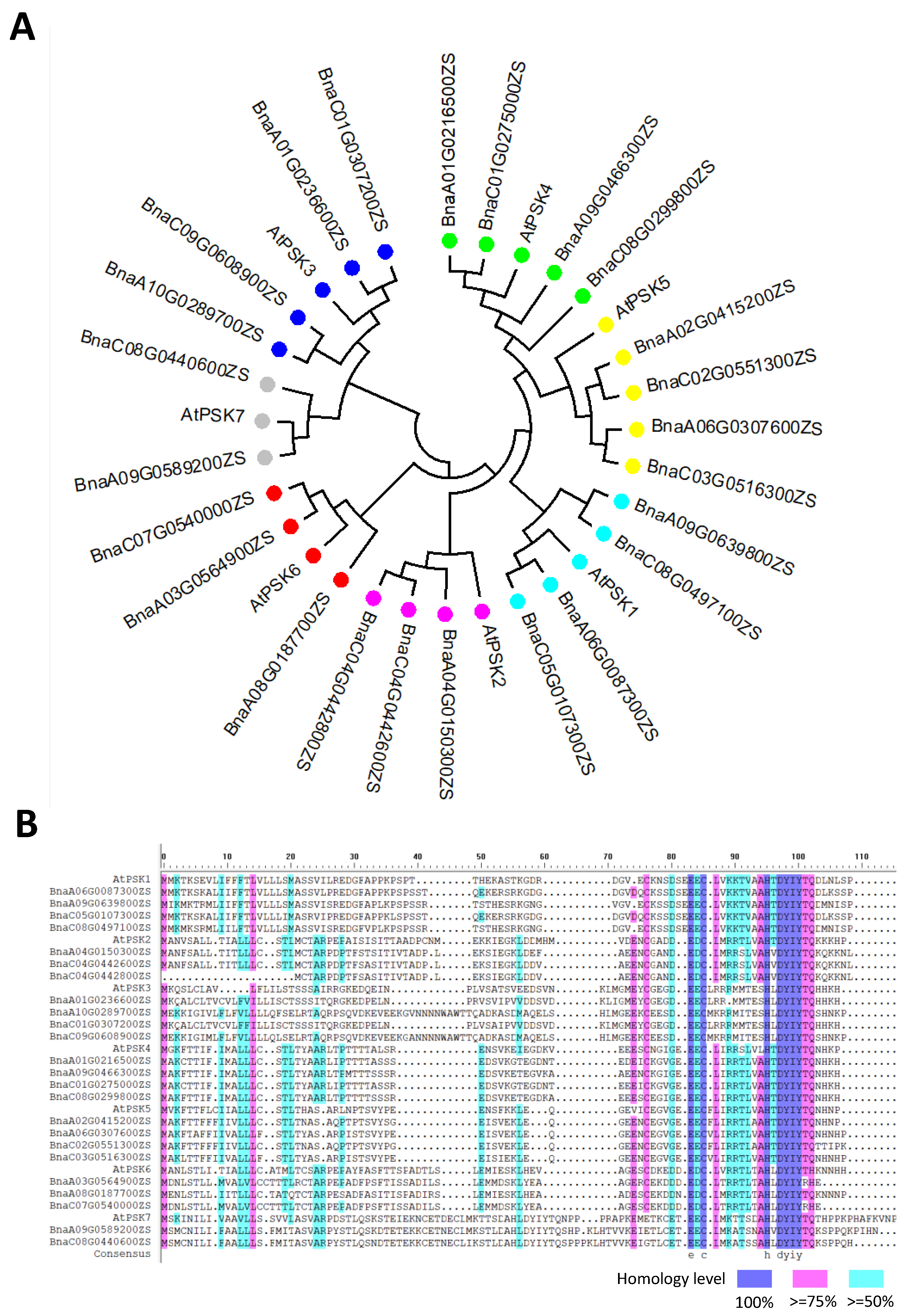
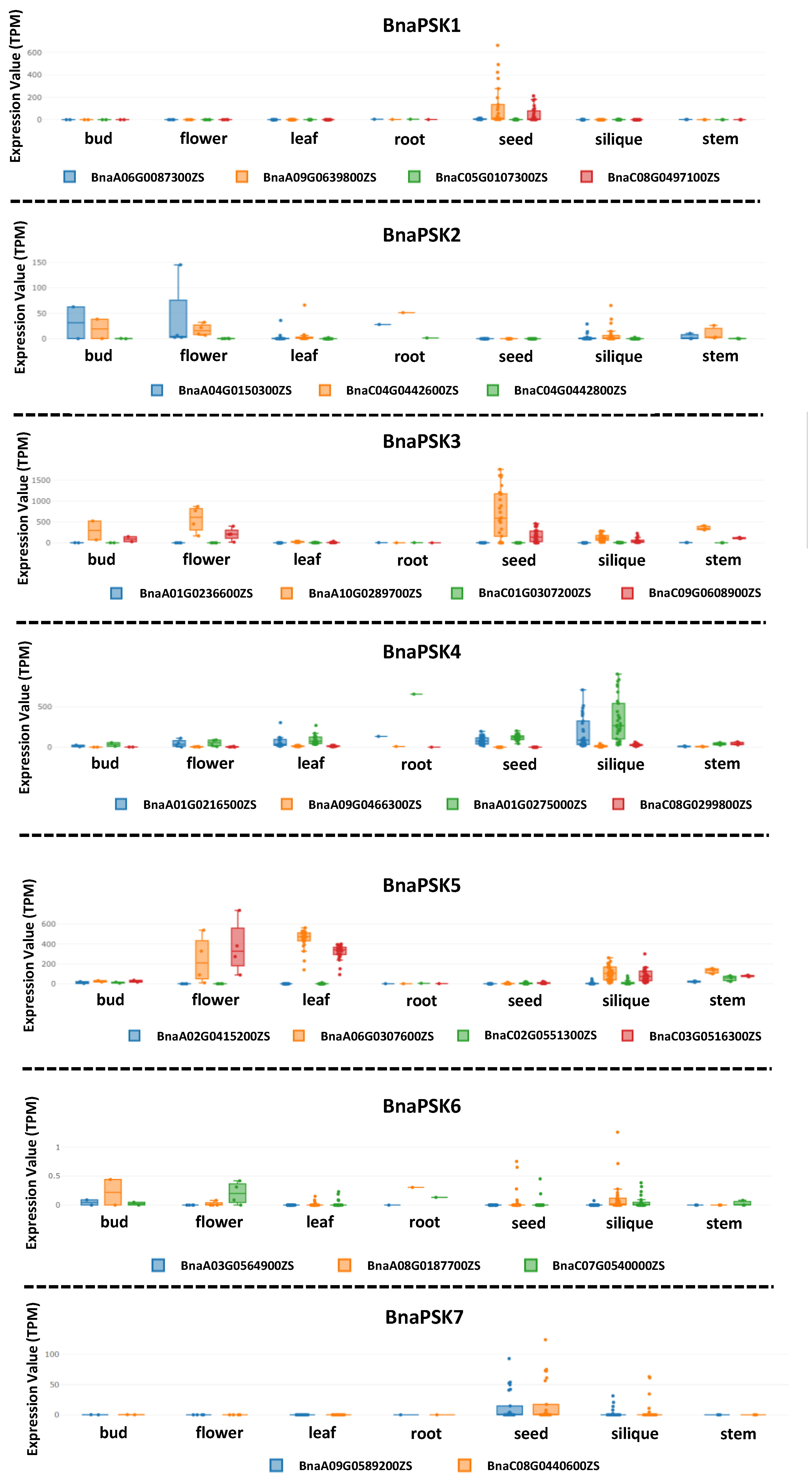
4. PSK-Related Genetic Correlation between Rapeseed and Arabidopsis

To date, the model plant Arabidopsis has remained the focus of most PSK research. Rapeseed and Arabidopsis have a close genetic link, and both are members of the Brassicaceae family. As a result, the knowledge gained from research on Arabidopsis can be further transferred to rapeseed. The development of bioinformatics has allowed us to obtain sequence information regarding PSK across different rapeseed genotypes.
Seven independent PSK precursor proteins with the PSK signature pattern YIYTQ/H have been identified in Arabidopsis and their corresponding genes are expressed in various plant tissues. PSK precursor genes are a relatively conservative gene family, with a high sequence similarity between Arabidopsis and rapeseed, based on the sequence alignment and phylogenetic tree analysis (Figure 2). The number of precursor genes in rapeseed that correspond to AtPSK1-AtPSK7 in Arabidopsis is 4, 3, 4, 4, 4, 3, and 2 (Figure 2). According to the online database, these genes are expressed in various rapeseed tissues and organs. BnaPSK1 is mostly expressed in seed, while BnaPSK2 is expressed in flower buds, flowers, and roots. BnaPSK3 is universally expressed in flower buds, flowers, and seeds. BnaPSK4 is primarily expressed in pod, and BnaPSK5 is mostly expressed in flower, leaf, and pod. In all tissues, the expression of BnaPSK6 and BnaPSK7 is comparatively low (Figure 3). The expressional patterns of the PSK precursor genes in rapeseed further confirm the idea that PSK signaling is present throughout rapeseed plants. This raises the prospect of enhancing the PSK content of particular tissues in rapeseed. Future work might explore the possibility of enhancing the promoter activity of various PSK precursor genes to boost the expression level of the corresponding genes.

5. Conclusions and Perspectives

5.1. Bottleneck and Gaps of PSK-Related Research

In Arabidopsis, substantial advances have been made in PSK biosynthesis, PSKR-dependent signal transduction, and PSK-related physiological functions. Nevertheless, it is necessary to address several concerns related to PSK in detail, as follows: (1) Of the 56 members of the SBT family of enzymes, only AtSBT1.1 and AtSBT3.8 were found to specifically cleave AtPSK4 and AtPSK1 propeptide. The modification or cleavage of PSK preproproteins most likely also involves additional SBTs or unidentified proteases [6,13]; (2) The precise regulatory mechanism of PSK signaling transduction is still unknown, despite the identification of several regulatory elements in the PSKR-dependent signal transduction; (3) Both phytohormones and PSK are essential for the regulation of plant development, while the interplay between PSK and these phytohormones is so far unclear; (4) The physiological applications of PSK to most crops are currently poorly studied due to the majority of PSK research being concentrated on Arabidopsis; (5) It has been recently revealed that PSKR1 interacts with the plant U-box E3 ligases PUB12 and PUB13, and the abundance and protein stability of PSKR are controlled by the ubiquitin/proteasome degradation pathway. It is, therefore, anticipated that the PSKR protein can be stabilized by repressing the activity of PUB12 or PUB13 for the improvement of plant disease resistance. However, it is unclear whether the knockout of PUB12 or PUB13 causes any other negative impacts on plants.

5.2. Putative Function of PSK on Callus Formation during Genetic Transformation in B. napus

Rapeseed is a crucial oil crop that stabilizes oil output on a global scale. Meanwhile, rapeseed plants at vegetative phases can also be served as edible vegetables. The largest and most popular variety of rapeseed grown worldwide is B. napus. Therefore, both domestically and globally, research into relevant economic and adaptive traits in B. napus is gaining popularity.
In rapeseed, B. napus exhibits a higher rate of genetic transformation than other Brassica species [65,66]. To date, the genetic transformation of B. napus still relies on agrobacterium-mediated hypocotyl transformation, and the transformation efficiency varies among different varieties. The regeneration rate of four commercial rapeseed varieties (Faisal canola, Punjab canola, Aari canola, and Nifa Gold) as well as the model variety (Westar) have been tested. Aari canola shows the highest transformation efficiency at 50.6%, followed by Westar with a transformation rate of 37.3%. The transformation efficiencies of other genotypes are still less than 33% [66]. In other academic research, the transformation efficiency of four different rapeseed varieties (GY284, WH3417, ZS9, and ZS11) after co-incubation with agrobacterium strain was found to be 3.7–5.6%, 1.1–3.5%, 0–0.87%, and 0, respectively [67], indicating that the genotypes in rapeseed continue to play a significant role in the efficiency of the agrobacterium-mediated transformation. It has been made clear that, in addition to genotypes, the transformation efficiency is also strongly correlated with the ratio of various hormones and the status of explants [68].
Previous research reported that a tiny amount of PSK considerably increases the transformation efficiency of various plants [47,48,62]. In order to improve the effectiveness of genetic transformation independent of genotypes in rapeseed, PSK can thus be evaluated in the genetic transformation. Experimental investigation is still needed to determine the optimal amount of PSK during tissue culture. In the meantime, it is anticipated that the overexpression of PSK precursor genes in rapeseed plants might bring about comparable outcomes.

5.3. Potential Use of PSK-Related Genes in Agricultural Breeding

PSK serves a variety of functions and is crucial for increasing crop yield and stress tolerance [7,50]. However, rapeseed has not exhibited these helpful attributes. The breakthrough in bioinformatics has made it possible for us to acquire sequence data about PSK across multiple rapeseed genotypes. It may be conceivable to investigate PSK-related quantitative trait loci (QTLs) or optimal haplotypes to improve plant growth and stress tolerance by combining phenotypic and molecular biology investigations in rapeseed. This work is closely associated with agricultural output in the future.
PSKR1 is mainly ubiquitinated by PUB13 through Lys-748 and Lys-905 sites. The Lys-748 and Lys-905 sites of PSKR, which are important protein sites, can be manipulated as an alternative strategy to confer disease resistance. These modifications largely rely on base editing or prime editing technology. Future work will likely focus on the identification of PUB members that interact with PSKR1 in rapeseed. It is anticipated that the crucial ubiquitination sites can be modified to maintain the abundance and stability of PSKR1, which may help accelerate the cultivation of rapeseed resistant to bacterial and fungal infections. It is also reasonable to consider, at the molecular level, how to optimize the activity of the enzymes involved in posttranslational modifications, such as TPST and SBTs. Future studies need to meticulously evaluate the inhibitors that control the activity of these enzymes. The accurate genome editing of these suppressors aids in enhancing PSK activity or content, which may be crucial in the growth and development of rapeseed.

Author Contributions

C.L. conceived the outline of the review. X.S., N.S. and C.L. created the illustrations and wrote the manuscript. All authors have read and agreed to the published version of the manuscript.

Funding

The support by the Development of Jiangsu Higher Education Institutions to CL, by Outstanding Ph.D. program in Yangzhou (grant no. YZLYJFJH2022YXBS147 to CL) and by the General Project of Basic Scientific Research to colleges and universities in Jiangsu Province (grant no. 22KJB210019 to CL) is greatly acknowledged.

Institutional Review Board Statement

Not applicable.

Informed Consent Statement

Not applicable.

Data Availability Statement

Not applicable.

Conflicts of Interest

The authors declare no conflict of interest.

References

  1. Matsubayashi, Y.; Sakagami, Y. Phytosulfokine, sulfated peptides that induce the proliferation of single mesophyll cells of Asparagus officinalis L. Proc. Natl. Acad. Sci. USA 1996, 93, 7623–7627. [Google Scholar] [CrossRef] [PubMed]
  2. Yang, H.; Matsubayashi, Y.; Nakamura, K.; Sakagami, Y. Oryza sativa PSK gene encodes a precursor of phytosulfokine-α, a sulfated peptide growth factor found in plants. Proc. Natl. Acad. Sci. USA 1999, 96, 13560–13565. [Google Scholar] [CrossRef] [PubMed]
  3. Liu, Y.; Zhang, D.; Li, M.; Yan, J.; Luo, L.; Yu, L. Overexpression of PSK-γ in Arabidopsis promotes growth without influencing pattern-triggered immunity. Plant Signal. Behav. 2019, 14, 1684423. [Google Scholar] [CrossRef]
  4. Di, Q.; Li, Y.; Zhang, D.; Wu, W.; Zhang, L.; Zhao, X.; Luo, L.; Yu, L. A novel type of phytosulfokine, PSK-ε, positively regulates root elongation and formation of lateral roots and root nodules in Medicago truncatula. Plant Signal.Behav. 2022, 17, 2134672. [Google Scholar] [CrossRef] [PubMed]
  5. Hanai, H.; Nakayama, D.; Yang, H.; Matsubayashi, Y.; Hirota, Y.; Sakagami, Y. Existence of a plant tyrosylprotein sulfotransferase: Novel plant enzyme catalyzing tyrosine O-sulfation of preprophytosulfokine variants in vitro. FEBS Lett. 2000, 470, 97–101. [Google Scholar] [CrossRef]
  6. Srivastava, R.; Liu, J.X.; Howell, S.H. Proteolytic processing of a precursor protein for a growth-promoting peptide by a subtilisin serine protease in Arabidopsis. Plant J. 2008, 56, 219–227. [Google Scholar] [CrossRef]
  7. Stuhrwohldt, N.; Buhler, E.; Sauter, M.; Schaller, A. Phytosulfokine (PSK) precursor processing by subtilase SBT3.8 and PSK signaling improve drought stress tolerance in Arabidopsis. J. Exp. Bot. 2021, 72, 3427–3440. [Google Scholar] [CrossRef]
  8. Ladwig, F.; Dahlke, R.I.; Stuhrwohldt, N.; Hartmann, J.; Harter, K.; Sauter, M. Phytosulfokine regulates growth in Arabidopsis through a response module at the plasma membrane that includes CYCLIC NUCLEOTIDE-GATED CHANNEL17, H+-ATPase, and BAK1. Plant Cell 2015, 27, 1718–1729. [Google Scholar] [CrossRef]
  9. Matsubayashi, Y.; Sakagami, Y. Peptide hormones in plants. Annu. Rev. Plant Biol. 2006, 57, 649–674. [Google Scholar] [CrossRef]
  10. Stührwohldt, N.; Schaller, A. Regulation of plant peptide hormones and growth factors by post-translational modification. Plant Biol. 2019, 21 (Suppl. S1), 49–63. [Google Scholar] [CrossRef]
  11. Yang, H.; Matsubayashi, Y.; Nakamura, K.; Sakagami, Y. Diversity of Arabidopsis genes encoding precursors for phytosulfokine, a peptide growth factor. Plant Physiol. 2001, 127, 842–851. [Google Scholar] [CrossRef]
  12. Lorbiecke, R.; Sauter, M. Comparative analysis of PSK peptide growth factor precursor homologs. Plant Sci. 2002, 163, 321–332. [Google Scholar] [CrossRef]
  13. Stuhrwohldt, N.; Scholl, S.; Lang, L.; Katzenberger, J.; Schumacher, K.; Schaller, A. The biogenesis of CLEL peptides involves several processing events in consecutive compartments of the secretory pathway. eLife 2020, 9, 19. [Google Scholar] [CrossRef] [PubMed]
  14. Sauter, M. Phytosulfokine peptide signalling. J. Exp. Bot. 2015, 66, 5161–5169. [Google Scholar] [CrossRef]
  15. Matsubayashi, Y.; Takagi, L.; Sakagami, Y. Phytosulfokine-α, a sulfated pentapeptide, stimulates the proliferation of rice cells by means of specific high- and low-affinity binding sites. Proc. Natl. Acad. Sci. USA 1997, 94, 13357–13362. [Google Scholar] [CrossRef] [PubMed]
  16. Matsubayashi, Y.; Sakagami, Y. Characterization of specific binding sites for a mitogenic sulfated peptide, phytosulfokine-α, in the plasma-membrane fraction derived from Oryza sativa L. Eur. J. Biochem. 1999, 262, 666–671. [Google Scholar] [CrossRef]
  17. Matsubayashi, Y.; Sakagami, Y. 120- and 160-kDa receptors for endogenous mitogenic peptide, phytosulfokine-α, in rice plasma membranes. J. Biol. Chem. 2000, 275, 15520–15525. [Google Scholar] [CrossRef]
  18. Matsubayashi, Y.; Ogawa, M.; Morita, A.; Sakagami, Y. An LRR receptor kinase involved in perception of a peptide plant hormone, phytosulfokine. Science 2002, 296, 1470–1472. [Google Scholar] [CrossRef]
  19. Wang, J.; Li, H.; Han, Z.; Zhang, H.; Wang, T.; Lin, G.; Chang, J.; Yang, W.; Chai, J. Allosteric receptor activation by the plant peptide hormone phytosulfokine. Nature 2015, 525, 265–268. [Google Scholar] [CrossRef]
  20. Morillo, S.A.; Tax, F.E. Functional analysis of receptor-like kinases in monocots and dicots. Curr. Opin. Plant Biol. 2006, 9, 460–469. [Google Scholar] [CrossRef]
  21. Kaufmann, C.; Sauter, M. Pull-down assay to characterize Ca2+/calmodulin binding to plant receptor kinases. Methods Mol. Biol. 2017, 1621, 151–159. [Google Scholar] [CrossRef] [PubMed]
  22. Matsubayashi, Y.; Ogawa, M.; Kihara, H.; Niwa, M.; Sakagami, Y. Disruption and overexpression of Arabidopsis phytosulfokine receptor gene affects cellular longevity and potential for growth. Plant Physiol. 2006, 142, 45–53. [Google Scholar] [CrossRef] [PubMed]
  23. Amano, Y.; Tsubouchi, H.; Shinohara, H.; Ogawa, M.; Matsubayashi, Y. Tyrosine-sulfated glycopeptide involved in cellular proliferation and expansion in Arabidopsis. Proc. Natl. Acad. Sci. USA 2007, 104, 18333–18338. [Google Scholar] [CrossRef] [PubMed]
  24. Kwezi, L.; Ruzvidzo, O.; Wheeler, J.I.; Govender, K.; Iacuone, S.; Thompson, P.E.; Gehring, C.; Irving, H.R. The phytosulfokine (PSK) receptor is capable of guanylate cyclase activity and enabling cyclic GMP-dependent signaling in plants. J. Biol. Chem. 2011, 286, 22580–22588. [Google Scholar] [CrossRef]
  25. Hartmann, J.; Fischer, C.; Dietrich, P.; Sauter, M. Kinase activity and calmodulin binding are essential for growth signaling by the phytosulfokine receptor PSKR1. Plant J. 2014, 78, 192–202. [Google Scholar] [CrossRef]
  26. Stuhrwohldt, N.; Dahlke, R.I.; Steffens, B.; Johnson, A.; Sauter, M. Phytosulfokine-α controls hypocotyl length and cell expansion in Arabidopsis thaliana through phytosulfokine receptor 1. PLoS ONE 2011, 6, e21054. [Google Scholar] [CrossRef]
  27. Talke, I.N.; Blaudez, D.; Maathuis, F.J.; Sanders, D. CNGCs: Prime targets of plant cyclic nucleotide signalling? Trends Plant Sci. 2003, 8, 286–293. [Google Scholar] [CrossRef]
  28. Hu, Z.; Fang, H.; Zhu, C.; Gu, S.; Ding, S.; Yu, J.; Shi, K. Ubiquitylation of PHYTOSULFOKINE RECEPTOR 1 modulates the defense response in tomato. Plant Physiol. 2023, 192, 2507–2522. [Google Scholar] [CrossRef]
  29. Eun, C.-H.; Ko, S.-M.; Matsubayashi, Y.; Sakagami, Y.; Kamada, H. Phytosulfokine-α requires auxin to stimulate carrot non-embryogenic cell proliferation. Plant Physiol. Biochem. 2003, 41, 447–452. [Google Scholar] [CrossRef]
  30. Yamakawa, S.; Sakuta, C.; Matsubayashi, Y.; Sakagami, Y.; Kamada, H.; Satoh, S. The promotive effects of a peptidyl plant growth factor, phytosulfokine-α, on the formation of adventitious roots and expression of a gene for a root-specific cystatin in cucumber hypocotyls. J. Plant Res. 1998, 111, 453–458. [Google Scholar] [CrossRef]
  31. Igasaki, T.; Akashi, N.; Ujino-Ihara, T.; Matsubayashi, Y.; Sakagami, Y.; Shinohara, K. Phytosulfokine stimulates somatic embryogenesis in Cryptomeria japonica. Plant Cell Physiol. 2003, 44, 1412–1416. [Google Scholar] [CrossRef]
  32. Hao, Z.; Wu, H.; Zheng, R.; Li, R.; Zhu, Z.; Chen, Y.; Lu, Y.; Cheng, T.; Shi, J.; Chen, J. The plant peptide hormone phytosulfokine promotes somatic embryogenesis by maintaining redox homeostasis in Cunninghamia lanceolata. Plant J. 2023, 113, 716–733. [Google Scholar] [CrossRef] [PubMed]
  33. Stahl, Y.; Simon, R. Plant stem cell niches. Int. J. Dev. Biol. 2005, 49, 479–489. [Google Scholar] [CrossRef] [PubMed]
  34. Kumpf, R.; Thorstensen, T.; Rahman, M.A.; Heyman, J.; Nenseth, H.Z.; Lammens, T.; Herrmann, U.; Swarup, R.; Veiseth, S.V.; Emberland, G.; et al. The ASH1-RELATED3 SET-domain protein controls cell division competence of the meristem and the quiescent center of the Arabidopsis primary root. Plant Physiol. 2014, 166, 632–643. [Google Scholar] [CrossRef]
  35. Rovere, F.D.; Fattorini, L.; Ronzan, M.; Falasca, G.; Altamura, M.M. The quiescent center and the stem cell niche in the adventitious roots of Arabidopsis thaliana. Plant Signal. Behav. 2016, 11, e1176660. [Google Scholar] [CrossRef] [PubMed]
  36. Mussig, C.; Shin, G.H.; Altmann, T. Brassinosteroids promote root growth in Arabidopsis. Plant Physiol. 2003, 133, 1261–1271. [Google Scholar] [CrossRef]
  37. Wildwater, M.; Campilho, A.; Perez-Perez, J.M.; Heidstra, R.; Blilou, I.; Korthout, H.; Chatterjee, J.; Mariconti, L.; Gruissem, W.; Scheres, B. The RETINOBLASTOMA-RELATED gene regulates stem cell maintenance in Arabidopsis roots. Cell 2005, 123, 1337–1349. [Google Scholar] [CrossRef]
  38. Hartmann, J.; Stuhrwohldt, N.; Dahlke, R.I.; Sauter, M. Phytosulfokine control of growth occurs in the epidermis, is likely to be non-cell autonomous and is dependent on brassinosteroids. Plant J. 2013, 73, 579–590. [Google Scholar] [CrossRef]
  39. Komori, R.; Amano, Y.; Ogawa-Ohnishi, M.; Matsubayashi, Y. Identification of tyrosylprotein sulfotransferase in Arabidopsis. Proc. Natl. Acad. Sci. USA 2009, 106, 15067–15072. [Google Scholar] [CrossRef]
  40. Han, J.; Tan, J.; Tu, L.; Zhang, X. A peptide hormone gene, GhPSK promotes fibre elongation and contributes to longer and finer cotton fibre. Plant Biotechnol. J. 2014, 12, 861–871. [Google Scholar] [CrossRef]
  41. Geng, Y.; Jian, C.; Xu, W.; Liu, H.; Hao, C.; Hou, J.; Liu, H.; Zhang, X.; Li, T. miR164-targeted TaPSK5 encodes a phytosulfokine precursor that regulates root growth and yield traits in common wheat (Triticum aestivum L.). Plant Mol. Biol. 2020, 104, 615–628. [Google Scholar] [CrossRef]
  42. Yu, L.; Liu, Y.; Zeng, S.; Yan, J.; Wang, E.; Luo, L. Expression of a novel PSK-encoding gene from soybean improves seed growth and yield in transgenic plants. Planta 2019, 249, 1239–1250. [Google Scholar] [CrossRef] [PubMed]
  43. Chen, Y.F.; Matsubayashi, Y.; Sakagami, Y. Peptide growth factor phytosulfokine-α contributes to the pollen population effect. Planta 2000, 211, 752–755. [Google Scholar] [CrossRef]
  44. Tsuwamoto, R.; Fukuoka, H.; Takahata, Y. Identification and characterization of genes expressed in early embryogenesis from microspores of Brassica napus. Planta 2007, 225, 641–652. [Google Scholar] [CrossRef] [PubMed]
  45. Stuhrwohldt, N.; Dahlke, R.I.; Kutschmar, A.; Peng, X.; Sun, M.X.; Sauter, M. Phytosulfokine peptide signaling controls pollen tube growth and funicular pollen tube guidance in Arabidopsis thaliana. Physiol. Plantarum 2015, 153, 643–653. [Google Scholar] [CrossRef]
  46. Kou, X.; Liu, Q.; Sun, Y.; Wang, P.; Zhang, S.; Wu, J. The peptide PbrPSK2 from phytosulfokine family induces reactive oxygen species (ROS) production to regulate pear pollen tube growth. Front. Plant Sci. 2020, 11, 601993. [Google Scholar] [CrossRef]
  47. Kiełkowska, A.; Adamus, A. Peptide growth factor phytosulfokine-α stimulates cell divisions and enhances regeneration from B. oleracea var. capitata L. protoplast culture. J. Plant Growth Regul. 2018, 38, 931–944. [Google Scholar] [CrossRef]
  48. Matsubayashi, Y.; Goto, T.; Sakagami, Y. Chemical nursing: Phytosulfokine improves genetic transformation efficiency by promoting the proliferation of surviving cells on selective media. Plant Cell Rep. 2004, 23, 155–158. [Google Scholar] [CrossRef]
  49. Yamakawa, S.; Matsubayashi, Y.; Sakagami, Y.; Kamada, H.; Satoh, S. Promotive effects of the peptidyl plant growth factor, phytosulfokine-α, on the growth and chlorophyll content of Arabidopsis seedlings under high night-time temperature conditions. Biosci. Biotechnol. Biochem. 1999, 63, 2240–2243. [Google Scholar] [CrossRef]
  50. Nagar, P.; Kumar, A.; Jain, M.; Kumari, S.; Mustafiz, A. Genome-wide analysis and transcript profiling of PSKR gene family members in Oryza sativa. PLoS ONE 2020, 15, e0236349. [Google Scholar] [CrossRef] [PubMed]
  51. Reichardt, S.; Piepho, H.P.; Stintzi, A.; Schaller, A. Peptide signaling for drought-induced tomato flower drop. Science 2020, 367, 1482–1485. [Google Scholar] [CrossRef] [PubMed]
  52. Soleimani Aghdam, M.; Sayyari, M.; Luo, Z. Exogenous phytosulfokine α application delays senescence and promotes antioxidant nutrient accumulation in strawberry fruit during cold storage by triggering endogenous phytosulfokine α signaling. Postharvest Biol. Technol. 2021, 175, 111473. [Google Scholar] [CrossRef]
  53. Aghdam, M.S.; Ebrahimi, A.; Sheikh-Assadi, M. Phytosulfokine α (PSKα) delays senescence and reinforces SUMO1/SUMO E3 ligase SIZ1 signaling pathway in cut rose flowers (Rosa hybrida cv. Angelina). Sci. Rep. 2021, 11, 23227. [Google Scholar] [CrossRef]
  54. Aghdam, M.S.; Flores, F.B. Employing phytosulfokine α (PSKα) for delaying broccoli florets yellowing during cold storage. Food Chem. 2021, 355, 129626. [Google Scholar] [CrossRef]
  55. Wang, D.; Huang, H.; Jiang, Y.; Duan, X.; Lin, X.; Soleimani Aghdam, M.; Luo, Z. Exogenous phytosulfokine α (PSKα) alleviates chilling injury of banana by modulating metabolisms of nitric oxide, polyamine, proline, and gamma-aminobutyric acid. Food Chem. 2022, 380, 132179. [Google Scholar] [CrossRef] [PubMed]
  56. Jiao, C. PpCBF6 is involved in phytosulfokine α-retarded chilling injury by suppressing the expression of PpLOX5 in peach fruit. Front. Plant Sci. 2022, 13, 874338. [Google Scholar] [CrossRef] [PubMed]
  57. Mosher, S.; Seybold, H.; Rodriguez, P.; Stahl, M.; Davies, K.A.; Dayaratne, S.; Morillo, S.A.; Wierzba, M.; Favery, B.; Keller, H.; et al. The tyrosine-sulfated peptide receptors PSKR1 and PSY1R modify the immunity of Arabidopsis to biotrophic and necrotrophic pathogens in an antagonistic manner. Plant J. 2013, 73, 469–482. [Google Scholar] [CrossRef] [PubMed]
  58. Zhang, H.; Hu, Z.; Lei, C.; Zheng, C.; Wang, J.; Shao, S.; Li, X.; Xia, X.; Cai, X.; Zhou, J.; et al. A plant phytosulfokine peptide initiates auxin-dependent immunity through cytosolic Ca2+ signaling in tomato. Plant Cell 2018, 30, 652–667. [Google Scholar] [CrossRef]
  59. Loivamaki, M.; Stuhrwohldt, N.; Deeken, R.; Steffens, B.; Roitsch, T.; Hedrich, R.; Sauter, M. A role for PSK signaling in wounding and microbial interactions in Arabidopsis. Physiol. Plant 2010, 139, 348–357. [Google Scholar] [CrossRef]
  60. Yang, W.; Zhang, B.; Qi, G.; Shang, L.; Liu, H.; Ding, X.; Chu, Z. Identification of the phytosulfokine receptor 1 (OsPSKR1) confers resistance to bacterial leaf streak in rice. Planta 2019, 250, 1603–1612. [Google Scholar] [CrossRef]
  61. Igarashi, D.; Tsuda, K.; Katagiri, F. The peptide growth factor, phytosulfokine, attenuates pattern-triggered immunity. Plant J. 2012, 71, 194–204. [Google Scholar] [CrossRef] [PubMed]
  62. Grzebelus, E.; Szklarczyk, M.; Greń, J.; Śniegowska, K.; Jopek, M.; Kacińska, I.; Mrożek, K. Phytosulfokine stimulates cell divisions in sugar beet (Beta vulgaris L.) mesophyll protoplast cultures. Plant Growth Regul. 2012, 67, 93–100. [Google Scholar] [CrossRef]
  63. Yasuda, M.; Ishikawa, A.; Jikumaru, Y.; Seki, M.; Umezawa, T.; Asami, T.; Maruyama-Nakashita, A.; Kudo, T.; Shinozaki, K.; Yoshida, S.; et al. Antagonistic interaction between systemic acquired resistance and the abscisic acid-mediated abiotic stress response in Arabidopsis. Plant Cell 2008, 20, 1678–1692. [Google Scholar] [CrossRef] [PubMed]
  64. Boller, T.; Felix, G. A renaissance of elicitors: Perception of microbe-associated molecular patterns and danger signals by pattern-recognition receptors. Annu. Rev. Plant Biol. 2009, 60, 379–406. [Google Scholar] [CrossRef]
  65. Murata, M.; Orton, T.J. Callus initiation and regeneration capacities in Brassica species. Plant Cell Tissue Organ Cult. 1987, 11, 111–123. [Google Scholar] [CrossRef]
  66. Farooq, N.; Nawaz, M.A.; Mukhtar, Z.; Ali, I.; Hundleby, P.; Ahmad, N. Investigating the in vitro regeneration potential of commercial cultivars of Brassica. Plants 2019, 8, 558. [Google Scholar] [CrossRef]
  67. Dai, C.; Li, Y.; Li, L.; Du, Z.; Lin, S.; Tian, X.; Li, S.; Yang, B.; Yao, W.; Wang, J.; et al. An efficient Agrobacterium-mediated transformation method using hypocotyl as explants for Brassica napus. Mol. Breed. 2020, 40, 96. [Google Scholar] [CrossRef]
  68. Maheshwari, P.; Selvaraj, G.; Kovalchuk, I. Optimization of Brassica napus (canola) explant regeneration for genetic transformation. New Biotechnol. 2011, 29, 144–155. [Google Scholar] [CrossRef]
Figure 1. Schematic model of PSK biosynthesis and signaling cascade uncovered in Arabidopsis. The N-terminus of the PSK precursor protein contains a hydrophobic amino acid residue, and the characteristic sequence YIYTQ/H is located at the C-terminus of the precursor protein. YIYTQ/H is surrounded by two pairs of alkaline amino acid residues that might be important for precursor processing. The precursor protein is first sulfonated by TPST localized on the Golgi apparatus membrane to modify two tyrosines of sequence YIYTQ/H. The sulfonated preproprotein is further cleaved by SBTs in the cytoplasmic matrix. After maturation of PSK, it is secreted into the extracellular apoplast and recognized by the extracellular LRR domains of PSKR. PSKR also contains cyclic guanylate kinase domain and a calmodulin (CaM) binding site in the intracellular kinase domain of PSKR; the production of cGMP is therefore stimulated after the binding of PSK and PSKR. Finally, the downstream signal controls cell growth and development through gene expressional regulation, while the control of kinase activity occurs through the binding of calcium ions and calmodulin (CaM). In addition, potassium ions are also indispensable for PSK signal transduction. cGMP acts as the second messenger downstream of PSKR1 to activate CNGC17. Instead of a direct interaction between PSKR1 and CNGC17, they rather form a complex module with the H+-ATPases AHA1 and AHA2 in the plasma membrane, as well as BRI-associated receptor kinase 1 (BAK1). This schematic view was modified by Sauter and Ladwig [8,14].
Plants 12 03075 g001
Figure 2. Alignment of PSK precursor proteins in A. thaliana and B. napus. (A) The Neighbor-Joining (NJ) phylogenetic tree regarding the PSK precursor proteins was constructed with MEGA7 software, based on amino acid alignment between A. thaliana and B. napus. Dots with the same color represent homologous proteins. This is an originally developed figure. (B) The amino acid alignment reveals the conserved domain in the PSK precursor proteins. The amino acid sequence was obtained from an online database (http://yanglab.hzau.edu.cn/BnTIR (accessed on 20 June 2023)). Sequence alignment was performed with DNAman8 software. This is an originally developed figure.
Plants 12 03075 g002
Figure 3. Expressional profiles of PSK precursor genes in different plant tissues of B. napus. The data of the expressional profiles were obtained from an online database with slight modifications (http://yanglab.hzau.edu.cn/BnTIR (accessed on 20 June 2023)).
Plants 12 03075 g003
Disclaimer/Publisher’s Note: The statements, opinions and data contained in all publications are solely those of the individual author(s) and contributor(s) and not of MDPI and/or the editor(s). MDPI and/or the editor(s) disclaim responsibility for any injury to people or property resulting from any ideas, methods, instructions or products referred to in the content.

