Article

Chemical Composition and In Vitro Evaluation of Antioxidant and Antiproliferative Effects of Volatile Oils Hydrodistilled from Onobrychis carduchorum C.C. Towns., a Kurdish Traditional Plant

by
Hawraz Ibrahim M. Amin
1,2,*,
Kamaran Abdoulrahman
1,
Azad S. Sadraddin
3,
Heman A. Smail
1,
Zanko Hassan Jawhar
4,
Kovan Dilawer Issa
5,
Chabaco Armijos
6 and
Giovanni Vidari
5,7
1
Department of Chemistry, College of Science, Salahaddin University-Erbil, Erbil 44001, Iraq
2
Department of Medical Biochemical Analysis, Cihan University-Erbil, Erbil 44001, Iraq
3
Department of Chemistry, College of Education, Salahaddin University-Erbil, Erbil 44001, Iraq
4
Department of Science, College of Health Science, Lebanese French University, Erbil 44001, Iraq
5
Department of Medical Analysis, Faculty of Applied Science, Tishk International University, Erbil 44001, Iraq
6
Departamento de Química, Universidad Técnica Particular de Loja (UTPL), Loja 1101608, Ecuador
7
Department of Chemistry, University of Pavia, Via Taramelli 12, 27100 Pavia, Italy
*
Author to whom correspondence should be addressed.
Plants 2023, 12(16), 3013; https://doi.org/10.3390/plants12163013
Submission received: 20 June 2023 / Revised: 13 August 2023 / Accepted: 18 August 2023 / Published: 21 August 2023

Abstract

:
The volatile oils hydrodistilled from the aerial parts and roots of O. carduchorum C.C Towns. (Fabaceae) have been chemically characterized for the first time. A total of 43 constituents with an abundance >0.03% were identified and quantified in the two oils by GC/MS and GC/FID analyses. They comprise 38 components (98.58%) of the oil isolated from the aerial parts (OCA) and 34 components (93.33%) of the oil from the roots (OCR). Six constituents, α-pinene (23.11 ± 0.1%), β-elemene (17.33 ± 0.1%), 1,8-cineole (12.15 ± 0.2%), furfural (7.91 ± 0.1%), terpineol-4-ol (6.32 ± 0.2%), and limonene (4.13 ± 0.1%), accounted for about 75% of the total OCA oil. On the other hand, 1,8-cineole (15.79 ± 0.1%), furfural (10.44 ± 0.1%), β-elemene (10.14 ± 0.2%), α-terpineol (7.74 ± 0.1%), linalool (7.45 ± 0.1%), and α-pinene (4.76 ± 0.1%) made up about 60% of the OCR oil. The IC50 values of the scavenging activities of the OCA and OCR oils towards the DPPH radical and H2O2 were 79.8 ± 0.5 and 153.3 ± 0.6 μg/mL and 394.09 ± 0.2 and 311.67 ± 0.4 μg/mL, respectively. In addition, in the MTS assay, the OCA and OCR oils showed significant antiproliferative effects against T47D, MDA-MB-453, BG-1, and A549 human cancer cells that were more powerful than those against two normal human cell lines, HEK-293 and HFF-1. The abundant presence of β-elemene as an antiproliferative component of the two oils suggested the existence of a new chemotype of O. carduchorum.

1. Introduction

Only a few ethnobotanical and phytochemical investigations have been conducted so far on plants growing in the Kurdistan Region of Iraq. Thus, most uses of local traditional plants are not sustained by scientific evidence [1]. The plant Onobrychis carduchorum C.C. Towns. (OC) is widely distributed in Iran, Iraq, Syria, and Turkey [2,3,4,5]. In Iraq, it grows mainly on Halgurd Mountain in the Kurdistan region [6]. It is commonly used as an anti-inflammatory and chronic wound healing remedy by Kurds living in a few villages located in the northern region of Choman-Erbil. In medicinal applications, shredded OC leaves are put on skin lesions. In addition, injured skin is cleaned and treated with an infusion prepared by boiling an aqueous suspension of aerial parts for about fifteen minutes. O. carduchorum, together with O. altissima Grossh., is also mentioned in a detailed survey of medicinal plants used in the Çatak region of Turkey, where the two plants are traditionally used to cure hemorrhages, wounds, and cuts [7]. The morphological, taxonomical, genetic, and ecogeographical characteristics of O. carduchorum are well described in the literature [8]. Thus, O. carduchorum is easily distinguished from other species belonging to the Onobrychis sect. In Onobrychis, the length of the keels are considerably shorter than standards [4,5,8].
The genus Onobrychis Mill. is the largest in the tribe Hedysareae (family Fabaceae) [8,9,10], which includes more than 170 annual or perennial species. Moreover, more taxa are continuously added to the genus, while others are revised, using both morphological and molecular evidence using the nuclear ribosomal DNA internal transcribed spacer (nrDNA ITS) [10]. Wild Onobrychis species grow mainly in Southwest Asia and the Mediterranean region, in different environments. Iran and Anatolia are biodiversity centers for the genus [10], while fourteen species grow in Iraq [3]. A few species, for example, O. viciifolia (sainfoin), are also cultivated as important fodder or ornamental plants [11].
Only a few Onobrychis species have been subjected to chemical investigations, among which most phytochemical studies were dedicated to O. viciifolia. This plant contained condensed tannins, cinnamic acids, flavonoids, isoflavonoids, and acylated flavonol glycosides [12,13]. The aerial parts of O. galegifolia Boiss., O. albiflora Hub.–Mor., O. tournefortii (Willd.) Desv., and O. argyrea Boiss. subsp. argyrea Boiss. exhibited antidiabetic activity in alloxan-induced diabetic mice [14]. The extracts of flowers and roots of O. armena Boiss & Huet displayed antioxidant and antimicrobial activity against some human and fish pathogens [15]. Strong antioxidant activity was also exhibited by the methanol extracts of O. sosnowiec, O. viciifolia, and O. melanotricha [16], while the aerial parts of O. oxyodonta Boiss. showed moderate antibacterial and antitumor properties [17]. Several prenylated polyphenols, including isoflavone genistein derivatives, flavanone naringenin derivatives, and dihydro-stilbenes exhibiting a remarkable wound healing activity, were isolated from the crude acetone extract of leaves and flowers of O. carduchorum collected in Iraq [18].
A few investigations have been carried out on non-volatile metabolites isolated from crude extracts of Onobrychis species; by contrast, little is known about the composition of volatile fractions. To our knowledge, only the essential oil from O. armena has been analyzed so far [19]. Thus, we found it interesting to study the volatile fractions isolated from O. carduchorum. In fact, there is an increasing interest in the analysis of essential oils, due to the characteristic chemical composition and the wide variety of biological activities, which include antibacterial, antiviral, antioxidant, and anticancer properties. Not less important are the industrial applications of essential oils [20,21,22,23,24,25,26,27,28]. In this context, the study of essential oils isolated from species of little-investigated genera, such as Onobrychis, is attracting worldwide attention nowadays. Moreover, we considered that some volatile components of O. carduchorum may contribute to the wound healing effects of the plant, as they can be released by rubbing the leaves on skin lesions.
In this paper, we report, for the first time, the chemical–physical properties and the composition of the essential oils isolated from the aerial parts (OCA) and from the roots (OCR) of O. carduchorum (Figure 1c) collected on Mount Halgurd in the province of Erbil of the Kurdistan Autonomous Region, Iraq (Figure 1a,b). Moreover, a preliminary evaluation of the antiradical and antiproliferative effects of the two oils is also described. In fact, some essential oils (EOs) have been labeled as promising anticancer agents and are currently being investigated for their cytotoxic and antiproliferative activities in cancer cell lines or experimental animals [24,26,27,28]. Different mechanisms have been reported for the cytotoxic effects of EOs or their constituents. These include induction of cell death by apoptosis and/or necrosis; antimutagenic, antiproliferative, and antioxidant properties; cell cycle arrest; and loss of key organelle functions [27].

2. Results and Discussion

2.1. Physical Properties of the Essential Oils

The essential oils from aerial parts (OCA) and roots (OCR) of O. carduchorum had a yellowish color and a pleasant aromatic odor. The oils’ isolated yields were 0.15 ± 0.01 and 0.06 ± 0.01% (w/w), respectively, on air-dried plant material; the specific optical rotations of OCA and OCR oils were [ α ] D 20 +1.44 and [ α ] D 20 +2.73 (c = 0.1, CH2Cl2), respectively; the relative densities were 0.81 ± 0.02 and 0.83 ± 0.03 g/L, respectively.

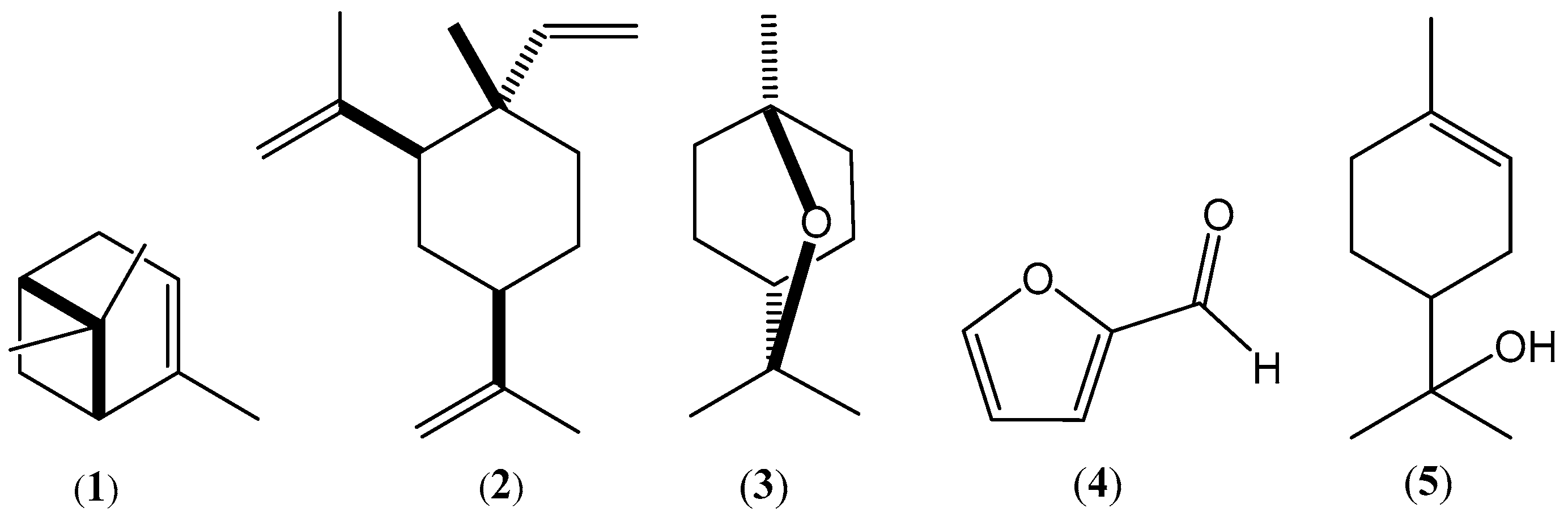
2.2. Chemical Composition of the Essential Oils

The compounds identified in the OCA and OCR oils, with the corresponding percentages, are listed in Table 1, together with the corresponding retention times (RTs) and determined linear retention indices (LRIexps).
The terpenoids identified in the two oils have been grouped in families in Table 2, while the other identified compounds have been divided according to chemical class. Compounds occurring in traces were not considered.

2.3. Antiproliferative Activity

The in vitro growth-inhibitory effects of OCA and OCR oils on T-47D human breast cancer cells, MDA-MB-453 human breast adenocarcinoma, BG-1 human ovarian carcinoma, A549 human adenocarcinoma alveolar basal cells, and two normal cell lines, namely, human embryonic kidney cells (HEK-293) and human fibroblast cells (HFF-1), were evaluated by the MTS assay [36]. The well-known anticancer drug cisplatin (cis-diamminedichloroplatinum (II)) was the reference compound. The results are reported in Table 3. The IC50 values ± SDs (μg/mL) were calculated as the means of three replicates. The goodness-of-fit of the probit regression model was assessed using the Pearson χ2 test.

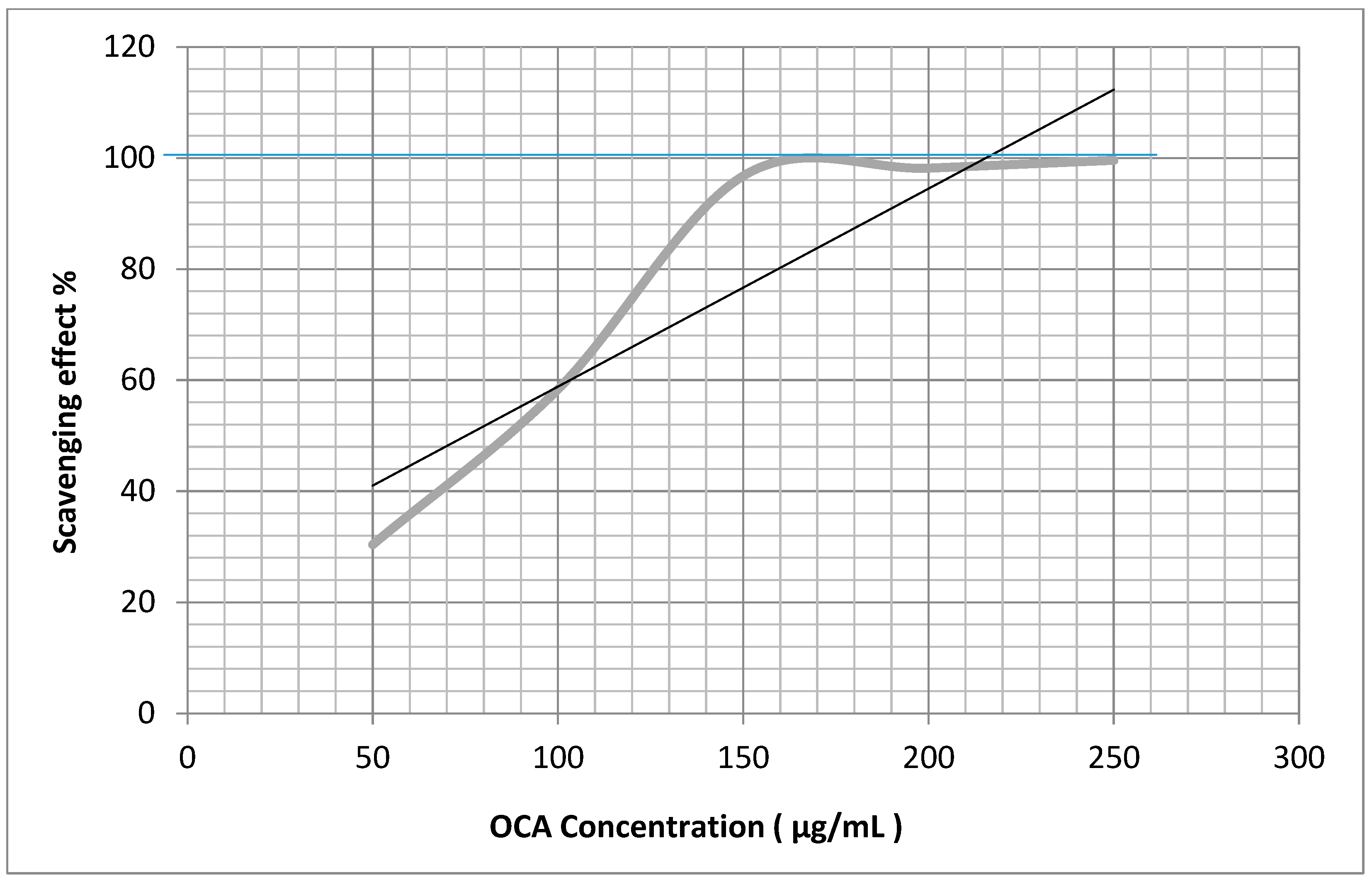
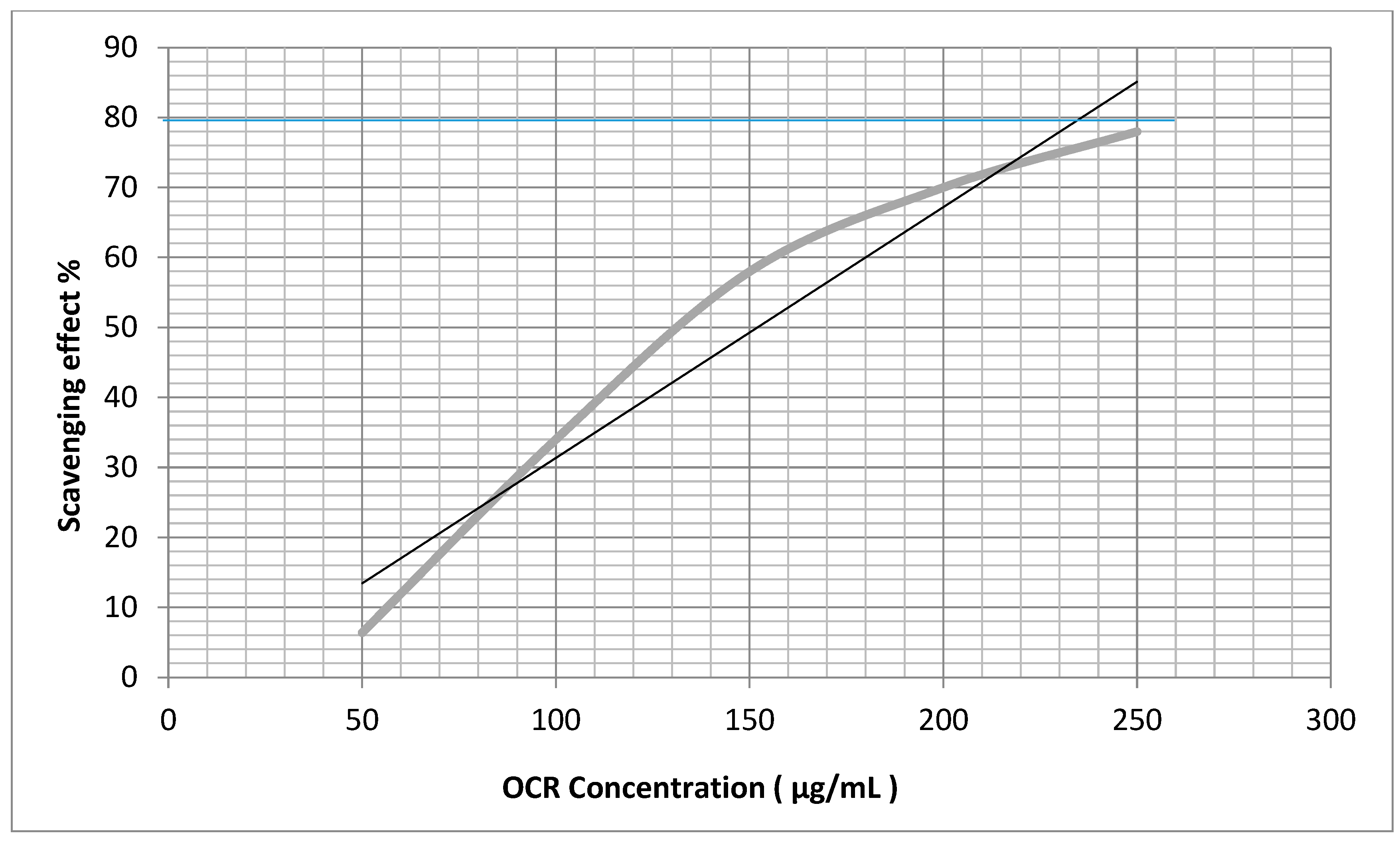
2.4. Antiradical Activities

The antioxidant and antiradical activities of the OCA and OCR oils were evaluated by measuring the scavenging properties towards H2O2 and the stable DPPH (2,2-diphenyl-1-picrylhydrazyl) hydroxyl radical (Figure 2 and Figure 3), using ascorbic acid as a positive control. The EC50 values ± SDs (μg/mL) are reported in Table 4 and were calculated as the means of three replicates.

3. Discussion

The most abundant constituents of the OCA oil were monoterpene hydrocarbons, followed by oxygenated monoterpenoids, while oxygenated monoterpenoids were largely predominant in the OCR oil. α-Pinene (1), β-elemene (2), 1,8-cineole (3), furfural (4) (Figure 4), terpineol-4-ol, and limonene were, in that order, the most abundant constituents of the OCA oil, accounting for about 70% of the oil composition (Table 1). On the other hand, 1,8-cineole (3), furfural (4), β-elemene (2), α-terpineol (5) (Figure 4), and linalool were, in that order, the most abundant constituents of the OCR oil, making up about 60% of the oil composition (Table 1).
Our findings indicated that the compositions of the two essential oils isolated from O. carduchorum aerial parts and roots differed greatly from that of the essential oil isolated from Onobrychis armena, which was rich in high-molecular-weight hydrocarbons and fatty acids [19]. Thus, the occurrence of α-pinene (1), β-elemene (2), 1,8-cineole (3), and furfural (4) markedly characterized the essential oils from O. carduchorum collected in Iraqi Kurdistan, although the percentages of these constituents varied between the two oils. Such variations were likely the basis of the small differences between the oils’ biological activities and the odors. Thus, the main contributor to the smell of the OCA oil was possibly the most abundant component, α-pinene (23.11 ± 0.1%), which has a characteristic intense turpentine scent. By contrast, the smell of the OCR oil was probably due to a combination of the smells of different components.
On the other hand, the presence of large amounts of β-elemene (2) in the two oils suggested the existence of a chemotype of O. carduchorum grown in Iraqi Kurdistan.
Regarding the antiproliferative activity of the oils against cancer cells in the MTS assay [36] (Table 3), the OCA oil exhibited high activity towards A549, BG-1, and T-47D human cancer cell lines, although the effects were significantly weaker than cisplatin. Of note, the minimal activity displayed against MDA-MB-453 cells appeared to exclude a non-specific antiproliferative activity against cancer cells. On the other hand, the OCR oil possessed significant antiproliferative activity against T-47D and MDA-MB-453 cell lines. In contrast, the efficacy of OCA and OCR oils towards two normal human cell lines, HEK-293 and HFF-1, was weaker than against cancer cells. Taken all together, the results of the MTS assay (Table 3) suggested that the two oils had a specific antiproliferative activity against certain cancer cells.
We considered the toxicity of the two oils quite interesting. In fact, based on the criteria of the U.S. National Cancer Institute (NCI), the cytotoxicity of essential oils is classified as follows: IC50 ≤ 20 µg/mL = highly cytotoxic, IC50 range between 21 and 200 µg/mL = moderately cytotoxic, IC50 range between 201 and 500 µg/mL = weakly cytotoxic, IC50 > 501 µg/mL = non-cytotoxic [37]. The cytotoxicity of the two oils likely resulted from a complex interaction of various oil components, acting with cellular structures and processes both alone and in synergy [27,28]. Moreover, some oil constituents could reduce the concentration of active components or have antagonist effects. The terpenoids limonene, 1,8-cineole (3), linalool, and the powerful antitumor agent β-elemene (2) [38,39], which was highly abundant (10–17%) in both oils, were possibly the main constituents responsible for the observed antiproliferative effects. It is remarkable that the sesquiterpene β-elemene (2) and various derivatives inhibited tumor cell growth even in vivo and demonstrated significant efficacy in clinical trials with cancer patients [40].
The oxidation process is a chemical reaction that produces free radicals, leading to chain reactions. A chain-breaking antiradical/antioxidant agent has the capacity to scavenge free radicals, typically by donating a hydrogen atom and forming a relatively stable radical which is unable to propagate the chain reaction. Alternately, the antioxidant undergoes autoxidation characterized by a very fast termination process [41]. It must be noted that the terms antiradical and antioxidant are often considered synonyms. However, strictly speaking, an antioxidant is an antiradical agent capable of quenching the radical species involved in oxidative chain carrying, that is, a peroxyl radical [42].
It has been proposed that antioxidants may play a role in the prevention and treatment of certain diseases, such as inflammation, aging, brain dysfunction, cancer, heart disease, arthritis, and the decline of the immune system. In fact, among the causes of the etiology and progression of these pathologies, evidence has indicated the adverse effects of an excess of free radicals and reactive oxygen species, such as H2O2, that react with biomacromolecules and cause severe damages to cells [42,43]. This pathological condition is called oxidative stress, which corresponds to a serious imbalance between the production of free radicals and the antioxidant inherent defense system of an organism [43]. In this situation, to prevent oxidation, the addition of either synthetic or natural antioxidants to fats, foods, and cosmetics is a common practice. Many essential oils have antioxidant properties, and their use as natural antioxidants is a field of growing interest because some synthetic antioxidants are now suspected to be potentially harmful to human health.
To obtain a preliminary indication of the antioxidant/antiradical properties of the OCA and OCR oils isolated from O. cardochorum, we used two standard tests (Table 4), although we were aware of the limited significance of such methods. In fact, according to some authors, the results would indicate a “radical trapping power” rather than true antioxidant activity [41]. The two oils showed comparable radical scavenging activities in the two tests, although they were less potent than ascorbic acid, which was used as a reference (Table 4). It is very difficult to attribute these effects to one or some active principles and to identify a specific pathway of molecular action because the oils contained mixtures of different components which can act with different mechanisms, both individually and synergistically [24,41]. Monoterpene alcohols, such as linalool; ketones; aldehydes; hydrocarbons, such as limonene; and ethers, such as 1,8-cineole (3), are probably the main contributors to the free radical scavenging activity of the OCA and OCR oils [21,44]; however, the activity of the strong antioxidant phenols cresol and carvacrol, although minor components of the oils, may not be negligible.

4. Materials and Methods
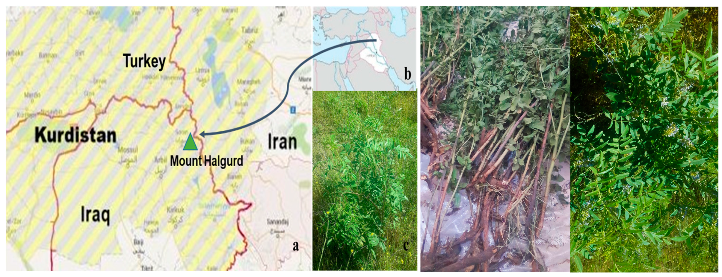
4.1. Plant Material

Aerial parts (flowers, leaves, and stems) and roots of Onobrychis carduchorum were collected in April 2022 at an altitude of 2100 m a.s.l. on Mount Mountain, Choman District, in Erbil province, Kurdistan Autonomous Region of Iraq. The plant was identified by A. Al-Khayyat, Professor of Botany at the Salahaddin University in Erbil; a voucher specimen has been deposited at the Herbarium of the Salahaddin University with the accession number 7235. Freshly cut aerial parts and roots were air-dried for a few hours at room temperature in the shade with an unforced ventilation.

4.2. Isolation of Volatile Fractions

Air-dried aerial parts (100 g) and roots (150 g) of O. carduchorum were separately hydrodistilled for approximately 5 h using a conventional Clevenger-type apparatus; this procedure was performed in triplicate for each sample. After decantation, each layer of oil was separated from the aqueous phase using a pipette; subsequently, the oils were dried over anhydrous sodium sulphate, filtered, and kept in the dark at −20 °C until analysis. The density, optical rotation, and yield of the oils were determined by standard methods [45].

4.3. GC-MS Analysis

GC-MS analyses were performed on a Thermo Scientific Focus GC instrument coupled to a DSQ mass spectrometer detector operating in an electron-impact (EI) mode with a voltage of ionization of 70 eV. A slightly polar Agilent J&W HP-5 fused silica capillary column (5% phenylmethylpolysiloxane, 30 m × 0.25 mm i.d.; 0.25 µm film thickness; Agilent Technologies Italia S.p.A, Cernusco sul Naviglio, MI, Italy) was used. The carrier gas was He at a constant flow rate of 1.0 mL/min. Each essential oil was dissolved in CH2Cl2 to a dilution of 1 mg/10 mL, and 1 µL of the solution was injected in split mode (20:1). The oven temperature was maintained at 60 °C for 1 min, then it was increased to 260 °C at a rate of 5 °C/min and held at 260 °C for 5 min. Acquisition mass range: m/z 41–350 amu; injector and transfer-line temperatures: 250 °C. Data were analyzed with MSD ChemStation software.

4.4. GC-FID Analysis

GC separations were carried out on a Perkin Elmer GC 2400 instrument (Perkin Elmer Italia SPA, Milan, Italy). A slightly polar Agilent J&W HP-5 fused silica capillary column (5% phenylmethylpolysiloxane, 25 m × 0.32 mm i.d.; 1.05 µm film thickness; Agilent Technologies Italia S.p.A, Cernusco sul Naviglio, MI, Italy) was used. The carrier gas was He at a constant flow rate of 1.0 mL/min. The injector temperature was 250 °C, and the FID detector was set at 260 °C; the oven temperature was maintained at 60 °C for 3 min, then it was increased to 260 °C at a rate of 5 °C/min and held at 260 °C for 1 min. A quantity of 1 µL of each essential oil solution (1 mg/10 mL in CH2Cl2) was manually injected in split mode (27:1). The relative amount of each oil component (Table 1) was calculated as the percent of the corresponding FID peak area with respect to the total area of peaks, without applying a correcting response factor. Mean % abundances and standard deviations were determined from the results of three replicates for each oil. Data were collected with HP3398A GC Chemstation software (Hewlett–Packard, Rev. A.01.01).

4.5. Identification of the Essential Oil Components

Each chemical component of the two oils (Table 1) was identified by comparing the corresponding mass spectrum with the spectra contained in the Adams [33] and NIST 08 [34] libraries, as well as by comparing the calculated linear retention index (LRIexp) with the literature [30,31,32,33,34,35]. Each LRI was calculated relative to a homologous series of standard n-alkanes (Sigma-Aldrich, Milan, Italy, No. CE 203-777-6), from n-octane (C8) to n-tricosane (C23), according to the van Den Dool and Kratz method [29]. The identification of most oil components was confirmed by coelution with authentic standards (Sigma-Aldrich, Milan, Italy).

4.6. Determination of the Antiproliferative Effects of the Oils (MTS Assay)

The MTS assay for evaluating the in vitro antiproliferative effects of a sample is based on the reduction of the yellow-colored MTS inner salt [3-(4,5-dimethylthiazol-2-yl)-5-(3-carboxymethoxyphenyl)-2-(4-sulfophenyl)-2H-tetrazolium] by NAD(P)H-dependent dehydrogenase enzymes in metabolically active cells to a purple formazan salt that is soluble in cell culture media [36]. The formazan salt has an absorbance maximum near 490 nm. When cells die, they lose the ability to convert MTS into formazan; color change thus serves as a convenient marker of only the viable (living) cells, and the measure of the absorbance can be directly related to the number of viable cells.

4.6.1. Cell Cultures

Four human cancer cell lines, breast cancer T-47D, apocrine breast cancer MDA-MB-453, lung cancer A549, and ovarian adenocarcinoma BG-1, and two normal cell lines, namely, human embryonic kidney cells (HEK-293) and human fibroblast cells (HFF-1), obtained from the American Type Culture Collection (ATCC) and Sigma Aldrich, were used in the test. The cells were cultured in RPMI-1640 or DMEM/F-12 medium (Euroclone, S.p.A., Milan, Italy) supplemented with 10% fetal bovine serum (FBS), 100 μg/mL penicillin/streptomycin, and 2 mM L-glutamine (Life Technologies, Milan, Italy) at 37 °C under a humidified atmosphere in the presence of 5% CO2, changing the liquid growth medium whenever needed. When a cell culture reached 80% confluence, a small amount of trypsin was added to the medium to separate the cells from the flask; after 3 min of incubation at 37 °C, 1 mL of FBS was added to stop the action of trypsin and to avoid degradation of cell membranes. Subsequently, the cell-containing medium was transferred into a cell strainer and centrifuged (ALC 4232 Centrifuge) at 1000 rpm for 10 min. The resulting pellet was resuspended in the growth medium (1 mL), and the cells were separated using an automatic pipette and counted using a counting chamber and trypan blue as dye. Quadruple cell samples were grown in 96-well flat-bottom microtiter plates (Cellstar, Greiner bio-one) at a density of 5 × 10⁵ cells/mL of growth medium in each well. After 2 h of incubation, the medium was replaced with 100 µL of test medium (RPMI 1640, added with 0.005% L-glutamine, penicillin, and streptomycin), and the microplates were left in the incubator for an additional 24 h.

4.6.2. MTS Assay of the OCA and OCR Oils

To determine the antiproliferative effects, six solutions of the OCA and OCR oils and cisplatin were prepared separately. Their concentrations ranged from 0.5 to 50 µg/mL in dimethyl sulfoxide (DMSO). The solvent DMSO was also tested for possible inhibitory effect by adjusting its concentration to be the same as the working concentration. The medium in the wells containing cultured cells was replaced with a solution (100 µL) of increasing sample concentration. Three replicates were performed for each sample dilution. The microplates were then incubated for 24 h in a humidified atmosphere of 5% CO2 at 37 °C; subsequently, the sample-containing medium was replaced with fresh test medium (100 µL) and 20 µL of MTS tetrazolium reagent (CellTiter 96®–AQueous One Solution Cell Proliferation Assay, Promega Italia S.R.L., Milan, Italy) was added. The plates were incubated for 2 h at 37 °C and the extent of MTS reduction was measured spectrophotometrically at 490 nm using a plate reader (BioRAD Model 550 Microplate Reader). Experiments were conducted in triplicate at room temperature. Cytotoxicity was expressed as the concentration of compound inhibiting cell growth by 50% (IC50). The IC50 values (µg/mL) (Table 3) were calculated by probit analysis (p < 0.05, χ2 test) with the GraphPad Prism 4 computer program (GraphPad Software, S. Diego, CA, USA).

4.7. Evaluation of Antiradical and Antioxidant Activities

The in vitro antiradical and antioxidant potential of the OCA and OCR oils were evaluated by the 1,1-diphenyl-2-picrylhydrazyl radical (DPPH) scavenging test [46,47] and by the H2O2 scavenging assay [48].

4.7.1. DPPH Test

The method described by Vani [47] was used with a few modifications. Eight solutions (3 mL) of ascorbic acid or an oil at 5, 10, 25, 50, 100, 150, 200, and 300 µg/mL concentrations in MeOH-H2O, 9:1, were added individually to a fresh solution of DPPH in MeOH (1 mL, 0.3 mM). Subsequently, each mixture was shaken vigorously and incubated for 30 min at 22 °C in the dark until a deep violet color and a stable absorbance value (A) at 517 nm was observed. The absorbance was measured against the blank using a UV-Visible spectrophotometer (Lambda 25 UV/Vis spectrometer N.3903, Perkin Elmer instruments, Waltham, MA, USA). A lower absorbance of the reaction mixture indicated a higher free radical (DPPH.) scavenging (FRS) activity. The DPPH solution (1 mL) added by 10% aqueous MeOH (3 mL) was used as the control. The FRS% was calculated using the formula: [1 − (Asample/Acontrol)] × 100, where Acontrol is the absorbance of the control at t = 0 min and Asample is the absorbance at t = 30 min in the presence of the oil or ascorbic acid (Sigma-Aldrich, Milan, Italy), which was used as the standard. The curve of the % scavenging activity against the concentration was plotted for each sample (Figure 3 and Figure 4) using the MS Excel-based program to calculate the value of the EC50, i.e., the concentration (µg/mL) of the sample required to scavenge 50% of the DPPH concentration. Each analysis was carried out in triplicate at room temperature. The lower the EC50 value, the higher the antiradical activity of the sample.

4.7.2. H2O2 Test

The method described in reference [48] was used with a few adjustments. After preliminary experiments, methanol solutions of the tested oil or ascorbic acid (2 mL; 100, 250, 500, and 1000 μg/mL) were added to a fresh hydrogen peroxide solution (0.6 mL, 40 mM) in phosphate buffer (pH 7.4). Ten minutes later, the absorbance of hydrogen peroxide at 230 nm was determined against a blank solution containing phosphate buffer without H2O2. The buffered H2O2 solution (0.6 mL, 40 mM) added by MeOH (2 mL) was used as the control. The H2O2 scavenging activity (%) was then calculated using the following equation: H2O2 scavenging effect % = [(Acontrol − Asample)/Acontrol)] × 100, where Acontrol is the absorbance of the control and Asample is the absorbance in the presence of the oil or ascorbic acid (Sigma-Aldrich, Milan, Italy), which was used as the standard. The curve of the % scavenging activity against the concentration was plotted for each sample using the MS Excel-based program to calculate the EC50 value, i.e., the concentration (µg/mL) of the sample required to scavenge 50% of the H2O2 concentration. Each analysis was carried out in triplicate at room temperature. The lower the EC50 value, the higher the H2O2 scavenging activity of the sample.

4.8. Statistical Analysis

All statistical data shown were expressed as means ± standard deviations (SDs) of three independent experiments (n = 3) and were calculated by probit analysis. To determine the half maximal inhibitory concentration (IC50/EC50) values, we employed probit analysis for analyzing dose–response data. The observed responses were transformed into probits, and a regression analysis was performed to estimate the IC50/EC50 values and the slopes of the dose–response curves. The goodness-of-fit of the probit regression model was assessed using the Pearson χ2 test. The one-way analysis of variance (ANOVA) test was used to compare the efficacies of compounds. The software GraphPad Prism 4 (GraphPad Software Inc., San Diego, CA, USA) was used for the analyses. The probability value of (* p < 0.05) was considered to denote a statistically significance difference.

5. Conclusions

The first determination of the chemical compositions of the essential oils hydrodistilled from the aerial parts and roots of Onobrychis carduchorum contributes to the still poor phytochemical knowledge of the genus Onobrychis. Moreover, the two oils showed significant antioxidant and radical quenching potential in vitro and interesting specific antiproliferative effects against a few human cancer cell lines in the MTS assay. However, before considering these oils as potential targets for the research of new antineoplastic agents, more investigations are needed, such as evaluation of cell morphology, exploration of the mechanisms of action, and in vivo studies aimed at defining the pharmacokinetic profile, safety, and toxicity of the oils.
As a conclusive remark, the wide range of biological activities determined for the essential oils from O. carduchorum, together with the properties previously found for the non-volatile metabolites, gives scientific support to the use of the plant in traditional Kurdish medicine.

Author Contributions

Conceptualization, H.I.M.A., C.A. and G.V.; methodology, H.I.M.A., A.S.S. and K.A.; software, Z.H.J. and K.D.I.; validation, H.I.M.A., C.A. and G.V.; formal analysis, H.I.M.A. and H.A.S.; investigation, H.I.M.A.; resources, H.I.M.A. and C.A.; data curation, H.I.M.A., K.A. and A.S.S.; writing—original draft preparation, H.I.M.A.; writing—review and editing, G.V. and C.A.; visualization, H.I.M.A., C.A. and G.V.; supervision, H.I.M.A. and G.V.; project administration, H.I.M.A. and G.V. All authors have read and agreed to the published version of the manuscript.

Funding

This research received no external funding.

Data Availability Statement

Not applicable.

Acknowledgments

The authors are grateful to A. Al Khayyat at the Salahaddin University-Erbil (Iraq) for the plant identification.

Conflicts of Interest

The authors declare no conflict of interest.

References

  1. Pieroni, A.; Sõukand, R.; Amin, H.I.M.; Zahir, H.; Kukk, T. Celebrating Multi-Religious Co-Existence in Central Kurdistan: The Bio-Culturally Diverse Traditional Gathering of Wild Vegetables among Yazidis, Assyrians, and Muslim Kurds. Hum. Ecol. 2018, 46, 217–227. [Google Scholar] [CrossRef]
  2. Aktoklu, E. Two new varieties and a new record in Onobrychis from Turkey. Turk. J. Bot. 2001, 25, 359–363. [Google Scholar]
  3. Townsend, C.C.; Guest, E. Flora of Iraq. In Leguminales; Ministry of Agriculture and Agrarian Reform: Baghdad, Iraq, 1974; Volume 3, pp. 471–493. [Google Scholar]
  4. Rechinger, K.H. Onobrychis (Hedysareae-Papilionaceae II). In Flora Iranica; Rechinger, K.H., Ed.; Akademische Druck: Graz & Wien, Austria, 1984; Volume 157, pp. 387–464. [Google Scholar]
  5. Davis, P.; Miller, R.; Kit, T. Flora of Turkey; Davis, P., Kit, T., Eds.; Edinburgh University Press: Edinburgh, UK, 1988; Volume 10, pp. 129–131. [Google Scholar]
  6. Amin, H.I.M.; Ibrahim, M.F.; Hussain, F.H.S.; Sardar, A.S.; Vidari, G. Phytochemistry and ethnopharmacology of some medicinal plants used in the Kurdistan region of Iraq. Nat. Prod. Commun. 2016, 11, 291–296. [Google Scholar] [CrossRef] [PubMed]
  7. Mükemre, M.; Behçet, L.; Çakılcıoğlu, U. Ethnobotanical study on medicinal plants in villages of Çatak (Van-Turkey). J. Ethnopharm. 2015, 166, 361–374. [Google Scholar] [CrossRef] [PubMed]
  8. Toluei, Z. Iranian Onobrychis carduchorum (Fabaceae) populations: Morphology, ecology and phylogeography. Plant Ecol. Evol. 2013, 146, 53–67. [Google Scholar] [CrossRef]
  9. Mabberley, D.J. Mabberley’s Plant-Book: A Portable Dictionary of Plants, Their Classification and Uses, 4th ed.; Cambridge University Press: Cambridge, UK, 2017. [Google Scholar] [CrossRef]
  10. Amirahmadii, A.; Khoshsokhan-Mozaffar, M. Molecular and morphological evidence of Onobrychis avanakensis, a new species from Iran. Phytotaxa 2021, 487, 75–81. [Google Scholar] [CrossRef]
  11. Yakovlev, G.P.; Sytin, A.K.; Roskov, Y.R. Legumes of Northern Eurasia: A Checklist; Royal Botanic Gardens, Kew: Richmond, UK, 1996. [Google Scholar]
  12. Lu, Y.; Sun, Y.; Foo, Y.L.; McNabb, W.C.; Molan, A.L. Phenolic glycosides of forage legume Onobrychis viciifolia. Phytochemistry 2000, 55, 67–75. [Google Scholar] [CrossRef]
  13. Veitch, N.C.; Regos, I.; Kite, G.C.; Treutter, D. Acylated flavonol glycosides from the forage legume, Onobrychis viciifolia (sainfoin). Phytochemistry 2011, 72, 423–429. [Google Scholar] [CrossRef]
  14. Ozbek, H.; Acikara, O.B.; Oz, B.E.; Ozbilgin, S.; Kirmizi, N.I.; Ozrenk, B.C.; Tekin, M.; Saltan, G. Antidiabetic activity evaluation of Onobrychis species on alloxan-induced diabetic mice. Braz. J. Pharm. Sci. 2019, 55, e18157. [Google Scholar] [CrossRef]
  15. Karakoca, K.; Asan-Ozusaglam, M.; Cakmak, Y.S.; Teksen, M. Phenolic compounds, biological and antioxidant activities of Onobrychis armena Boiss. & Huet flower and root extracts. Chiang Mai J. Sci. 2015, 42, 376–392. [Google Scholar]
  16. Karamian, R.; Asadbegy, M. Antioxidant activity, total phenolic and flavonoid contents of three Onobrychis species from Iran. Pharm. Sci. 2016, 22, 112–119. [Google Scholar] [CrossRef]
  17. Usta, C.; Yildirim, A.B.; Turker, A.U. Antibacterial and antitumour activities of some plants grown in Turkey. Biotechnol. Biotechnol. Equip. 2014, 28, 306–315. [Google Scholar] [CrossRef] [PubMed]
  18. Clericuzio, M.; Hussain, F.H.S.; Amin, H.I.M.; Bona, E.; Gamalero, E.; Novello, G.; Lappano, R.; Talia, M.; Maggiolini, M.; Bazzicalupo, M.; et al. Cytotoxic, anti-bacterial, and wound-healing activity of prenylated phenols from the Kurdish traditional medicinal plant Onobrychis carduchorum (Fabaceae). Planta Med. Intern. Open 2020, 07, e106–e113. [Google Scholar]
  19. Karatas, S.; Aktumsek, A.; Uysal, S. Investigation of antioxidant properties, essential oil, and fatty acid composition of Onobrychis armena Boiss. & Huet. Istanbul. J. Pharm. 2022, 52, 164–172. [Google Scholar] [CrossRef]
  20. Mozaniel, S.O.; Oliviu, V.; Daniela, R.; Eloisa, H. Editorial: Bioactive compounds present in essential oils: Advances and pharmacological applications. Front.Pharmacol. 2023, 14, 1130097. [Google Scholar] [CrossRef]
  21. Ni, Z.-J.; Wang, X.; Shen, Y.; Thakur, K.; Han, J.; Zhang, J.-G.; Hu, F.; Wei, Z.-J. Recent updates on the chemistry, bioactivities, mode of action, and industrial applications of plant essential oils. Trends Food Sci. Technol. 2021, 110, 78–89. [Google Scholar] [CrossRef]
  22. Tit, D.M.; Bungau, S.G. Antioxidant Activity of essential oils. Antioxidants. 2023, 12, 383. [Google Scholar] [CrossRef]
  23. Adorjan, B.; Gerhard Buchbauer, G. Biological properties of essential oils: An updated review. Flavour Fragr. J. 2010, 25, 407–426. [Google Scholar] [CrossRef]
  24. Eid, A.M.; Jaradat, N.; Shraim, N. Assessment of anticancer, antimicrobial, antidiabetic, anti-obesity and antioxidant activity of Ocimum basilicum seeds essential oil from Palestine. BMC Complement Med. Ther. 2023, 23, 221. [Google Scholar] [CrossRef]
  25. Ibrahim, M.F.; Robustelli della Cuna, F.; Villa, C.; Corti, M.; Amin, H.I.M.; Faris, P.; Grisoli, P.; Brusotti, G. A chemometric assessment and profiling of the essential oils from Hibiscus sabdariffa L. from Kurdistan, Iraq. Nat. Prod. Res. 2022, 36, 2409–2412. [Google Scholar] [CrossRef]
  26. Narayanankutty, A.; Visakh, N.U.; Sasidharan, A.; Pathrose, B.; Olatunji, O.J.; Al-Ansari, A.; Alfarhan, A.; Ramesh, V. Chemical composition, antioxidant, anti-bacterial, and anti-cancer activities of essential oils extracted from Citrus limetta Risso peel waste remains after commercial use. Molecules 2022, 27, 8329. [Google Scholar] [CrossRef] [PubMed]
  27. Blowman, K.; Magalhães, M.; Lemos, M.F.L.; Cabral, C.; Pires, I.M. Anticancer properties of essential oils and other natural products. Evid. Based Complement. Altern. Med. 2018, 2018, 3149362. [Google Scholar] [CrossRef] [PubMed]
  28. Sharma, M.; Grewal, K.; Jandrotia, R.; Batish, D.R.; Singh, H.P.; Kohli, R.K. Essential oils as anticancer agents: Potential role in malignancies, drug delivery mechanisms, and immune system enhancement. Biomed. Pharmacother. 2022, 146, 112514. [Google Scholar] [CrossRef] [PubMed]
  29. Van Den Dool, H.; Kratz, P.D. A generalization of the retention index system including linear temperature programmed gas-liquid partition chromatography. J. Chromatogr. A 1963, 11, 463–471. [Google Scholar] [CrossRef] [PubMed]
  30. Khan, M.; Abdullah, M.M.S.; Mousa, A.A.; Alkhathlan, H.Z. Chemical composition of vegetative parts and flowers essential oils of wild Anvillea garcinii grown in Saudi Arabia. Rec. Nat. Prod. 2016, 10, 251–256. [Google Scholar]
  31. Khan, M.; Al-Saleem, M.S.M.; Alkhathlan, H.Z. A detailed study on chemical characterization of essential oil components of two Plectranthus species grown in Saudi Arabia. J. Saudi Chem. Soc. 2016, 20, 711–721. [Google Scholar] [CrossRef]
  32. Khan, M.; Al-Mansour, M.A.; Mousa, A.A.; Alkhathlan, H.Z. Compositional characteristics of the essential oil of Myrtus communis grown in the central part of Saudi Arabia. J. Essent. Oil Res. 2014, 26, 13–18. [Google Scholar] [CrossRef]
  33. Adams, R.P. Identification of Essential Oil Components by Gas Chromatography/Mass Spectrometry, 4th ed.; Allured Publishing Corporation: Carol Stream, IL, USA, 2007. [Google Scholar]
  34. NIST 08, Mass Spectral Library (NIST/EPA/NIH); National Institute of Standards and Technology: Gaithersburg, MD, USA, 2008.
  35. Babushok, V.I.; Linstrom, P.J.; Zenkevich, I.G. Retention indices for frequently reported compounds of plant essential oils. J. Phys. Chem. Ref. Data 2011, 40, 043101. [Google Scholar] [CrossRef]
  36. Riss, T.L.; Moravec, R.A.; Niles, A.L.; Duellman, S.; Benink, H.A.; Worzella, T.J.; Minor, L. Cell Viability Assays. In Assay Guidance Manual [Internet]; 2013 May 1 [updated 2016 July 1]; Markossian, S., Grossman, A., Brimacombe, K., Arkin, M., Auld, D., Austin, C., Baell, J., Chung, T.D.Y., Coussens, N.P., Dahlin, J.L., Eds.; Eli Lilly & Company and the National Center for Advancing Translational Sciences: Bethesda, MD, USA, 2004. [Google Scholar]
  37. Geran, R.I.; Greenberg, N.H.; Macdonald, M.M.; Schumacher, A.M. Protocols for screening chemical agents and natural products against animal tumors and other biological systems. Cancer Chemother. Rep. 1972, 3, 59–61. [Google Scholar]
  38. Li, X.; Wang, G.; Zhao, J.; Ding, H.; Cunningham, C.; Flynn, D.C.; Reed, E.; Li, Q.Q. Antiproliferative effect of β-elemene in chemoresistant ovarian carcinoma cells is mediated through arrest of the cell cycle at the G2-M phase. CMLS Cell. Mol. Life Sci. 2005, 62, 894–904. [Google Scholar] [CrossRef]
  39. Dai, Z.J.; Tang, W.; Lu, W.F.; Gao, J.; Kang, H.F.; Ma, X.B.; Min, W.L.; Wang, X.J.; Wu, W.Y. Antiproliferative and apoptotic effects of β-elemene on human hepatoma HepG2 cells. Cancer Cell Int. 2013, 13, 27. [Google Scholar] [CrossRef] [PubMed]
  40. Bai, Z.; Yao, C.; Zhu, J.; Xie, Y.; Ye, X.Y.; Bai, R.; Xie, T. Anti-tumor drug discovery based on natural product β-elemene: Anti-tumor mechanisms and structural modification. Molecules 2021, 26, 1499. [Google Scholar] [CrossRef] [PubMed]
  41. Amorati, R.; Foti, M.C.; Valgimigli, L. Antioxidant activity of essential oils. J. Agric. Food Chem. 2013, 61, 10835–10847. [Google Scholar] [CrossRef] [PubMed]
  42. Yang, S.; Lian, G. ROS and diseases: Role in metabolism and energy supply. Mol. Cell Biochem. 2020, 467, 1–12. [Google Scholar] [CrossRef]
  43. Wells, P.G.; McCallum, G.P.; Chen, C.S.; Henderson, J.T.; Lee, C.J.; Perstin, J.; Preston, T.J.; Wiley, M.J.; and Wong, A.W. Oxidative stress in developmental origins of disease: Teratogenesis, neurodevelopmental deficits, and cancer. Toxicol. Sci. 2009, 108, 4–18. [Google Scholar] [CrossRef] [PubMed]
  44. Shaaban, H.A.E.; El-Ghorab, A.H.; Shibamoto, T. Bioactivity of essential oils and their volatile aroma components: Review. J. Essent. 2012, 24, 203–212. [Google Scholar] [CrossRef]
  45. Amin, H.I.M.; Amin, A.A.; Tosi, S.; Mellerio, G.G.; Hussain, F.H.S.; Picco, A.M.; Vidari, G. Chemical composition and antifungal activity of essential oils from flowers, leaves, rhizomes, and bulbs of the wild Iraqi Kurdish plant Iris persica. Nat. Product Commun. 2017, 12, 441–444. [Google Scholar] [CrossRef]
  46. Amin, H.I.M.; Hussain, F.H.S.; Najmaldin, S.K.; Thu, Z.M.; Ibrahim, M.F.; Gilardoni, G.; Vidari, G. Phytochemistry and bological activities of Iris species growing in Iraqi Kurdistan and phenolic constituents of the traditional plant Iris postii. Molecules 2021, 26, 264. [Google Scholar] [CrossRef]
  47. Vani, T.; Rajani, M.; Sarkar, S.; Shishoo, C.J. Antioxidant properties of the ayurvedic formulation Triphala and its constituents. Int. J. Pharmac. 1997, 35, 313–317. [Google Scholar] [CrossRef]
  48. Al-Amiery, A.A.; Al-Majedy, Y.K.; Kadhum, A.A.H.; Mohamad, A.B. Hydrogen peroxide scavenging activity of novel coumarins synthesized using different approaches. PLoS ONE 2015, 10, e0132175. [Google Scholar] [CrossRef]
Figure 1. (a) Geographical position of Mount Halgurd in Iraqi Kurdistan; (b) position of the region in the Middle East; (c) Onobrychis carduchorum (photos taken by one of the authors, H.I.M.A.).
Plants 12 03013 g001
Figure 2. % DPPH radical scavenging effect vs. concentration (μg/mL) of the essential oil isolated from the aerial parts of O. carduchorum (OCA).
Plants 12 03013 g002
Figure 3. % DPPH radical scavenging effect vs. concentration (μg/mL) of the essential oil isolated from the roots of O. carduchorum (OCR).
Plants 12 03013 g003
Figure 4. Chemical structures of the main components of the essential oils hydrodistilled from the aerial parts and roots of O. carduchorum: α-pinene (1), β-elemene (2), 1,8-cineole (3), furfural (4), and α-terpineol (5).
Plants 12 03013 g004
Table 1. Chemical composition of the essential oils from aerial parts and roots of O. carduchorum.
No. of Identified CompoundCompound Name aRT bLRIexp cLRIlit d% MS Match e% in the OCA Oil f ± SD% in the OCR Oil g ± SD
1n-Octane h2.12801800942.26 ± 0.010.32 ± 0.02
2Hexanal h3.88803801930.13 ± 0.022.22 ± 0.11
3Furfural h3.98825828937.91 ± 0.0610.44 ± 0.08
42-Methylbutanoic acid h4.11831832870.77 ± 0.030.34 ± 0.03
5Ethyl isovalerate h4.3184584989Tr0.55 ± 0.02
6(2E)-Hexenol h5.1785585490Tr0.05 ± 0.01
71-Hexanol h5.53862863890.83 ± 0.020.24 ± 0.02
8Heptanal h6.34903901892.01 ± 0.010.83 ± 0.03
9Methional h7.03911909901.45 ± 0.03Tr
102-Acetylfuran h7.2191590990Tr2.14 ± 0.06
11α-Pinene h7.939359328823.11 ± 0.064.76 ± 0.12
123-Methylcyclohexanone h7.98946945780.77 ± 0.01Tr
13Benzaldehyde h8.01959952950.32 ± 0.020.54 ± 0.04
14β-Pinene h8.18971974900.29 ± 0.024.37 ± 0.14
15Isomaltol8.26985980940.07 ± 0.010.03 ± 0.01
161,4-Cineole h8.34996991830.17 ± 0.01Tr
Unidentified9.111006--Tr0.43 ± 0.02
17α-Terpinene h9.8110181014901.65 ± 0.04Tr
18Limonene h10.2310221024884.13 ± 0.062.89 ± 0.07
191,8-Cineole h10.59102410269312.15 ± 0.0515.79 ± 0.12
Unidentified11.221045--0.54 ± 0.030.67 ± 0.03
20m-Cresol h11.3910691072740.54 ± 0.03Tr
21cis-Linalool oxide h (furanoic) 11.7410781067831.23 ± 0.041.33 ± 0.06
22trans-Linalool oxide h (furanoic) 12.0910811084670.32 ± 0.02Tr
Unidentified13.191089--0.42 ± 0.020.34 ± 0.03
23Linalool h13.6711001095802.70 ± 0.047.45 ± 0.06
24Nonanal h13.7411051100640.43 ± 0.02Tr
25Methyl octanoate h13.911130112368Tr2.3 ± 0.07
261,4-Dimethoxybenzene h14.0411631161820.28 ± 0.040.07 ± 0.01
27Octanoic acid h14.2211711167891.41 ± 0.012.45 ± 0.03
28Terpinen-4-ol h14.3911771174906.32 ± 0.053.61 ± 0.07
29α-Terpineol h15.5711901186811.32 ± 0.057.74 ± 0.07
30Decanal h15.8912051201900.08 ± 0.020.05 ± 0.01
31(Z)-Ocimenone16.1212311226901.32 ± 0.04Tr
Unidentified16.881243--Tr0.67 ± 0.02
322-Phenylethyl acetate h17.6512551260791.30 ± 0.020.15 ± 0.01
33Cinnamaldehyde h17.771258126751Tr0.18 ± 0.02
34Carvacrol h17.9012971298941.06 ± 0.036.41 ± 0.05
35Undecanal h17.9812991305840.23 ± 0.040.03 ± 0.01
Unidentified18.941365--Tr1.77 ± 0.09
36β-Elemene h18.98138513899117.33 ± 0.0710.14 ± 0.12
37γ-Curcumene19.0914821481950.03 ± 0.01Tr
38β-Ionone h19.3314951486821.35 ± 0.032.65 ± 0.03
Unidentified20.771520--Tr0.65 ± 0.04
39α-Cadinene21.7315401537880.04 ± 0.010.11 ± 0.01
40Zierone22.4515801574830.11 ± 0.030.17 ± 0.03
Unidentified23.111583--0.12 ± 0.030.42 ± 0.02
41Salvial-4(14)-en-1-one24.2515911594810.03 ± 0.010.04 ± 0.01
42Tetradecanoic acid25.3317771767880.98 ± 0.050.55 ± 0.02
43Octadecanoic acid h25.9721652169952.15 ± 0.062.39 ± 0.07
a Compounds are listed in order of their elution from a HP-5 column; the names of major compounds in both oils are bolded. b Compound retention time (min) on a HP-5 column. c Linear retention index on an HP-5 column, experimentally determined using a standard homologous series (C8–C23) of n-alkanes [29]. d Linear retention index taken from the literature [30,31,32,33,34,35] for a slightly polar column. e % match of the experimental mass spectrum with the literature [33,34]. Identical mass spectra would produce a match of 100%. f,g % content ± SD of each component (n = 3) in the corresponding oil, calculated from the corresponding peak area in the FID gas chromatogram. Tr = trace amount (mean value below 0.03%). h Coeluted with a standard.
Table 2. Chemical classes of the compounds identified in the OCA and OCR oils.
Chemical Classes of Identified Compounds (Total Number in the OCA + OCR Oils)% in OCA% in OCR
Terpenoids:
Monoterpene hydrocarbons (4)29.1812.02
Oxygenated monoterpenoids (8)25.5335.92
Sesquiterpene hydrocarbons (3)17.4010.25
Oxygenated sesquiterpenoids (2)0.140.21
Others:
Hydrocarbons (1)2.260.32
Aldehydes (9)12.5614.29
Ketones (4)2.194.82
Alcohols (2)0.830.29
Carboxylic acids (4)5.315.73
Esters (3)1.303.00
Miscellaneous aromatic derivatives (3)1.886.48
Total identified compounds (43)98.5893.33
Table 3. In vitro antiproliferative activity (IC50 values ± SDs, µg/mL) of the essential oils hydrodistilled from aerial parts (OCA) and roots (OCR) of O. carduchorum.
SampleT-47D aMDA-MB-453 bBG-1 cA549 dHEK-293 eHFF-1 f
OCA12.1 ± 0.11 *>5011.2 ± 0.2 *10.2 ± 0.3 *26.1 ± 0.21 *>50
OCR16.3 ± 0.15 *14.5 ± 0.2 *23.4 ± 0.6 *16.4 ± 0.4 *30.8 ± 0.1 *32.5 ± 0.3 *
Cisplatin5.10 ± 0.04 3.09 ± 0.063.69 ± 0.113.96 ± 0.087.3 ± 0.077.09 ± 0.11
a Human breast cancer cell line. b Human breast cancer cell line. c Human ovarian carcinoma cell line. d Human adenocarcinoma alveolar basal epithelial cells. e Human embryonic kidney cells. f Human fibroblast cells (* p < 0.05).
Table 4. In vitro scavenging activity (EC50 ± SD, μg/mL) of the essential oils isolated from the aerial parts (OCA) and roots (OCR) of O. carduchorum.
SampleDPPHH2O2
OCA79.8 ± 0.5 *394.1 ± 0.2 *
OCR153.3 ± 0.6 *311.7 ± 0.5 *
Ascorbic acid19.84 ± 0.1236.51 ± 0.10
* p < 0.05.
Disclaimer/Publisher’s Note: The statements, opinions and data contained in all publications are solely those of the individual author(s) and contributor(s) and not of MDPI and/or the editor(s). MDPI and/or the editor(s) disclaim responsibility for any injury to people or property resulting from any ideas, methods, instructions or products referred to in the content.

