Article

PhCHS5 and PhF3′5′H Genes Over-Expression in Petunia (Petunia hybrida) and Phalaenopsis (Phalaenopsis aphrodite) Regulate Flower Color and Branch Number

1
Development Centre of Plant Germplasm Resources, College of Life Sciences, Shanghai Normal University, Shanghai 200234, China
2
Shanghai Key Laboratory of Plant Molecular Sciences, College of Life Sciences, Shanghai Normal University, Shanghai 200234, China
*
Author to whom correspondence should be addressed.
These authors contributed equally to this work.
Plants 2023, 12(11), 2204; https://doi.org/10.3390/plants12112204
Submission received: 10 March 2023 / Revised: 9 May 2023 / Accepted: 22 May 2023 / Published: 2 June 2023
(This article belongs to the Special Issue Innovation and Advanced Technology for Orchid Research)

Abstract

:
Flower breeders are continually refining their methods for producing high-quality flowers. Phalaenopsis species are considered the most important commercially grown orchids. Advances in genetic engineering technology have provided researchers with new tools that can be used along with traditional breeding methods to enhance floral traits and quality. However, the application of molecular techniques for the breeding of new Phalaenopsis species has been relatively rare. In this study, we constructed recombinant plasmids carrying flower color-related genes, Phalaenopsis Chalcone synthase (PhCHS5) and/or Flavonoid 3′,5′-hydroxylase (PhF3′5′H). These genes were transformed into both Petunia and Phalaenopsis plants using a gene gun or an Agrobacterium tumefaciens-based method. Compared with WT, 35S::PhCHS5 and 35S::PhF3′5′H both had deeper color and higher anthocyanin content in Petunia plants. Additionally, a phenotypic comparison with wild-type controls indicated the PhCHS5 or PhF3′5′H-transgenic Phalaenopsis produced more branches, petals, and labial petals. Moreover, PhCHS5 or PhF3′5′H-transgenic Phalaenopsis both showed deepened lip color, compared with the control. However, the intensity of the coloration of the Phalaenopsis lips decreased when protocorms were co-transformed with both PhCHS5 and PhF3′5′H. The results of this study confirm that PhCHS5 and PhF3′5′H affect flower color in Phalaenopsis and may be relevant for the breeding of new orchid varieties with desirable flowering traits.

1. Introduction

Orchidaceae is the largest angiosperm family, with more than 29,000 species, accounting for 7–10% of all angiosperms. A previous study confirmed that the orchids form one of the largest and most evolved and diverse taxa in the plant kingdom [1]. Floriculture is one of the most economically important industries, with Phalaenopsis hybrida currently the most desired ornamental orchids worldwide. High-quality flowers can enhance the ornamental value of plants, while also promoting the economic growth of the floral industry. Over the past decade, researchers have explored the applicability of genetic engineering for modifying floricultural plants [2,3,4]. Flower color is one of the most important quality traits among angiosperms, and most insect-pollinated plants have large, brightly colored petals, which influence the pollination behavior of insects. Additionally, the anthocyanin content of plants is related to the success of sexual reproduction and the continued propagation of species [5]. Approximately 33% of Orchidaceae species rely on deceptive pollination, with their petal colors and patterns important for attracting insects [6]. Floral fragrance in the orchid family also attracts insect pollination [7]. Phalaenopsis species are excellent model plants for studying the molecular mechanisms regulating anthocyanin biosynthesis because of their natural variations in flower color. The availability of the complete Phalaenopsis equestris genome sequence and some orchid transcriptase gene sequencing data has facilitated the identification of candidate genes [8].
In this study, we investigated the key enzymes in the anthocyanin biosynthesis pathway. Anthocyanin biosynthesis is regulated by various enzymes, including chalcone synthase (CHS) (EC.2.3.1.74). The production of CHS determines the flavonoid diversity and content in plants, with implications for flower colors and/or patterns [9]. Thus, silencing, overexpressing, or mutating CHS genes will directly or indirectly affect anthocyanin biosynthesis and floral coloration [10]. In an earlier study, the CHS gene of Torenia hybrida was silenced by RNA interference (RNAi), which caused the flowers to change from blue to white or pale colors [11]. Chalcone synthase, which is the first enzyme in the anthocyanin biosynthesis pathway, catalyzes the synthesis of chalcone, which is converted to dihydrokaempferol (DHK) by consecutive reactions catalyzed by chalcone isomerase (CHI) and flavanone 3-hydroxylase (F3H). Flavonoid 3′-hydroxylase (F3′H) and flavonoid 3′,5′-hydroxylase (F3′5′H) respectively convert DHK to dihydroquercetin and dihydromyricetin, which are two types of dihydroflavonols [12]. Therefore, the F3H, F3′H, and F3′5′H activities determine anthocyanin structures, making them important enzymes for the coloration of flowers [13].
Previous research revealed F3′5′H belongs to the CYP75B subfamily of the cytochrome P450 superfamily, and is specifically produced in flowers, wherein it is crucial for the blue coloration of petals [14]. The regulation of enzymes and transcription factors involved in anthocyanin biosynthesis can significantly alter flower colors. The insertion of the F3′5′H gene into various flowers, such as roses and carnations, results in delphinidin production, which turns flowers purple or violet [15]. In chrysanthemums transformed with the butterfly pea gene CtA3′5′GT and the Canterbury bells gene CamF3′5′H, which contribute to anthocyanin structural modifications via B-ring hydroxylations and glucosylations, delphinidin-based anthocyanin acumulate to produce blue flowers [16]. Engineering of the rose flavonoid biosynthetic pathway successfully generated blue-hued flowers accumulating delphinidin [13]. A gene expression analysis by quantitative real-time PCR indicated that F3′5′H gene expression is followed by anthocyanin accumulation. Additionally, F3′5′H activity is positively correlated with the anthocyanin content [17,18]. isolated the cytochrome P450 family F3′5′H gene involved in the synthesis of anthocyanin in blue and fuchsia Phalaenopsis flowers, after which they transferred the gene into a red-flowered Phalaenopsis species. An analysis of the genetically modified flowers revealed that the petals changed from red to purple in 48 h, indicating that the F3′5′H gene can affect anthocyanin biosynthesis [18]. Additionally, blue-hued carnations or roses can be produced by eliminating the competition from the endogenous enzymes F3′H, DFR, and FLS. Accordingly, down-regulating the expression of the genes encoding these enzymes and inserting the F3′5′H gene into the genome can result in the accumulation of substantial amounts of delphinidin-type anthocyanin [19]. For example, the genetic engineering of chrysanthemum via the RNAi-based silencing of the endogenous F3′H gene and the insertion of the pansy F3′5′H gene coding region under the control of the rose flower-specific CHS promoter reportedly results in transgenic plants that produce blue flowers [20]. Additionally, RNAi technology was applied to target the gentian F3′5′H and 5/3′AT genes, which changed the flower color from blue to light blue [21].
We previously cloned three Phalaenopsis CHS-encoding genes (PhCHS3, PhCHS4, and PhCHS5), and confirmed that the PhCHS5 expression level is related to the anthocyanidin accumulation of the petals and lip during the floral pigment accumulation period [22]. Additionally, we cloned the PhF3′5′H gene, which is transcribed during a late petal development stage, coinciding with the anthocyanin production in petals [23].
Petunia hybrida is an important horticultural ornamental plant species, which has long been used as a genetic model system because its rapid growth and obvious biological characteristics make it easy to conduct molecular analyses. Moreover, it is well suited for comparative genomics studies. Thus, P. hybrida has become a useful model plant species for investigating flowers [24].
In the current study, we transferred the flower color-related Phalaenopsis genes (PhCHS5 and PhF3′5′H) to Petunia, after which the phenotypes and anthocyanin contents of the transgenic plants were analyzed. Additionally, transforming Phalaenopsis with endogenous PhF3′5′H or PhCHS5 showed deepened lip color. However, a co-transformation with both genes resulted in a faded lip color. Moreover, transgenic Phalaenopsis plants transformed with PhCHS5, PhF3′5′H, or PhCHS5 + PhF3′5′H exhibited morphological changes, including increased leaf differentiation and the production of multiple heads and many branches. The characterization of the PhCHS5 and PhF3′5′H genes described herein may be important for improving the flower color quality of new varieties of Phalaenopsis and other ornamental plants.

2. Results

2.1. Phylogenetic Analysis of CHS5 and F3′5′H in Phalaenopsis Species

Anthocyanin are the colored end products of flavonoid synthesis. Flavonoid-3′5′-hydroxylase is the key enzyme in the synthesis of 3′5′-hydroxyanthocyanin; it catalyzes the hydroxylation of flavonoid at 3′, 5′ position of B ring to produce blue delphinide; chalcone synthase (CHS) is the first specific enzyme in the flavonoid synthesis pathway, which is an important rate limiting step in the whole flavonoid synthesis pathway. For further study and analysis of PhF3′5′H and PhCHS5, we selected 15 different species for cluster analysis. The result showed that F3′5′Hs from different species are mainly divided into two groups, monocotyledons and dicotyledons (Figure S1a). The phylogenetic relationship between PhF3′5′H and Dendrobium moniliforme is the closest, and the nucleotide similarity rate is up to 82%. CHS5 in Phalaenopsis clustered together with Oryza sativa, Zea mays, Dendrobium hybrid cultivar, Cymbidium hybrid cultivar, and Bromheadia finlaysoniana. Among them, PhCHS5 had the closest genetic relationship with that in Bromheadia finlaysoniana, and the nucleotide similarity rate reached 95% (Figure S1b).
The PhCHS5 open reading frame (ORF) with an Xba I restriction site at the 5′ and 3′ ends was generated by PCR as previously described. The amplified 1404-bp PhCHS5 fragment was inserted into the pCAMBIA2301 vector (Figure 1a). The resulting recombinant plasmid, pCAMBIA2301-sense-CHS5, was inserted into A. tumefaciens strain GV3101 cells. Additionally, the PhF3′5′H ORF with the Xho I and Bgl II restriction sites added to the 5′ and 3′ ends, respectively, was also amplified by PCR. A. tumefaciens cells were transformed with the assembled plasmids carrying the target fragment. The transformation was confirmed by the amplification of specific fragments (approximately 1.7 kb) by PCR (Figure 1b). The PhCHS5 sequence was inserted into Petunia plants via an A. tumefaciens-mediated transformation method (Figure 1c,d). The PhF3′5′H-transformed Petunia plants were differentiated and rooted (Figure 1e,f). A PCR was completed with the NPTII gene primers (Figure 1g), and the amplified fragment was the expected size (approximately 600 bp), implying the transformation was successful. Similarly, a PCR analysis of the transgenic Petunia with the F3′5′H primers revealed an amplified fragment that was consistent with the target fragment, indicating the Petunia plants were correctly transformed (Figure 1h).

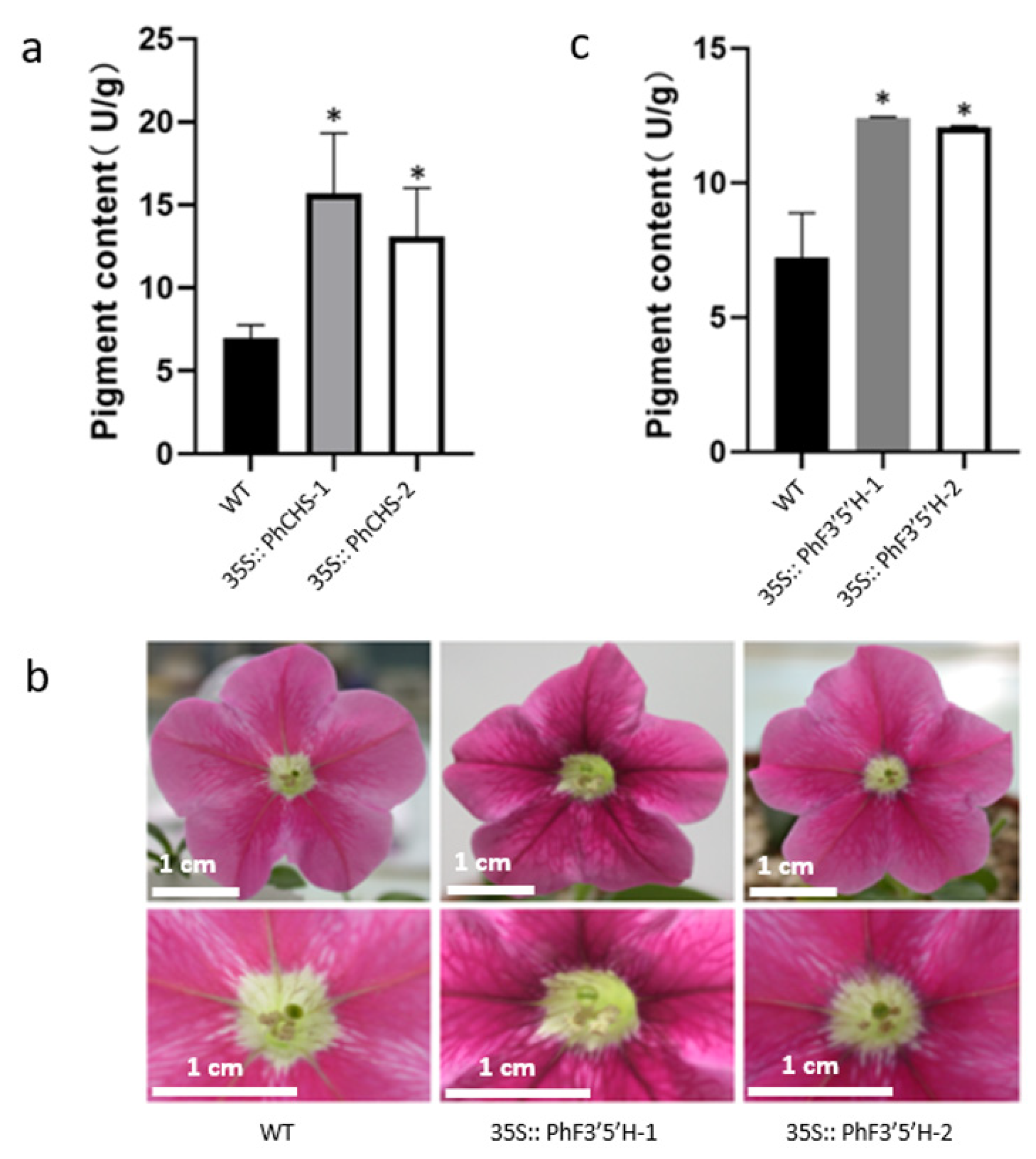
2.2. Analyses of the Phenotypes and Anthocyanin Contents of Genetically Modified Petunia hybrida

In order to further study how PhCHS5 and PhF3′5′H regulate flower color formation, Petunia hybrida was overexpressed with PhCHS5 and PhF3′5′H to obtain genetically-modified lines. Compared with WT, PhCHS5-overexpressing lines (PhCHS5-1 and PhCHS5-2) had deeper flower color and higher anthocyanin content (Figure 2a). Two genetically-modified lines 35S::PhF3′5′H-1 and 35S::PhF3′5′H-2 were obtained by overexpression of PhF3′5′H. Compared with WT, 35S::PhF3′5′H-1 and 35S::PhF3′5′H-2 both had deeper color and higher anthocyanin content (Figure 2b,c). The above results indicated that PhCHS5 and PhF3′5′H could deepen flower color and increase anthocyanin content in the pathway of flower color formation.

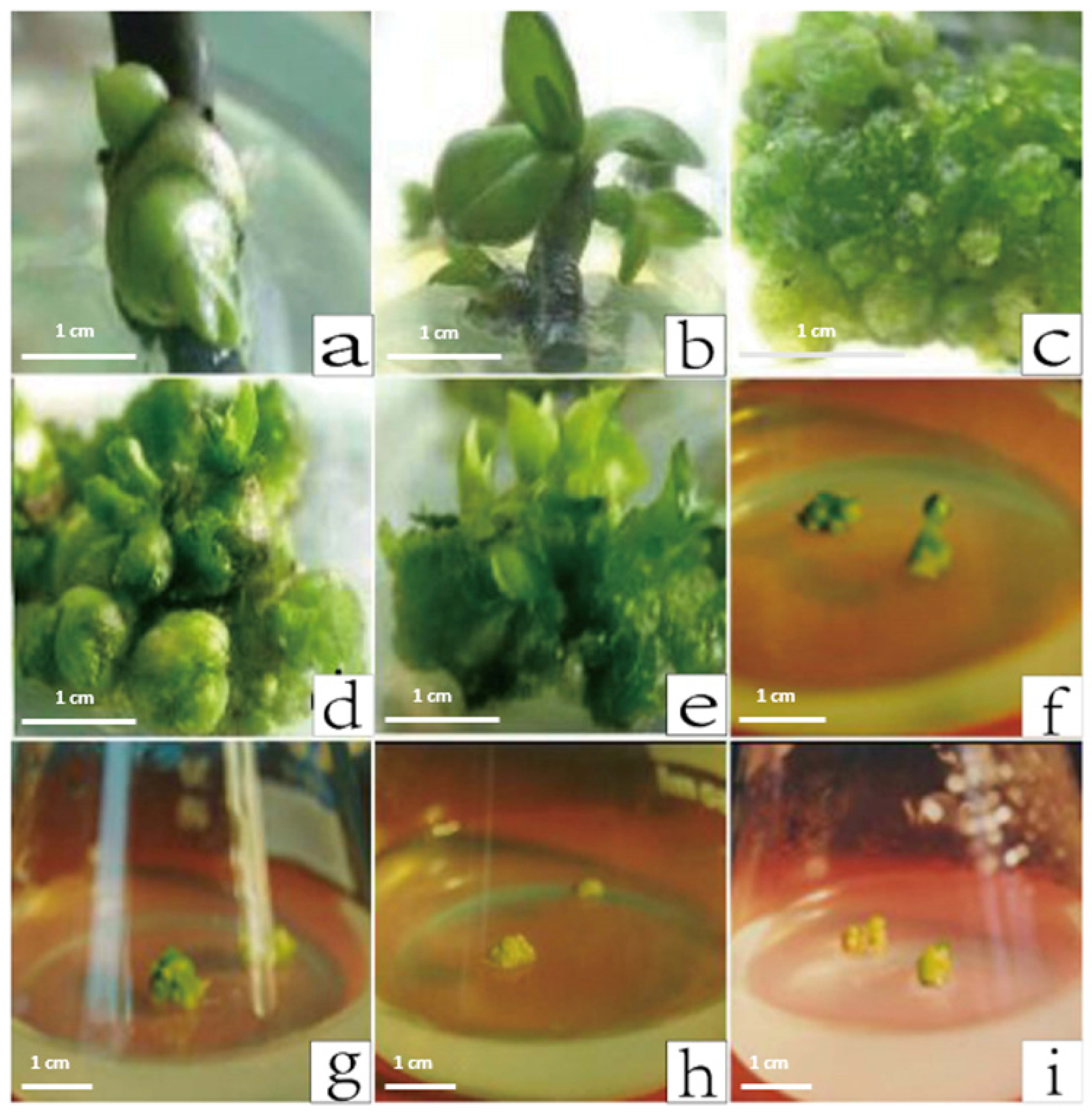
2.3. Induction and Cultivation of Phalaenopsis Protocorms

The Phalaenopsis protocorms required for the transgenic study were mainly generated from axillary bud explants. Specifically, the Phalaenopsis axillary bud explants were added to the Phalaenopsis induction medium in culture bottles. To produce high-quality materials and minimize contamination, four or five Phalaenopsis axillary bud segments were added to each culture bottle. Within two weeks of inoculation, the axillary buds began to swell (Figure 3a). The above materials were subcultured twice under aseptic conditions, after which the axillary buds began to elongate and produce leaves (Figure 3b). The leaves that grew to approximately 1 cm long were cut and transferred to the same induction medium. Many protocorms formed after two months (Figure 3c). Additionally, we examined the effects of different BA and NAA concentrations on the induction of protocorms. Ensuring the auxin and cytokinin contents are balanced is critical for plant morphological development. The protocorm induction efficiency of YD3 (3 ppm BA and 0.1 ppm NAA) was 80% (Table 1). In the YD3 medium, the original stem formed buds within 4 weeks (Figure 3d,e), which was efficient for the production of genetically modified materials. Increasing the NAA concentration (Figure 3f,g) or decreasing the BA concentration (Figure 3h,i) inhibited the production of protocorms and buds formation.

2.4. Screening of PhCHS5 and PhF3′5′H in Transformed Phalaenopsis Protocorms

The PhCHS5 and PhF3′5′H genes were cloned and determined to be involved in anthocyanin biosynthesis. Thus, we analyzed the expression of these two genes to clarify their roles related to flower coloration. The pCAMBIA2301-PhCHS5 and pCAMBIA2301-PhF3′5′H recombinant plasmids were constructed for the subsequent analysis of the overexpression of the genes in the Phalaenopsis hybrid ‘Formosa Rose’ background through A. tumefaciens infection and gene gun. The PhCHS5-transformed protocorms and PhF3′5′H-transformed protocorms were screened for cefotaxim resistance. GUS staining showed PhCHS5 and PhF3′5′H were successfully transferred into Phalaenopsis protocorms (Figure 4a,c). Those that grew well in the presence of cefotaxim were cultured to eventually produce leaves (Figure S2). A PCR analysis of the leaf in transgenic Phalaenopsis with the Kan gene primers revealed an amplified fragment that was consistent with the target fragment, indicating Phalaenopsis plants were correctly transformed (Figure 4b,d).

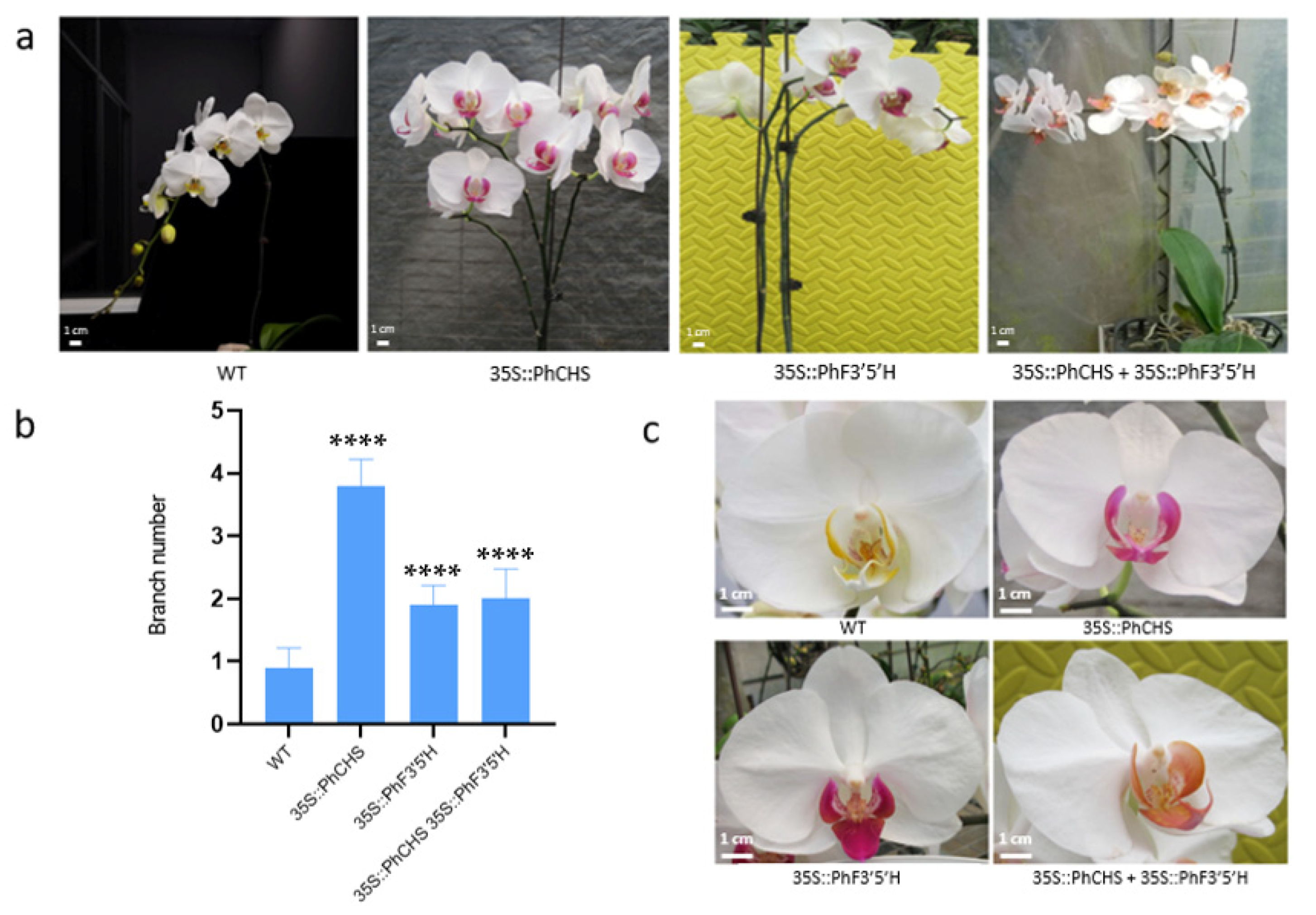
2.5. Regulatory Effects of PhCHS5 and PhF3′5′H Expression on the Branching of Phalaenopsis Stems and the Color of Phalaenopsis Lips

Transgenic seedlings with PhCHS5, PhF3′5′H, or PhCHS5 + PhF3′5′H grew well after the rooting, transplanting, and cultivation of whole plants (Figure S3). After two months’ growth, partially of the PhCHS5-transformed plants (Figure S3a′–c′), a few of the PhF3′5′H-transformed plants (Figure S3d′–f′) and some of PhCHS5 + PhF3′5′H transgenic plants were observed to grow abnormally, with asymmetrical plants and diverse leaf shapes. However, most of the transformed plants with normal shapes and grew well for further evaluation.
The splitting of the single stem of PhCHS5-transgenic plants resulted in multiple branches and Phalaenopsis plants with multiple heads. The stem of the PhF3′5′H-transformed plants also appeared to split into two (Figure 5a,b), with an increasing number of flowers with increases in the number of flowering branches (Figure 5a,b). The transgenic plants carrying PhCHS5 and PhF3′5′H produced many branches and multiple heads (Figure 5a,b). Additionally, PhCHS5 or PhF3′5′H-transgenic plants both showed deepened lip color, compared with the control (Figure 5c). However, the intensity of the coloration of the Phalaenopsis lips decreased when protocorms were co-transformed with both PhCHS5 and PhF3′5′H (Figure 5c). Accordingly, both genes are likely necessary for the regulation of Phalaenopsis lip coloration and branch development.

3. Materials and Methods

3.1. Materials and Growth Conditions

Petunia is laboratory reserved variety with red petals which were grown in a greenhouse of Shanghai Normal University (SHNU) and Phalaenopsis hybrid cv. Formosa roses with white petals are collected from the Vegetable and Horticulture Research Institute of the Shanghai Academy of Agricultural Sciences. Petunia was cultivated at a temperature of 24–26 °C with light for 16 h and dark for 8 h, the relative humidity is maintained at 74%, and the light intensity is 5000 lux. Phalaenopsis was cultured at a temperature of 18–20 °C with light for 16 h and dark for 8 h with a light intensity of 2500 lux and a relative humidity maintained at 80%.

3.2. Preliminary Treatment and Cultivation of Phalaenopsis Tissue Culture

The well-grown shoot segments of Phalaenopsis hybrid (Formosa rosa) were selected as materials, and the leaves outside the axillary buds were carefully peeled off and cut into 1 cm stem segments. Soak in soapy water and wash it many times. Rinse in running tap water and dry. It was immersed and sterilized by 5% NaClO on a clean bench for 15 min, and then rinsed 6 times with sterile water. The materials are cultured in the culture room after being inoculated into the sterilized medium. Medium formulation of induction and differentiation were shown in Supplementary Table S1.

3.3. Construction of PhCHS5 and PhF3′5′H Prokaryotic Expression Vectors

PCR was performed using forward primers for the 5′end of PhCHS5 is 5′GCTCTAGAGGAGAGGGAGTTAATGGC3′ and the 3′end reverse primer is 5′GCATTTTGTGGTTTTATTGGACT3′ (Xba I linker is added to both ends of the forward primer and reverse primer). The forward primer for the 5′end of PhF3′5′H is 5′CCGCTCGAGATGTCCATCTTCCTCATCATCACACACACCC3′, and the reverse primer at the 3′end of PhF3′5′H is 5′GAAGATCTTCAAACAACCCCATACGCCGCCG3′ (with Bgl II restriction site) (Shanghai Bioengineering Co., Ltd., located in Shanghai, China). Afterwards, the PCR products of PhCHS5 and the pCAMBIA2301 vector were double-digested with Xba I, Xho I; the PCR products of PhF3′5′H and the pCAMBIA2301 vector were double-digested with Xho I, Bgl II.

3.4. Agrobacterium-Mediated Leaf Disc Method Transgenic to Obtain Transgenic Petunia Expressing PhCHS5 and PhF3′5′H Gene

By transgenic leaf discs, Petunia leaves were cut into 0.8 × 0.8 cm (1 cm) squares, placed in MS0 solution containing Agrobacterium for infection, and incubated in the dark on MS1 at 22 °C for three days [25]. Afterward, the bacterial cells are washed and excess water is absorbed with absorbent paper, the leaves are transferred to MS2 and cultured at 25 °C for subculture every 20 days, and the Seedlings differentiated from calluses are thoroughly washed with sterile distilled water after they grow, and then transferred to plastic pots containing peat-based soil. Seedlings are incubated in a growth chamber at 25 ± 2 °C at 85% relative humidity for 2–3 days. The seedlings are then transferred to plastic pots filled with peat-based soil and grown in greenhouses under controlled conditions. The medium formula is as follows:
Differentiation medium (MS1) formulation: MS, 6-BA 1 mg/L, NAA 0.1 mg/L, plant gel concentration 0.25%, sucrose final concentration 3%, pH 6.0. Differentiation liquid medium (MS0) formulation is without plant gel concentration. Rooting medium formulation: 1/2MS, plant gel concentration 0.25%, sucrose final concentration 3%, pH 5.8. Resistance screening medium formulation: on the basis of normal differentiation and rooting medium for mula, Kan 25 mg/mL + Cef 250 mg/mL (MS2) or Kan 25 mg/mL + Cef 500 mg/mL (MS3) were added respectively.

3.5. Construction of Transgenic Phalaenopsis by Gene Gun

After osmotic treatment for 1 h, the protocorms of Phalaenopsis were cut into pieces and pre-cultured in a 60 mm Petri dish containing YD3 medium (see Supplementary Table S1). The plasmid was purified by QIAGEN Plasmid Purification Kit (Qiagen, Shanghai, China) and precipitated on gold particles 1.0-(m). In a vacuum environment, the Bio-Rad system was used, and bombarded with 1100 kPa helium, bombarded twice per dish. The protocoms after bombardment were transferred to new YD3, and cultures (containing corresponding antibiotics) were screened after 2–10 days. Data were obtained from at least three biological replicates and blank controls per sample.

3.6. Construction of Transgenic Phalaenopsis by Agrobacterium Infection

Shake overnight (not less than 7 h, 28 °C, 200 rpm). Make several holes in the protocom used for transgene. The protocol was placed in the bacterial solution, and the bacteria were shaken for 30 min. Appropriately remove excess bacteria. The protocom was clamped to YD3 (see Supplementary Table S1) solid medium covered with a layer of sterile filter paper and cultured for 3 days in the dark. After three days, the excess bacterial solution was washed away, and the protocom was washed by 3–5 times by YD3T with antibiotics, at last, were placed on a medium of YD3 (see Supplementary Table S1) + cef (250 mg/L) for 6 weeks. The newly grown protocorm-likebodies (PLB) were then transferred to YD3 and YDR rooting medium (supplemented with antibiotics) for Proliferation and rooting.

3.7. Analysis of Anthocyanin in the Petals of Transgenic Petunia

Put the petals in liquid nitrogen and grind thoroughly. Add 1 mL of the prepared acidic methanol (methanol: HCl = 24:1) to each test tube and let it shake well on the shaker. To disperse petal fragments. At 4 °C and 100 rpm, the vibrator vibrates for 2 days, during which it vibrates twice on the vibrator. After that, the anthocyanin content was measured, and the atmosphere was diluted to 2 mL with 800 mL, and then the absorbance of A530 and A657 was measured with a spectrophotometer to calculate the anthocyanin content (U/g).

3.8. Molecular Identification and Phenotype Analysis of Transgenic Phalaenopsis Strains

Kan gene was used in PCR to screen transgenic strains, designing primers based on its genetic sequences, The forward primer for the 5′end of Kan is 5′ATTTTCTCCCAATCAGGCTTGATCC3′, and the reverse primer at the 3′end of Kan is 5′CACCTATGATGTGGAACGGGAAAAG3′. DNA extraction was performed by the method [26]. After the plant grows to a certain stage, it is subjected to flower treatment to perform plant phenotype identification.

3.9. Statistical Analysis

The date including anthocyanin content, rate of protocorm and branch number was used in t-test analysis and the Dumcans multiplerenge test method.

4. Discussion

Both CHS5 and F3′5′H are key enzymes contributing to anthocyanin biosynthesis in plants, with crucial regulatory functions related to plant coloration. To date, the effects of CHS and F3′5′H genes on plant colors have mostly been investigated in Rosa rugosa Thunb and Petunia hybrida Vilm [27]. Due to their varied colors, orchids have become the ideal plant materials for studying the mechanism underlying the development of flower colors. Increases in the commercial production of Phalaenopsis species have coincided with increasing molecular research on these important orchids. However, these species have not been as well characterized as some other widely grown crops, including rice and corn. In China, much of the relevant research currently involves Phalaenopsis tissue cultures, but there are few reports describing research on the genetic basis of flower coloration [28].
The F3′5′H gene has been studied by several researchers. For example, the overexpression of the violet F3′5′H gene in Rosa chinensis ‘Lavande’ increased the delphinidin accumulation by up to 44.2% [29]. Nakano and Tanaka produced purple lilies by generating plants constitutively expressing the Campanula F3′5′H gene under the control of the CaMV 35S promoter [19,30]. In another study, the expression of the Phalaenopsis F3′5′H gene in lily flowers caused the petals to change from pink to blue [31]. Additionally, purple carnations and roses accumulating delphinidin-based anthocyanin following the introduction of the F3′5′H gene have been commercialized globally [32]. The results of the current study are similar to those of earlier investigations. The flower colors of the PhF3′5′H-transformed Petunia were relatively intense (Figure 2), implying that PhF3′5′H likely induces the accumulation of anthocyanin.
In an earlier investigation, the expression of antisense CHS genes in Petunia led to the production of completely white flowers [33]. Additionally, the overexpression of the CHS gene was expected to deepen the flower colors. In the current study, PhCHS5-transformed Petunia showed deeper flower color, while Phalaenopsis plants exhibited deepened lip color (Figure 5c), which was consistent with the expectation. To our interest, the intensity of the coloration of the Phalaenopsis lips decreased when protocorms were co-transformed with both PhCHS5 and PhF3′5′H, compared with the plants transformed with PhCHS5 or PhF3′5′H alone (Figure 5c), which suggested that there might be conjugate effect between PhCHS5 and PhF3′5′H, in which interaction cofactor between them should be further studied.
The transgenes analyzed in this study had no effect on the production of white petals (Figure 5), possibly because of an endogenous mechanism that prevents anthocyanin synthesis and accumulation. In a previous study, a rare mutation in white flowers due to DFR inactivation was detected in a Solanaceae species [34]. Most Caryophyllales species are unable to accumulate anthocyanin because of a lack of DFR and ANS activity [35]. Phalaenopsis ‘Formosa Rose’ petals may lack the substrate for anthocyanin biosynthesis or key enzymes (i.e., DFR and ANS), which may explain their inability to synthesize anthocyanin or their production of only colorless anthocyanin. However, our analysis of the flowers of Phalaenopsis plants transformed with both PhCHS5 and PhF3′5′H revealed a deepening of the lip color, indicative of the accumulation of colored anthocyanin in the lip (Figure 5). Moreover, the transformation of the endogenous genes contributed to the increased anthocyanin accumulation. Accordingly, the expression levels of PhCHS5 and PhF3′5′H may be related, possibly due to the regulatory effects of specific transcription factors.
In the current study, the application of exogenous and endogenous genes to synthesize anthocyanin was insufficient for enhancing the petal coloration of white Phalaenopsis flowers. There are published reports indicating that the transformation of Phalaenopsis plants producing pink flowers with the Commelina communis F3′5′H gene generates transgenic plants with bluish flowers [15,30]. Thus, we will need to evaluate the effects of pH, metal ions, transcription factors, and genetic modifications to determine how to obtain blue Phalaenopsis flowers. In Petunia, seven genetic loci (PH1-PH7) regulating the pH of petals have been identified [36]. Under acidic and alkaline conditions, the petals will be reddish and bluish, respectively. Metal ion transporters can also influence flower coloration. The tulip TgVit1 gene and its homolog in Centaurea cyanus (CcVit) encode vacuolar iron transporters, which promote the accumulation of iron ions in the vacuole, resulting in blue petals [37]. Changes to the transcription factors associated with the petal anthocyanin biosynthesis pathway may alter flower colors. The results of these earlier investigations may be relevant for developing novel methods for improving flower colors. However, enhancing the floral coloration of Phalaenopsis species requires a more thorough elucidation of the underlying mechanism. The data derived from recent whole-genome sequencing efforts may provide a solid foundation for future comprehensive analyses of the mechanism mediating the formation of floral patterns. Additionally, like other non-model plant species, many ornamental plants are associated with specific problems adversely affecting molecular research, including a low genetic transformation efficiency, a long juvenile stage, the development of abnormal petals, and substantial variability in transgene expression. These problems must be overcome to facilitate the generation of new commercially valuable flower varieties. Our study findings described herein may provide the basis for future research on Phalaenopsis floral coloration, specifically regarding the effects of PhF3′5′H and PhCHS5 on lip color.

Supplementary Materials

The following supporting information can be downloaded at: https://www.mdpi.com/article/10.3390/plants12112204/s1, Figure S1 Phylogenetic analysis of CHS5 and F3’5’H in different plant species. (a) CHS5; (b) F3’5’H; Figure S2 Hygromycin screening and cluster seedlings of the transgenic Phalaenopsis samples; Figure S3 Leaf phenotypes of the transgenic Phalaenopsis samples. (a’–c’) PhCHS5-transformed plants. (d’–f’) PhF3′5′H-transformed plants. (g’–i’) PhCHS5 + PhF3′5′H-transformed plants; Table S1 Medium information for protocorm-likebodies (PLB) induction and differentiation and Agrobacterium infection.

Author Contributions

F.M. proposed research ideas and designed research plans; Y.L. and Q.Z. conducted experiments together, wrote and revised the paper together. X.Y. and Q.X. completed the collation and revision of the paper; W.W. and R.G. completed the editing and revision of the pictures of the paper. All authors have read and agreed to the published version of the manuscript.

Funding

This work was supported by National Key R&D Program of China (2018YFD1000400), Science and Technology Commission of Shanghai Municipality (18DZ2260500), Shanghai Engineering Research Center of Plant Germplasm Resources (17DZ2252700).

Data Availability Statement

Not applicable.

Conflicts of Interest

The authors declare that they have no conflict of interest.

References

  1. Christenhusz, M.J.M.; Byng, J.W. The number of known plants species in the world and its annual increase. Phytotaxa 2016, 261, 201–217. [Google Scholar] [CrossRef] [Green Version]
  2. Azadi, P.; Bagheri, H.; Nalousi, A.M.; Nazari, F.; Chandler, S.F. Current status and biotechnological advances in genetic engineering of ornamental plants. Biotechnol. Adv. 2016, 34, 1073–1090. [Google Scholar] [CrossRef] [PubMed]
  3. Anwar, M.; Chen, L.; Xiao, Y.; Wu, J.; Zeng, L.; Li, H.; Wu, Q.; Hu, Z. Recent Advanced Metabolic and Genetic Engineering of Phenylpropanoid Biosynthetic Pathways. Int. J. Mol. Sci. 2021, 22, 9544. [Google Scholar] [CrossRef] [PubMed]
  4. Tomizawa, E.; Ohtomo, S.; Asai, K.; Ohta, Y.; Takiue, Y.; Hasumi, A.; Nishihara, M.; Nakatsuka, T. Additional betalain accumulation by genetic engineering leads to a novel flower color in lisianthus (Eustoma grandiflorum). Plant Biotechnol. 2021, 38, 323–330. [Google Scholar] [CrossRef]
  5. Cardona, J.; Lara, C.; Ornelas, J.F. Pollinator divergence and pollination isolation between hybrids with different floral color and morphology in two sympatric Penstemon species. Sci. Rep. 2020, 10, 8126. [Google Scholar] [CrossRef] [PubMed]
  6. Zhang, Z.B.; Cheng, J.; Yang, M.; Cui, J.; Chen, Y.M.; Deng, Z.H.; Zhao, X.H. Study on foodborne deceptive pollination of qinliwandai LAN. J. Beijing For. Univ. 2015, 37, 100–106. [Google Scholar]
  7. Ray, H.A.; Stuhl, C.J.; Gillett-Kaufman, J.L. Floral fragrance analysis of Prosthechea cochleata (Orchida-ceae), an endangered native, epiphytic orchid, in Florida. Plant Signal. Behav. 2018, 13, e1422461. [Google Scholar] [CrossRef] [Green Version]
  8. Wang, L.M.; Zhang, J.; Dong, X.Y.; Fu, Z.Z.; Jiang, H.; Zhang, H.C. Identification and functional analysis of anthocyanin biosynthesis genes in Phalaenopsis hybrids. Biol. Plant. 2018, 62, 45–54. [Google Scholar] [CrossRef]
  9. Lin, L.; Tang, H.R.; Chen, Q.; Lu, M.; Jia, H.F.; Li, Y.G.; Zhang, X.N. Cloning and sequence analysis of chalcone synthase gene in ornamental peach. Acta Hortic. Sin. 2012, 39, 581–587. [Google Scholar]
  10. Xu, W.; Yu, X.Y.; Chen, J.; Fu, H.Y.; Hu, B.; Chen, Y.B.; Li, D. Cloning and sequence analysis of CHS gene from Loropetalum rubrum. Chin. Agric. Sci. Bull. 2012, 29, 24–28. [Google Scholar]
  11. Fukusaki, E.I.; Kawasaki, K.; Kajiyama, S.I.; An, C.I.; Suzuki, K.; Tanaka, Y.; Kobayashi, A. Flower color modulations of Torenia hybrida by downregulation of chalcone synthase genes with RNA interference. J. Biotechnol. 2004, 111, 229–240. 9. [Google Scholar] [CrossRef] [PubMed]
  12. Koes, R.E.; Spelt, C.E.; Elzen, P.J.; Mol, J.N. Cloning and molecular characterization of the chalcone synthase multigene family of Petunia hybrida. Gene 1989, 81, 245–257. [Google Scholar] [CrossRef] [PubMed]
  13. Tanaka, Y.; Sasaki, N.; Ohmiya, A. Biosynthesis of plant pigments: Anthocyanin, betalains and carotenoids. Plant J. 2008, 54, 733–749. [Google Scholar] [CrossRef] [PubMed]
  14. Shi, S.C.; Gao, Y.K.; Zhang, X.M.; Sun, J.Q.; Zhao, L.L.; Wang, Y. Progress on plant genes involved in biosynthetic pathway of Anthocyanin. Bull. Bot. Res. 2011, 31, 633–640. [Google Scholar]
  15. Noda, N. Recent advances in the research and development of blue flowers. Breed Sci. 2018, 68, 79–87. [Google Scholar] [CrossRef] [Green Version]
  16. Noda, N.; Yoshioka, S.; Kishimoto, S.; Nakayama, M.; Douzono, M.; Tanaka, Y.; Aida, R. Generation of blue chrysanthemums by anthocyanin B-ring hydroxylation and glucosylation and its coloration mechanism. Sci. Adv. 2017, 3, e1602785. [Google Scholar] [CrossRef] [Green Version]
  17. Xiao, J.P.; Yang, X.N.; Guo, H.C. Bioinformatics and expression analysis of flavonoids-3-5-hydroxylase (F3′5′H) gene in color potato variety ‘Zhuanxinwu’. Genom. Appl. Biol. 2015, 34, 1494–1502. [Google Scholar]
  18. Su, V.; Hsu, B.D. Cloning and expression of a putative cytochrome P450 gene that influences the color of Phalaenopsis flowers. Biotechnol. Lett. 2003, 25, 1933–1939. [Google Scholar] [CrossRef]
  19. Tanaka, Y.; Nakamura, N.; Kobayashi, H.; Okuhara, H.; Kondo, M.; Koike, Y.; Hoshi, Y.; Nomizu, T. Method for Cultivating Lilies Containing Delphinidin in the Petals Thereof. European Patent WO2012036290, 22 March 2012. [Google Scholar]
  20. Feng, H.; Wang, J.H.; Wang, M.L.; Li, Y.; Li, Y. Study on genetic transformation of chalcone synthase gene in Petunia. J. Tianjin Agric. 2007, 14, 9–12. [Google Scholar]
  21. Nakatsuka, T.; Mishiba, K.; Kubota, A.; Abe, Y.; Yamamura, S.; Nakamura, N. Genetic engineering of novel flower color by suppression anthocyanin modification genes in gentian. J. Plant Physiol. 2010, 167, 231–237. [Google Scholar] [CrossRef]
  22. Han, Y.Y.; Ming, F.; Wang, J.W.; Wen, J.G.; Ye, M.M.; Shen, D.L. Cloning and characterization of a novel chalcone synthase gene from Phalaenopsis hybrida orchid flowers. Russ. J. Plant Physiol. 2006, 53, 250–258. [Google Scholar] [CrossRef]
  23. Wang, J.W.; Ming, F.; Han, Y.Y.; Shen, D.L. Flavonoid -3′5′-hydroxylase from Phalaenopsis: cDNA cloning, endogenous expression and molecular modeling. Biotechnol. Lett. 2006, 28, 327–334. [Google Scholar] [CrossRef] [PubMed]
  24. Vandenbussche, M.; Chambrier, P.; Rodrigues Bento, S.; Morel, P. Petunia, Your Next Supermodel. Front. Plant Sci. 2016, 7, 72. [Google Scholar] [CrossRef] [PubMed] [Green Version]
  25. Ai, T.N.; Naing, A.H.; Arun, M.; Jeon, S.M.; Kim, C.K. Expression of RsMYB1 in Petunia enhances antho-cyanin production in vegetative and floral tissues. Sci. Hortic. 2017, 214, 58–65. [Google Scholar] [CrossRef]
  26. Xia, Y.; Chen, F.; Du, Y.; Liu, C.; Bu, G.; Xin, Y.; Liu, B. A modified SDS-based DNA extraction method from raw soybean. Biosci. Rep. 2019, 39, BSR20182271. [Google Scholar] [CrossRef] [Green Version]
  27. Qi, Y.; Zhao, L.Y. Cloning and bioinformatics analysis of chalcone synthase CHS gene from Rose. J. Agric. 2015, 5, 91–96. [Google Scholar]
  28. Sun, R.; Yu, N.; Sun, P. Advances in genetic engineering of Phalaenopsis. North. Hortic. 2014, 12, 177–180. [Google Scholar]
  29. Katsumoto, Y.; Fukuchi, M.M.; Fukui, Y. Engineering of the rose flavonoid biosynthetic pathway successfully generated blue-hued flowers accumulating delphinidin. Plant Cell Physiol. 2007, 48, 1589–1600. [Google Scholar] [CrossRef]
  30. Nakano, M.; Mii, M.; Kobayashi, H.; Otani, M.; Yagi, M. Molecular approaches to flower breeding. Breed. Res. 2016, 18, 34–40. [Google Scholar] [CrossRef] [Green Version]
  31. Qi, Y.; Lou, Q.; Quan, Y.; Liu, Y.; Wang, Y. Flower-specific expression of the Phalaenopsis flavonoid 3′,5′-hydoxylase modifies flower color pigmentation in Petunia and Lilium. Plant Cell Tissue Organ Cult. 2013, 115, 263–273. [Google Scholar] [CrossRef]
  32. Tanaka, Y.; Brugliera, F. Flower colour and cytochromes p450. Phytochem. Rev. 2013, 5, 283–291. [Google Scholar] [CrossRef]
  33. Van der Krol, A.; Lenting, P.; Veenstra, J.; Van der Meer, I.; Koes, R.; Gerats, A.; Mol, J.; Stuitje, A. An anti-sense chalcone synthase gene in transgenic plants inhibits flower pigmentation. Nature 1988, 333, 866–869. [Google Scholar] [CrossRef]
  34. Coburn, R.A.; Griffin, R.H.; Smith, S.D. Genetic basis for a rare floral mutant in an Andean species of Solanaceae. Am. J. Bot. 2015, 102, 264–272. [Google Scholar] [CrossRef] [PubMed] [Green Version]
  35. Shimada, S.; Otsuki, H.; Sakuta, M. Transcriptional control of anthocyanin biosynthetic genes in the Caryophyllales. J. Exp. Bot. 2007, 58, 957–967. [Google Scholar] [CrossRef] [PubMed] [Green Version]
  36. Van Houwelingen, A. Analysis of flower pigmentation mutants generated by random transposon mutagenesis in Petunia hybrida. Plant J. 1998, 13, 39–50. [Google Scholar]
  37. Momonoi, K.; Yoshida, K.; Mano, S.; Takahashi, H.; Nakamori, C.; Shoji, K.; Nitta, A.; Nishimura, M. A vacuolar iron transporter in tulip, TgVit1, is responsible for blue coloration in petal cells through iron accumulation. Plant J. 2009, 59, 437–447. [Google Scholar] [CrossRef]
Figure 1. Construction of recombinant plasmids carrying the PhCHS5 and PhF3′5′H sequences for the transformation of Petunia. (a) Confirmation of the construction of the pCAMBIA2301-sense-CHS5 plasmid by PCR. Regarding the plasmid with PhCHS5 in the forward direction, lane 1 presents the fragment amplified with the Cav35s-primerF and CHS-XR’ primers. Lane 2 presents the approximately 750-bp non-specific band amplified by PCR with the Cav35s-primerF and CHS-XF primers. (b) Confirmation of the transformation of A. tumefaciens cells with PhF3′5′H by PCR. Lanes 1 to 8 represent transformed A. tumefaciens colonies that were analyzed by PCR. (c) Petunia leaf explants during the transformation procedure. (d) Mature transgenic plant. (e) Transgenic Petunia tissue culture. (f) Rooting of transgenic Petunia plants. (g) Preliminary identification of PhCHS5-transformed Petunia plants by PCR. Each lane represents a different Petunia strain. The red arrow indicates the PhCHS5 stripe; WT for wild type; +stand for vector with NPTII gene (h) Preliminary identification of PhF3′5′H-transformed Petunia plants by PCR. Each lane represents a different Petunia strain. The black arrow indicates the PhF3′5′H stripe; WT for wild type; Ev stand for vector with only NPTII gene without PhF3′5′H.
Plants 12 02204 g001
Figure 2. Phenotypic analysis and anthocyanin content determination of Petunia mutants PhCHS5 and PhF3′5′H. (a) Anthocyanin content of Petunia hybrida transformed by PhCHS5. (b) The phenotype of Petunia transformed by PhF3′5′H. (c) Anthocyanin content of Petunia hybrida transformed by PhF3′5′H. WT: Wild Type. The data were expressed as mean ± standard error and repeated three times for each sample. * p < 0.05.
Plants 12 02204 g002
Figure 3. Induction of the Phalaenopsis protocorm. (a) Swollen axillary bud. (b) Seedling generated directly from an axillary bud. (c) Protocorm development. (d) Protocorm enlargement. (e) Seedling cluster. (fi) Protocorms from YD4 (f,g), YD2 (h) and YD1 (i), respectively.
Plants 12 02204 g003
Figure 4. Screening of Phalaenopsis protocorms carrying PhCHS5 and PhF3′5′H after transformations. (a) PhCHS5-transgenic Phalaenopsis protocorms. The image on the right is a close-focus image of the image on the left (b) Confirmation of the transformation of Phalaenopsis protocorms with PhCHS5 by PCR for Kan gene.M stands for marker, 1–10 stands for transformation seedlings. (c) PhF3′5′H-transgenic Phalaenopsis protocorms. Figure 1 is a close-focus image, and Figure 2, Figure 3 and Figure 4 are three representative repeating telefocal images (d) Molecular confirmation of the presence of PhF3′5′H in the transformed samples by PCR for Kan gene. M stands for marker, 1–5 stands for transformation seedlings.
Plants 12 02204 g004
Figure 5. Phenotypes of the transgenic Phalaenopsis samples. (a,b) Branch number of the transgenic plants. (c) Flower phenotypes of the transgenic plants. WT: Wild Type. The data were expressed as mean ± standard error and repeated three times for each sample. **** p < 0.0001.
Plants 12 02204 g005
Table 1. Effect of different hormones on the induction rate of protocorm.
NumberingBA/NAA (ppm) ConcentrationProtocorm Induction Rate (%)Unit Axillary Buds Get the Number of Bushes
YD12/0.250 ± 1.2 b14 ± 1.4 b
YD22/2.040 ± 2.6 c25 ± 3.4 a
YD33/0.180 ± 2.0 a10 ± 1.6 bc
YD43/2.010 ± 1.8 d4 ± 2.5 c
All data is the mean of four repetitions. Using the Dumcans multiplerenge test method, different letters in the same column indicate significant differences (p < 0.05, n = 3).
Disclaimer/Publisher’s Note: The statements, opinions and data contained in all publications are solely those of the individual author(s) and contributor(s) and not of MDPI and/or the editor(s). MDPI and/or the editor(s) disclaim responsibility for any injury to people or property resulting from any ideas, methods, instructions or products referred to in the content.

