Article

Duplex Real-Time PCR Assays for the Simultaneous Detection and Quantification of Botryosphaeriaceae Species Causing Canker Diseases in Woody Crops

by
Laura Romero-Cuadrado
1,
Carlos José López-Herrera
2,
Ana Aguado
1 and
Nieves Capote
1,*
1
Andalusian Institute of Agricultural and Fisheries Research and Training (IFAPA), Center Las TorresAlcalá del Río, 41200 Seville, Spain
2
Instituto de Agricultura Sostenible, CSIC, C/ Alameda del Obispo s/n, 14004 Córdoba, Spain
*
Author to whom correspondence should be addressed.
Plants 2023, 12(11), 2205; https://doi.org/10.3390/plants12112205
Submission received: 24 March 2023 / Revised: 11 May 2023 / Accepted: 31 May 2023 / Published: 2 June 2023

Abstract

:
Woody canker diseases caused by fungi of the Botryosphaeriaceae family are producing increasing losses in many economically important woody crops, including almond. To develop a molecular tool for the detection and quantification of the most aggressive and threatening species is of main importance. This will help to prevent the introduction of these pathogens in new orchards and to conveniently apply the appropriate control measures. Three reliable, sensitive and specific duplex qPCR assays using TaqMan probes have been designed for the detection and quantification of (a) Neofusicoccum parvum and the Neofusicoccum genus, (b) N. parvum and the Botryosphaeriaceae family and (c) Botryosphaeria dothidea and the Botryosphaeriaceae family. The multiplex qPCR protocols have been validated on artificially and naturally infected plants. Direct systems to process plant materials, without DNA purification, allowed high-throughput detection of Botryosphaeriaceae targets even in asymptomatic tissues. These results validate the qPCR using the direct sample preparation method as a valuable tool for Botryosphaeria dieback diagnosis allowing a large-scale analysis and the preventive detection of latent infection.

1. Introduction

Almond (Prunus dulcis (Mill.) D.A. Webb) is one the most important nut crops in the world. California (USA) is the leader in almond production followed by Spain and Australia (FAOSTAT, 2021; http://www.fao.org/faostat/en/#data (accessed on 10 March 2023). The implementation of new management techniques, such as high-density cultivation, prune intensification, drip irrigation and fertilization, mechanical harvest, use of more productive varieties and the cultivation in agronomically and environmentally more favorable cropping areas, has increased the fruit production in the last decades [1]. However, this new scenario, together with the current climate change situation, have favored the increase in almond diseases [2,3,4]. Among them, woody canker diseases, associated with fungi of the Botryosphaeriaceae family, affect a great number of agronomically important woody crops such as olive, grapevine, avocado, blueberry, stone fruit, citrus and nut crops, including almond [5,6,7,8,9,10,11,12]. These diseases affect the trunk and branches of young and mature almond trees causing cankers, extensive gumming, dieback, discoloration and necrosis of internal tissues and, especially in severe cases and young trees, plant death. Almond canker diseases have been described in California [13,14,15,16,17], Iran [18], Turkey [19], Morocco [20] and Spain [5,21,22,23], causing important economic losses [24].
Fungi of the Botryosphaeriaceae family are one of the most prevalent pathogens in almond orchards [13,16,21]. Many Botryosphaeriaceae species have been associated with almond cankers worldwide (Botryosphaeria dothidea, Diplodia corticola, D. gallae, D. mutila, D. olivarum, D. seriata, Dothiorella iberica, Do. prunicola, Do. sarmentorum, Do. viticola, Lasiodiplodia theobromae, Macrophomina phaseolina, Neofusicoccum arbuti, N. australe, N. luteum, N. mediterraneum, N. nonquaesitum, N. parvum, N. vitifusiforme and Neoscytalidium dimidiatum) [5,13,15,16,18,21,22,23,25] with Neofusicoccum being the prevailing genus [24]. Regarding other nut crops, N. parvum is also the dominant species in walnut orchards in Australia, California, China, Chile, Italy, Iran and Spain [18,26,27,28,29,30,31,32], and N. mediterraneum prevails in Spanish and Californian pistachio orchards [33,34]. The infection of the host plant occurs through natural wounds or those caused by pruning or injury or through other openings such as lenticels and stomata. These fungi can act as saprophytic, endophytic or latent pathogens and disease symptoms usually appear when the host is under stress conditions [35,36]. Therefore, the latent infections allow the “silent” introduction of the pathogens in new plantations and a late diagnosis of the disease, making control measures less efficient. Diagnosis of the disease by symptoms visualization in the field is not accurate because canker symptoms caused by other non-Botryosphaeriaceae species can produce similar symptoms and, in addition, co-infection of a single plant by different pathogenic species usually occurs [16]. Traditional methods for the isolation and identification of Botryosphaeriaceae species are based on culture of symptomatic plant tissues in the appropriate culture medium, followed by obtention of monoconidial or hyphal tip isolates and further characterization based on morphological characteristics (colony aspect or microscopic features). This process is laborious and time-consuming and not always accurate. In addition, many of the Botryosphaeriaceae species do not produce conidia in culture medium [37], making identification difficult.
For the preventive detection and an accurate diagnosis of fungal diseases, the use of molecular tools based on real-time PCR is one of the most promising strategies. PCR-based methods (conventional PCR and nested PCR) have been designed to detect species of Botryosphaeriaceae in almonds [38] and in other crops [39] or for the specific detection of Botryosphaeriaceae family [40]. In addition, qPCR-based methods have also been designed for the detection of some species or genera in the Botryosphaeriaceae family [41,42,43,44]. Most of the previously designed qPCR protocols used SYBR Green chemistry, which is cheaper and extensively used for the detection of high number of targets. However, the use of hydrolysis probes such as TaqMan probes has been demonstrated to be more advantageous due to their higher specificity (an additional specific primer, the TaqMan probe, is used, while SYBR Green can unspecifically join to dsDNA and/or generate by products), higher sensitivity and, in addition, the possibility to accurately quantify the fungal target [45].
Among the Botryosphaeriaceae family, Neofusicoccum parvum and Botryosphaeria dothidea have been reported as the most aggressive and most prevailing species in Spanish almond crop, respectively [21,23]. However, since the accurate identification of the causal agents is not easy and canker diseases can be caused by more than one pathogen, the objectives of this work were (i) to develop sensitive and reliable qPCR methods based on TaqMan probes for the simultaneous detection of (a) N. parvum and the Neofusicoccum genus, (b) N. parvum and the Botryosphaeriaceae family and (c) B. dothidea and the Botryosphaeriaceae family, in almond; (ii) to validate the designed protocols in artificially and naturally infected almonds by using two methods of sample preparation: extraction of DNA from infected tissues and direct preparation of plant crude extracts; and (iii) to apply the designed duplex qPCRs for the detection of the latent infection of Botryosphaeriaceae species in almond.

2. Results

2.1. Design of Primers and Probes

Primers and probes for the specific detection of N. parvum and B. dothidea were designed based on sequences from the translation elongation factor 1-alpha gene (tef1). Primers and probes for the multispecies detection of Neofusicoccum spp. and Botryosphaeriaceae family were designed based on sequences from the beta tubulin gene (tub2) (Table 1). For the duplex detection, TaqMan probes specific for N. parvum and B. dothidea were labelled with FAM fluorophore and TaqMan probes for the detection of Neofusicocum spp. and Botryosphaeriaceae family were marked with SUN fluorophore.

2.2. Analytical Specificity—Inclusivity and Exclusivity—And Limit of Detection

Simplex qPCR protocols for the specific detection of N. parvum and B. dothidea did not detect other Neofusicoccum or Botryosphaeria species used in the specificity tests. Furthermore, BLAST analyses showed that N. parvum-specific qPCR protocol would not detect Neofusicoccum species associated to trunk diseases of walnut (N. luteum, N. mediterraneum, N. nonquaesitum, N. vitifusiforme) and pistachio (N. australe, N. hellenicum, N. mediterraneum, N. pistaciae N. vitifusiforme) [24,46] nor Neofusicoccum species affecting other Mediterranean crops, such as Citrus spp. and olive [47]. However, primers and probe for the detection of N. parvum could detect some of the phylogenetically closest species in the N. parvum species complex, such as N. ribis and N. kwambonambiense, which affect Mediterranean woody crops (Table S2). The B. dothidea simplex qPCR protocol could also detect B. corticis and B. fabicerciana described in Prunus and other woody crops in Mediterranean areas but not reported in almond [48,49] (Table S2). On the contrary, primers and probes designed for the specific detection of the genus Neofusicoccum and the Botryosphaeriaceae family could detect, respectively, all the Neofusicoccum and Botryosphaeriaceae species described in almond and Mediterranean crops. No amplification was detected using DNA from non-target species or negative controls.
Simplex and duplex qPCR reactions showed efficiencies between 90 and 110% and R2 about 0.99 (Figure 1). Duplex qPCR reactions did not decrease the sensitivity of detection nor decrease R2 values compared with simplex qPCR reactions.
The limit of detection of N. parvum, B. dothidea, Neofusicoccum spp. and Botryosphaeriaceae family was 10 fg of genomic DNA from pure fungal colonies (see Material and Methods). Although it was possible to detect as low as 1 fg of genomic DNA in some of the replicates, the limit of detection was established from 10 fg of genomic DNA due to the reliability of the repetitions (Figure 1).

2.3. Detection of Botryosphaeria Species in Naturally and Artificially Infected Plants

Thirteen out of eighteen samples from naturally infected almonds cvs. ‘Soleta’, ‘Lauranne’, ‘Marcona’ and ‘Belona’ rendered positive results by using the developed qPCR methods (Table 2). B. dothidea and N. parvum were detected in 62.5% and 12.5% of the affected trees, respectively, by simplex and duplex qPCRs. Mixed infections of Botryosphaeriaceae species were not detected in any of the analyzed trees. Three ‘Soleta’ and two ‘Lauranne’ trees which presented canker symptoms (Sol 5, Sol 6, Lau 3 and Lau 4) rendered negative results for Botryosphaeriaceae species detection by qPCR, but Cytospora sp. was isolated from these trees by isolation and sequence identification. The simplex and the duplex qPCR for detection of N. parvum and Neofusicoccum genera yielded negative results when applied to almond trees naturally infected with B. dothidea. Similarly, simplex B. dothidea-specific qPCR yielded negative results when applied to trees naturally infected with N. parvum. Asymptomatic trees from ‘Soleta’ (Sol 10) and ‘Lauranne’ (Lau 6) cultivars were negative for simplex and duplex qPCR detection. The detection of Botryosphaeriaceae species by simplex or duplex qPCR was 100% consistent with the results obtained by isolation and sequencing (Table 2).
Artificially inoculated almond twigs and naturally infected samples were used to compare qPCR detection of Botryosphaeriaceae species by using DNA extracted with a commercial kit or directly prepared plant crude extracts. Detection of N. parvum, B. dothidea, Neofusicoccum genus and Botryosphaeriaceae family by direct qPCR using 1:10 dilution of plant crude extracts of artificially and naturally infected samples had 100% of coincident results with qPCR using DNA extractions, demonstrating high agreement between the two sample preparation techniques. The sensitivity of detection when using plant crude extracts was one order of magnitude lower than when using DNA extracted with a commercial kit. Additionally, plant crude extracts used without dilution (1:1; v:v) did not always give amplification signal. Therefore, a 1:10 (v:v) dilution of the plant crude extract is advisable for direct qPCR detection (Table 3).

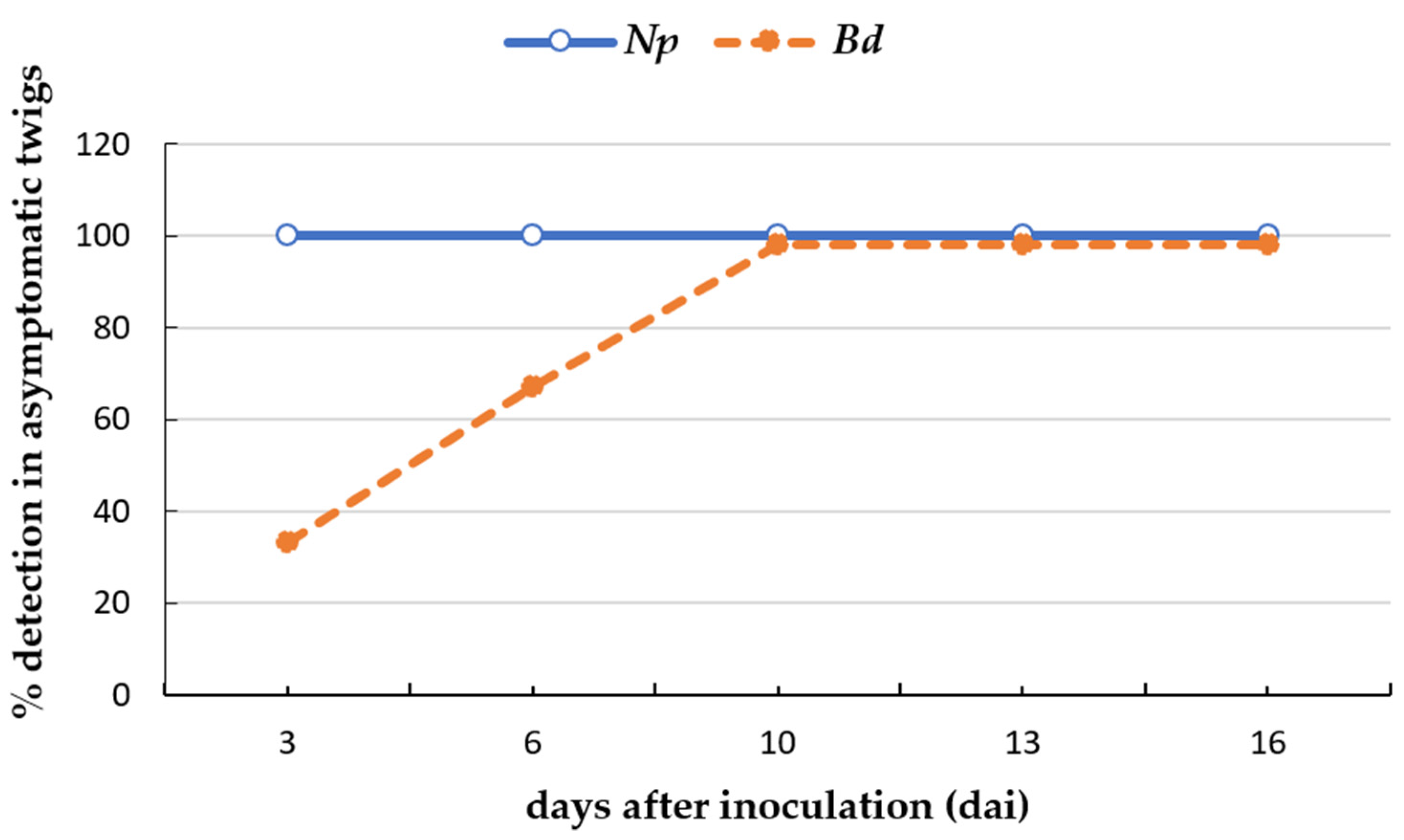
2.4. Detection of Botryosphaeriaceae Species on Asymptomatic Plant Tissues

The qPCR protocols developed using dilutions of plant crude extracts as sample could detect N. parvum and B. dothidea in artificially inoculated asymptomatic plant tissues. The first necrotic lesions appeared on some of the inoculated twigs 10 days after inoculation (dai) for N. parvum and B. dothidea but only the asymptomatic twigs were selected for the qPCR analysis. N. parvum was detected from 3 dai to 16 dai in all asymptomatic twigs assayed. B. dothidea was detected in 33% of the asymptomatic twigs analyzed at 3 dai and in 100% of the asymptomatic twigs from 10 to 16 dai (Figure 2). The amount of fungal DNA quantified in the asymptomatic twigs ranged from 11.6 fg at 3 dai to 1.09 ng at 16 dai for N. parvum and from 18.2 fg at 3 dai to 1.1 pg at 16 dai for B. dothidea. Both fungi were respectively detected from 10 dai in symptomatic twigs.

3. Discussion

Wood canker diseases are a major threat to global nut crop productivity. Among them, canker diseases caused by Botryosphaeriaceae fungi have not been given the importance they deserve and the role of these fungi in wood diseases has been overlooked. The fact that Botryosphaeriaceae species have a wide host range and produce latent infections has led to their “silent” introduction into crop fields through adjacent infected crops and infected but asymptomatic nursery plant material, to later disperse through rain, wind, insects or pruning and harvest tools. In fact, the number of reports associated with trunk diseases caused by Botryosphaeriaceae in numerous woody crops worldwide has significantly increased in the past years [50,51]. The preventive detection of the causal agents is one of the most important and effective strategies for the control of fungal diseases. For that reason, the development of an accurate, sensitive and rapid diagnostic method is of great importance. In this work, molecular tools based on TaqMan-qPCR protocols have been developed for the specific detection and quantification of the two most important Botryosphaeriaceae species of the almond crop: N. parvum and B. dothidea. However, as these two species are not the only ones that infect almond, and more than one species is usually detected in a producing orchard and in a single infected plant, duplex qPCR have been designed to detect the genus Neofusicoccum and the family Botryosphaeriaceae, thus giving information about the presence of other unknown Botryosphaeriacae species.
One of the main advantages of the designed qPCR protocols is their high sensitivity of detection compared with other PCR-based methods previously reported. Conventional and nested PCR performed in multiplex reached a detection limit of 1 pg and 0.1 pg of N. parvum gDNA, respectively [38,39,40] and 100 pg of B. dothidea gDNA [38]. These methods are time consuming and require visualization of the results in agarose gels. This entails the risk of cross contamination and, therefore, the appearance of false positives. Previously reported qPCR methods for detection of canker-causing pathogens generally used SYBR Green chemistry [42,44]. Those using hydrolysis probes were more sensitive and were able to detect as low as 200 fg of N. parvum gDNA [43]. Our methods have improved the sensitivity of detection, even allowing the detection of Botryosphaeriaceae species in asymptomatic tissues. In addition, the implementation of a duplex qPCR for the simultaneous detection of several targets at the same time and the use of a direct sample preparation method allow the analysis of a large number of samples, saving costs. One of the main constraints of the designed qPCR protocols is the impossibility to distinguish between N. parvum and some of the phylogenetically closest species within the N. parvum species complex such as N. ribis and N. kwambonambiense or between B. dothidea and B. corticis and B. fabicerciana. These species are not described in almond but affect some Mediterranean woody crops such as blueberry, grapevine, citrus and avocado [25,49,52,53]. If a precise identification is required, isolation of the pathogen and subsequent morphological identification or sequencing of another genome locus would be necessary.
Several studies have evidenced the endophytic phase of Botryosphaeriaceae fungi in nursery plant material [54,55,56,57]. In this sense, the high sensitivity of the developed qPCR tools allows for the early detection of these pathogens even before the appearance of disease symptoms, providing evidence of the latent infection of these canker-causing pathogens in woody plant tissues. Even more, using the direct sample preparation method described in this study allows the friendly and more economic analysis of a large number of samples. Detection of the target fungi failed when using non diluted plant crude extracts probably due to the presence in the reaction of high amounts of PCR inhibitors of plant origin. Using bovine serum albumin (BSA, 0.1 mg/mL) in the qPCR reaction helped to detect more true positives, but not all. Therefore, the possibility of detecting false negatives makes it advisable to use 1:10 dilutions and BSA which showed high agreement with the DNA extraction method. Therefore, these tools could be applied for the preventive detection of Botryosphaeriaceae fungi in recently planted young trees and in nursery plant material, even being certified as free of the aforementioned pathogens, ensuring the sale of healthy plants to the farmer and, at the same time, avoiding the introduction and dispersion of these pathogens in production fields.
The qPCR protocols described in this work are useful not only for the accurate pathogen identification and disease diagnostics in almond, but also for their application in epidemiological studies to determine the source of inoculum (air, irrigation water, neighboring crops) and quantify the seasonal variation of the inoculum level and the patterns of dispersion of Botryosphaeriaceae spores in almond orchards as previously described [43]. Additionally, this knowledge will permit to establish the correct schedules for the application of control measures, thus avoiding economic losses and damage to the environment. Validation of these qPCR protocols for detection of Botryosphaeriaceae groups in environmental samples should be addressed. In addition, these molecular tools could be applied to evaluate the efficacy of fungicide or biological control agents treatments, by quantifying the time-course concentration of fungal inoculum before and after treatments, as previously reported in other pathosystems [58].
In conclusion, this study provides an accurate, sensitive and easy-to-use molecular tool for the detection of the most important botryosphaeriaceous canker-causing pathogens, N. parvum and B. dothidea, along with any other species from the Neofusicoccum genus or Botryosphaeriaceae family in almond. These qPCR protocols could be potentially also applied to other woody crops affected by these diseases, after appropriate validation, i.e., the verification that the variation in the specific matrix does not affect the test performance.

4. Materials and Methods

4.1. Surveys of Almond Orchards and Fungal Isolation

Three commercial almond orchards affected by almond trunk diseases located in Alcalá del Río, La Rinconada and Jerez de la Frontera locations (southwestern Spain) were surveyed in 2021 and 2022. Symptomatic plant material exhibiting trunk cankers, gummosis and internal necrosis were taken to be analyzed in the laboratory. Samples were used for both the isolation of Botryosphaeriaceae fungi in culture medium and for the detection of Botryosphaeriaceae species using qPCR protocols. For the isolation of fungal isolates, plant tissues were surface-disinfected in 1.5% sodium hypochlorite solution for 2 min, rinsed twice in sterile distilled water, and left to air dry in a laminar flow cabinet. Small pieces were plated on potato dextrose agar supplemented with 0.5 g/L of streptomycin sulphate (Sigma-Aldrich, St. Louis, MO, USA) (PDAS). Petri dishes were incubated at 25 °C in darkness for 7–10 d and any Botryosphaeriaceae-like colony was individually transferred to PDA plates. All isolates were hyphal-tipped and maintained at −80 °C in cryovials containing 20% glycerol and at 4 °C in vials containing sterile distilled water, in the fungal collection of IFAPA Centro Las Torres (Seville, Spain).
The rest of fungal isolates used in this study were Botryosphaeriaceae species isolated from other woody crops, such as avocado and blueberry affected by trunk diseases [8,10], fungal genera phylogenetically close to the target species, and non-Botryosphaeriaceae pathogenic or endophytic species from almond (Table 4).

4.2. Fungal DNA Extraction and Identification by Sequencing

Fungal genomic DNA was extracted from pure fungal cultures using 0.1 mg of mycelium scraped from PDA plates incubated at 25 °C in darkness for 5–7 d using the HigherPurity Plant DNA Purification Kit (Canvax Biotech, S.L., Córdoba, Spain) and following the instructions of the manufacturer. DNA concentration was quantified using a NanoDrop 2000 Spectrophotometer (Thermo Scientific, Wilmington, DE, USA).
For identification, sequencing of the internal transcribed spacer (ITS) nuclear rDNA using ITS1 and ITS4 primers [59] and a portion of the tef1 gene using EF446f and EF1035r primers [60] was performed. The sequences generated were deposited in the GenBank (Table S1) and compared with available sequences by BLAST analysis.

4.3. Design of qPCR Protocols

Firstly, simplex qPCR protocols were designed for the single detection of N. parvum, B. dothidea, Neofusicoccum spp. and the Botryosphaeriaceae family, respectively. Then, conditions for duplex qPCR for the simultaneous detection of N. parvum/Neofusicoccum spp., N. parvum/Botryosphaeriaceae family and B. dothidea/Botryosphaeriaceae family were optimized:

4.3.1. Design of Specific Primers and Probes

Sequences of the ITS region, tef1 gene, and tub2 gene from Botryosphaeriaceae species were retrieved form the GenBank and aligned using Mega 7.0 software [61]. Nucleotide sequences among the three loci that showed the highest specific consensus for each fungal target and not for the remaining species were selected and used to design forward/reverse primers and TaqMan probes using the software from Integrated DNA Technologies, Inc. (IDT) under its default settings. A BLASTn query against the NCBI GenBank database was used to ensure the specificity of the primers and probes in silico (Table S2). TaqMan probes were labeled with 6-carboxy-fluorescein, FAM (for N. parvum and B. dothidea) and SUN (for Neofusicoccum spp. and Botryosphaeriaceae family) reporter dyes at the 5′-end. TaqMan probes also harbored an internal ZEN quencher and an Iowa Black FQ quencher (IBFQ) at the 3′-end.

4.3.2. Optimization of qPCR Conditions

Combination of several primers and probes concentrations were tested to obtain the best efficiency reaction and higher sensitivity. Final concentrations of 300, 600 and 900 nM of primers and 100, 150, 250 nM of probes were assayed. Each DNA sample was run in simplex and duplex reactions to compare reaction efficiency and sensitivity. qPCR assays were performed in 96-well plates using a CFX Connect thermocycler (Bio-Rad, Hercules, CA, USA) in a 20 μL reaction volume. Reaction cocktails contained sample template (5 μL of extracted DNA from a pure fungal colony or from infected plant tissue or 1:10 (v:v) dilution of plant crude extracts), iTaq Universal Probes Supermix (1×) (Bio-Rad, Hercules, CA, USA), forward and reverse primers for N. parvum and B. dothidea (600 nM) or for Neofusicoccum spp. and Botryosphaeriaceae family (300 nM), TaqMan probes for N. parvum and B. dothidea (250 nM) or for Neofusicoccum spp. (100 nM) and Botryosphaeriaceae family (200 nM). Amplifications were performed at 95 °C for 5 min, then 45 cycles of 5 s at 95 °C, followed by 40 s at 60 °C. In each run, sterile distilled water was used instead of DNA as a no-template control. The data were analyzed using the CFX Maestro software 2.3 (Bio-Rad, Hercules, CA, USA). Once simplex qPCR reactions were optimized, duplex qPCRs were performed for the simultaneous detection of (a) N. parvum and Neofusicoccum genus, (b) N. parvum and Botryosphaeriaceae family and (c) B. dothidea and Botryosphaeriaceae family. Conditions for duplex qPCR were the same as for simplex qPCR.

4.4. Analytical Specificity and Analytical Sensitivity of the qPCR Reactions

The analytical specificity, inclusivity and exclusivity of each individual and duplex qPCR were tested on DNA from 81 isolates including species of the Botryosphaeriaceae family and other fungal species pathogenic and non-pathogenic to almond (Table 4). In addition, BLAST analysis was also performed using forward and reverse primers and probe sequences of each Botryosphaeriaceae target separately on the corresponding sequences of all described Botryosphaeriaceae species affecting almond and other Mediterranean crops (Table S2).
The analytical sensitivity of the qPCR assays was evaluated using eight ten-fold decreasing concentrations of DNA obtained from pure colonies of N. parvum NpALM2 isolate and B. dothidea BdALM2 isolate (0.2 ng/μL to 0.2 fg/μL) diluted in a background of DNA (10 ng/μL) extracted from the subcortical tissue of a healthy almond plant. An amount of 5 μL of each dilution was used as template in the qPCR reactions (1 ng to 1 fg of total genomic DNA/qPCR reaction). For each dilution point, we amplified three replicates (from 1 ng to 100 fg/qPCR reaction) and six replicates (for 10 and 1 fg/qPCR reaction) and the values obtained for each dilution series were used to generate standard curves for quantification of the respective fungal targets in simplex and duplex qPCR reactions. Each qPCR assay was repeated at least three times. Linear regression between the quantification cycle (Cq) and the log value of DNA concentration was performed to obtain the corresponding quantification values.

4.5. Plant DNA Extraction and Direct Sample Preparation Method

Samples from almond trees (small pieces of inoculated twigs and trunk subcortical tissue) were divided into two halves: one half was grounded in a mortar in the presence of liquid nitrogen and 0.1 mg of the homogenized tissue were used for DNA isolation following the instructions of the DNeasy Plant Pro kit (Qiagen, Hilden, Germany). DNA concentration was measured using a NanoDrop 2000 Spectrophotometer (Thermo Scientific, Wilmington, DE, USA) and stored at −20 °C for further analysis; the other half was introduced into plastic bags containing a soft net (Bioreba, Reinach, Switzerland) and grounded with the help of a manual homogenizer (or a hammer in the case of trunk subcortical tissues), in the presence of 1:20 (w:v) PBS buffer, pH 7.2 supplemented with 2% (w:v) polyvinyl pyrrolidone and 0.2% (w:v) sodium diethyl dithiocarbamate to obtain a plant crude extract. An amount of 5 μL of kit-extracted DNA and 5 μL of plant crude extracts were used in the qPCR reactions. Serial dilutions (1:1 to 1:107 v:v) of extracted DNA and plant crude extracts from the same tissue were used for qPCR approaches to compare the sensitivities of the two methods.

4.6. Validation of the Assays in Naturally and Artificially Infected Tissues

For validation of the qPCR protocols in naturally infected plants, subcortical tissue from the trunk of symptomatic and asymptomatic (negative control) almond trees were collected, surface sterilized in 1.5% sodium hypochlorite for 2 min, rinsed twice in sterile distilled water and air-dried. Small pieces (0.5 cm) were taken for both, the reisolation of inoculated fungi in PDA plates and qPCR detection. The pieces selected for fungal reisolation were placed on PDA plates and incubated at 25 °C for 7–10 days in darkness. The pieces selected for qPCR detection were divided into two halves, one for DNA extraction and the other for the preparation of plant crude extracts (see Section 4.6). For all samples, qPCR reactions were performed for the simplex detection of N. parvum and B. dothidea and the duplex detection of N. parvum + Neofusicoccum spp., N. parvum + Botryosphaeriaceae family and B. dothidea + Botryosphaeriaceae family.
For validation of the qPCR assays in artificially infected almonds, one-year-old twigs from the ‘Guara’ cultivar were inoculated with representative isolates of N. parvum (isolate NpALM2) and B. dothidea (isolate BdALM2). For this, the almond twigs were cut to 12 cm in length, surface sterilized by spraying with 75% ethanol to run off and air dried in a laminar flow cabinet under sterile conditions. Two wounds per twig were made with a 5-millimeter-diameter cork borer. Five-millimeter mycelium plugs from colonies actively growing on PDA were placed in the wounds and sealed with parafilm. Inoculated twigs were individually inserted in glass tubes containing 2 mL of sterile water. Tubes were capped and incubated in a growth chamber at 25 °C. Eight twigs per isolate were inoculated and twigs with non-colonized PDA plugs were used as negative controls. Two weeks after inoculation, the twigs were observed and small pieces from the margin of necrotic lesions were taken for both the reisolation of inoculated fungi in PDA plates and DNA extraction for qPCR detection, as explained above.
To confirm the specificity of the qPCR protocols, plant crude extracts and gDNA extracted form plant material artificially and naturally infected with N. parvum or B. dothidea were used as template in all simplex and duplex qPCR reactions.

4.7. Detection of Botryosphaeriaceae Fungi in Asymptomatic Tissues

To ascertain whether the qPCR protocols designed were able to detect Botryosphaeriaceae fungi in asymptomatic plant tissues, N. parvum NpALM2 and B. dothidea BdALM2 isolates were artificially inoculated on respective detached twigs of almond cv. Guara as explained above. Thirty twigs were inoculated per pathogen isolate. Successive sampling was performed at 3, 6, 10, 13 and 16 dai. At each sampling time, 5 twigs without symptoms were selected. Samples consisted of pieces (1 × 0.5 mm) of subcortical tissue without epidermis taken from the left and right of the inoculation site, 2 mm apart from the inoculation point. The sampled tissue fragments were processed to extract DNA and prepare plant crude extracts, as described above. Duplex qPCR protocols were applied to detect N. parvum and B. dothidea, respectively, and the percentage of detection in asymptomatic twigs was calculated.

Supplementary Materials

The following supporting information can be downloaded at: https://www.mdpi.com/article/10.3390/plants12112205/s1, Table S1. GenBank Accession number of rRNA internal transcribed spacer (ITS) and translation elongation factor 1 alpha (tef1) gene of Botryosphaeriaceae isolates from almond, avocado and blueberry. Table S2. In silico analysis of the specificity of qPCR primers and probes for the detection of Neofusicoccum parvum and Botryosphaeria dothidea affecting Mediterranean woody crops. Reference [62] is cited in the supplementary materials.

Author Contributions

Conceptualization, N.C.; methodology, N.C. and L.R.-C.; software, N.C. and L.R.-C.; validation, N.C. and L.R.-C.; formal analysis, N.C. and L.R.-C.; investigation, N.C. and L.R.-C.; resources, N.C., C.J.L.-H. and A.A.; writing—original draft preparation, N.C.; writing—review and editing, C.J.L.-H., L.R.-C. and A.A.; supervision, N.C., C.J.L.-H. and A.A.; project administration, N.C. and C.J.L.-H.; funding acquisition, N.C. and C.J.L.-H. All authors have read and agreed to the published version of the manuscript.

Funding

This research was funded by the State Plan for Scientific and Technical Research and Innovation 2017–2020 of the Ministry of Science and Innovation and European Regional Development Fund (ERDF), grant number PID2020-115639RR-I00 and L.R.-C. has a contract of Technical Support Personnel (PTA) from the Spanish Ministry of Science and Innovation.

Data Availability Statement

Not applicable.

Acknowledgments

The authors would like to thank A. Azpilicueta, B. De los Santos, C. Borrero, F.T. Arroyo, J. Armengol and J. Luque for the supply of Botryosphaeriaceae isolates or DNA.

Conflicts of Interest

The authors declare no conflict of interest. The funders had no role in the design of the study; in the collection, analyses, or interpretation of data; in the writing of the manuscript, or in the decision to publish the results.

References

  1. Iglesias, I.; Foiles, P.; Oliveira, C. El almendro en España y Portugal: Situación, innovación tecnológica, costes, rentabilidad y perspectivas. Fruticultura 2021, 81, 6–49. [Google Scholar]
  2. Doll, D.A.; Michailides, T.J.; Rolshausen, P.E. Botryosphaeriaceae associated with almond trunk cankers: A threat to the almond industry? Phytopathology 2013, 103, 13. [Google Scholar]
  3. Velasquez, A.C.; Castroverde, C.D.M.; He, S.Y. Plant-Pathogen Warfare under Changing Climate Conditions. Curr. Biol. 2018, 28, R619–R634. [Google Scholar] [CrossRef] [Green Version]
  4. Fernandez, O.; Lemaitre-Guillier, C.; Songy, A.; Robert-Siegwald, G.; Lebrun, M.H.; Schmitt-Kopplin, P.; Larignon, P.; Adrian, M.; Fontaine, F. The Combination of Both Heat and Water Stresses May Worsen Botryosphaeria Dieback Symptoms in Grapevine. Plants 2023, 12, 753. [Google Scholar] [CrossRef]
  5. Gramaje, D.; Agusti-Brisach, C.; Perez-Sierra, A.; Moralejo, E.; Olmo, D.; Mostert, L.; Damm, U.; Armengol, J. Fungal trunk pathogens associated with wood decay of almond trees on Mallorca (Spain). Persoonia 2012, 28, 1–13. [Google Scholar] [CrossRef] [PubMed]
  6. Urbez-Torres, J.R.; Peduto, F.; Vossen, P.M.; Krueger, W.H.; Gubler, W.D. Olive Twig and Branch Dieback: Etiology, Incidence, and Distribution in California. Plant Dis. 2013, 97, 231–244. [Google Scholar] [CrossRef] [Green Version]
  7. Carlucci, A.; Cibelli, F.; Lops, F.; Raimondo, M.L. Characterization of Botryosphaeriaceae Species as Causal Agents of Trunk Diseases on Grapevines. Plant Dis. 2015, 99, 1678–1688. [Google Scholar] [CrossRef] [Green Version]
  8. Arjona-Girona, I.; Ruano-Rosa, D.; Lopez-Herrera, C.J. Identification, pathogenicity and distribution of the causal agents of dieback in avocado orchards in Spain. Span. J. Agric. Res. 2019, 17, e1003. [Google Scholar] [CrossRef] [Green Version]
  9. Bezerra, J.D.P.; Crous, P.W.; Aiello, D.; Gullino, M.L.; Polizzi, G.; Guarnaccia, V. Genetic Diversity and Pathogenicity of Botryosphaeriaceae Species Associated with Symptomatic Citrus Plants in Europe. Plants 2021, 10, 492. [Google Scholar] [CrossRef]
  10. Aviles, M.; de los Santos, B.; Borrero, C. Increase of canker disease severity in blueberries caused by Neofusicoccum parvum or Lasiodiplodia theobromae due to interaction with Macrophomina phaseolina root infection. Eur. J. Plant Pathol. 2021, 159, 655–663. [Google Scholar] [CrossRef]
  11. Zezlina, I.; Rot, M.; Kac, M.; Trdan, S. Causal Agents of Stone Fruit Diseases in Slovenia and the Potential for Diminishing Their Economic Impact—A Review. Plant Prot. Sci. 2016, 52, 149–157. [Google Scholar] [CrossRef] [Green Version]
  12. Hernandez, D.; Garcia-Perez, O.; Perera, S.; Gonzalez-Carracedo, M.A.; Rodriguez-Perez, A.; Siverio, F. Fungal Pathogens Associated with Aerial Symptoms of Avocado (Persea americana Mill.) in Tenerife (Canary Islands, Spain) Focused on Species of the Family Botryosphaeriaceae. Microorganisms 2023, 11, 585. [Google Scholar] [CrossRef] [PubMed]
  13. Inderbitzin, P.; Bostock, R.M.; Trouillas, F.P.; Michailides, T.J. A six locus phylogeny reveals high species diversity in Botryosphaeriaceae from California almond. Mycologia 2010, 102, 1350–1368. [Google Scholar] [CrossRef] [PubMed]
  14. Nouri, M.T.; Lawrence, D.P.; Yaghmour, M.A.; Michailides, T.J.; Trouillas, F.P. Neoscytalidium dimidiatum Causing Canker, Shoot Blight and Fruit Rot of Almond in California. Plant Dis. 2018, 102, 1638–1647. [Google Scholar] [CrossRef] [Green Version]
  15. Jimenez Luna, I.; Doll, D.; Ashworth, V.E.T.M.; Trouillas, F.P.; Rolshausen, P.E. Comparative Profiling of Wood Canker Pathogens from Spore Traps and Symptomatic Plant Samples Within California Almond and Walnut Orchards. Plant Dis. 2022, 106, 2182–2190. [Google Scholar] [CrossRef]
  16. Holland, L.A.; Trouillas, F.P.; Nouri, M.T.; Lawrence, D.P.; Crespo, M.; Doll, D.A.; Duncan, R.A.; Holtz, B.A.; Culumber, C.M.; Yaghmour, M.A.; et al. Fungal Pathogens Associated with Canker Diseases of Almond in California. Plant Dis. 2021, 105, 346–360. [Google Scholar] [CrossRef]
  17. Luo, Y.; Niederholzer, F.J.A.; Lightle, D.M.; Felts, D.; Lake, J.; Michailides, T.J. Limited Evidence for Accumulation of Latent Infections of Canker-Causing Pathogens in Shoots of Stone Fruit and Nut Crops in California. Phytopathology 2021, 111, 1963–1971. [Google Scholar] [CrossRef]
  18. Sohrabi, M.; Mohammadi, H.; Leon, M.; Armengol, J.; Banihashemi, Z. Fungal pathogens associated with branch and trunk cankers of nut crops in Iran. Eur. J. Plant Pathol. 2020, 157, 327–351. [Google Scholar] [CrossRef]
  19. Ozer, G.; Turkolmez, S.; Dervis, S. First report of Lasiodiplodia theobromae causing dieback on almond (Prunus dulcis) in Turkey. J. Plant Pathol. 2022, 104, 445–446. [Google Scholar] [CrossRef]
  20. Goura, K.; Lahlali, R.; Bouchane, O.; Baala, M.; Radouane, N.; Kenfaoui, J.; Ezrari, S.; El Hamss, H.; El Alami, N.; Amiri, S.; et al. Identification and Characterization of Fungal Pathogens Causing Trunk and Branch Cankers of Almond Trees in Morocco. Agronomy 2023, 13, 130. [Google Scholar] [CrossRef]
  21. Olmo, D.; Armengol, J.; Leon, M.; Gramaje, D. Characterization and Pathogenicity of Botryosphaeriaceae Species Isolated from Almond Trees on the Island of Mallorca (Spain). Plant Dis. 2016, 100, 2483–2491. [Google Scholar] [CrossRef] [PubMed] [Green Version]
  22. Munoz, R.M.; Lerma, M.L.; Castillo, P.; Tolosa, V.M.; Olmo, D.; Trapero, A.; Agusti-Brisach, C. First report of Lasiodiplodia theobromae causing crown canker of almond in Spain. J. Plant Pathol. 2022, 104, 411–412. [Google Scholar] [CrossRef]
  23. Agusti-Brisach, C.; Moldero, D.; Raya, M.d.C.; Lorite, I.J.; Orgaz, F.; Trapero, A. Water Stress Enhances the Progression of Branch Dieback and Almond Decline under Field Conditions. Plants 2020, 9, 1213. [Google Scholar] [CrossRef]
  24. Moral, J.; Morgan, D.; Trapero, A.; Michailides, T.J. Ecology and Epidemiology of Diseases of Nut Crops and Olives Caused by Botryosphaeriaceae Fungi in California and Spain. Plant Dis. 2019, 103, 1809–1827. [Google Scholar] [CrossRef] [PubMed] [Green Version]
  25. Zhang, W.; Groenewald, J.Z.; Lombard, L.; Schumacher, R.K.; Phillips, A.J.L.; Crous, P.W. Evaluating species in Botryosphaeriales. Persoonia 2021, 46, 63–115. [Google Scholar] [CrossRef]
  26. Moral, J.; Munoz-Diez, C.; Gonzalez, N.; Trapero, A.; Michailides, T.J. Characterization and Pathogenicity of Botryosphaeriaceae Species Collected from Olive and Other Hosts in Spain and California. Phytopathology 2010, 100, 1340–1351. [Google Scholar] [CrossRef] [Green Version]
  27. Lopez-Moral, A.; Lovera, M.; del Carmen Raya, M.; Cortes-Cosano, N.; Arquero, O.; Trapero, A.; Agusti-Brisach, C. Etiology of Branch Dieback and Shoot Blight of English Walnut Caused by Botryosphaeriaceae and Diaporthe Species in Southern Spain. Plant Dis. 2020, 104, 533–550. [Google Scholar] [CrossRef]
  28. Ali, H.; Alam, S.; Attanayake, R.; Rahman, M.; Chen, W. Population Structure and Mating Type Distribution of the Chickpea Blight Pathogen Ascochyta Rabiei from Pakistan and the United States. J. Plant Pathol. 2012, 94, 99–108. [Google Scholar]
  29. Chen, S.; Morgan, D.P.; Hasey, J.K.; Anderson, K.; Michailides, T.J. Phylogeny, Morphology, Distribution, and Pathogenicity of Botryosphaeriaceae and Diaporthaceae from English Walnut in California. Plant Dis. 2014, 98, 636–652. [Google Scholar] [CrossRef] [Green Version]
  30. Gusella, G.; Giambra, S.; Conigliaro, G.; Burruano, S.; Polizzi, G. Botryosphaeriaceae species causing canker and dieback of English walnut (Juglans regia) in Italy. For. Pathol. 2021, 51, e12661. [Google Scholar] [CrossRef]
  31. Luna, I.J.; Besoain, X.; Saa, S.; Peach-Fine, E.; Morales, F.C.; Riquelme, N.; Larach, A.; Morales, J.; Ezcurra, E.; Ashworth, V.E.; et al. Identity and pathogenicity of Botryosphaeriaceae and Diaporthaceae from Juglans regia in Chile. Phytopathol. Mediterr. 2021, 61, 69–74. [Google Scholar]
  32. Antony, S.; Billones-Baaijens, R.; Stodart, B.J.; Steel, C.C.; Lang, M.D.; Savocchia, S. Incidence and distribution of Botryosphaeriaceae species associated with dieback in walnut orchards in Australia. Plant Pathol. 2023, 72, 610–622. [Google Scholar] [CrossRef]
  33. Nouri, M.T.; Lawrence, D.P.; Holland, L.A.; Doll, D.A.; Kallsen, C.E.; Culumber, C.M.; Trouillas, F.P. Identification and Pathogenicity of Fungal Species Associated with Canker Diseases of Pistachio in California. Plant Dis. 2019, 103, 2397–2411. [Google Scholar] [CrossRef] [PubMed]
  34. Lopez-Moral, A.; del Carmen Raya, M.; Ruiz-Blancas, C.; Medialdea, I.; Lovera, M.; Arquero, O.; Trapero, A.; Agusti-Brisach, C. Aetiology of branch dieback, panicle and shoot blight of pistachio associated with fungal trunk pathogens in southern Spain. Plant Pathol. 2020, 69, 1237–1269. [Google Scholar] [CrossRef]
  35. Slippers, B.; Wingfield, M. Botryosphaeriaceae as endophytes and latent pathogens of woody plants: Diversity, ecology and impact. Fungal Biol. Rev. 2007, 21, 90–106. [Google Scholar] [CrossRef]
  36. Rathnayaka, A.R.; Chethana, K.W.T.; Phillips, A.J.L.; Liu, J.K.; Samarakoon, M.C.; Jones, E.B.G.; Karunarathna, S.C.; Zhao, C.L. Re-Evaluating Botryosphaeriales: Ancestral State Reconstructions of Selected Characters and Evolution of Nutritional Modes. J. Fungi 2023, 9, 184. [Google Scholar] [CrossRef]
  37. Crous, P.W.; Slippers, B.; Groenewald, J.Z.; Wingfield, M.J. Botryosphaeriaceae: Systematics, pathology, and genetics. Fungal Biol. 2017, 121, 305–306. [Google Scholar] [CrossRef]
  38. Avenot, H.F.; Jaime-Frias, R.; Travadon, R.; Holland, L.A.; Lawrence, D.P.; Trouillas, F.P. Development of PCR-Based Assays for Rapid and Reliable Detection and Identification of Canker-Causing Pathogens from Symptomatic Almond Trees. Phytopathology 2022, 112, 1710–1722. [Google Scholar] [CrossRef]
  39. Ni, H.-F.; Yang, H.-R.; Chen, R.-S.; Hung, T.-H.; Liou, R.-F. A nested multiplex PCR for species-specific identification and detection of Botryosphaeriaceae species on mango. Eur. J. Plant Pathol. 2012, 133, 819–828. [Google Scholar] [CrossRef]
  40. Ridgway, H.J.; Amponsah, N.T.; Brown, D.S.; Baskarathevan, J.; Jones, E.E.; Jaspers, M.V. Detection of botryosphaeriaceous species in environmental samples using a multi-species primer pair. Plant Pathol. 2011, 60, 1118–1127. [Google Scholar] [CrossRef]
  41. Pouzoulet, J.; Rolshausen, P.E.; Schiavon, M.; Bol, S.; Travadon, R.; Lawrence, D.P.; Baumgartner, K.; Ashworth, V.E.; Comont, G.; Corio-Costet, M.-F.; et al. A Method to Detect and Quantify Eutypa late and Diplodia seriata-Complex DNA in Grapevine Pruning Wounds. Plant Dis. 2017, 101, 1470–1480. [Google Scholar] [CrossRef] [Green Version]
  42. Haidar, R.; Yacoub, A.; Pinard, A.; Roudet, J.; Fermaud, M.; Rey, P. Synergistic effects of water deficit and wood-inhabiting bacteria on pathogenicity of the grapevine trunk pathogen Neofusicoccum parvum. Phytopathol. Mediterr. 2020, 59, 473–484. [Google Scholar] [CrossRef]
  43. Billones-Baaijens, R.; Urbez-Torres, J.R.; Liu, M.; Ayres, M.; Sosnowski, M.; Savocchia, S. Molecular Methods to Detect and Quantify Botryosphaeriaceae Inocula Associated with Grapevine Dieback in Australia. Plant Dis. 2018, 102, 1489–1499. [Google Scholar] [CrossRef] [PubMed] [Green Version]
  44. Luo, Y.; Gu, S.; Felts, D.; Puckett, R.D.; Morgan, D.P.; Michailides, T.J. Development of qPCR systems to quantify shoot infections by canker-causing pathogens in stone fruits and nut crops. J. Appl. Microbiol. 2017, 122, 416–428. [Google Scholar] [CrossRef] [PubMed]
  45. Capote, N.; Pastrana, P.; Aguado, A.; Sánchez-Torres, P. Molecular tools for detection of plant pathogenic fungi and fungicide resistance. In Plant Pathology; InTech-Open Access Publisher: London, UK, 2012; pp. 151–202. [Google Scholar]
  46. Garcia, J.F.; Lawrence, D.P.; Morales-Cruz, A.; Travadon, R.; Minio, A.; Hernandez-Martinez, R.; Rolshausen, P.E.; Baumgartner, K.; Cantu, D. Phylogenomics of Plant-Associated Botryosphaeriaceae Species. Front. Microbiol. 2021, 12, 652802. [Google Scholar] [CrossRef] [PubMed]
  47. Manetti, G.; Brunetti, A.; Lumia, V.; Sciarroni, L.; Marangi, P.; Cristella, N.; Faggioli, F.; Reverberi, M.; Scortichini, M.; Pilotti, M. Identification and Characterization of Neofusicoccum stellenboschiana in Branch and Twig Dieback-Affected Olive Trees in Italy and Comparative Pathogenicity with N. mediterraneum. J. Fungi 2023, 9, 292. [Google Scholar] [CrossRef] [PubMed]
  48. Hilario, S.; Lopes, A.; Santos, L.; Alves, A. Botryosphaeriaceae species associated with blueberry stem blight and dieback in the Centre Region of Portugal. Eur. J. Plant Pathol. 2020, 156, 31–44. [Google Scholar] [CrossRef]
  49. Xiao, X.E.; Wang, W.; Crous, P.W.; Wang, H.K.; Jiao, C.; Huang, F.; Pu, Z.X.; Zhu, Z.R.; Li, H.Y. Species of Botryosphaeriaceae associated with citrus branch diseases in China. Persoonia 2021, 47, 106–135. [Google Scholar] [CrossRef]
  50. Batista, E.; Lopes, A.; Alves, A. What Do We Know about Botryosphaeriaceae? An Overview of a Worldwide Cured Dataset. Forests 2021, 12, 313. [Google Scholar] [CrossRef]
  51. Guarnaccia, V.; Kraus, C.; Markakis, E.; Alves, A.; Armengol, J.; Eichmeier, A.; Compant, S.; Gramaje, D. Fungal trunk diseases of fruit trees in Europe: Pathogens, spread and future directions. Phytopathol. Mediterr. 2022, 61, 563–599. [Google Scholar] [CrossRef]
  52. Urbez-Torres, J.R.; Peduto, F.; Striegler, R.K.; Urrea-Romero, K.E.; Rupe, J.C.; Cartwright, R.D.; Gubler, W.D. Characterization of fungal pathogens associated with grapevine trunk diseases in Arkansas and Missouri. Fungal Divers. 2012, 52, 169–189. [Google Scholar] [CrossRef]
  53. Ru, S.; Ding, S.; Oliver, J.E.; Amodu, A. A Review of Botryosphaeria Stem Blight Disease of Blueberry from the Perspective of Plant Breeding. Agriculture 2023, 13, 100. [Google Scholar] [CrossRef]
  54. Halleen, F.; Crous, P.W.; Petrini, O. Fungi associated with healthy grapevine cuttings in nurseries, with special reference to pathogens involved in the decline of young vines. Australas. Plant Pathol. 2003, 32, 47–52. [Google Scholar] [CrossRef]
  55. Aroca, A.; Gramaje, D.; Armengol, J.; Garcia-Jimenez, J.; Raposo, R. Evaluation of the grapevine nursery propagation process as a source of Phaeoacremonium spp. and Phaeomoniella chlamydospora and occurrence of trunk disease pathogens in rootstock mother vines in Spain. Eur. J. Plant Pathol. 2010, 126, 165–174. [Google Scholar] [CrossRef]
  56. Berlanas, C.; Ojeda, S.; Lopez-Manzanares, B.; Andres-Sodupe, M.; Bujanda, R.; del Pilar Martinez-Diz, M.; Diaz-Losada, E.; Gramaje, D. Occurrence and Diversity of Black-Foot Disease Fungi in Symptomless Grapevine Nursery Stock in Spain. Plant Dis. 2020, 104, 94–104. [Google Scholar] [CrossRef]
  57. van der Merwe, R.; Halleen, F.; van Dyk, M.; Jacobs, V.G.; Mostert, L. Occurrence of Canker and Wood Rot Pathogens on Stone Fruit Propagation Material and Nursery Trees in the Western Cape of South Africa. Plant Dis. 2021, 105, 3586–3599. [Google Scholar] [CrossRef] [PubMed]
  58. Arjona-Lopez, J.M.; Capote, N.; Melero-Vara, J.M.; Lopez-Herrera, C.J. Control of avocado white root rot by chemical treatments with fluazinam in avocado orchards. Crop Prot. 2020, 131, 105100. [Google Scholar] [CrossRef]
  59. White, T.J.; Bruns, T.; Lee SJ, W.T.; Taylor, J. Amplification and direct sequencing of fungal ribosomal RNA genes for phylogenetics. In PCR Protocols: A Guide to Methods and Applications; Academic Press: Cambridge, MA, USA, 1990; pp. 315–322. [Google Scholar]
  60. Inderbitzin, P.; Harkness, J.; Turgeon, B.G.; Berbee, M.L. Lateral transfer of mating system in Stemphylium. Proc. Natl. Acad. Sci. USA 2005, 102, 11390–11395. [Google Scholar] [CrossRef] [Green Version]
  61. Kumar, S.; Stecher, G.; Tamura, K. MEGA7: Molecular Evolutionary Genetics Analysis Version 7.0 for Bigger Datasets. Mol. Biol. Evol. 2016, 33, 1870–1874. [Google Scholar] [CrossRef] [Green Version]
  62. Mondragón-Flores, A.; Rodríguez-Alvarado, G.; Gómez-Dorantes, N.; Guerra-Santos, J.J.; Fernández-Pavía, S.P. Botryosphaeriaceae: A complex, diverse and cosmopolitan family of fungi. Rev. Mex. Cienc. Agrícolas 2021, 12. [Google Scholar]
Figure 1. Standard curves in simplex and duplex qPCR for the quantitative detection of (a) Neofusicoccum parvum; (b) Botryosphaeria dothidea; (c) Neofusicoccum genus; (d) Botryosphaeriaceae family; (e) N. parvum and Neofusicoccum genus; (f) N. parvum and Botryosphaeriaceae family; and (g) B. dothidea and Botryosphaeriaceae family. N. parvum and B. dothidea were detected with FAM fluorophore. Neofusicoccum genus and Botryosphaeriaceae family were detected with SUN fluorophore. Ten-fold dilutions of genomic DNA from pure colonies of N. parvum and B. dothidea (1 ng to 10 fg) were amplified in three or six (10 fg point, limit of detection) replicates. Efficiency (E), coefficient of determination (R2) and regression equations of standard curves are shown for each qPCR reaction.
Plants 12 02205 g001
Figure 2. Incidence of latent infection of Neofusicoccum parvum (Np) and Botryosphaeria dothidea (Bd) in asymptomatic almond twigs artificially inoculated with isolates NpALM2 and BdALM2, respectively, measured at 3, 6, 10, 13 and 16 days after inoculation (dai).
Plants 12 02205 g002
Table 1. Primers and TaqMan probes designed for the specific detection of Botryosphaeriaceae species by simplex and duplex qPCR.
Target OrganismOligo
Name
Oligo TypeSequence 5’–3’Target
Gene 1
Botryosphaeria dothideaBd-F1 Forward primer CGCCGAATTTGCCTTATCAtef1
Bd-R1Reverse primer TTAGCATATGGTCGCATAGAC
Bd-PProbeFAM-TCACCAACG/ZEN/CTTCCAGCCACTCA-IABkFQ
Neofusicoccum parvumNp-F1 Forward primer GAAGTTCGAGAAGGTAAGAAAGTtef1
Np-R1Reverse primer TGAGTGCGGGAACCC
Np-PProbeFAM-CTGCACGCG/ZEN/CTGGGTGCCAG-IABkFQ
Neofusicoccum spp.Nspp-F Forward primer GGCCTGGACGGCTCTtub2
Nspp-R1Reverse primer AGTGAGAGAGTACCTCGTTGAAG
Nspp-PProbeSUN-GCGCGAATG/ZEN/GCAATGGCTGACC-IABkFQ
Botryosphaeriaceae familyBot-F1 Forward primer GTATGGCAATCTTCTGAACGtub2
Bot-R2Reverse primer GAARAGCTGGCCRAAGG
Bot-PProbeSUN-TCGAGCCCG/ZEN/GCACSATGGAT-IABkFQ
1 tef1: translation elongation factor 1-alpha; tub2: beta-tubulin 2. IABkFQ: Iowa Black®FQ.
Table 2. Concordance between molecular and traditional methods (simplex and duplex qPCR using extracted DNA as sample and isolation methods) for the detection of Botryosphaeriaceae species in subcortical tissue of naturally infected almond trees.
Almond Tree Samples 1Simplex qPCR 2Duplex qPCR 2Isolation
and
Sequencing
NpBdNp + Nspp.Np + Bot FamilyBd + Bot Family
Mar 1+++B. dothidea
Sol 2+++B. dothidea
Sol 3+++B. dothidea
Sol 5Cytospora sp.
Sol 6Cytospora sp.
Sol 7++++N. parvum/Cytospora sp.
Sol 9++++N. parvum
Sol 11+++B. dothidea
Lau 2+++B. dothidea
Lau 3Cytospora sp.
Lau 4Cytospora sp.
Lau 7+++B. dothidea
Bel 3+++B. dothidea
Bel 4+++B. dothidea
Bel 5+++B. dothidea
Bel 6+++B. dothidea
Sol 10 *NI
Lau 6 *NI
1 Almond trees of cultivars ‘Marcona’ (Mar), ‘Soleta’ (Sol), ‘Lauranne’ (Lau) and ‘Belona’ (Bel). Asterisks indicate asymptomatic almond trees; 2 + Positive qPCR amplification; − Negative qPCR amplification. NI: no canker-causing fungi isolation. Np: Neofusicoccum parvum; Bd: Botryosphaeria dothidea; Nspp.: Neofusicoccum genus; Bot: Botryosphaeriaceae family.
Table 3. Comparison of real-time PCR (qPCR) sensitivities for the detection of Botryosphaeriaceae fungi in serial dilutions of DNA from an artificially inoculated almond twig extracted with a commercial kit (DNA extraction) or by direct sample preparation method (plant crude extracts).
Target OrganismDilutionReal-Time PCR (Cq) 1
DNA ExtractionPlant Crude Extracts
Neofusicoccum parvum1:1+ (22.92 ± 0.10)-
1:10+ (26.35 ± 0.04)+ (29.24 ± 0.02)
1:102+ (29.70 ± 0.07)+ (31.52 ± 0.42)
1:103+ (32.97 ± 0.09)+ (35.18 ± 0.98)
1:104+ (36.08 ± 0.43)+ (38.88 ± 0.00)
1:105+ (39.12 ± 0.00)+/−
1:106+/−
1:107
Botryosphaeria dothidea1:1+ (22.73 ± 0.07)
1:10+ (26.80 ± 0.13)+ (32.71 ± 0.20)
1:102+ (30.44 ± 0.11)+ (35.90 ± 0.28)
1:103+ (33.63 ± 0.14)+ (38.84 ± 0.16)
1:104+ (36.91 ± 0.29)+ (42.85 ± 0.00)
1:105+ (39.41 ± 0.45)
1:106+/−
1:107
1 + Positive amplification; − negative amplification; +/− amplification in one or two out of the three replicates. Cq values (threshold cycle of the real-time PCR) ± standard error of three replicates is in parenthesis.
Table 4. Fungal isolates used in this study.
SpeciesIsolate IDHostYear of IsolationCountry
Botryosphaeria dothideaBd ALM1Prunus dulcis2016Spain
Bd ALM2Prunus dulcis2016Spain
Bd ALM3Prunus dulcis2016Spain
Bd ALM4Prunus dulcis2021Spain
Bd ALM6Prunus dulcis2021Spain
Bd ALM7Prunus dulcis2021Spain
Bd ALM8Prunus dulcis2021Spain
Bd ALM9Prunus dulcis2021Spain
Bd ALM10Prunus dulcis2022Spain
Bd ALM11Prunus dulcis2022Spain
Bd ALM12Prunus dulcis2022Spain
Bd ALM13Prunus dulcis2022Spain
Bd ALM14Prunus dulcis2022Spain
Bd ALM15Prunus dulcis2022Spain
Bd ALM16Prunus dulcis2022Spain
Bd ALM17Prunus dulcis2022Spain
ALM TOR1Prunus dulcis2022Spain
Bo.13.2Vaccinium corymbosum2009Spain
Bd1141Vitis vinifera-Spain
Bd1143Vitis vinifera-Spain
Botryosphaeria sp.Bo.11Vaccinium corymbosum2009Spain
Neofusicoccum australeBo.8Vaccinium corymbosum2009Spain
Neofusicoccum luteumNF 146Persea americana2013Spain
Neofusicoccum mediterraneumNm ALM3Prunus dulcis2020Spain
CJL 593Pistacia vera2005Spain
Neofusicoccum parvumNp ALM1Prunus dulcis2018Spain
Np ALM2Prunus dulcis2019Spain
Np ALM5Prunus dulcis2022Spain
NF 152Persea americana2013Spain
NF 161Persea americana2013Spain
Bo.2Vaccinium corymbosum2009Spain
Bo.4.1Vaccinium corymbosum2009Spain
Bo.4.2Vaccinium corymbosum2009Spain
Bo.6.1Vaccinium corymbosum2009Spain
Bo.7Vaccinium corymbosum2009Spain
Bo.9Vaccinium corymbosum2009Spain
Bo.10Vaccinium corymbosum2009Spain
Bo.13.3Vaccinium corymbosum2009Spain
Bo.14.2Vaccinium corymbosum2009Spain
Bo.16Vaccinium corymbosum2009Spain
Bo.17.1Vaccinium corymbosum2009Spain
Diplodia cortícolaCJL 165Quercus suber1995Spain
CJL 166Quercus suber1995Spain
Diplodia cupresiiGIHF 321Vitis vinifera2021Spain
Diplodia mutilaCJL 456Fraxinus excelsior2003Spain
Diplodia seriataDs ALM1Prunus dulcis2019Spain
CJL 398Vitis vinifera2003Spain
Dothiorella fraxiniGIHF 132Fraxinus angustifolia2016Spain
Dothiorella ibericaCJL 218Quercus ilex1999Spain
CJL 220Quercus ilex1999Spain
Dothiorella viticolaCJL 570Vitis vinifera2004Spain
CJL 572Vitis vinifera2004Spain
Lasiodiplodia theobromaeL.2Vaccinium corymbosum2017Spain
GIHF 272Vitis vinifera2019Spain
Macrophomina phaseolinaMp ALM 1Prunus dulcis2018Spain
Mp ALM 2Prunus dulcis2019Portugal
Mp ARA11Vaccinium corymbosum2018Spain
Mp ARA12Vaccinium corymbosum2019Spain
TOR 872Vaccinium corymbosum2017Spain
TOR 956Vaccinium corymbosum2020Spain
Cytospora acaciaeCa ALM1Prunus dulcis2022Spain
Ca ALM2Prunus dulcis2022Spain
Ca ALM3Prunus dulcis2022Spain
Botrytis cinereaBc ARA1Vaccinium corymbosum2021Spain
Bc ALM1Prunus dulcis2016Spain
Monilia fructicolaMf CIR1Prunus salicina2011Spain
Monilia laxaMl CIR1Prunus salicina2011Spain
Diaporthe amygdaliDAL-65Prunus dulcis2017Spain
Diaporthe foeniculinaDAL-69Prunus dulcis2017Spain
Diaporthe phaseolorumDAL-222Prunus dulcis2018Spain
Collectotrichum accutatum20,240CECT-Spain
Verticilium dahliaeVd ALM1Prunus dulcis2017Spain
Cylindrocladiella variabilisAL139Prunus dulcis2019Spain
Dactylonectria macrodidymaAL150Prunus dulcis2019Spain
Dactylonectria novozelandicaAL84Prunus dulcis2019Spain
Dactylonectria torresensisAL3Prunus dulcis2019Spain
Ilyonectria liriodendriAL79Prunus dulcis2019Spain
Neonectria quercicolaAL141Prunus dulcis2019Spain
Rhizoctonia solaniRs ALM4Prunus dulcis2020Spain
Epicoccum nigrumEn ALM5Prunus dulcis2020Spain
Alternaria alternataAl ALM1Prunus dulcis2020Spain
CECT: Colección Española de Cultivos Tipo (https://www.uv.es/uvweb/coleccion-espanola-cultivos-tipo/es/coleccion-espanola-cultivos-tipo-1285872233521.html (accessed on 6 February 2023).
Disclaimer/Publisher’s Note: The statements, opinions and data contained in all publications are solely those of the individual author(s) and contributor(s) and not of MDPI and/or the editor(s). MDPI and/or the editor(s) disclaim responsibility for any injury to people or property resulting from any ideas, methods, instructions or products referred to in the content.

