Article

Cultivable Fungal Endophytes in Roots, Rhizomes and Leaves of Posidonia oceanica (L.) Delile along the Coast of Sicily, Italy

1
Department of Agricultural, Food and Forest Sciences, University of Palermo, 90128 Palermo, Italy
2
Institute of Life Sciences, Scuola Superiore Sant’Anna, 56127 Pisa, Italy
3
Department of Earth and Sea Sciences, University of Palermo, 90128 Palermo, Italy
4
Italian Association for Water and Soil Bio—Engineering, Interregional Section Sicily—Calabria, 90139 Palermo, Italy
5
Regional Agency for Environmental Protection of Sicily, 90149 Palermo, Italy
*
Author to whom correspondence should be addressed.
Plants 2022, 11(9), 1139; https://doi.org/10.3390/plants11091139
Submission received: 25 February 2022 / Revised: 20 April 2022 / Accepted: 20 April 2022 / Published: 22 April 2022

Abstract

:
The presence of endophytic fungi in the roots, rhizomes, and leaves of Posidonia oceanica was evaluated in different localities of the Sicilian coast. Samples of roots, rhizomes, and leaves were submitted to isolation techniques, and the obtained fungal colonies were identified by morphological and molecular (rRNA sequencing) analysis. Fungal endophytes occurred mainly in roots and occasionally in rhizomes and leaves. Lulwoana sp. was the most frequent of the isolated taxa, suggesting a strong interaction with the host. In addition, eight other fungal taxa were isolated. In particular, fungi of the genus Ochroconis and family Xylariaceae were identified as endophytes in healthy plants at all sampling stations, whereas Penicillium glabrum was isolated at only one sampling station. Thus, several organs, especially roots of Posidonia oceanica, harbor endophytic fungi, potentially involved in supporting the living host as ascertained for terrestrial plants.

1. Introduction

Posidonia oceanica (Linnaeus) Delile is a Mediterranean paleoendemic in the infralittoral zone, where it forms extensive marine meadows from the surface to a depth of more than 50 m. [1], supporting highly diverse communities [2,3,4] and providing important ecosystem functions and services [5,6]. Most studies on seagrass-associated biodiversity have focused mainly on macro fauna and flora, revealing the high complexity of their communities, in accordance with the epi-hypogeal compartments of seagrass and the physicochemical gradient that they provide [7]. The microbioma associated with P. oceanica was studied from the end of the 20th century [8,9,10,11,12,13,14,15,16]. The results of these studies show that, like terrestrial plants, symbiotic bacteria and fungi colonize the host and that their populations vary according to organ, season, and environment.
The first studies of the presence of endophytic fungi in P. oceanica have highlighted the presence of the species of Lulwoana Kohlm et al. in the roots of plants of the central Mediterranean Sea [17], but none of the isolates have been identified as L. uniseptata. The new genus Lulwoana, which was previously included in the larger genera Lulworthia, typical marine ascomycetes include only one described species: Lulwoana uniseptata (Nakagiri) Kohlm et al. with the anamorph Zalerion maritimum (Linder) Anastasiou. L. uniseptata produces fusiform asci which are curved, deliquescing early and containing eight ascospores, which are filiform, hyaline, and one septate shorter than 150 µm [18]. In the root sections, inter- and intracellular melanized hyphae and microsclerotia, typical features of dark septate endophytes (DSEs) [19,20], were also observed. Subsequent studies have highlighted the presence of other endophytic fungi in the roots of the host and also detected Lulwoana sp. [21,22,23,24,25,26]. In most cases, the taxonomy, biology, and ecology of many of the isolated fungal taxa are not well-known. The detection of endophytic fungal populations in marine phanerogams (and in terrestrial ones too) could also help in understanding the type of symbiotic plant–fungus association (reciprocal, neutral, or antagonistic), considering the species of microorganism, the vegetative state of the colonized organ, and the environmental characteristics of the ecosystem. In particular, the fungal endophytes condition implies the absence of evident symptoms in the host; thus, it is important to assess the health state of the plants [27,28]. In this study the fungal endophytic symbiosis in P. oceanica was studied. Roots, rhizomes, and leave samples were collected from different marine meadows growing in three Sicilian seas, in order to (i) detect the presence of endophytic fungi also in these organs, (ii) confirm the presence of Lulwoana sp., and (iii) identify any other fungal taxa. The study of the endophyte community was designed in order to assess also the healthy vegetative state of the plant host by using leaf biometry analyses.

2. Results

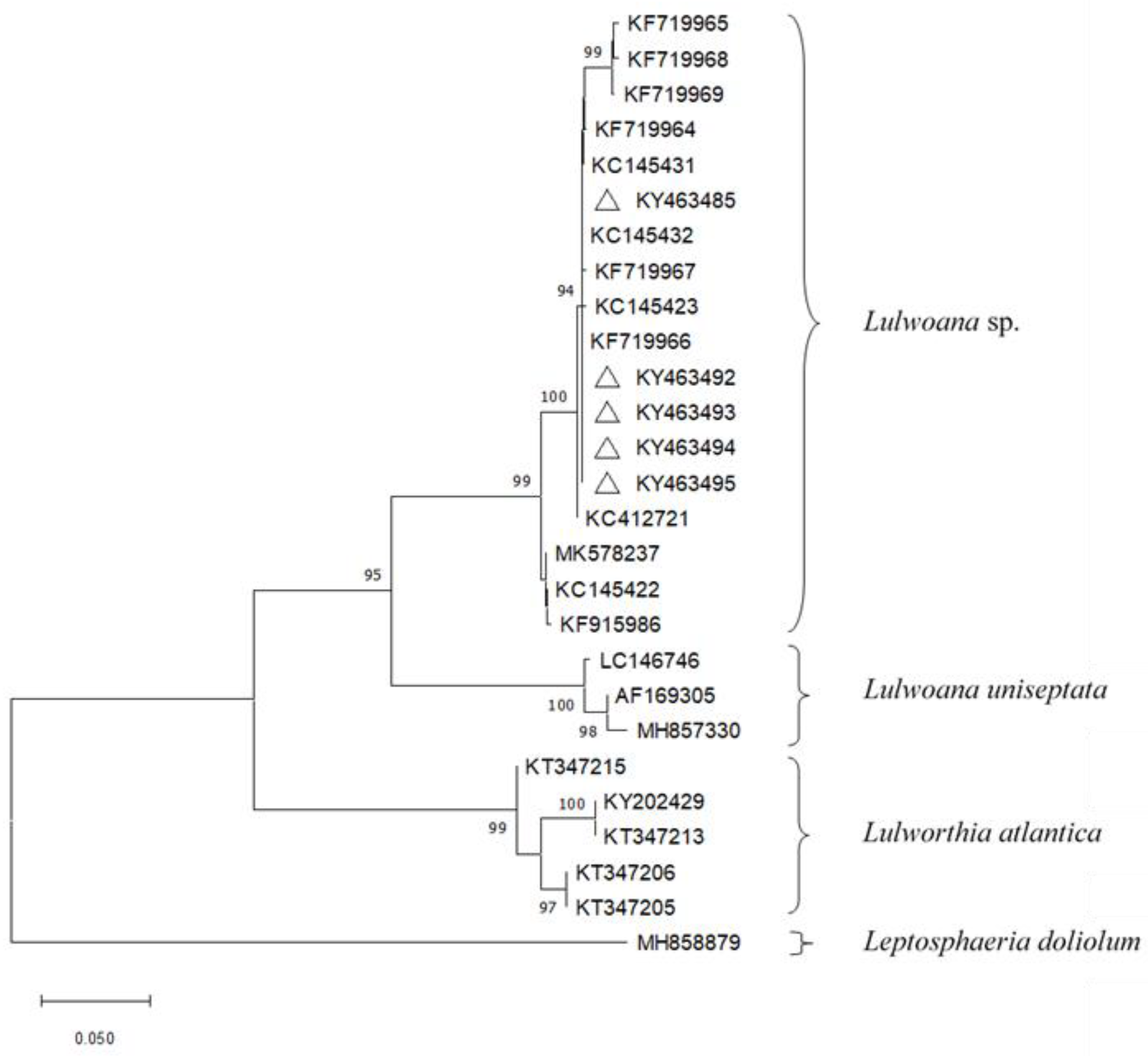
2.1. Fungal Isolation and Identification

The agar plates used to test the efficacy of the surface sterilization technique showed no growth of epiphytic bacterial or fungal colonies, proving the adequacy of the employed method. The growth of fungal colonies from the vegetal fragments was detected up to 5 weeks after the assays were set up. From 7800 P. oceanica fragments, we obtained 504 fungi (absolute isolation frequency, IF = 6.41%). The total fungal population showed IFs (isolation frequency for the sites) of 12.3% for Bonagia, 78% for Ognina and 9.7% for Sciacca, while the IFo (isolation frequency for the organs) was 81% for roots, 17.9% for rhizomes, and only 0.4% for leaf samples (exclusively from Sciacca). Fungal isolates developing reproductive structures were morphologically identified at family, genus, or species level. Molecular analysis confirmed morphological characterization and was the basis for grouping fungal isolates into nine taxa (Table 1 and Table 2).
Most of the fungal isolates (98.6%) were represented by 4 taxa: Penicillium glabrum (Wehmer) Westling (65.7%), Lulwoana sp. (23%), Xylariaceae Tul. & C. Tul (7.9%), Ochroconis de Hoog & Arx sp. (2%). All isolates of P. glabrum were obtained from plant roots taken from one of the two Ognina stations. Considering sampling sites and detected organs, the populations of Lulwoana sp., Ochroconis sp., P. glabrum, and Xylariaceae were distributed as reported in Table 3.
Phylogenetic analyses, based on ITS sequences exclusively, showed that the isolates of Lulwoana formed a separate subclade, with high bootstrap support, within a clade containing Lulwoana uniseptata Kohlm et al., (Table 4; Figure 1).

2.2. Detection of Fungal Endophytes in P. oceanica Organs
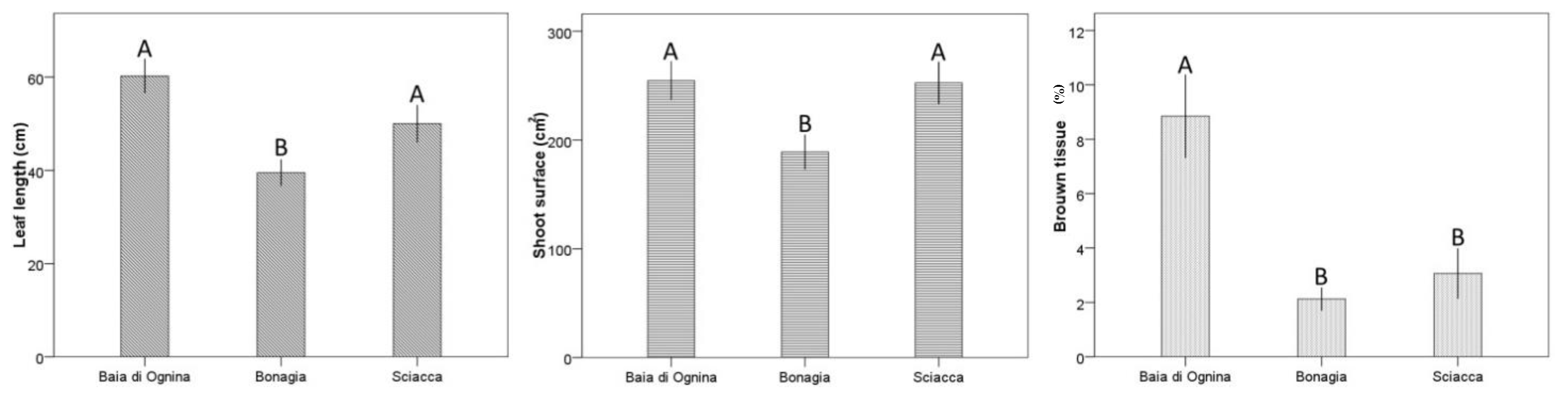
All the observed sections obtained from the sampled organs revealed the occurrence of several fungal structures, such as more or less melanized hyphae and microsclerotia, improved by the use of acid fuchsin. In particular, in almost all the observed root sections, melanized, septate, and thick straight hyphae on the surface produced extensive hyphal sheaths. Rhizodermal cells rarely contained single fungal hyphae, while most of the fungal colonization occurred inter- and intracellularly in the hypodermic and exodermal tissues, showing brown microsclerotia such as those described by [17] (Figure 2).
Sections of the rhizomes from the three sites showed both melanized and grayish hyphae surrounding and enveloping the host cells, from the thin cuticle to the central stele. In the stained sections, the hyphae were coloured red. In a few cases, fungal propagative structures were observed, both outside and inside the detected organs (Figure 3).
Regarding fungal endophytic colonization in leaves, fungal structures were only detected in the sections taken from Sciacca samples. Thin hyphae infecting mesophyll tissue and epidermal cells (red-stained grayish compact microsclerotia in epidermal and, occasionally, in fibrous cells and hyaline-greyish hyphae in mesophyll tissues and in lacunar spaces were observed (Figure 4).

2.3. Leaf Biometry

The leaf biometry in the detected P. oceanica plants showed that leaf length varies from 39.5 ± 2.8 to 60.3 ± 3.7 cm and shoot surface from 189.0 ± 16.0 to 254.5 ± 17.7 cm2, while brown tissue is generally low, ranging from 2.1 ± 0.4 to 8.8 ± 1.5% (Figure 5). The low brown tissues are a normal feature in old leaves of P. oceanica in early summer.
ANOVA detected statistical differences in the leaf length of shoots sampled from the different localities (P < 0.01), with higher mean values at Ognina and Sciacca than at Bonagia (P < 0.001; Table 5, Figure 5). Similar comparison results were detected for shoot surface (P < 0.05; Table 5), while brown tissue was bigger at Ognina compared to the other two localities (P < 0.05; Table 5). Biometric characteristics of leaf bundles showed on average values comparable to those recorded in a previous study [29] for the same three coastal sectors during the same season (current study: shoot surface 232 cm2 vs. 281 cm2 in [29]).

3. Discussion

Nine different fungal taxa were associated with seagrass, but only three of these recurred in the host as a result of isolation and identification tests. In particular, Lulwoana sp., Ochroconis sp., and some colonies ascribable to the Xylariaceae were differently distributed in the host population. Among these, only Lulwoana is known as a genus of obligate marine fungi [30], isolated also from submerged wood and driftwood [31,32].
The marine meadows chosen for this study are exposed to good or high environmental conditions according to a macroalgae-based index [33,34]. The slight reduction (less than 20%) compared to previous studies carried out in the same sector and period [29] is likely quite normal, considering that the study examined meadows growing mainly on sand and matte, which are known for 30% higher leaf length and shoot surface values compared to those growing on rock, as in our study [35,36,37]. Instead, observed morphometric variability between localities is a frequent phenomenon for P. oceanica meadows located hundreds of kilometres apart, reflecting differences in habitat type at the locations, such as wave exposure, temperature regime, and grazing pressure [37]. Moreover, among the three sampling areas, P. oceanica was more endophytized in the Ionian Sea (Ognina).
The observation revealed that fungal colonization in plants is more abundant in roots, less in rhizomes, and only occasional in leaves. The study confirmed the presence of Lulwoana sp. in healthy P. oceanica roots in the Tyrrhenian Sea (Bonagia), as ascertained for the first time by [17], and in two other sectors: the Ionian Sea (Ognina) and the Sicilian Strait (Maragani). Moreover, the fungal endophyte is also associated with all the asymptomatic plant organs as well as the roots and rhizomes in all the tested meadows, and occasionally in the leaves (only in the Sciacca stand). Ocrhoconis sp. was associated with rhizomes (Bonagia and Ognina), roots (Bonagia), and leaves (Sciacca), whereas colonies of Xylariaceae were associated with roots (Bonagia and Ognina) and rhizomes (Bonagia). The distribution of these endophytes in the host is common to nonclavicipitaceous endophytes included in class II by [38] and, as such, could also play a fundamental role in maintaining the optimal vegetative state of the host. Moreover, the number of Penicillium glabrum colonies isolated from the roots of the plants in a spatial replicate of Bonagia could be due to special ecological conditions and require further study. The presence of all these fungal taxa as endophytes in P. oceanica is reported for the first time.
Although ITS sequences could not clearly discriminate all fungal taxa at species level, phylogenetic analyses showed that our strains of Lulwoana sp. isolated from P. oceanica belong to a similar cluster represented by L. uniseptata. Our Lulwoana sp. isolates clustered with other strains were isolated from P. oceanica in Italy, France, Croatia, Spain [17,22,39] and from driftwood in Italy [31]. Further analysis using more specific primers is clearly needed for better identification of the species to which our isolates belong. Moreover, considering the studies of [24,32], Lulwoana sp., like the other members of the Lulwortiaceae, produce cellulolytic enzymes, breaking down complex lignocellulosic compounds and thus contributing to the recycling of nutrients.
Typical fungal structures of dark septate endophytes (DSEs) described by [20,30], such as inter- and intracellular melanized hyphae and microsclerotia, were detected in all the asymptomatic roots of the host, thus confirming their first detection [17]. Similar fungal structures were also detected in rhizomes. Other studies, subsequent to [17], also detected DSEs across the Mediterranean Sea in P. oceanica, including sites along the Sicilian coasts [26]. These results suggest that the occurrence of the fungal endophyte is more widespread than previously known.
The genus Ochroconis is characterized by melanized fungi producing rust-coloured to brown colonies, with brownish conidiophores bearing small numbers of septate, ellipsoidal conidia, mostly rough-walled, with sympodial conidiogenesis and rhexolytic liberation. All the species belonging to the genus are mesophilic and often oligotrophic, often reported from wet areas in the domestic environment or isolated from soil, water, humans, and animals such as insects and fish (Salmo salar L.) [40].
After recent taxonomic revisions [41,42], the Xylariaceae sensu stricto presently comprise 33 genera and over 1000 (and up to 1230) species, of which more than 50% belong to the genus Xylaria Hill ex Schrank. Most species display saprobic, pathogenic, or endophytic (in wood, leaves and fruits) behaviour or are associated with insect vectors, exhibiting their highest diversity in the tropics. The family Xylariaceae is also one of the most prolific lineages of secondary metabolite producers [43]. [44] described two new saprobic species of Ascotricha Berk belonging to Xylariaceae on the brown alga Padina tetrastromatica Hauck.
Although P. glabrum is a well-known pathogen and post-harvest agent of fruit and vegetable rots, frequently isolated from different matrices [45], some authors have also reported its presence in association with several hosts in marine environments. [46] detected it in soft coral, but it was also isolated from Gracilaria lemaneiformis (Bory) Greville (=Gracilariopsis lemaneiformis (Bory) Dawson, Acleto & Foldvik), Sargassum thunbergii (Mertens ex Roth) Kuntze [47], Tethya aurantium Pallas [48], and stems and rhizomes of the seagrass Zostera marina L. [49]. Regarding its ecological role, some studies have shown that P. glabrum can solubilize calcium and iron phosphates and have a xylanase activity [50], thus suggesting that it could play a role in the solubilizing and adsorption of these minerals, and in the decaying of dead plant tissue.
For decades, studies of the presence and distribution of endophytic microorganisms and fungi in host plants, in particular, have contributed to understanding symbiotic relationships in nature. Their presence also appears to influence, among other things, the health of the plant they colonize. The importance of endophytic fungi in the evolution of plant communities was highlighted following the discovery of their relevant effects on the ecology, physiology, and development of each plant host. Above all, thorough research is needed to unravel the molecular mechanisms and signalling pathways undertaken by endophytic fungi to exert their effects on plants [27,28].
Also in marine environments, fungal communities could be considered as new frontiers in ecology, physiology, and chemistry studies. Fungal biodiversity has recently been detected in P. oceanica. This variability could be related to various factors (characteristics of the host, season, environment, etc.) and undoubtedly plays a well-defined role, which could involve the entire marine meadow. Here we show that P. oceanica organs represent a source of endophytic fungal biodiversity, prevalent in the roots and progressively decreasing towards the epigeal compartment, thus further corroborating the hypothesis of their involvement in substrate–host interaction [17,51]. It cannot be excluded that these results could also apply to other marine phanerogams.
Therefore, it would be interesting to examine whether these fungal variability patterns are common trends within marine phanerogams. Further studies on this subject could provide information on the interaction between fungal endophytes and plants, which would be useful for gaining a better understanding of the functional implications at seagrass ecosystem level in healthy environments.

4. Materials and Methods

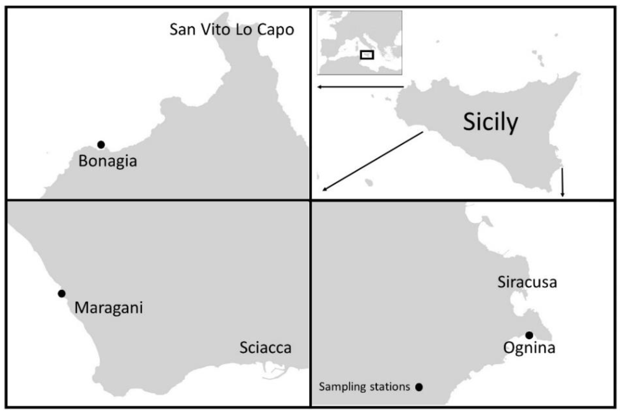
4.1. Study and Sampling Areas

In the summer of 2014, the study was carried out at three different localities along the coasts of Sicily (Italy), namely, Ognina on 14 July 2014(Siracusa, Ionian Sea: 526,863 E, 4,096,103 N 323,103 E, 4,155,293 N), Bonagia 21 July 2014(S. Vito Lo Capo, Tyrrhenian Sea: 287,646 E, 4,216,808 N), and Maragani 07 August 2014 (Sciacca, Strait of Sicily, Mediterranean Sea: 323,103 E, 4,155,293 N) (Figure 6). In these localities P. oceanica is exposed to very low human pressures because it is far from urban or industrial activities and from harbours or fish farms. Studies carried out in 2018 classified the water body of sampling stations on a scale ranging from “good” to “high” according to PREI and CARLIT indices, respectively [33,34]. At each locality, shoot samples were taken from meadows on a rock substrate at a depth of about 6 m at two stations located within an area of about 500 m2. Rocky substrates were chosen for sampling because increased fungal colonization has previously been observed on such substrates [17].
In particular, at each station, 5 asymptomatic P. oceanica shoots, with apparently healthy roots, rhizomes, and leaf bundles were randomly collected. The shoots were transported to the laboratory in tanks containing seawater and analysed within 24 h.

4.2. Isolation of Fungal Endophytes from P. oceanica Organs

After careful washing with tap water, P. oceanica roots, rhizomes, and leaf bundles were divided into two parts for use in isolation assays and cytological analyses, respectively. The first part was subdivided into fragments that were surface-sterilized by sequential washing in 5% NaOCl for 5 min, 95% EtOH for 1 min, and 5% H2O2 for 3 min, and then rinsed three times in distilled sterile water [52]. Prior to plating, a subset of samples was imprinted onto a fresh medium to check the surface-sterilization procedure. Previous investigations have shown that among the different artificial substrates Potato Dextrose Agar (PDA), Malt Extract Agar (MEA) [17], and Sea Water Agar (SWA, unpublished data), MEA was more suitable for the growth of fungal colonies. Therefore, sample fragments of 2 mm length were placed on 2% MEA (Oxoid, Milano, Italy) in Petri dishes (10 cm Ø, five fragments per dish). In total, for each plant the study analysed 75, 50, and 135 fragments of leaf, rhizome, and root, respectively [53]. The Petri dishes containing the fragments were incubated at 20 ± 1 °C in the dark, then observed on a daily basis for fungal development during the first 2 weeks and then weekly for 3 months. Subsequently, the dishes were checked occasionally, until the substrate dried. Hyphal tips from the developed fungal colonies were transferred to 2% MEA in order to obtain pure fungal colonies, morphologically identified macroscopically and microscopically using a light microscope (Axioskop Zeiss, Oberkochen, Germany) [54,55,56]. The isolation frequency of the fungal colonies (IF) was calculated using the formula: IF = (Nif/Ntf) × 100, where Nif is the number of colonies and Ntf is the total number of isolations attempted × 100. The isolation frequency for site(IFs) and the isolation frequency for organ (IFo) were also calculated. All the obtained fungal colonies were grouped by morphotype [53] according to their macroscopic and microscopic features.

4.3. Molecular Identification and Phylogenetic Analyses

One isolate for each morphotype was used for genomic DNA extraction applying the standard cetyltrimethylammoniumbromide (CTAB)-based protocol [57]. According to the standard method for fungal identification, the internal transcribed spacer (ITS) regions of the ribosomal DNA were amplified and sequenced with primers ITS1/ITS4 [58]. PCR amplification and sequencing of amplicons was carried out as described by [59]. Sequences were edited with Sequencher v 4.7 (Gene Codes Corporation, Ann Arbor, MI, USA) and compared with sequences deposited in GenBank through BLASTn searches. New sequences were deposited in GenBank (Table 1). Sequences of Lulworthiales currently known from cultures were retrieved from GenBank (Table 2) and aligned with sequences of the isolates obtained in this study. Phylogenetic analyses were performed as described by [60]. Alignments were made using ClustalX v. 1.83 [61] and, if necessary, manually edited using MEGA6 [62]. Maximum likelihood (ML) analyses were performed on a Neighbour-Joining starting tree automatically generated by MEGA6. Nearest-Neighbour-Interchange (NNI) was used as the heuristic method for tree inference, and 1,000 bootstrap replicates were performed.

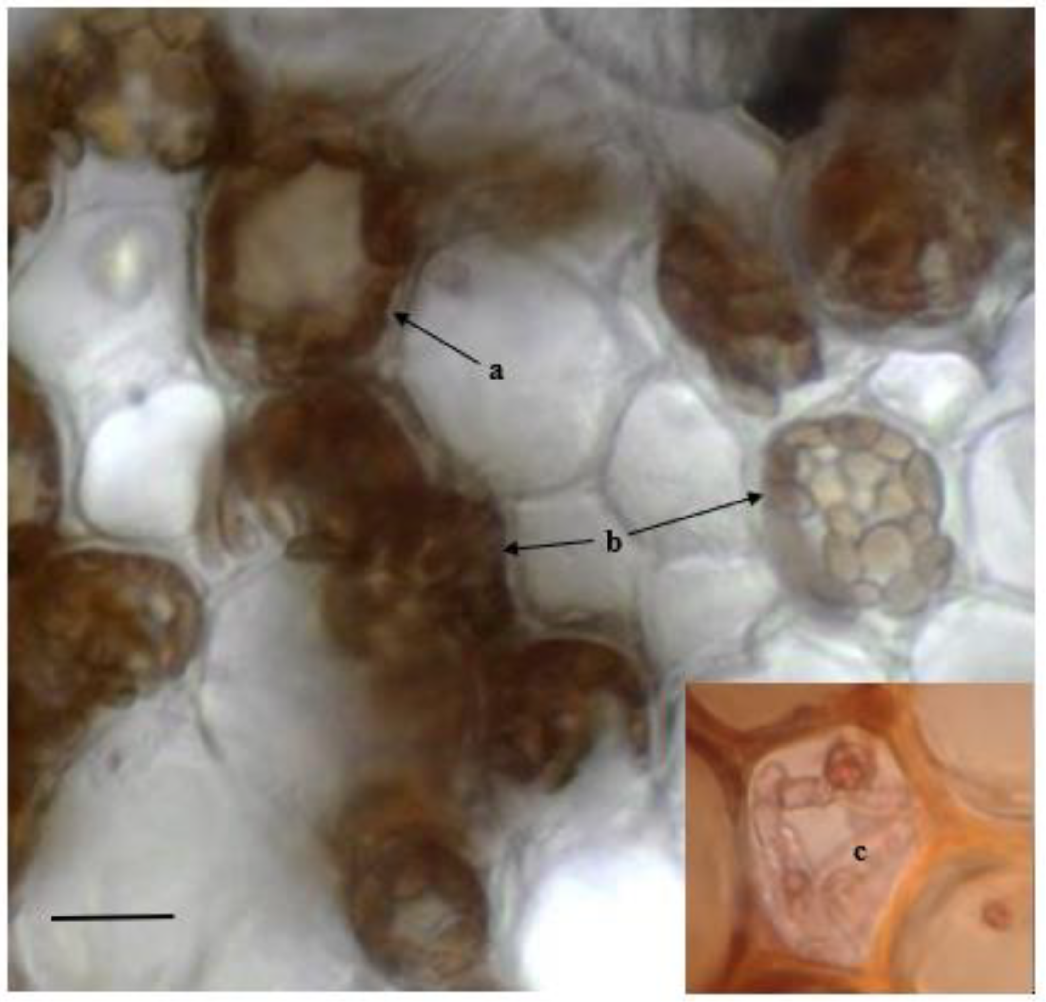
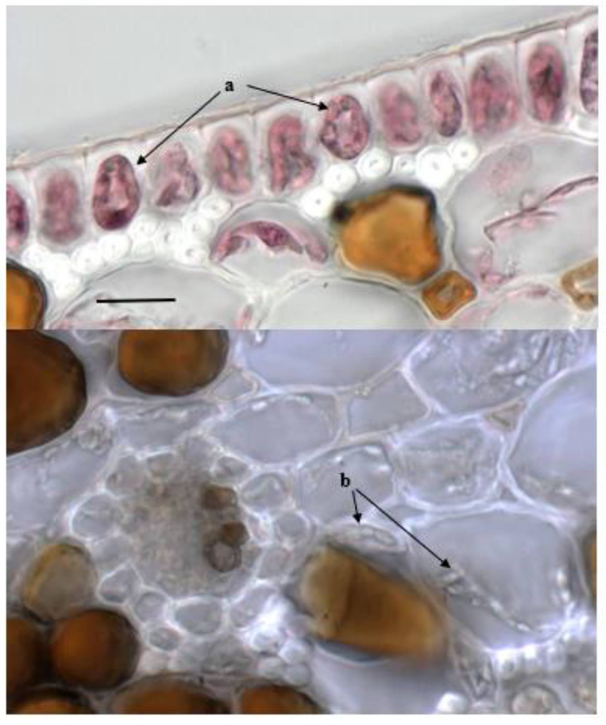
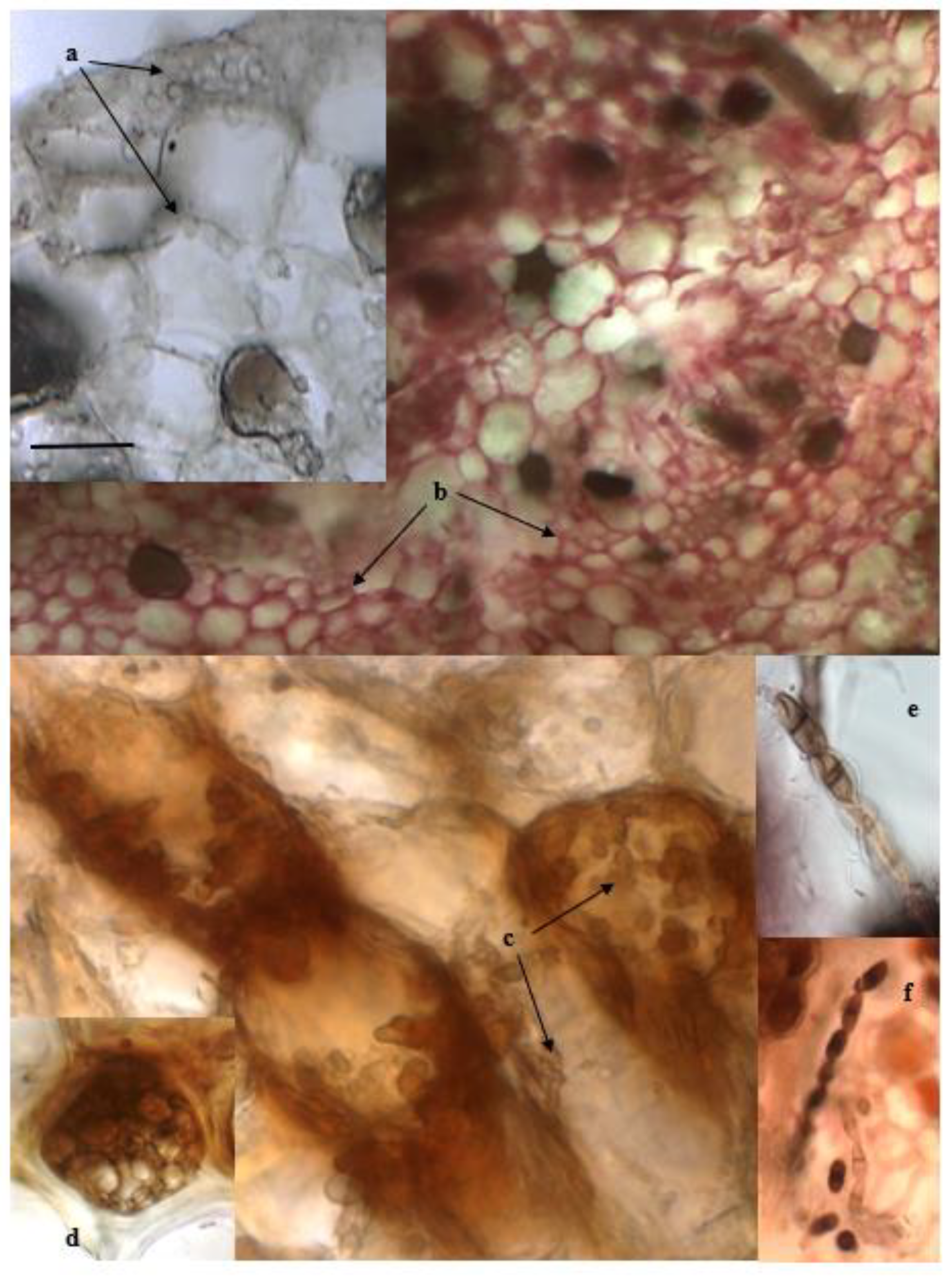
4.4. Microscopic Observations

The second part of each organ was manually dissected in transverse sections (20 µm thick) and stored in an Eppendorf tube with a fixative solution (Formalin–Acetic acid–Alcohol, F.A.A.: 95% ethanol, 40% formaldehyde, glacial acetic acid, distilled water, respectively 30, 8, 2, and 60%). To visualize fungal structures, some sections were mounted in a lactophenol solution (25 mL distilled water, 25 mL glycerin, 25 mL lactic acid, 25 g phenol crystals) and directly observed. Other sections were acidified by rapid immersion in 10% (v/v) HCl in water, kept at room temperature in a lactophenol solution containing 1 gL−1 acid fuchsin for 10 min, washed with lactophenol solution, and mounted. Excess stain was removed by washing with lactophenol without dye [17]. All sections (20 for each organ and site) were observed using a light microscope (Axioskop; Zeiss, Oberkochen, Germany) coupled with an AxioCam MRc5 (Zeiss, Oberkochen, Germany) digital camera. Images were captured using the AxioVision 4.6 software package (Zeiss, Oberkochen, Germany).

4.5. Leaf Biometry of P. oceanica Samples

In order to obtain information on the status of the shoots under investigation, biometric analyses according to [63,64] were carried out on leaf bundles of P. oceanica sampled shoots. In detail, all leaves from each shoot were counted and characterized as ‘adult,’ ‘intermediate,’ or ‘juvenile’ according to the classes proposed by [63], and estimates of morphometric parameters were made following the procedures described by [65]. Measurements of leaf length and width and leaf necrosis were recorded. In this way, the mean shoot surface and the percentage of brown tissue with respect to the total leaf length were calculated.

4.6. Statistical Analysis

One way ANOVA [66] was used to detected differences in morphological variables. In particular, the factor of locality was fixed, with three levels: 1 Ognina, 2 Bonagia, 3 Sciacca. A post hoc mean comparison test (HSD Tukey) was performed, and significant differences were found (P < 0.05) by ANOVA. Prior to the analysis, the data were checked for homoscedasticity (Levene’s test) and transformed when necessary. The SPSS 14 package was used for statistical analysis.

Author Contributions

Conceptualization: L.T., S.B., S.C. and A.T.; Methodology: L.T., S.B., S.C. and A.T.; Investigation: L.T., S.B., S.G., G.P., G.C., G.M., M.P., G.B. and R.C.; Data curation: L.T., S.B., S.G., G.C., G.M., S.C. and A.T.; Writing original draft preparation: L.T., S.B., S.C. and A.T.; Writing review and editing: L.T., R.C., G.M., S.C. and A.T.; Supervision: L.T., S.C. and A.T. All authors have read and agreed to the published version of the manuscript.

Funding

This research was funded by PON-Programma Operativo Nazionale “Ricerca e Competitività 2007–2013 per le Regioni della Convergenza” Progetto PON03PE_00203_1 Progetto di ricerca Marine Hazard sviluppo di tecnologie innovative per l’identificazione, monitoraggio, remediation di sorgenti di contaminazione naturale e antropica.

Conflicts of Interest

The authors declare no conflict of interest.

References

  1. Procaccini, G.; Buia, M.C.; Gambi, M.C.; Perez, M.; Pergent, G.; Pergent-Martini, C.; Romero, J. Seagrass status and extent along the Mediterranean coasts of Italy, France and Spain. In World Atlas of Seagrasses; Green, E.P., Short, F.T., Eds.; University of California Press: Berkeley, CA, USA, 2003; pp. 48–58. [Google Scholar]
  2. Hemminga, M.A.; Duarte, C.M. Seagrass Ecology; Cambridge University Press: Cambridge, UK, 2000. [Google Scholar]
  3. Diaz, A.E. Patch dynamics of the Mediterranean seagrass Posidonia oceanica: Implications for recolonisation process. Aquat. Bot. 2008, 89, 397–403. [Google Scholar] [CrossRef]
  4. Boudouresque, C.F.; Bernard, G.; Bonhomme, P.; Charbonnel, E.; Diviacco, G.; Meinesz, A.; Pergent, G.; Pergent-Martini, C.; Ruitton, S.; Tunesi, L. Protection and Conservation of Posidonia oceanica Meadows; RAMOGE and RAC/SPA Publications: Tunis, Tunisia, 2012; p. 202. [Google Scholar]
  5. Vassallo, P.; Paoli, C.; Rovere, A.; Montefalcone, M.; Morri, C.; Bianchi, N.B. The value of the seagrass Posidonia oceanica: A natural capital assessment. Mar. Pollut. Bull. 2013, 75, 157–167. [Google Scholar] [CrossRef] [PubMed]
  6. Boudouresque, C.F.; Pergent, G.; Pergent-Martini, C.; Ruitton, S.; Thibaut, T.; Verlaque, V. The necromass of the Posidonia oceanica seagrass meadow: Fate, role, ecosystem services and vulnerability. Hydrobiologia 2016, 781, 25–42. [Google Scholar] [CrossRef] [Green Version]
  7. Mazzella, L.; Buia, M.C.; Gambi, M.C.; Lorenti, M.; Russo, G.F.; Scipione, M.B.; Zupo, V. Plant-animal trophic relationships in the Posidonia oceanica ecosystem of the Mediterranean Sea: A review. In Plant-Animal Interactions in Marine Benthos; John, D.M., Hawkins, S.J., Pric, J.H., Eds.; Clarendon Press: Oxford, UK, 1992; Volume 46, pp. 165–187. [Google Scholar]
  8. Montagne, C. Exploration Scientifique de l’Algérie Pendant les Années 1840, 1841, 1842; Durieu De Maisonneuve, M.C., Ed.; Imprimerie Royale: Paris, France, 1846; pp. 1–197. [Google Scholar]
  9. Johnson, T.W., Jr.; Sparrow, F.K. Marine Biology: Fungi in Oceans and Estuaries; J. Cramer, Lehere: Weinheim, Germany, 1961; pp. xxiv + 668. [Google Scholar]
  10. Kohlmeyer, J. Zwei neue Ascomycetes Gat tungen auf Posidonia-rhizomen. Nova Hedwig. 1963, 6, 5–13. [Google Scholar]
  11. Kohlmeyer, J. Intertidal and phycophilous fungi from Tenerife (Canary Islands). Trans. Br. Mycol. Soc. 1967, 50, 137–147. [Google Scholar] [CrossRef]
  12. Kohlmeyer, J.; Kohlmeyer, E. Marine fungi from tropical America and Africa. Mycologia 1971, 63, 831–861. [Google Scholar] [CrossRef]
  13. Cuomo, V.; Vanzanella, F.; Fresi, E.; Mazzella, L.; Scipione, M.B. Micoflora delle fenerogame dell ‘Isolad’ Ischia: Posidonia oceanica (L.) Delile Cymodocea nodosa (Ucria) Aschers; Bulletin Musea Institute Biologia, Universiti Genova: Genoa, Italy, 1982; Volume 50, pp. 162–166. [Google Scholar]
  14. Cuomo, V.; Vanzanella, F.; Fresi, E.; Cinelli, F.; Mazzella, L. Fungal flora of Posidonia oceanica and its ecological significance. Trans. Br. Mycol. Soc. 1985, 84, 35–40. [Google Scholar] [CrossRef]
  15. Garcias-Bonet, N.; Arrieta, J.M.; de Santana, C.N.; Duarte, C.M.; Marba, N. Endophytic bacterial community of a Mediterranean marine angiosperm (Posidonia oceanica). Front. Microbiol. 2012, 3, 342. [Google Scholar] [CrossRef] [Green Version]
  16. Panno, L.; Bruno, M.; Voyron, S.; Anastasi, A.; Gnavi, G.; Miserere, L.; Varese, G.C. Diversity, ecological role and potential biotechnological applications of marine fungi associated to the seagrass Posidonia oceanica. New Biotechnol. 2013, 30, 686–694. [Google Scholar] [CrossRef]
  17. Torta, L.; Lo Piccolo, S.; Piazza, G.; Burruano, S.; Colombo, P.; Ottonello, D.; Perrone, R.; Di Maida, G.; Pirrotta, M.; Tomasello, A.; et al. Lulwoana sp., a dark septate endophyte in roots of Posidoniaoceanica (L.) Delile seagrass. Plant Biol. 2015, 17, 505–511. [Google Scholar] [CrossRef] [PubMed] [Green Version]
  18. Campbell, J.; Volkmann-Kohlmeyer, B.; Gräfenhan, T.; Spatafora, J.W.; Kohlmeyer, J. A re-evaluation of Lulworthiales: Relationships based on 18S and 28S rDNA. Mycol. Res. 2005, 109, 556–568. [Google Scholar] [CrossRef] [PubMed] [Green Version]
  19. Jumpponen, A.; Trappe, J.M. Dark septate endophytes: A review of facultative biotrophic root-colonizing fungi. New Phytol. 1998, 140, 295–310. [Google Scholar] [CrossRef] [PubMed]
  20. Sieber, T.N.; Grünig, C.R. Fungal Root Endophytes. In Plant Roots—The Hidden Half, 4th ed.; Eshel, A., Beeckman, T., Eds.; CRC Press, Taylor & Francis Group: Boca Raton, FL, USA, 2013; pp. 31–49. [Google Scholar]
  21. Vohník, M.; Borovec, O.; Župan, I.; Vondrášek, D.; Petrtýl, M.; Sudová, R. Anatomically and morphologically unique dark septate endophytic association in the roots of the Mediterranean endemic seagrass Posidonia oceanica. Mycorrhiza 2015, 25, 663–672. [Google Scholar] [CrossRef]
  22. Vohník, M.; Borovec, O.; Kolařík, M. Communities of cultivable root mycobionts of the seagrass Posidonia oceanica in the Northwest Mediterranean Sea are dominated by a hitherto undescribed pleosporalean dark septate endophyte. Microb. Ecol. 2016, 71, 442–451. [Google Scholar] [CrossRef]
  23. Vohník, M.; Borovec, O.; Župan, I.; Kolařík, M.; Sudová, R. Fungal root symbionts of the seagrass Posidonia oceanica in the central Adriatic Sea revealed by microscopy, culturing and pyrosequencing. Mar. Ecol. Prog. Ser. 2017, 583, 107–112. [Google Scholar] [CrossRef]
  24. Lefebvre, L.; Gobert, S. How Does Endophytic Fungi Transform the Posidonia oceanica (L.) Delile (1813) Meadow into the Aegagropiles? CIBM: Liège, Belgium, 2018. [Google Scholar]
  25. Tomasello, A.; Perrone, R.; Colombo, P.; Pirrotta, M.; Calvo, S. Variability of root hair anatomy and morphology in Posidonia oceanica (L.) Delile and substratum typology: First novel on spiral form. Aquat. Bot. 2018, 145, 45–48. [Google Scholar] [CrossRef]
  26. Vohník, M. Are lulworthioid fungi dark septate endophytes of the dominant Mediterranean seagrass Posidonia oceanica? Plant Biol. 2021, 24, 127–133. [Google Scholar] [CrossRef]
  27. Saikkonen, K.; Saari, S.; Helander, M. Defensive mutualism between plants and endophytic fungi? Fungal Divers. 2010, 41, 101–113. [Google Scholar] [CrossRef]
  28. Sarkar, S.; Dey, A.; Kumar, V.; El-Saber, B.G.; El-Esawi, M.A.; Tomczyk, M.; Ray, P. Fungal endophyte: An interactive endosymbiont with the capability of modulating host physiology. Front. Plant Sci. 2021, 12, 701800. [Google Scholar] [CrossRef]
  29. Calvo, S.; Tomasello, A.; Di Maida, G.; Pirrotta, M.; Buia, M.C.; Cinelli, F.; Cormaci, M.; Furnari, G.; Giaccone, G.; Luzzu, F.; et al. Seagrasses along the Sicilian coasts. Chem. Ecol. 2010, 26, 249–266. [Google Scholar] [CrossRef]
  30. Jones, E.B.J.; Sakayaroj, J.; Suetrong, S.; Somrithipol, S.; Pang, K. Classification of marine Ascomycota, anamorphic taxa and Basidiomycota. Fungal Divers. 2009, 35, 1–187. [Google Scholar]
  31. Garzoli, L.; Gnavi, G.; Tamma, F.; Tosi, S.; Varese, G.C.; Picco, A.M. Sink or swim: Updated knowledge on marine fungi associated with wood substrates in the Mediterranean Sea and hints about their potential to remediate hydrocarbons. Progr. Oceanogr. 2015, 137, 140–148. [Google Scholar] [CrossRef]
  32. Poli, A.; Prigione, V.; Bovio, E.; Perugini, I.; Varese, G.C. Insights on Lulworthiales inhabiting the Mediterranean Sea and description of three novel species of the genus Paralulworthia. J. Fungi. 2021, 7, 940. [Google Scholar] [CrossRef] [PubMed]
  33. Bellissimo, G.; Sirchia, B.; Ruvolo, V. Monitoring of Posidonia oceanica meadows in the Sicilian coasts under the Water Framework Directive (WFD). In Proceedings of the 8th International Symposium “Monitoring of Mediterranean coastal areas: Problems and measurement techniques”, Livorno, Italy, 16–18 June 2020; Firenze University Press: Livorno, Italy; pp. 510–518. [Google Scholar]
  34. Bellissimo, G.; Sirchia, B.; Ruvolo, V. Assessment of the ecological status of Sicilian coastal waters according to a macroalgae based index (CARLIT). In Proceedings of the 8th International Symposium “Monitoring of Mediterranean coastal areas: Problems and measurement techniques”, Livorno, Italy, 16–18 June 2020; Firenze University Press: Livorno, Italy; pp. 519–528. [Google Scholar] [CrossRef]
  35. Giovannetti, E.; Lasagna, R.; Montefalcone, M.; Bianchi, C.N.; Albertelli, G.; Morri, C. Inconsistent responses to substratum nature in Posidonia oceanica meadows: An integration through complexity levels? Chem. Ecol. 2008, 24, 83–91. [Google Scholar] [CrossRef]
  36. Di Maida, G.; Tomasello, A.; Sciandra, M.; Pirrotta, M.; Milazzo, M.; Calvo, S. Effect of different substrates on rhizome growth: Leaf biometry and shoot density of Posidonia oceanica. Mar. Environ. Res. 2013, 87–88, 96–102. [Google Scholar] [CrossRef]
  37. Balestri, E.; Cinelli, F.; Lardicci, C. Spatial variation in Posidonia oceanica structural, morphological and dynamic features in a northwestern Mediterranean coastal area: A multi-scale analysis. Mar. Ecol. Prog. Ser. 2003, 250, 51–60. [Google Scholar] [CrossRef]
  38. Rodriguez, R.J.; White, J.F., Jr.; Arnold, A.E.; Redman, R.S. Fungal endophytes: Diversity and functional roles. New Phytol. 2009, 182, 314–330. [Google Scholar] [CrossRef]
  39. Poli, A.; Bovio, E.; Ranieri, L.; Varese, G.C.; Prigione, V. Fungal diversity in the Neptune Forest: Comparison of the mycobiota of Posidonia oceanica, Flabellia petiolata, and Padina pavonica. Front. Microbiol. 2020, 11, 933. [Google Scholar] [CrossRef]
  40. Samerpitak, K.A.; Duarte, A.P.M.; Attili-Angelis, D.; Pagnocca, F.C.; Heinrichs, G.; Rijs, A.J.M.M.; Alfjorden, A.; Gerrits van den Ende, A.H.G.; Menken, S.B.J.; de Hoog, G.S. A new species of the oligotrophic genus Ochroconis (Sympoventuriaceae). Mycol. Progress. 2015, 14, 6. [Google Scholar] [CrossRef] [Green Version]
  41. Hyde, K.D.; Norphanphoun, C.; Maharachchikumbura, S.S.N.; Bhat, D.J.; Jones, E.B.G.; Bundhun, D.; Chen, Y.J.; Bao, D.F.; Dayarathne, M.C.; Devedatha, B.; et al. Refined families of Sordariomycetes. Mycosphere 2020, 11, 305–1059. [Google Scholar] [CrossRef]
  42. Wijayawardene, N.N.; Hyde, K.D.; Al-Ani, L.K.T.; Tedersoo, L.; Haelewaters, D.; Rajeshkumar, K.C.; Zhao, R.L.; Aptroot, A.; Leontyev, D.V.; Saxena, R.K.; et al. Outline of fungi and fungus-like taxa. Mycosphere 2020, 11, 1060–1456. [Google Scholar] [CrossRef]
  43. Becker, K.; Stadler, M. Recent progress in biodiversity research on the Xylariales and their secondary metabolism. J. Antibiot. 2021, 74, 1–23. [Google Scholar] [CrossRef]
  44. Cheng, X.; Li, W.; Cai, L. Molecular phylogeny of Ascotricha, including two new marine algae-associated species. Mycologia 2015, 107, 490–504. [Google Scholar] [CrossRef] [PubMed]
  45. Houbraken, J.; Visagie, C.M.; Meijer, M.; Frisvad, J.C.; Busby, P.E.; Pitt, J.I.; Seifert, K.A.; Louis-Seize, G.; Demirel, R.; Yilmaz, N.; et al. A taxonomic and phylogenetic revision of Penicillium section Aspergilloides. Stud. Mycol. 2014, 78, 373–451. [Google Scholar] [CrossRef] [PubMed] [Green Version]
  46. Zhang, H.; Lei, X.; Shao, S.; Zhou, X.; Li, Y.; Yang, B. Azaphilones and meroterpenoids from the soft coral-derived fungus Penicillium glabrum glmu003. Chem. Biodivers. 2021, 18, e2100663. [Google Scholar] [CrossRef]
  47. Zhang, Y.; Mu, J.; Feng, Y.; Kang, Y.; Zhang, J.; Gu, P.; Wang, Y.; Ma, L.; Zhu, Y. Broad-spectrum antimicrobial epiphytic and endophytic fungi from marine organisms: Isolation, bioassay and taxonomy. Mar. Drugs 2009, 7, 97–112. [Google Scholar] [CrossRef] [Green Version]
  48. Wiese, J.; Ohlendorf, B.; Blümel, M.; Schmaljohann, R.; Imhoff, J.F. Phylogenetic identification of fungi isolated from the marine sponge Tethya aurantium and identification of their secondary metabolites. Mar. Drugs 2011, 9, 561–585. [Google Scholar] [CrossRef]
  49. Kirichuka, N.N.; Pivkin, M.V. Filamentous fungi associated with the seagrass Zostera marina Linnaeus, 1753 of Rifovaya Bay (Peter the Great Bay, the Sea of Japan). Russ. J. Mar. Biol. 2015, 41, 351–355. [Google Scholar] [CrossRef]
  50. Knob, A.; Beitel, S.M.; Fortkamp, D.; Terrasan, C.R.; de Almeida, A.F. Production, purification, and characterization of a major Penicillium glabrum xylanase using Brewer’s spent grain as substrate. BioMed Res. Int. 2013, 2013, 728735. [Google Scholar] [CrossRef] [Green Version]
  51. Borovec, O.; Vohník, M. Ontogenetic transition from specialized root hairs to specific root-fungus symbiosis in the dominant Mediterranean seagrass Posidonia oceanica. Sci. Rep. 2018, 8, 10773. [Google Scholar] [CrossRef] [PubMed] [Green Version]
  52. Lo Piccolo, S.; Mondello, V.; Giambra, S.; Conigliaro, G.; Torta, L.; Burruano, S. Arthrinium phaeospermum, Phoma cladoniicola and Ulocladium consortiale, new olive pathogens in Italy. J. Phytopathol. 2014, 162, 258–263. [Google Scholar] [CrossRef]
  53. Ragazzi, A.; Mancini, F.; Dellavalle, I.; Capretti, P.; Moricca, S. Endophytic fungi in Quercus cerris: Isolation frequency in relation to phenological phase, tree health and the organ affected. Phytopathol. Mediterr. 2001, 40, 165–171. [Google Scholar] [CrossRef]
  54. Domsch, K.H.; Gams, W.; Anderson, T.H. Compendium of Soil Fungi; Academic Press: London, UK, 1980; p. 406. [Google Scholar]
  55. Barnett, H.L.; Hunter, B. Illustrated Genera of Imperfect Fungi; APS Press: St. Paul, MN, USA, 1998; p. 220. [Google Scholar]
  56. Pitt, J.I.; Hocking, A.D. Fungi and Food Spoilage, 2nd ed.; Aspen Publishers Inc.: Gaithersburg, MD, USA, 1999; p. 234. [Google Scholar]
  57. O’Donnell, K.; Cigelnik, E.; Nirenberg, H.I. Molecular systematics and phylogeography of the Gibberella fujikuroi species complex. Mycologia 1998, 90, 465–493. [Google Scholar] [CrossRef] [Green Version]
  58. White, T.J.; Bruns, T.; Lee, S.; Taylor, J. Amplification and direct sequencing of fungal ribosomal RNA genes for phylogenetics. In PCR Protocols: A Guide to Methods and Applications; Innis, M.A., Gelfand, D.H., Sninsky, J.J., White, T.J., Eds.; Academic Press: San Diego, CA, USA, 1990; pp. 315–322. [Google Scholar]
  59. Alves, A.; Correia, A.; Luque, J.; Phillips, A. Botryosphaeria corticola, sp. nov. on Quercus species, with notes and description of Botryosphaeria stevensii and its anamorph, Diplodia mutila. Mycologia 2004, 96, 598–613. [Google Scholar] [CrossRef]
  60. Giambra, S.; Piazza, G.; Alves, A.; Mondello, V.; Berbegal, M.; Burruano, S. Botryosphaeriaceae species associated with diseased loquat trees in Italy and description of Diplodia rosacearum sp. nov. Mycosphere 2016, 7, 978–989. [Google Scholar] [CrossRef]
  61. Thompson, J.D.; Gibson, T.J.; Plewniak, F.; Jeanmougin, F.; Higgins, D.G. The ClustalX windows interface: Flexible strategies for multiple sequence alignment aided by quality analysis tools. Nucleic Acids Res. 1997, 25, 4876–4882. [Google Scholar] [CrossRef] [Green Version]
  62. Tamura, K.; Stecher, G.; Peterson, D.; Filipski, A.; Kumar, S. MEGA6: Molecular evolutionary genetics analysis version 6.0. Mol. Biol. Evol. 2013, 30, 2725–2729. [Google Scholar] [CrossRef] [Green Version]
  63. Giraud, G. Essai de classement des herbiers de Posidonia oceanica (L.) Delile. Bot. Mar. 1977, 20, 487–491. [Google Scholar] [CrossRef]
  64. Pergent-Martini, C.; Leoni, V.; Pasqualini, V.; Ardizzone, G.D.; Balestri, E.; Bedini, R.; Belluscio, A.; Belsher, T.; Borg, J.A.; Bordouresque, C.; et al. Descriptors of Posidonia oceanica meadows: Use and application. Ecol. Indic. 2005, 5, 213–230. [Google Scholar] [CrossRef]
  65. Pergent, G.; Pergent-Martini, C.; Boudouresque, C. Utilisation de l’herbier à Posidonia oceanica comme indicateur biologique de la qualité du milieu littoral en Méditerranée: État des connaissances. Mésogée 1995, 54, 3–27. [Google Scholar]
  66. Underwood, A.J. Experiments in Ecology: Their Logical Design and Interpretation Using Analysis of Variance; Cambridge University Press: Cambridge, UK, 1997; p. 524. [Google Scholar]
Figure 1. Neighbour-joining tree based on phylogenetic analysis of the ITS1-5.8S rDNA-ITS2 sequences of Lulworthiales. Bootstrap percentages calculated from 1000 re-samplings are indicated at nodes. GenBank numbers with triangles represent the sequences obtained in this study and deposited at the GenBank Database.
Plants 11 01139 g001
Figure 2. Radial section of P. oceanica root: intracellular fungal colonization by melanized and septate hyphae (a), brown microsclerotia (b) and young red-stained intracellular hyphae (c). Bar = 20 µm.
Plants 11 01139 g002
Figure 3. Section of rhizomes of P. oceanica. Melanized (a) and red-stained (b) hyphae surrounding and infecting exodermal and cortical cells, respectively. Detail of intracellular fungal infection by melanized and septate hyphae (c) and of a microsclerotium (d). Chains of melanized, two-cellular conidial structures outside (e) and inside (f) the rhizomal tissue. Bar: a = 50 µm; b = 100 µm; c, d = 20 µm; e, f = 10 µm.
Plants 11 01139 g003
Figure 4. Section of leaves of P. oceanica from Sciacca: hyphae in epidermal (red-stained, (a)) and in mesophyll (not stained, (b)) cells. Bar = 20 µm.
Plants 11 01139 g004
Figure 5. Mean (±SE) values of biometric variables at the sampling stations. Letters over the bars indicate a homogeneous group checked by a post hoc test.
Plants 11 01139 g005
Figure 6. P. oceanica sampling locations along the coasts of Sicily.
Plants 11 01139 g006
Table 1. List of fungal endophytes isolated from Posidonia oceanica meadows in three Sicilian seas with their corresponding GenBank accession numbers and Blast results, obtained from GenBank.
Isolate CodeIsolate IdentityGenBank Accession No.Blast Match Sequence
Reference Accession No.Coverage (%)Identity (%)
PO1CordycipitaceaeKY463483MH2312489996
PO2Fusarium sp. KY463484MK589327100100
PO3Lulwoana sp.KY463485KF719966100100
PO4Ochroconis sp.KY463486MH063201100100
PO5Paecilomyces sp.KY463487KF87146010099
PO6Penicillium glabrumKY463488MH864674100100
PO7SordariomycetesKY463489GQ153240100100
PO8Thielavia microsporaKY463490JN709490100100
PO9Xylariaceae sp.KY463491MK334345100100
PO10Lulwoana sp. KY463492KF719966100100
PO11Lulwoana sp. KY463493KF719966100100
PO12Lulwoana sp.KY463494KF719966100100
PO13Lulwoana sp. KY463495KF719966100100
Table 2. Composition of the fungal population (504 colonies) isolated from the different P. oceanica organs in the three study sites.
TaxaNo. of IsolatesIF%
Penicillium glabrum33165.7
Lulwoana sp.11623.0
Xylariaceae407.9
Ochroconis sp.102.0
Cordycipitaceae30.6
Fusarium sp.10.2
Paecilomyces sp.10.2
Sordariomycetes10.2
Thielavia microspora10.2
Table 3. Distribution of Lulwoana sp., Ochroconis sp., P. glabrum and Xylariaceae population, as a function of sampling site and P. oceanica organ.
TaxaNo. of IsolatesIFo% IFs%
RootsRhizomeLeafBonagiaOgninaSciacca
P. glabrum3311000001000
Lulwoana sp.11644.854.30.919.843.137.1
Xylariaceae4042.557.5072.527.50
Ochroconis sp.10702010801010
Table 4. Isolates included in this study. The newly generated sequences are indicated in bold font.
Isolate NumberIsolate IdentityHostCountryITS GenBank
Acc. No.
RP2Lulwoana sp.Posidonia oceanicaItalyKF719965
RP5Lulwoana sp.Posidonia oceanicaItalyKF719968
RP6Lulwoana sp.Posidonia oceanicaItalyKF719969
RP1Lulwoana sp.Posidonia oceanicaItalyKF719964
P12LulworthialesPosidonia oceanicaSpainKC145431
P03Lulwoana sp.Posidonia oceanicaItalyKY463485
P13LulworthialesPosidonia oceanicaFranceKC145432
RP4Lulwoana sp.Posidonia oceanicaItalyKF719967
P03LulworthialesPosidonia oceanicaSpainKC145423
RP3Lulwoana sp.Posidonia oceanicaItalyKF719966
P010Lulwoana sp.Posidonia oceanicaItalyKY463492
P011Lulwoana sp.Posidonia oceanicaItalyKY463493
PO12Lulwoana sp.Posidonia oceanicaItalyKY463494
PO13Lulwoana sp.Posidonia oceanicaItalyKY463495
P32LulworthialesPosidonia oceanicaCroatiaKC412721
MUT 5413Lulwoana sp.Posidonia oceanicaItalyMK578237
P02LulworthialesPosidonia oceanicaItalyKC145422
MUT 1483Lulwoana sp.DriftwoodItalyKF915986
NBRC 32137Lulwoana uniseptataSubmerged woodJapanLC146746
ATCC62580Zalerion maritimumDriftwoodU.S.A.AF169305
CBS 280.54Lulwoana uniseptataUnknownUnknownMH857330
FCUL210208SF10Lulworthia atlanticaSea waterPortugalKT347215
CBS 139632Lulworthia atlanticaFagus sylvaticaPortugalKY202429
FCUL090707CF10Lulworthia atlanticaSea waterPortugalKT347213
FCUL061107CP4Lulworthia atlanticaSea waterPortugalKT347206
FCUL210208SP4Lulworthia atlanticaSea waterPortugalKT347205
CBS 541.66Leptosphaeria doliolumRudbeckia sp.NetherlandsMH858879
Table 5. ANOVA results on leaf length, shoot surface, and brown tissue.
Leaf LengthShoot SurfaceBrown Tissue
Source of VariationDfMSFDfMSFDfMSF
Locality21863.99.5 ***224,980.14.8 *26.14.9 *
RES47197.1 475155.8 331.3
Levene’s testns Ns *
Transformation None Ln
Post hoc test
(1)
Ognina
(2)
Bonagia
(3)
Sciacca
1 = 3 > 21 = 3 > 2 1 > 2 = 3
RES = residual, DF = degrees of freedom, MS = mean square, F = ratio, *** = P < 0.001, * = P < 0.05, ns = P > 0.05.
Publisher’s Note: MDPI stays neutral with regard to jurisdictional claims in published maps and institutional affiliations.

