
Morphology Characterization, Molecular Identification, and Pathogenicity of Fungal Pathogen Causing Kaffir Lime Leaf Blight in Northern Thailand

 ,
,  ,
,  ,
,  and
and
Abstract
1. Introduction
2. Results
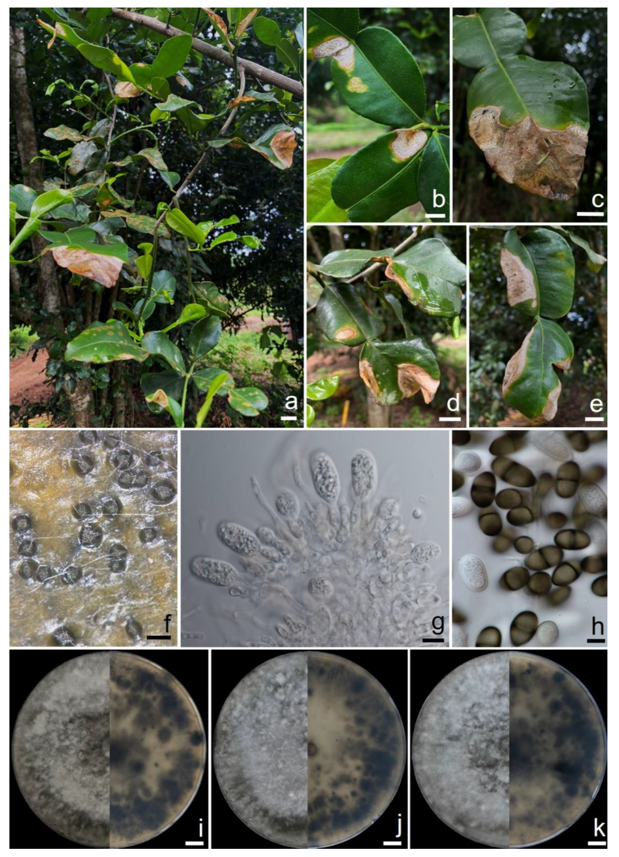
2.1. Sample Collection and Disease Symptoms
2.2. Fungal Isolation and Morphological Study
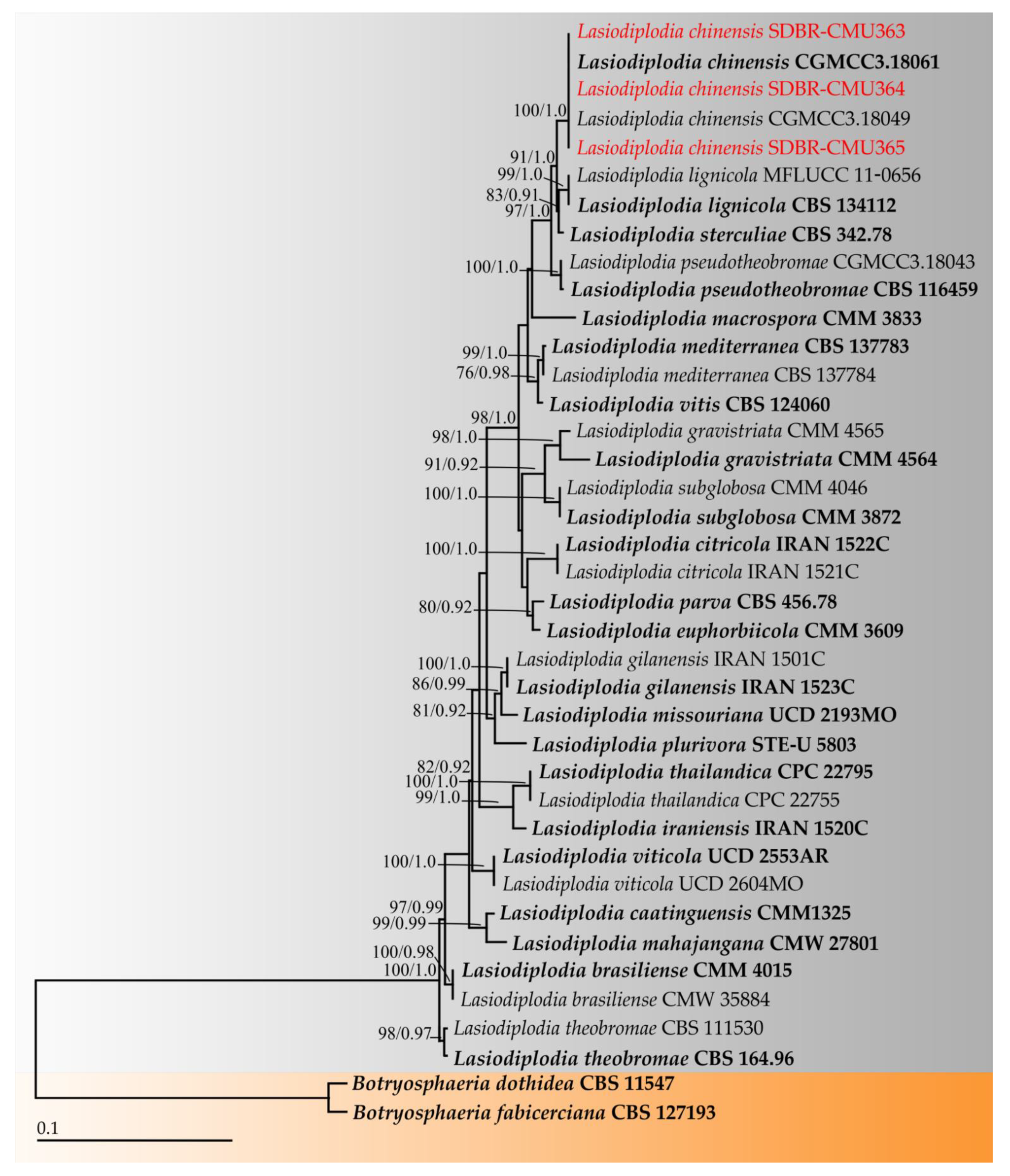
2.3. Phylogenetic Results
2.4. Pathogenicity Test
3. Discussion
4. Materials and Methods
4.1. Sample Collection
4.2. Fungal Isolation and Morphological Study
4.3. Molecular Study
4.3.1. DNA Extraction, PCR Amplification, and Sequencing
4.3.2. Sequence Alignment and Phylogenetic Analyses
4.4. Pathogenicity Tests
5. Conclusions
Author Contributions
Funding
Institutional Review Board Statement
Informed Consent Statement
Data Availability Statement
Acknowledgments
Conflicts of Interest
References
- Chanthaphon, S.; Chanthachum, S.; Hongpattarakere, T. Antimicrobial activities of essential oils and crude extracts from tropical Citrus spp. against food-related microorganisms. Songklanakarin J. Sci. Technol. 2008, 30, 125–131. [Google Scholar]
- Agouillal, F.; Taher, Z.; Moghrani, H.; Nasrallah, N.; El Enshasy, H. A review of genetic taxonomy, biomolecules chemistry and bioactivities of Citrus hystrix DC. Biosci. Biotechnol. Res. Asia 2017, 14, 285–305. [Google Scholar] [CrossRef]
- Le, X.T.; Ha, P.T.H.; Phong, H.X.; Hien, T.T.; Ngan, T.T.K. Extraction of essential oils and volatile compounds of kaffir lime (Citrus hystrix D.C) by hydrodistillation method. IOP Conf. Ser. Mater. Sci. Eng. 2020, 991, 012024. [Google Scholar] [CrossRef]
- Chueahongthong, F.; Ampasavate, C.; Okonogi, S.; Tima, S.; Anuchapreeda, S. Cytotoxic effects of crude kaffir lime (Citrus hystrix DC.) leaf fractional extracts on leukemic cell line. J. Med. Plant Res. 2011, 5, 3097–3105. [Google Scholar]
- Budiarto, R.; Poerwanto, R.; Santosa, E.; Efendi, D.; Agusta, A. Production, post-harvest and marketing of kaffir lime (Citrus hystrix DC) in Tulungagung, Indonesia. J. Trop. Crop Sci. 2019, 6, 138–143. [Google Scholar] [CrossRef]
- Wongpornchai, S. Kaffir lime leaf. In Handbook of Herbs and Spices, 2nd ed.; Peter, K.V., Ed.; Woodhead Publishing Limited: Cambridge, UK, 2012; pp. 319–328. [Google Scholar]
- Norkaew, O.; Pitija, K.; Pripdeevech, P.; Sookwong, P.; Wongpornchai, S. Supercritical fluid extraction and gas chromatographic-mass spectrometric analysis of terpenoids in fresh kaffir lime leaf oil. Chiang Mai J. Sci. 2013, 40, 240–247. [Google Scholar]
- Kidarn, S.; Saenjum, C.; Hongwiset, D.; Phrutivorapongkul, A. Furanocoumarins from kaffir lime and their inhibitory effects on inflammatory mediator production. Cogent Chem. 2018, 4, 1529259. [Google Scholar] [CrossRef]
- Md Othman, S.N.A.; Hassan, M.A.; Nahar, L.; Basar, N.; Jamil, S.; Sarker, S.D. Essential oils from the Malaysian Citrus (Rutaceae) medicinal plants. Medicines 2016, 3, 13. [Google Scholar] [CrossRef] [PubMed]
- Waikedre, J.; Dugay, A.; Barrachina, I.; Herrenknecht, C.; Cabalion, P.; Fournet, A. Chemical composition and antimicrobial activity of the essential oils from new caledonian Citrus macroptera and Citrus hystric. Chem. Biodivers. 2010, 7, 871–877. [Google Scholar] [CrossRef]
- Buakaew, W.; Pankla Sranujit, R.; Noysang, C.; Thongsri, Y.; Potup, P.; Nuengchamnong, N.; Suphrom, N.; Usuwanthim, K. Phytochemical constituents of Citrus hystrix DC. leaves attenuate inflammation via NF-κB signaling and NLRP3 inflammasome activity in macrophages. Biomolecules 2021, 11, 105. [Google Scholar] [CrossRef]
- Wulandari, Y.W.; Darmadji, P.; Anwar, C.; Supriyadi, S. Comparison between hydrodistillation with steam explosion and conventional hydrodistillation in kaffir lime oil extraction. Agritech 2019, 39, 306–314. [Google Scholar] [CrossRef]
- Wilkinson, K.; Grant, W.P.; Green, L.E.; Hunter, S.; Jeger, M.J.; Lowe, P.; Medley, G.F.; Mills, P.; Phillipson, J.; Poppy, G.M.; et al. Infectious diseases of animals and plants: An interdisciplinary approach. Phil. Trans. R. Soc. B 2011, 366, 1933–1942. [Google Scholar] [CrossRef]
- Jain, A.; Sarsaiya, S.; Wu, Q.; Lu, Y.; Shi, J. A review of plant leaf fungal diseases and its environment speciation. Bioengineered 2019, 10, 409–424. [Google Scholar] [CrossRef] [PubMed]
- Nuangmek, W.; Aiduang, W.; Kumla, J.; Lumyong, S.; Suwannarach, N. Evaluation of a newly identified endophytic fungus, Trichoderma phayaoense for plant growth promotion and biological control of gummy stem blight and wilt of muskmelon. Front. Microbiol. 2021, 12, 634772. [Google Scholar] [CrossRef]
- Rai, J.N. Leaf blight disease of Citrus acida var. variegata. Proc. Indian Acad. Sci. 1956, 43, 325–333. [Google Scholar] [CrossRef]
- Burnett, H.C.; Nemec, S.; Patterson, M. A review of Florida citrus blight and its association with soil edaphic factors, nutrition and Fusarium solani. Trop. Pest Manag. 1982, 28, 416–422. [Google Scholar] [CrossRef]
- Derrick, K.S.; Timmer, L.W. Citrus blight and other diseases of recalcitrant etiology. Annu. Rev. Phytopathol. 2000, 38, 181–205. [Google Scholar] [CrossRef]
- Derrick, K.S.; Barthe, G.A.; Hewitt, B.G.; Lee, R.F.; Albrigo, L.G. Detection of citrus blight by serological assays. Proc. Fla. State Hort. Soc. 1992, 105, 26–28. [Google Scholar]
- Timmer, L.W.; Lee, L.F.; Brlansky, R.H.; Graham, J.H.; Albrigo, L.G.; Derrick, K.S.; Tucker, D.P.H. The infectious nature of citrus blight. Proc. Fla. State Hort. Soc. 1992, 105, 21–26. [Google Scholar]
- Marques, M.W.; Lima, N.B.; de Morais, M.A., Jr.; Barbosa, M.A.G.; Souza, B.O.; Michereff, S.J.; Phillips, A.J.L.; Câmara, M.P.S. Species of Lasiodiplodia associated with mango in Brazil. Fungal Divers. 2013, 61, 181–193. [Google Scholar] [CrossRef]
- Cruywagen, E.M.; Slippers, B.; Roux, J.; Wingfield, M.J. Phylogenetic species recognition and hybridisation in Lasiodiplodia: A case study on species from baobabs. Fungal Biol. 2017, 121, 420–436. [Google Scholar] [CrossRef]
- Coutinho, I.B.L.; Freire, F.C.O.; Lima, C.S.; Lima, J.S.; Gonçalves, F.J.T.; Machado, A.R.; Silva, A.M.S.; Cardoso, J.E. Diversity of genus Lasiodiplodia associated with perennial tropical fruit plants in northeastern Brazil. Plant Pathol. 2017, 66, 90–104. [Google Scholar] [CrossRef]
- Dou, Z.P.; He, W.; Zhang, Y. Lasiodiplodia chinensis, a new holomorphic species from China. Mycosphere 2017, 8, 521–532. [Google Scholar] [CrossRef]
- Abdollahzadeh, J.; Javadi, A.; Goltapeh, E.M.; Zare, R.; Phillips, A.J. Phylogeny and morphology of four new species of Lasiodiplodia from Iran. Persoonia 2010, 25, 1–10. [Google Scholar] [CrossRef]
- Machado, A.R.; Pinho, D.B.; Pereira, O.L. Phylogeny, identification and pathogenicity of the Botryosphaeriaceae associated with collar and root rot of the biofuel plant Jatropha curcas in Brazil, with a description of new species of Lasiodiplodia. Fungal Divers. 2014, 67, 231–247. [Google Scholar] [CrossRef]
- Netto, M.S.B.; Lima, W.G.; Correia, K.C.; Da Silva, C.F.B.; Thon, M.; Martins, R.B.; Miller, R.N.G.; Michereff, S.J.; Câmara, M.P.S. Analysis of phylogeny, distribution, and pathogenicity of Botryosphaeriaceae species associated with gummosis of Anacardium in Brazil, with a new species of Lasiodiplodia. Fungal Biol. 2017, 121, 437–451. [Google Scholar] [CrossRef]
- Liu, J.K.; Phookamsak, R.; Doilom, M.; Wikee, S.; Li, Y.M.; Ariyawansha, H.; Boonmee, S.; Chomnunti, P.; Dai, D.Q.; Bhat, J.D.; et al. Towards a natural classification of Botryosphaeriales. Fungal Divers. 2012, 57, 149–210. [Google Scholar] [CrossRef]
- Begoude, B.A.D.; Slippers, B.; Wingfield, M.J.; Roux, J. Botryosphaeriaceae associated with Terminalia catappa in Cameroon, South Africa and Madagascar. Mycol. Prog. 2010, 9, 101–123. [Google Scholar] [CrossRef]
- Linaldeddu, B.T.; Deidda, A.; Scanu, B.; Franceschini, A.; Serra, S.; Berraf-Tebbal, A.; Zouaoui Boutiti, M.; Ben Jamâa, M.L.; Phillips, A.J.L. Diversity of Botryosphaeriaceae species associated with grapevine and other woody hosts in Italy, Algeria and Tunisia, with descriptions of Lasiodiplodia exigua and Lasiodiplodia mediterranea sp. nov. Fungal Divers. 2015, 71, 201–214. [Google Scholar] [CrossRef]
- Urbez-Torres, J.R.; Peduto, F.; Striegler, R.K.; Urrea-Romero, K.E.; Rupe, J.C.; Cartwright, R.D.; Gubler, W.D. Characterization of fungal pathogens associated with grapevine trunk diseases in Arkansas and Missouri. Fungal Divers. 2012, 52, 169–189. [Google Scholar] [CrossRef]
- Alves, A.; Crous, P.W.; Correia, A.; Phillips, A.J.L. Morphological and molecular data reveal cryptic speciation in Lasiodiplodia theobromae. Fungal Divers. 2008, 28, 1–13. [Google Scholar]
- Damm, U.; Crous, P.W.; Fourie, P.H. Botryosphaeriaceae as potential pathogens of Prunus species in South Africa, with descriptions of Diplodia africana and Lasiodiplodia plurivora sp. nov. Mycologia 2007, 99, 664–680. [Google Scholar] [CrossRef] [PubMed]
- Yang, T.; Groenewald, J.Z.; Cheewangkoon, R.; Jami, F.; Abdollahzadeh, J.; Lombard, L.; Crous, P.W. Families, genera, and species of Botryosphaeriales. Fungal Biol. 2017, 121, 322–346. [Google Scholar] [CrossRef]
- Trakunyingcharoen, T.; Cheewangkoon, R.; To-anun, C.; Crous, P.W.; van Niekerk, J.M.; Lombard, L. Botryosphaeriaceae associated with diseases of mango (Mangifera indica). Australasian Plant Pathol. 2014, 43, 425–438. [Google Scholar] [CrossRef]
- Trakunyingcharoen, T.; Lombard, L.; Groenewald, J.Z.; Cheewangkoon, R.; To-Anun, C.; Crous, P.W. Caulicolous Botryosphaeriales from Thailand. Persoonia 2015, 34, 87–99. [Google Scholar] [CrossRef] [PubMed]
- Phillips, A.; Alves, A.; Correia, A.; Luque, J. Two new species of Botryosphaeria with brown, 1-septate ascospores and Dothiorella anamorphs. Mycologia 2005, 97, 513–529. [Google Scholar] [CrossRef] [PubMed]
- Slippers, B.; Crous, P.W.; Denman, S.; Coutinho, T.A.; Wingfield, B.D.; Wingfield, M.J. Combined multiple gene genealogies and phenotypic characters differentiate several species previously identified as Botryosphaeria dothidea. Mycologia 2004, 96, 83–101. [Google Scholar] [CrossRef][Green Version]
- Chen, S.F.; Pavlic, D.; Roux, J.; Slippers, B.; Xie, Y.J.; Wingfield, M.J.; Zhou, X.D. Characterization of Botryosphaeriaceae from plantation-grown Eucalyptus species in South China. Plant Pathol. 2011, 60, 739–751. [Google Scholar] [CrossRef]
- Chen, S.F.; Morgan, D.P.; Hasey, J.K.; Anderson, K.; Michailides, T.J. Phylogeny, morphology, distribution, and pathogenicity of Botryosphaeriaceae and Diaporthaceae from English walnut in California. Plant Dis. 2014, 98, 636–652. [Google Scholar] [CrossRef]
- Li, G.Q.; Liu, F.F.; Li, J.Q.; Liu, Q.L.; Chen, S.F. Botryosphaeriaceae from Eucalyptus plantations and adjacent plants in China. Persoonia 2018, 40, 63–95. [Google Scholar] [CrossRef]
- De Silva, N.I.; Phillips, A.J.L.; Liu, J.K.; Lumyong, S.; Hyde, K.D. Phylogeny and morphology of Lasiodiplodia species associated with Magnolia forest plants. Sci. Rep. 2019, 9, 14355. [Google Scholar] [CrossRef]
- Chen, J.; Zhu, Z.; Fu, Y.; Cheng, J.; Xie, J.; Lin, Y. Identification of Lasiodiplodia pseudotheobromae causing fruit rot of citrus in China. Plants 2021, 10, 202. [Google Scholar] [CrossRef]
- Wang, Y.; Zhang, Y.; Bhoyroo, V.; Rampadarath, S.; Jeewon, R. Multigene phylogenetics and morphology reveal five novel Lasiodiplodia species associated with blueberries. Life 2021, 11, 657. [Google Scholar] [CrossRef]
- Tennant, P.F.; Robinson, D.; Fisher, L.; Bennett, S.; Hutton, D.; Coates-Beckford, P.; Laughlin, W.M. Diseases and pests of citrus (Citrus spp.). Tree For. Sci. Biotech. 2009, 3, 81–107. [Google Scholar]
- Aglave, B. Citrus. In Handbook of Plant Disease Identification and Management; CRC Press: Boca Raton, FL, USA, 2018; pp. 129–175. [Google Scholar]
- Dwiastuti, M.E.; Wuryantini, S.; Sugiyatno, A.; Supriyanto, A. Seed health evaluation in the process of free-virus citrus seed production on Kampar regency, Riau province of Indonesia. RJOAS 2019, 2, 273–282. [Google Scholar] [CrossRef]
- Afloukou, F.; Zinsou, V.; Onelge, N. Citrus in Benin Republic: Past, present, and future challenges. Citrus Res. Technol. 2020, 41, e1060. [Google Scholar] [CrossRef]
- Jaouad, M.; Moinina, A.; Ezrari, S.; Lahlali, R. Key pests and diseases of citrus trees with emphasis on root rot diseases: An overview. Mor. J. Agri. Sci. 2020, 1, 149–160. [Google Scholar]
- Burgess, T.I.; Barber, P.A.; Mohali, S.; Pegg, G.; de Beer, W.; Wingfield, M.J. Three new Lasiodiplodia spp. from the tropics, recognized based on DNA sequence comparisons and morphology. Mycologia 2006, 98, 423–435. [Google Scholar] [CrossRef]
- Slippers, B.; Wingfield, M.J. Botryosphaeriaceae as endophytes and latent pathogens of woody plants: Diversity, ecology and impact. Fungal Biol. Rev. 2007, 21, 90–106. [Google Scholar] [CrossRef]
- Úrbez-Torres, J.R.; Leavitt, G.M.; Guerrero, J.C.; Guevara, J.; Gubler, W.D. Identification and pathogenicity of Lasiodiplodia theobromae and Diplodia seriata, the causal agents of bot canker disease of grapevines in Mexico. Plant Dis. 2008, 92, 519–529. [Google Scholar] [CrossRef]
- Chen, S.; Li, G.; Liu, Q.; Li, J.; Liu, F. Characteristics of Lasiodiplodia theobromae from Rosa rugosa in South China. Crop Prot. 2016, 79, 51–55. [Google Scholar] [CrossRef]
- Bautista-Cruz, M.A.; Almaguer-Vargas, G.; Leyva-Mir, S.G.; Colinas-León, M.T.; Correia, K.C.; Camacho-Tapia, M.; Robles-Yerena, L.; Michereff, S.J.; Tovar-Pedraza, J.M. Phylogeny, distribution, and pathogenicity of Lasiodiplodia species associated with cankers and dieback symptoms of Persian lime in Mexico. Plant Dis. 2019, 103, 1156–1165. [Google Scholar] [CrossRef]
- Fayyaz, A.; Bonello, P.; Tufail, M.R.; Amrao, L.; Habib, A.; Gai, Y.; Sahi, S.T. First report of citrus withertip (tip dieback), a disease complex caused by Colletotrichum siamense and Lasiodiplodia iraniensis, on Citrus reticulata cv. Kinnow in Punjab, Pakistan. Plant Dis. 2018, 102, 2659. [Google Scholar] [CrossRef]
- Ahmed, M.Z.; Shafique, M.S.; Anwaar, H.A.; Sarfraz, S.; Tufail, M.R.; Fayyaz, A.; Muntaha, S.; Haque, K.; Ghuffar, S.; Amrao, L. First report of Lasiodiplodia pseudotheobromae causing trunk cankers in Citrus reticulata in Pakistan. Plant Dis. 2020, 104, 2522. [Google Scholar] [CrossRef]
- Gui, Q.; Zhao, J.; Yu, Z.; Sun, W.; Mo, J.; Li, Q.; Guo, T.; Tang, L.; Huang, S.; Hsiang, T. First report of trunk canker and gummosis of kumquat caused by Lasiodiplodia theobromae in China. Plant Dis. 2020, 104, 971. [Google Scholar] [CrossRef]
- Xiao, X.E.; Wang, W.; Crous, P.W.; Wang, H.K.; Jiao, C.; Huang, F.; Pu, Z.X.; Zhu, Z.R.; Li, H.Y. Species of Botryosphaeriaceae associated with citrus branch diseases in China. Persoonia 2021, 47, 106–135. [Google Scholar] [CrossRef]
- Adesemoye, A.O.; Mayorquin, J.S.; Wang, D.H.; Twizeyimana, M.; Lynch, S.C.; Eskalen, A. Identification of species of Botryosphaeriaceae causing bot gummosis in citrus in California. Plant Dis. 2014, 98, 55–61. [Google Scholar] [CrossRef]
- Guajardo, J.; Riquelme, N.; Tapia, L.; Larach, A.; Torres, C.; Camps, R.; Besoain, X. First report of Lasiodiplodia theobromae causing bot gummosis in Citrus limon in Chile. Plant Dis. 2018, 102, 818. [Google Scholar] [CrossRef]
- Daengsuwan, W.; Wonglom, P.; Sunpapao, A. First report of Lasiodiplodia theobromae causing spadix rot in Anthurium andraeanum. J. Phytopathol. 2020, 168, 129–133. [Google Scholar] [CrossRef]
- Suwannarach, N.; Khuna, S.; Kumla, J.; Tanruean, K.; Lumyong, S. First report of Lasiodiplodia theobromae causing fruit rot on melon (Cucumis melo) in Thailand. Plant Dis. 2019, 104, 280. [Google Scholar] [CrossRef]
- Pipattanapuckdee, A.; Boonyakait, D.; Tiyayon, C.; Seehanam, P.; Ruangwong, O. Lasiodiplodia pseudotheobromae causes postharvest fruit rot of longan in Thailand. Australas. Plant Dis. Notes 2019, 14, 21. [Google Scholar] [CrossRef]
- Chantarasiri, A.; Boontanom, P. Fusarium solani and Lasiodiplodia pseudotheobromae, fungal pathogens causing stem rot disease on durian trees (Durio zibethinus) in Eastern Thailand. New Dis. Rep. 2021, 44, e12026. [Google Scholar] [CrossRef]
- Gomdola, D.; Jeewon, R.; Jayawardena, R.S.; Pem, D.; Harishchandra, D.L. A new record of Lasiodiplodia pseudotheobromae causing leaf spot of Cynometra malaccensis in Thailand. Plant Pathol. Quar. 2020, 10, 223–237. [Google Scholar] [CrossRef]
- Trakunyingcharoen, T.; Cheewangkoon, R.; Toanun, C. Phylogeny and pathogenicity of fungal species in the family Botryosphaeriaceae associated with mango (Mangifera indica) in Thailand. Int. J. Agric. Technol. 2013, 9, 1535–1543. [Google Scholar]
- Choi, Y.W.; Hyde, K.D.; Ho, W.H. Single spore isolation of fungi. Fungal Divers. 1999, 3, 29–38. [Google Scholar]
- White, T.J.; Bruns, T.; Lee, S.; Taylor, J. Amplification and direct sequencing of fungal ribosomal RNA genes for phylogenetics. In PCR Protocols: A Guide to Methods and Applications; Innis, M.A., Gelfand, D.H., Sninsky, J.J., White, T.J., Eds.; Academic Press: San Diego, CA, USA, 1990; pp. 315–322. [Google Scholar]
- O’Donnell, K.; Lutzoni, F.M.; Ward, T.J.; Benny, G.L. Evolutionary relationships among mucoralean fungi Zygomycota: Evidence for family polyphyly on a large scale. Mycologia 2001, 93, 286–297. [Google Scholar] [CrossRef]
- Glass, N.L.; Donaldson, G.C. Development of primer sets designed for use with the PCR to amplify conserved genes from filamentous ascomycetes. Appl. Environ. Microbiol. 1995, 61, 1323–1330. [Google Scholar] [CrossRef]
- Edgar, R.C. MUSCLE: A multiple sequence alignment method with reduced time and space complexity. BMC Bioinform 2004, 5, 1–19. [Google Scholar] [CrossRef]
- Hall, T. Bioedit Version 6.0.7. 2004. Available online: http://www.mbio.ncsu.edu/bioedit/bioedit.html (accessed on 20 November 2021).
- Felsenstein, J. Confidence intervals on phylogenetics: An approach using bootstrap. Evolution 1985, 39, 783–791. [Google Scholar] [CrossRef]
- Stamatakis, A. RAxML-VI-HPC: Maximum likelihood-based phylogenetic analyses with thousands of taxa and mixed models. Bioinformatics 2006, 22, 2688–2690. [Google Scholar] [CrossRef] [PubMed]
- Miller, M.A.; Pfeiffer, W.; Schwartz, T. Creating the cipres science gateway for inference of large phylogenetic trees. In Proceedings of the 2010 Gateway Computing Environments Workshop (GCE), New Orleans, LA, USA, 14 November 2010; IEEE: Manhattan, NY, USA; pp. 1–8. [Google Scholar]
- Ronquist, F.; Teslenko, M.; van der Mark, P.; Ayres, D.L.; Darling, A.; Höhna, S.; Larget, B.; Liu, L.; Suchard, M.A.; Huelsenbeck, J.P. MrBayes 3.2: Efficient Bayesian phylogenetic inference and model choice across a large model space. Syst. Biol. 2012, 61, 539–542. [Google Scholar] [CrossRef] [PubMed]
- Rambaut, A. FigTree Tree Figure Drawing Tool Version 131, Institute of Evolutionary 623 Biology, University of Edinburgh. Available online: http://treebioedacuk/software/figtree/ (accessed on 20 October 2021).
- Suwannarach, N.; Sujarit, K.; Kumla, J.; Bussaban, B.; Lumyong, S. First report of leaf spot disease on oil palm caused by Pestalotiopsis theae in Thailand. J. Gen. Plant Pathol. 2013, 79, 277–279. [Google Scholar] [CrossRef]


| Fungal Taxa | Strain/Isolate | GenBank Accession Number | Reference | |||
|---|---|---|---|---|---|---|
| ITS | tef-1 | tub | rpb2 | |||
| Lasiodiplodia brasiliense | CMM 4015 T | JX464063 | JX464049 | − | − | [21] |
| L. brasiliense | CMW 35884 | KU887094 | KU886972 | KU887466 | KU696345 | [22] |
| L. caatinguensis | CMM1325 T | KT154760 | KT008006 | KT154767 | − | [23] |
| L. chinensis | CGMCC3.18061 T | KX499889 | KX499927 | KX500002 | KX499965 | [24] |
| L. chinensis | CGMCC3.18049 | KX499878 | KX499916 | KX499991 | KX499954 | [24] |
| L. chinensis | SDBR-CMU363 | OL989102 | OL989839 | OL989842 | OL989845 | This study |
| L. chinensis | SDBR-CMU364 | OL989137 | OL989840 | OL989843 | OL989846 | This study |
| L. chinensis | SDBR-CMU365 | OL989141 | OL989841 | OL989844 | OL989847 | This study |
| L. citricola | IRAN 1522C T | GU945354 | GU945340 | KU887505 | KU696351 | [22,25] |
| L. citricola | IRAN 1521C | GU945353 | GU945339 | KU887504 | KU696350 | [22,25] |
| L. euphorbiicola | CMM 3609 T | KF234543 | KF226689 | KF254926 | − | [26] |
| L. gilanensis | IRAN 1523C T | GU945351 | GU945342 | KU887511 | KU696357 | [22,25] |
| L. gilanensis | IRAN 1501C | GU945352 | GU945341 | KU887510 | KU696356 | [22,25] |
| L. gravistriata | CMM 4564 T | KT250949 | KT250950 | − | − | [27] |
| L. gravistriata | CMM 4565 | KT250947 | KT266812 | − | − | [27] |
| L. iraniensis | IRAN 1520C T | GU945348 | GU945336 | KU887516 | KU696363 | [22,25] |
| L. lignicola | CBS 134112 T | JX646797 | KU887003 | JX646845 | KU696364 | [22,28] |
| L. lignicola | MFLUCC 11-0656 | JX646798 | − | JX646846 | − | [28] |
| L. macrospora | CMM 3833 T | KF234557 | KF226718 | KF254941 | − | [26] |
| L. mahajangana | CMW 27801 T | FJ900595 | FJ900641 | FJ900630 | KU696365 | [29] |
| L. mediterranea | CBS 137783 T | KJ638312 | KJ638331 | KU887521 | KU696368 | [22,30] |
| L. mediterranea | CBS 137784 | KJ638311 | KJ638330 | KU887522 | KU696369 | [22,30] |
| L. missouriana | UCD 2193MO T | HQ288225 | HQ288267 | HQ288304 | KU696370 | [22,31] |
| L. parva | CBS 456.78 T | EF622083 | EF622063 | KU887523 | KU696372 | [22,32] |
| L. plurivora | STE-U 5803 T | EF445362 | EF445395 | KU887524 | KU696374 | [22,33] |
| L. pseudotheobromae | CBS 116459 T | EF622077 | EF622057 | EU673111 | KU696376 | [22,32] |
| L. pseudotheobromae | CGMCC3.18043 | KX499872 | KX499910 | KX499985 | KX499948 | [24] |
| L. sterculiae | CBS 342.78 T | KX464140 | KX464634 | KX464908 | KX463989 | [34] |
| L. subglobosa | CMM 3872 T | KF234558 | KF226721 | KF254942 | − | [26] |
| L. subglobosa | CMM 4046 | KF234560 | KF226723 | KF254944 | − | [26] |
| L. thailandica | CPC 22795 T | KJ193637 | KJ193681 | − | − | [35] |
| L. thailandica | CPC 22755 | KM006433 | KM006464 | − | − | [36] |
| L. theobromae | CBS 164.96 T | AY640255 | AY640258 | KU887532 | KU696383 | [22,37] |
| L. theobromae | CBS 111530 | EF622074 | EF622054 | KU887531 | KU696382 | [22,32] |
| L. viticola | UCD 2553AR T | HQ288227 | HQ288269 | HQ288306 | KU696385 | [22,31] |
| L. viticola | UCD 2604MO | HQ288228 | HQ288270 | HQ288307 | KU696386 | [22,31] |
| L. vitis | CBS 124060 T | KX464148 | KX464642 | KX464917 | KX463994 | [34] |
| Botryosphaeria dothidea | CBS 115476 T | AY236949 | AY236898 | AY236927 | EU339577 | [38] |
| B. fabicerciana | CBS 127193 T | HQ332197 | HQ332213 | KF779068 | MF410137 | [39,40,41] |
| Gene | Primer Name | Primer Sequence | The Obtained Length (bp) | ||
|---|---|---|---|---|---|
| SDBR-CMU363 | SDBR-CMU364 | SDBR-CMU365 | |||
| ITS | ITS4 | 5′-TCCTCCGCTTATTGATATGC-3′ | 540 | 522 | 529 |
| ITS5 | 5′-GGAAGTAAAAGTCGTAACAAGG-3′ | ||||
| tef-1 | EF1-983F | 5′-GCYCCYGGHCAYCGTGAYTTYAT-3′ | 955 | 943 | 932 |
| EF1-2218R | 5′-ATGACACCRACRGCRACRGTYTG-3′ | ||||
| tub | Bt2a | 5′-GGTAACCAAATCGGTGCTGCTTTC-3′ | 890 | 810 | 850 |
| Bt2b | 5′-ACCCTCAGTGTAGTGACCCTTGGC-3′ | ||||
| rbp2 | RPB2-LasF | 5′-GGTAGCGACGTCACTCCT-3′ | 593 | 580 | 591 |
| RPB2-LasR | 5′-GCGCAAATACCCAGAATCAT-3′ | ||||
Publisher’s Note: MDPI stays neutral with regard to jurisdictional claims in published maps and institutional affiliations. |
© 2022 by the authors. Licensee MDPI, Basel, Switzerland. This article is an open access article distributed under the terms and conditions of the Creative Commons Attribution (CC BY) license (https://creativecommons.org/licenses/by/4.0/).
Share and Cite
Suwannarach, N.; Khuna, S.; Kumla, J.; Cheewangkoon, R.; Suttiprapan, P.; Lumyong, S. Morphology Characterization, Molecular Identification, and Pathogenicity of Fungal Pathogen Causing Kaffir Lime Leaf Blight in Northern Thailand. Plants 2022, 11, 273. https://doi.org/10.3390/plants11030273
Suwannarach N, Khuna S, Kumla J, Cheewangkoon R, Suttiprapan P, Lumyong S. Morphology Characterization, Molecular Identification, and Pathogenicity of Fungal Pathogen Causing Kaffir Lime Leaf Blight in Northern Thailand. Plants. 2022; 11(3):273. https://doi.org/10.3390/plants11030273
Chicago/Turabian StyleSuwannarach, Nakarin, Surapong Khuna, Jaturong Kumla, Ratchadawan Cheewangkoon, Piyawan Suttiprapan, and Saisamorn Lumyong. 2022. "Morphology Characterization, Molecular Identification, and Pathogenicity of Fungal Pathogen Causing Kaffir Lime Leaf Blight in Northern Thailand" Plants 11, no. 3: 273. https://doi.org/10.3390/plants11030273
APA StyleSuwannarach, N., Khuna, S., Kumla, J., Cheewangkoon, R., Suttiprapan, P., & Lumyong, S. (2022). Morphology Characterization, Molecular Identification, and Pathogenicity of Fungal Pathogen Causing Kaffir Lime Leaf Blight in Northern Thailand. Plants, 11(3), 273. https://doi.org/10.3390/plants11030273