Article

Mummified Wood of Juniperus (Cupressaceae) from the Late Miocene of Taman Peninsula, South Russia

by
Anna V. Stepanova
1,*,
Anastasia A. Odintsova
2,
Alena I. Rybkina
2,
Yuliana V. Rostovtseva
2,3 and
Alexei A. Oskolski
1,4
1
Komarov Botanical Institute of the Russian Academy of Science, Professor Popov Street 2, 197376 St. Petersburg, Russia
2
Geophysical Center of the Russian Academy of Sciences (GC RAS), Molodezhnaya Street 3, 119296 Moscow, Russia
3
Department of Petroleum Sedimentology and Marine Geology, Faculty of Geology, Lomonosov Moscow State University, Leninskie Gory GSP-1, 119991 Moscow, Russia
4
Department of Botany and Plant Biotechnology, University of Johannesburg, Auckland Park 2006, Johannesburg P.O. Box 524, South Africa
*
Author to whom correspondence should be addressed.
Plants 2022, 11(15), 2050; https://doi.org/10.3390/plants11152050
Submission received: 11 July 2022 / Revised: 1 August 2022 / Accepted: 2 August 2022 / Published: 5 August 2022

Abstract

:
Juniperus L. is the second-largest genus of conifers, having the widest distribution of all conifer genera. Its phytogeographic history is, however, obscure due to its very poor fossil record. We described a wood of Juniperus sp. from the lower Maeotian sediments of the Popov Kamen section, Taman Peninsula, South Russia, in order to clarify its taxonomic position shedding light on the phytogeographic history of the genus. This fossil wood was well-preserved by mummification, which allowed for it to be studied by the same methods as used for the anatomical examination of modern woods. The wood from the Popov Kamen section shows the greatest similarity to the extant Mediterranean species J. excelsa, belonging to the section Sabina. This is the first reliable macrofossil evidence of the sect. Sabina from Eurasia convincingly dated to the Miocene. The age of the mummified wood from the Popov Kamen section is consistent with molecular dating of diversification of the lineage comprising juniper species of the sect. Sabina from Europe, Asia and eastern Africa. The wood of Juniperus sp. has not been buried in situ, as it was found in the relatively deep-water marine sediments. The available coeval pollen series and macrofossils of Cupressaceae from the surrounding regions suggest that this wood was likely transferred by sea current from the northwestern side of the Black Sea, which was a part of the Eastern Paratethys.

1. Introduction

Juniperus L. is the second-largest genus of conifers, and the largest member of the family Cupressaceae. Comprising 75 species [1], Juniperus has the widest distribution of all conifer genera [2]. Most species of this genus are confined to forests and to shrubby vegetations occurring in arid and semiarid regions throughout the Northern Hemisphere, with a single species crossing the Equator in eastern Africa [3]. Adams [1] recognized three monophyletic sections within this genus: sect. Calocedrus Endl., with a single species in the Mediterranean; sect. Juniperus, with fourteen species in East Asia and the Mediterranean plus one circumboreal species, J. communis L.; and sect. Sabina Spach, with 60 species distributed in southwestern North America, Asia and the Mediterranean as well as in eastern Africa and Macaronesia.
Molecular dating with fossil calibration shows that the Juniperus diverged from other Cupressaceae in the late Paleocene to Eocene [4,5]. Axelrod [6] suggested that diversification of junipers occurred within warm temperate semiarid vegetation of the Madrean–Tethyan belts that ran along the southern areas of Eurasia and North America during Eocene and Oligocene. This scenario has been confirmed by molecular evidence [4]. The fossil record for Juniperus is still too poor for comprehensive reconstructions of the history of this genus.
The most ancient fossils of Juniperus belonging to the sect. Sabina have been described from the Eocene–Oligocene boundary in the Czech Republic [7]. Other fossils attributed to this section were reported from the Oligocene to the middle Miocene deposits of North America [8,9,10,11], from the Miocene–Pliocene boundary of Bulgaria [12] and from the Pliocene of Bashkortostan, Russia [13]. At the same time, no reliable macrofossil evidence for diversification of the sect. Sabina during the Miocene has been found in Eurasia. As for the sections Juniperus and Calocedrus, their fossils are known only from the middle Miocene and the Pliocene of Europe [12,14,15,16].
In this study, we present anatomical investigations of mummified wood belonging to the genus Juniperus from the Late Miocene sediments of the Popov Kamen section, Taman Peninsula, South Russia. We aim to clarify the taxonomic position of this fossil wood in order to shed light on phytogeographical history of the section Sabina during the Neogene.

2. Results

2.1. Systematic Description

Order Coniferales Gorozhankin, 1904
Family Cupressaceae Gray, 1822
Genus Juniperus Linnaeus, 1753 [type: Juniperus communis L.]
Section Sabina Spach, 1841
Juniperus sp.
Material: PK-2020, three fragments of well-preserved mummified wood from an entire fossil stem, discovered from the Late Miocene sediments of the Popov Kamen section, Taman Peninsula, South Russia, deposited at the Laboratory of Paleobotany, Komarov Botanical Institute, St. Petersburg, Russia. Duplicates of these samples were deposited at the Department of Paleontology, Geological Faculty, Moscow State University, Moscow, Russia (Figure 1).

2.2. Description

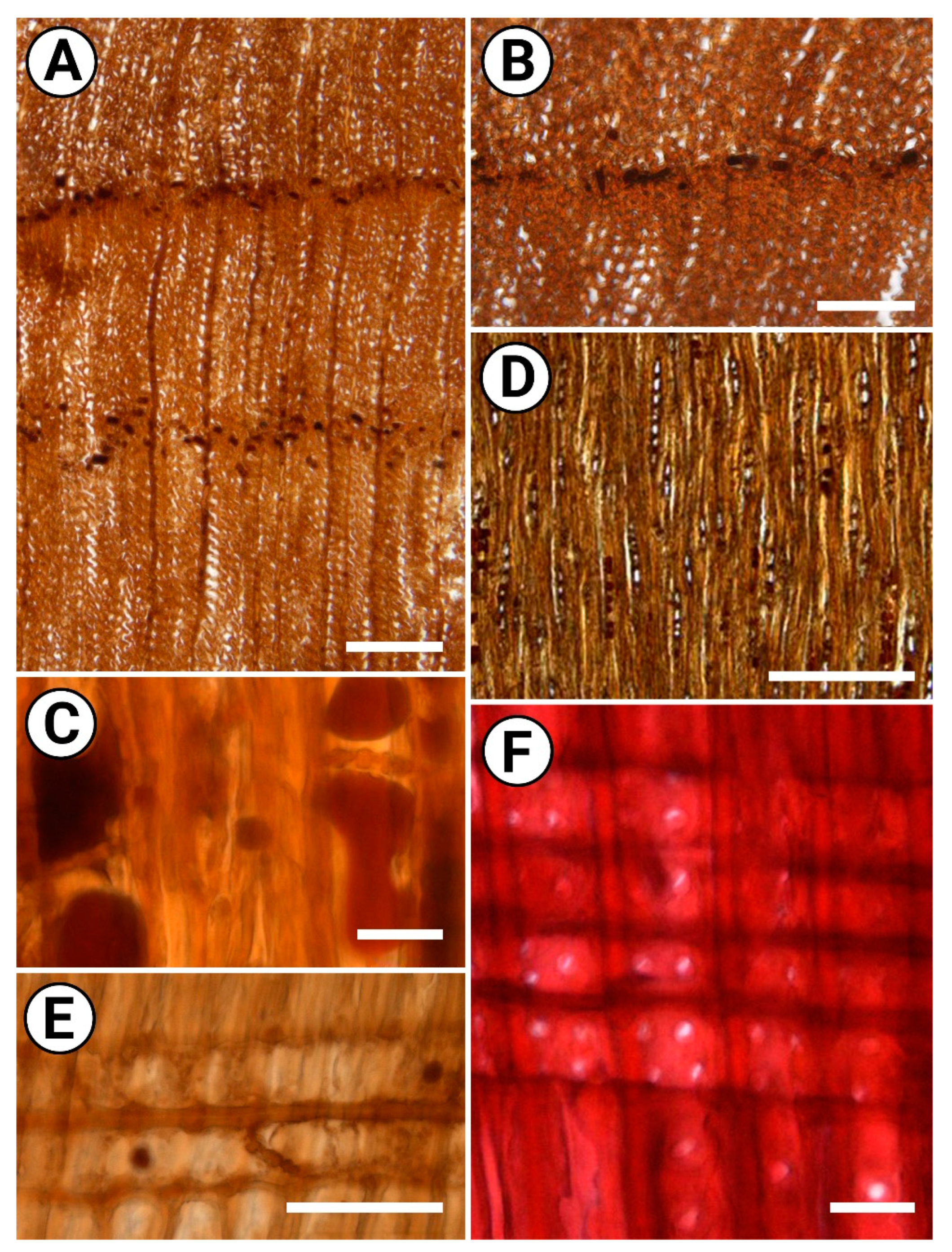
Growth rings are distinct, 0.19–1.60 mm wide; the transition from earlywood to latewood is gradual (Figure 2A). Earlywood tracheids are thin-walled (1.8–3.6 μm thick), polygonal to oval in outline, 13–31 μm (mean 22 μm) in tangential diameter. Latewood tracheids are thin- to moderately thick-walled (2.1–3.8 μm thick), circular to oval in outline, 12–24 μm (mean 11 μm) in tangential diameter. False growth rings occur. Normal and traumatic axial resin ducts are not found. The bordered pits on the radial tracheid walls (Figure 2F) are uniseriate, circular to oval in outline, with 5–9 μm in diameter and prominent tori. Small bordered pits (3–5 μm) also occur on the tangential walls of the tracheids. Warty layer, crassulae, helical and callitroid thickenings on the tracheid walls are not found.
The axial parenchyma is abundant, tangentially zonate (Figure 2A) and occasionally also marginal (Figure 2B); in strands of 2–4 cells with thickened pitted longitudinal walls and nodular transverse walls (Figure 2C), sometimes with distinct indentures. Dark deposits commonly occur in axial parenchyma cells (Figure 2C).
Rays are exclusively uniseriate (Figure 2D), completely composed of parenchyma cells (ray tracheids absent), 1–9 cells high (mean 3.8 cells); ray cells are 17–28 μm (mean 22.7 μm) in height. The horizontal walls of ray parenchyma cells are moderately thick and distinctly pitted (Figure 2E); the end walls of ray parenchyma cells are nodular, with distinct indentures. Cross-field pits are cupressoid (Figure 2F) and taxodioid, circular to oval, 4–6 μm in diameter of; cross-fields mostly have 2–4 pits (up to 5) pits on marginal portions of rays, and mostly 1–2 (up to 4) pits in central regions of rays. Radial resin ducts are not found.
Dark-stained compounds are common in axial parenchyma cells and occur in ray cells. Crystals are not found.

2.3. Comparison with Modern Woods

The fossil sample from Taman Peninsula represents a typical homoxylic wood, showing the tracheids bearing large (up to 24 µm in diameter) circular bordered pits on radial walls as well as exclusively uniseriate rays. This suite of traits occurs only in conifers. The InsideWood [17] search for the combination of distinct growth-ring boundaries (40p), absence of helical thickenings on tracheid walls (61a), tangentially zonate axial parenchyma (72p, 74p), absence of ray tracheids (80p), distinctly pitted end walls of ray parenchyma cells (86p), cupressoid cross-field pits (93p), average ray height < 4 cells (102p, 103a, 104a, 105a), absence of axial canals (109a), radial canals (110a) and traumatic canals (111a) returns Fitzroya cupressoides (Molina) I.M. Johnst. and eight speices of Juniperus (J. californica Carrière, J. chinensis L., J. drupacea Labill. J. excelsa M.Bieb., J. oxycedrus L., J. phoenicia L., J. rigida Siebold & Zucc., J. squamata Buch.-Ham. ex D.Don.). Among these species, the presence of marginal axial parenchyma has been reported only in J. drupacea [18], J. excelsa and J. oxycedrus [19,20]. J. drupacea and J. oxycedrus are distinctive, however, from the fossil wood from Taman Peninsula by larger pits on tangential tracheid walls (6–10 µm and 8–10 µm in diameter, respectively, vs. 3–5 µm in the sample under study) and also by the occurrence of higher (>10 cells in height, up to 18 cells in J. drupacea) rays. The latter species also differs from the wood sample under study by fewer (mostly 1–2) pits per cross-fields [21]. J. excelsa shows greater similarity with the Miocene wood, but it differs from the fossil wood in thinner horizontal walls of the ray cells, and the occasional occurrence of biseriate rays [19,20,21].
As the presence of marginal axial parenchyma is a prominent feature of the fossil sample from Taman Peninsula, which is uncommon in the wood of conifers, we compared it with other extant Juniperus species having this trait. Apart from J. drupacea, J. excelsa and J. oxycedrus, marginal axial parenchyma has been reported in J. conferta [18], J. monosperma (Engelm.) Sarg., J. thurifera L., J. scopulorum Sarg. and J. tibetica Kom. [19,20]. J. scopulorum and J. thurifera differ from the fossil wood from Taman Peninsula by the occurrence of larger pits (>10 µm in diameter) on tangential walls of tracheids [21]. J. thurifera is also distinctive by much more numerous rays (190–200 rays per mm2 vs. 93–117 rays per mm2, respectively), lower ray cells (10–18 µm vs. 17–28 µm in height, respectively), and the lack of indentures [19,21]. Unlike our fossil wood, J. conferta has mostly unpitted end walls of ray cells [18,21]. Then, J. scopulorum, J. monosperma, and J. tibetica share the occurrence of higher rays (>10 cells in height, up to 16 cells in J. scopulorum) than the fossil wood from Taman Peninsula. The latter two species and J. conferta are also distinctive from the sample under study in having fewer (up to three) pits on cross-fields [21].
In summary, our fossil wood belongs to the genus Juniperus, but it cannot be convincingly placed into any modern species that have been examined to date by wood anatomists. It shows the greatest similarity to the Mediterranean species J. excelsa, belonging to the section Sabina.

2.4. Comparison with Fossil Woods

The fossil homoxylic woods showing a combination of spaced arrangement of circular pits on radial tracheid walls, cupressoid cross-field pits, nodular transverse walls of axial parenchyma cells and usually also nodular walls of ray cells have been ascribed to the genera Juniperoxylon Houlbert [22,23] and Juniperus [20,24]. Juniperoxylon pottoniense (Stopes) Kräusel from the early Cretaceous of England [25] and the Eocene of Denmark [26] and J. wagneri Süss & Rathner from the Miocene of Germany [27] are distinctive, however, from other congeneric species and from the fossil wood under study in having smooth horizontal walls of ray cells. The latter species as well as other members of Juniperoxylon, including J. zamunerae Ruiz & Bodnar from the Middle Triassic of Argentina [23,28]; J. breviparenchmatosum Watari & Nishida from the Eocene of Hokkaido, Japan [29]; Juniperoxylon acarcae Akkemik, from the early Miocene of the central Turkey [20]; and four species from the Miocene deposits of Germany J. juniperoides (Kownas) Huard, J. pachyderma (Göppert) Kräusel, J. rhenanum Burgh from North Rhine-Westphalia [30] and J. schneiderianum Dolezych from Lusatia [31], differ from the fossil wood from the Taman Peninsula by the presence of biseriate pitting on the radial tracheid walls, and by the occurrence of biseriate rays. Finally, two fossil woods of Juniperus sp. from the early Miocene of the Galatean Volcanic Province, northwestern Turkey [24,32], show greatest resemblance to the studied wood sample, but both are distinctive in the lack of marginal axial parenchyma.
Overall, the mummified wood from the late Miocene deposits of the Taman Peninsula shows a close affinity to some extant species from the section Sabina of the genus Juniperus, as well as to the early Miocene woods from the northwestern Turkey assigned to the Juniperus sp. Although the studied sample is distinctive from any woods of modern or extinct junipers described to date, its anatomical traits are not sufficient for its reliable taxonomic attribution. Thus, we do not consider the fossil wood as a new species of this genus, but designate it as Juniperus sp. seemingly belonging to the section Sabina.

3. Discussion

The mummified wood of Juniperus sp. from the lower Maeotian sediments of Taman Peninsula is the first reliable macrofossil evidence of the section Sabina from Eurasia whose age is convincingly dated to the Miocene. The most ancient fossils of junipers attributed to this group have been described from the Eocene/Oligocene boundary of north Bohemia, the Czech Republic [7]. More recent Neogene record of the section Sabina includes three extinct species from the Oligocene to the middle Miocene of the North America [8,9,10,11] as well as the fossil twigs ascribed to the extant J. foetidissima from the Miocene–Pliocene boundary from the Sofia Basin, Bulgaria [12], and the seeds of J. sabina from Bashkortostan, Russia [13]. The mummified wood of Juniperus sp. from the Popov Kamen section shows that the species of this lineage occurred in the regions adjacent to Eastern Paratethys at least since the early Maeotian age.
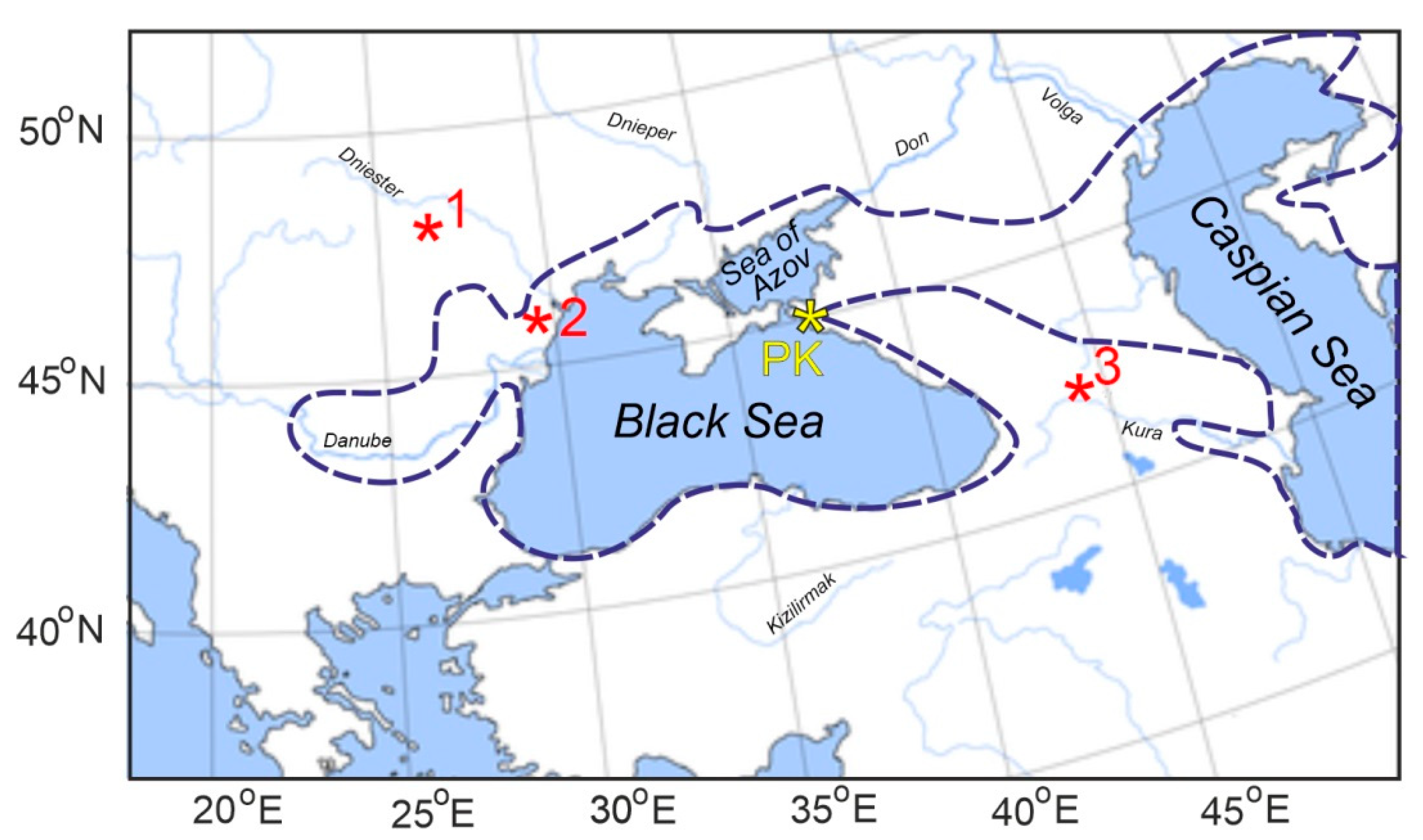
Obviously, the wood of Juniperus sp. has not been buried in situ, as it was found in relatively deep-water marine sediments. The only coeval occurrence of the pollen grains ascribed to Juniperus sp. has been reported from the lower Maeotian deposits of Odessa Oblast, southern Ukraine [33]. More ancient pollen evidence for this genus was found in the Sarmatian deposits of Kartli, eastern Georgia [34]. As for the macrofossils, the cone of J. bessarabica Negru has been described from the lower Sarmatian of Moldova [14] (Figure 3). This extinct species shows greatest affinity to the section Juniperus, i.e., to another lineage of junipers than the fossil wood under study attributed to the section Sabina. Cupressaceae have not been reported, however, in other pollen series studied in the Sarmatian and Maeotian deposits of the Eastern Paratethys regions, including those from the Taman Peninsula [35,36], the Lower Don [37], Bulgaria [38], Abkhazia [39] and several localities of Georgia [40]. No macrofossils of Juniperus have also been found in coeval paleofloras in southern Ukraine [33], Krasnodar Krai of Russia [41] and Georgia [40,42,43]. The available data suggest, therefore, that the wood of Juniperus sp. was likely transferred by sea current from the northwestern side of the Black Sea, which was a part of the Eastern Paratethys in the Miocene. Transportability of driftwoods over large distances has been supported by strong evidence [44].
The fossil wood of Juniperus sp. shows the greatest similarity to the extant Mediterranean species J. excelsa, belonging to a well-supported “clade IV” within the section Sabina [4]. This lineage also comprises the European species J. thurifera, the Asian J. chinensis and J. polycarpus, as well as J. procera from east Africa and south Arabia. As suggested by molecular dating [4], this lineage has been diversified during the Miocene. This estimation is consistent with the age of the fossil wood of Juniperus sp. from the Popov Kamen section. The reported fossil records of this group, up to now, are too sparse, however, for detailed reconstruction of its phytogeographic history.
Figure 3. Locations of the Popov Kamen section (PK, yellow asterisk) and coeval fossils of Juniperus from the regions adjacent to the Black Sea (red asterisks). The Paratethys area configuration at the Late Miocene is marked by dotted line. 1: The cone of Juniperus bessarabica Negru from the lower Sarmatian of Moldova [14]. 2: Pollen of Juniperus sp. from the lower Maeotian of southern Ukraine [33]. 3: Pollen of Juniperus sp. from the Sarmatian of eastern Georgia [34].
Plants 11 02050 g003

4. Materials and Methods

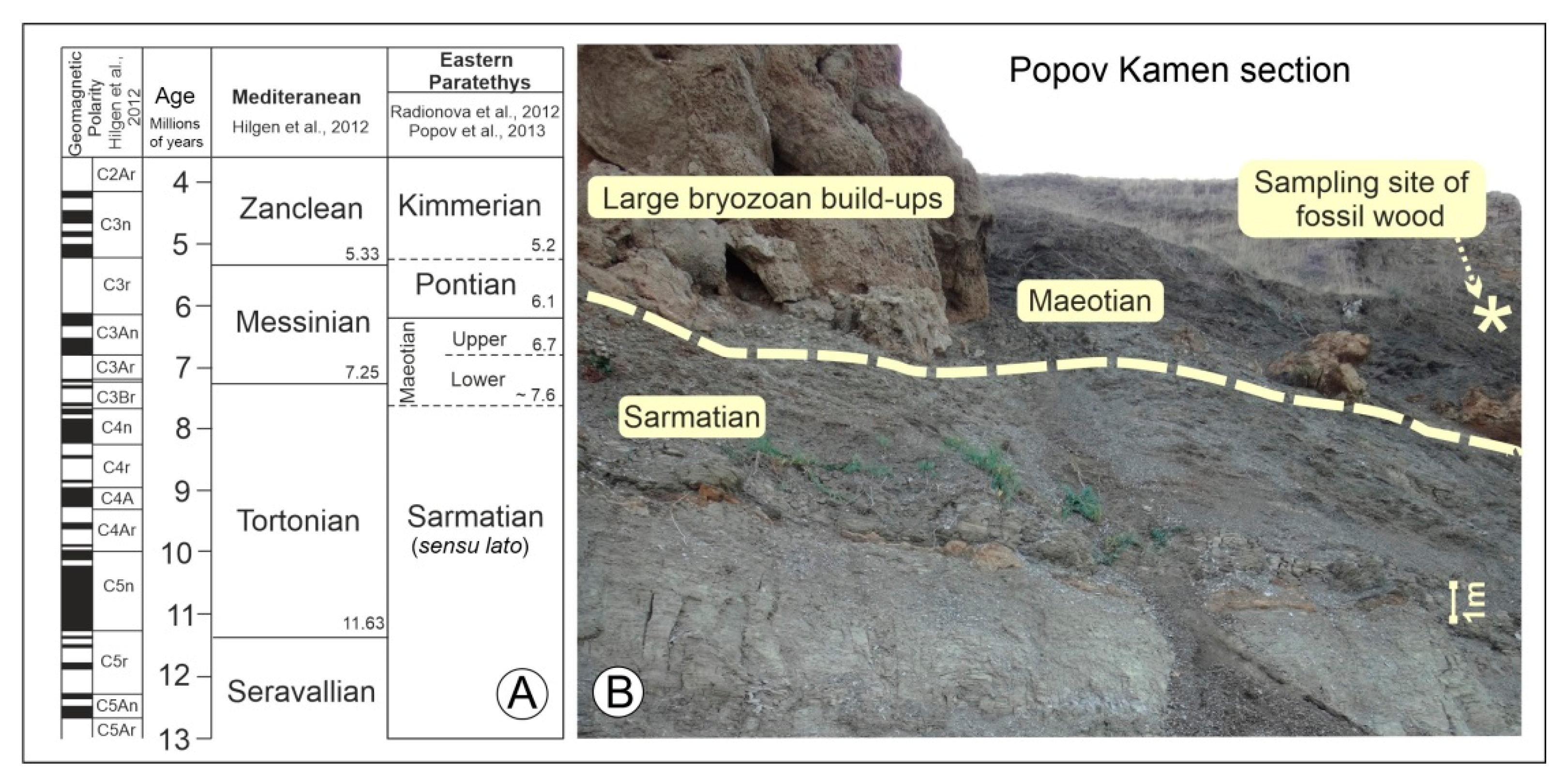
Three fragments of totally mummified portion of entire tree trunk, 23 cm in length and 7 cm in diameter, without any traces of organisms feeding (Figure 1), were collected from the Upper Miocene sediments of the Popov Kamen section (Figure 3 and Figure 4A,B). This trunk shows about 50 growth rings (some rings are hardly detectable) on its cross section.
Since its first description by Andrusov [45], this geological section has been extensively studied, using paleomagnetic, paleontological, and lithological methods of investigation [45,46,47,48,49,50,51,52]. The Popov Kamen section is located on the Black Sea coast of Taman Peninsula (45°16′01.8″ N, 36°61′97.6″ E, Russia) and comprises well-exposed Upper Sarmatian as well as Lower and Upper Maeotian sediments of the Eastern Paratethys. These sediments mainly represent clays with sporadic diatomite and limestone layers. The large bryozoan build-ups are located at the base of the Lower Maeotian, which directly underlie and overlie clays. The studied fossil wood was found in the clays laying 1.5–2 m above the top of the large bryozoan build-ups; these clays contain no fauna. The Lower Maeotian sediments of the Popov Kamen section accumulated in relatively deep-water environments (at depths of 50–75 m [46,47]). The Maeotian started with a marine transgression, which increased salinity back to 18‰ and flooded marginal areas of the Eastern Paratethys. At the Late Sarmatian, the Eastern Paratethys was mainly isolated. The sea level became unstable and a regression caused exposure of marginal parts of the basin, as well as a significant decrease in salinity down to 4–9‰ [53].
Figure 4. Geological setting of the mummified wood’s locality. (A) The time scale for Mediterranean and Eastern Paratethys (Black Sea). The ages of boundaries between the stages are indicated (millions of years). (B) The location of fossil woods; the boundary between the Sarmatian and Maeotian sediments was marked by yellow hashed line [50,51,52,54,55].
Plants 11 02050 g004
The fossil wood in this study was well-preserved by mummification. This specimen was processed and sectioned using the same methods as used for modern wood. The wood samples were boiled in water for about one hour and sectioned with a sledge microtome. Transverse, radial and tangential microtome sections of 20–30 µm in thickness were stained with alcian blue/safranin [56] or left unstained, dehydrated in gradient series of alcohol, and then mounted in Euparal. Then, the sections were examined with a light microscope (Olympus BX53). Wood anatomical measurements and anatomical terminology used for the descriptions in this paper follow the recommendations of the International Association of Wood Anatomists (IAWA) list of Microscopic Features for Softwood Identification [56]. The taxonomic position of fossil woods is determined by comparative work with similar modern and fossil softwood structures. This comparative work is based on modern wood slices and reference materials, particularly the computerized InsideWood database [57], in which we can search for similar modern softwoods.

Author Contributions

A.A.O. (Alexei A. Oskolski), A.A.O. (Anastasia A. Odintsova) and A.I.R., conception and initiation of the study; A.I.R. and Y.V.R., materials collecting; A.V.S., anatomical examination of fossil wood; A.A.O. (Alexei A. Oskolski), A.V.S., Y.V.R., A.A.O. (Anastasia A. Odintsova) and A.I.R., data interpretation, writing, manuscript preparation; A.I.R. and A.A.O. (Anastasia A. Odintsova), project administration. All authors have read and agreed to the published version of the manuscript.

Funding

This research was funded by the University of Johannesburg for A. Oskolski, by the Komarov Botanical Institute of the Russian Academy of Sciences (institutional research projects # AAAA-A17-117051810115-1 for A.V.S., and by budgetary funding of the Geophysical Center of RAS, adopted by the Ministry of Science and Higher Education of the Russian Federation (project 075-00764-22-00) for A. Odintsova and A.I.R.

Institutional Review Board Statement

Not applicable.

Informed Consent Statement

Not applicable.

Data Availability Statement

Not applicable.

Conflicts of Interest

The authors declare no conflict of interest.

References

  1. Adams, R.P. Junipers of the World: The Genus Juniperus, 4th ed.; Trafford Publishing: Victoria, Australia, 2014; pp. 1–422. [Google Scholar]
  2. Farjon, A.; Filer, D. An Atlas of the World’s Conifers: An Analysis of Their Distribution, Biogeography, Diversity and Conservation Status; Brill: Leiden, The Netherlands, 2013; pp. 1–524. [Google Scholar]
  3. Farjon, A. A Monograph of Cupressaceae and Sciadopitys; Royal Botanic Gardens: Kew, Australia, 2005; pp. 1–648. [Google Scholar]
  4. Mao, K.S.; Hao, G.; Liu, J.Q.; Adams, R.P.; Milne, R.I. Diversification and biogeography of Juniperus (Cupressaceae): Variable diversification rates and multiple intercontinental dispersals. New Phytol. 2010, 188, 254–272. [Google Scholar] [CrossRef] [PubMed]
  5. Leslie, A.B.; Beaulieu, J.; Holman, G.; Campbell, C.S.; Mei, W.B.; Raubeson, L.R.; Mathews, S. An overview of extant conifer evolution from the perspective of the fossil record. Am. J. Bot. 2018, 105, 1531–1544. [Google Scholar] [CrossRef] [PubMed]
  6. Axelrod, D.I. Evolution and biogeography of Madrean-Tethyan sclerophyll vegetation. Ann. Mo. Bot. Gard. 1975, 62, 280–334. [Google Scholar] [CrossRef]
  7. Kvaček, Z. A new juniper from the Palaeogene of Central Europe. Feddes Repert. 2002, 113, 492–502. [Google Scholar] [CrossRef]
  8. Axelrod, D.I. Mio-Pliocene Floras from West-Central Nevada; University California Press: Oakland, CA, USA, 1956; Volume 33, pp. 1–316. [Google Scholar]
  9. Axelrod, D.I. The Late Oligocene Creede Flora, Colorado; University California Press: Oakland, CA, USA, 1987; Volume 130, pp. 1–235. [Google Scholar]
  10. Axelrod, D.I. The Early Miocene Buffalo Canyon Flora of Western Nevada; University California Press: Oakland, CA, USA, 1991; Volume 135, pp. 1–76. [Google Scholar]
  11. Wolfe, J.A. Miocene Floras from Fingerrock Wash, Southwestern Nevada; US Government Printing Office: Washington, DC, USA, 1964; Volume 454–N, pp. 1–36.
  12. Palamarev, E.; Bozukov, V.; Uzunova, K.; Petkova, A.; Kitanov, G. Catalogue of the Cenozoic plants of Bulgaria (Eocene to Pliocene). Phytol. Balc. 2005, 11, 215–364. [Google Scholar]
  13. Dorofeev, P.I. On the Pliocene flora of Bashkiria. Botanicheskii Zhurnal 1962, 47, 787–801. (In Russian) [Google Scholar]
  14. Negru, A.G. Lower Sarmatian Flora of Northeastern Moldavia; Shtiintsa: Kishinev, Moldova, 1972; pp. 1–203. (In Russian) [Google Scholar]
  15. Palamarev, E. Paleobotanical evidences of the Tertiary history and origin of the Mediterranean sclerophyll dendroflora. Plant. Syst. Evol. 1989, 162, 93–107. [Google Scholar] [CrossRef]
  16. Kovar-Eder, J.; Kvaček, Z.; Martinetto, E.; Roiron, P. Late Miocene to Early Pliocene vegetation of southern Europe (7–4Ma) as reflected in the megafossil plant record. Palaeogeogr. Palaeoclim. Palaeoecol. 2006, 238, 321–339. [Google Scholar] [CrossRef]
  17. InsideWood. 2004–Onwards. Available online: http://insidewood.lib.ncsu.edu/ (accessed on 28 March 2022).
  18. Román-Jordán, E.; Esteban, L.G.; de Palacios, P.; Fernández, F.G. Comparative wood anatomy of the Cupressaceae and correspondence with phylogeny, with special reference to the monotypic taxa. Plant. Sys. Evol. 2017, 303, 203–219. [Google Scholar] [CrossRef]
  19. Esteban, L.G.; de Palacios, P.; Casasús, A.G.; Fernández, F.G. Characterisation of the xylem of 352 conifers. Investig. Agrar. Sist. Recur. For. 2004, 13, 152–178. [Google Scholar]
  20. Akkemik, Ü. A new species of Juniperoxylon from the Early Miocene of Northwestern Turkey. Acta Palaeontol. Pol. 2021, 17, 15–26. [Google Scholar] [CrossRef]
  21. Greguss, P. Xylotomische Bestimmung der heute Lebenden Gymnospermen; Akadémiai Kiadó: Budapest, Hungary, 1955; pp. 1–700. [Google Scholar]
  22. Philippe, M.; Bamford, M.K. A key to morphogenera used for Mesozoic conifer-like woods. Rev. Palaeobot. Palyno. 2008, 148, 184–207. [Google Scholar] [CrossRef]
  23. Ruiz, D.P.; Bodnar, J. The oldest record of Juniperoxylon, a cupressaceous fossil wood from the Middle Triassic of Argentina. Acta Palaeontol. Pol. 2019, 64, 481–488. [Google Scholar] [CrossRef] [Green Version]
  24. Akkemik, Ü.; Arslan, M.; Poole, I.; Tosun, S.; Köse, N.; Karlıoğlu Kılıç, N.; Aydın, A. Silicified woods from two previously undescribed early Miocene forest sites near Seben, northwest Turkey. Rev. Palaeobot. Palynol. 2016, 235, 31–50. [Google Scholar] [CrossRef]
  25. Stopes, M.C. Catalogue of the Mesozoic Plants in the British Museum (Natural History); Part II: Lower Greensand (Aptian) Plants of Britain; Trustees of British Museum: London, UK, 1915; pp. 1–360. [Google Scholar]
  26. Kräusel, R. Die fossilen Koniferen-Hölzer (Unter Ausschluß von Araucarioxylon Kraus). II: Kritische Untersuchungen zur Diagnostik lebender und fossiler Koniferen-Hölzer. Palaeontogr. B 1949, 89, 83–203. [Google Scholar]
  27. Süss, H.; Rathner, U. Ein neues fossiles Holz, Juniperoxylon wagneri sp. nova, aus der miozänen Braunkohle von Wetro (Oberlausitdsachsen, Deutschland). Feddes Repert. 1998, 109, 15–24. [Google Scholar] [CrossRef]
  28. Bodnar, J.; Ruiz, D.P.; Artabe, A.E.; Morel, E.M.; Ganuza, D. Voltziales y Pinales (=Coniferales) de la Formación Cortaderita (Triásico Medio), Argentina, y su implicancia en la reconstrucción de las coníferas triásicas. Rev. Bras. Paleontol. 2015, 18, 141–160. [Google Scholar] [CrossRef]
  29. Watari, S.; Nishida, M. Juniperoxylon from the Tertiary of Hokkaido. J. Jpn. Bot. 1973, 48, 154–159. [Google Scholar]
  30. Van der Burgh, J. Hölzer der niederrheinischen Braunkohlen formation, 2. Hölzer der Braunkohlengruben “Maria Theresia” zu Herzogenrath, “Zukunft West” zu Eschweiler und “Victor” (Zülpich Mitte) zu Zülpich. Nebst einer systematisch– anatomischen Bearbeitung der Gattung pinus L. Rev. Palaeobot. Palynol. 1973, 15, 73–275. [Google Scholar]
  31. Dolezych, M. A remarkable extinct wood from Lusatia (central Europe)—Juniperoxylon schneiderianum sp. nov. with affinity to Cupressospermum saxonicum Mai. Palaeontographica B 2016, 295, 5–31. [Google Scholar] [CrossRef]
  32. Acarca Bayam, N.N.; Akkemik, Ü.; Poole, I.; Akarsu, F. Further Contributions to the early Miocene forest vegetation of the Galatean Volcanic Province, Turkey. Palaeobot. Electron. 2018, 21, 1–42. [Google Scholar]
  33. Shchekina, N.A. History of Flora and Vegetation of the Southern European Part of the USSR in the Late Miocene and Early Pliocene; Naukova Dumka: Kyiv, Ukraine, 1979; pp. 1–198. (In Russian) [Google Scholar]
  34. Shatilova, I.; Maissuradze, L.; Koiava, K.; Mchedlishvili, N.; Rukhadze, L.; Spezzaferri, S.; Strasser, A. Foraminifers and palynomorphs in the Sarmatian deposits of Kartli (Eastern Georgia): Stratigraphical and palaeoclimatological implications. Proc. Georgian Acad. Sci. Biol. Ser. B 2008, 6, 65–76. [Google Scholar]
  35. Philippova, N.J. Spores, pollen and organic-walled phytoplankton from Neogene deposits of the Zheleznyi Rog Key Section (Taman Peninsula). Stratigr. Geol. Correl. 2002, 10, 80–92. (In Russian) [Google Scholar]
  36. Razumkova, E.S. Palynological record of the Sarmatian deposits of the Eastern Paratethys (Section Zelenskii Mountain-Cape Panagiya, Taman Peninsula). Stratigr. Geol. Correl. 2012, 20, 108–119. (In Russian) [Google Scholar] [CrossRef]
  37. Ananova, E.N. Pollen in the Neogene Deposits of the Southern Part of Russian Plain; Leningrad State University: Leningrad, Russia, 1974; pp. 1–196. (In Russian) [Google Scholar]
  38. Ivanov, D. Climate and vegetation change during the late Miocene in southwest Bulgaria based on pollen data from the Sandanski Basin. Rev. Palaeobot. Palynol. 2015, 221, 128–137. [Google Scholar] [CrossRef]
  39. Shatilova, I.I.; Maissuradze, L.S.; Kokolashvili, I.M.; Bruch, A.A. The palaeobiological basis of the stratigraphcal subdivision of Meotian deposits of Abkhazia. Bull. Georgian Natl. Acad. Sci. 2019, 13, 118–125. [Google Scholar]
  40. Shatilova, I.I.; Maissuradze, L.S.; Koiava, K.P.; Kokolashvili, I.M.; Bukhsianidze, M.G.; Bruch, A.A. The Environmental History of Georgia During the Late Miocene Based of Foraminifera and Pollen; Georgian National Museum: Tbilisi, Georgia, 2020; pp. 1–84. [Google Scholar]
  41. Kutuzkina, E.F. The Sarmatian flora of Armavir. Paleobotanika 1964, 5, 148–229. (In Russian) [Google Scholar]
  42. Uznadze, M.D. Neogene Flora of Georgia; Metsniereba: Tbilisi, Georgia, 1965; pp. 1–180. (In Russian) [Google Scholar]
  43. Uznadze, M.D.; Tsagareli, E.A. The Sarmatian Flora of the Dzindza River Ravine (Goderdzi Flora); Metsniereba: Tbilisi, Georgia, 1979; pp. 1–165. (In Russian) [Google Scholar]
  44. Murphy, E.; Nistor, I.; Cornett, A.; Wilson, J.; Pilechi, A. Fate and transport of coastal driftwood: A critical review. Mar. Pollut. Bull. 2021, 170, 112649. [Google Scholar] [CrossRef]
  45. Andrusov, N. Geological researches at the Taman Peninsula. Mater. Geol. Russ. 1903, 21, 257–383. (In Russian) [Google Scholar]
  46. Popov, S.V.; Zastrozhnov, A. Neogene Key Sections of the Eastern Paratethys (Taman Peninsula); Tour Guide: Volgograd-Taman, Russia, 1998; pp. 1–20. (In Russian) [Google Scholar]
  47. Rostovtseva, Y.V.; Goncharova, I. The structure of relatively deep-water sediments of the Lower Maeotian Black Sea region (Taman Peninsula: Section Popov Kamen), Biostratigraphic basis for the development of stratigraphic patterns in Ukraine. Proc. Inst. Geol. Sci. NAS Ukr. 2008, 4, 270–275. (In Russian) [Google Scholar]
  48. Rostovtseva, Y.V. Lower Maeotian facies of the Taman trough. Lithol. Miner. Resour. 2009, 44, 451–464. [Google Scholar] [CrossRef]
  49. Radionova, E.P.; Golovina, L.A. Upper Maeotian–Lower Pontian “Transitional Strata” in the Taman Peninsula: Stratigraphic position and paleogeographic interpretations. Geol. Carpath. 2011, 2, 62–100. [Google Scholar] [CrossRef] [Green Version]
  50. Trubikhin, V.M.; Pilipenko, O.V. Rock magnetism and paleomagnetism of Maeotian deposits of the Popov Kamen Reference Section (Taman Peninsula). Izvestiya. Phys. Solid Earth 2011, 47, 233–245. (In Russian) [Google Scholar] [CrossRef]
  51. Radionova, E.P.; Golovina, L.A.; Filippova, N.Y.; Trubikhin, V.M.; Popov, S.V.; Goncharova, I.A.; Vernigorova, Y.V.; Pinchuk, T.N. Middle–Upper Miocene stratigraphy of the Taman Peninsula, Eastern Paratethys. Cent. Eur. J. Geosci. 2012, 4, 188–204. [Google Scholar] [CrossRef]
  52. Popov, S.V.; Akhmetiev, M.A.; Golovina, L.A.; Goncharova, I.A.; Radionova, E.P.; Filippova, N.Y.; Trubichin, V.M. Neogene regiostage stratigraphic scale of the South Russia: Current state and perspectives. In General Stratigraphic Scale of Russia: Current State and Ways of Perfection; Fedonkin, M.A., Ed.; Geological Institute of RAS: Moscow, Russia, 2013; pp. 356–360. (In Russian) [Google Scholar]
  53. Iljina, L.B.; Nevesskaya, L.A.; Paramonova, N.P. Patterns of the Mollusks Development in Brackish Sea Basins of Eurasia; Nedra: Moscow, Russia, 1976; pp. 1–288. (In Russian) [Google Scholar]
  54. Hilgen, F.J.; Lourens, L.J.; Van Dam, J.A. The Neogene Period. In The Geological Time Scale 2012; Gradstein, F., Ogg, J., Schmitz, M., Ogg, G., Eds.; Cambridge University Press: Cambridge, UK, 2012; pp. 923–979. [Google Scholar]
  55. Popov, S.V.; Rostovtseve, Y.V.; Fillippova, N.Y.; Golovina, L.A.; Radionova, E.P.; Vernyhorvoa, Y.V.; Dykan, N.I.; Pinchuk, T.N.; Iljina, L.B.; Koromyslova, A.V.; et al. Paleontology and stratigraphy of the Middle-Upper Miocene of the Taman Peninsula: Part 1. Description of key sections and benthic fossil groups. Paleontol. J. 2016, 50, 1039–1206. [Google Scholar] [CrossRef]
  56. Jansen, S.; Choat, B.; Vinckier, S.; Lens, F.; Schols, P.; Smets, E. Intervascular pit membranes with a torus in the wood of Ulmus (Ulmaceae) and related genera. New Phytol. 2004, 163, 51–59. [Google Scholar] [CrossRef] [PubMed] [Green Version]
  57. IAWA Committee. IAWA List of microscopic features for softwood identification. IAWA J. 2004, 25, 1–70. [Google Scholar] [CrossRef]
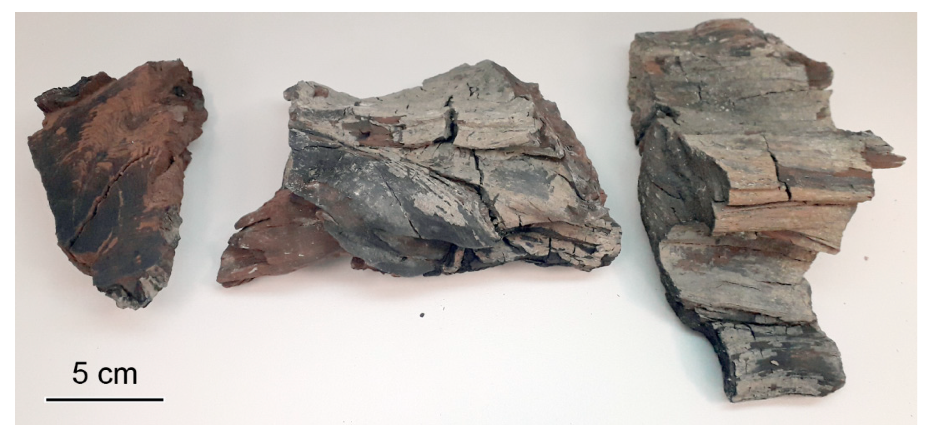
Figure 1. Fragments of fossil woody stem examined in the present study.
Plants 11 02050 g001
Figure 2. Wood structure of Juniperus sp., light microscopy. (A) Transverse section (TS), distinct growth-ring boundary, gradual transition from earlywood to latewood, tangentially zonate axial parenchyma. (B) TS, distinct boundary of growth ring, marginal axial parenchyma. (C) Tangential longitudinal section (TLS), portions of axial parenchyma strands, nodular transverse wall, dark deposits in axial parenchyma cells. (D) TLS, exclusively uniseriate low rays (up to 8 cells in height). (E) Radial longitudinal section (RLS), ray cells with thickened pitted horizontal walls and nodular end walls. (F) RLS, bordered pits on radial tracheid walls, cross-fields with 2–4 cupressoid pits. Scale bars: 200 µm for (A,D), 100 µm for (B), 50 µm for (C,E), 20 µm for (F).
Plants 11 02050 g002
Publisher’s Note: MDPI stays neutral with regard to jurisdictional claims in published maps and institutional affiliations.