
The In Vitro and In Vivo Synergistic Antimicrobial Activity Assessment of Vacuum Microwave Assisted Aqueous Extracts from Pomegranate and Avocado Fruit Peels and Avocado Seeds Based on a Mixtures Design Model

 ,
,  ,
, 

Abstract
:
1. Introduction
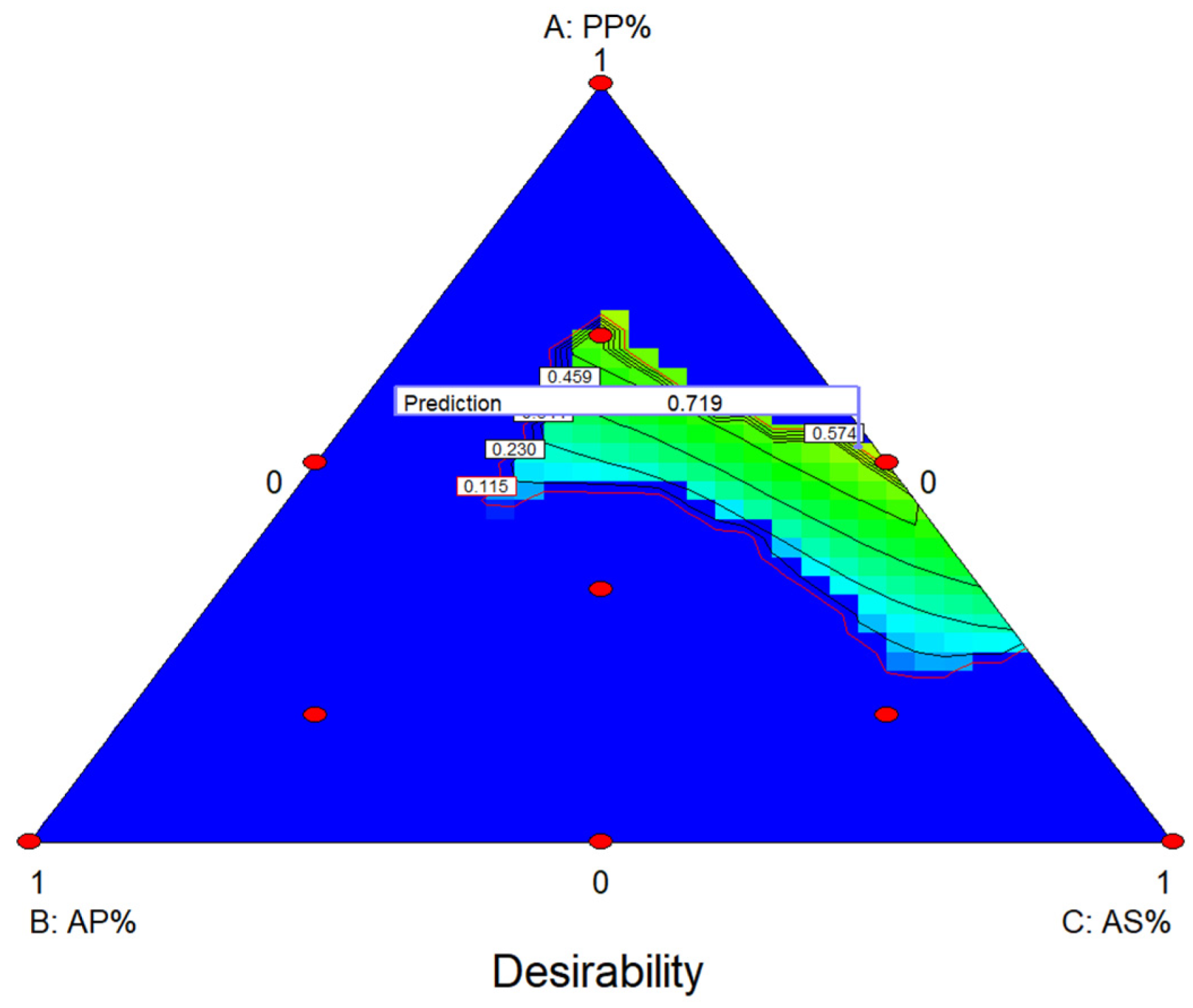
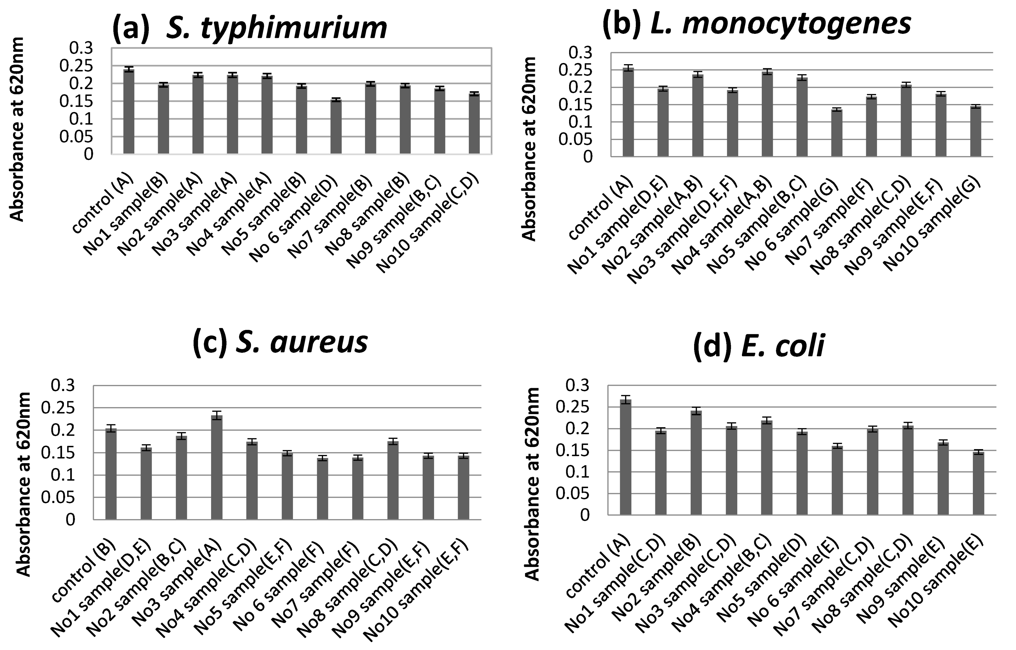
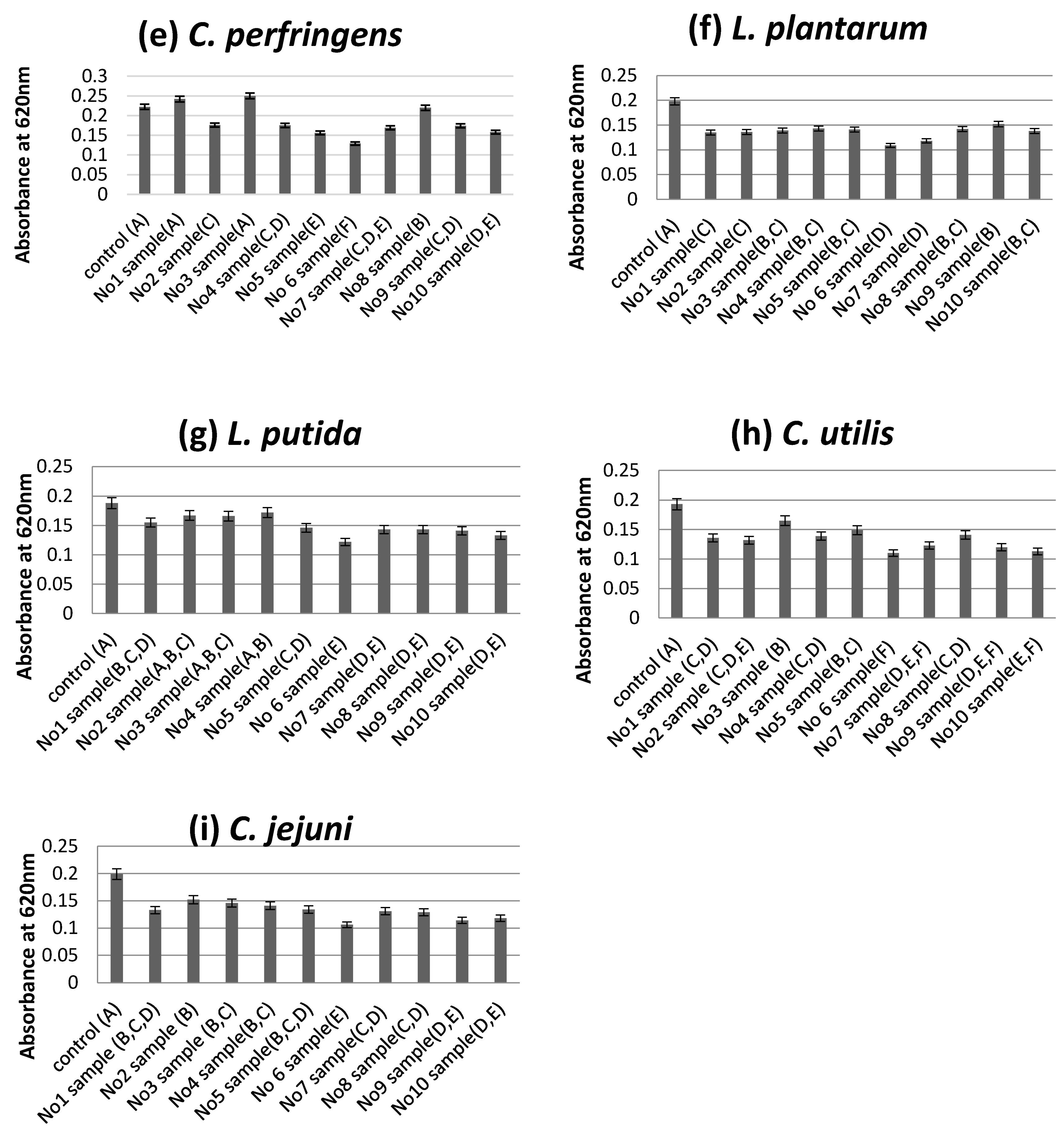
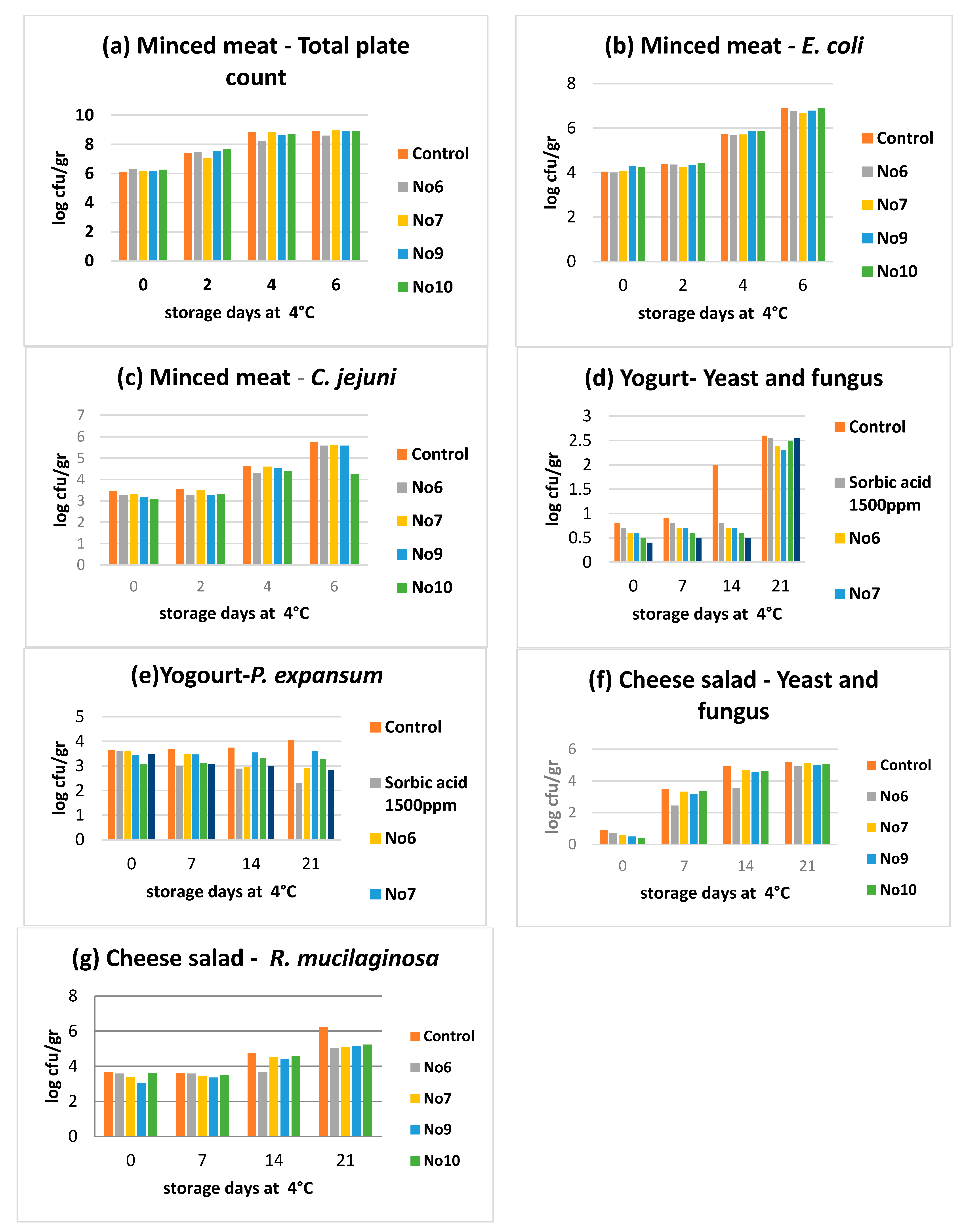
2. Results
3. Discussion
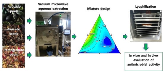
4. Materials and Methods
4.1. Plant Materials
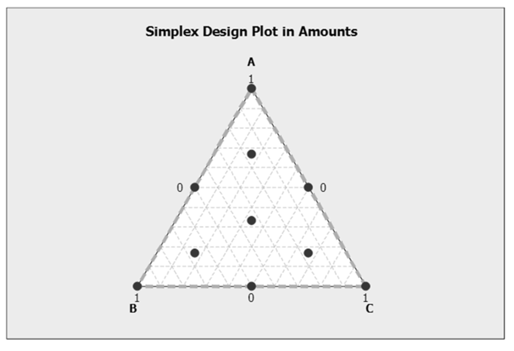
4.2. Mixture Design and Antimicrobial Extracts/Samples Composition and Coding
4.3. Encapsulation and Lyophilisation of the Extracted Pomegranate and Avocado Peel and Seed Material Mixtures
4.4. Chemicals
4.5. Determination of Total Phenols Content (TPC) of the Extracts
4.6. Determination of Antioxidant Capacity of the Samples (DPPH• Method)
4.7. Microorganisms Used for the Determination of Antimicrobial Activity
4.8. Antimicrobial Assays
4.8.1. Well Diffusion Assay
4.8.2. MIC/MBC Assay
4.8.3. Optical Density Assay
4.8.4. Preparation of Foods Used in the Food Model
4.9. Statistical Analysis
5. Conclusions
Supplementary Materials
Author Contributions
Funding
Institutional Review Board Statement
Informed Consent Statement
Acknowledgments
Conflicts of Interest
References
- Castro-Rosas, J.; Ferreira-Grosso, C.R.; Gómez-Aldapa, C.A.; Rangel-Vargas, E.; Rodríguez-Marín, M.L.; Guzmán-Ortiz, F.A.; Falfan-Cortes, R.N. Recent Advances in Microencapsulation of Natural Sources of Antimicrobial Compounds Used in Food—A Review. Food Res. Int. 2017, 102, 575–587. [Google Scholar] [CrossRef]
- Protti, M.; Gualandi, I.; Mandrioli, R.; Zappoli, S.; Tonelli, D.; Mercolini, L. Journal of Pharmaceutical and Biomedical Analysis Analytical Profiling of Selected Antioxidants and Total Antioxidant Capacity of Goji (Lycium spp.) Berries. J. Pharm. Biomed. Anal. 2017, 143, 252–260. [Google Scholar] [CrossRef]
- Bhuyan, D.J.; Alsherbiny, M.A.; Perera, S.; Low, M.; Basu, A.; Devi, O.A.; Barooah, M.S.; Li, C.G.; Papoutsis, K. The Odyssey of Bioactive Compounds in Avocado (Persea americana) and Their Health Benefits. Antioxidants 2019, 8, 426. [Google Scholar] [CrossRef] [PubMed] [Green Version]
- Skenderidis, P.; Mitsagga, C.; Giavasis, I.; Petrotos, K.; Lampakis, D.; Leontopoulos, S.; Hadjichristodoulou, C.; Tsakalof, A. The in vitro Antimicrobial Activity Assessment of Ultrasound Assisted Lycium barbarum Fruit Extracts and Pomegranate Fruit Peels. J. Food Meas. Charact. 2019, 13, 2017–2031. [Google Scholar] [CrossRef]
- Rodrigues, L.A.; da Silva, M.L.C.P.; Alvarez-Mendes, M.O.; dos Reis Coutinho, A.; Thim, G.P. Phenol Removal from Aqueous Solution by Activated Carbon Produced from Avocado Kernel Seeds. Chem. Eng. J. 2011, 174, 49–57. [Google Scholar] [CrossRef]
- Rajha, H.N.; Mhanna, T.; El Kantar, S.; El Khoury, A.; Louka, N.; Maroun, R.G. Innovative Process of Polyphenol Recovery from Pomegranate Peels by Combining Green Deep Eutectic Solvents and a New Infrared Technology. LWT 2019, 111, 138–146. [Google Scholar] [CrossRef]
- Tan, C.X. Virgin Avocado Oil: An Emerging Source of Functional Fruit Oil. J. Funct. Foods 2019, 54, 381–392. [Google Scholar] [CrossRef]
- Cowan, A.K.; Wolstenholme, B.N. Avocado. In Encyclopedia of Foods and Health, 1st ed.; Caballero, B., Finglas, P.M., Toldrá, F.B.T., Eds.; Academic Press: Oxford, UK, 2016; pp. 294–300. ISBN 978-0-12-384953-3. [Google Scholar]
- Dreher, M.L.; Davenport, A.J.; Dolci, G.; Catenacci, A.; Malpei, F.; Grosso, M. Hass Avocado Composition and Potential Health Effects. Crit. Rev. Food Sci. Nutr. 2013, 53, 738–750. [Google Scholar] [CrossRef] [PubMed] [Green Version]
- Zafar, T.; Sidhu, J.S. Avocado: Production, Quality, and Major Processed Products. In Handbook of Vegetables and Vegetable Processing; Sinha, N.K., Ed.; Blackwell Publishing Ltd: Hoboken, NJ, USA, 2011; pp. 525–543. [Google Scholar]
- Duarte, P.F.; Chaves, M.A.; Borges, C.D.; Mendonça, C.R.B. Avocado: Characteristics, Health Benefits, and Uses. Int. News Fats Oils Relat. Mater. 2017, 28, 28–32. [Google Scholar] [CrossRef] [Green Version]
- Rodríguez-Carpena, J.G.; Morcuende, D.; Estévez, M. Avocado By-Products as Inhibitors of Color Deterioration and Lipid and Protein Oxidation in Raw Porcine Patties Subjected to Chilled Storage. Meat Sci. 2011, 89, 166–173. [Google Scholar] [CrossRef] [PubMed]
- Ranade, S.S.; Thiagarajan, P. A Review on Persea americana Mill. (Avocado)- Its Fruit and Oil. Int. J. PharmTech Res. 2015, 8, 72–77. [Google Scholar]
- Melgar, B.; Dias, M.I.; Ciric, A.; Sokovic, M.; Garcia-Castello, E.M.; Rodriguez-Lopez, A.D.; Barros, L.; Ferreira, I.C.R.F. Bioactive Characterization of Persea americana Mill. by-Products: A Rich Source of Inherent Antioxidants. Ind. Crop. Prod. 2018, 111, 212–218. [Google Scholar] [CrossRef] [Green Version]
- Araújo, R.G.; Rodriguez-Jasso, R.M.; Ruiz, H.A.; Pintado, M.M.E.; Aguilar, C.N. Avocado By-Products: Nutritional and Functional Properties. Trends Food Sci. Technol. 2018, 80, 51–60. [Google Scholar] [CrossRef]
- Olaeta, J.A.; Schwartz, M.; Undurraga, P.; Contreras, S. Use of Hass Avocado (Persea americana Mill.) Seed as a Processed Product. In Proceedings of the VI World Avocado Congress, Viña Del Mar, Chile, 12–16 November 2007; pp. 1–8. [Google Scholar]
- Oelrichs, P.B.; Ng, J.C.; Seawright, A.A.; Ward, A.; Schäffeler, L.; Macleod, J.K. Isolation and Identification of a Compound from Avocado (Persea americana) Leaves Which Causes Necrosis of the Acinar Epithelium of the Lactating Mammary Gland and the Myocardium. Nat. Toxins 1995, 3, 344–349. [Google Scholar] [CrossRef] [PubMed]
- Celik, I.; Temur, A.; Isik, I.; John, K.M.M.; Bhagwat, A.A.; Luthria, D.L.; Fang, Z.; Bhandari, B.; Hemaiswarya, S.; Kruthiventi, A.K. Hepatoprotective Role and Antioxidant Capacity of Pomegranate (Punica granatum) Flowers Infusion against Trichloroacetic Acid-Exposed in Rats. Phytomedicine 2013, 21, 639–652. [Google Scholar] [CrossRef] [PubMed]
- John, K.M.M.; Bhagwat, A.A.; Luthria, D.L. Swarm Motility Inhibitory and Antioxidant Activities of Pomegranate Peel Processed under Three Drying Conditions. Food Chem. 2017, 235, 145–153. [Google Scholar] [CrossRef]
- Li, Y.; Guo, C.; Yang, J.; Wei, J.; Xu, J.; Cheng, S. Evaluation of Antioxidant Properties of Pomegranate Peel Extract in Comparison with Pomegranate Pulp Extract. Food Chem. 2006, 96, 254–260. [Google Scholar] [CrossRef]
- Sood, A.; Gupta, M. Extraction Process Optimization for Bioactive Compounds in Pomegranate Peel. Food Biosci. 2015, 12, 100–106. [Google Scholar] [CrossRef]
- Kalaycıoğlu, Z.; Erim, F.B. Total Phenolic Contents, Antioxidant Activities, and Bioactive Ingredients of Juices from Pomegranate Cultivars Worldwide. Food Chem. 2017, 221, 496–507. [Google Scholar] [CrossRef]
- Fischer, U.A.; Carle, R.; Kammerer, D.R. Identification and Quantification of Phenolic Compounds from Pomegranate (Punica granatum L.) Peel, Mesocarp, Aril and Differently Produced Juices by HPLC-DAD-ESI/MS(n). Food Chem. 2011, 127, 807–821. [Google Scholar] [CrossRef]
- Lampakis, D.; Skenderidis, P.; Leontopoulos, S. Technologies and Extraction Methods of Polyphenolic Compounds Derived from Pomegranate (Punica granatum) Peels. A Mini Review. Processes 2021, 9, 236. [Google Scholar] [CrossRef]
- Skenderidis, P.; Leontopoulos, S.; Petrotos, K.; Giavasis, I. Optimization of Vacuum Microwave-Assisted Extraction of Pomegranate Fruits Peels by the Evaluation of Extracts’ Phenolic Content and Antioxidant Activity. Foods 2020, 9, 1655. [Google Scholar] [CrossRef]
- Skenderidis, P.; Leontopoulos, S.; Petrotos, K.; Giavasis, I. Vacuum Microwave-Assisted Aqueous Extraction of Polyphenolic Compounds from Avocado (Persea americana) Solid Waste. Sustainability 2021, 13, 2166. [Google Scholar] [CrossRef]
- Munin, A.; Edwards-Lévy, F. Encapsulation of Natural Polyphenolic Compounds; a Review. Pharmaceutics 2011, 3, 793–829. [Google Scholar] [CrossRef] [Green Version]
- Petrotos, K.B.; Karkanta, F.K.; Gkoutsidis, P.E.; Giavasis, I.; Papatheodorou, N.; Ntontos, A.C. Production of Novel Bioactive Yogurt Enriched with Olive Fruit Polyphenols. Int. J. Nutr. Food Eng. 2012, 6, 170–175. [Google Scholar]
- Fang, Z.; Bhandari, B. Encapsulation of Polyphenols—A Review. Trends Food Sci. Technol. 2010, 21, 510–523. [Google Scholar] [CrossRef]
- Robert, P.; Fredes, C. The Encapsulation of Anthocyanins from Berry-Type Fruits. Trends in Foods. Molecules 2015, 20, 5875–5888. [Google Scholar] [CrossRef]
- Hemaiswarya, S.; Kruthiventi, A.K.; Doble, M. Synergism between Natural Products and Antibiotics against Infectious Diseases. Phytomedicine 2008, 15, 639–652. [Google Scholar] [CrossRef]
- Lee, C.-C.; Shen, S.-R.; Lai, Y.-J.; Wu, S.-C. Rutin and Quercetin, Bioactive Compounds from Tartary Buckwheat, Prevent Liver Inflammatory Injury. Food Funct. 2013, 4, 794–802. [Google Scholar] [CrossRef]
- Ku, S.K.; Seo, B.I.; Park, J.H.; Park, G.Y.; Seo, Y.B.; Kim, J.S.; Lee, H.S.; Roh, S.S. Effect of Lonicerae Flos Extracts on Reflux Esophagitis with Antioxidant Activity. World J. Gastroenterol. 2009, 15, 4799–4805. [Google Scholar] [CrossRef]
- Calderón-Oliver, M.; Escalona-Buendía, H.B.; Medina-Campos, O.N.; Pedraza-Chaverri, J.; Pedroza-Islas, R.; Ponce-Alquicira, E. Optimization of the Antioxidant and Antimicrobial Response of the Combined Effect of Nisin and Avocado Byproducts. LWT Food Sci. Technol. 2016, 65, 46–52. [Google Scholar] [CrossRef]
- Rodríguez-Carpena, J.G.; Morcuende, D.; Andrade, M.J.; Kylli, P.; Estevez, M. Avocado (Persea americana Mill.) Phenolics, in vitro Antioxidant and Antimicrobial Activities, and Inhibition of Lipid and Protein Oxidation in Porcine Patties. J. Agric. Food Chem. 2011, 59, 5625–5635. [Google Scholar] [CrossRef]
- Alexandre, E.M.C.; Silva, S.; Santos, S.A.O.; Silvestre, A.J.D.; Duarte, M.F.; Saraiva, J.A.; Pintado, M. Antimicrobial Activity of Pomegranate Peel Extracts Performed by High Pressure and Enzymatic Assisted Extraction. Food Res. Int. 2019, 115, 167–176. [Google Scholar] [CrossRef] [PubMed] [Green Version]
- Debnath, S.; Habibur Rahman, S.M.; Deshmukh, G.; Duganath, N.; Pranitha, C.; Chiranjeevi, A. Antimicrobial screening of various fruit seed extracts. Pharmacogn. J. 2011, 3, 83–86. [Google Scholar] [CrossRef] [Green Version]
- Skenderidis, P.; Petrotos, K.; Leontopoulos, S. Functional Properties of Goji Berry (Lycium barbarum) Fruit Extracts. In Phytochemicals in Goji Berries; CRC Press: Boca Raton, FL, USA, 2020; pp. 181–224. ISBN 9780429021749. [Google Scholar]
- Daglia, M. Polyphenols as Antimicrobial Agents. Curr. Opin. Biotechnol. 2012, 23, 174–181. [Google Scholar] [CrossRef] [PubMed]
- Torres, E.; García, A.; Aranda, M.; Saéz, V.; Zúñiga, F.; Alarcón, J.; Avello, M.; Pastene, E. One step purification of two semi-synthetic epicatechin adducts prepared from avocado peels procyanidins by centrifugal partition chromatography and evaluation of their anti-inflammatory effects on adenocarcinoma gastric cells infected with with Helicobacter pylori. J. Chil. Chem. Soc. 2018, 63, 4222–4228. [Google Scholar] [CrossRef] [Green Version]
- Segovia, F.J.; Corral-Pérez, J.J.; Almajano, M.P. Avocado Seed: Modeling Extraction of Bioactive Compounds. Ind. Crop. Prod. 2016, 85, 213–220. [Google Scholar] [CrossRef]
- Dixon, R.A.; Xie, D.-Y.; Sharma, S.B. Proanthocyanidins—A Final Frontier in Flavonoid Research? New Phytol. 2005, 165, 9–28. [Google Scholar] [CrossRef] [Green Version]
- Kähkönen, M.P.; Hopia, A.I.; Heinonen, M. Berry Phenolics and Their Antioxidant Activity. J. Agric. Food Chem. 2001, 49, 4076–4082. [Google Scholar] [CrossRef]
- Puupponen-Pimiä, R.; Nohynek, L.; Meier, C.; Kähkönen, M.; Heinonen, M.; Hopia, A.; Oksman-Caldentey, K.-M. Antimicrobial Properties of Phenolic Compounds from Berries. J. Appl. Microbiol. 2001, 90, 494–507. [Google Scholar] [CrossRef]
- Kanatt, S.R.; Chander, R.; Sharma, A. Antioxidant and Antimicrobial Activity of Pomegranate Peel Extract Improves the Shelf Life of Chicken Products. Int. J. Food Sci. Technol. 2010, 45, 216–222. [Google Scholar] [CrossRef]
- Agourram, A.; Ghirardello, D.; Rantsiou, K.; Zeppa, G.; Belviso, S.; Romane, A.; Oufdou, K.; Giordano, M. Phenolic Content, Antioxidant Potential, and Antimicrobial Activities of Fruit and Vegetable by-Product Extracts. Int. J. Food Prop. 2013, 16, 1092–1104. [Google Scholar] [CrossRef]
- Li, G.; Xu, Y.; Wang, X.; Zhang, B.; Shi, C.; Zhang, W.; Xia, X. Tannin-Rich Fraction from Pomegranate Rind Damages Membrane of Listeria monocytogenes. Foodborne Pathog. Dis. 2014, 11, 313–319. [Google Scholar] [CrossRef]
- Xu, Y.; Li, G.; Zhang, B.; Wu, Q.; Wang, X.I.N.; Xia, X. Tannin-Rich Pomegranate Rind Extracts Reduce Adhesion to and Invasion of Caco-2 Cells by Listeria Monocytogenes and Decrease Its Expression of Virulence Genes. J. Food Prot. 2015, 78, 128–133. [Google Scholar] [CrossRef]
- Cushnie, T.P.T.; Hamilton, V.E.S.; Chapman, D.G.; Taylor, P.W.; Lamb, A.J. Aggregation of Staphylococcus aureus Following Treatment with the Antibacterial Flavonol Galangin. J. Appl. Microbiol. 2007, 103, 1562–1567. [Google Scholar] [CrossRef] [PubMed] [Green Version]
- Scheffé, H. Experiments with Mixtures. J. R. Stat. Soc. Ser. B (Methodol.) 1958, 20, 344–360. [Google Scholar] [CrossRef]
- Wang, X.; Yang, G.; Li, F.; Feng, Y.; Ren, G.; Han, X. Evaluation of Two Statistical Methods for Optimizing the Feeding Composition in Anaerobic Co-Digestion: Mixture Design and Central Composite Design. Bioresour. Technol. 2013, 131, 172–178. [Google Scholar] [CrossRef]
- Muteki, K.; MacGregor, J.F. Sequential Design of Mixture Experiments for the Development of New Products. J. Chemom. 2007, 21, 496–505. [Google Scholar] [CrossRef]
- Haslam, E. Natural Polyphenols (Vegetable Tannins) as Drugs: Possible Modes of Action. J. Nat. Prod. 1996, 59, 205–215. [Google Scholar] [CrossRef]
- Belay, Z.A.; Caleb, O.J.; Mahajan, P.V.; Opara, U.L. Application of Simplex Lattice Mixture Design for Optimization of Active Modified Atmosphere for Pomegranate Arils (Cv. Wonderful) Based on Microbial Criteria. Food Packag. Shelf Life 2017, 14, 12–17. [Google Scholar] [CrossRef]
- Martinello, T.; Kaneko, T.M.; Velasco, M.V.R.; Taqueda, M.E.S.; Consiglieri, V.O. Optimization of Poorly Compactable Drug Tablets Manufactured by Direct Compression Using the Mixture Experimental Design. Int. J. Pharm. 2006, 322, 87–95. [Google Scholar] [CrossRef]
- Zorba, Ö.; Kurt, Ş.; Gençcelep, H. The Effects of Different Levels of Skim Milk Powder and Whey Powder on Apparent Yield Stress and Density of Different Meat Emulsions. Food Hydrocoll. 2005, 19, 149–155. [Google Scholar] [CrossRef]
- Skenderidis, P.; Kerasioti, E.; Karkanta, E.; Stagos, D.; Kouretas, D.; Petrotos, K.; Hadjichristodoulou, C. Assessment of the Antioxidant and Antimutagenic Activity of Extracts from Goji Berry of Greek Cultivation. Toxicol. Rep. 2018, 5, 251–257. [Google Scholar] [CrossRef]
- Brand-Williams, W.; Cuvelier, M.E.; Berset, C. Use of a Free Radical Method to Evaluate Antioxidant Activity. Food Sci. Technol. 1995, 28, 25–30. [Google Scholar] [CrossRef]
- Reller, L.B.; Weinstein, M.; Jorgensen, J.H.; Ferraro, M.J. Antimicrobial Susceptibility Testing: A Review of General Principles and Contemporary Practices. Clin. Infect. Dis. 2009, 49, 1749–1755. [Google Scholar] [CrossRef]
- Leontopoulos, S.V.; Giavasis, I.; Petrotos, K.; Kokkora, M.; Makridis, C. Effect of Different Formulations of Polyphenolic Compounds Obtained from OMWW on the Growth of Several Fungal Plant and Food Borne Pathogens. Studies in vitro and in vivo. Agric. Agric. Sci. Procedia 2015, 4, 327–337. [Google Scholar] [CrossRef] [Green Version]
- Baydar, N.G.; Sagdic, O.; Ozkan, G.; Cetin, S. Determination of Antibacterial Effects and Total Phenolic Contents of Grape (Vitis vinifera L.) Seed Extracts. Int. J. Food Sci. Technol. 2006, 41, 799–804. [Google Scholar] [CrossRef]
- CLSI. Methods for Dilution Antimicrobial Susceptibility Tests for Bacteria That Grow Aerobically; Approved Standard, 9th ed.; CLSI: Wayne, PA, USA, 2012; Volume 32, ISBN 1562387839. [Google Scholar]
- Dalgaard, P.; Ross, T.; Kamperman, L.; Neumeyer, K.; McMeekin, T.A. Estimation of Bacterial Growth Rates from Turbidimetric and Viable Count Data. Int. J. Food Microbiol. 1994, 23, 391–404. [Google Scholar] [CrossRef]


| Number of Samples * | TPC (mgGAE/g Extract DW) | ΕC50 of DPPH (μg/mL) |
|---|---|---|
| No1 | 20.9 ± 1.11 e | 1410 ± 19 f |
| No2 | 10.13 ± 1.86 f | 2200 ± 21 h |
| No3 | 7.08 ± 1.44 f | 2950 ± 28 j |
| No4 | 8.45 ± 1.36 f | 2750 ± 35 i |
| No5 | 31.2 ± 1.26 d | 1200 ± 19 e |
| No6 | 77.54 ± 4.56 a | 38.3 ± 2.3 a |
| No7 | 43.77 ± 2.11 c | 650 ± 11 d |
| No8 | 19.35 ± 1.47 e | 1520 ± 17 g |
| No9 | 54.59 ± 3.14 b | 310 ± 14 b |
| No10 | 42.57 ± 2.89 c | 450 ± 16 c |
| Tested Microorganism | Days | Zone of Inhibition per Sample Number (in mm) | |||||||||
|---|---|---|---|---|---|---|---|---|---|---|---|
| No1 | No2 | No3 | No4 | No5 | No6 | No7 | No8 | No9 | No10 | ||
| A. niger | 3 | 5.76 ± 0.18 c,d | 5.77 ± 0.16 c,d | 6.35 ± 0.15 a,b | 6.18 ± 0.15 a,b,c | 5.81 ± 0.19 c,d | 4.37 ± 0.16 e | 5.44 ± 0.11 d | 6.00 ± 0.14 b,c | 6.53 ± 0.13 a | 5.49 ± 0.15 d |
| 5 | 10.72 ± 0.12 c | 11.63 ± 0.14 b | 10.39 ± 0.09 c | 11.63 ± 0.19 b | 11.50 ± 0.15 b | 8.33 ± 0.17 d | 6.35 ± 0.16 e | 10.51 ± 0.15 c | 12.19 ± 0.12 a | 10.3 ± 0.17 c | |
| 7 | 12.55 ± 0.08 c | 14.35 ± 0.09 a | 12.39 ± 0.25 c | 14.07 ± 0.16 a,b | 13.65 ± 0.13 b | 11.82 ± 0.19 d | 12.41 ± 0.11 c | 13.62 ± 0.19 b | 13.8 ± 0.14 b | 14.14 ± 0.32 a,b | |
| P. expansum | 3 | 5.53 ± 0.15 d,e | 6.52 ± 0.14 a | 5.94 ± 0.17 b,c,d | 6.54 ± 0.13 a | 6.16 ± 0.23 a,b,c | 4.59 ± 0.19 f | 5.61 ± 0.08 d,e | 5.91 ± 0.21 c,d | 5.13 ± 0.16 e | 6.41 ± 0.15 a,b |
| 5 | 10.51 ± 0.15 c | 10.39 ± 0.39 c | 11.7 ± 0.61 a | 10.3 ± 0.22 c | 11.5 ± 0.11 a,b | 10.72 ± 0.16 b,c | 6.35 ± 0.16 d | 12.19 ± 0.27 a | 10.33 ± 0.23 c | 11.63 ± 0.22 a | |
| 7 | 13.62 ± 0.37 b | 12.39 ± 0.15 c | 13.8 ± 0.19 a,b | 14.14 ± 0.11 a,b | 13.65 ± 0.12 b | 12.41 ± 0.22 c | 9.34 ± 0.19 d | 14.07 ± 0.14 a,b | 12.55 ± 0.22 c | 14.35 ± 0.19 a | |
| Tested Microorganism | Zone of Inhibition per Sample Number (in mm) | |||||||||
|---|---|---|---|---|---|---|---|---|---|---|
| No1 | No2 | No3 | No4 | No5 | No6 | No7 | No8 | No9 | No10 | |
| L.monocytogenes | N.D. | N.D. | N.D. | N.D. | N.D. | 2.95 ± 0.6 a | N.D. | N.D. | 2.75 ± 0.98 a | N.D. |
| C. perfringens | 3.34 ± 0.1 c,d,e | 3.03 ± 0.12 e,f | 2.75 ± 0.11 f | 1.70 ± 0.19 g | 3.65 ± 0.09 b,c | 3.27 ± 0.08 d,e | 4.6 ± 0.13 a | 3.67 ± 0.16 b,c | 3.93 ± 0.09 b | 3.43 ± 0.07 c,d |
| Tested Micro-Organism | Sample Concentration mg/mL | Sample No1 | Sample No2 | Sample No3 | Sample No4 | Sample No5 | Sample No6 | Sample No7 | Sample No8 | Sample No9 | Sample No10 | ||||||||||
|---|---|---|---|---|---|---|---|---|---|---|---|---|---|---|---|---|---|---|---|---|---|
| MIC | MBC | MIC | MBC | MIC | MBC | MIC | MBC | MIC | MBC | MIC | MBC | MIC | MBC | MIC | MBC | MIC | MBC | MIC | MBC | ||
| E. coli | 5 | + | + | + | + | + | + | + | + | + | + | ||||||||||
| 10 | + | + | + | + | + | + | + | + | + | + | |||||||||||
| 25 | + | + | + | + | + | − | + | + | + | − | + | − | + | ||||||||
| 50 | + | + | + | + | + | − | + | + | + | − | + | − | + | ||||||||
| S. aureus | 5 | + | + | + | + | + | + | + | + | + | + | ||||||||||
| 10 | + | + | + | + | + | + | + | + | + | + | |||||||||||
| 25 | + | + | + | + | + | − | + | − | + | + | − | + | − | + | |||||||
| 50 | − | + | + | + | + | − | + | − | + | − | + | + | − | + | − | + | |||||
| S. typhimurium | 5 | + | + | + | + | + | + | + | + | + | + | ||||||||||
| 10 | + | + | + | + | + | + | + | + | + | + | |||||||||||
| 25 | + | + | + | + | + | + | + | + | + | + | |||||||||||
| 50 | + | + | + | + | + | − | + | + | + | + | − | + | |||||||||
| L. monocytogenes | 5 | + | + | + | + | + | + | + | + | + | + | ||||||||||
| 10 | + | + | + | + | + | + | + | + | + | + | |||||||||||
| 25 | + | + | + | + | + | + | + | + | + | + | |||||||||||
| 50 | + | + | + | + | + | − | + | + | + | − | + | − | + | ||||||||
| C. perfringens | 5 | + | + | + | + | + | + | + | + | + | + | ||||||||||
| 10 | + | + | + | + | + | + | + | + | + | + | |||||||||||
| 25 | + | + | + | + | + | + | + | + | + | + | |||||||||||
| 50 | + | + | + | + | + | − | + | + | + | + | − | + | |||||||||
| C. jejuni | 5 | + | + | + | + | + | + | + | + | + | + | ||||||||||
| 10 | + | + | + | + | + | + | + | + | + | + | |||||||||||
| 25 | − | + | − | + | − | + | − | + | − | + | − | + | − | + | − | + | − | + | − | + | |
| 50 | − | + | − | + | − | + | − | + | − | + | − | + | − | + | − | + | − | + | − | + | |
| L. plantarum | 5 | + | + | + | + | + | + | + | + | + | + | ||||||||||
| 10 | + | + | + | + | + | + | + | + | + | + | |||||||||||
| 25 | − | + | − | + | − | + | − | + | − | + | − | + | − | + | + | − | + | − | + | ||
| 50 | − | + | − | + | − | + | − | + | − | + | − | + | − | + | − | + | − | + | − | + | |
| A. niger | 5 | + | + | + | + | + | + | + | + | + | + | ||||||||||
| 10 | + | + | + | + | + | + | + | + | + | + | |||||||||||
| 25 | − | + | + | + | + | − | + | − | + | − | + | + | − | + | + | ||||||
| 50 | − | + | + | + | + | − | + | − | + | − | + | − | + | − | + | + | |||||
| P. expansum | 5 | + | + | + | + | + | + | + | + | + | + | ||||||||||
| 10 | + | + | + | + | + | + | + | + | + | + | |||||||||||
| 25 | + | + | + | + | + | + | − | + | + | − | + | + | |||||||||
| 50 | + | − | + | + | + | + | − | + | − | + | + | − | + | + | |||||||
| P. putida | 5 | + | + | + | + | + | + | + | + | + | + | ||||||||||
| 10 | + | + | + | + | + | + | + | + | + | + | |||||||||||
| 25 | + | + | + | + | + | + | + | + | + | + | |||||||||||
| 50 | + | + | + | + | + | + | + | + | + | + | |||||||||||
| C. utilis | 5 | + | + | + | + | + | + | + | + | + | + | ||||||||||
| 10 | + | + | + | + | + | + | + | + | + | + | |||||||||||
| 25 | + | + | + | + | + | + | + | + | + | + | |||||||||||
| 50 | + | + | + | + | + | − | + | − | + | + | + | − | + | ||||||||
| Tested Micro-Organism | Pearson Correlation with TPC | p-Value |
|---|---|---|
| E. coli | 0.8 | 0.006 |
| S. aureus | 0.759 | 0.011 |
| S. typhimurium | 0.652 | 0.041 |
| L. monocytogenes | 0.8 | 0.006 |
| C. perfigenes | 0.652 | 0.041 |
| A. niger | 0.542 | 0.106 |
| C. utilis | 0.691 | 0.027 |
| Number of Samples | Pomegranate Peel (PP) % | Avocado Peel (AP) % | Avocado Seed (AS) % |
|---|---|---|---|
| 1 | 16.7 | 66.7 | 16.7 |
| 2 | 0 | 100 | 0 |
| 3 | 0 | 0 | 100 |
| 4 | 0 | 50 | 50 |
| 5 | 33.3 | 33.3 | 33.3 |
| 6 | 100 | 0 | 0 |
| 7 | 50 | 50 | 0 |
| 8 | 16.7 | 16.7 | 66.7 |
| 9 | 66.7 | 16.7 | 16.7 |
| 10 | 50 | 0 | 50 |
Publisher’s Note: MDPI stays neutral with regard to jurisdictional claims in published maps and institutional affiliations. |
© 2021 by the authors. Licensee MDPI, Basel, Switzerland. This article is an open access article distributed under the terms and conditions of the Creative Commons Attribution (CC BY) license (https://creativecommons.org/licenses/by/4.0/).
Share and Cite
Skenderidis, P.; Leontopoulos, S.; Petrotos, K.; Mitsagga, C.; Giavasis, I. The In Vitro and In Vivo Synergistic Antimicrobial Activity Assessment of Vacuum Microwave Assisted Aqueous Extracts from Pomegranate and Avocado Fruit Peels and Avocado Seeds Based on a Mixtures Design Model. Plants 2021, 10, 1757. https://doi.org/10.3390/plants10091757
Skenderidis P, Leontopoulos S, Petrotos K, Mitsagga C, Giavasis I. The In Vitro and In Vivo Synergistic Antimicrobial Activity Assessment of Vacuum Microwave Assisted Aqueous Extracts from Pomegranate and Avocado Fruit Peels and Avocado Seeds Based on a Mixtures Design Model. Plants. 2021; 10(9):1757. https://doi.org/10.3390/plants10091757
Chicago/Turabian StyleSkenderidis, Prodromos, Stefanos Leontopoulos, Konstantinos Petrotos, Chrysanthi Mitsagga, and Ioannis Giavasis. 2021. "The In Vitro and In Vivo Synergistic Antimicrobial Activity Assessment of Vacuum Microwave Assisted Aqueous Extracts from Pomegranate and Avocado Fruit Peels and Avocado Seeds Based on a Mixtures Design Model" Plants 10, no. 9: 1757. https://doi.org/10.3390/plants10091757
APA StyleSkenderidis, P., Leontopoulos, S., Petrotos, K., Mitsagga, C., & Giavasis, I. (2021). The In Vitro and In Vivo Synergistic Antimicrobial Activity Assessment of Vacuum Microwave Assisted Aqueous Extracts from Pomegranate and Avocado Fruit Peels and Avocado Seeds Based on a Mixtures Design Model. Plants, 10(9), 1757. https://doi.org/10.3390/plants10091757