Article

Regulation of Botrytis cinerea Infection and Gene Expression in Cut Roses by Using Nano Silver and Salicylic Acid

1
Division of Horticulture and Medicinal Plant, Andong National University, Andong 36729, Korea
2
Department of Plant Medicine, Andong National University, Andong 36729, Korea
*
Author to whom correspondence should be addressed.
Plants 2021, 10(6), 1241; https://doi.org/10.3390/plants10061241
Submission received: 11 May 2021 / Revised: 5 June 2021 / Accepted: 16 June 2021 / Published: 18 June 2021
(This article belongs to the Special Issue Plant Cell Signalling under Stress)

Abstract

:
Botrytis cinerea (B. cinerea) is one of the necrotrophic pathogens resulting in the heaviest commercial losses in cut rose flowers, and the severity of gray mold disease partly depends on the presence of ethylene during the storage and transport. The effectiveness of nano silver (NS) and salicylic acid (SA) was assessed as a novel control agent in protecting the cut rose flowers against B. cinerea infection and ethylene damages. The efficacy of NS and SA was compared with an inoculated control (CON). A non-treated control (NT) was also used to evaluate the natural infection process. The results indicated that pretreatment with 20 mg L−1 NS significantly reduced B. cinerea growth in rose petals during vase periods. NS effectively suppressed the mRNA levels of ethylene biosynthesis genes (RhACS2, RhACS4, and RhACO1) and the reduction in expression levels of ethylene receptor genes (RhETR1, RhETR2, and RhETR5) and the downstream regulator RhCTR2 in rose petals after B. cinerea inoculation. NS application also decreased the expression of the B. cinerea snod-prot-like 1 (Bcspl1) gene which acts as the virulence factor in cut roses. In NS flowers, the maximum quantum yield of PSII (Fv/Fm) value was higher and the leaf temperature was lower on day 1, suggesting that these factors can be used for detecting B. cinerea infection and water stress in cut rose flowers. Furthermore, NS improved water relations and extended the vase life of cut rose flowers by 3.3 d, compared with that of NT flowers. In contrast, SA had no inhibitive effects on both B. cinerea growth and ethylene response in cut roses. The findings from the present study highlight NS as a promising new candidate for preventing B. cinerea infection and ethylene damages and for improving the postharvest quality of cut roses exported overseas.

Graphical Abstract

1. Introduction

Roses (Rosa hybrida L.) are grown worldwide because of their fragrances, colors, and economic and ornamental values. However, cut rose flowers are prone to infection with gray mold disease which is caused by B. cinerea under suitable conditions such as high humidity and low temperature during export and distribution [1]. In rose flowers, B. cinerea infection often appears and forms necrotic lesions on the petals and spreads to the entire flower, resulting in failure of the flower opening and petal abscission and complete loss of economic value [1,2,3]. The infection frequently occurs in cut flowers during storage and transport to flower markets, although the petals of cut flowers sometimes do not exhibit disease symptoms at the harvest stage [1]. Additionally, once the rose flowers are infected, the infection rapidly increases and is difficult to control. Therefore, it is important to develop an effective method for suppressing gray mold disease and for improving postharvest quality of cut rose flowers.
The postharvest quality and longevity of cut roses are also influenced by flower senescence which is primarily caused by ethylene synthesis in various floral organs. The perception of ethylene by ethylene receptors is essential for commencing and sustaining the ethylene responses during flower senescence and resistance to diseases [4,5]. A previous study has shown that cut flowers that are susceptible to gray mold disease show considerably increased ethylene synthesis in floral organs, which may contribute to accelerating senescence symptoms in rose flowers [6]. B. cinerea infection induces the expression levels of the ethylene biosynthesis genes (ACS and ACO) in the host plants [7,8]. Another study also indicated that the severity of B. cinerea infection symptoms is correlated with ethylene production by leaves and petals in different cut rose cultivars [2,6]. The expression of the B. cinerea snod-prot-like 1 (Bcspl1) gene, a putative pathogenicity fungal factor, is also enhanced in plants after B. cinerea inoculation. In B. cinerea- infected plants, the expression of Bcspl1 is positively correlated with ethylene production [9].
The current literature indicates that chemical fungicides, gamma irradiation, and biological control have been used to control gray mold disease in rose flowers. However, none of these methods have consistently effective control of this necrotrophic pathogen [10,11,12]. Salicylic acid (SA) is one kind of defense-related plant hormone that can induce resistance against many fungal pathogens in plants [13]. Treatment with exogenous SA was found to decrease the incidence of disease and the diameter of lesions caused by P. expansum, B. cinerea, F. oxysporum, and X. axonopodis in many plants [13]. Nano silver (NS) is a non-residual chemical and is widely used in the medical industry, in water purification, and in vegetable and fruit disinfection. NS is effective against fungal diseases on plants, such as those caused by B. cinerea and Fusarium sp. [14]. Recently, NS has also been used for extending the postharvest longevity of cut flowers via inhibition of bacteria and ethylene biosynthesis [15,16]. Compared to synthetic fungicides, NS and SA are used for relatively safe evaluation of various plant pathogens [13,14]. However, not much research has been conducted on the effectiveness of NS and SA for treating gray mold disease and the expression of ethylene-related genes in cut roses under export conditions.
In the present study, we examined the effects of NS and SA on B. cinerea growth and gene expression in cut rose flowers. We monitored the B. cinerea infection rate and the mRNA levels of ethylene biosynthesis, receptor, signaling, and Bcspl1 genes in rose petals after B. cinerea inoculation. Additionally, we also investigated the effectiveness of NS and SA on physiological changes in cut roses after B. cinerea infection. Our findings may provide a theoretical basis for developing an effective preservative solution to control the gray mold disease and ethylene damages and for improving postharvest longevity of cut roses during export processes.

2. Results

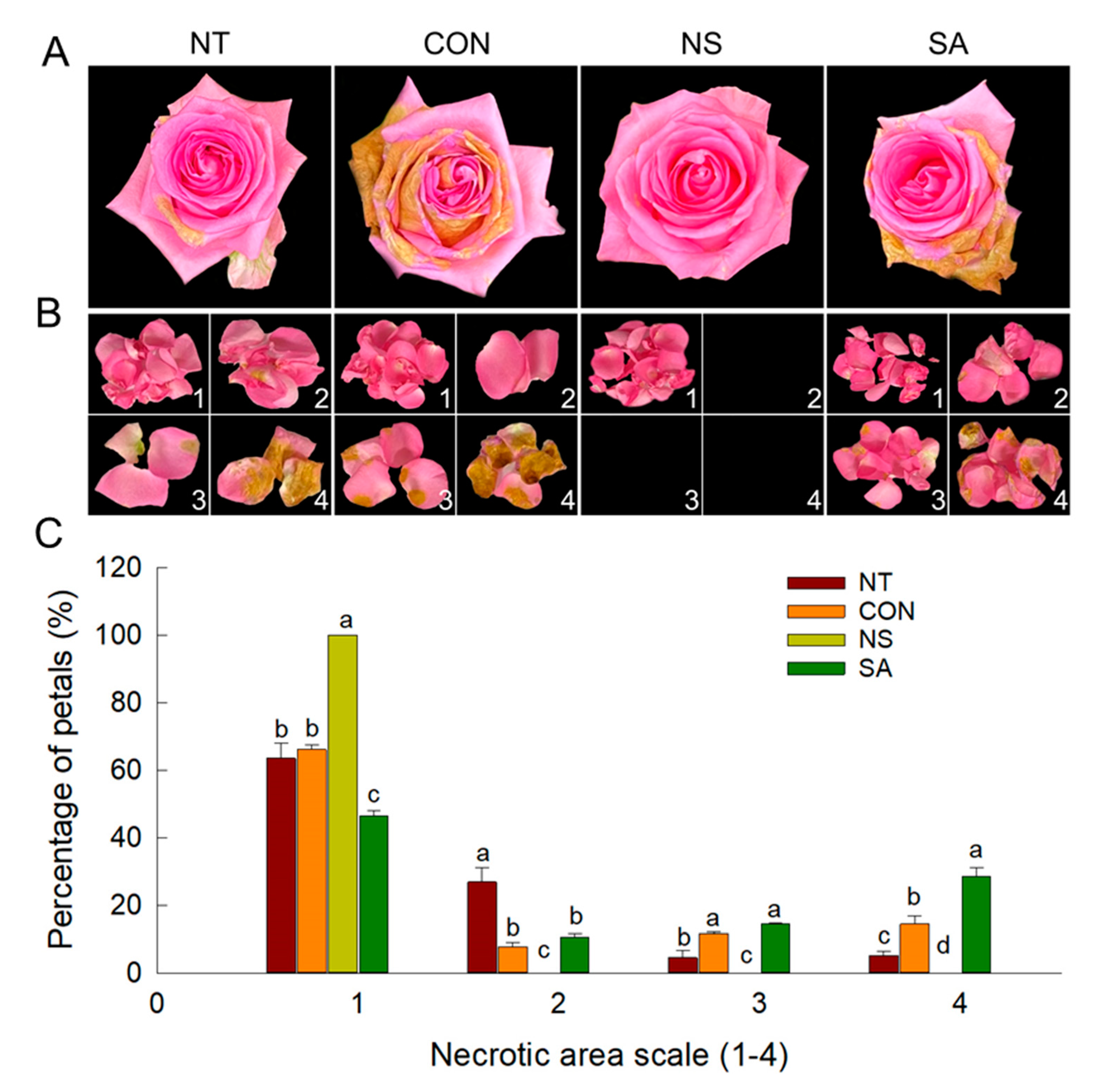
2.1. B. cinerea Infection Rate, Vase Life and Flower Opening of Cut Roses

After B. cinerea inoculation and transport simulation, visual symptoms of B. cinerea infection on rose plants were recorded on day 1 of vase period in control and SA flowers and on day 5 in NT flowers (data not shown). Control and SA flowers exhibited the highest infection rate (100%) of gray mold disease on rose plants on day 7 of vase life (Table 1). In contrast, 80% of the NT flowers were infected with B. cinerea after seven days of the treatment (Table 1). Application of NS significantly decreased the B. cinerea infection rate of rose flowers during vase life (Table 1 and Figure 1A).
The lesions in the petals of cut flowers were also recorded and observed daily to assess the effectiveness of NS and SA in inhibiting gray mold disease. The extent of the petal area infected by B. cinerea varied according to the treatments. The incidence of infection corresponding to 3 and 4 on the relative scale was higher in control and SA flowers than in NT flowers (Figure 1B,C). The extent of the infected petal area of NS flowers was suppressed during the vase period (Figure 1B,C). These results showed that the NS could completely inhibit gray mold disease on cut rose flowers, whereas SA did not inhibit lesion development.
Early termination of vase life of cut ‘Revival’ roses was due to gray mold disease in control (5.5 d) and SA (6.2 d) treatments (Table 1). Compared to NT flowers (8.6 d), the vase life of NS flowers was significantly prolonged by 3.3 days (Table 1). Interestingly, the maximum flower diameter was largest in NS flowers, whereas it was smaller in control and SA flowers during vase life (Table 1), indicating that the severe gray mold disease on petals caused the failure of full flower opening.

2.2. Expression Patterns of Ethylene Biosynthesis and Bcspl1 Genes

The expression of five ethylene biosynthesis genes and Bcspl1 in rose petals was detected using quantitative real time PCR (qRT-PCR) on days 1, 3, 5, and 7 after B. cinerea inoculation. The mRNA levels of three ethylene biosynthesis genes, RhACS2, RhACS4, and RhACO1 were higher in control and SA flowers than in NT and NS flowers (Figure 2B,D,E). The expression of these genes was increased in NT flowers on days 5 and 7; however, there were no changes in NS flowers during vase period (Figure 2B,D,E). The expression of RhACS3 in petals was induced in all treatments on days 5 and 7 of vase life (Figure 2C). The mRNA levels of RhACS1 were decreased in control flowers and not affected in other treatments during vase periods. However, the accumulation of RhACS1 in petals was higher in NT and SA flowers than that of CON and NS flowers on days 3, 5, and 7 (Figure 2A).
A higher level of Bcspl1 expression was detected in the control and SA flowers during the vase period (Figure 2F). In NT flowers, the expression of Bcspl1 was enhanced on days 5 and 7 after B. cinerea inoculation (Figure 2F). In NS flowers, transcript levels of Bcspl1 were not detected on days 1 and 3, and remained very low on days 5 and 7 of the vase life (Figure 2F). Among the ethylene biosynthesis genes, RhACS2, RhACS4, and RhACO1 exhibited expression patterns similar to those of Bcspl1 in petals (Figure 2B,D–F), indicating that these genes might be closely correlated with B. cinerea growth in roses.

2.3. Expression Patterns of Ethylene Receptor and Signaling Genes

To further assess the effectiveness of NS and SA on gray mold disease and ethylene responses in cut roses, the expression patterns of five ethylene receptors (RhETR1-RhETR5), two RhCTRs (RhCTR1 and RhCTR2), and three RhEIN3s (RhEIN3-1-RhEIN3-3) were determined in the petals. After B. cinerea infection, the mRNA levels of three ethylene receptor genes, RhETR1, RhETR2, and RhETR5, and the downstream protein kinase, RhCTR2 were down-regulated in control and SA flowers during vase life (Figure 3A,B,E,G). In contrast, the decrease in the expression of these genes was suppressed in NS flowers (Figure 3A,B,E,G). In NT flowers, the expression levels of RhETR1, RhETR2, RhETR5, and RhCTR2 were decreased on days 5 and 7 of the vase period (Figure 3A,B,E,G). The expression of RhETR3 and RhETR4 increased during the senescence stage (day 7) in regardless of the treatments (Figure 3C,D). The expression pattern of RhCTR1 in cut flowers was the opposite of that noted for RhCTR2 (Figure 3F). In addition, the mRNA levels of RhCTR1 were higher in control and SA flowers than that of NT and NS flowers (Figure 3F). The RhEIN3s expression pattern was similar to that of the ethylene biosynthesis gene RhACS2, RhACS4, and RhACO1. The growth of B. cinerea led to a significant increase in the mRNA levels of RhEIN3s in the petals of the control and SA flowers (Figure 3H–J). NS treatment effectively suppressed the ethylene-inducible increase in RhEIN3s in cut rose flowers (Figure 3H–J).

2.4. Changes in the Fv/Fm, Leaf Temperature, and Water Relations of Cut Roses
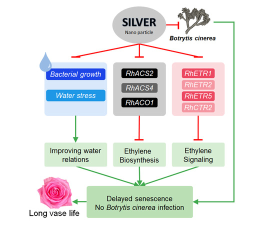
The maximum quantum yield of PSII (Fv/Fm) was measured using dark-adapted rose leaves, which were subsequently exposed to actinic light on day 1. The B. cinerea infection occurring on day 1 decreased the Fv/Fm values in the leaves of control and SA flowers (Figure 4A). The decrease in maximum quantum yield of PSII values was significantly suppressed in NT and NS flowers (Figure 4A). Under light conditions, the temperature difference between the leaf (TL) and the reference plate (TR) (TL − TR) in control and SA flowers on day 1 exhibited positive values (Figure 4B). This finding indicates that the temperature of B. cinerea-infected roses increased prior to the appearance of the visual symptoms.
Changes in the water relations of the cut flowers were also analyzed daily to evaluate the effect of NS and SA on postharvest quality of cut flowers after B. cinerea infection. Under the light conditions, stomatal opening rate and transpiration of control and SA flowers were significantly lower than those of NT and NS flowers (Figure 4C,D). The number of days that flowers retained their initial fresh weight and positive water balance was shortest in control and SA flowers but longest in NS flowers, compared to NT flowers (Figure 5A,B). This result showed that NS not only suppressed the B. cinerea growth on cut roses but also effectively improved the water relations of cut flowers during vase period.

3. Discussion

Gray mold caused by B. cinerea, a necrotrophic pathogen, decreases the economic and ornamental value of cut roses during storage and transportation. After harvest, cut roses are still susceptible to B. cinerea infection even under very low temperature conditions because of latent penetration and ability to grow at low temperatures [12]. Thus, early detection and suppression are required to effectively prevent the spread of gray mold in cut flowers.
A previous study showed that the silver ions which are released from nano silver can attack a wide range of biological processes in microorganisms [17]. Therefore, in the current study, NS effectively suppressed the growth of B. cinerea spores and prevented the development of the lesions in rose petals during vase life. Previous research showed the effectiveness of SA in inhibiting fungal disease and inducing the disease resistance in several plants [18]. To examine the role of SA in B. cinerea resistance, we applied 0.5 mM exogenous SA to cut ‘Revival’ roses and then inoculated the flowers with B. cinerea. However, we found no significant difference between the SA-treated flowers and control flowers. Exogenous SA application did not decrease the B. cinerea infection rate in rose petals in this study. The postharvest longevity of the cut rose flowers treated with SA was considerably decreased, comparing with NT flowers. SA has resistance effects on various microbial pathogen, but the function of SA in plant defense against the necrotrophic pathogens varies according to plant species [13]. Additionally, the function of SA in the fungal infection process appears to be complex, including at least three strategies: signalization, degradation of SA by the fungal pathogen, and growth inhibition of the pathogen [19].
The development of gray mold disease is related to an increase in ethylene synthesis from the infected tissues [6]. Along with jasmonic acid and SA, ethylene plays an important role in plant defense response to pathogens [20]. When plants are infected with a microorganism, ethylene production by infected tissues is induced for defense against pathogens [8,21]. The results of the present study revealed that the high transcript levels of RhACS2, RhACS4, and RhACO1 were associated with the severity of the gray mold disease in rose petals after B. cinerea infection. Among ethylene biosynthesis genes in roses, RhACS1 and RhACS3 are mainly related to flower development and flower opening, while RhACS2, RhACS4, and RhACO1 are responsible for flower senescence and disease defense [22,23]. The increased expression of RhACS2, RhACS4, and RhACO1 in control and SA flowers after B. cinerea inoculation might lead to endogenous ethylene synthesis and a decrease in mRNA levels of RhETR1, RhETR2, and RhETR5 in rose petals. This led to a reduction in the expression level of the downstream protein kinase, RhCTR2, thereby resulting in the ethylene response in the cut flowers and acceleration of necrosis development in petals [24]. A previous study also indicated that the expression of both LeACO1 and LeACS2 was increased in B. cinerea-infected leaves in tomatoes after four days of inoculation and their hybridization intensity was similar to the pattern of BcactA, which is a marker for actively growing B. cinerea [8]. Simultaneous pretreatment with SA and ethylene has been found to lead to notably increase the transcript levels of LeACO1 in infected tissues in tomatoes [8]. Consistent with these findings, the present study indicated that the expression of Bcspl1 was highly induced in control and SA flowers, but strongly inhibited by NS after B. cinerea inoculation. In addition, Bcspl1 exhibited the expression pattern similar to those of RhACS2, RhACS4, and RhACO1 in the petals after B. cinerea inoculation. Notably, the expression of Bcspl1 was strongly induced in the petals in the later stage of the B. cinerea infection. It has been well known that the fungal infection was thought to depend on its capacity to kill the host plants and destroy plant tissues. The interaction between B. cinerea and host plants is subtle and often evaluated by virulence traits and quantitative susceptibility [25]. The protein encoded by Bcspl1 might act as an elicitor of plant defense in the early infection phase (penetration and primary lesion formation), whereas it might act as a virulence factor to kill and extract nutrition from host plant tissues during a later phase (plant colonization) [9,26,27]. Moreover, the expression of Bcspl1 in B. cinerea has been found to be induced by ethylene especially during early infection stages and stimulated during later infection stages [9]. In the current study, the increase in mRNA levels of ethylene biosynthesis genes caused by B. cinerea infection in rose petals could induce the expression of Bcspl1, which acted as the virulence factor and rapidly terminated the vase life of control and SA flowers.
Recently, NS has been applied as an ethylene antagonist and found to interfere with ethylene biosynthesis in many plants [15,16,28]. NS decreased ethylene synthesis by inhibiting the mRNA levels of the ethylene biosynthesis genes, such as DcACS1, DcACO1, AtACS7, and AtACO2 in carnation flowers and Arabidopsis seedlings [15,16,28]. The silver ion, which is released from nano silver, can replace the cofactor Cu2+ for the ethylene receptor and thereby effectively suppresses ethylene binding [29]. In the current study, the suppression of ethylene action by NS could prevent the degradation of ethylene receptors and the downstream protein kinase by maintaining the mRNA levels of these genes, resulting in the inhibition of the ethylene responses and the necrosis development in rose petals.
Disease infection often causes disorder of physiological metabolism in the infected plant tissues. The stomatal opening rate, leaf temperature, maximum quantum yield of PSII of leaves, and water relations of the control and SA flowers were negatively affected by B. cinerea infection. Stomata can be considered to participate in the plant innate immune responses via regulation of stomatal closure. Bacteria and fungi can trigger stomatal closure through pathogen-associated molecular patterns, which prevents penetration through these pores [30,31,32]. Thus, the B. cinerea infection could induce stomatal closure in control and SA flowers to prevent infiltration through stomatal pores. Stomatal closure reduced the transpiration rate in the leaves of the cut flowers and led to an increased leaf temperature. Our results are consistent with a previous study showing that the infection of Pseudoperonospora cubensis caused an increase in leaf temperature via the regulation of stomatal transpiration in cucumbers [33]. Interaction of SA and abscisic acid was found to be required for stomata responses to disease infection in plants [34]. SA is also a potent inducer of stomatal closure [32]. These previous findings strongly supported our results indicating that flowers treated with SA exhibited a smallest stomatal opening rate, lowest transpiration, and highest leaf temperature after B. cinerea inoculation.
Among the different chlorophyll fluorescence parameters, Fv/Fm is the most important value that is used as a plant stress indicator, since it declines under most stress conditions and reflects the maximum photochemical efficiency [35,36,37,38]. Decrease in Fv/Fm is often observed before the visual appearance of disease or stress symptoms; therefore Fv/Fm has been used to predict and detect those abnormal stress status in many plant species [36,37,39]. A highly significant negative correlation was observed between leaf temperature and Fv/Fm in peanut and cotton under stress conditions, suggesting that higher photodamage in the photosynthetic apparatus of peanut due to increase in leaf temperature [37]. The reduction in Fv/Fm in control and SA flowers indicated that downregulation in maximum quantum yield of PSII was induced in the tissues by B. cinerea infection at an early stage. This finding coincided with an increase in leaf temperature, which was likely due to stomatal closure. Consistent with these findings, previous studies also observed the considerable decrease in Fv/Fm in tobacco plants infected with B. cinerea [40], maize and banana plants infected with Fusarium [41], and in melon infected by Dickeya dadantii [41]. Our results also revealed that the Fv/Fm value and leaf temperature can be used for detecting infection of plant tissues by B. cinerea at an early stage of the infection process.
In summary, our results indicated that the B. cinerea growth on rose petals considerably increased the expression of ethylene biosynthesis genes, leading to accumulation of the virulence factor Bcspl1. B. cinerea infection also decreased the transcript levels of ethylene receptor genes and of the downstream regulator RhCTR2, thereby inducing the plant responses to ethylene. The growth of the B. cinerea caused the downregulation in maximum quantum yield of PSII via regulation of stomatal function and leaf temperature. Nano silver significantly inhibited B. cinerea growth and suppressed the ethylene responses in rose petals by inhibiting the expression of ethylene biosynthesis and signaling genes. Moreover, nano silver effectively maintained the Fv/Fm in leaves, improved water relations, and reduced the incidence of senescence of cut flowers, thereby prolonging the vase life of cut roses. Thus, our results show that nano silver can be a promising chemical for preventing B. cinerea infection and ethylene damages, and for improving the postharvest quality of cut roses that are exported overseas.

4. Materials and Methods

4.1. Plant Materials

Cut roses (Rosa hybrida L. cv ‘Revival’) were harvested at commercial maturity stage from a commercial greenhouse in Daegu, Korea in June 2020. Rose plants were provided with a nutrient solution including calcium, nitrogen, phosphorus, magnesium, potassium, and other trace substances. Changes in environmental conditions in the greenhouse such as light density, relative humidity (RH), and air temperature were monitored during the year using data loggers. After harvest, the cut flowers were kept in buckets containing tap water and transferred to the laboratory within 2 h for further treatments.

4.2. Preparation of B. cinerea Conidial Suspension

B. cinerea used in this study was obtained from Agricultural Genetic Resource Information Center, Korea. Prior to treatments, the isolated B. cinerea conidia were transferred and grown on potato dextrose agar media at 25 °C for 14 days. To collect the B. cinerea conidia, 25 mL sterile distilled water was added to the culture petri dishes and gently filtered by using four layers of sterile gauze. The concentration of B. cinerea conidia was determined using a hemocytometer, and the final concentration was adjusted to 10⁵ conidia mL−1 for the experiments.

4.3. Nano Silver, Salicylic Acid, and B. cinerea Treatments

Based on our previous study [42], cut rose flowers were placed and sprayed with 20 mg L−1 NS (ShangHai HuZheng Nano Tech Co., Ltd., ShangHai, China) or 0.5 mM SA (Daejung Chemicals, Korea). NT and CON flowers were placed in distilled water without treatment solutions. CON, NS, and SA flowers were inoculated with the B. cinerea conidial suspension (10⁵ conidia mL−1) to induce gray mold development, whereas NT flowers were not inoculated with B. cinerea and used to assess the natural infection process. Subsequently, all cut flowers were held at 10 ± 2 °C and 80–90% RH under dark conditions for 4 days for simulation of export conditions. After the transport simulations, cut flowers were trimmed to 50 cm of the length along with three upper leaves. Each cut rose was placed in a glass vase containing 350 mL distilled water. For vase life evaluation, the flowers were maintained in a controlled environmental room at 25 ± 1 °C, 50–55% relative humidity, and a photoperiod of 12 h with light supplied by fluorescent tubes at 20 µmol m−2 s−1 light intensity.

4.4. Evaluation of B. cinerea Infection and the Morphological and Physiological Characteristics of Cut Flowers

Number of rose stems with flower and leaf damage and visual symptoms of B. cinerea in rose petals were recorded daily to evaluate the effectiveness of NS and SA on inhibition of B. cinerea infection in cut roses. The extent of development of visual symptoms of B. cinerea was assessed on a relative scale of 1–4 according to the extent of the infected rose petal area, as follows: 1, no infection; 2, ≤25%; 3, 26–50%; and 4, 51–100%. The vase life of cut roses was considered terminated when cut flowers showed B. cinerea growth on petals that corresponded to 3 or 4 points on the relative scale. Other senescence symptoms, such as bent neck, bluing, or wilting of leaf and flower were also observed when the cut flowers reached the senescent stage.
Changes in morphological and physiological characteristics of cut flowers such as vase life, flower opening, fresh weight, and water uptake were also determined daily at 9:30 am. The water balance of cut flowers was determined by deducting the daily transpiration rate from daily water uptake. Leaf stomatal sizes were measured under light conditions (after 45 min at 20 mol−2 s−1 light intensity) on day 1 of the vase life. Stomata were obtained from the abaxial surface of the uppermost leaf of the cut flowers using Suzuki’s Universal Micro-Printing method. The stomatal width and length were measured by using the Image J software (Version 1.49p, NIH, Bethesda, MD, USA) and the stomatal size was determined using the equation:
S = π × r1 × r2
where S is stomatal size, r1 is half of the width, and r2 is half of the length.

4.5. Measurement of Leaf Temperature and Chlorophyll Fluorescence (CF) Parameters

The uppermost leaves of the cut flowers were used for measuring the leaf temperature and CF parameters. The CF parameters of these leaves were measured using the FluorPen FP110/D device (Photon Systems Instruments, Brno, Czech Republic). A dark-adapted leaf was obtained by covering the leaf with a leaf-clip for 15 min. The clip was then opened and the minimal fluorescence (F0) and maximal fluorescence (Fm) of the leaf were measured. The maximal quantum efficiency of PSII (Fv/Fm) of the leaf was automatically calculated from the F0 and Fm values.
To measure the temperature of the rose leaves, thermal images of cut rose flowers were obtained using a thermal camera (FLIR, VUE™ PRO R, FLIR systems) after morphological measurement. For each treatment, thermal images of cut flowers were taken under the similar conditions. To reduce the effect of distance variation on the results, the distance between the rose leaves and the camera lens was set as 30 cm for all flowers during the measurements. A reference plate (10 cm × 10 cm × 3 mm) was also placed at the same height as the leaf measured. Thermal images of the cut roses were exported to the computer by using FLIR tools for measuring the leaf temperature. The temperature difference between the leaf (TL) and the reference plate (TR) was calculated as TL − TR.

4.6. RNA Isolation and cDNA Synthesis

Petals were detached from cut rose flowers after the transport simulation on days 1, 3, 5 and 7. These rose petals were immediately frozen in liquid nitrogen and then stored at −80 °C until RNA extraction. The rose petals were ground with liquid nitrogen to a fine powder (200 mg) using a prechilled mortar and pestle and total RNA was isolated from the fine powder by using the GeneJET plant RNA Purification Mini Kit (Thermo Fisher Scientific Baltics UBA V.A. Graiciuno, Vilnius, Lithuania) with a slight modification of the manufacturer’s protocol. The total RNA was quantified at 260 nm/280 nm using a Nanodrop spectrophotometer (NanoDrop 2000, Thermo Scientific, Waltham, MA, USA). Total RNA (0.1 µg) was used to synthesize the first strand of cDNA using Power cDNA Synthesis Kit (INTRON Biotechnology, Inc., Gyeonggi-do, Republic of Korea) as per the manufacturer’s instruction, in a Bio-Rad PTC-100 Programmable Thermal Controller (MJ Research Inc., Hercules, CA, USA).

4.7. qRT-PCR

The mRNA levels of ethylene biosynthesis, receptor, and signaling and B. cinerea snod-prot-like 1 (Bcspl1) genes in rose petals were detected using the BIO-RAD CFX Connect Real-Time System (Life Science, Hercules, CA, USA). The sequences of forward and reverse primers used for qRT-PCR are listed in Table 2. The gene-specific primers were designed by an online version of Primer 3 software, and then synthesized by Cosmogenetech (Seoul, Korea). Rosa hybrida actin (RhACT1) and B. cinerea actinA (BcactA) genes were used as an internal control to confirm the amount of template RNA. The reaction mixtures (20 µL) contained 5 µL cDNA as a template, 10 µL iQ™ SYBR® Green Supermix (Life Science, Hercules, CA, USA), 1 µL forward and reverse primers (10 µM), and 4 µL water. The qRT-PCR reactions were conducted using the following PCR program: 95 °C for 60 s for pre-denaturation (one cycle) and followed by 40 cycles of 95 °C for 10 s (denaturation), 65 °C for 10 s (annealing), and 72 °C for 40 s (extension). The final threshold cycle value was the mean of three independent biological replications and the coefficient of variance for each gene was also calculated. The relative level of the gene expression was measured as the absolute integrated absorbency normalized to the relative actin.

4.8. Statistical Analysis

The vase life and disease incidence data were designed with nine replicates for each treatment and one flower per replication. qRT-PCR analysis, stomatal size, CF parameters, and leaf temperature measurements were performed with three biological replicates. All data were presented as mean ± standard error (SE). One-way analysis of variance was conducted for data. When significant effects were detected, post hoc pairwise comparisons of group means were executed with Duncan’s multiple range test, with a significance level of p = 0.05. Least significant difference (LSD; p = 0.05) values were calculated for mean separation using the critical values of t for two-tailed tests. Data analysis was performed by using SPSS version 22.0 (IBM, Somers, NY, USA).

Author Contributions

Data curation, S.T.T.H. and Y.-T.K.; Formal analysis, S.T.T.H. and B.-C.I.; Funding acquisition, B.-C.I.; Investigation, S.T.T.H. and Y.-T.K.; Methodology, Y.H.J., H.W.C. and B.-C.I.; Project administration, B.-C.I.; Writing—original draft, S.T.T.H.; Writing—review and editing, Y.H.J., H.W.C. and B.-C.I. All authors have read and agreed to the published version of the manuscript.

Funding

This study was supported partially by the Basic Research Program (2018R1D1A3B07045695) through the National Research Foundation of Korea (NRF) funded by the Ministry of Education, Science and Technology and a research grant (PJ0150142020) from the Rural Development Administration of Korea (RDA).

Data Availability Statement

All data generated and analyzed during this study are included in this published article.

Acknowledgments

We thank the members of the Postharvest Physiology Lab for their assistance during measurements, and the Agricultural Genetic Resource Information Center for providing B. cinerea.

Conflicts of Interest

The authors declare no conflict of interest.

References

  1. Williamson, B.; Duncan, G.H.; Harrison, J.G.; Harding, L.A.; Elad, Y.; Zimand, G. Effect of humidity on infection of rose petals by dry-inoculated conidia of Botrytis cinerea. Mycol. Res. 1995, 99, 1303–1310. [Google Scholar] [CrossRef]
  2. Elad, Y.; Volpin, H. The involvement of ethylene and calcium in gray mold of pelargonium, ruscus, and rose plants. Phytoparasitica 1988, 16, 119–131. [Google Scholar] [CrossRef]
  3. Muñoz, M.; Faust, J.E.; Schnabel, G. Characterization of Botrytis cinerea From Commercial Cut Flower Roses. Plant Dis. 2019, 103, 1577–1583. [Google Scholar] [CrossRef]
  4. Borochov, A.; Woodson, W. Physiology and Biochemistry of Flower Petal Senescence. Hortic. Rev. 2011, 11, 15–43. [Google Scholar]
  5. Pantelides, I.S.; Tjamos, S.E.; Paplomatas, E.J. Insights into the role of ethylene perception in tomato resistance to vascular infection by Verticillium dahliae. Plant Pathol. 2010, 59, 130–138. [Google Scholar] [CrossRef]
  6. Elad, Y. Involvement of ethylene in the disease caused by Botrytis cinerea on rose and carnation flowers and the possibility of control. Ann. Appl. Biol. 1988, 113, 589–598. [Google Scholar] [CrossRef]
  7. Benito, E.P.; ten Have, A.; van’t Klooster, J.W.; van Kan, J.A.L. Fungal and plant gene expression during synchronized infection of tomato leaves by Botrytis cinerea. Eur. J. Plant Pathol. 1998, 104, 207–220. [Google Scholar] [CrossRef]
  8. Díaz, J.; ten Have, A.; van Kan, J.A.L. The Role of Ethylene and Wound Signaling in Resistance of Tomato to Botrytis cinerea. Plant Physiol. 2002, 129, 1341–1351. [Google Scholar] [CrossRef] [Green Version]
  9. Chagué, V.; Danit, L.V.; Siewers, V.; Schulze-Gronover, C.; Tudzynski, P.; Tudzynski, B.; Sharon, A. Ethylene sensing and gene activation in Botrytis cinerea: A missing link in ethylene regulation of fungus-plant interactions? Mol. Plant Microbe Interact. Mpmi 2006, 19, 33–42. [Google Scholar] [CrossRef] [Green Version]
  10. Elad, Y.; Shtienberg, D. Botrytis cinerea in greenhouse vegetables: Chemical, cultural, physiological and biological controls and their integration. Integr. Pest Manag. Rev. 1995, 1, 15–29. [Google Scholar] [CrossRef]
  11. Chu, E.-H.; Shin, E.-J.; Park, H.-J.; Jeong, R.-D. Effect of gamma irradiation and its convergent treatment for control of postharvest Botrytis cinerea of cut roses. Radiat. Phys. Chem. 2015, 115, 22–29. [Google Scholar] [CrossRef] [Green Version]
  12. Williamson, B.; Tudzynski, B.; Tudzynski, P.; van Kan, J.A. Botrytis cinerea: The cause of grey mould disease. Mol. Plant Pathol. 2007, 8, 561–580. [Google Scholar] [CrossRef]
  13. Koo, Y.M.; Heo, A.Y.; Choi, H.W. Salicylic Acid as a Safe Plant Protector and Growth Regulator. Plant Pathol. J. 2020, 36, 1–10. [Google Scholar] [CrossRef]
  14. Kim, S.W.; Jung, J.H.; Lamsal, K.; Kim, Y.S.; Min, J.S.; Lee, Y.S. Antifungal Effects of Silver Nanoparticles (AgNPs) against Various Plant Pathogenic Fungi. Mycobiology 2012, 40, 53–58. [Google Scholar] [CrossRef] [Green Version]
  15. Naing, A.H.; Win, N.M.; Han, J.S.; Lim, K.B.; Kim, C.K. Role of Nano-silver and the Bacterial Strain Enterobacter cloacae in Increasing Vase Life of Cut Carnation ‘Omea’. Front. Plant Sci. 2017, 8, 1590. [Google Scholar] [CrossRef] [PubMed] [Green Version]
  16. Park, D.Y.; Naing, A.H.; Ai, T.N.; Han, J.-S.; Kang, I.-K.; Kim, C.K. Synergistic Effect of Nano-Sliver with Sucrose on Extending Vase Life of the Carnation cv. Edun. Front. Plant Sci. 2017, 8, 1601. [Google Scholar] [CrossRef] [Green Version]
  17. Dakal, T.C.; Kumar, A.; Majumdar, R.S.; Yadav, V. Mechanistic Basis of Antimicrobial Actions of Silver Nanoparticles. Front. Microbiol. 2016, 7. [Google Scholar] [CrossRef] [PubMed] [Green Version]
  18. Hua, L.; Yong, C.; Zhanquan, Z.; Boqiang, L.; Guozheng, Q.; Shiping, T. Pathogenic mechanisms and control strategies of Botrytis cinerea causing post-harvest decay in fruits and vegetables. Food Qual. Saf. 2018, 2, 111–119. [Google Scholar] [CrossRef] [Green Version]
  19. Dieryckx, C.; Gaudin, V.; Dupuy, J.W.; Bonneu, M.; Girard, V.; Job, D. Beyond plant defense: Insights on the potential of salicylic and methylsalicylic acid to contain growth of the phytopathogen Botrytis cinerea. Front. Plant Sci. 2015, 6. [Google Scholar] [CrossRef] [Green Version]
  20. Wu, L.; Huang, Z.; Li, X.; Ma, L.; Gu, Q.; Wu, H.; Liu, J.; Borriss, R.; Wu, Z.; Gao, X. Stomatal Closure and SA-, JA/ET-Signaling Pathways Are Essential for Bacillus amyloliquefaciens FZB42 to Restrict Leaf Disease Caused by Phytophthora nicotianae in Nicotiana benthamiana. Front. Microbiol. 2018, 9, 847. [Google Scholar] [CrossRef] [Green Version]
  21. Bleecker, A.B.; Kende, H. Ethylene: A gaseous signal molecule in plants. Annu. Rev. Cell Dev. Biol. 2000, 16, 1–18. [Google Scholar] [CrossRef] [Green Version]
  22. Ma, N.; Cai, L.; Lu, W.; Tan, H.; Gao, J. Exogenous ethylene influences flower opening of cut roses (Rosa hybrida) by regulating the genes encoding ethylene biosynthesis enzymes. Sci. China. Ser. C Life Sci. 2005, 48, 434–444. [Google Scholar] [CrossRef]
  23. In, B.-C.; Ha, S.T.T.; Lee, Y.S.; Lim, J.H. Relationships between the longevity, water relations, ethylene sensitivity, and gene expression of cut roses. Postharvest Biol. Technol. 2017, 131, 74–83. [Google Scholar] [CrossRef]
  24. Lund, S.T.; Stall, R.E.; Klee, H.J. Ethylene regulates the susceptible response to pathogen infection in tomato. Plant Cell 1998, 10, 371–382. [Google Scholar] [CrossRef]
  25. Veloso, J.; Van Kan, J.A.L. Many shades of grey in Botrytis-Host plant interactions. Trends Plant Sci. 2018, 23, 613–622. [Google Scholar] [CrossRef]
  26. Tudzynski, P.; Sharon, A. Fungal pathogenicity genes. Appl. Mycol. Biotechnol. 2003, 3, 187–212. [Google Scholar]
  27. Wolpert, T.J.; Dunkle, L.D.; Ciuffetti, L.M. Host-selective toxins and avirulence determinants: What’s in a name? Annu. Rev. Phytopathol. 2002, 40, 251–285. [Google Scholar] [CrossRef] [Green Version]
  28. Syu, Y.-Y.; Hung, J.-H.; Chen, J.-C.; Chuang, H.-W. Impacts of size and shape of silver nanoparticles on Arabidopsis plant growth and gene expression. Plant Physiol. Biochem. 2014, 83, 57–64. [Google Scholar] [CrossRef]
  29. Rodríguez, F.I.; Esch, J.J.; Hall, A.E.; Binder, B.M.; Schaller, G.E.; Bleecker, A.B. A copper cofactor for the ethylene receptor ETR1 from Arabidopsis. Science 1999, 283, 996–998. [Google Scholar] [CrossRef]
  30. Melotto, M.; Zhang, L.; Oblessuc, P.R.; He, S.Y. Stomatal Defense a Decade Later. Plant Physiol. 2017, 174, 561–571. [Google Scholar] [CrossRef] [Green Version]
  31. Melotto, M.; Underwood, W.; He, S.Y. Role of stomata in plant innate immunity and foliar bacterial diseases. Annu. Rev. Phytopathol. 2008, 46, 101–122. [Google Scholar] [CrossRef] [Green Version]
  32. Gudesblat, G.E.; Torres, P.S.; Vojnov, A.A. Stomata and pathogens: Warfare at the gates. Plant Signal. Behav. 2009, 4, 1114–1116. [Google Scholar] [CrossRef]
  33. Oerke, E.-C.; Steiner, U.; Dehne, H.-W.; Lindenthal, M. Thermal imaging of cucumber leaves affected by downy mildew and environmental conditions. J. Exp. Bot. 2006, 57, 2121–2132. [Google Scholar] [CrossRef]
  34. Prodhan, M.Y.; Munemasa, S.; Nahar, M.N.-E.-N.; Nakamura, Y.; Murata, Y. Guard Cell Salicylic Acid Signaling Is Integrated into Abscisic Acid Signaling via the Ca2+/CPK-Dependent Pathway. Plant Physiol. 2018, 178, 441–450. [Google Scholar] [CrossRef] [Green Version]
  35. Rolfe, S.A.; Scholes, J.D. Chlorophyll fluorescence imaging of plant-pathogen interactions. Protoplasma 2010, 247, 163–175. [Google Scholar] [CrossRef]
  36. Shahenshah; Isoda, A. Effects of Water Stress on Leaf Temperature and Chlorophyll Fluorescence Parameters in Cotton and Peanut. Plant Prod. Sci. 2010, 13, 269–278. [Google Scholar] [CrossRef]
  37. Rousseau, C.; Belin, E.; Bove, E.; Rousseau, D.; Fabre, F.; Berruyer, R.; Guillaumès, J.; Manceau, C.; Jacques, M.-A.; Boureau, T. High throughput quantitative phenotyping of plant resistance using chlorophyll fluorescence image analysis. Plant Methods 2013, 9, 17. [Google Scholar] [CrossRef] [Green Version]
  38. Kim, J.H.; Bhandari, S.R.; Chae, S.Y.; Cho, M.C.; Lee, J.G. Application of maximum quantum yield, a parameter of chlorophyll fluorescence, for early determination of bacterial wilt in tomato seedlings. Hortic. Environ. Biotechnol. 2019, 60, 821–829. [Google Scholar] [CrossRef]
  39. Pineda, M.; Soukupová, J.; Matouš, K.; Nedbal, L.; Barón, M. Conventional and combinatorial chlorophyll fluorescence imaging of tobamovirus-infected plants. Photosynthetica 2008, 46, 441–451. [Google Scholar] [CrossRef]
  40. Rossi, F.R.; Krapp, A.R.; Bisaro, F.; Maiale, S.J.; Pieckenstain, F.L.; Carrillo, N. Reactive oxygen species generated in chloroplasts contribute to tobacco leaf infection by the necrotrophic fungus Botrytis cinerea. Plant J. Cell Mol. Biol. 2017, 92, 761–773. [Google Scholar] [CrossRef] [Green Version]
  41. Kuckenberg, J.; Tartachnyk, I.; Noga, G. Temporal and spatial changes of chlorophyll fluorescence as a basis for early and precise detection of leaf rust and powdery mildew infections in wheat leaves. Precis. Agric. 2009, 10, 34–44. [Google Scholar] [CrossRef]
  42. Ha, S.T.T.; Kim, Y.T.; In, B.C. Assessment of the preservative solutions for reducing Botrytis cinerea infection in cut roses. Flower Res. J. 2020, 28, 279–284. [Google Scholar] [CrossRef]
Figure 1. Effect of B. cinerea infection on visual appearance (A) and the B. cinerea infection rate in petals (B,C) of cut roses on day 7 after the transport simulation. Scale of the extent of the infected rose petal area by B. cinerea: 1, no infection; 2, ≤25%; 3, 26–50%; and 4, 51–100%. NT, non-treated flowers; CON, flowers were held in distilled water; NS and SA, cut flowers were treated with nano silver and salicylic acid. CON, NS, and SA flowers were sprayed with B. cinerea conidial suspension to induce gray mold development. All flowers were then stored at 10 ± 2°C and 80–90% RH in dark conditions for 4 d for export simulation. Different letters above bars indicate statistically significant differences among treatments at p = 0.05 based on Duncan’s multiple range test. Vertical bars show standard errors of the means (n = 9).
Plants 10 01241 g001
Figure 2. Effect of B. cinerea infection on transcript levels of RhACS1 (A), RhACS2 (B), RhACS3 (C), RhACS4 (D), RhACO1 (E), and Bcspl1 (F) in petals of cut roses. mRNA levels of ethylene biosynthesis and Bcspl1 genes were detected on days 1, 3, 5, and 7 after the transport simulation. NT, non-treated flowers; CON, flowers were held in distilled water; NS and SA, cut flowers were treated with nano silver and salicylic acid. CON, NS, and SA flowers were sprayed with B. cinerea conidial suspension to induce gray mold development. All flowers were then stored at 10 ± 2 °C and 80–90% RH in dark conditions for 4 d for export simulation. Different letters above bars indicate statistically significant differences among treatments at p = 0.05 based on Duncan’s multiple range test. Vertical bars show standard errors of the means (n = 3).
Plants 10 01241 g002
Figure 3. Effect of B. cinerea infection on transcript levels of ethylene receptor (AE) and signaling (FJ) genes in petals of cut roses. mRNA levels of ethylene receptor and signaling genes were detected on days 1, 3, 5, and 7 after the transport simulation. NT, non-treated flowers; CON, flowers were held in distilled water; NS and SA, cut flowers were treated with nano silver and salicylic acid. CON, NS, and SA flowers were sprayed with B. cinerea conidial suspension to induce gray mold development. All flowers were then stored at 10 ± 2 °C and 80–90% RH in dark conditions for 4 d for export simulation. Different letters above bars indicate statistically significant differences among treatments at p = 0.05 based on Duncan’s multiple range test. Vertical bars show standard errors of the means (n = 3).
Plants 10 01241 g003
Figure 4. Effect of B. cinerea infection on maximum quantum yield of PSII (Fv/Fm) (A), leaf temperature (B), stomata size (C), and transpiration (D) of cut roses on day 1 after the transport simulation. NT, non-treated flowers; CON, flowers were held in distilled water; NS and SA, cut flowers were treated with nano silver and salicylic acid. Leaf stomatal size and transpiration of cut flowers were measured on day 1 of vase life. CON, NS, and SA flowers were sprayed with B. cinerea conidial suspension to induce gray mold development. All flowers were then stored at 10 ± 2 °C and 80–90% RH in dark conditions for 4 d for export simulation. Different letters above bars indicate statistically significant differences among treatments at p = 0.05 based on Duncan’s multiple range test. Vertical bars show standard errors of the means (n = 3 for (AC) and 9 for (D)).
Plants 10 01241 g004
Figure 5. Effect of B. cinerea infection on initial fresh weight (A) and water balance (B) of cut roses. NT, non-treated flowers; CON, flowers were held in distilled water; NS and SA, cut flowers were treated with nano silver and salicylic acid. CON, NS, and SA flowers were sprayed with B. cinerea conidial suspension to induce gray mold development. All flowers were then stored at 10 ± 2 °C and 80–90% RH in dark conditions for 4 d for export simulation. Data were presented as the mean ± standard errors (n = 9).
Plants 10 01241 g005
Table 1. Infection rate, vase life, and flower opening of cut rose flowers after B. cinerea inoculation.
TreatmentB. cinerea Incidence (%) ˣVase Life (Days)Maximum Flower Diameter (%) ʸ
Non-treatment (NT)80.0 ± 0.0 b ᶻ8.6 ± 0.3 b121.1 ± 1.3 ab
Control (CON)100.0 ± 0.0 a5.5 ± 0.4 c109.3 ± 1.9 c
Nano silver (NS)0.0 ± 0.0 c11.9 ± 0.8 a125.7 ± 2.3 a
Salicylic acid (SA)100 ± 0.0 a6.2 ± 0.4 c108.6 ± 5.8 c
ˣ The incidence of B. cinerea in cut roses was evaluated on day 7 of vase life. ʸ Maximum flower diameter (percentage of the initial diameter) was measured and compared among the treatments on day 4 of vase life. ᶻ Different letters among the treatments indicate the statistically significant differences at p = 0.05 based on Duncan’s multiple range test (n = 9).
Table 2. Primer sequences used in qRT-PCR for gene analysis in rose petals.
Gene (Accession Number)Forward PrimerReverse PrimerSize
RhACS1 (AY378152.1)5′-CAGTGAGAAAGGGGAGCTTG-3′5′-TGTATTGAACCGGGATGGTT-3′102
RhACS2 (AY803737.1)5′-GCGAACAGGGGTACAACTTC-3′5′-GGGTTTGAGGGGTTGGTAAT-3′147
RhACS3 (AY803738.1)5′-CAGTGAGAAAGGGGAGCTTG-3′5′-AACCATCCCGGTTCAATACA-3′142
RhACS4 (AY525068.1)5′-GCTTCCAACTTGGGATCAAA-3′5′-GCTCCATGAAACTTGCCATT-3′100
RhACO1 (AF441282.1)5′CGTTCTACAACCCAGGCAAT-3′5′-TTGAGGCCTGCATAGAGCTT-3′130
RhETR1 (AY953869.1)5′-TGACTGGCCTGATGTCTCTG-3′5′-GGCAACTGGTGAAAAGGAAA-3′158
RhETR2 (AF127220.1)5′-CTGCGTTAGAGCAGCAACTG-3′5′-GGAATTCGGCGATATCTTCA-3′131
RhETR3 (AY953392.1)5′-CCATGAGTTGAAAGGGAGGA-3′5′-GGCTCACCAAAATCACCACT-3′156
RhETR4 (AF159172.1)5′-TTGAAGTCGTTGCAGACCAG-3′5′-TCATGACAGCAAGGAAGTCG-3′168
RhETR5 (AF441283.1)5′-TGTGTGGAGCGACACATCTT-3′5′-TGAGGGCAGTAGCACATGAC-3′120
RhEIN3-1 (AF443783)5′-TGCTGAAGATGATGGAGGTG-3′5′-GCAGGGCCATTCTTATCAAA-3′142
RhEIN3-2 (AY919867.1)5′-ATTGAACTTGGCCAATCAGG-3′5′-GCAGTCATCTTGTCCTGCAA-3′168
RhEIN3-3 (KC484653.1)5′-GCCAGTGGATCTTTGGTGAT-3′5′-ACTTGAAGCCCTTCCCTCAT-3′149
RhCTR1 (AY032953.1)5′-GGCTCTGATGTTGCTGTGAA-3′5′-CAAGTTTGGGGGCTTTGTAA-3′150
RhCTR2 (AY029067.1)5′-CGAGCAACCCCACTATTGTT-3′5′-TTATGTTTCAAGCGCGACAG-3′109
RhACT1 (KC514918.1)5′-GTTCCCAGGAATCGCTGATA-3′5′-ATCCTCCGATCCAAACACTG-3′116
Bcspl1 (XM024691684.1)5′-CCTACGACGTTGGCTACGAT-3′5′-CCTCAAGAACTTCCCCAACA-3′123
BcactA(XM024697950.1)5′-GCACCACCCGAGAGAAAATA-3′5′-AAGAGTACGACGAGTCCGGA-3′169
Publisher’s Note: MDPI stays neutral with regard to jurisdictional claims in published maps and institutional affiliations.

