Article

Phytochemical Analysis of Anvillea garcinii Leaves: Identification of Garcinamines F–H and Their Antiproliferative Activities

by
Hanan Y. Aati
1,
Shagufta Perveen
1,*,
Raha Orfali
1,
Areej M. Al-Taweel
1,
Jiangnan Peng
2,
Sobia Tabassum
3,
Maged S. Abdel-Kader
4,
Hasan Soliman Yusufoglu
4 and
Orazio Taglialatela-Scafati
5
1
Department of Pharmacognosy, College of Pharmacy, King Saud University, P.O. Box 22452, Riyadh 11495, Saudi Arabia
2
Department of Chemistry, School of Computer, Mathematical and Natural Sciences, Morgan State University, Baltimore, MD 21251, USA
3
Interdisciplinary Research Centre in Biomedical Materials (IRCBM), Lahore Campus, COMSATS University Islamabad, Islamabad 44000, Pakistan
4
Department of Pharmacognosy, College of Pharmacy, Prince Sattam Bin Abdulaziz University, P.O. Box 173, Al-Kharj 11942, Saudi Arabia
5
Department of Pharmacy, School of Medicine and Surgery, University of Naples Federico II, Via Montesano 49, 80131 Naples, Italy
*
Author to whom correspondence should be addressed.
Plants 2021, 10(6), 1130; https://doi.org/10.3390/plants10061130
Submission received: 26 April 2021 / Revised: 26 May 2021 / Accepted: 27 May 2021 / Published: 2 June 2021

Abstract

:
Anvillea garcinii is a medicinal plant used in the Arab region for intestinal diseases, lung and liver diseases, digestive problems, and as an antidiabetic agent. Repeated chromatographic purifications of A. garcinii leaves led to the isolation of three undescribed guaiane sesquiterpene derivatives, named garcinamines F–H, characterized by the presence of an amino acid unit, along with five known sesquiterpene lactones (garcinamines B–E and 9β-hydroxyparthenolide). The structures of the new compounds were established using spectroscopic (1D and 2D NMR) and spectrometric methods (ESIMS). Garcinamine H possesses a double bond at the Δ1,10 position, a structural feature rarely reported in guaianolide-type sesquiterpenes. The antiproliferative activity of the isolated sesquiterpenes was screened against three different cancer cell lines, and 9β-hydroxyparthenolide and garcinamines C and D displayed significant effects against lung carcinoma (A549), colon carcinoma (LoVo), and breast carcinoma (MCF7) cell lines.

Graphical Abstract

1. Introduction

Cancer is the second-most life-threatening disease, with a worldwide incidence leading to nearly 9.5 million deaths and 18 million cases in 2018, with an expected doubling of cases and deaths by 2050 [1,2]. The Arabian Peninsula makes no exception to these general figures: it has been found that the number of known cancer cases increased by 136% from 1999 to 2015 and is projected to rise by a further 63% in 2030. Breast, colorectal and thyroid cancers, and non-Hodgkin’s lymphoma accounted for the largest contributions to these increased rates [3]. Cancer therapy faces numerous challenges, since most chemotherapeutic drugs are not fully effective and are often associated with many unpleasant side effects such as myelosuppression, peripheral neuropathy, and leukopenia [4,5]. Since about 83% of non-biological anticancer drugs are related to natural products [6], researchers have been investigating the possible additions of plant extracts or pure phytochemicals to enhance their therapeutic potency or decrease the adverse effects [7,8].
Anvillea garcinii (Burm.f.) DC. (family Asteraceae) is one of the distinctive ethnomedicinal plants used in the Arabian Peninsula to cure many illnesses, such as cold, digestive problems, pulmonary infections, and liver diseases [9]. A. garcinii is a small woody shrub that is densely branched with triangular leaves, is green-grey in color, and has yellow flowers. It usually flowers in spring but can also flower at other times in the year. The predominant classes of secondary metabolites characterized by previous phytochemical investigations carried on this plant were flavonoids and sesquiterpene lactones [10,11] that most likely work synergistically to achieve the interesting biological activities exerted by A. garcinii. Our previous investigations on A. garcinii specimens growing in Saudi Arabia revealed the presence of sesquiterpene lactones of the guaianolide and germacranolide types, as well as of flavonoid glycosides [12,13,14,15].
During the last decades, sesquiterpene lactones have attracted wide attention because of their multi-faceted biological activities, including antitumor and anti-inflammatory properties [16]. In several cases, these activities have been rationalized based on the presence of the α-methylene-γ-butyrolactone moiety in their structures, which could act as an electrophilic agent in Michael-type reactions, especially with free cysteine residues. The biological activities could derive from the reversible or irreversible inhibition of enzymes and other functional proteins [16]. It should be noted that the presence of the electrophilic moiety is not sufficient to completely describe the activity, since in many cases the sesquiterpene skeleton plays a crucial modulation role. For example, guaianolides (sesquiterpenes with a 5/7 ring architecture) have been shown in some cases to possess a higher antiproliferative activity compared to other classes of sesquiterpene lactones [17].
Herein we report the result of a phytochemical characterization of the sesquiterpene profile of a specimen of A. garcinii and the evaluation of the antiproliferative activity of the isolated compounds on lung carcinoma (A549), colon carcinoma (LoVo), and breast carcinoma (MCF7) cell lines, which are three of the most represented human cancers.

2. Results and Discussion

Previous investigations on the aerial parts of A. garcinii [12,13,14,15] identified the n-butanol fraction of the ethanol extract as the richest in polar sesquiterpenoids. Its chromatographic separation was achieved using a combination of Sephadex LH-20, silica gel, and RP-18 column chromatography, and yielded three new (13) and five known compounds (48) (Figure 1). The structures of these metabolites were elucidated by spectroscopic analysis, mainly one-dimensional (1D) and two-dimensional (2D) NMR spectroscopy and electrospray ionization mass spectrometry (ESI-MS) (Figure S1–S18). Compounds 48 were identified as garcinamine E (4), 9β-hydroxyparthenolide (5), garcinamine B (6), garcinamine D (7), and garcinamine C (8) by comparison of their spectroscopic data with those reported in the literature [12,13,14,15].

2.1. Structural Elucidation of Garcinamines F–H (13)

Garcinamine F (1) was obtained as an optically active yellow gum with the molecular formula C20H28NO6, established by ESI-MS (negative ions). The 13C NMR and DEPT spectra of 1 (see Supplementary Materials) showed twenty carbon atoms, encompassing only one methyl group (δC 22.5), eight methylene carbons (seven sp3 and one sp2 at δC 108.5), seven sp3 methines, and four unprotonated carbon signals, including an ester carbonyl (δC 171.9) and a carboxylic acid (δC 177.0) (Table 1).
The 1H NMR spectrum of garcinamine F (Table 2) showed signals for a methyl singlet (δH 1.33), two exomethylene sp2 protons (δH 5.17 and 5.51), two oxymethine protons (δH 3.99 and 4.23), and two downfield methylene protons (δH 3.53/3.63 and 3.25/3.86). The COSY spectrum allowed the organization of all the multiplets of the 1H NMR spectrum within a large spin system (Figure 2), which was strongly reminiscent of a guaianolide-type sesquiterpene lactone based on comparison with data of related compounds from the same source [12,13,14,15]. The 1H NMR spectra also showed typical signals of an L-proline moiety including the nitrogen-linking methine at δH 4.02 and methylene at δH 3.25 and 3.86. The planar structure of garcinamine F was then unambiguously defined based on the long-range H/C correlations evidenced by the 2D HMBC NMR spectrum (Figure 2).
The key HMBC correlations were those from H3-15 to C-3, C-4, and C-5; from H2-14 to C-1, C-9, and C-10; from H-6 to C-12; and from H2-13 to the proline carbons C-2′ and C-5′. ROESY correlations of H-6 with H3-15 and H-11, of H-5 with H-1 and H-7, and of H-7 with H-9 defined the relative configuration of the stereogenic centers.
Garcinamine G (2) showed the same molecular formula C20H28NO6 as garcinamine F and also very similar 1H (Table 2) and 13C (Table 1) NMR spectra (see Supplementary Materials). Analysis of COSY, HSQC, and HMBC 2D NMR spectra led to the assignment of all the H/C resonances and to the definition of a planar structure identical to that of garcinamine F, clearly pointing to a diastereomeric relationship between the two compounds. The most significant differences in the 13C NMR spectra of garcinamines F and G were confined to the signals of C-8, C-9, C-10, and C-14. Similarly, the 1H NMR spectra of these two compounds mainly differed for the resonances of the seven-membered ring protons. These spectral data strongly suggest that garcinamine G (2) should be the 9-epimer of garcinamine F. The marked change in the coupling constant pattern of H-9 and the ROESY cross-peaks of H-6 and H-9 with H-8β support this conclusion.
Additionally, garcinamine H (3) showed the molecular formula C20H28NO6, the same as garcinamines F and G, suggesting a relationship among these compounds. The 13C NMR and DEPT spectra of 3 (see Supplementary Materials) showed twenty carbon atoms but with a pattern that differed from that of garcinamine F. Indeed, it included two methyl groups, seven methylene carbons (all sp3), six sp3 methines, and five unprotonated carbon signals (Table 1). The multiplets of the 1H NMR spectrum (Table 2) were arranged through the COSY spectrum into three spin systems (Figure 2), evidencing the lack of proton connection between the H2-2/H2-3 fragment and the remaining protons of the guaiane skeleton. Key HMBC correlations from H3-15 to C-3, C-4, and C-5 and from H2-2 to C-1 defined the structure of the five-membered ring. Correlations from the allylic H3-14 to the oxygenated C-9 and to the sp2 C-1 and C-10 defined the structure of the seven-membered ring with a double bond between the two latter carbons. Finally, correlations from H-6 to C-12 and from H2-13 to the proline carbons C-2′ and C-5′ completed the structure of garcinamine H (3). ROESY correlations of garcinamine H paralleled those of garcinamine G for common stereocenters and allowed the assignment of the relative configuration to 3. Thus, garcinamine H is an isomer of garcinamine G showing a somewhat rare Δ1,10 double bond in place of the Δ10,14 one. This is the first finding of this structural feature in A. garcinii.
The sesquiterpenoid derivatives isolated in this study bear an evident biosynthetic relationship. For example, the newly isolated garcinamine G could derive from the epoxide-driven cyclization of the germacranolide garcinamine C. Hydration of the exocyclic double bond of garcinamine G produces garcinamine E that, upon dehydration, would give garcinamine H (3) (Figure 3).

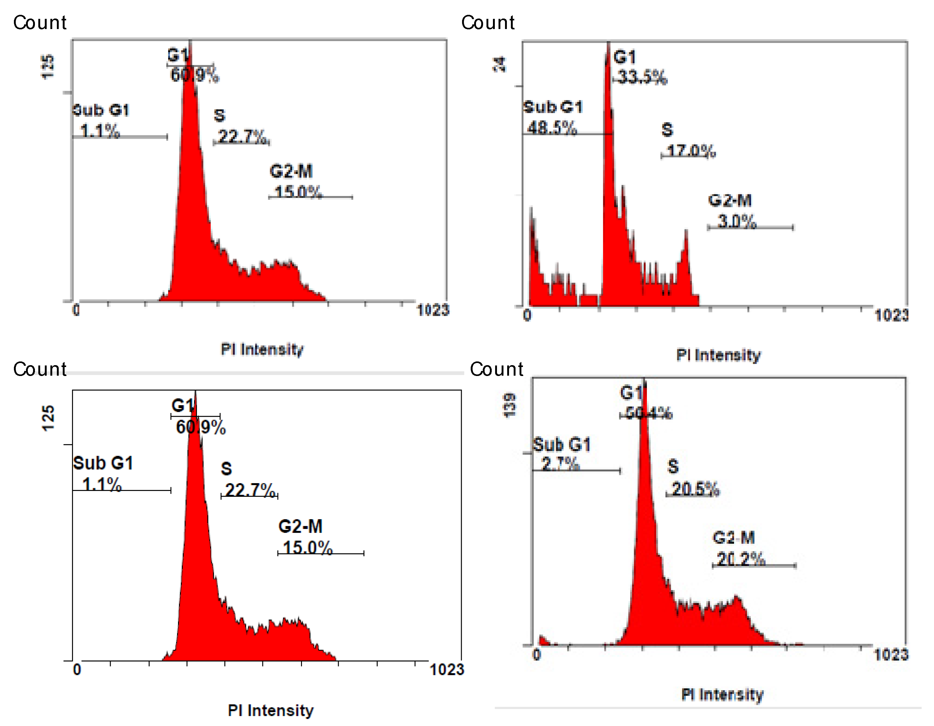
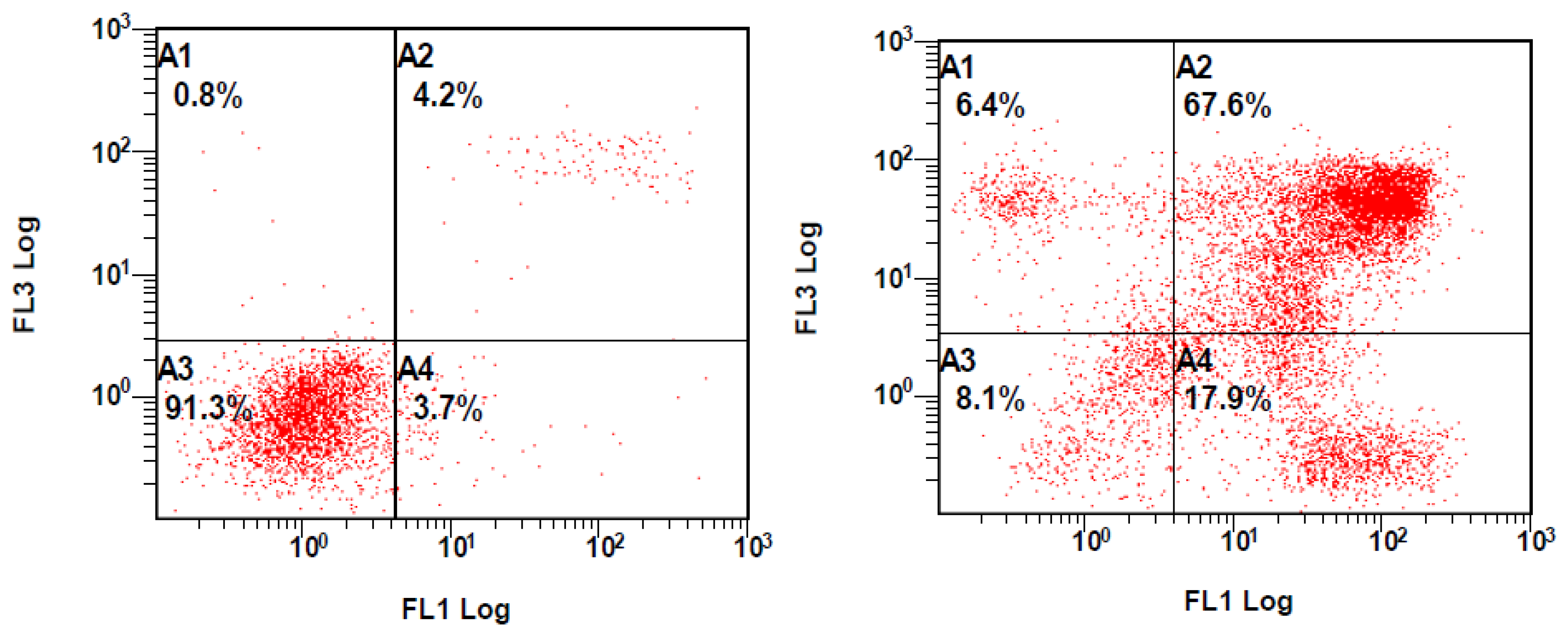
2.2. Biological Activity

The antiproliferative activity of the isolated compounds 18 was evaluated on A549 (lung), MCF-7 (breast), and LoVo (colon) carcinoma cell lines using the MTT assay. Table 3 shows IC50 values measured after 48 h of treatments. The results evidenced that 9β-hydroxyparthenolide and garcinamines C and D exerted cytotoxic effects in a dose- and time-dependent manner. The activity of 9β-hydroxyparthenolide was not unexpected since it had already been described in the literature [18]. However, better activity was obtained for garcinamine D against A549 cells and thus it was selected for cell cycle analysis by PI staining and apoptosis induction by Annexin-FITC/PI assays.
The effects of garcinamine D on A549 cell cycle phases were evaluated using PI staining in FACS analysis to quantify the cell percentages in the different phases. For this analysis, A549 cells were treated with garcinamine D (IC50 concentration) for 48 h and later stained with PI (Figure 4, top). The results revealed the accumulation of cells in the sub-G1 phase. The number of cells in the sub-G1 phase was 1.1% in the untreated cells, and 48.5% in the treated cells. Furthermore, the cells were treated with one of the non-active compounds (garcinamine E) at the corresponding IC50, and as expected, it did not show clear activity (Figure 4, bottom).
The effect of garcinamine D on the growth inhibition of A549 cells was also investigated by Annexin V/PI assay to show its apoptotic effects. After 48 h of the treatment with the IC50 concentration of garcinamine D, 17.9% and 67.6% of cells moved into early and late apoptosis, respectively (Figure 5), demonstrating the apoptosis-inducing effect of garcinamine D. On the other hand, the non-active compound garcinamine E did not show any prominent activity at the corresponding concentration (data not shown).
The close structural similarity within the series of compounds tested for antiproliferative activity can stimulate interesting comments on structure–activity relationships (SARs). Firstly, contrary to other reported cases (see the Introduction), germacrane compounds seemed to be more active than the corresponding guaianes, which is evident if we compare the bioactivities of 7/8 with those of the parallel 1/2. Interestingly, although bioactivity of sesquiterpenoid γ-lactones is commonly associated with the presence of an electrophilic exomethylene group [19], compounds 7/8 were equally active (or even slightly more active) than the exomethylene-containing 9β-hydroxyparthenolide (5). Finally, the amino acid unit included in these adducts appears to play an important role, as evident from the comparison between the inactive valine-bearing garcinamine B (6) and the corresponding active proline-bearing garcinamine C (8). This observation is not unprecedented, and the antiulcer activity of saussureamines is also modulated by the amino acid unit present in the adduct [20]. In summary, the antiproliferative activity of sesquiterpene-amino acid adducts seems to follow independent SAR compared to simple sesquiterpenoids, and still needs to be studied in detail.

3. Materials and Methods

3.1. General

The MX-500 Bruker spectrometer (Billerica, MA, USA) was used to measure one-dimensional (1D) and two-dimensional (2D) nuclear magnetic resonance (NMR) spectra. The chemical shifts (δ) were calculated (ppm) relative to TMS, based on the residual solvent signal and J scalar coupling constants reported in Hertz (Hz). ESI-MS analyses were carried out on an Agilent (Santa Clara, CA, USA) Triple Quadrupole 6410 QQQ LC/MS mass spectrometer with ESI ion source (60 psi nebulizer pressure, 350 °C gas temperature and 12 L/min flow rate), operating in the negative and positive scan modes of ionization through the direct infusion method using methanol:water (4:6 v/v) at a flow rate of 0.5 mL/ min. Separations and purifications of secondary metabolites were carried by using column chromatography with either silica gel 70–230 mesh or RP-18 (Merck, Darmstadt, Germany). The TLC plates were RP-18 (Merck) and pre-coated silica gel 60 F254, and the spots were detected by UV light and by spraying with ceric sulphate and sulfuric acid reagents followed by heating on a hot plate (TLC plate heater III CAMAG, USA). Dragendorff’s spraying reagent was used to give a dark orange color. Analytical-grade solvents and reagents were obtained from Sigma-Aldrich (St. Louis, MO, USA). NMR deuterated methanol (CD3OD) and dimethylsulfoxide (DMSO-d6) were purchased from Cambridge Isotope Laboratories (Tewksbury, MA, USA).

3.2. Plant Material, Extraction and Isolation

A. garcinii fresh leaves (1.0 kg) were collected manually in March 2020 from Riyadh city, Saudi Arabia and authenticated by Dr. M. Atiqur Rahman, College of Pharmacy, King Saud University, KSA. A voucher specimen (# 12811) was deposited at the Herbarium of the College of Pharmacy, King Saud University, KSA.
Fresh leaves (1.0 kg) were dried in the shade for up to 2–3 weeks and then crushed into a fine powder. The plant powder was soaked in 80% methanol at room temperature (3 × 15 L), with frequent shaking. The extract was then filtered using filter paper (Whatman No. 1). The filtrate was concentrated under reduced pressure using a rotary evaporator (Büchi Rotavapor RII, Flawil, Switzerland). The obtained crude extract (102.2 g) was suspended in H2O (800 mL) and fractionated successively with organic solvent, and the residual water fraction was lyophilized. The n-butanol-soluble fraction (75.8 g) was exposed to a silica gel open column and eluted with a gradient of CH2Cl2:CH3OH (9.5:0.5 to 1.0:9.0), to afford eight major fractions (I–VIII) based on their TLC pattern. Fraction I (0.6 g) was isolated at CH2Cl2:CH3OH (9.0:1.0) and further exposed to purification via using normal silica column to give 5 (7.2 mg), CH2Cl2:CH3OH (8.0:2.0). Furthermore, fractions II and III were eluted by using CH2Cl2:CH3OH (8.0:2.0 and 7.5:2.5, respectively) and led to the isolation of compounds 2 and 7 (8.4 mg and 6.9 mg, respectively). From the major silica column, solvent system CH2Cl2:CH3OH (6.5:3.5) gave fraction IV which was further purified by using the RP-C18 column and a mobile phase mixture of water/methanol was used with gradient elution and decreasing polarity to afford compound 4, 6.0:4.0 H2O:MeOH (11.3 mg). Fraction VI was obtained at 6.0:4.0 CH2Cl2:CH3OH gradient elution and further loaded on the RP C-18 column for chromatography, eluted under medium pressure with a gradient of water/methanol (7.5:2.5→9.0:1.0), to obtain two sub-fractions which was further purified by loading on a second RP C-18 column and eluted with a gradient mixture of water/methanol to yield compounds 1 and 3 (9.0 mg and 10.5 mg, respectively). Fraction VII (1.2 g) was eluted with increasing polarity by the addition of methanol to dichloromethane until subfraction VII-a was afforded (4.0:6:0), which was further loaded on an RP-C18 column the elution was continued using gradient polarity reduction, by adding methanol to water gradually, until compounds 6 (9.4 mg) and 8 (12.8 mg) were yielded (4.0:6.0 and 3.0:7.0, H2O:MeOH).
Garcinamine F (1). Yellow gummy solid; [α]25 D-27.2 (c 0.10, CH3OH); (+) ESIMS m/z 380.2076 [M + H]+ (calcd. for C20H30NO6, 380.2073); 1H and13C NMR data, see Table 1 and Table 2.
Garcinamine G (2). Yellow gummy solid; [α]25D-22.5 (c 0.15, CH3OH); (+) ESIMS m/z 380.2080 [M + H]+ (calcd. for C20H30NO6, 380.2073); 1H and13C NMR data, see Table 1 and Table 2.
Garcinamine H (3). Yellow gummy solid; [α]25D-29.3 (c 0.15, CH3OH); (+) ESIMS m/z 380.2069 [M + H]+ (calcd. for C20H30NO6, 380.2073); 1H and13C NMR data, see Table 1 and Table 2.

3.3. Biological Activity

Cell proliferation assay was carried out using MTT. Three different types of human cancer cell lines (A549 lung carcinoma, MCF-7 breast carcinoma, and LoVo colon carcinoma) were used in this study. The cells were counted and plated in a 24-well cell culture plate with a density of 5 × 10⁴ cells/well and allowed to grow and attach to the plate for 24 h. The compounds were dissolved in dimethyl sulfoxide (DMSO) to prepare stock solution (3 mg/mL). The serial dilutions of compounds (1–30 µM) were added to triplicate wells; mock-treated (0.1% DMSO) cells were used as negative control and doxorubicin was used as positive control. Cells were treated with the appropriate compounds for 48 h. At the end of the treatment period, 1 mL of 5 mg/mL MTT was added to each well, and the plates were further incubated at 37 °C in the presence of 5% CO2 for 2–4 h. The media were removed, 0.01 N HCl in isopropanol was added to each well, and the plates were placed in a shaker for 10 min. The absorbance was read at 570 nm using a microplate reader (BioTek, Winooski, VT, USA). The concentration at which cell viability was reduced by 50% (IC50) was calculated using Origin Pro 8.5 software.
The antiproliferative effect of compounds 18 against breast (MCF-7), colon (LoVo), and lung (A549) cell lines were examined using MTT assay. The collected cells were cultured in Dulbecco’s modified Eagle’s medium (DMEM) with 10% heat-inactivated fetal bovine serum, HEPES buffer, 1% L-glutamine, and 50 µg/mL gentamycin, and then placed in 96-well plates at 50,000 cells/well. Compounds were dissolved in DMSO and the cells were treated with different concentrations of each compound for 48 h, and then 3-(4,5-dimethyltiazol-2yl)-2,5 diphenyl tetrazolium bromide (MTT) (5 mg/mL in saline, 10 µL) was added and cells were incubated for an additional 4 h at 37 °C. Thereafter, cells were lysed and dark blue crystals of the formazan product were dissolved with isopropanol and the absorbance intensity was measured at 570 nm using a microplate reader The cell survival percent was calculated as the mean absorbance of treated sample/mean absorbance of control × 100. The dose–response curve of the compounds was analyzed using OriginPro 8.5 software. Doxorubicin was used as a positive control.
Cellular DNA content was analyzed using propidium iodide staining by flow cytometry. In brief, A549 cells were seeded into a 6-well plate in 2 mL culture medium with an IC50 concentration and DMSO as a vehicle for 48 h. Thereafter, cells were harvested using trypsin and centrifuged and fixed in 70% ethanol for 4 h at 4 °C. Ethanol-fixed cells were pelleted, washed with ice-cold PBS, and re-suspended in a staining solution containing 50 μg/mL PI and 100 μg/mL RNase. After incubation in the dark for 30 min, the cells were analyzed by flow cytometry.
The apoptotic cells were detected using flow cytometry with the Annexin V-FITC/PI double labeling method. Briefly, after 48 h of treatment with 7 at IC50 values, A549 cells were collected, washed and resuspended in Annexin binding buffer. Thereafter, cell staining was performed with FITC and PI (5 µL of each), according to the manufacturer’s instructions (BioLegend, San Diego, CA, USA). Flow cytometric analysis was performed immediately after 20 min of staining. Data acquisition and analysis were performed in a flow cytometer (Cytomics FC 500; Beckman Coulter, Brea, CA, USA).

4. Conclusions

Phytochemical investigation of the aerial parts of Anvillea garcinii allowed us to expand the knowledge on the sesquiterpenoid composition of this plant with the addition of three new guaianolide-proline adducts named garcinamines F–H. These compounds bear an evident biogenetic relationship with germacranolides, previously isolated from the same plant. All the isolated compounds were evaluated for their antiproliferative activity against three human carcinoma cell lines. Garcinamines C and D and 9β-hydroxyparthenolide exerted cytotoxic effects in a dose- and time-dependent manner. The mechanism of action of garcinamine D was investigated, revealing a marked pro-apoptotic effect with an accumulation of cells in the sub-G1 phase. These results further confirm that plants constitute an immense casket of chemodiversity that can be beneficially used to discover new hits for drug development.

Supplementary Materials

The following are available online at https://www.mdpi.com/article/10.3390/plants10061130/s1, Figures S1–S18: 1D and 2D NMR spectra of garcinamines F–H.

Author Contributions

Conceptualization, H.Y.A., S.P. and R.O.; methodology, A.M.A.-T. and J.P.; formal analysis, S.T., H.S.Y. and M.S.A.-K.; investigation, S.P. and R.O.; writing—original draft, S.P. and O.T.-S.; writing—review and editing, S.P., O.T.-S. and H.Y.A. All authors have read and agreed to the published version of the manuscript.

Funding

This research project was supported by a grant from the “Research Center of the Female Scientific and Medical Colleges”, Deanship of Scientific Research, King Saud University.

Institutional Review Board Statement

Not applicable.

Informed Consent Statement

Not Applicable.

Data Availability Statement

Not Applicable.

Conflicts of Interest

The authors declare no conflict of interest.

References

  1. Bray, F.; Ferlay, J.; Soerjomataram, I.; Siegel, R.L.; Torre, L.A.; Jemal, A. Global cancer statistics 2018: GLOBOCAN estimates of incidence and mortality worldwide for 36 cancers in 185 countries. CA Cancer J. Clin. 2018, 68, 394–424. [Google Scholar] [CrossRef] [PubMed] [Green Version]
  2. Jemal, A.; Center, M.M.; DeSantis, C.; Ward, E.M. Global Patterns of Cancer Incidence and Mortality Rates and Trends. Cancer Epidemiol. Biomark. Prev. 2010, 19, 1893–1907. [Google Scholar] [CrossRef] [PubMed] [Green Version]
  3. Jazieh, A.R.; Da’ar, O.B.; Alkaiyat, M.; Zaatreh, Y.A.; Saad, A.A.; Bustami, R.; Alrujaib, M.; Alkattan, K. Cancer incidence trends from 1999 to 2015 and contributions of various cancer types to the overall burden: Projections to 2030 and extrapolation of economic burden in Saudi Arabia. Cancer Manag. Res. 2019, 11, 9665–9674. [Google Scholar] [CrossRef] [PubMed] [Green Version]
  4. Hsieh, T.C.; Chen, S.S.; Wang, X.; Wu, J.M. Regulation of androgen receptor (AR) and prostate specific antigen (PSA) expres-sion in the androgen-responsive human prostate LNCaP cells by ethanolic extracts of the Chinese herbal preparation, PC-SPES. IUBMB Life 1997, 42, 535–544. [Google Scholar] [CrossRef] [PubMed]
  5. André, T.; Boni, C.; Navarro, M.; Tabernero, J.; Hickish, T.; Topham, C.; Bonetti, A.; Clingan, P.; Bridgewater, J.; Rivera, F.; et al. Improved Overall Survival With Oxaliplatin, Fluorouracil, and Leucovorin As Adjuvant Treatment in Stage II or III Colon Cancer in the MOSAIC Trial. J. Clin. Oncol. 2009, 27, 3109–3116. [Google Scholar] [CrossRef] [PubMed] [Green Version]
  6. Newman, D.J.; Cragg, G.M. Natural Products as Sources of New Drugs from 1981 to 2014. J. Nat. Prod. 2016, 79, 629–661. [Google Scholar] [CrossRef] [PubMed] [Green Version]
  7. Wang, C.-Z.; Luo, X.; Zhang, B.; Song, W.-X.; Ni, M.; Mehendale, S.; Xie, J.-T.; Aung, H.H.; He, T.-C.; Yuan, C.-S. Notoginseng enhances anti-cancer effect of 5-fluorouracil on human colorectal cancer cells. Cancer Chemother. Pharmacol. 2007, 60, 69–79. [Google Scholar] [CrossRef] [PubMed] [Green Version]
  8. Koh, Y.-C.; Ho, C.-T.; Pan, M.-H. Recent advances in cancer chemoprevention with phytochemicals. J. Food Drug Anal. 2020, 28, 14–37. [Google Scholar] [CrossRef] [PubMed]
  9. Aati, H.; El-Gamal, A.; Shaheen, H.; Kayser, O. Traditional use of ethnomedicinal native plants in the Kingdom of Saudi Arabia. J. Ethnobiol. Ethnomed. 2019, 15, 1–9. [Google Scholar] [CrossRef] [PubMed]
  10. Tyson, R.L.; Chang, C.J.; McLaughlin, J.L.; Cassady, J.M. A novel sesquiterpene lactone from Anvillea garcinii (Burm.). J. Nat. Prod. 1979, 42, 680–681. [Google Scholar]
  11. Ulubelen, A.; Mabry, T.J.; Aynehchi, Y. Flavonoids of Anvillea garcinii. J. Nat. Prod. 1980, 42, 624–626. [Google Scholar] [CrossRef]
  12. Perveen, S.; Al-Taweel, A.M.; Yusufoglu, H.S.; Fawzy, G.A.; Foudah, A.; Abdel-Kader, M.S. Hepatoprotective and cytotoxic activities of Anvillea garcinii and isolation of four new secondary metabolites. J. Nat. Med. 2017, 72, 106–117. [Google Scholar] [CrossRef] [PubMed]
  13. Perveen, S.; Fawzi, G.A.; Al-Taweel, A.M.; Orfali, R.S.; Yusufoglu, H.S.; Abdel-Kader, M.S.; Al-Sabbagh, R.M. Antiulcer activ-ity of different extract of Anvillea garcinii and isolation of two new secondary metabolites. Open Chem. 2018, 16, 437–445. [Google Scholar] [CrossRef] [Green Version]
  14. Perveen, S.; Alqahtani, J.; Orfali, R.; Al-Taweel, A.M.; Yusufoglu, H.S.; Abdel-Kader, M.S.; Taglialatela-Scafati, O. Antimicro-bial guaianolide sesquiterpenoids from leaves of the Saudi Arabian plant Anvillea garcinii. Fitoterapia 2019, 134, 129–134. [Google Scholar] [CrossRef] [PubMed]
  15. Perveen, S.; Alqahtani, J.; Orfali, R.; Aati, H.Y.; Al-Taweel, A.M.; Ibrahim, T.A.; Khan, A.; Yusufoglu, H.S.; Abdel-Kader, M.S.; Taglialatela-Scafati, O. Antibacterial and Antifungal Sesquiterpenoids from Aerial Parts of Anvillea garcinii. Molecules 2020, 25, 1730. [Google Scholar] [CrossRef] [PubMed]
  16. Akram, G.A.; Gali-Muhtasib, H.; Vuorela, H.; Saliba, N.A.; Darwiche, N. Sesquiterpene lactones isolated from indigenous Middle Eastern plants inhibit tumor promoter-induced transformation of JB6 cells. Drug Discov. Today 2010, 15, 668–678. [Google Scholar]
  17. González, A.G.; Bermejo, J.; Breton, J.L.; Massanet, G.M.; Domínguez, B.; Amaro, J.M. The chemistry of the Compositae. Part XXX1. Absolute configuration of the sesquiterpene lactones centaurepensin (chlorohyssopifolin A), acroptilin (chlorohys-sopifolin C), and repin. J. Chem. Soc. Perkin Trans. I 1976, 115, 1663–1666. [Google Scholar] [CrossRef]
  18. El Bouakher, A.; Jismy, B.; Allouchi, H.; Duverger, E.; Barkaoui, L.; El Hakmaoui, A.; Daniellou, R.; Guillaumet, G.; Akssira, M. Synthetic Modification of 9α- and 9β-Hydroxyparthenolide by Heck or Acylation Reactions and Evaluation of Cytotoxic Activities. Planta Med. 2016, 83, 661–671. [Google Scholar] [CrossRef] [PubMed]
  19. Chadwick, M.; Trewin, H.; Gawthrop, F.; Wagstaff, C. Sesquiterpenoids Lactones: Benefits to Plants and People. Int. J. Mol. Sci. 2013, 14, 12780–12805. [Google Scholar] [CrossRef] [PubMed] [Green Version]
  20. Yoshikawa, M.; Hatakeyama, S.; Inoue, Y.; Yamahara, J. Saussureamines A, B, C, D, and E, new anti-ulcer principles from Chinese Saussureae Radix. Chem. Pharm. Bull. 1993, 41, 214–216. [Google Scholar] [CrossRef] [PubMed] [Green Version]
Figure 1. The sesquiterpenoid derivatives isolated from A. garcinii.
Plants 10 01130 g001
Figure 2. Selected HMBC (→) and COSY ( Plants 10 01130 i001
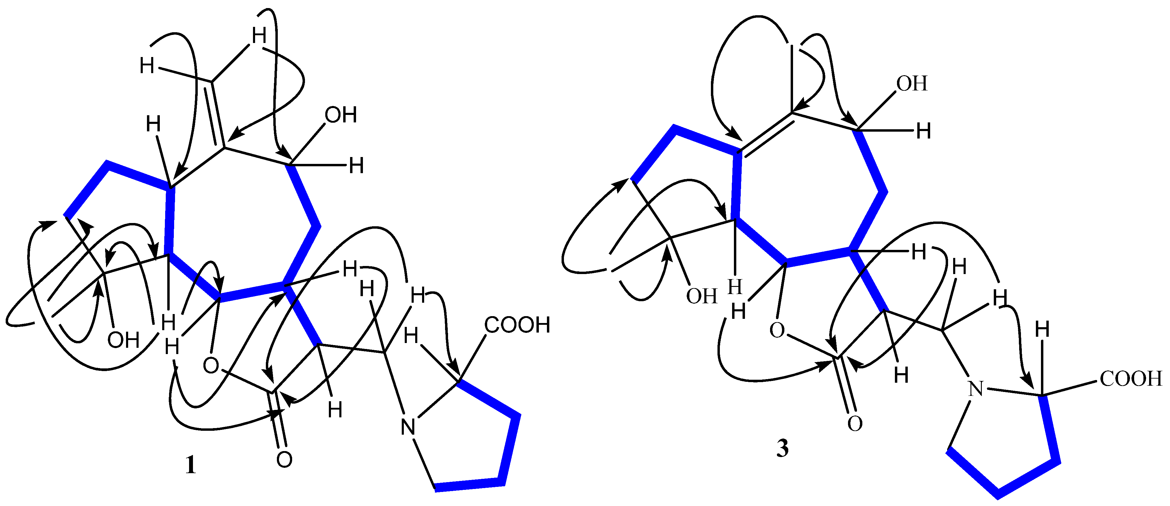
) NMR correlations of garcinamines F (1) and H (3).
Plants 10 01130 g002
Figure 3. Biosynthetic relationship among some of the isolated sesquiterpenoids.
Plants 10 01130 g003
Figure 4. Effect of compounds garcinamine D (top) and garcinamine E (bottom) on the cell cycle distribution of A549 cells. After treatment with the IC50 concentration of garcinamine D (right); Untreated (left). Distribution of cell cycle phases was performed after 48 h of treatment and was quantitated based on flow cytometric analysis.
Plants 10 01130 g004
Figure 5. Assessment of apoptosis induction in A549 cells treated with garcinamine D: (left) control and (right) treated A549 (IC50, 48 h). Quarters (A4) represent early apoptosis, (A3) viable cells, (A2) late apoptosis, and (A1) necrosis. The rate of apoptosis was determined by flow cytometry, using the Annexin V-FITC/PI staining assay.
Plants 10 01130 g005
Table 1. 13C (125 MHz) NMR data for garcinamines F–H (13) in CD3OD.
PositionsGarcinamine F (1)Garcinamine G (2)Garcinamine H (3)
δH, typeδH, typeδH, type
135.4, CH35.4, CH136.2, C
226.0, CH225.6, CH233.0, CH2
341.4, CH241.4, CH239.2, CH2
479.5, C79.6, C80.3, C
555.1, CH54.9, CH56.6, CH
684.1, CH84.6, CH83.3, CH
743.0, CH41.4, CH47.0, CH
839.5, CH238.5, CH235.7, CH2
974.6, CH73.2, CH71.4, CH
10152.6, C150.8, C131.3, C
1141.8, CH41.6, CH41.6, CH
12177.0, C177.1, C177.0, C
1353.0, CH253.1, CH252.5, CH2
14108.5, CH2111.1, CH215.0, CH3
1522.5, CH322.5, CH321.8, CH3
1′171.9, C171.9, C172.0, C
2′70.8, CH70.6, CH70.6, CH
3′28.6, CH228.6, CH228.6, CH2
4′23.5, CH223.4, CH223.4, CH2
5′54.1, CH254.1, CH254.2, CH2
Table 2. 1H (500 MHz) NMR data for garcinamines F–H (13) in CD3OD.
PositionsGarcinamine F (1) Garcinamine G (2)Garcinamine H (3)
δH (multiplicity, J in Hz)δH (multiplicity, J in Hz)δH (multiplicity, J in Hz)
13.04 (dd, 4.0, 11.0)3.56 m-
2a1.92 (m)1.90 (m)2.12 (m)
2b1.83, overlapped1.78 (m)1.70 (m)
31.83–1.85 (m)1.82 (m)1.76 (m)
52.30 (t, 11.0)2.28 (t, 11.0)2.70 (d, 10.5)
64.23 (d, 11.0)4.26 (d, 11.0)4.02 (bd, 10.5)
72.30 (m)2.52 (m)2.31 (dd, 3.0, 13.0)
2.39 (dd, 4.0, 9.0)2.24 (m)2.06 (d, 11.5)
1.42 (m)1.65 (m)1.65 (d, 11.5)
93.99 (dd, 4.0, 7.0)4.56 (bs)4.23 (bs)
113.0 (m)3.07 (dd, 4.0, 11.5)3.13 (dd, 3.5, 12.5)
13a3.63 (dd, 11.0, 12.5)3.60 (dd, 11.0, 12.5)3.65 (dd, 10.5, 12.5)
13b3.53 (dd, 4.0, 12.5)3.45 (dd, 4.0, 12.5)3.57 (dd, 3.5, 10.5)
14a5.51 (bs)5.15 (bs)1.77 (s)
14b5.17 (bs)5.05 (bs)-
151.33 (s)1.30 (s)1.36 (s)
2′4.02 (dd, 5.0, 9.5)4.02 (dd, 5.0, 9.5)4.06 (dd, 6.0, 10.0)
3′a2.46 (dd, 3.5, 9.5)2.45 (dd, 3.5, 9.5)2.46 (dd, 3.5, 10.0)
3′b2.26 (dd, 5.0, 3.5)2.24 (dd, 5.0, 3.5)2.26 (m)
4′a2.17 (m)2.15 (m)2.17 (m)
4′b1.98 (m)1.97 (m)1.99 (m)
5′a3.86 (dd, 4.0, 7.5)3.85 (dd, 4.0, 7.5)3.88 (dd, 4.0, 7.5)
5′b3.25 (dd, 7.5, 10.5)3.24 (dd, 7.5, 10.5)3.27 (dd, 7.5, 10.5)
Table 3. Antiproliferative activity (IC50 values) of compounds 18 against different cancer cells a.
CompoundCell Lines and IC50 (µM)
A549LoVoMCF-7
1NANANA
2NANANA
3NANANA
4NANANA
583.7 ± 0.3121.4 ± 3.275.0 ± 5.3
6NANANA
731.5 ± 3.139.2 ± 3.036.0 ± 3.4
875.8 ± 1.438.8 ± 2.171.6 ± 5.2
Doxorubicin0.98 ± 0.025.5 ± 0.52.3 ± 0.1
a Values are the mean ± SD (n = 3); NA = No activity at 100 µM (highest concentration tested). Half maximal inhibitory concentration (IC50) values were calculated using OriginPro 8.5 software.
Publisher’s Note: MDPI stays neutral with regard to jurisdictional claims in published maps and institutional affiliations.

