Article

The Relationship between AGAMOUS and Cytokinin Signaling in the Establishment of Carpeloid Features

by
Andrea Gómez-Felipe
1,†,
Daniel Kierzkowski
2 and
Stefan de Folter
1,*
1
Unidad de Genómica Avanzada (UGA-Langebio), Centro de Investigación y de Estudios Avanzados del Instituto Politécnico Nacional (CINVESTAV-IPN), Irapuato CP 36824, Guanajuato, Mexico
2
Department of Biological Sciences, Plant Biology Research Institute, University of Montreal, Montreal, QC H1X 2B2, Canada
*
Author to whom correspondence should be addressed.
Present address: Plant Biology Research Institute, Department of Biological Sciences, University of Montreal, Montreal, QC H1X 2B2, Canada.
Plants 2021, 10(5), 827; https://doi.org/10.3390/plants10050827
Submission received: 23 February 2021 / Revised: 9 April 2021 / Accepted: 15 April 2021 / Published: 21 April 2021
(This article belongs to the Special Issue Plant Reproductive Development)

Abstract

:
Gynoecium development is dependent on gene regulation and hormonal pathway interactions. The phytohormones auxin and cytokinin are involved in many developmental programs, where cytokinin is normally important for cell division and meristem activity, while auxin induces cell differentiation and organ initiation in the shoot. The MADS-box transcription factor AGAMOUS (AG) is important for the development of the reproductive structures of the flower. Here, we focus on the relationship between AG and cytokinin in Arabidopsis thaliana, and use the weak ag-12 and the strong ag-1 allele. We found that cytokinin induces carpeloid features in an AG-dependent manner and the expression of the transcription factors CRC, SHP2, and SPT that are involved in carpel development. AG is important for gynoecium development, and contributes to regulating, or else directly regulates CRC, SHP2, and SPT. All four genes respond to either reduced or induced cytokinin signaling and have the potential to be regulated by cytokinin via the type-B ARR proteins. We generated a model of a gene regulatory network, where cytokinin signaling is mainly upstream and in parallel with AG activity.

Graphical Abstract

1. Introduction

Angiosperms produce flowers during their reproductive phase. Floral organs develop from pluripotent stem cells located in specialized tissues called floral meristems (FMs) [1]. In Arabidopsis, each FM gives rise to four types of organs arranged in concentric whorls: four sepals in whorl 1, four petals in whorl 2, six stamens in whorl 3, and one gynoecium that consists of two fused carpels in whorl 4 [2,3]. Many genetic studies over the last 30 years have identified important transcription factors that orchestrate floral development (reviewed in: [4,5,6,7,8]. One very well-known transcription factor is AGAMOUS (AG) of the MADS-box family, which fulfills the C-function of the ABC model specifying the reproductive organs [9,10,11]. AG controls several processes such as stamen identity, carpel identity, FM determinacy, microsporogenesis, organ maturation, and prevents the misexpression of the A-function genes in the third and fourth whorls [12]. The lack of AG activity causes alterations in the third and fourth whorl organs: petals develop at the positions of stamens, and at the position of the gynoecium, the FM remains active and a new flower develops. This process repeats itself indeterminately, resulting in the flower-in-flower phenotype [9,10,13]. Constitutive expression of AG results in carpeloid sepals and staminoid petals in the first and second whorl [14]. It is known that AG activity inhibits AP2 function and, vice versa, AP2 function represses AG activity [15]. For instance, whorls 1 and 2 of ap2 mutant flowers have carpeloid features due to ectopic AG activity [9,12,13]. These data support that carpeloid features are regulated through AG targets. However, flowers of the ap2-2 ag-1 double mutant have still carpeloid features. Based on genetics, it has been shown that CRABS CLAW (CRC) and SPATULA (SPT) also promote carpel tissue development in parallel with AG [16,17,18]. In the ag-1 null mutant no carpeloid features are observed due to ectopic AP2 function, and AP2 represses CRC and SPT function [16]. Furthermore, the AG paralog SHATTERPROOF2 (SHP2) gene is also able to induce carpel development, in an AG-independent manner [19,20]. In the quadruple mutant ap2 ag shp1 shp2, all carpeloid features were absent. Furthermore, ectopic SHP2 expression can complement the ag-1 mutant and causes ectopic carpeloid structures, something that CRC and SPT do not [19,21,22]. Based on genetics, it can be suggested that AG and SHP1/2 redundantly regulate SPT and CRC expression [16,19,23]. The transcription factors CRC and SHP2 are direct targets of AG [24,25]. SPT has not been demonstrated to be a direct target of AG. Altogether, these data indicate that AG is important, though not fully required to generate all carpel properties.
Phytohormones such as auxin and cytokinin are important factors during plant development. This holds also for carpel initiation and gynoecium development (for reviews: [8,26,27,28,29,30,31,32,33]). In general, cytokinin is important for meristem formation and maintenance, and auxin is important for organ differentiation [34,35]. It is well known that phytohormones interact to maintain their fine-turned regulatory functions, and auxin and cytokinin are also referred to as the ´yin and yang´ of plant development. In recent years, substantial progress has been made in elucidating the molecular mechanisms of auxin and cytokinin signaling during carpel initiation and early gynoecium development (e.g., [8,26,36,37,38,39,40,41,42,43,44,45,46,47,48,49,50,51,52,53]).
However, there is still a gap in understanding how hormones and transcription factors interact. A few recent studies related AG and cytokinin signaling [42,43,44]. It has been reported that AG activity has a negative effect on cytokinin signaling [42,44]. In another study, it has been shown that the cytokinin signaling type-B ARR proteins physically bind to the second intron of AG to control carpel regeneration from calli [43]. Here, we further addressed the relationship between AG and cytokinin signaling. Furthermore, we also addressed the involvement of the three known carpel development promoting transcription factors, CRC, SHP2, and SPT. We found that cytokinin induces carpeloid structures in an AG-dependent manner and the expression of the transcription factors CRC, SHP2, and SPT, which are involved in carpel development. Furthermore, AG, CRC, SHP2, and SPT respond to altered cytokinin signaling and have the potential to be directly regulated by type-B ARR proteins. The cytokinin-mediated induction of AG and SPT seems to be importantly, or entirely, dependent on ARR1, ARR10 and/or ARR12. We propose a model of a gene regulatory network where cytokinin signaling acts mainly upstream and in parallel with AG activity during early stages of flower development.

2. Materials and Methods

2.1. Plant Growth Conditions

Arabidopsis thaliana plants used in this study were the ag-12 weak mutant (T-DNA insertion line SALK_014999) in the Col-0 background [54], the ag-1 null mutant in the Ler background [10], the type-B ARR arr1-3 arr10-5 arr12-1 (arr1 arr10 arr12) triple mutant (CS39992; [55]), and the wild type accessions Col-0 and Ler. Seeds were germinated on soil under long-day conditions (16/8 h, light/dark) in a growth chamber at 22°C. One week after germination, plants were transferred to the greenhouse with a temperature range from 22 to 28 °C, with long-day conditions (13/11 h, light/dark) and natural light.

2.2. Hormone Treatments

One week after bolting, the ag-12 mutant, the ag-1 mutant and wild type inflorescences were treated for ten consecutive days with BAP solution (100 µM 6-benzylaminopurine (Sigma, Toluca, Mexico) and 0.015% (v/v) Silwet L-77) (Lehle Seeds, Round Rock, US) or mock solution (0.015% (v/v) Silwet L-77 in water) by submerging the inflorescence for ~1 min.

2.3. Scanning Electron Microscopy

One week after the last BAP or mock treatment, flowers from BAP and mock were scanned using a Zeiss EVO40 environmental scanning electron microscope (Carl Zeiss; Oberkochen, Germany) with 25 kV beam, and the signal was collected using the SE detector. Each plant tissue was collected and directly observed in the microscope.

2.4. qRT-PCR Analysis

For qRT-PCR analysis, ag-12, ag-1 and wild type inflorescences were treated once with BAP or mock solution by submerging the inflorescences for ~1 min. 24 h later, young floral buds from 10 individual plants for each line were harvested and frozen in liquid nitrogen. Three biological replicates were sampled. Immediately after harvesting, total RNA was extracted using the Quick-RNA MicroPrep Kit (Zymo Research, Irvine, CA, USA). Samples were treated with DNase I, included in the kit. Reverse transcription and amplification were performed using a KAPA SYBR FAST One-Step qRT-PCR Kit (kapa Biosystems, Cape Town, South Africa). qRT-PCR was performed on a StepOneTM thermocycler (Applied Biosystems, Foster City, CA, USA). Target gene expression levels were normalized to ACTIN 2 and calibrated to the average ΔCt of the wild type. Data was analyzed using the 2−ΔΔCT method [56]. Primers used are listed in the supplementary Table S1.

3. Results and Discussion

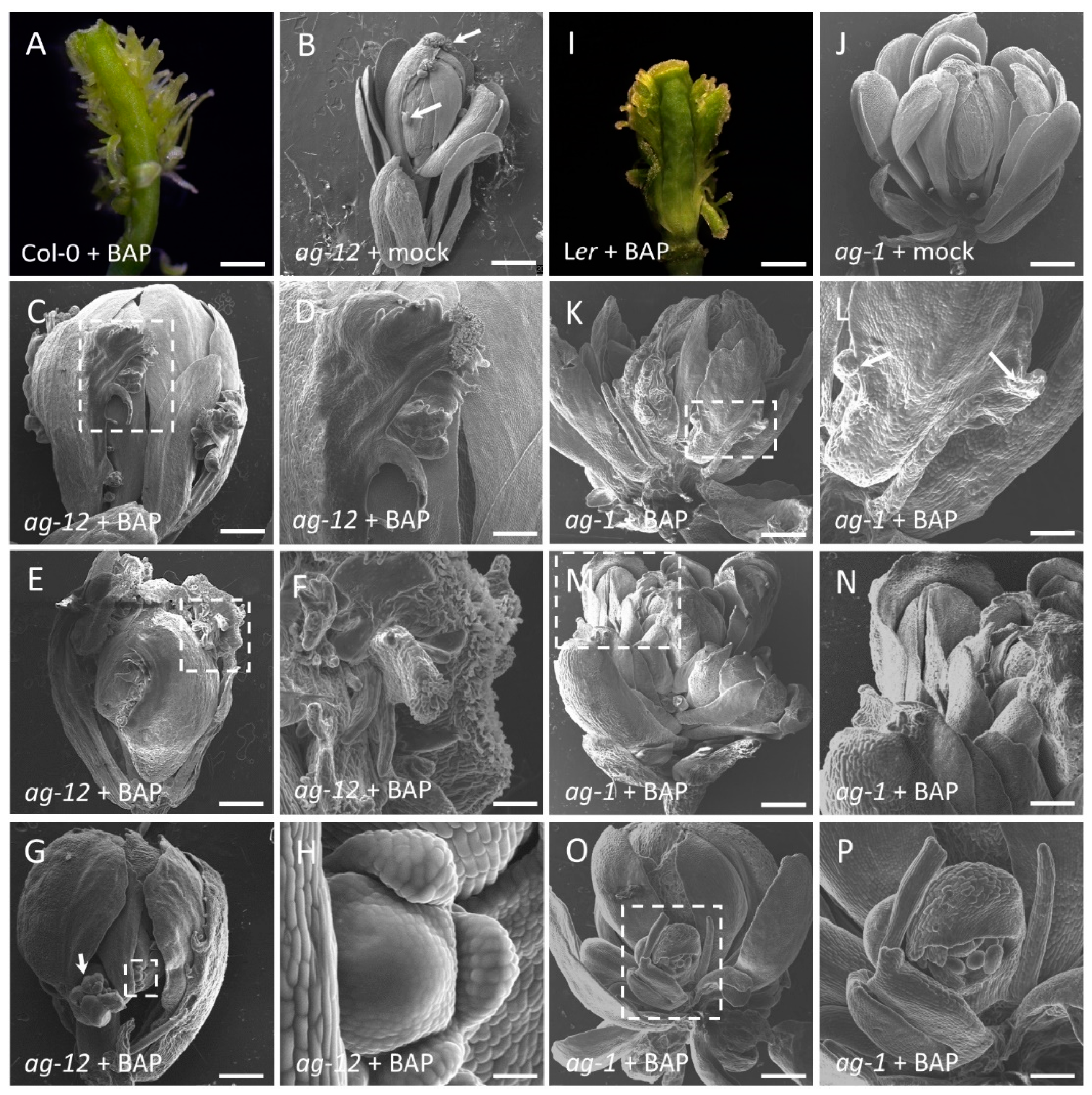
3.1. Exogenous Cytokinin Induces Carpeloid Features in an AG-Dependent Manner

We have previously shown that cytokinin is involved in gynoecium development [8,39,45,49,53]. Repeated exogenous applications of cytokinin (6-benzylamonopurine; BAP) to wild type inflorescences induces ectopic proliferative tissue from the replum, which has at its tip stigmatic-like characteristics (Figure 1A,I; [49]). We decided to analyze the relationship between the transcription factor AGAMOUS (AG) and cytokinin signaling in the establishment of carpeloid features using the same pharmacological assay.
To achieve this, we used two recessive mutants, ag-12 and ag-1, that show replacement of stamens and carpels by petals and sepals, resulting in a flower within a flower phenotype, characteristic of AG loss-of-function phenotypes [9,10,42]. ag-12 and ag-1 are different in phenotypic severity, with ag-12 being a weaker allele compared to the strong ag-1 null allele. In the absence of exogenous BAP, ag-1 does not present any carpeloid structures (Figure 1J), while ag-12 presents some ectopic ovules and stigmatic papillae (Figure 1B), likely due to the fact that this mutant still produces full length transcripts, though at reduced levels compared to wild-type plants [57]. Furthermore, ag-12 shows an elongated pedicel resulting in a separation of the internal flowers, which has been attributed to ag-12 and ag-1 being in different genetic backgrounds [10].
Upon exogenous BAP treatment, we observed in the ag-12 mutant proliferation displaying stigmatic papillae at the apex (85% of 80 observed flowers, Figure 1C–F). In BAP-treated ag-1 plants, 75% of 80 observed flowers showed marginal outgrowth in the sepals, but no stigmatic papillae or any other carpeloid structure (Figure 1K–N). In both mutant alleles, we observed the formation of secondary floral buds in the axil of the sepals (Figure 1G,H,O,P), which has been reported for the strong ag-1 allele [46]. As expected, the mock treatment did not induce any carpeloid structures in ag-12 and ag-1, nor induced the formation of secondary floral buds (Figure 1B,J). These results suggest that the residual AG activity in ag-12 is sufficient to induce carpeloid structures, and that BAP-treatment induces tissue proliferation, as it does in the wild type (Figure 1A,I), and this tissue acquires carpeloid characteristics only in the presence of AG.

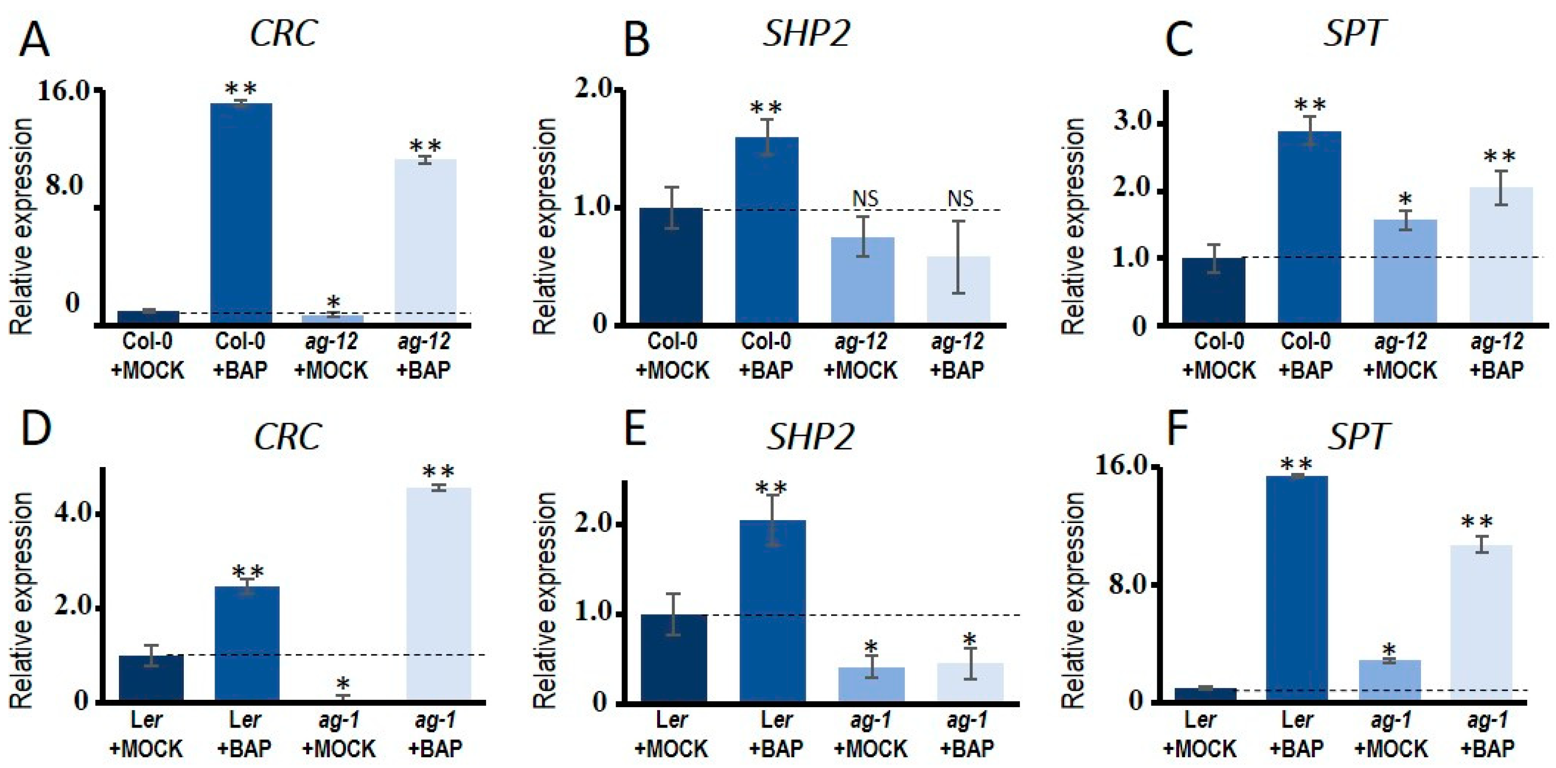
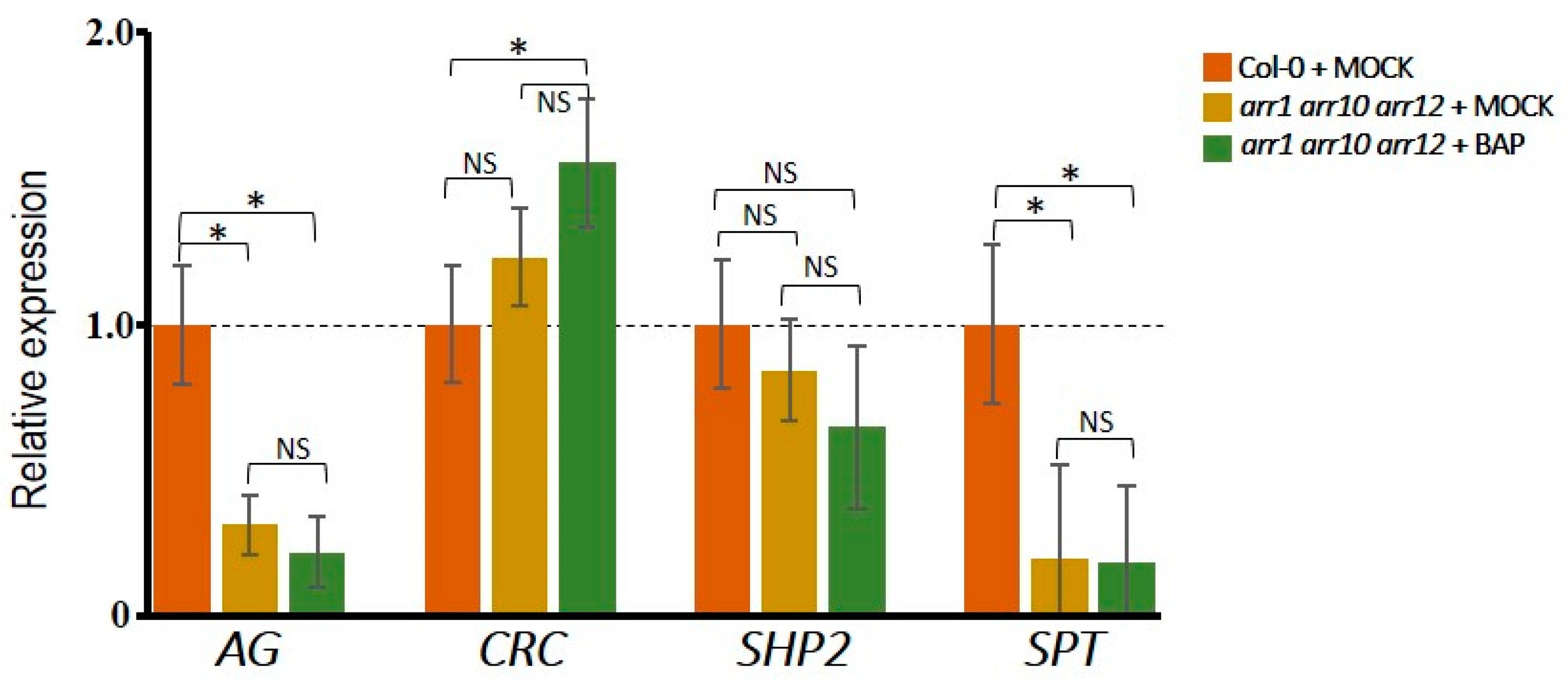
3.2. Cytokinin Induces AG Targets Involved in Gynoecium Development

Three genes have been reported to induce carpel features independently of AG: CRABS CLAW (CRC), SPATULA (SPT), and SHATTERPROOF 2 (SHP2) [16,19]. We wondered whether one or more of these genes could be induced by cytokinin, thus explaining the observed carpeloid features in the ag-12 BAP-treated flowers. In wild type flowers, either Col-0 or Ler, all three genes are induced 24 h after BAP treatment, compared to their corresponding mock controls (Figure 2A–F). In the mock-treated ag-1 null mutant flowers, CRC and SHP2 showed significantly reduced expression compared to the mock-treated wild type flowers (Figure 2D,E). In the weak ag-12 mutant, CRC and SHP2 showed a slightly reduced expression, though SHP2 reduction is not significantly different, compared to the mock-treated wild type flowers. This suggests that AG induces the expression of CRC and SHP2, and the less reduced expression in ag-12 is probably due to the residual AG activity in this mutant. On the other hand, the expression of SPT was increased in both ag mutants compared to the wild type, suggesting that SPT expression is independent of AG (Figure 2C,F). In the BAP-treated ag mutants, CRC and SPT were induced compared to the wild type, while SHP2 expression was not significantly altered by BAP.
This suggests that SHP2 response to cytokinin is dependent on the presence of AG, and that CRC and SPT can respond to cytokinin even with a reduced or absent AG function. Furthermore, these results suggest that the carpeloid structures observed in the BAP-treated weak ag-12 mutant could be due to mainly the presence of residual AG activity, and perhaps in addition to the presence of SHP2 expression. The BAP-induction of CRC and SPT expression alone is not sufficient, because in the strong ag-1 allele no BAP-induced carpeloid features were observed.

3.3. Different Effects of Type-B ARR Proteins on Carpel Promoting Genes

The cytokinin signaling pathway affects gene expression via the type-B ARR transcription factors. We hypothesized that the observed positive effect of cytokinin on CRC, SPT and SHP2 expression could be regulated through the type-B cytokinin response regulators (type-B ARRs). Therefore, we tested by qRT-PCR the expression of CRC, SPT and SHP2 in the arr1 arr10 arr12 triple mutant, which is severely affected in cytokinin signaling [58,59,60,61]. As a positive control, we evaluated the expression of AG, which has already been tested previously in the arr1 arr10 arr12 triple mutant [43]. As reported by Rong and colleagues, AG expression was reduced in the arr triple mutant (Figure 3). Furthermore, also SPT showed a strong reduction in expression (Figure 3). In contrast, the AG direct targets CRC and SHP2 showed no significant change in expression, meaning they are also regulated by other genes (Figure 3). Next, we analyzed the gene expression after 24 h of BAP-treatment. Interestingly, though to be expected, none of the four genes responded to cytokinin in the arr1 arr10 arr12 triple mutant when compared to the mock-treatment of the arr triple mutant (Figure 3). Note, a slight but statistically significant BAP-induction of CRC expression was observed in the arr triple mutant when compared to mock-treated wild type plants. This could suggest that other type-B ARR proteins might regulate CRC expression as well.
As described above, expression of CRC, SPT, and SHP2 in wild type plants is induced upon cytokinin-treatment (Figure 2). So, this may be due to direct or indirect regulation by the transcription factors ARR1, ARR10, and ARR12. Recently, it has been shown that AG is a direct target of ARR1 and ARR10 [43]. Additionally, based on analysis of ChIP-seq data of ARR1, ARR10 and ARR12 [61], we observed type-B ARR binding to the CRC, SPT and SHP2 promoter regions (Figure S1). It is noteworthy that seedlings were used to generate the ChIP-seq data [62], however, clear signals can be observed in the regulatory regions of AG, CRC, SPT, and SHP2 (Figure S1). The ARR1 binding to the second intron of AG that has been previously reported using ChIP by Rong and colleagues [43], can clearly be observed in the ChIP-seq tracks (Figure S1).
Altogether, this indicates that AG, CRC, SPT, and SHP2 have the potential to be directly regulated by type-B ARR proteins. The expression levels of AG and SPT seem to be more dependent on type-B ARRs ARR1, ARR10 and ARR12 function than CRC and SHP2. Concerning the ability to respond to exogenous cytokinin, all four genes are dependent on the type-B ARRs ARR1, ARR10 and ARR12 proteins. Finally, the SHP2 gene responds to cytokinin in an AG-dependent manner and has clear ChIP-seq signals for the type-B ARR proteins, suggesting that SHP2 might be cooperatively regulated by AG and ARRs in this condition.

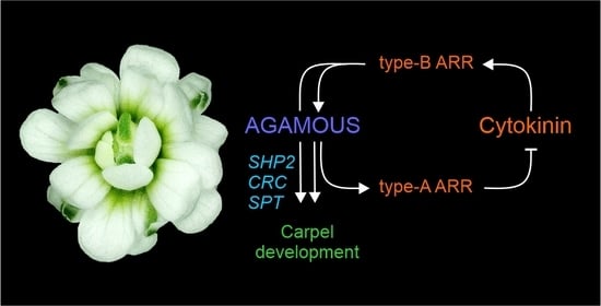
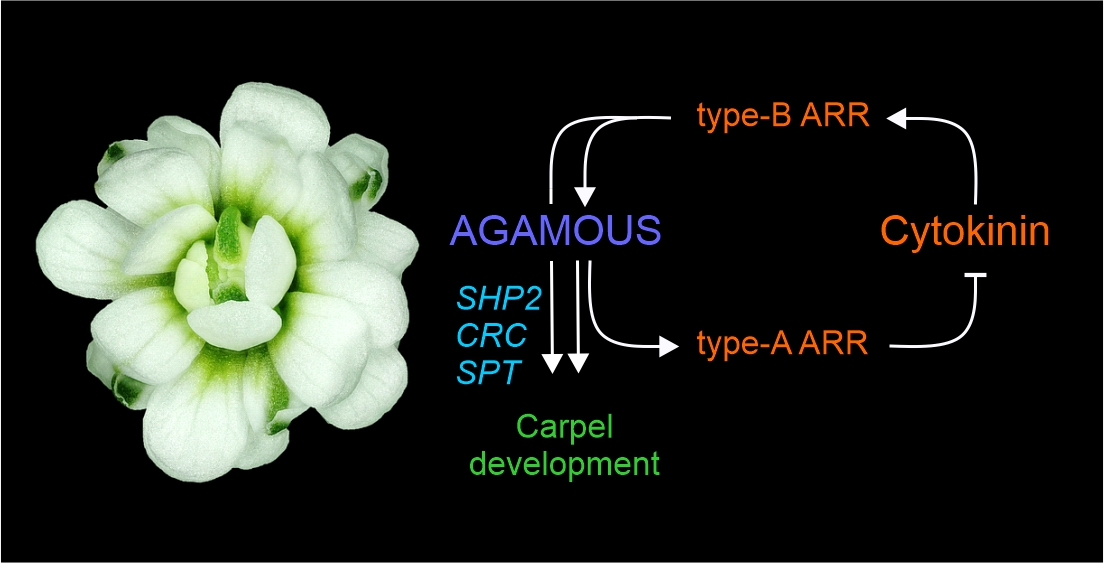
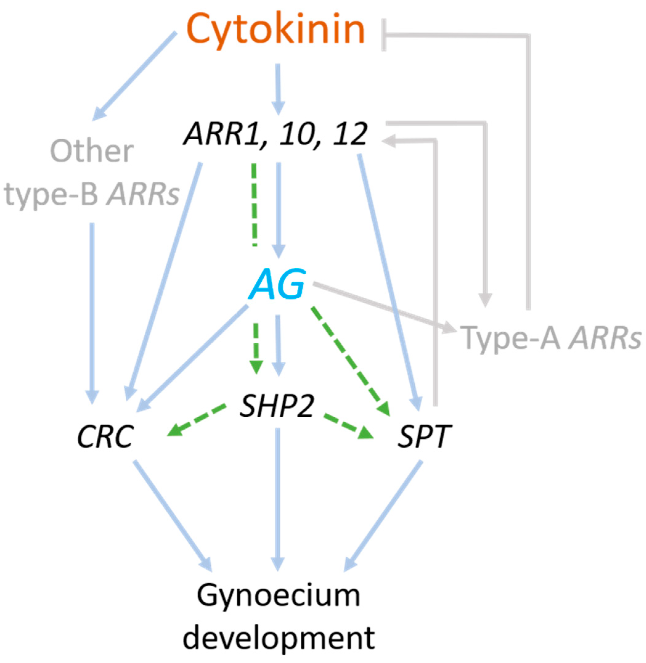
3.4. Model of Regulatory Network between AG and Cytokinin

We generated a model of a possible gene regulatory network where cytokinin signaling acts mainly upstream and in parallel with AG activity during carpel differentiation (Figure 4). AG is important for gynoecium development, and contributes to regulating or directly regulates CRC, SHP2, and SPT. All four genes respond to either reduced or induced cytokinin signaling. Cytokinin-mediated induction of AG and SPT is dependent on the type-B ARR1, ARR10, and ARR12 proteins. Furthermore, cytokinin-induced expression of CRC, SHP2, and SPT is dependent on type-B ARR protein activity, and SHP2 cytokinin-induced expression is also dependent on AG activity. Results also suggest that CRC is probably also regulated by other type-B ARR proteins. In addition, it is known that AG regulates type-A ARR genes, which negatively affect cytokinin signaling, meaning that cytokinin signaling is also downstream of AG [42,62]. It is clear that various pathways in parallel regulate carpel differentiation by controlling the hormonal balance between cytokinin and auxin. In the moment when cytokinin signaling goes down in the FM, probably the auxin pathway takes over to induce carpel differentiation, which can happen by the activity of AG and CRC [41,62], including SPT [39]; all induce auxin biosynthetic genes. In addition, the direct AG target ETT, which also responds to auxin, represses cytokinin signaling [44]. On the other hand, during carpel differentiation, SPT activates the type-B ARR proteins for cytokinin signaling [39]. Likely, all these regulatory interactions are dynamic, and depend on the developmental time, tissue type, and transcription factor complexes. Future research will elucidate more details on how AG and other transcription factors are involved in cytokinin and auxin homeostasis to ensure proper carpel initiation and differentiation.

4. Conclusions

We found that cytokinin induces carpeloid features in an AG-dependent manner and the expression of the transcription factors CRC, SHP2, and SPT that are involved in carpel development. AG is important for gynoecium development, and contributes to regulating, or else directly regulates CRC, SHP2, and SPT. All four genes respond to either reduced or induced cytokinin signaling and have the potential to be regulated by cytokinin via the type-B ARR proteins. We generated a model of a gene regulatory network, where cytokinin signaling is mainly upstream and in parallel with AG activity.

Supplementary Materials

The following are available online at https://www.mdpi.com/article/10.3390/plants10050827/s1, Figure S1. Type-B ARR binding events in the AG, CRC, SPT and SHP2 regulatory regions. Table S1. Oligonucleotides used in this study.

Author Contributions

Conceptualization and designed the experiments: A.G.-F., D.K. and S.d.F.; performed the experiments, A.G.-F.; writing and original draft preparation, A.G.-F., D.K. and S.d.F. All authors have read and agreed to the published version of the manuscript.

Funding

This research was funded by the Mexican National Council of Science and Technology (CONACyT) for a PhD fellowship to A.G.F. Work in the S.D.F. laboratory was financed by the CONACyT grants CB-2012-177739, CB-2017-2018-A1-S-10126, and FC-2015-2/1061. D.K. acknowledges the Discovery Grant RGPIN-2018-04897 from the Natural Sciences and Engineering Council of Canada. A.G.F., S.D.F., and D.K. thank Mitacs Globalink Research Award no. IT16047 of Canada.

Institutional Review Board Statement

Not applicable.

Informed Consent Statement

Not applicable.

Data Availability Statement

No new data were created or analyzed in this study. Data sharing is not applicable to this article.

Acknowledgments

We are grateful to Lino Sánchez-Segura for initial technical assistance with scanning electron microscopy, and Victor M. Zúñiga-Mayo for exploratory work on the ag-12 mutant. We thank the ABRC stock center and Frank Wellmer for seeds. We are indebted to the reviewers who helped during the review process significantly improving this story.

Conflicts of Interest

The authors declare no conflict of interest.

References

  1. Gaillochet, C.; Lohmann, J.U. The never-ending story: From pluripotency to plant developmental plasticity. Development 2015, 142, 2237–2249. [Google Scholar] [CrossRef] [Green Version]
  2. Smyth, D.R.; Bowman, J.L.; Meyerowitz, E.M. Early flower development in Arab. Plant Cell 1990, 2, 755–767. [Google Scholar] [PubMed] [Green Version]
  3. Alvarez-Buylla, E.R.; Benítez, M.; Corvera-Poiré, A.; Chaos Cador, A.; de Folter, S.; De Buen Gamboa, A.; Garay-Arroyo, A.; García-Ponce, B.; Jaimes-Miranda, F.; Pérez-Ruiz, R.V.; et al. Flower Development. Arab. Book 2010, 8, 1–58. [Google Scholar] [CrossRef] [PubMed] [Green Version]
  4. Bowman, J.L.; Smyth, D.R.; Meyerowitz, E.M. The ABC model of flower development: Then and now. Development 2012, 4098, 4095–4098. [Google Scholar] [CrossRef] [PubMed] [Green Version]
  5. Denay, G.; Chahtane, H.; Tichtinsky, G.; Parcy, F. A flower is born: An update on Arabidopsis floral meristem formation. Curr. Opin. Plant Biol. 2017, 35, 15–22. [Google Scholar] [CrossRef] [PubMed]
  6. Wils, C.R.; Kaufmann, K. Gene-regulatory networks controlling inflorescence and flower development in Arabidopsis thaliana. Biochim. Biophys. Acta-Gene Regul. Mech. 2017, 1860, 95–105. [Google Scholar] [CrossRef]
  7. Thomson, B.; Wellmer, F. Molecular regulation of flower development. Curr. Topics Dev. Biol. 2019, 131, 185–210. [Google Scholar]
  8. Zúñiga-Mayo, V.M.; Gómez-Felipe, A.; Herrera-Ubaldo, H.; de Folter, S. Gynoecium development: Networks in Arabidopsis and beyond. J. Exp. Bot. 2019, 70, 1447–1460. [Google Scholar] [CrossRef]
  9. Bowman, J.L.; Smyth, D.R.; Meyerowitz, E.M. Genes directing flower development in Arabidopsis. Plant Cell 1989, 1, 37–52. [Google Scholar]
  10. Yanofsky, M.F.; Ma, H.; Bowman, J.L.; Drews, G.N.; Feldmann, K.A.; Meyerowitz, E.M. The protein encoded by the Arabidopsis homeotic gene agamous resembles transcription factors. Nature 1990, 346, 35–39. [Google Scholar] [CrossRef]
  11. Coen, E.S.; Meyerowitz, E.M. The war of the whorls: Genetic interactions controlling flower development. Nature 1991, 353, 31–37. [Google Scholar] [CrossRef]
  12. Dreni, L.; Kater, M.M. MADS reloaded: Evolution of the AGAMOUS subfamily genes. New Phytol. 2014, 201, 717–732. [Google Scholar] [CrossRef] [PubMed]
  13. Bowman, J.L.; Smyth, D.R.; Meyerowitz, E.M. Genetic interactions among floral homeotic genes of Arabidopsis. Development 1991, 112, 1–20. [Google Scholar] [PubMed]
  14. Mizukami, Y.; Ma, H. Ectopic expression of the floral homeotic gene AGAMOUS in transgenic Arabidopsis plants alters floral organ identity. Cell 1992, 71, 119–131. [Google Scholar] [CrossRef]
  15. Bowman, J.L.; Drews, G.N.; Meyerowitz, E.M. Expression of the Arabidopsis floral homeotic gene AGAMOUS is restricted to specific cell types late in flower development. Plant Cell 1991, 3, 749–758. [Google Scholar]
  16. Alvarez, J.; Smyth, D.R. CRABS CLAW and SPATULA, two Arabidopsis genes that control carpel development in parallel with AGAMOUS. Development 1999, 126, 2377–2386. [Google Scholar] [PubMed]
  17. Bowman, J.L.; Smyth, D.R. CRABS CLAW, a gene that regulates carpel and nectary development in Arabidopsis, encodes a novel protein with zinc finger and helix-loop-helix domains. Development 1999, 126, 2387–2396. [Google Scholar]
  18. Heisler, M.G.B.; Atkinson, A.; Bylstra, Y.H.; Walsh, R.; Smyth, D.R. SPATULA, a gene that controls development of carpel margin tissues in Arabidopsis, encodes a bHLH protein. Development 2001, 128, 1089–1098. [Google Scholar]
  19. Pinyopich, A.; Ditta, G.S.; Savidge, B.; Liljegren, S.J.; Baumann, E.; Wisman, E.; Yanofsky, M.F. Assessing the redundancy of MADS-box genes during carpel and ovule development. Nature 2003, 424, 85–88. [Google Scholar] [CrossRef]
  20. Favaro, R.; Pinyopich, A.; Battaglia, R.; Kooiker, M.; Borghi, L.; Ditta, G.; Yanofsky, M.F.; Kater, M.M.; Colombo, L. MADS-Box protein complexes control carpel and ovule developement in Arabidopsis. Plant Cell 2003, 15, 2603–2611. [Google Scholar] [CrossRef] [Green Version]
  21. Groszmann, M.; Paicu, T.; Alvarez, J.P.; Swain, S.M.; Smyth, D.R. SPATULA and ALCATRAZ, are partially redundant, functionally diverging bHLH genes required for Arabidopsis gynoecium and fruit development. Plant J. 2011, 68, 816–829. [Google Scholar] [CrossRef]
  22. Gross, T.; Broholm, S.; Becker, A. CRABS CLAW acts as a bifunctional transcription factor in flower development. Front. Plant Sci. 2018, 9, 1–13. [Google Scholar] [CrossRef] [Green Version]
  23. Ferrándiz, C.; Fourquin, C.; Prunet, N.; Scutt, C.P.; Sundberg, E.; Trehin, C.; Vialette-Guiraud, A.C.M. Carpel development. Adv. Bot. Res. 2010, 55, 1–73. [Google Scholar] [CrossRef]
  24. Gómez-Mena, C.; de Folter, S.; Costa, M.M.R.; Angenent, G.C.; Sablowski, R. Transcriptional program controlled by the floral homeotic gene AGAMOUS during early organogenesis. Development 2005, 132, 429–438. [Google Scholar] [CrossRef] [Green Version]
  25. Ó’Maoiléidigh, D.S.; Wuest, S.E.; Rae, L.; Raganelli, A.; Ryan, P.T.; Kwaśniewska, K.; Das, P.; Lohan, A.J.; Loftus, B.; Graciet, E.; et al. Control of reproductive floral organ identity specification in arabidopsis by the C function regulator AGAMOUS. Plant Cell 2013, 25, 2482–2503. [Google Scholar] [CrossRef] [PubMed] [Green Version]
  26. Larsson, E.; Roberts, C.J.; Claes, A.R.; Franks, R.G.; Sundberg, E. Polar auxin transport is essential for medial versus lateral tissue specification and vascular-mediated valve outgrowth in Arabidopsis gynoecia. Plant Physiol. 2014, 166, 1998–2012. [Google Scholar] [CrossRef] [Green Version]
  27. Reyes-Olalde, J.I.; Zuñiga-Mayo, V.M.; Chávez Montes, R.A.; Marsch-Martínez, N.; de Folter, S. Inside the gynoecium: At the carpel margin. Trends Plant Sci. 2013, 18, 644–655. [Google Scholar] [CrossRef]
  28. Hawkins, C.; Liu, Z. A model for an early role of auxin in Arabidopsis gynoecium morphogenesis. Front. Plant Sci. 2014, 5, 1–12. [Google Scholar] [CrossRef] [Green Version]
  29. Sehra, B.; Franks, R.G. Auxin and cytokinin act during gynoecial patterning and the development of ovules from the meristematic medial domain. Wiley Interdiscip. Rev. Dev. Biol. 2015, 4, 555–571. [Google Scholar] [CrossRef]
  30. Marsch-Martínez, N.; de Folter, S. Hormonal control of the development of the gynoecium. Curr. Opin. Plant Biol. 2016, 29, 104–114. [Google Scholar] [CrossRef]
  31. Reyes-Olalde, J.I.; de Folter, S. Control of stem cell activity in the carpel margin meristem (CMM) in Arabidopsis. Plant Reprod. 2019, 32, 123–136. [Google Scholar] [CrossRef]
  32. Simonini, S.; Østergaard, L. Female reproductive organ formation: A multitasking endeavor. Curr. Top. Dev. Biol. 2019, 131, 337–371. [Google Scholar] [CrossRef] [PubMed]
  33. Xu, Y.; Yamaguchi, N.; Gan, E.S.; Ito, T. When to stop: An update on molecular mechanisms of floral meristem termination. J. Exp. Bot. 2019, 70, 1711–1718. [Google Scholar] [CrossRef] [PubMed]
  34. Skoog, F.; Miller, C.O. Chemical regulation of growth and organ formation in plant tissue cultured in vitro. Symp. Soc. Exp. Biol. 1957, 11, 118–131. [Google Scholar] [PubMed]
  35. Schaller, G.E.; Bishopp, A.; Kieber, J.J. The yin-yang of hormones: Cytokinin and auxin interactions in plant development. Plant Cell 2015, 27, 44–63. [Google Scholar] [CrossRef] [PubMed] [Green Version]
  36. Lucero, L.E.; Uberti-Manassero, N.G.; Arce, A.L.; Colombatti, F.; Alemano, S.G.; Gonzalez, D.H. TCP15 modulates cytokinin and auxin responses during gynoecium development in Arabidopsis. Plant J. 2015, 84, 267–282. [Google Scholar] [CrossRef]
  37. Schuster, C.; Gaillochet, C.; Lohmann, J.U. Arabidopsis HECATE genes function in phytohormone control during gynoecium development. Development 2015, 142, 3343–3350. [Google Scholar] [CrossRef] [Green Version]
  38. Müller, C.J.; Larsson, E.; Spíchal, L.; Sundberg, E. Cytokinin-auxin crosstalk in the gynoecial primordium ensures correct domain patterning. Plant Physiol. 2017, 175, 1144–1157. [Google Scholar] [CrossRef]
  39. Reyes-Olalde, J.I.; Zúñiga-Mayo, V.M.; Serwatowska, J.; Chavez Montes, R.A.; Lozano-Sotomayor, P.; Herrera-Ubaldo, H.; Gonzalez-Aguilera, K.L.; Ballester, P.; Ripoll, J.J.; Ezquer, I.; et al. The bHLH transcription factor SPATULA enables cytokinin signaling, and both activate auxin biosynthesis and transport genes at the medial domain of the gynoecium. PLoS Genet. 2017, 13, e1006726. [Google Scholar] [CrossRef]
  40. Simonini, S.; Bencivenga, S.; Trick, M.; Østergaard, L. Auxin-induced modulation of ETTIN activity orchestrates gene expression in arabidopsis. Plant Cell 2017, 29, 1864–1882. [Google Scholar] [CrossRef] [Green Version]
  41. Yamaguchi, N.; Huang, J.; Xu, Y.; Tanoi, K.; Ito, T. Fine-tuning of auxin homeostasis governs the transition from floral stem cell maintenance to gynoecium formation. Nat. Commun. 2017, 8. [Google Scholar] [CrossRef] [Green Version]
  42. Ó‘Maoiléidigh, D.S.; Stewart, D.; Zheng, B.; Coupland, G.; Wellmer, F. Floral homeotic proteins modulate the genetic program for leaf development to suppress trichome formation in flowers. Development 2018, 145. [Google Scholar] [CrossRef] [PubMed] [Green Version]
  43. Rong, X.F.; Sang, Y.L.; Wang, L.; Meng, W.J.; Zou, C.H.; Dong, Y.X.; Bie, X.M.; Cheng, Z.J.; Zhang, X.S. Type-B ARRs control carpel regeneration through mediating AGAMOUS expression in Arabidopsis. Plant Cell Physiol. 2018, 59, 761–769. [Google Scholar] [CrossRef] [Green Version]
  44. Zhang, K.; Wang, R.; Zi, H.; Li, Y.; Cao, X.; Li, D.; Guo, L.; Tong, J.; Pan, Y.; Jiao, Y.; et al. AUXIN RESPONSE FACTOR3 regulates floral meristem determinacy by repressing cytokinin biosynthesis and signaling. Plant Cell 2018, 30, 324–346. [Google Scholar] [CrossRef] [Green Version]
  45. Cerbantez-Bueno, V.E.; Zúñiga-Mayo, V.M.; Reyes-Olalde, J.I.; Lozano-Sotomayor, P.; Herrera-Ubaldo, H.; Marsch-Martinez, N.; de Folter, S. Redundant and non-redundant functions of the AHK cytokinin receptors during gynoecium development. Front. Plant Sci. 2020, 11, 1–13. [Google Scholar] [CrossRef]
  46. Venglat, S.P.; Sawhney, V.K. Benzylaminopurine induces phenocopies of floral meristem and organ identity mutants in wild-type Arabidopsis plants. Planta 1996, 198, 480–487. [Google Scholar] [CrossRef]
  47. Bartrina, I.; Otto, E.; Strnad, M.; Werner, T.; Schmülling, T. Cytokinin regulates the activity of reproductive meristems, flower organ size, ovule formation, and thus seed yield in Arabidopsis thaliana. Plant Cell 2011, 23, 69–80. [Google Scholar] [CrossRef] [Green Version]
  48. Nibau, C.; Di Stilio, V.S.; Wu, H.M.; Cheung, A.Y. Arabidopsis and Tobacco SUPERMAN regulate hormone signalling and mediate cell proliferation and differentiation. J. Exp. Bot. 2011, 62, 949–961. [Google Scholar] [CrossRef] [PubMed] [Green Version]
  49. Marsch-Martínez, N.; Ramos-Cruz, D.; Irepan Reyes-Olalde, J.; Lozano-Sotomayor, P.; Zúñiga-Mayo, V.M.; de Folter, S. The role of cytokinin during Arabidopsis gynoecia and fruit morphogenesis and patterning. Plant J. 2012, 72, 222–234. [Google Scholar] [CrossRef]
  50. Xing, S.; Salinas, M.; Garcia-Molina, A.; Höhmann, S.; Berndtgen, R.; Huijser, P. SPL8 and miR156-targeted SPL genes redundantly regulate Arabidopsis gynoecium differential patterning. Plant J. 2013, 75, 566–577. [Google Scholar] [CrossRef] [PubMed]
  51. Martínez-Fernández, I.; Sanchís, S.; Marini, N.; Balanzá, V.; Ballester, P.; Navarrete-Gómez, M.; Oliveira, A.C.; Colombo, L.; Ferrándiz, C. The effect of NGATHA altered activity on auxin signaling pathways within the Arabidopsis gynoecium. Front. Plant Sci. 2014, 5, 1–11. [Google Scholar] [CrossRef] [Green Version]
  52. Moubayidin, L.; Østergaard, L. Dynamic control of auxin distribution imposes a bilateral-to-radial symmetry switch during gynoecium development. Curr. Biol. 2014, 24, 2743–2748. [Google Scholar] [CrossRef] [PubMed] [Green Version]
  53. Zúñiga-Mayo, V.M.; Reyes-Olalde, J.I.; Marsch-Martinez, N.; de Folter, S. Cytokinin treatments affect the apical-basal patterning of the Arabidopsis gynoecium and resemble the effects of polar auxin transport inhibition. Front. Plant Sci. 2014, 5, 1–8. [Google Scholar] [CrossRef] [Green Version]
  54. Urbanus, S.L.; de Folter, S.; Shchennikova, A.V.; Kaufmann, K.; Immink, R.G.; Angenent, G.C. In planta localisation patterns of MADS domain proteins during floral development in Arabidopsis thaliana. BMC Plant Biol. 2009, 9, 1–16. [Google Scholar] [CrossRef] [Green Version]
  55. Argyros, R.D.; Mathews, D.E.; Chiang, Y.H.; Palmer, C.M.; Thibault, D.M.; Etheridge, N.; Argyros, D.A.; Mason, M.G.; Kieber, J.J.; Schallera, G.E. Type B response regulators of Arabidopsis play key roles in cytokinin signaling and plant development. Plant Cell 2008, 20, 2102–2116. [Google Scholar] [CrossRef] [Green Version]
  56. Livak, K.J.; Schmittgen, T.D. Analysis of relative gene expression data using real-time quantitative PCR and the 2-ΔΔCT method. Methods 2001, 25, 402–408. [Google Scholar] [CrossRef]
  57. Gao, Y.; Zhao, Y. Epigenetic suppression of T-DNA insertion mutants in Arabidopsis. Mol. Plant 2013, 6, 539–545. [Google Scholar] [CrossRef] [Green Version]
  58. Mason, M.G.; Mathews, D.E.; Argyros, D.A.; Maxwell, B.B.; Kieber, J.J.; Alonso, J.M.; Ecker, J.R.; Schaller, G.E. Multiple type-B response regulators mediate cytokinin signal transduction in Arabidopsis. Plant Cell 2005, 17, 3007–3018. [Google Scholar] [CrossRef] [Green Version]
  59. Yokoyama, A.; Yamashino, T.; Amano, Y.I.; Tajima, Y.; Imamura, A.; Sakakibara, H.; Mizuno, T. Type-B ARR transcription factors, ARR10 and ARR12, are implicated in cytokinin-mediated regulation of protoxylem differentiation in roots of Arabidopsis thaliana. Plant Cell Physiol. 2007, 48, 84–96. [Google Scholar] [CrossRef] [PubMed] [Green Version]
  60. Ishida, K.; Yamashino, T.; Yokoyama, A.; Mizuno, T. Three type-B response regulators, ARR1, ARR10 and ARR12, play essential but redundant roles in cytokinin signal transduction throughout the life cycle of Arabidopsis thaliana. Plant Cell Physiol. 2008, 49, 47–57. [Google Scholar] [CrossRef] [PubMed] [Green Version]
  61. Xie, M.; Chen, H.; Huang, L.; O’Neil, R.C.; Shokhirev, M.N.; Ecker, J.R. A B-ARR-mediated cytokinin transcriptional network directs hormone cross-regulation and shoot development. Nat. Commun. 2018, 9, 1–13. [Google Scholar] [CrossRef] [Green Version]
  62. Yamaguchi, N.; Huang, J.; Tatsumi, Y.; Abe, M.; Sugano, S.S.; Kojima, M.; Takebayashi, Y.; Kiba, T.; Yokoyama, R.; Nishitani, K.; et al. Chromatin-mediated feed-forward auxin biosynthesis in floral meristem determinacy. Nat. Commun. 2018, 9, 1–7. [Google Scholar] [CrossRef]
Figure 1. Cytokinin-induced carpeloid structures and secondary floral buds in flowers of plants lacking AG activity. Scanning electron micrographs of floral phenotypes of ag mutant plants treated with mock or cytokinin (100 µM BAP) solution; pictures are of one week after the treatment. (A) Micrograph of a gynoecium Col-0 treated with cytokinin solution. (B) Mock-treated ag-12 flower showing ectopic ovules and stigmatic papillae structures (as indicated by the white arrows). (CH) BAP-treated ag-12 flowers, stigmatic and carpeloid structures are visible. (D,F) Close up from dotted white square in (C) and (E), respectively. (G) BAP-treated ag-12 flower showing floral buds (as indicated by the white arrow). (H) Close up from dotted white square in (G) showing a floral meristem. (I) Micrograph of a gynoecium Ler treated with cytokinin solution. (J) Mock treated ag-1 flowers without carpeloid structures (some sepals and petals were removed). (KN) BAP-treated ag-1 flowers (some sepals and petals were removed) showing only some proliferating tissue (as indicated by the white arrows). (L,N) Close up from dotted white square in (K) and (M), respectively. (O) BAP-treated ag-1 flower showing a floral bud (as indicated by the dotted white square). (P) Close up from dotted white square in (O) showing a floral bud. (D,F,H,L,N,P) Close up from dotted white square with a higher magnification. Scale bars: (BG, JO) 200 µm, (A,I,P) 100 µm, (H) 30 µm.
Plants 10 00827 g001
Figure 2. Expression levels of CRC, SPT and SHP2 in ag mutant backgrounds and wild type. (AF) qRT-PCR expression data for CRC, SPT and SHP2 in floral buds after 24 h of cytokinin-treatment (100 µM BAP) or mock-treatment in the ag-12 mutant (AC) and in the ag-1 mutant (DF). The results were obtained for three independent biological replicates with three technical replicates for each one. One-way ANOVA was used to evaluate the significant differences. Significant values are indicated as follow: (*) p ˂ 0.05, (**) p ˂ 0.01, not significant (NS), compared to mock-treated wild type.
Plants 10 00827 g002
Figure 3. Expression levels of AG, CRC, SPT and SHP2 in the type-B arr1 arr10 arr12 triple mutant. qRT-PCR expression data for AG, CRC, SPT and SHP2 in floral buds after 24 h of cytokinin-treatment (100 µM BAP) or mock-treatment in the type-B arr1 arr10 arr12 triple mutant. The results were obtained for three independent biological replicates with three technical replicates for each one. One-way ANOVA was used to evaluate the significant differences. Significant values are indicated as follow: (*) p ˂ 0.05, not significant (NS), compared to mock-treated wild type.
Plants 10 00827 g003
Figure 4. Model of a gene regulatory network of the relationship between AG, CRC, SPT, SHP2, and cytokinin signaling during early gynoecium development. AG is important for gynoecium development, and contributes to regulating or directly regulates CRC, SHP2, and SPT. All four genes respond to either reduced or induced cytokinin signaling. Cytokinin-mediated induction of AG and SPT is dependent on type-B ARR proteins (ARR1, ARR10, ARR12). The SHP2 gene responds to cytokinin in an AG-dependent manner and might be cooperatively regulated by AG and ARR1 in this condition. AG regulates type-A ARR genes, which negatively feedbacks to cytokinin signaling (in grey colored lines); a negative feedback loop is important for homeostasis. The second negative feedback loop is activated by the type-B ARR proteins. Green dashed lines are suggested interactions and might be direct or indirect regulatory interactions. Not indicated, but all transcription factors are also regulated by other factors.
Plants 10 00827 g004
Publisher’s Note: MDPI stays neutral with regard to jurisdictional claims in published maps and institutional affiliations.

