Review

Small Non-Coding RNAs at the Crossroads of Regulatory Pathways Controlling Somatic Embryogenesis in Seed Plants

1
BioISI—Biosystems & Integrative Sciences Institute, Faculty of Sciences, University of Lisboa, 1749-016 Lisboa, Portugal
2
Instituto de Tecnologia Química e Biológica António Xavier, Universidade Nova de Lisboa, 2780-157 Oeiras, Portugal
3
Centre for Functional Ecology, Department of Life Sciences, University of Coimbra, Calçada Martim de Freitas, 3000-456 Coimbra, Portugal
4
iBET, Instituto de Biologia Experimental e Tecnológica, Apartado 12, 2781-901 Oeiras, Portugal
*
Author to whom correspondence should be addressed.
Plants 2021, 10(3), 504; https://doi.org/10.3390/plants10030504
Submission received: 5 February 2021 / Revised: 26 February 2021 / Accepted: 1 March 2021 / Published: 9 March 2021

Abstract

:
Small non-coding RNAs (sncRNAs) are molecules with important regulatory functions during development and environmental responses across all groups of terrestrial plants. In seed plants, the development of a mature embryo from the zygote follows a synchronized cell division sequence, and growth and differentiation events regulated by highly regulated gene expression. However, given the distinct features of the initial stages of embryogenesis in gymnosperms and angiosperms, it is relevant to investigate to what extent such differences emerge from differential regulation mediated by sncRNAs. Within these, the microRNAs (miRNAs) are the best characterized class, and while many miRNAs are conserved and significantly represented across angiosperms and other seed plants during embryogenesis, some miRNA families are specific to some plant lineages. Being a model to study zygotic embryogenesis and a relevant biotechnological tool, we systematized the current knowledge on the presence and characterization of miRNAs in somatic embryogenesis (SE) of seed plants, pinpointing the miRNAs that have been reported to be associated with SE in angiosperm and gymnosperm species. We start by conducting an overview of sncRNA expression profiles in the embryonic tissues of seed plants. We then highlight the miRNAs described as being involved in the different stages of the SE process, from its induction to the full maturation of the somatic embryos, adding references to zygotic embryogenesis when relevant, as a contribution towards a better understanding of miRNA-mediated regulation of SE.

1. Introduction

Small non-coding RNAs (sncRNAs) are usually 20–24 nt long RNA molecules with important regulatory functions during development and environmental responses across all groups of land plants, from bryophytes like Physcomitrella patens [1] and Marchantia polymorpha [2], to the gymnosperms [3] and angiosperms [4]. These small molecules are classified into several classes according to their biogenesis and roles [5], the micro RNAs (miRNAs) being the best characterized group of sncRNAs, and the short-interfering RNAs (siRNAs) the other broad category of plant sncRNAs. miRNAs are typically molecules with 21–22 nt produced from a MIR gene. Commonly, these tiny molecules act at the post-transcriptional level based on nearly perfect sequence complementary recognition of their target mRNAs, leading to the cleavage or translation inhibition of the target. The most abundant subclasses of siRNAs, with 24-nt or 21–22-nt, can either lead to transcriptional or post-transcriptional gene silencing depending on if they derive from transposons or repeats (heterochromatic siRNAs), or if they are triggered by specific miRNAs (trans-acting siRNAs) [6]. The involvement of these classes of molecules in several plant developmental processes, including embryogenesis, and in response to environmental conditions has been often reported and reviewed [4,7,8,9].
Somatic embryogenesis (SE) refers to the process whereby somatic cells can be induced to undergo a series of developmental stages that mirror the development of a zygotic embryo within the seed and has been widely used as an experimental model to study zygotic embryogenesis (ZE). In Arabidopsis thaliana (Arabidopsis), the distinct stages of embryo development are characterized in detail and include the zygotic, globular, heart, torpedo and cotyledonary (mature) embryo stages [10]. At the zygotic stage, asymmetric cell division occurs, originating a small apical cell from which most parts of the mature embryo will form, and a basal cell whose divisions later originate the distal part of the root apex and the suspensor. The globular embryo results from a series of cell divisions, and at the 16-celled proembryo it is possible to identify a superficial cell layer, the protoderm, that will become the epidermal tissue. Polar auxin transport mediated by the PIN proteins is critical for establishing the distal part of the root apical meristem and later on for the formation of the two cotyledon primordia at the heart stage. At the latest stages of embryogenesis, the torpedo and mature embryo, the basic plan of the future plant is formed, bearing the root apical meristem at one end and the shoot apical meristem localized between the two cotyledons at the other end.
Given the distinct features found right from the very initial stages of embryogenesis in gymnosperms and angiosperms, it is relevant to investigate to what extent such differences emerge from differential post-transcriptional regulation mediated by sncRNAs. As an example, the critical event in early embryogenesis of angiosperms whereby the asymmetric cell division of the zygote originates the apical and the basal cell, which will follow different cell-fate specification pathways, does not occur in gymnosperms. In these species, the zygote goes through several rounds of nuclear duplication without cytokinesis followed by cellularization to form two tiers of cells and a proembryo with eight cells [11]. Recent work uncovering cell lineage-specific transcriptome in Arabidopsis shows distinct molecular pathways at the apical and basal cell [12] following the first cell division, a stage that has no morphological equivalent in gymnosperm embryogenesis.
Another study with Arabidopsis mutants in genes required for miRNA biogenesis (dcl1, se-1 and hyl1-2) provided evidence that miRNAs are required at the zygote stage [13]. Some of these mutants showed loss of zygote polarity, presenting a more symmetric pattern of the first cell division and subsequent horizontal division of the apical cell, instead of vertical division. These observations lead to several pertinent questions that can start to be addressed by gathering the evidence available in several reports in the literature. For instance, it is important to determine if initial zygotic/somatic embryo development in gymnosperms is regulated by miRNAs, if the regulation is mediated by the same miRNA families in both gymnosperms and angiosperms and if the roles of these miRNAs are conserved, i.e., if their target transcripts show conserved functions.
The same kinds of questions apply to other stages of embryo development, where distinct features are evident in the two plant groups. In fact, in addition to the occurrence of cleavage polyembryony, a common phenomenon in gymnosperms during early embryogeny, the differentiation of multiple cotyledons, instead of only two cotyledons, around the shoot apical meristem (SAM) during late embryogeny, is also a highly distinctive characteristic that confers radial symmetry to the embryo instead of the bilateral symmetry observed in angiosperms [14]. Furthermore, it is also important to focus on the events that occur prior to embryo development, during the induction of SE, where a somatic cell changes its fate in order to become an embryo.
Here, we gather the current knowledge on the presence and characterization of sncRNAs, especially miRNAs, during embryogenesis, focusing on these two major groups of plants, the angiosperms and gymnosperms. Despite the aforementioned differences in embryo development, highlighting the similarities and the main differences already identified so far will help to address if regulation of SE by sncRNAs occurs in a similar way in these two groups. Within the gymnosperms, SE is especially relevant in conifers where it offers potential benefits in the clonal propagation of selected germplasm. However, one of the main obstacles to its industrial application has been the limited range of tissues from which SE can be induced. This fact can be hypothetically related to the repertoire of transcripts, coding and non-coding, present in the tissues at the time of excision (explant preparation). In the sections below, we start by providing a broad overview of the sncRNA landscape in embryo tissues, followed by a more detailed description of already characterized miRNAs related to several stages of embryogenesis, from the induction phase to later stages of embryo development, focusing on SE.

2. Small RNA Expression Profiles in Embryonic Tissues

Seed tissues, including the embryos, show characteristic expression profiles. In Arabidopsis, a recent study highlighted the uniqueness of embryo transcriptomes when compared to other post-embryonic tissues [15]. Given the role of sncRNAs in the control of gene expression, it is not surprising that sncRNA transcriptomes of embryos have revealed characteristic features underling some of the regulatory pathways that operate at specific developmental stages. While diverse sncRNA profiles have been detected in seed tissues, based mostly on available data from angiosperms, there seems to be a general tendency for a higher abundance and diversity of 24-nt sncRNAs regardless of the developmental stage of the seed [16]. In some gymnosperm species, like maritime pine (Pinus pinaster), 24-nt sncRNAs were also highly expressed in embryos when compared to vegetative tissues [3,17] and more abundant in somatic embryos than in zygotic embryos at equivalent stages of development. In Picea glauca, another gymnosperm species, both 21-nt and 24-nt sncRNAs were abundant in embryos [18], this profile being in contrast to that of buds [19], confirming that the 24-nt sncRNAs are mostly present in reproductive tissues. It has been proposed that such abundance is associated with epigenetic safeguard mechanisms for controlling the expression of transposons and heterochromatic repeats during embryo development [16,20]. A recent study [21] showed that siRNAs generated from transposable elements (TEs), highly numerous in Arabidopsis embryos and typically 24-nt long, originate both from euchromatic TEs, being required not only during embryogenesis as a way of silencing TEs, but also from heterochromatic TEs specifically required during embryogenesis to help establish TE methylation de novo, which is then maintained during post-embryogenesis independently of siRNAs.
Despite the recognized importance of siRNAs during embryogenesis and its apparent increased abundance in somatic embryos, most probably as a response to artificially-provided environmental conditions, these sequences remain mostly uncharacterized, with most of the attention being focused on miRNAs, especially those that are conserved among angiosperms.
Although many miRNAs are conserved and significantly represented across angiosperms and other seed plants during development, including embryogenesis, some miRNA families exist that are specific to some plant lineages (Table 1).
Some of these specific or non-conserved miRNAs might play yet uncovered roles in embryo development. For example, in the conifer P. pinaster, in addition to miRNA families conserved across land plants, eleven miRNA families, such as miR1316 and miR3699, were detected in embryos and megagametophytes that are conifer-specific. Among these, miR946, miR947, miR950, miR951, miR1311, miR1312, miR1313 and miR3701 were also found in the transition from dormancy to germination of Japanese larch (Larix leptolepis (syn. L. kaempferi)) embryos [22], and miR1315 was identified in embryogenic tissues of Picea balfouriana [23]. Other conifer-specific miRNAs are miR3702 and miR3704, which were identified in SE-related tissues of L. leptolepis [24]. In turn, miR1314 family is gymnosperm-specific, since in addition to being identified in P. pinaster embryos and megagametophytes [16] and L. leptolepis embryos [22], it was found in Ginkgo biloba leaves [25]. Similar observations have been documented for other species [18]. For instance, some Poaceae-specific miRNAs, such as miR531, miR1139, miR1878 and miR5049, were reported as being involved in the embryogenic callus development [26], as well as the angiosperm-specific miR444 that has revealed a differential accumulation during SE induction in maize (Zea mays) [27].

3. Induction and Early Somatic Embryogenesis

The transition from somatic cells to embryonic tissues is a widely reviewed process due to its biotechnological importance, from the micropropagation of superior genotypes to the production of transgenic plants. While zygotic embryo development starts with the formation of the zygote following fertilization, somatic cells acquire embryogenic competence as a result of forced chemical and physical stimuli that induce the reprogramming of gene expression patterns [35,36,37].
The plant embryogenesis induction mechanisms are complex but quite similar among different plant species: (i) it is required that the explant cells have the potential to express totipotency, (ii) these cells must be competent to respond to exogenous stimuli and (iii) the competent cells, induced by specific stimuli, become committed to an embryogenic fate. Embryogenesis commitment is preceded by a dedifferentiation/transdifferentiation stage [38], in which differentiated cells from the explant lose their specificity and acquire a meristematic-like behavior [39]. Then, depending on the culture conditions provided, those cells can start a new developmental fate and regenerate embryos and eventually a complete plant. Thereby, the SE starts with embryonic induction in which the expression of a set of genes is promoted by specific stimuli, most commonly the application of exogenous plant growth regulators (PGRs) or stress conditions, to form the embryogenic cells. Several studies have focused on the earliest stages of SE induction [40,41,42,43,44,45], trying to clarify the endogenous and exogenous factors associated with the embryonic switch in order to better understand the molecular mechanisms involved in the developmental cell plasticity and improve plant regeneration systems. However, SE can be initiated in different ways depending on the species, the explant type and the provided stimuli. Somatic embryos can initiate through direct and indirect embryogenesis, and from the organization of groups of cells relying on gradients of auxin and cytokinin and simultaneous establishment of meristem organizing centers [38]. Therefore, it should be pointed out that the underlying molecular mechanisms involved, including those associated with miRNA functions, cannot be generalized and may differ depending on the developmental pathway followed.
The crucial role of miRNAs in a great number of developmental processes in plants has been reported, including ZE, in which miRNAs were identified to be fundamental to the proper patterning and morphology of the embryos [46]. Taking into account the substantial impact of miRNAs in ZE regulation and their reported roles in plant cell response to hormones or stress conditions, it is expected that these molecules are also crucial in the regulation of SE, from its induction phase up to the development of a fully mature embryo. In fact, the expression of miRNAs during SE induction was reported for several species, within both angiosperms and gymnosperms; however, a comprehensive functional characterization of the specific miRNAs identified is still limited. Interestingly, it has been reported that MIR gene promoters are enriched in regulatory sequences that control hormone and stress responses [47,48]. A recent study focused on the regulation of Arabidopsis SE [49] reported a vast number of active MIR genes (98%), some of which were specifically expressed in the induction phase. From these, a high number of differentially expressed MIR genes during early SE (56%) and advanced SE (58%) stages were identified, but their expression patterns differed sharply between the two SE phases. During early SE induction (0–5 days), a majority of MIR genes were downregulated, and a large part of them was found to be highly repressed. In opposition to early SE induction, most MIR genes were predominantly upregulated in advanced SE. These observations support previous evidence of the miRNA contribution to cellular differentiation during embryonic development through the regulation of transcription factor (TF) genes in both SE and ZE [50]. In fact, the differential expression of a substantial number of miRNAs [49] is accompanied by an extensive modulation of TF genes in Arabidopsis embryogenic cultures [49,51]. Furthermore, dcl1 mutants, harboring a deficiency in the DICER-LIKE1 (DCL1) gene with a crucial role in miRNA biogenesis, were reported as unable for SE induction [52].
Moreover, evidence of the involvement of miRNAs in in vivo asexual embryogenesis, a good model to understand in vitro somatic embryogenesis, has emerged. While studying nucellar embryony (initiation of asexual embryos directly from nucellar cells surrounding the embryo sac) in citrus [53], the authors of the study found that from the approximately 150 miRNAs, including ~90 conserved and ~60 novel miRNAs, expressed in the ovules of both poly and monoembryonic ovules, two of them were differentially expressed. The novel miRNA named miRN23-5p was repressed in the polyembryonic ovules, and its target showed a reciprocal expression pattern, suggesting putative involvement in the process.

3.1. Plant Growth Regulators and Stress Signaling Associated miRNAs

A deep knowledge of the physiological and molecular mechanisms underlying induction of SE is fundamental for its manipulation. There are several factors commonly reported as promoting the induction of SE, including culture conditions such as medium composition, high concentrations of PGRs and the wounding of the explant. The type of explant, the genotype, the cellular density and the explant age are additional factors that influence embryogenic potential acquisition [44].
Considering the numerous protocols found in the literature, SE can be induced from different explant types, like seedlings, petioles, leaves, roots, shoot meristems, seeds, cotyledons and zygotic embryos. Nevertheless, immature zygotic embryos are the most frequently used, allowing one to induce SE in species considered for many years to be recalcitrant, such as conifers [54] and many angiosperms, including Arabidopsis [40]. SE-responsive explants in many species seem to contain a higher indole-3-acetic acid (IAA) content than non-responsive explants, suggesting a positive correlation between explant-responsiveness and IAA content. However, in other species including conifers, the SE-recalcitrant tissues also presented high level of IAA, implying that the specific endogenous auxin content that presumably enables SE induction seems to be genotype- and tissue-dependent [45].
In combination with genotype and tissue specificity, the effect of different stimuli is very important in triggering the molecular mechanisms underpinning SE induction. Accordingly, in Arabidopsis, several genes that include many targets of miRNAs associated with embryogenic response (e.g., miR156, mir157, miR158, miR159, miR160, miR164, miR166, miR169, miR319, miR390, miR393, miR396, miR398) are annotated to the functional category response to stimuli, including those involved in plant responses to PGRs, especially auxin, abscisic acid (ABA), gibberellic acid (GA3) and ethylene, osmotic stress and radiation [49].
The miR169 is a stress-related candidate possibly involved in the regulation of SE, given its high expression in response to different stresses in distinct plants like Arabidopsis, tomato (Solanum lycopersicum) and rice (Oryza sativa) [55]. Consistently, in Arabidopsis this can be corroborated by the down-regulation of NF-YA gene family members (NF-YA1, NF-YA8 and NF-YA10, encoding the HAP2-type TFs), established as the main targets of miR169 family [49,56,57,58], in the early stages of SE induction, simultaneously with the high expression of miR169h-n [49].
Similarly, the differential expression of miR319 during Arabidopsis SE suggests that this miRNA, involved in the control of the general plant stress-responses, also plays a role in SE induction by repressing the auxin response inhibitor AtIAA3 and by indirectly interfering with auxin signaling [59]. During advanced stages of SE induction, with a large emphasis on somatic cell differentiation, miR319 was reported to control TCP4 and TCP10 (TEOSINTE BRANCHED1/CYCLOIDEA/PROLIFERATING CELL FACTOR) genes encoding TFs that are involved in the organ-specific regulation of cell growth and differentiation [49,60].
Additionally, miR398, with differential accumulation in SE, was reported to control plant responses to stress, contributing to SE induction via activation of a stress reaction [61]. Recently, this hypothesis was supported in Arabidopsis by evidence of down-regulated expression of miR398 in early SE followed by a significant up-regulation of the CSD1 gene (Cu/Zn SUPEROXIDASE DISMUTASE1) that encodes an enzyme involved in the response to oxidative stress [49]. The downregulation of miR398 linked with the up regulation of the CSD genes was also reported in embryogenic cultures in other angiosperms like longan (Dimocarpus longan) [32] and conifers, like L. leptolepis [24].
The miR164, targeting genes of the NAC transcription factor family, including the CUP-SHAPED COTYLEDON1 (CUC1) and CUC2 genes, has previously been reported as required for normal embryonic development contributing to the separation of adjacent developing organs [62]. In addition to this role, miRNA164 has been suggested to have a regulatory role during SE induction in Arabidopsis [49]. While CUC1 and CUC2 are highly expressed, miR164 is downregulated during induction. However, it should not be excluded that other NAC TFs previously reported to be involved in auxin signaling [63] or stress responses [64,65] are targeted by members of the miR164 family during SE induction.
In what concerns miR159, it has been highlighted in SE induction both in Arabidopsis, showing a differential accumulation during SE induction [49] and in conifer species. In the conifer L. leptolepis [66], the maintenance of the embryogenic potential in proliferating embryogenic cultures has been reported as associated to the regulation of LaMYB33 transcript levels by its targeting miR159. This is an example of a miRNA that seems to be important not only in maintaining the ability for embryogenic potential but also in the somatic embryo maturation process, depending on its expression level. A low expression of miR159, and a corresponding increased expression of LaMYB33, was found in non-embryogenic cultures, which may be associated with ABA signaling commonly associated with embryo maturation (see Section 4). Additionally, in P. balfouriana, possible regulation of GAMYB-like gene expression by miR159 was associated to embryogenic ability through mediation of GA3 levels in cultures subject to different cytokinin treatments [23]. In the same study, the AP2 domain-containing transcription factor family, which includes, for example, BABY BOOM (BBM), with well-established roles in SE induction [67], has been implicated in miRNA-mediated regulation associated to embryogenic competence, namely involving miR1160.
Finally, it is important to mention a recent study in Arabidopsis [68] showing that the control of miRNA pathways can be mediated by histone acetylation during the embryogenic reprogramming. This involved the transcription factor AGAMOUS-LIKE 15 (AGL15), which is capable of regulating miR156 both by transcriptional activation of MIR156 or containment of its levels through repression of the miRNA biogenesis genes DCL1, SERRATE and HEN1. In experiments involving the use of an inhibitor of the HDAC histone deacetylases, the authors showed that histone deacetylation is associated with the repression of miRNA processing, and this is mediated by AGL15 [68].

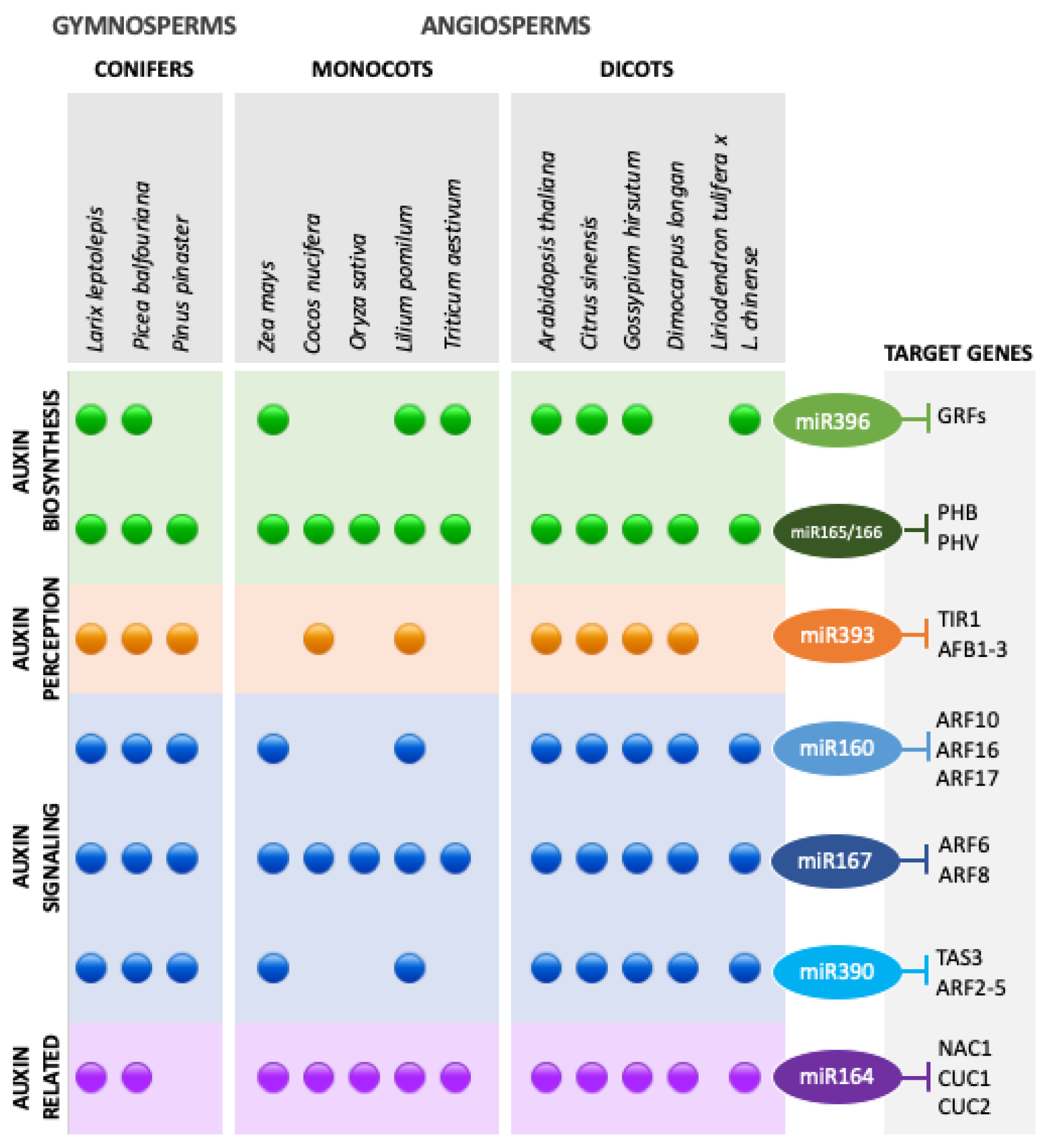
3.2. Auxins and miRNA–ARF Interactions

As mentioned above, auxin content has been pointed out as one of the most important factors required for the transition of a somatic cell into an embryo [45]. High concentrations of auxin are often related to SE stimulation in plants. Auxin signaling is activated by high levels of exogenously supplemented auxin to explant tissues. This network is crucial for the reprogramming of gene expression cascades during somatic cells to embryo transition [69]. The most used synthetic auxin for SE induction is 2,4-dichlorophenoxyacetic acid (2,4-D), structurally and functionally analogous to the natural auxin IAA, being applied in almost 78% of the published protocols, either alone or in combination with other PGRs [45].
Such as for the Arabidopsis model system, most frequently used for better understanding the regulation of SE, many differentially expressed miRNAs have also been identified in other embryogenic cultures of different gymnosperms [18], mono- and dicotyledonous plants [15,21,26,27]. From these studies, a large number of miRNAs has been associated with SE target genes related to auxin perception, signaling and biosynthesis, as presented in Figure 1.
The most represented auxin-related miRNAs in embryogenic cultures are miR165/166 and miR167, which have been found in all SE studies, miR160, miR164 and miR390, which have been identified in most SE transcriptomes and miR395, with expression in some of the embryogenic cultures. In addition to these widely conserved miRNAs, an angiosperm-specific miRNA, miR827, has been suggested to have a role in the embryogenic ability acquisition through the regulation of auxin metabolism in D. longan callus [30].
Given that several TF binding sites were identified in the MIR promoters, regulatory feedback loop mechanisms between TFs and MIR genes are expected to operate during SE [47]. In fact, Hewezi and Baum (2012) [81] found that in Arabidopsis, the GROWTH RESPONSE FACTOR1 (GRF1) and GRF3, both targets of miR396, which were differentially expressed in SE, were also reported to repress the expression of miR396a and miR396b. Furthermore, AUXIN RESPONSE FACTORS (ARFs) might control the expression of their targeting miRNAs, MIR160, MIR167 and MIR390, during SE, on account of the presence of AUXIN RESPONSE ELEMENTS (AREs) detected in the promoters of these genes [49,82].
miR160 has been indicated as an important auxin-related miRNA targeting ARF10, ARF16 and ARF17 [42,83,84], and being involved as regulator of several developmental processes in Arabidopsis, including embryogenesis. As reported in the conifer L. leptolepis [24] and in D. longan [32,85], the downregulation of miR160 was also observed in Arabidopsis SE [42,49].
The association of miR165/166 with the control of auxin biosynthesis during SE has been reported, corroborating the enhanced embryogenic response of the adaxial vs. abaxial side of cotyledon explants in Arabidopsis immature zygotic embryos [86]. Such a response was promoted by the restriction of PHABULOSA/PHAVOLUTA (PHB/PHV) transcripts to the adaxial cotyledon side by the action of miR165/166, resulting in a side-specific auxin accumulation, caused by the stimulation of LEAFY COTYLEDON2 (LEC2) expression mediated by PHB/PHV [42]. Additionally, ARF10 and ARF16 are referred as possible negative regulators of PHB/PHV since there is an increase of PHB transcript levels in arf10arf16 and a decrease in ARF16, miR160b and miR160c. An interaction between miR160 and miR165/166 during the SE induction process was supported by an up-regulation of ARF10 and ARF16 transcript levels in phb, phv, phb1-d and short tandem target mimic (STTM) 165/166 lines [42]. The LEC1 and LEC2 targets diversity suggested that these proteins are important contributors for several auxin-related processes, such as auxin biosynthesis, and members of the Aux/IAA family were indicated as regulators of LEC1 and LEC2 during embryo development [87]. Despite this information, the exact nature of miR160 and miR165/166 pathways interaction needs to be enlightened in future studies.
In Arabidopsis, a significant increase in the expression of ARF5, ARF6, ARF8, ARF10 and ARF16 is observed during the ZE and SE processes [41]. In a recent study with cotton (Gossypium hirsutum) SE, an increase of ARF6 and ARF8 expression was reported using miR167 mimic transformed lines (MIM167) for miR167 downregulation, in comparison with the control. In the same study, different genes showed significant differences in expression in MIM167 transformed lines when compared to control, namely auxin-response GRETCHEN HAGEN 3 (GH3), auxin transporter AUX1, auxin influx/efflux carrier LAX3, encoding an AUX1-like protein 3, and PIN-FORMED1 (PIN1) and PIN2 genes, suggesting that diminution of miR167 led to enhanced cellular auxin signaling. Other miRNAs associated with auxin signaling, such as miR160, miR393, miR166, miR156 and miR157 were differentially expressed in MIM167 lines, suggesting that the expression of these miRNAs is influenced by the diminution of miR167 [88].
In a recent study, it was found that MIR167A acts as a maternal gene for embryo development in Arabidopsis mainly through the targeted ARF6 and ARF8 genes [89]. Although it seems that miR167A is the key DCL1-processed miRNA involved in embryo development control from maternal sporophytic tissues, the embryogenesis and seed development defects in mir167a mutants are 100% penetrant while dcl1 mutants were not, suggesting that other DCL proteins may also be involved in miRNA167 precursors biogenesis [89]. Another recent research reported an increased auxin biosynthesis in the integuments at early embryogenesis stages, this maternally produced auxin being required for embryo development [90]. A lack of miR167 in the integuments will lead to an expected ARF6 and ARF8 target transcript accumulation, eventually disrupting auxin signaling and consequently seed development. Apart from this influence from maternal tissues, MIR167A and MIR167B were also found expressed in globular embryos [13], which corroborates its regulatory role of the auxin response in embryos thus contributing to normal embryo development. In what concerns SE, the involvement of these miRNAs in embryo development has not yet been reported. However, low levels of miR167 expression in cotton embryogenic calli as compared with non-embryogenic have been associated to a higher magnitude of SE in vitro [80], which is consistent with the results obtained for miR167-mimic transgenic calli in a recent study [88].
Regarding miR390, it regulates auxin signaling by directing the production of trans-acting small interfering RNAs (tasiRNAs) that down-regulate the expression of ARF2-4 genes [91,92]. Even though miR390 had a significantly modulated expression in SE of Arabidopsis and in other plant species such as L. leptolepis [24], D. longan [32] and Citrus sinensis [93], results in Arabidopsis suggest that the regulatory complex miR390-TAS3-ARFs seems to operate during early SE induction and the regulation of ARF2 and ARF3 mediated by miR390 contributes to auxin signaling during embryogenic transition in induced somatic cells [49]. Additionally, miR393, targeting the auxin receptors TIR1 and AFB2, contributes to embryogenesis transition modulating tissue sensitivity to auxin treatments. Corroborating this assumption, the authors reported a relation between the embryogenic response of explant tissue to the level of expressed miR393 and the concentration of 2,4-D used in the medium for SE induction [52].

4. Late Somatic Embryogenesis

Somatic embryo full development and maturation prior to germination is a crucial step to achieve plant formation through SE. As it would be expected, the regulatory networks involved in the acquisition of the features characteristic of a mature embryo are different from those operating in the initial phases of SE induction and morphogenic embryo development. During this period, embryo cells undergo various physiological changes, which become evident by the deposition of storage materials, repression of germination and acquisition of desiccation tolerance. At the cotyledonary stage, energy requirements reach a maximum, suggesting the relevance of primary metabolite production, such as amino acids and fatty acids, whereas fermentation could constitute an alternative source of energy at the early stages (i.e., globular stage) of somatic embryo development [94]. This intense cellular reprogramming usually requires the removal of the auxin exogenously supplied in the induction step, and other PGRs assume critical roles, such as ethylene and ABA [95]. Exogenous ABA treatments are often used to increase somatic embryo maturation efficiency both in gymnosperms and angiosperms. In germinating Arabidopsis seeds, ABA induces the accumulation of miR159 in an ABI3-dependent fashion, and miR159 mediates cleavage of MYB101 and MYB33 transcripts in vitro and in vivo. In L. leptolepis, miR169 was described as involved in the maturation of the somatic embryo, by responding to ABA in addition to a possible role in the maintenance of embryogenic or non-embryogenic potential and the maturation of the somatic embryo [96].
In the model species Arabidopsis, the study of dcl1 mutants evidence the crucial role of miRNAs, some of them not yet identified, as key inhibitors of the maturation program during embryogenesis, in part by repressing the master regulators LEC2 and FUSCA3 [97]. The trihelix TFs ARABIDOPSIS 6B-INTERACTING PROTEIN1-LIKE1 (ASIL1) and ASIL2 and the histone deacetylase HDA6/SIL1 were also identified as components that act downstream of miRNAs to repress the maturation program early in embryogenesis [97].
In several species, somatic embryo maturation normally occurs under high sucrose concentrations, not only within the conifers but also the angiosperms, which can be considered a strong stress for the plant cells. According to previous studies, high levels of miR397 and miR408 in developing somatic embryos can be related to such stress conditions [98].
In conifers, several miRNAs known to contribute to normal and synchronized development of somatic embryos were pointed out as potential biomarkers of SE, including miR156, miR159, miR166, miR167, miR168, miR171, miR397 and miR398 [24,96,99]. Nevertheless, there is evidence that a specific miRNA can participate at several stages of the SE process in different plant species. For instance, in maize, miR156 regulates embryogenic callus differentiation, in cotton it is necessary for globular embryo development whereas in D. longan and L. leptolepis it is important for cotyledonary embryo development [100]. In fact, miR156, one of the most conserved plant miRNAs families, is involved in the regulation of several crucial biological processes like fertility, juvenile to adult transition phase, root, leaf and fruit development and secondary metabolism, targeting SQUAMOSA PROMOTOR BINDING PROTEIN LIKE (SPL) transcription factor genes [101]. It has been considered a major regulator of early embryogenesis due to the highly repressed expression of SPLs in the eight-cell embryo in mutants [102]. The repression of SPL10 and SPL11 transcripts by miR156 leads to an inhibition of the expression of seed maturation genes, even if the embryo is not at a proper developmental stage for that to occur [50]. Additionally, miR156 has also been associated with SE induction in sweet orange (C. sinensis) [78] and csi-miR156a was the highest expressed miRNA during SE. The overexpression of this isomiR, leading to a respective downregulation of SPLs, was confirmed to play an important role in citrus SE by direct involvement in stress responses and hormone signaling pathways [103].
Beyond targeting PHB/PHV during the SE induction stage, miR165/166 has been associated with the repression of WUSCHEL-RELATED HOMEOBOX5 (WOX5) TF, thus causing an impact in later stages of the SE process, given the role of WOX5 in the maintenance of the root apical meristem in Arabidopsis somatic embryos [77,104].
Other examples that reinforce the plant species dependency on the particular patterns of miRNAs expression during SE and embryo development, is the low expression of miR171, miR390 and miR398 before induction of embryo differentiation from Oryza sativa [105] and L. leptolepis [106] embryogenic calli, in contrast with their increase during the differentiation process in citrus species [78].
The dicot-specific miR403 has been described to accumulate during the late transition phase and persist in mature green embryos in Arabidopsis, which also suggests a role in embryo maturation [4]. Similarly, miR161, a miRNA only found in the Brassicaceae family, has shown accumulation during the transition phase, despite its low levels in mature embryos in Arabidopsis. Another Brassicaceae-specific, the miR824 targeting AGAMOUS-LIKE16, was reported to increase its levels during mid-embryogenesis, especially in the heart and early torpedo embryonic stages [4].
The miR482/miR2118 superfamily, characterized in P. abies [107], was found up-regulated in P. pinaster cotyledonary and mature somatic embryos when compared to zygotic embryos at the same stages of development [17]. This superfamily has been associated with the regulation of siRNAs biogenesis from the nucleotide-binding site-leucine-rich repeat (NB-LRR) gene family and triggering siRNA production in reproductive tissue, suggesting a dual function in gymnosperms. These functions were also individually retained in monocots and angiosperms [108,109,110,111]. The role that this family is playing in SE is still not clear; however, it was reported that NB-LRR genes play a role in hormonal responses to environmental stress [112], which could be in line with the conditions of reduced water availability applied for somatic embryo maturation SE protocols in these species [113].

5. Conclusions and Perspectives

The extensive studies on embryogenic cultures of seed plants within most taxonomic groups, but particularly on the model plant Arabidopsis, have to some extent disclosed the complexity of the interactions between miRNA-mediated regulation and hormone and stress signaling, of which the auxin-related pathways have been found to play a central role (Figure 1).
Despite the similarities between the genetic regulation of SE and ZE, recent RNAseq data of embryogenic cultures from both angiosperms (Arabidopsis) and gymnosperms (P. pinaster) reported that the transcriptomes of somatic embryos differ from the transcriptomes of zygotic embryo at equivalent stages of development, with somatic embryos bearing a resemblance to the gene expression pattern of germinating seeds [15,17]. This is not too surprising, given the artificial and mostly stressful conditions required for the differential gene expression that triggers SE. In fact, the analysis of Arabidopsis embryo coding transcriptomes revealed significant differences between zygotic and somatic embryos [15]. Several key regulators such as WOX2, WOX8, LEC1 and LEC2, were less abundant in somatic embryos, while other such as PLETHORA 1/2/3 were more abundant. A high correlation between the expression patterns of somatic embryos and germinating seeds was also found, suggesting that the timing of gene expression is altered in somatic embryos [15]. In fact, also the sncRNA profiles in maritime pine shows that although somatic and zygotic embryos express roughly the same repertoire of conserved and novel miRNAs, their expression profiles along development are different [17]. However, it should be pointed out that few reports have addressed the potential role of the zygotic embryo surrounding tissues (endosperm or megagametophyte), lacking in SE, in the miRNA-mediated regulation of the developing embryo. For instance, in P. pinaster, the megagametophytes show a rich non-coding transcriptome [17] which may have yet undiscovered roles in embryo development. The molecular networks that characterize the early vs. the later stages of SE [7,15] indicate that the most significant variations between distinct taxonomic groups are most likely to occur at the earliest stages, namely during induction of embryogenic competence. This emphasizes the challenge involved in the identification of the central components of the common molecular regulatory pathways that confer embryogenic capacity and, consequently, embryonic development from zygotic and somatic cells. Within these central components, miRNAs have been associated with the regulation of key TFs that modulate SE responses across all groups of land plants. Several questions, such as the miRNA roles, if any, at the initial embryo cell divisions in gymnosperms, following distinct patterns from those observed in angiosperms, remain unanswered.
In the past recent years, several studies have pointed out that most miRNAs are highly conserved throughout the plant kingdom, some of which were identified as crucial regulators of embryogenesis. Some miRNA families seem to be involved in the regulation of embryo development in both plant groups. However, other families, present throughout the embryogenic process, seem to be specific to each group (Table 1). According to specialized repositories such as miRbase (http://www.mirbase.org/) [114] (Accessed on 23 January 2021) and PmiREN (http://www.pmiren.com/) [115] (Accessed on 23 January 2021), some miRNA families are gymnosperm-specific, while others are angiosperm-specific, and within these, some are only found in monocots (such as O. sativa, Z. mays and Triticum aestivum) or in dicots (such as Arabidopsis). Based on a literature survey, Table 1 presents the different group-specific miRNA families reported as differentially expressed in tissues undergoing SE, although the roles of some of them, such as the Poaceae-specific miR5067 and the dicot-specific miR163, are yet to be determined. It is expected that the publication of more and larger miRNA datasets in the near future will significantly extend our understanding of their roles in embryogenesis. Furthermore, the release of new genome sequences and improved genome annotations will be important to address questions that still remain unanswered.
The adoption of novel approaches such as single cell transcriptomics should lead to a better resolution in the characterization of cell type-specific expression patterns, which are usually diluted in the tissue samples used in most of the reports published up to now. In any case, it will be always a major challenge to study SE due to the wide variety of different experimental conditions used to trigger embryo development from somatic cells. Slight variations in the concentration of applied PGRs may result in significant differences in miRNA expression [71], making it difficult to find common trends, and this is further hindered by genotype dependence. At last, it should be pointed out that currently, many studies are performed on the assumption that specific miRNA target transcripts, and their functions, are largely conserved between species. However, in many cases this may not be the case. The availability of bioinformatic tools with improved miRNA target prediction algorithms may help to address if the roles of relevant miRNAs are conserved, i.e., if their target transcripts show conserved functions. This could contribute to significant advances in the functional characterization of the large number of miRNAs identified in transcriptomic studies. Furthermore, in vivo validation of the miRNA-target interactions should be pursued, thus contributing to a better understanding of miRNA-mediated regulation of SE.

Author Contributions

C.M. and S.C. conceived the idea. A.A., D.C., S.C. and C.M. designed the structure of the review. A.A. prepared the original draft. All authors contributed to the writing, review and editing. All authors have read and agreed to the published version of the manuscript.

Funding

This research was funded by Fundação para a Ciência e a Tecnologia (FCT/MCTES), grant numbers UIDB/04046/2020 and UIDP/04046/2020 to BioISI, UIDB/04004/2020 to Centre for Functional Ecology—Science for People and the Planet (CFE), UID/Multi/04551/2013 to GREEN-IT; and the doctoral fellowships SFRH/BD/128827/2017 (to AA) and SFRH/BD/136925/2018 (to DC).

Conflicts of Interest

The authors declare no conflict of interest.

References

  1. Cho, S.H.; Coruh, C.; Axtell, M.J. miR156 and miR390 Regulate tasiRNA Accumulation and Developmental Timing in Physcomitrella patens. Plant Cell 2012, 24, 4837–4849. [Google Scholar] [CrossRef] [Green Version]
  2. Tsuzuki, M.; Futagami, K.; Shimamura, M.; Inoue, C.; Kunimoto, K.; Oogami, T.; Tomita, Y.; Inoue, K.; Kohchi, T.; Yamaoka, S.; et al. An Early Arising Role of the MicroRNA156/529-SPL Module in Reproductive Development Revealed by the Liverwort Marchantia polymorpha. Curr. Biol. 2019, 29, 3307–3314. [Google Scholar] [CrossRef]
  3. Perdiguero, P.; Rodrigues, A.S.; Chaves, I.; Costa, B.; Alves, A.; de María, N.; Vélez, M.D.; Díaz-Sala, C.; Cervera, M.T.; Miguel, C.M. Comprehensive analysis of the isomiRome in the vegetative organs of the conifer Pinus pinaster under contrasting water availability. Plant. Cell Environ. 2020, 44, 706–728. [Google Scholar] [CrossRef] [PubMed]
  4. Plotnikova, A.; Kellner, M.J.; Mosiolek, M.; Schon, M.A.; Nodine, M.D. “MicroRNA Dynamics and Functions During Arabidopsis Embryogenesis. Plant Cell 2019, 31, 2929–2946. [Google Scholar] [CrossRef] [PubMed]
  5. Axtell, M.J. Classification and Comparison of Small RNAs from Plants. Annu. Rev. Plant Biol. 2013, 64, 137–159. [Google Scholar] [CrossRef] [PubMed] [Green Version]
  6. Vazquez, F.; Vaucheret, H.; Rajagopalan, R.; Lepers, C.; Gasciolli, V.; Mallory, A.; Hilbert, J.L.; Bastel, D.P.; Crété, P. Endogenous trans-acting siRNAs regulate the accumulation of arabidopsis mRNAs. Mol. Cell 2004, 16, 69–79. [Google Scholar] [CrossRef] [PubMed]
  7. Li, C.; Zhang, B. MicroRNAs in Control of Plant Development. J. Cell. Physiol. 2016, 231, 303–313. [Google Scholar] [CrossRef]
  8. Shriram, V.; Kumar, V.; Devarumath, R.M.; Khare, T.S.; Wani, S.H. Micrornas as potential targets for abiotic stress tolerance in plants. Front. Plant Sci. 2016, 7, 817. [Google Scholar] [CrossRef]
  9. Alves, A.; Rodrigues, A.S.; Miguel, C. microRNAs in Plant Embryogenesis. In Plant microRNAs. Concepts and Strategies in Plant Sciences, 1st ed.; Miguel, C., Dalmay, T., Chaves, I., Eds.; Springer: Cham, Switzerland, 2020; pp. 99–120. [Google Scholar]
  10. ten Hove, C.A.; Lu, K.J.; Weijers, D. Building a plant: Cell fate specification in the early arabidopsis embryo. Development 2015, 142, 420–430. [Google Scholar] [CrossRef] [Green Version]
  11. Cairney, J.; Pullman, G.S. The cellular and molecular biology of conifer embryogenesis. New Phytol. 2007, 176, 511–536. [Google Scholar] [CrossRef] [PubMed]
  12. Zhou, X.; Liu, Z.; Shen, K.; Zhao, P.; Sun, M.X. Cell lineage-specific transcriptome analysis for interpreting cell fate specification of proembryos. Nat. Commun. 2020, 11, 1–13. [Google Scholar] [CrossRef]
  13. Armenta-Medina, A.; Lepe-Soltero, D.; Xiang, D.; Datla, R. Arabidopsis thaliana miRNAs promote embryo pattern formation beginning in the zygote. Dev. Biol. 2017, 431, 145–151. [Google Scholar] [CrossRef]
  14. von Arnold, S.; Clapham, D.; Abrahamsson, M. Embryology in conifers. In Advances in Botanical Research; Cánovas, F.M., Ed.; Academic Press: Cambridge, MA, USA, 2019; Volume 89, pp. 157–184. [Google Scholar]
  15. Hofmann, F.; Schon, M.A.; Nodine, M.D. The embryonic transcriptome of Arabidopsis thaliana. Plant Reprod. 2019, 32, 77–91. [Google Scholar] [CrossRef] [Green Version]
  16. Rodrigues, A.S.; Miguel, C.M. The pivotal role of small non-coding RNAs in the regulation of seed development. Plant Cell Rep. 2017, 36, 653–667. [Google Scholar] [CrossRef] [PubMed] [Green Version]
  17. Rodrigues, A.S.; Chaves, I.; Costa, B.V.; Lin, Y.C.; Lopes, S.; Milhinhos, A.; van de Peer, Y.; Miguel, C.M. Small RNA profiling in Pinus pinaster reveals the transcriptome of developing seeds and highlights differences between zygotic and somatic embryos. Sci. Rep. 2019, 9, 11327. [Google Scholar] [CrossRef] [Green Version]
  18. Liu, Y.; El-Kassaby, Y.A.; Liu, E.K. Global Analysis of Small RNA Dynamics during Seed Development of Picea glauca and Arabidopsis thaliana Populations Reveals Insights on their Evolutionary Trajectories. Front. Plant Sci. 2017, 8, 1719. [Google Scholar] [CrossRef]
  19. Källman, T.; Chen, J.; Gyllenstrand, N.; Lagercrantz, U. A significant fraction of 21-nucleotide small RNA originates from phased degradation of resistance genes in several perennial species. Plant Physiol. 2013, 162, 741–754. [Google Scholar] [CrossRef] [Green Version]
  20. Liu, Y.; El-Kassaby, Y.A. Landscape of fluid sets of hairpin-derived 21-/24-nt-long small RNAs at seed set uncovers special epigenetic features in Picea glauca. Genome Biol. Evol. 2017, 9, 82–92. [Google Scholar] [CrossRef] [PubMed] [Green Version]
  21. Papareddy, R.; Páldi, K.; Paulraj; Kao, S.P.; Nodine, M. Chromatin Regulates Bipartite-Classified Small RNA Expression to Maintain Epigenome Homeostasis in Arabidopsis. Genome Biol. 2020, 21, 251. [Google Scholar] [CrossRef]
  22. Zhang, J.; Zhang, S.; Han, S.; Li, X.; Tong, Z.; Qi, L. Deciphering small noncoding RNAs during the transition from dormant embryo to germinated embryo in larches (Larix leptolepis). PLoS ONE 2013, 8, e81452. [Google Scholar] [CrossRef] [PubMed]
  23. Li, Q.; Deng, C.; Xia, Y.; Kong, H.; Zhang, S.; Wang, J. Identification of novel miRNAs and miRNA expression profiling in embryogenic tissues of Picea balfouriana treated by 6-benzylaminopurine. PLoS ONE 2017, 12, e0176112. [Google Scholar] [CrossRef] [PubMed]
  24. Zhang, J.; Zhang, S.; Han, S.; Wu, T.; Li, X.; Li, W.; Qi, L. Genome-wide identification of microRNAs in larch and stage-specific modulation of 11 conserved microRNAs and their targets during somatic embryogenesis. Planta 2012, 236, 647–657. [Google Scholar] [CrossRef]
  25. Wang, L.; Zhao, J.; Zhang, M.; Li, X.; Luo, K.; Lu, Z.; Zhang, C.; Jin, B. Identification and characterization of microrna expression in Ginkgo biloba L. Leaves. Int. J. Doc. Anal. Recognit. 2015, 11, 76. [Google Scholar] [CrossRef]
  26. Chu, Z.; Chen, J.; Xu, H.; Dong, Z.; Chen, F.; Cui, D. Identification and comparative analysis of microRNA in wheat (Triticum aestivum L.) callus derived from mature and immature embryos during In vitro culture. Front. Plant Sci. 2017, 30, 1302. [Google Scholar] [CrossRef] [PubMed] [Green Version]
  27. Juárez-González, V.T.; López-Ruiz, B.A.; Baldrich, P.; Luján-Soto, E.; Meyers, B.C.; Dinkova, T.D. The explant developmental stage profoundly impacts small RNA-mediated regulation at the dedifferentiation step of maize somatic embryogenesis. Sci. Rep. 2019, 9, 14511. [Google Scholar] [CrossRef] [Green Version]
  28. Zhang, J.; Wu, T.; Li, L.; Han, S.; Li, X.; Zhang, S.; Qi, L. Dynamic expression of small RNA populations in larch (Larix leptolepis). Planta 2013, 237, 89–101. [Google Scholar] [CrossRef]
  29. Zhang, J.W.; Wu, T.; Han, S.Y.; Qi, L.W.; Zhang, S.G. Expression Analysis of 12 miRNA Families Specific to Conifers during Somatic Embryogenesis of Larch. Forest Res. 2012, 25, 411–418. [Google Scholar]
  30. Xu, X.; Chen, X.; Chen, Y.; Zhang, Q.; Su, L.; Chen, X.; Chen, Y.; Zhang, Z.; Lin, Y.; Lai, Z. Genome-wide identification of miRNAs and their targets during early somatic embryogenesis in Dimocarpus longan Lour. Sci. Rep. 2020, 10, 4626. [Google Scholar] [CrossRef] [Green Version]
  31. Zhang, J.; Xue, B.; Gai, M.; Song, S.; Jia, N.; Sun, H. Small RNA and transcriptome sequencing reveal a potential miRNA-mediated interaction network that functions during somatic embryogenesis in Lilium pumilum DC. Fisch. Front. Plant Sci. 2017, 8, 566. [Google Scholar] [CrossRef]
  32. Lin, Y.; Lai, Z. Comparative analysis reveals dynamic changes in miRNAs and their targets and expression during somatic embryogenesis in longan (Dimocarpus longan Lour.). PLoS ONE 2013, 8, e60337. [Google Scholar] [CrossRef] [Green Version]
  33. Chen, Y.; Si, L.; Chen, X.; Zhang, S.; Xu, X.; Zhang, Z.; Chen, Y.; XuHan, X.; Lin, Y.; Lai, Z. Genome-wide identification and characterization of long non-coding RNAs involved in the early somatic embryogenesis in Dimocarpus longan Lour. BMC Genomics 2018, 19, 805. [Google Scholar] [CrossRef] [PubMed] [Green Version]
  34. Zhai, L.; Xu, L.; Wang, Y.; Huang, D.; Yu, R.; Limera, C.; Gong, Y.; Liu, L. Genome-Wide Identification of Embryogenesis-Associated microRNAs in Radish (Raphanus sativus L.) by High-Throughput Sequencing. Plant Mol. Biol. Rep. 2014, 32, 900–915. [Google Scholar] [CrossRef]
  35. Elhiti, M.; Stasolla, C.; Wang, A. Molecular regulation of plant somatic embryogenesis. Vitr. Cell. Dev. Biol. Plant 2013, 49, 631–642. [Google Scholar] [CrossRef]
  36. Fehér, A. Somatic embryogenesis—Stress-induced remodeling of plant cell fate. Biochim. Biophys. Acta Gene Regul. Mech 2015, 1849, 385–402. [Google Scholar] [CrossRef]
  37. Fehér, A.; Pasternak, T.P.; Dudits, D. Transition of somatic plant cells to an embryogenic state. Plant Cell Tiss Org. 2003, 74, 201–228. [Google Scholar] [CrossRef]
  38. Fehér, A. Callus, Dedifferentiation, Totipotency, Somatic Embryogenesis: What These Terms Mean in the Era of Molecular Plant Biology? Front. Plant Sci. 2019, 10, 536. [Google Scholar] [CrossRef] [PubMed] [Green Version]
  39. Elhiti, M.; Tahir, M.; Gulden, R.H.; Khamiss, K.; Stasolla, C. Modulation of embryo-forming capacity in culture through the expression of Brassica genes involved in the regulation of the shoot apical meristem. J. Exp. Bot. 2010, 61, 4069–4085. [Google Scholar] [CrossRef] [Green Version]
  40. Gaj, M.D. Factors influencing somatic embryogenesis induction and plant regeneration with particular reference to Arabidopsis thaliana (L.) Heynh. Plant Growth Regul. 2004, 43, 27–47. [Google Scholar] [CrossRef]
  41. Wójcikowska, B.; Gaj, M.D. Expression profiling of AUXIN RESPONSE FACTOR genes during somatic embryogenesis induction in Arabidopsis. Plant Cell Rep. 2017, 36, 843–858. [Google Scholar] [CrossRef] [Green Version]
  42. Wójcik, A.M.; Nodine, M.D.; Gaj, M.D. miR160 and miR166/165 Contribute to the LEC2-Mediated Auxin Response Involved in the Somatic Embryogenesis Induction in Arabidopsis. Front. Plant Sci 2017, 8, 2024. [Google Scholar] [CrossRef] [Green Version]
  43. Horstman, A.; Li, M.; Heidman, I.; Weeman, M.; Chen, B.; Muino, J.S.; Angenent, G.C.; Boutilier, K. The BABY BOOM transcription factor activates the LEC1-ABI3-FUS3-LEC2 network to induce somatic embryogenesis. Plant Physiol. 2017, 175, 848–857. [Google Scholar] [CrossRef] [Green Version]
  44. Méndez-Hernández, H.A.; Ledezma-Rodriguez, M.; Avilez-Montalvo, R.N.; Juárez-Gómez, Y.L.; Skeete, A.; Avilez-Montavo, J.; De-la-Peña, C.; Loyola-Vargas, V. Signaling overview of plant somatic embryogenesis. Front. Plant Sci. 2019, 10, 77. [Google Scholar] [CrossRef] [Green Version]
  45. Wójcik, A.M.; Wójcikowska, B.; Gaj, M.D. Current perspectives on the auxin-mediated genetic network that controls the induction of somatic embryogenesis in plants. Int. J. Mol. Sci 2020, 21, 1333. [Google Scholar] [CrossRef] [Green Version]
  46. Vashisht, D.; Nodine, M.D. MicroRNA functions in plant embryos. Biochem. Soc. Trans. 2014, 42, 352–357. [Google Scholar] [CrossRef]
  47. Megraw, M.; Baev, V.; Rusinov, V.; Jensen, S.T.; Kalantidis, K.; Hatzigeorgiou, A.G. MicroRNA promoter element discovery in Arabidopsis. RNA 2006, 12, 1612–1619. [Google Scholar] [CrossRef] [Green Version]
  48. Zhao, X.; Li, L. “Comparative Analysis of MicroRNA Promoters in Arabidopsis and Rice. Genom Proteom. Bioinf. 2013, 11, 56–60. [Google Scholar] [CrossRef] [PubMed] [Green Version]
  49. Szyrajew, K.; Bielewicz, D.; Dolata, J.; Wójcik, A.M.; Nowak, K.; Szczygiel-Sommer, A.; Szweykowska-Kulinska, Z.; Jarmolowski, A.; Gaj, M.D. MicroRNAs Are Intensively Regulated during Induction of Somatic Embryogenesis in Arabidopsis. Front. Plant Sci. 2017, 8, 18. [Google Scholar] [CrossRef] [PubMed]
  50. Nodine, M.D.; Bartel, D.P. MicroRNAs prevent precocious gene expression and enable pattern formation during plant embryogenesis. Genes Dev. 2010, 24, 2678–2692. [Google Scholar] [CrossRef] [PubMed] [Green Version]
  51. Gliwicka, M.; Nowak, K.; Balazadeh, S.; Mueller-Roeber, B.; Gaj, M.D. Extensive Modulation of the Transcription Factor Transcriptome during Somatic Embryogenesis in Arabidopsis thaliana. PLoS ONE 2013, 8, e69261. [Google Scholar] [CrossRef] [Green Version]
  52. Wójcik, A.M.; Gaj, M.D. miR393 contributes to the embryogenic transition induced in vitro in Arabidopsis via the modification of the tissue sensitivity to auxin treatment. Planta 2016, 244, 231–243. [Google Scholar] [CrossRef] [Green Version]
  53. Long, J.M.; Liu, Z.; Wu, X.M.; Fang, Y.N.; Jia, H.H.; Xie, Z.Z.; Deng, X.X.; Guo, W.W. Genome-scale mRNA and small RNA transcriptomic insights into initiation of citrus apomixis. J. Exp. Bot. 2016, 67, 5743–5756. [Google Scholar] [CrossRef] [Green Version]
  54. von Arnold, S.; Clapham, D.; Egertsdotter, U.; Mo, L.H. Somatic embryogenesis in conifers—A case study of induction and development of somatic embryos in Picea abies. Plant Growth Regul. 1996, 20, 3–9. [Google Scholar] [CrossRef]
  55. Zhang, X.; Zou, Z.; Gong, P.; Zhang, J.; Ziaf, K.; Li, H.; Xiao, F.; Ye, Z. Over-expression of microRNA169 confers enhanced drought tolerance to tomato. Biotechnol. Lett. 2011, 33, 403–409. [Google Scholar] [CrossRef] [PubMed]
  56. Zhao, B.; Ge, L.; Liang, R.; Li, W.; Ruan, K.; Lin, H.; Jin, Y. Members of miR-169 family are induced by high salinity and transiently inhibit the NF-YA transcription factor. BMC Mol. Biol. 2009, 10, 29. [Google Scholar] [CrossRef] [PubMed] [Green Version]
  57. Ding, Q.; Zeng, J.; He, X.Q. MiR169 and its target PagHAP2-6 regulated by ABA are involved in poplar cambium dormancy. J. Plant Physiol. 2016, 198, 1–9. [Google Scholar] [CrossRef] [PubMed]
  58. Luan, M.; Xu, M.; Lu, Y.; Zhang, L.; Fan, Y.; Wang, L. Expression of zma-miR169 miRNAs and their target ZmNF-YA genes in response to abiotic stress in maize leaves. Gene 2015, 555, 178–185. [Google Scholar] [CrossRef] [PubMed]
  59. Koyama, T.; Mitsuda, N.; Seki, M.; Shinozaki, K.; Ohme-Takagi, M. TCP transcription factors regulate the activities of ASYMMETRIC LEAVES1 and miR164, as well as the auxin response, during differentiation of leaves in Arabidopsis. Plant Cell 2010, 22, 3574–3588. [Google Scholar] [CrossRef] [Green Version]
  60. Palatnik, J.F.; Allen, E.; Wu, X.; Schommer, C.; Schwab, R.; Carrington, J.C.; Weigel, D. Control of leaf morphogenesis by microRNAs. Nature 2003, 425, 257–263. [Google Scholar] [CrossRef] [Green Version]
  61. Sunkar, R.; Kapoor, A.; Zhu, J.K. Posttranscriptional induction of two Cu/Zn superoxide dismutase genes in Arabidopsis is mediated by downregulation of miR398 and important for oxidative stress tolerance. Plant Cell 2006, 18, 2415. [Google Scholar] [CrossRef] [Green Version]
  62. Mallory, A.C.; Dugas, D.V.; Bartel, D.P.; Bartel, B. MicroRNA Regulation of NAC-Domain Targets Is Required for Proper Formation and Separation of Adjacent Embryonic, Vegetative, and Floral Organs. Curr. Biol. 2004, 14, 1035–1046. [Google Scholar] [CrossRef] [Green Version]
  63. Guo, H.-S.; Xie, Q.; Fei, J.-F.; Chua, N.-H. MicroRNA Directs mRNA Cleavage of the Transcription Factor NAC1 to Downregulate Auxin Signals for Arabidopsis Lateral Root Development. Plant Cell 2005, 17, 1376–1386. [Google Scholar] [CrossRef] [Green Version]
  64. Akdogan, G.; Tufekci, E.D.; Uranbey, S.; Unver, T. miRNA-based drought regulation in wheat. Funct. Integr. Genomics 2016, 16, 221–233. [Google Scholar] [CrossRef]
  65. Hernandez, Y.; Goswami, K.; Sanan-Mishra, N. Stress induced dynamic adjustment of conserved miR164:NAC module. Plant-Environ. Interact. 2020, 1, 134–151. [Google Scholar] [CrossRef]
  66. Li, W.F.; Shou-Gong, Z.; Han, S.Y.; Wu, T.; Zhang, J.H.; Qi, L.W. Regulation of LaMYB33 by miR159 during maintenance of embryogenic potential and somatic embryo maturation in Larix kaempferi (Lamb.) Carr. Plant Cell Tiss Org. 2013, 113, 131–136. [Google Scholar] [CrossRef]
  67. Boutilier, K.; Offringa, R.; Sharma, V.K.; Kief, H.; Ouellet, T.; Zhang, L.; Hattori, J.; Liu, C.M.; van Lammeren, A.A.M.; Miki, B.L.A.; et al. Ectopic expression of BABY BOOM triggers a conversion from vegetative to embryonic growth. Plant Cell 2002, 14, 1737–1749. [Google Scholar] [CrossRef] [PubMed] [Green Version]
  68. Nowak, K.; Moronczyk, J.; Wójcik, A.; Gaj, M.D. AGL15 Controls the Embryogenic Reprogramming of Somatic Cells in Arabidopsis through the Histone Acetylation-Mediated Repression of the miRNA Biogenesis Genes. Int. J. Mol. Sci. 2020, 21, 6733. [Google Scholar] [CrossRef] [PubMed]
  69. Jiménez, V.M. Involvement of plant hormones and plant growth regulators on in vitro somatic embryogenesis. Plant Growth Regul. 2005, 47, 91–110. [Google Scholar] [CrossRef]
  70. Li, S.G.; Li, W.F.; Han, S.Y.; Yang, W.H.; Qi, L.W. Stage-specific regulation of four HD-ZIP III transcription factors during polar pattern formation in Larix leptolepis somatic embryos. Gene 2013, 522, 177–183. [Google Scholar] [CrossRef] [PubMed]
  71. Li, Q.; Deng, C.; Zhu, T.; Ling, J.; Zhang, H.; Kong, L.; Zhang, S.; Wang, J.; Chen, X. Dynamics of physiological and miRNA changes after long-term proliferation in somatic embryogenesis of Picea balfouriana. Trees Struct. Funct. 2019, 33, 469–480. [Google Scholar] [CrossRef]
  72. Shen, Y.; Jiang, Z.; Lu, S.; Lin, H.; Gao, S.; Peng, H.; Yuan, G.; Xhang, Z.; Zhao, M.; Rong, T.; et al. Combined small RNA and degradome sequencing reveals microRNA regulation during immature maize embryo dedifferentiation. Biochem. Biophys. Res. Commun. 2013, 441, 425–430. [Google Scholar] [CrossRef]
  73. López-Ruiz, B.A.; Juárez-González, V.T.; Chávez-Hernández, E.C.; Dinkova, T.D. MicroRNA Expression and Regulation During Maize Somatic Embryogenesis. Methods Mol. Biol. 2018, 1815, 397–410. [Google Scholar]
  74. Sabana, A.A.; Antony, G.C.; Rahul, U.; Rajesh, M.K. In silico identification of microRNAs and their targets associated with coconut embryogenic calli. Agri Gene 2018, 7, 59–65. [Google Scholar] [CrossRef]
  75. Sinha, A.; Solanki, M.; Shukla, L.I. Evidences for differential expression of miR167d-5p, target, positional nucleotide preference, and its role in somatic and different stages of regenerating calli of Oryza sativa. Plant Cell Tiss Org. 2019, 136, 537–548. [Google Scholar] [CrossRef]
  76. Chen, C.J.; Liu, Q.; Zhang, Y.C.; Qu, L.H.; Chen, Y.Q.; Gautheret, D. Genome-wide discovery and analysis of microRNAs and other small RNAs from rice embryogenic callus. RNA Biol. 2011, 8, 538–547. [Google Scholar] [CrossRef] [PubMed] [Green Version]
  77. Su, Y.H.; Liu, Y.B.; Zhou, C.; Li, X.M.; Zhang, X.S. The microRNA167 controls somatic embryogenesis in Arabidopsis through regulating its target genes ARF6 and ARF8. Plant Cel. Tiss Org. 2016, 124, 405–417. [Google Scholar] [CrossRef]
  78. Wu, X.M.; Liu, M.Y.; Ge, X.X.; Xu, Q.; Guo, W.W. Stage and tissue-specific modulation of ten conserved miRNAs and their targets during somatic embryogenesis of Valencia sweet orange. Planta 2011, 233, 495–505. [Google Scholar] [CrossRef] [PubMed]
  79. Jiang, S.C.; Mei, C.; Lian, S.; Yu, Y.-T.; Lu, K.; Wu, Z.; Wang, X.F.; Zhang, D.P. Crucial roles of the pentatricopeptide repeat protein SOAR1 in Arabidopsis response to drought, salt and cold stresses. Plant Mol. Biol. 2015, 88, 369–385. [Google Scholar] [CrossRef] [PubMed] [Green Version]
  80. Yang, X.; Wang, L.; Yuan, D.; Lindsey, K.; Zhang, X. Small RNA and degradome sequencing reveal complex miRNA regulation during cotton somatic embryogenesis. J. Exp. Bot. 2013, 64, 1521–1536. [Google Scholar] [CrossRef]
  81. Hewezi, T.; Baum, T.J. Complex feedback regulations govern the expression of miRNA396 and its GRF target genes. Plant Signal. Behav. 2012, 7, 749–751. [Google Scholar] [CrossRef] [Green Version]
  82. Marin, E.; Jouannet, V.; Herz, A.; Lokerse, A.S.; Weijers, D.; Vaucheret, H.; Nussaume, L.; Crespi, M.D.; Maizel, A. mir390, Arabidopsis TAS3 tasiRNAs, and their AUXIN RESPONSE FACTOR targets define an autoregulatory network quantitatively regulating lateral root growth. Plant Cell 2010, 22, 1104–1117. [Google Scholar] [CrossRef] [Green Version]
  83. Liu, P.P.; Montgomery, T.A.; Fahlgren, N.; Kasschau, K.D.; Nonogaki, H.; Carrington, J.C. Repression of AUXIN RESPONSE FACTOR10 by microRNA160 is critical for seed germination and post-germination stages. Plant J. 2007, 52, 133–146. [Google Scholar] [CrossRef] [PubMed]
  84. Liu, Q.; Chen, Y.Q. A new mechanism in plant engineering: The potential roles of microRNAs in molecular breeding for crop improvement. Biotechnol. Adv. 2010, 28, 301–307. [Google Scholar] [CrossRef] [PubMed]
  85. Lin, Y.; Lai, Z.; Tian, Q.; Lin, L.; Lai, R.; Yang, M.; Zhang, D.; Chen, Y.; Zhang, Z. Endogenous target mimics down-regulate miR160 mediation of ARF10, -16, and -17 cleavage during somatic embryogenesis in Dimocarpus longan Lour. Front. Plant Sci. 2015, 6, 956. [Google Scholar] [CrossRef] [PubMed]
  86. Wójcikowska, B.; Botor, M.; Moronczyk, J.; Wójcik, A.M.; Nodzynski, T.; Karcz, J.; Gaj, M.G. Trichostatin a triggers an embryogenic transition in arabidopsis explants via an auxin-related pathway. Front. Plant Sci. 2018, 9, 1353. [Google Scholar] [CrossRef] [PubMed]
  87. Zheng, Q.; Zheng, Y.; Ji, H.; Burnie, W.; Perry, S.E. Gene regulation by the AGL15 transcription factor reveals hormone interactions in somatic embryogenesis. Plant Physiol. 2016, 172, 2374–2387. [Google Scholar] [CrossRef] [PubMed] [Green Version]
  88. Arora, S.; Singh, A.K.; Chaudhary, B. Target-mimicry based miRNA167-diminution ameliorates cotton somatic embryogenesis via transcriptional biases of auxin signaling associated miRNAs and genes. Plant Cell. Tiss Org. 2020, 141, 511–531. [Google Scholar] [CrossRef]
  89. Yao, X.; Chen, J.; Zhou, J.; Yu, H.; Ge, C.; Zhang, M.; Gao, X.; Dai, X.; Yang, Z.N.; Zhao, Y. An Essential Role for miRNA167 in Maternal Control of Embryonic and Seed Development. Plant Physiol. 2019, 180, 453–464. [Google Scholar] [CrossRef] [PubMed] [Green Version]
  90. Robert, H.S.; Park, C.; Guitérrez, C.L.; Wójcikowska, B.; Pencik, A.; Novak, O.; Chen, J.; Grunewald, W.; Dresselhaus, T.; Friml, J.; et al. Maternal auxin supply contributes to early embryo patterning in Arabidopsis. Nat. Plants 2018, 4, 548–553. [Google Scholar] [CrossRef]
  91. Allen, E.; Xie, Z.; Gustafson, A.M.; Carrington, J.C. microRNA-Directed Phasing during Trans-Acting siRNA Biogenesis in Plants. Cell 2005, 121, 207–221. [Google Scholar] [CrossRef] [Green Version]
  92. Axtell, M.J.; Jan, C.; Rajagopalan, R.; Bartel, D.P. A Two-Hit Trigger for siRNA Biogenesis in Plants. Cell 2006, 127, 565–577. [Google Scholar] [CrossRef] [Green Version]
  93. Wu, X.M.; Kou, S.J.; Liu, Y.L.; Fang, Y.N.; Xu, Q.; Guo, W.W. Genomewide analysis of small RNAs in nonembryogenic and embryogenic tissues of citrus: microRNA-and siRNA-mediated transcript cleavage involved in somatic embryogenesis. Plant Biotechnol. J. 2015, 13, 383–394. [Google Scholar] [CrossRef]
  94. Ge, X.; Zhang, C.; Wang, Q.; Yang, Z.; Wang, Y.; Zhang, X.; Wu, Z.; Hou, Y.; Wu, J.; Li, F. ITRAQ protein profile differential analysis between somatic globular and cotyledonary embryos reveals stress, hormone, and respiration involved in increasing plantlet regeneration of Gossypium hirsutum L. J. Proteome Res. 2015, 14, 268–278. [Google Scholar] [CrossRef]
  95. Corredoira, E.; Merkle, S.A.; Martínez, M.T.; Toribio, M.; Canhoto, J.M.; Correia, S.I.; Ballester, A.; Vieitez, A.M. Non-Zygotic Embryogenesis in Hardwood Species. Crit. Rev. Plant Sci. 2019, 38, 29–97. [Google Scholar] [CrossRef]
  96. Zhang, L.; Li, W.; Xu, H.; Qi, L.; Han, S. Cloning and characterization of four differentially expressed cDNAs encoding NFYA homologs involved in responses to ABA during somatic embryogenesis in Japanese larch (Larix leptolepis). Plant Cell Tiss Org. 2014, 117, 293–304. [Google Scholar] [CrossRef]
  97. Willmann, M.R.; Mehalick, A.J.; Packer, R.L.; Jenik, P.D. MicroRNAs Regulate the Timing of Embryo Maturation in Arabidopsis. Plant Physiol. 2011, 155, 1871–1884. [Google Scholar] [CrossRef] [Green Version]
  98. Ma, C.; Burd, S.; Lers, A. MiR408 is involved in abiotic stress responses in Arabidopsis. Plant J. 2015, 84, 169–187. [Google Scholar] [CrossRef]
  99. Miguel, C.M.; Rupps, A.; Raschke, J.; Rodrigues, A.S.; Trontin, J.F. Impact of molecular studies on somatic embryogenesis development for implementation in conifer multi-varietal forestry. In Vegetative Propagation of Forest Trees; Park, Y.S., Bonga, J.M., Moon, H.K., Eds.; National Institute of Forest Science: Seoul, Korea, 2016; pp. 373–421. [Google Scholar]
  100. Siddiqui, Z.H.; Abbas, Z.K.; Ansari, M.W.; Khan, M.N. The role of miRNA in somatic embryogenesis. Genomics 2019, 111, 1026–1033. [Google Scholar] [CrossRef] [PubMed]
  101. Rhoades, M.W.; Reinhart, B.J.; Lim, L.P.; Burge, C.B.; Bartel, B.; Bartel, D.P. Prediction of plant microRNA targets. Cell 2002, 110, 513–520. [Google Scholar] [CrossRef] [Green Version]
  102. Yu, N.; Niu, Q.W.; Ng, K.H.; Chua, N.H. The role of miR156/SPLs modules in Arabidopsis lateral root development. Plant J. 2015, 83, 673–685. [Google Scholar] [CrossRef] [PubMed]
  103. Long, J.; Liu, C.; Feng, M.; Liu, Y.; Wu, X.; Guo, W. miR156- SPL modules regulate induction of somatic embryogenesis in citrus callus. J. Exp Bot. 2018, 69, 2979–2993. [Google Scholar] [CrossRef] [Green Version]
  104. Wang, L.; Chong, K. The essential role of cytokinin signaling in root apical meristem formation during somatic embryogenesis. Front. Plant Sci. 2016, 6, 1196. [Google Scholar] [CrossRef] [Green Version]
  105. Luo, Y.C.; Zhou, H.; Chen, Y.L.; Yang, J.H.; Chen, Y.Q.; Qu, L.H. Rice embryogenic calli express a unique set of microRNAs, suggesting regulatory roles of microRNAs in plant post-embryogenic development. FEBS Lett. 2006, 580, 5111–5116. [Google Scholar] [CrossRef] [Green Version]
  106. Zhang, S.; Zhou, J.; Han, S.; Yang, W.; Li, W.; Wei, H.; Li, X.; Qi, L. Four abiotic stress-induced miRNA families differentially regulated in the embryogenic and non-embryogenic callus tissues of Larix leptolepis. Biochem. Biophys. Res. Commun. 2010, 398, 355–360. [Google Scholar] [CrossRef]
  107. Xia, R.; Xu, J.; Arikit, S.; Meyers, B.C. Extensive families of miRNAs and PHAS loci in Norway spruce demonstrate the origins of complex phasiRNA networks in seed plants. Mol. Biol. Evol. 2015, 32, 2905–2918. [Google Scholar] [CrossRef] [Green Version]
  108. Fei, Q.; Xia, R.; Meyers, B.C. Phased, secondary, small interfering RNAs in posttranscriptional regulatory networks. Plant Cell 2013, 25, 2400–2415. [Google Scholar] [CrossRef] [Green Version]
  109. Jeong, D.; Schmidt, A.A.; Rymarquis, L.A.; Park, S.; Ganssmann, M.; German, M.A.; Accerbi, M.; Zhai, J.; Fahlgren, N.; Fox, E.F.; et al. Parallel analysis of RNA ends enhances global investigation of microRNAs and target RNAs of Brachypodium distachyon. Genome Biol. 2013, 14, R145. [Google Scholar] [CrossRef] [PubMed] [Green Version]
  110. Johnson, M.A.; Bender, J. Reprogramming the epigenome during germline and seed development. Genome Biol. 2009, 10, 232. [Google Scholar] [CrossRef] [PubMed] [Green Version]
  111. Song, Q.X.; Liu, Y.F.; Hu, Y.X.; Zhang, W.K.; Ma, B.; Chen, S.Y.; Zhang, J.S. Identification of miRNAs and their target genes in developing soybean seeds by deep sequencing. BMC Plant Biol. 2011, 11, 5. [Google Scholar] [CrossRef] [PubMed] [Green Version]
  112. Sarazin, V.; Duclercq, J.; Mendou, B.; Aubanelle, L.; Nicolas, Y.; Aono, M.; Pilard, S.; Guerineau, F.; Sangwan-Norreel, B.; Sangwan, R.S. Arabidopsis BNT1, an atypical TIR-NBS-LRR gene, acting as a regulator of the hormonal response to stress. Plant Sci. 2015, 239, 216–229. [Google Scholar] [CrossRef] [PubMed]
  113. Lelu-Walter, M.A.; Klimaszewska, K.; Miguel, C.; Aronen, T.; Hargreaves, C.T.; Trontin, J.F. Somatic embryogenesis for more effective breeding and deployment of improved varieties in Pinus spp.: Bottlenecks and recent advances. In Somatic Embryogenesis: Fundamental Aspects and Applications; Springer: Cham, Switzerland, 2016; pp. 319–365. [Google Scholar]
  114. Kozomara, A.; Birgaoanu, M.; Griffiths-Jones, S. MiRBase: From microRNA sequences to function. Nucleic Acids Res. 2019, 47, D155–D162. [Google Scholar] [CrossRef]
  115. Guo, Z.; Kuang, Z.; Wang, Y.; Zhao, Y.; Tao, Y.; Cheng, C.; Yang, J.; Lu, X.; Hao, C.; Wang, T.; et al. PmiREN: A comprehensive encyclopedia of plant miRNAs. Nucleic Acids Res. 2020, 48, D1114–D1121. [Google Scholar] [CrossRef] [PubMed] [Green Version]
Figure 1. miRNAs reported as expressed (presence of dots) in SE transcriptomes of several seed plants within gymnosperms [17,70,71] and angiosperms groups, monocots [26,31,72,73,74,75,76] and dicots [32,42,52,66,77,78,79,80], involved in auxin biosynthesis (green), perception (orange), signaling (blue) and auxin-related (purple).
Plants 10 00504 g001
Table 1. Group-specific miRNAs differentially expressed in embryogenesis.
Somatic Embryogenesis (SE)-Related miRNA FamilyDescribed or Putative Targets(Putative) Role in EmbryogenesisReferences
Gymnosperm-specific miR1314Putative cellulose synthases (TIGR)Embryo dormancy and germination[28]
Conifer-
specific
miR946
miR947
miR951
miR1311
unknownEmbryo dormancy and germination[28]
miR950NB-ARC
miR1312GRF2, HB1
miR1313LRK1
miR1315receptor-like protein kinaseEmbryogenic ability[23]
miR1316LIP1, LIP2, TIR-NBS-LRR proteinsNotdetermined yet[17,24,29]
miR3699unknownNot determined yet[17]
miR3701NBS-LRR proteins, cellulose synthaseNot determined yet[17,29]
miR3702
miR3704
unknownNot determined yet[24,29]
Angiosperm-specific miR444MIKC-type MADS-boxSE induction[27]
miR827NLA and PHT5Regulate auxin metabolism in early SE[30]
Monocot-
specific (Poaceae-
specific)
miR531Wpk4 protein kinaseEmbryogenic callus and embryo development[26]
miR1139Myb1Embryogenic callus development[26]
miR1878NBS-LRR resistance protein-like
miR5049Photosystem 1 subunit 5
Hydrolase, mitochondrial
Embryogenic callus and embryo development[26]
miR5067Wpk4 protein kinaseNot determined yet[31]
Dicot-
specific
miR158SPINDLYGibberellic acid responses[4]
miR163SAMT family membersNot determined yet[32]
miR403AGO2, AGO3Embryo maturation[4]
miR406Spliceosomal proteinsEarly embryogenesis[33]
Brassicaceae-
specific
miR161EMB2654, ARFEmbryo maturation[4,34]
miR824AGAMOUS-LIKE16Embryo maturation[4]
Publisher’s Note: MDPI stays neutral with regard to jurisdictional claims in published maps and institutional affiliations.

