Review

A Review of Robotic Interfaces for Post-Stroke Upper-Limb Rehabilitation: Assistance Types, Actuation Methods, and Control Mechanisms

by
André Gonçalves
1,2,*,
Manuel F. Silva
1,3,
Hélio Mendonça
1,2 and
Cláudia D. Rocha
1
1
INESC TEC—Institute for Systems and Computer Engineering, Technology and Science, Rua Dr. Roberto Frias, 4200-465 Porto, Portugal
2
FEUP—Faculty of Engineering, University of Porto, Rua Dr. Roberto Frias, 4200-465 Porto, Portugal
3
ISEP—Instituto Superior de Engenharia do Porto, Instituto Politécnico do Porto, Rua Dr. António Bernardino de Almeida, 4249-015 Porto, Portugal
*
Author to whom correspondence should be addressed.
Robotics 2025, 14(10), 141; https://doi.org/10.3390/robotics14100141
Submission received: 4 August 2025 / Revised: 1 October 2025 / Accepted: 3 October 2025 / Published: 6 October 2025

Abstract

Stroke is a leading cause of long-term disability worldwide, with survivors often facing significant challenges in regaining upper-limb functionality. In response, robotic rehabilitation systems have emerged as promising tools to enhance post-stroke recovery by delivering precise, adaptable, and patient-specific therapy. This paper presents a review of robotic interfaces developed specifically for upper-limb rehabilitation. It analyses existing exoskeleton- and end-effector-based systems, with respect to three core design pillars: assistance types, control philosophies, and actuation methods. The review highlights that most solutions favor electrically actuated exoskeletons, which use impedance- or electromyography-driven control, with active assistance being the predominant rehabilitation mode. Resistance-providing systems remain underutilized. Furthermore, no hybrid approaches featuring the combination of robotic manipulators with actuated interfaces were found. This paper also identifies a recent trend towards lightweight, modular, and portable solutions and discusses the challenges in bridging research prototypes with clinical adoption. By focusing exclusively on upper-limb applications, this work provides a targeted reference for researchers and engineers developing next-generation rehabilitation technologies.

1. Introduction

The British National Health Service (NHS) states that a stroke occurs when blood flow to a part of the brain is interrupted [1], resulting in either a blockage (ischemic stroke) or a rupture of a blood vessel (hemorrhagic stroke) [2]. Common consequences include difficulties with speech, movement, coordination, and memory. The extent and nature of these impairments can vary widely between individuals. Recovery from a stroke often requires prolonged and multidisciplinary rehabilitation, which may involve physiotherapy, occupational therapy, and speech-language therapy, among others [1].

1.1. Stroke Incidence

According to both the World Stroke Organization (WSO) and the World Health Organization (WHO), stroke is one of the leading causes of death and disability worldwide [3,4]. Studies from the early 2000s predicted that the occurrence of strokes per year would increase by 30% [5], and as of 2024, this medical emergency affects one fourth of the world’s population in their lifetime. Each year, 12 million people worldwide experience their first stroke and over 100 million people today are living with the aftermath of a stroke [3], six million of whom live in Europe [6]. On October 2024, the WHO predicted that by 2050 over two billion people worldwide (roughly one quarter of the global population) will be over 60 years old. Of this group, almost 500 million will be octogenarians [7], and with the advanced age comes a dangerous increase in stroke incidence [3].
Using the Institute for Health Metrics and Evaluation’s Global Health Data Exchange tool [8], it is possible to have a dynamic overview of the 2021 Global Burden of Disease study [8]. Figure 1 shows the incidence (number of new cases) and prevalence (number of existing cases) of stroke worldwide through the years. These graphs corroborate the statistics displayed above. It is possible to verify the continuous rise in new cases each year, which, from 1999 to 2021, grew to be more than 70%. Thus, an ever growing prevalence is also possible to anticipate.
Even though the incidence of stroke significantly increases with age [9], the WSO’s Global Stroke Fact Sheet 2022, using data from 2019, states that over 60% of all strokes affect people under 70, and those under 50 years old suffer from 16% of all occurrences [10]. This population needs a fast, accessible, and effective post-stroke healthcare response in order to maximize recovery and reduce long-term disability. A significant social economic investment by governments is crucial to reduce healthcare costs and improve the general quality of life of the survivors [6].

1.2. Social and Economic Impact of Stroke

Studies suggest that the lack of quality and access to medical care is the main contributor to the higher prevalence of strokes, especially in Low/Middle-Income Countries (LMICs) [11]. A noticeable shift in stroke commonness from High-Income Countries (HICs) to LMICs has taken place [12], which is consistent with the multiple projections which state that by 2050, 80% of old people will be living in LMICs [7,13]. Although there is not a perfect match between stroke incidence and the average income of countries (i.e., Russia is considered a HIC but has a higher stroke incidence than India, which is an LMIC), it is possible to verify a strong correlation between both statistics. Also, studies suggest that Asian countries have a higher predisposition to stroke because of their high salt intake [14,15,16].
The impact of strokes not only affects each individual but also each country’s economy. Rajsic et al. (2018) [6] highlight some estimates which suggest that stroke costs the European Union’s (EU) economy over EUR 38 billion every year. This value amounts to nearly one-fifth of all the costs related to Cardiovascular Diseases [17]. Nearly half of this quota, around EUR 18.5 billion, accounts for direct medical costs, such as hospital and clinical treatments and procedures. Moreover, the Cerebrovascular Accident (CVA), another term for stroke, also exacts a huge financial toll on the victims and their families. The other half of the EU’s financial burden arises from either productivity losses or informal care costs. Strokes oftentimes lead to long-term disabilities and demanding rehabilitation periods. Both survivors and their caregivers may become unavailable to resume their regular jobs, thereby diminishing workforce productivity. Additionally, this care can involve assistance with daily activities, transportation, support in recovery, among others, often creating a financial and emotional burden [6,17]. The global economic impact of stroke currently represents 0.66% of the global Gross Domestic Product, and the total cost of stroke is estimated to tip the EUR 1 trillion mark by 2030 [3].

1.3. Stroke Repercussions

Although several HICs are reporting improvements in the survival rate of stroke victims [18,19], the WHO estimates that about half of those who survive a stroke experience lasting disability [20]. More localized data indicate that two out of three stroke victims suffer impairments, as seen in Portugal [21] and the United Kingdom [22].
Strokes can affect most aspects of human life, leaving survivors oftentimes struggling with several challenges [23]:
  • Communication Problems, when people are left with reading difficulties and sometimes aphasia [24], which is a language disorder that affects how people communicate [25];
  • Cognitive Challenges, such as memory loss and dementia [26];
  • Emotional Issues, like dementia or low self-esteem [27];
  • Sensorial Difficulties, because hearing and vision can deteriorate [28];
  • Physical Effects, causing fatigue, incontinence of difficulty swallowing, among others [29]. The most common physical effects of strokes are hemiplegia or hemiparesis (total or partial weakness or loss of ability to produce movement of a limb, respectively), changes in muscle tension and loss of awareness of a limb position [30].
Disability-Adjusted Life Years (DALYs), a metric defined by the WHO as the loss of one year of full health, is commonly used to assess the burden of diseases [31]. Between 2004 and 2021, the contribution of stroke to DALYs increased significantly, rising from 46.6 million to 160.5 million, accounting for approximately 6% of total DALYs [32,33]. Close to half of all stroke survivors make incomplete recoveries [34], and most of these have limitations in basic activities six months after the their CVA [35]. They cannot lead an independent life and may require healthcare services all the time [6,34,36].

1.4. Upper-Limb Impairment and Robotic Rehabilitation

The upper-limb (UL) can be severely impaired after stroke, with a reported prevalence of up to 80% in stroke survivors [37], of which, half might not ever recover their previous capabilities [38]. This functional decline leads to lower independence and more difficulty in the performance of activities of daily living (ADLs), such as washing, dressing, cooking, eating, and using the toilet [38]. This impairment, which typically exceeds that observed in lower-limb sequelae following stroke [39], strongly affects people’s lives, rendering patients unable to work or care for their loved ones [40].
In response to this clinical challenge, rehabilitation robotics has emerged as a promising therapeutic approach to improve UL recovery following stroke. The practice of robot-assisted physical therapy can influence the escalation of rehabilitation, since it allows healthcare professionals to treat multiple people at the same time. Several studies have shown encouraging results in robot-assisted stroke rehabilitation therapies [41,42,43], and some suggest that robot-assisted rehabilitation can be equally effective as human-assisted physical therapy [44,45]. For example, Mehrholz et al. [46] analyzed 45 randomized controlled trials with 1619 stroke participants, reporting significant improvements in Fugl–Meyer Assessment (FMA) [47] scores, activities of daily living, and muscle strength measurements. Wang et al. [48] study reveals that robot-assisted therapy can reduce muscle spasticity, improve arm and hand movement control, increase range of motion, and enhance performance on ADLs. Although the initial investment in robotic systems can be significant, their cost-effectiveness improves with greater utilization [49]. Evidence from studies reveal that a combined approach, involving both human therapists and robots, is the most cost-effective [50], while others suggest that robotic rehabilitation offers a greater value when compared to traditional therapy [51,52].
The United States of America’s National Institute of Health defines Neuroplasticity as the “ability of the nervous system to change its activity in response to intrinsic or extrinsic stimuli by reorganizing its structure, functions, or connections after injuries, such as a stroke” [53]. Studies show that neuroplasticity and limb recovery can be amplified by repetitive, intensive, and task-oriented exercises. These exercises can be effectively offered by rehabilitation robots which, unlike physical therapists, are tireless, precise, and capable of monitoring movements, while providing feedback to caregivers. Rehabilitation robots also allow intensity variation and accurate progress reporting [54].

1.5. Study Methodology

This review follows the PRISMA approach [55] to the literature, with the aim of providing a high-quality, complete, and transparent overview of technological developments in robotic interfaces for post-stroke UL rehabilitation.
This review may be framed within the Population–Intervention–Comparison–Outcome framework. The population encompasses patients with upper-limb impairments, including but not limited to post-stroke patients, and broader neurological conditions affecting arm, elbow, shoulder, or wrist function. The intervention focuses on robots, exoskeletons, and end-effectors for upper-limb applications, their actuation (sometimes mentioned as being active), and control. The comparison element is not systematically addressed in this review, as most studies focus on device development rather than comparative effectiveness against conventional therapy. The outcome encompasses rehabilitation.
Relevant studies, reviewed up to the 30th June 2025, were identified through both Scopus and IEEE’s IEEEXplore databases. To capture the publications, the following search string was used:
  • (upper limb OR upper-limb Or upper arm OR stroke OR funtion*) AND
  • (robot OR robotic OR end-effector OR exoskeleton) AND
  • (actuat* OR active OR control) AND
  • (rehabilitation)
The use of upper* englobes all the synonyms possible, such as upper-limb, upper limb, upper body, among others. Similarly, rehabilitat* englobes rehabilitation and rehabilitative and robot* englobes robot, robotic, among others. The broad anatomical terms combined with “stroke” in the search string reflects the review’s focus on capturing both stroke-specific devices and general upper-limb rehabilitation technologies that could be applicable to stroke patients. The results were obtained by searching all terms on the fields Title, Abstract, and Keywords.
The review emphasizes studies on the topic of robotic interfaces for post-stroke UL rehabilitation, published since 2009 to capture recent developments, while also including earlier seminal works where it is necessary to provide historical or conceptual context. It is important to note that projects which developed UL rehabilitation mechanisms for broader patient populations were also included, given their potential applicability to stroke rehabilitation. A total of 4487 records (2005 on IEEEXplore and 2482 on Scopus) were initially retrieved, duplicates were removed, and their abstracts were analyzed. Studies were excluded based on the following fundamental criteria: (a) non-English publications or non-peer-reviewed sources; (b) conference abstracts without accompanying full technical descriptions; (c) patents or technical reports lacking detailed design specifications; (d) studies focusing exclusively on lower-limb rehabilitation devices; (e) brain–computer interfaces without physical robotic components; (f) software-only solutions developed for existing commercial collaborative robotic manipulators where the interface design was not the primary contribution; (g) devices with zero actuated degrees of freedom (purely passive mechanical systems); (h) systems providing only sensory feedback without physical assistance capabilities; (i) studies describing devices substantially similar in design, actuation, and control philosophy to others already included; (j) review papers, surveys, or meta-analyses on existing rehabilitation devices; (k) studies focusing solely on clinical outcomes without adequate technical or design description; (l) insufficient technical detail regarding actuation methods, control philosophy, or assistance types; (m) duplicate publications describing incremental modifications of the same fundamental device architecture; and (n) articles which focused on the design of new whole-platform rehabilitation devices. After screening and full text-assessment, 20 publications were included in the final review.

1.6. Contributions and Paper Structure

The main aim of this review is to present a detailed and structured analysis of robotic interfaces for post-stroke UL rehabilitation, focusing specifically on three critical aspects: the types of assistance provided, the control mechanisms employed, and the actuation methods used. To the best of the authors’ knowledge, only Narayan et al. published a similar review in early 2021 [56]. However, the present work expands significantly upon their analysis by including a broader and more recent selection of robotic devices, reviewed up to June 2025, a detailed comparison of control philosophies and actuation technologies and a dedicated analysis of assistance types (passive assist, active assist, and active resist). This paper is thus positioned as a specialized and updated review that addresses gaps left by prior work and reflects the current technological and clinical trends in robot-assisted UL rehabilitation.
The structure of this paper is as follows: Section 2 introduces the anatomical and functional background of the human UL, and differentiates between exoskeleton- and end-effector-based interfaces; Section 3 categorizes the main types of robotic assistance used in therapy and highlights their intended applications; Section 4 presents and explains various control philosophies, from assistive to challenge-based, and haptic to non-contact systems; Section 5 reviews the main actuation methods—electric, pneumatic, and hydraulic—and compares their suitability for rehabilitation purposes; Section 6 discusses and tabulates robotic devices, analyzing them in terms of their assistance types, actuators, DOFs, control mechanisms, and testing status; finally, Section 7 summarizes the key findings, highlights limitations, and suggests directions for future research.

2. Fundamentals of Robot-Aided Upper-Limb Rehabilitation

2.1. Human Upper-Limb Anatomy

The UL begins at the shoulder with the humerus, connecting to the trunk (clavicle and scapula). This is a ball-and-socket joint and it is called the Glenohumeral Joint. This joint has three degrees of freedom (DOF), which result in three movements: shoulder flexion/extension (left), abduction/adduction (center), and rotation [57] (right), as visible in Figure 2.
Together with the back muscles, the humerus, clavicle, and scapula are known as a structure called the Scapulohumeral Rhythm, which can generate two more DOF: protraction/retraction (on the left) and elevation/depression (on the right) [58]. These movements are visible in Figure 3.
At the elbow, the humerus connects to the radius and ulna in the elbow joint. This joint, depicted in Figure 4, allows two DOF: flexion/extension (left) and pronation/supination (right) [59].
As the Figure 5 suggests, at the wrist joint, the bones of the hand connect to the radius and ulna, generating two DOF: flexion/extension (left) and radial/ulnar (right) deviation [60].

2.2. Exoskeleton-Based and End-Effector-Based Therapies

Regarding the recovery of functionality in stroke-affected UL, the orthopedic devices can be divided in two categories: exoskeletons and end-effectors [56]. The Encyclopedia of Biomedical Engineering [61], from 2018, defines exoskeletons as “powered devices that attach around and to a human or animal body and contain actuators that deliver mechanical power to aid movement”. Exoskeletons are composed of a structural mechanism that mirrors the skeletal structure of the user’s limb. They have a series of joints and links which are connected to the patient’s in multiple points, matching their axes with the human joint axes. The movement of a joint in the exoskeleton and the movement of a human’s joint are, hence, made at the same time. This way, exoskeletons resemble human limbs and can provide automated motor practice for rehabilitation purposes [62,63,64,65].
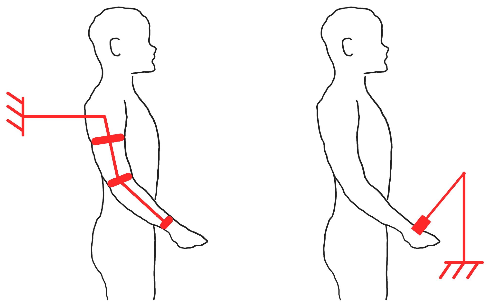
On the other hand, end-effectors do not mimic the human limbs at all. They are connected to robotic arms at their most distal extremity (the end in end-effector), and to the patient’s limb in a single point, like a grabbing mechanism. Forces generated at the end-effector change its position, thus changing the position of the limb it is attached to. Because the human limb is a connected mechanical chain, movements of the end-effector will result in the movement of other parts of the limb as well [64,65,66].
As can be seen in Figure 6, the difference between exoskeletons (left) and end-effectors (right) lies in how the movement is transferred from the device to the user’s limb [64]. While end-effectors are part of a robotic system, usually robotic collaborative manipulators, that interacts with the human body, exoskeletons are wearable devices that, in this case, support human movement.

3. Assistance Types in Robot-Aided Physical Therapy

In rehabilitation robotics, there are three major types of assistance: passive assist, active assist, and active resist [56,67,68], which are represented in Table 1.
Passive assistance is used in patients who have little to no mobility in their limbs. By moving the robotic device, and therefore the human limb attached to it, this approach can provide physical therapy without requiring any effort from the user. Often used in the early stages of rehabilitation, this assistance can improve muscle activation and blood circulation and prevent joint stiffness. This method is also able to provide resistance to the movements in order to stimulate muscle involvement [69].
Active assistance, also called Assist-As-Needed (AAN), is a type of physical therapy in which the patient actively participates in performing the exercises. The robotic device only delivers help if needed and does not move the limbs for the patient. This approach is used in patients who want to improve motor skills and rebuild strength. By having the patient perform the task and the robot deliver varying levels of assistance, this method can improve muscle power and coordination [70].
Active resist occurs when a robotic device can generate resistance in certain tasks performed by the patient. By applying a controlled resistance against the patient’s movement, this approach aims to enhance endurance and muscle strength. The patient actively participates in the exercises and an adjustable amount of resistance is applied to the movement, making it harder to complete. This occurs in patients who have regained a certain level of strength in their limbs [71].

4. Rehabilitation Robotic Devices’ Control Philosophy

In robot-aided physical therapy, there exists a plethora of control mechanisms [56,72,73], of which the most pertinent are described below, and also displayed in Figure 7.

4.1. Assistive Control

As the name suggests, assistive controllers aid the user in accomplishing the desired movements. Similarly to how rehabilitation therapists work, they let the patient move and only help as much as it is needed, in order for the patient to complete the task. There are four major types of assistive controllers: impedance-based, counter-balanced-based, electromyography (EMG)-based, and performance-based [72].

4.1.1. Impedance-Based

Electrical impedance is a measure of the opposition that a circuit presents to electric current [74], which, in the case of a motor, results in how much power is outputted. This allows the device to only help the subject when necessary, either when the user is deviating from the correct trajectory or is unable to complete the movement. When the task is being correctly done, the robot is passive, allowing the user to complete the exercise independently. Then, there usually is a dead-band set up, so that the patient can make small deviations without being disturbed. Once the user starts performing the task incorrectly, the motor generates, through mechanical impedance, the force necessary to the completion of the task [75]. These systems require individualized stiffness, damping, and inertia parameter tuning. Several approaches are possible, such as developing configuration-dependent optimal impedance control methods where parameters are assigned based on robot configuration during rehabilitation tasks [76] or implementing forgetting factor recursive least squares methods, to continuously adjust impedance parameters based on patient performance and reward functions [77].

4.1.2. Counter-Balanced-Based

Counter-balanced-based control utilizes weights as counter-balance to the user’s limb, thus reducing the strength necessary to complete certain movements (by decreasing the effect of gravity) [78]. Some examples used are arm supports, harnesses, and swimming pools (where buoyancy serves as the counterweight). This type of control requires the automatic customization of counterweights, adapting to every user. Research has documented different approaches for actively generating counter-balance forces through robot control systems, with the ability to partially cancel both limb and robot weights, to provide graduated resistance training [79]. Parameter selection involves assessment of compensation needs, with iterative learning control schemes developed to adjust gravity compensation based on patient-specific dynamics, in real time [80].

4.1.3. EMG-Based

Systems which provide an EMG-based therapy use surface EMG signals to drive the assistance. By measuring muscle activity, the device can generate assistance. When the EMG signal exceeds a given threshold, the system understands that movement is being performed and so helps the user [81]. However, EMG signals are sensitive to electrode placement and skin properties, so frequent calibrations are required. Furthermore, pathological EMG (like tremors and Parkinson’s) can lead to the augmentation of pathological motions. Research on activation thresholds is still being performed, with different thresholds required for different muscle groups and exercise intensities [82]. Pattern recognition approaches have been employed to decode patient movement intentions, though classification accuracy varies significantly between healthy subjects and stroke patients [83], because the EMG readings are differ from healthy to stroke impaired patients [84], for example.

4.1.4. Performance-Based

Performance-based therapies adapt themselves to certain parameters like force, time, trajectory, among others. If the user is performing a given exercise correctly, the device makes said exercise harder, by adapting one of the aforementioned parameters. Conversely, when the movement is being unsuccessful, the parameters are decreased, in order to make the task easier [85]. These devices require threshold setting for force, time, and trajectory parameters based on patient motor abilities. Simpler studies often use position tracking error and applied assistive force to dynamically adjust the desired task velocity [86]. Multi-mode adaptive control approaches have been developed that switch between robot-dominant and patient-dominant modes based on trajectory errors and assessed motor ability [87].

4.2. Challenge-Based Control

Contrary to the aforementioned assistive-based control, which helps the user and makes the tasks easier, challenge-based control makes the exercises harder and more challenging. Normally, this control philosophy is applied to less impaired patients, or those who have recovered past the need of assistance to complete their tasks [72]. It is important to use initial robotic assessments and ongoing performance monitoring to adapt exercise difficulty dynamically, keeping patients optimally challenged and engaged, such as the work developed by Metzger et al. (2014) [88].

4.2.1. Resistive-Based

In order to provide a challenge to the user, some devices follow a resistive-based approach. These devices restrict specified movements so as to increase the force applied by the user. By providing resistance to the participants’ movement, the devices force them to apply a larger force to finish the motion [89].

4.2.2. Restraint-Induced-Based

Restraint-induced-based control systems, as the name suggests, restraint the patient’s functional limb in order to encourage of the impaired limb [90].

4.2.3. Magnifying Error-Based

Magnifying error-based therapies increase deviations from a desired trajectory. When the user starts deviating from a given trajectory, devices with this control philosophy, trigger a force which pushes the patient with a disturbing force, requiring the patient to overcome this force to finish the exercise [91].

4.3. Haptic Interface Control

Haptic technology enables the user to interface with a virtual environment and provides a physical response such as a force, vibrations, or motions [92]. These devices can simulate ADLs to help patients recover their freedom and independence. One example of this type of control is using Virtual Reality rehabilitation for repetitive targeted motion [93], such as the use of serious games to engage the patients [94].

4.4. Non-Contact Monitoring Control

This control strategy does not rely on the physical interaction between a robot and a patient, but rather on observing the subject’s movement and instructing on what to do. These robots demonstrate how to perform the tasks and provide verbal guidance and encouragement [95,96].

4.5. General Considerations on Control Philosophy

It is important that robotic devices for UL rehabilitation take into consideration the heterogeneity of stroke patients and their varying impairment levels when designing control systems. Patient-specific factors significantly influence the effectiveness of different control philosophies and must be addressed to optimize therapeutic outcomes.
The severity of motor impairment, typically assessed through standardized measures such as the FMA score, directly impacts the most appropriate control strategy. Severely impaired patients often require passive assist control with high levels of robotic guidance, while patients with moderate impairment may benefit from assistive control that gradually reduces support.
Different muscle groups respond optimally to specific control approaches. Proximal muscles (shoulder and upper arm) typically benefit from impedance-based control with gravity compensation to support large reaching movements [97], while distal muscles (forearm and hand) often require more precise EMG-based control for fine motor tasks [98]. The abnormal muscle synergies common in post-stroke patients also necessitate control strategies that can selectively target individual muscle groups while discouraging compensatory movement patterns [99].

5. Actuation and Power Transmission in Robotic Devices

Actuators are devices used in robotics which convert the energy transmitted to them into motion or mechanical work [100,101]. There are a wide variety of actuation technologies used in robotic machines [56,102], but in rehabilitation devices, the most common are electric, pneumatic, and hydraulic actuators [102]. Several different types of actuators also exist, such as mechanical, piezoelectric, thermal, magnetic, rotary, and linear actuators, among others, but their presence and usage in rehabilitation devices are less frequent.

5.1. Electric Actuators

Electric actuators are the most widely used solution in rehabilitation devices because they can produce a large amount of torque, can produce a high precision motion and are easy to use. They can be made in different types, sizes, and capacities, suiting any robotic operation. On the other hand, electric actuators are heavyweight, which increases the total weight of the device [103].
The most common types of electric actuators are servomotors. Servomotors are controlled by an electric signal, producing an amount of movement, controlling speed, position, and torque which change frequently and sometimes abruptly [104,105]. There are two types of servomotors, alternating current (AC) and direct current (DC). AC servomotors are commonly used in robotics, and their speed is determined by the frequency of the applied voltage. On the other hand, DC servomotors’ speed is proportional to the supply voltage [106]. While AC servomotors are used in high-speed applications and are more efficient, DC servomotors are usually used in low-speed and high-torque applications. Due to their complexity, AC servomotors are usually heavier than DC servomotors [107].
There are also two types of DC servomotors: with brushes and brushless. Brushed DC motors work by using a spinning coil of wire (the rotor) inside a fixed magnetic field (the stator). To keep the motor spinning, small metal brushes touch a rotating part (the commutator), which switches the electrical current on and off in the right pattern. On the other hand, brushless DC motors don’t use brushes to switch the current. Instead, they rely on electronic circuits to control the power flow. While brushless motors are quieter, more efficient and have a longer lifetime (because there are no wearing brushes), they are more expensive than brushed servomotors [108].
Another type of brushless DC motor is the stepper motor. Unlike standard DC motors, stepper motors move in fixed increments called steps. These steps allow for accurate position control [109]. All these types of electric motors, along with their advantages and disadvantages are summarized in the Table 2.
In order to achieve a low impedance, electric motors are usually placed in series with an elastic element, called series elastic actuators (SEAs). This provides a securing safety, an accurate force control, tolerance to impact loads, and improved performance [110]. All these characteristics are beneficial for rehabilitation robots because most tasks require precise torque control and safety [111].

5.2. Pneumatic Actuators

Pneumatic actuators utilize pressurized air to produce motion and generate mechanical movement. They are flexible, lightweight, and easy to use. Because of their high bio-compatibility, this type of actuators are more comfortable to wear than rigid actuators. They have a higher force/volume ratio and a lower impedance compared to electric actuators. These pneumatic systems are also reliable, efficient and highly durable, requiring less maintenance. Pneumatic artificial muscles (PAMs) are a special type of pneumatic actuators because they are bio-inspired, replicating the working muscles of the human limb. They consist of an inflatable bladder and are normally used in pairs [103,112].

5.3. Hydraulic Actuators

Instead of using pressurized air, like pneumatic actuators, hydraulic actuators utilize high-viscosity and low compressibility fluids to generate movement, generating a higher power density. When comparing pneumatic and hydraulic cylinders of the same size, the forces generated by hydraulic actuators are 25 times greater than the forces generated by pneumatic actuators. They have a fast dynamic response, good controllability, offer high power/weight ratios, and a very large amount of torque. Regarding the force provided, hydraulic actuators out-perform current electric actuators. However, these devices are heavyweight, bulky, and noisy, have high impedance, and are prone to oil leaks which can lead to the need for complementary parts [103,113].

5.4. Comparison

In Table 3, the advantages and disadvantages of each actuator is displayed for easier understanding. Electric actuators offer high precision, substantial torque output, and minimal setup requirements. However, they tend to be relatively heavy, especially when compared to pneumatic actuators. Pneumatic actuators, in contrast, are lightweight, flexible, and often the most ergonomic option among the three types discussed. Their main drawback lies in the complexity of their setup, which typically involves compressed air systems. A similar challenge arises with hydraulic actuators: although they do not rely on air circulation, the use of fluid and bulky tubing introduces the risk of leakage and adds to the system’s overall complexity.

6. Discussion of Robotic Devices for Upper-Limb Rehabilitation

Before the discussion of the reviewed robotic devices, it is important to note that there are a plethora of rehabilitation outcome assessment measures available [93], like the FMA, which assesses a person’s motor impairment severity; the Action research arm test (ARAT) [114], which examines the UL functional recovery post cortical damage; and the Box and block test (BBT) [47], which evaluates gross manual dexterity, but each author opts for a different type of testing.

6.1. End-Effector-Based Devices

Life Science Robotics (LSR) [115] commercializes their robot Robert® which, although the official launch information is not specified in the available sources, appeared in the 2018 MEDICA Fair [116]. It is a rehabilitation robot which allows the therapy of the UL and uses Kuka’s LBR Med robotic collaborative manipulator [117] to provide physical rehabilitation. Their upper extremity module has a handle and a sleeve as its end-effector. The user slides their hand through the sleeve and holds onto the handle during the exercises. This end-effector provides an additional DOF to the manipulator. A healthcare professional attaches the end-effector to the patients’ impaired limb and the patient performs the exercises with the robot attached. The Robert® will then repeat those exercises, allowing the reduction of the physical therapist’s workload. It is capable of providing all aforementioned types of assistance and LSR also provides a set of exercises their robot is capable of. Neither of these devices has actuation on their end-effectors but LSR has developed an sEMG-triggered electrical stimulation device to support active mobilization [118].
Wang and Xu [119], in 2021, developed an end-effector-based wrist rehabilitation robot. This robot has three DOFs, both wrists’ DOFs and elbow supination/pronation, it is pneumatic-actuated, using PAMs, and it is also EMG-controlled. The wrist rehabilitation robot was tested through motion experiments covering flexion–extension, abduction/adduction, and supination/pronation training modes. Angle measurements verified kinematic accuracy, and surface EMG signals were analyzed using time-domain features and power spectrum ratios to evaluate muscle activation under different rehabilitation conditions.

6.2. Exoskeleton-Based Devices Actuated with Servomotors

Popescu et al. (2018) [120] developed an exoskeleton for UL rehabilitation based on their RAPMES virtual model [121]. This device was 3D-printed in order to be as light as possible, while maintaining the necessary strength. The user slides their arm through the opening and grabs the handle and, using Velcro-fastening systems, their limb is fastened to the exoskeleton. It has three actuated DOFs, using servomotors, which replicate the arms movements: both elbow’s DOFs and the wrist’s flexion/extension. This exoskeleton falls within the passive assist devices.
Yang Liu et al. (2021) [122] proposed a table-mounted lightweight elbow exoskeleton. This system possesses one actuated DOF, elbow extension/flexion, using a servomotor. Capable of delivering active resist, this exoskeleton utilizes sEMG signals to control the resistance. For that, they use the MYO armband [123]. The exoskeleton’s assist was tested by measuring arm muscle sEMG while holding weights, with and without it. sEMG data was classified to assess load recognition, and muscle activity changes were analyzed to evaluate assistance.
González-Mendoza et al. (2022) [124] developed an exoskeleton which is adaptable for different arm and forearm lengths, adjusted with Velcro straps, and is portable, working on a battery. It also has three DOFs, elbow flexion/extension and both wrist’s movements, actuated with servomotors. The exoskeleton is fitted with two load cells, which are capable of sensing forces applied by the user. Regarding assistance, this exoskeleton is capable of providing both passive (as of the writing of this paper, only for lightweight users) and active assistance. It is equipped with sEMG sensors and two different control systems. For passive therapy, a Proportional–Derivative controller delivers gravity compensation, and for active therapy, control is based on sEMG signals and system admittance.
Zhang et al. (2025) [125] developed an UL exoskeleton rehabilitation system that incorporates both active and passive rehabilitation modes for stroke patients. The system provides two degrees of freedom covering elbow flexion/extension (0º–150°) and wrist flexion/extension (−70° to 90°) with adjustable components accommodating different UL length. This adjustment is achieved using stepper motors, which lock the system in place. Regarding control, the system uses angle and force sensors, coupled to a servomotor. Zhang et al.’s device is capable of two types of assistance: in passive mode, the exoskeleton moves the impaired limb through predetermined trajectories; in active mode, angle sensors on the healthy limb guide synchronized movement of the impaired limb via wireless transmission. Safety mechanisms halt movement within 0.2 s when preset angle thresholds are exceeded. Testing with 20 healthy volunteers showed accurate angular precision and adaptability under different loads.

6.3. Exoskeleton-Based Devices Actuated with Brushed DC Motors

In 2009, Nef et al. published the development of the ARMin III [126]. This exoskeleton possesses the three DOFs of the shoulder and elbow flexion/extension. A different module can be attached and provide elbow pronation/supination and wrist flexion, totaling six DOFs. The ARMin III is equipped with brushed DC motors in all joints and controls its movements using impedance and position control. It also possesses a counter-balancing mechanism that, in case of a power cut, keeps the robot in place. The evaluation of the ARMin device followed a four-phase process. In Phase I, healthy volunteers tested the system for functionality, safety, and stability. Phase II extended the same assessments to chronic stroke and some spinal cord injury. Phase III involved a pilot study with eight chronic stroke patients undergoing 24–32 h of training over eight weeks. Results showed improvements UL functionality. The training was well-tolerated, with no missed sessions and high patient motivation. These promising but individual results led to Phase IV, a multicentre randomized clinical trial involving 88 chronic stroke patients: half receive ARMin therapy and half conventional therapy [127].
Lambelet et al. (2017) [128] developed the eWrist, a 3D printed light, portable and simple wrist exoskeleton, which is battery charged, and is coupled to the MYO armband. It is used in the rehabilitation of wrist flection/extension, providing one actuated DOF, with a DC motor. By using force sensors, the sEMG measures from the MYO armband and the angle and velocity of the wrist, this exoskeleton is capable of providing active assistance to its users. This AAN property is made possible through the use of admittance (inverse of impedance [74]) control. The device was evaluated for wearability, mechanical performance, range of motion, setup time, and control accuracy. Tests with healthy subjects showed improved movement efficiency and responsiveness under sEMG-based assistance.
In 2020, the team behind the MAHI Exo-II [129] used sEMG sensors to control this elbow, forearm and wrist rehabilitation device [130,131]. This exoskeleton was designed for the rehabilitation of people with Spinal Cord Injury (SCI) and has four electrically actuated DOF: two for the elbow and two for the wrist (all DOF for both). The creators of the MAHI Exo-II programmed their device so that it was able to provide all three types of assistance. The MAHI Exo-II has been tested with both able-bodied individuals and patients with SCI. These tests performed sEMG recordings during single- and multi-DOF movements, using different metrics to classify movement intentions. Clinical tests, like the ARAT, assessed hand and arm function, while robotic evaluations measured movement quality and control accuracy in both types of subjects.

6.4. Exoskeleton-Based Devices Actuated with Brushless DC Motors

Hocoma [132], in 2011, launched the world’s first commercially available robotic arm exoskeleton with a three dimensional (3D) workspace [133], Armeo®Power [134]. This device is equipped with a computer software along with serious game-like exercises, is capable of providing AAN rehabilitation and report user progress. It possesses seven DOFs, each with an electric motor [135]. It can also come with an actuated handle, the Manovo®Power [136], which enables the patients to exercise hand opening and closing.
In 2012, Bergamasco, Salsedo, and Lenzo patented their upper-body exoskeleton, Arm Light Exoskeleton—ALEx [137]. This robot has six DOFs: four actuated (shoulder abduction/adduction, rotation and flexion/extension, and elbow flexion/extension) and two passive (forearm pronation/supination and wrist flexion/extension). ALEx is equipped with brushless motors and angular position sensors located at the joints. Cables are utilized to transfer the torque to the joints. This device is capable of providing different levels of assistance: no assistance, in which the subject moves freely and the robot measures the movements; trajectory control; and AAN, where the exoskeleton helps the user finish the task. This system has been tested in healthy subjects’ UL rehabilitation, and positive results have been documented [138,139]. Both studies evaluated the ALEx exoskeleton through kinematic, EMG, and muscle synergy tests in healthy subjects. Kinematic results showed improved accuracy without compromising pace or smoothness. EMG analysis revealed reduced shoulder muscle activity and increased elbow flexor activation. Muscle synergy patterns remained largely consistent. Their conclusion was that, overall, ALEx supported natural movement and coordination, indicating its potential for post-stroke rehabilitation.
Based on their 2017 prototype [140], Zeiaee et al. (2021) [141] developed their CLEVERarm whole arm exoskeleton. CLEVERarm is lightweight, as it is made of carbon-fiber-reinforced 3D-printed plastic, and its upper-most part is connected to a stand. This exoskeleton possesses all the DOFs mentioned in Section 2.1, except for the wrists’ deviation. These eight DOFs, thus, mimic all shoulder and elbow movements. Of these eight DOFs, six are actuated: five in the shoulder and elbow flection/extension, and two are passive: forearm pronation/supination (elbow joint) and wrist deviation. All active joints utilize on-axis brushless electric motors. Also, every actuated joint is equipped with position and movement sensors and also torque sensors. As of 2021, this device is capable of providing anti-gravity assistance. It was also tested, through several metrics, such as rigidity using weight-induced deflection, joint dynamics via frequency response, range of motion with users, interaction forces for ergonomic alignment, friction compensation effectiveness, and tracking accuracy under disturbances.
In 2021, Islam et al. developed a passive rehabilitation robot that can operate both as an exoskeleton and as an end-effector, named u-Rob [142]. Islam et al. also wanted to incorporate a sensorized cuff to monitor the interaction forces at the upper arm. The u-Rob possesses seven actuated and two passive DOFs. The actuated DOF uses brushless DC motors to allow the three movements of the shoulder and both movements of the elbow. It also allows both wrists’ movements. As mentioned in Section 2.1, there are two more DOF in the UL, so this device uses the two passive DOF in order to achieve the maximum comfort for its wearers (in exoskeleton mode). Besides the force sensor on the cuff, which measures the user’s residual strength applied, the u-Rob is also equipped with joint position and velocity sensors. This robot was tested with five healthy subjects and was deemed adequate for performing passive rehabilitation of an impaired UL. These tests included single-joint and multi-joint movements as well as Cartesian space reaching tasks. Position, velocity, torque, and interaction force data were collected to assess tracking accuracy and user interaction.

6.5. Exoskeleton-Based Devices Actuated with SEAs

In 2016, Crea et al. presented their exoskeleton NESM [143]. This exoskeleton is mounted on a wheeled base and targets the shoulder and the elbow. It possesses eight passive and four active DOFs that are electrically actuated. These DOFs allow for the three movements of the shoulder and the flexion/extension of the elbow. All electric actuators are composed of SEAs coupled to a brushless DC motor. The NESM is equipped with angular and torque sensors and has two operating modes: joint position control mode, where the device moves the user’s arm passively, and joint torque control mode, providing ANN assistance to the patient.
In 2017, Kim and Deshpande released their new exoskeleton Harmony [144]. As of 2025, this exoskeleton, renamed Harmony SHR®, is the flagship product for the company Harmonic Bionics [145]. The Harmony SHR® is now a patented bilateral upper-extremity rehabilitation exoskeleton [146], with patented actuating mechanisms [147]. The Harmony SHR® provides all five DOFs of the shoulder and also both DOFs of the elbow. Additionally, it provides at least two DOFs for the back, allowing spine flexion, tilting and rotation. These DOFs are electrically actuated, using brushless DC motors and SEAs. The actuation mechanism allows for an accurate force and impedance control. Moreover, this exoskeleton is also equipped with position and motion sensors. Each patient wears four straps made of Naugahyde and Velcro, which are then attached to the robot, whose size can be adjusted to fit each patient’s proportions. Harmonic Bionics states that the Harmony SHR® has three different operating modes: Predefined Exercise Mode, a type of passive assistance; Active Freeform Mode; and Bilateral Sync Mode, which provides active assistance. The Harmony SHR® has been subject to many tests, with both healthy and disabled (stroke) patients [148,149,150,151], namely the FMA and the ARAT. It has been installed in Universities [152] and in April 2023 got approved by the American Food and Drug Administration (FDA) [153]. Since then, it has been installed in several hospitals [154,155]. However, it is important to note that in Harmonic Bionics’ website, on the bottom banner, is written “this product has not been approved, cleared, or authorized by the FDA and is Limited by Federal (or United States) law to investigational use”.
Chen et al. (2019) [156] developed an elbow exoskeleton whose electric motor was table-mounted. Then, using cables and SEAs, it provides two actuated DOFs to the elbow. Because the motor is placed separately from the exoskeleton, on a surface, this device is extremely light-weight. This exoskeleton can control the angles of the DOFs, calculate the strength applied by the user, and manage the amount of torque provided. These properties allow this robot to provide different levels of active resistance.
After their 2019 ANYexo [157], Zimmermann et al. developed the ANYexo 2.0 [158] exoskeleton in 2023. This device actuates in all nine DOFs of the UL, allowing all usual arm movements. All joints are electrically actuated and in the top five DOFs (shoulder and scapulohumeral rhythm) SEAs are used. The ANYexo 2.0 is also equipped with torque and position sensors and provides anti-gravity support. This exoskeleton was tested by its own authors in ADLs. The authors’ ANYexo 2.0’s joint range and workspace, structural stiffness under load, and control accuracy in following movement trajectories were examined. It performed key rehabilitation motions like flexion–extension and supination–pronation. Human subject tests validated usability and safety, with careful monitoring of interaction forces to prevent discomfort or injury.

6.6. Non-Electrically Actuated Exoskeleton-Based Devices

In 2015, Polygerinos et al. developed the finger exoskeleton [159]. This device is hydraulically actuated, using water to control its parts. In order to keep it light, Polygerinos et al. mounted the power supply, water reservoir and other heavy components in a waist belt pack, connected to the glove via tubing. The exoskeleton allows three DOFs for the index, middle, ring, and little fingers, allowing for flexion/extension movements. For the thumb, this exoskeleton also allows for three DOFs, two for flexion/extension and one for rotation. Polygerinos et al.’s glove possesses built-in fluidic pressure sensors and force/torque sensors. It is also equipped with position and movement sensors. This system was designed to help patients with ADLs and subsequent versions have been equipped with surface EMG (sEMG) sensors [160]. This newer version has been subject to tests with both healthy and disabled patients and its performance showed positive results. These tests showed that the robotic glove greatly reduced muscle effort during grasping, confirmed by EMG measurements. Pressure mapping demonstrated the glove can produce grip forces comparable to a natural hand. Functional tasks verified the glove’s EMG control effectively assisted hand movements, enabling grasp, hold, and release.
Using the MATLAB/Simulink Biomechanics of Bodies package [161], which is a biomechanical modeling package that contains a human musculoskeletal model, Copaci et al. have developed, from 2015 to 2019, an elbow rehabilitation exoskeleton which uses Shape Memory Alloys (SMA) [162,163,164,165,166]. SMA are materials that can undergo deformations but return to their original shape once subject to a stimuli, such as heat [167]. Copaci et al. use these alloys in order to avoid the high weight and noise of electric motors. Copaci et al.’s exoskeleton allows for two different DOFs: elbow flexion/extension and pronation/supination. It also has two modes of operation: one for data collection (the device is not actuated) and one for rehabilitation, providing active assistance. Throughout all the iterations of this project, Copaci et al. used several sensors to measure angles, force, position, pressure and temperature. Copaci et al. also experimented with an sEMG control mechanism. Since then, this team has also developed an sEMG controlled glove for hand rehabilitation [168].
Quan Liu et al. (2021) [169], in order to exploit the lightweight properties of pneumatic actuators, developed an UL exoskeleton using PAMs. This device is composed of two modules, one for wrist rehabilitation and another for elbow rehabilitation. These modules can be used together or separately. It is adjustable for all sizes and, when fully mounted, provides three DOFs, elbow flexion/extension and two for the wrist joint. The actuation is conducted using PAMs and the control is carried out with angle and force sensors in the joints. With this design, it is possible to deliver active assistance and position control.

6.7. Reviewed Devices Discussion and Comparison

Table 4 summarizes all the reviewed devices, with the pertinent information displayed, organized by year. It is important to note that when no name was identified for the device, the developer’s name was used.
Table 4 provides a valuable insight for the development of an electromechanical robot–human interface. Firstly, 18 out of 20 devices (90%) are exoskeletons, with only 2 actuated end-effectors being designed. Several comparison studies between both approaches have been published [54,171,172], but this review focused solely on actuated interfaces. Exoskeleton technologies, although capable of providing therapy to multiple joints and offering more DOFs, are more expensive, bigger, and more complex to produce. On the other hand, end-effector-based solutions, like the ROBERT® or the u-Rob in end-effector mode, are often limited to a specific motion and train the whole upper-arm, not focusing on specific joints. There is still debate and not enough information to decide which approach is the most effective towards stroke-recovering patients, as studies offer conflicting perspectives [65,173]. This 18:2 ratio of exoskeletons to end-effectors reveals a significant research bias toward wearable solutions despite end-effectors’ simpler mechanical design. The two end-effector systems reviewed (ROBERT® and Wang & Xu’s device) demonstrate markedly different approaches: one commercial system targeting multiple limbs versus one research prototype with 3 DOFs, suggesting underdeveloped standardization in this category.
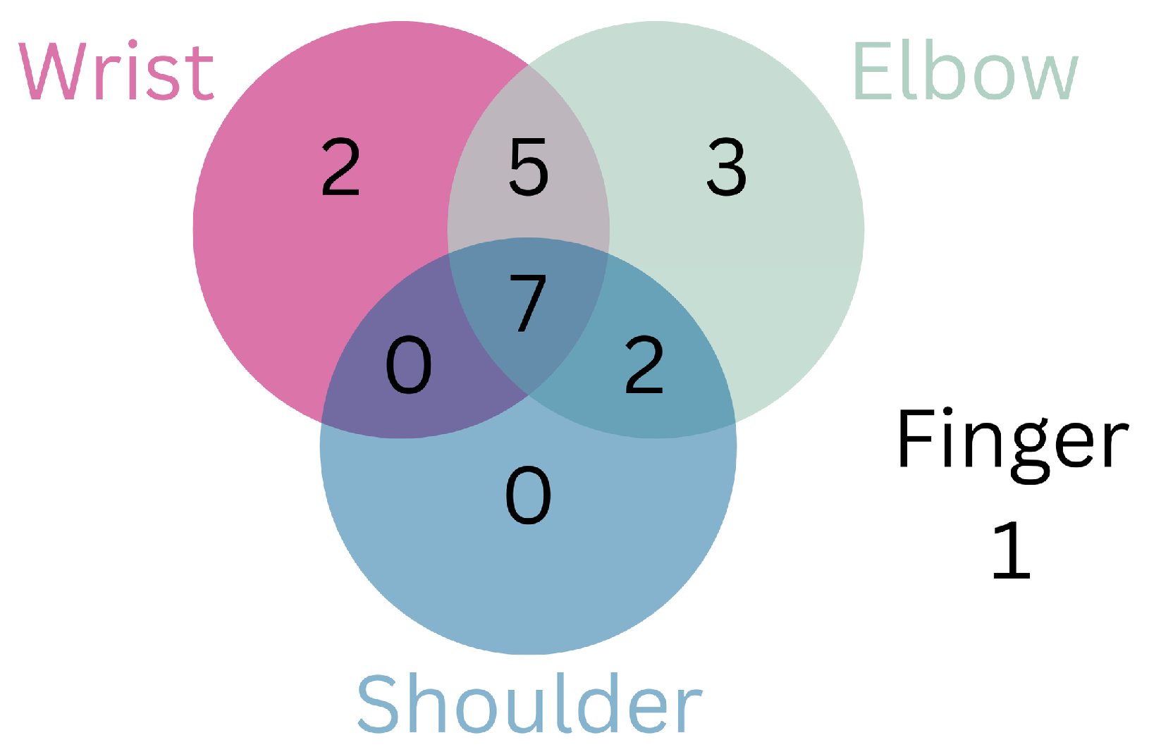
In Figure 8, it is possible to see a Venn diagram displaying how many of the reviewed devices target each portion of the UL. Additionally, 14 out of 20 robots (70%) target multiple sections of the UL, such as elbow and wrist, shoulder and elbow or the whole UL. The number of available DOFs increases to facilitate the rehabilitation of multiple areas. These devices, which target multiple portions of the UL, are more complex, thus requiring more sophisticated control mechanisms, and some even depend on ground- or table-mounted supports to hold their weight. Notably, the two commercially available systems (ROBERT® and Armeo®Power) represent opposite ends of this spectrum (1 DOF vs. 7-8 DOF), indicating no consensus on optimal complexity for clinical deployment. Furthermore, it is possible to infer that most devices target elbow rehabilitation (17 out of 20, 85%), which may happen because the movements of the elbow are less complex than those of the shoulder, but more crucial than those of the wrist. Finally, Polygerinos et al.’s glove stands alone, targeting only the fingers.
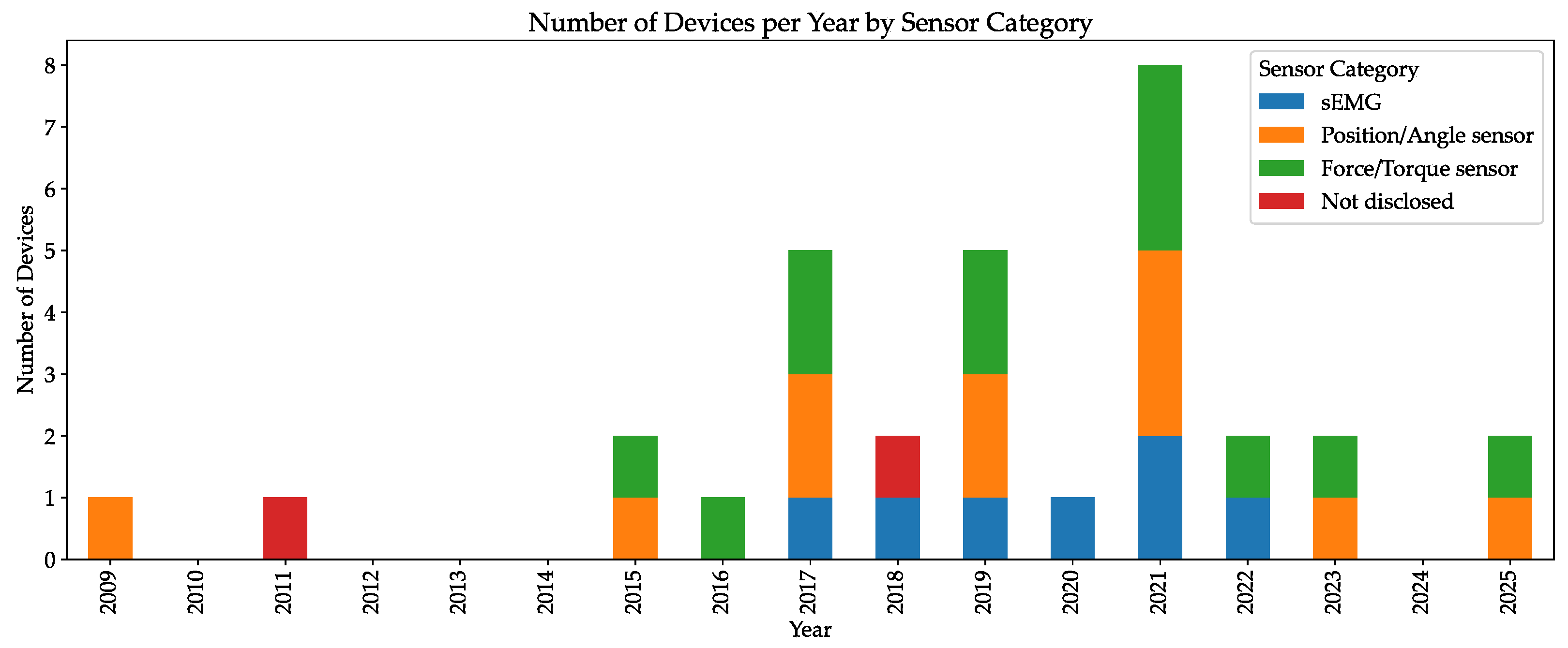
Figure 9 displays the reviewed devices, each one with a different color, and the type of sensors they use. The use of position or angle and force/torque sensors is quite common throughout all the reviewed devices, with 14 out of 20 devices (70%) opting for an impedance-based control. sEMG is also a frequent choice for the devices’ control, with its use being implemented in 7 out of 20 devices (35%), with higher frequency in more recent projects (all are from 2017 onwards), as seen in MAHI Exo-II and González-Mendoza’s design, possibly due to the wide availability of surface EMG sensors, such as the MYO armband, reflecting a shift towards user-intent-driven rehabilitation. Although these two control methods are widely adopted, their suitability varies across patient profiles. For instance, impedance control offers robust safety and compliance, but may lack the responsiveness to sudden user intent changes. EMG systems, while promising for user-driven interaction, often struggle with signal reliability, sensor placement variability and signal interpretation, especially in patients with low or erratic muscle activity.
As can be seen in Figure 10, 15 out of 20 devices (75%) opt for electric motors, particularly brushless DC motors and SEAs, as seen in Harmony SHR® and NESM. This observable preference happens likely because of their easy setup, high-torque capabilities, and reduced operating noise. However, they are generally heavy and require external supports for stability. In contrast, Polygerinos et al. hydraulic glove and Wang and Xu’s pneumatic actuator, offer lightweight and compliant interaction, better suited for wearable designs, though at the cost of added complexity, such as fluid handling and air compressing systems, and reduced positional accuracy. SMA-based systems attempt to bridge this gap but, currently, only 1 out of 20 projects (5%) involving this technology are available in this review. A future direction might involve hybrid actuation strategies to balance force, weight, and responsiveness. Furthermore, a recent trend can be observed in the use of servomotors, likely due to their precise position control, alongside an increasing adoption of SEAs and PAMs, which reduce rigidity and offer muscle-like interaction.
Moreover, 14 out of 20 devices (70%) focus on providing assistance, specially active assistance, while offering resistance is not the primary goal, with only 4 out of 20 devices (20%) performing this type of exercises. This is the case given that most robots are designed for the severely impaired, and active resistance is typically introduced at a later stage of rehabilitation when patients have regained some mobility and strength in their limb. Active resistance, however, becomes essential in later stages of rehabilitation, once patients have regained some mobility and strength. It plays a critical role in rebuilding muscle strength and endurance. Devices that cannot deliver active resistance may be unsuitable for full-spectrum rehabilitation, either excluding certain patients or requiring complementary therapies to achieve a complete recovery.
As can be seen in Figure 11, not all reviewed interfaces display information on their testing status (six devices not tested at all), but 8 out of 20 machines (40%) were tested on healthy volunteers while 4 out of 20 (20%) were tested on impaired patients (i.e., stroke patients). Only 2 out of 20 solutions (10%) are medically approved (i.e., FDA) and are commercially available in hospitals.
Electric actuator dominance correlates with testing advancement, 5 out of 6 patient-tested devices use electric motors, and both commercially available devices are electrically actuated. Nevertheless, despite these technological advancements, the potential of robotic rehabilitation remains far from fully realized. Clinical adoption faces several challenges: lack of standardized testing protocols and performance metrics, insufficient long-term clinical data, difficulties in customizing to patient anatomy, and high production costs. Moreover, 14 out of 20 reviewed systems (70%) remain in laboratory testing phases with limited field deployment, and only two devices (the Harmony and the MAHI Exo-II, 10%) have executed known and published outcome analyses tests. Comparative metrics—such as rehabilitation effectiveness, user compliance, and cost–benefit ratios—remain under-reported and vary across studies, making it difficult to establish clear benchmarks. The use of standardized performance metrics (such as the FMA and the ARAT) should be a goal for every researcher, as these tests allow for objective comparison with other devices and high-trust clinical validation. To advance beyond this stage, future research must prioritize modular design, compliance with medical device regulations, robust usability testing in diverse clinical environments, and a structured framework for evaluating robotic interfaces that integrates kinematic recovery metrics with patient-reported outcomes. Collaboration with clinicians and rehabilitation therapists from early development stages can facilitate this transition.
While this review presents an overview of robotic interfaces for post-stroke UL rehabilitation, several promising innovations and approaches are emerging. Most devices mentioned are bulky and unsuitable for in-home use. Miniaturization and portability should be considered key-factors in future developments. With the advent of Artificial Intelligence (AI), future works will most likely shift towards AI-driven systems coupled to biofeedback, possibly even brain–computer interfaces. These technologies offer a more specialized and adaptive rehabilitation, linking the user’s intent with robotic movement. Finally, as mentioned, no systems combining the use of robotic manipulators and actuated end-effector were found, so there is an opportunity to explore hybrid systems that combine the fine motion control of exoskeletons with the range and workspace flexibility of robotic manipulators.

7. Conclusions

Neurological diseases, such as stroke, remain a crucial area of study, for they comprise some of the greatest causes of disability, and the number of new cases is growing each year. Ever since the 1990s, the practice of robot-aided physical therapy has become more common, and its market is steadily growing. This paper presents a systematic review on robotic interfaces for UL rehabilitation, not limited to stroke survivors but also applicable to several disabled patients, based on the analysis of 20 papers from 2009 to June 2025. A detailed analysis of each device’s characteristics, including assistance types, control mechanisms, and actuation technologies, has been provided.
The overview presented in Section 6 allows for a clear identification of technological directions and gaps in the field of UL rehabilitation robotics. A first notable pattern is the predominance of exoskeleton-based designs, with 90% of the reviewed devices adopting this configuration. This highlights the importance attributed to joint alignment and segment-specific movement guidance, although it also introduces challenges related to donning, ergonomics and the need for more complex control mechanisms. End-effector devices, while less common, remain relevant because of their simpler mechanics, faster setup and lower cost, which can be advantageous in clinical context where usability is critical and also for easier and broader adoption. However, this comes at the expense of reduced precision and limited monitoring of individual joint movements compared to exoskeletons, which may compromise exercise quality and therapeutic outcomes.
Analysis of the relationship between device complexity and clinical advancement reveals a critical pattern in the field. Specifically, devices with moderate complexity achieve greater clinical success than either overly simple or highly complex alternatives. Figure 12 compares the reviewed devices’ complexity and their testing status. Device complexity scores were calculated using a standardized point system where each targeted UL portion (fingers, wrist, elbow, shoulder), control/sensor type, assistance type, and degree of freedom contributed one point, with non-electric actuated devices receiving an additional point. On their testing status, 0 is not tested; 1 is tested on healthy participants; 2 is tested on disabled patients; and 3 is commercially available.
As demonstrated in Figure 12, devices with moderate complexity levels (8–15) have achieved the most extensive clinical testing and validation, with the majority of commercially available (ROBERT® and Armeo®Power) and patient-tested devices clustering within this range. This concentration suggests an optimal complexity window where devices provide sufficient therapeutic functionality while remaining clinically feasible. Devices below complexity level 8 appear to be filtered out early in development, likely because their limited functionality cannot justify the substantial investment required for clinical trials and regulatory approval. Conversely, highly complex devices (above level 15) face different barriers: while they may offer superior therapeutic potential, they encounter significant regulatory, technical, and usability difficulties, which may impede clinical progression. The single outlier device exceeding complexity level 15, Polygerinos et al.’s glove, despite reaching patient testing in 2015, remains commercially unavailable, illustrating these implementation challenges. The absence of commercially successful devices below complexity level 8 and the rarity of complex devices on clinical practice suggests that sustainable clinical translation requires achieving sufficient therapeutic capacity while maintaining regulatory and practical feasibility.
Conversely, only one device exceeds complexity level 15, and while it has been tested on impaired patients (in 2015), it represents an outlier rather than a trend, suggesting that highly complex devices face significant barriers to development and clinical progression. The pattern reveals that no devices below complexity level 8 have achieved commercial status, suggesting there may be a minimum complexity threshold required for commercial viability, though this does not reflect on the technical sophistication of these devices, which could be high regardless of their complexity level.
Comparative analysis reveals clear performance trade-offs across device categories. Rigid skeletons like the Armin III and the Harmony SHR® demonstrate superior force output and joint stability, earning them their FDA approval, but can face acceptance challenges due to weight and cost limitations. Conversely, soft robotics approaches such as Polygerinos et al.’s glove show higher patient satisfaction and bio-compatibility but present limited force generation capabilities. Hybrid systems are beginning to show superior clinical outcomes by adaptively adjusting assistance levels according to patient progression, though they remain underrepresented in current research.
The analysis also reveals diversity in actuation choices: electric motors, whether brushed or brushless (often combined with SEAs) remain the most widely adopted solution, due to their controllability and high precision, but add significant weight to devices. Pneumatic systems offer lighter, safer, and more compliant interaction but suffer from reduced controllability and setup complexity. The fact that alternative actuation methods like PAMs and smart materials remain less prevalent indicates ongoing technological challenges in balancing force output, weight, reliability, control difficulties, and slow response times.
Control strategies show a similar trend towards greater personalization. While many early devices used position or velocity control alone, more recent systems integrate sensors, either sEMG or force/torque sensors. EMG-based control allows for personalized, intent-driven assistance but suffers from signal noise, electrode placement variability, and reliability issued, particularly in patients with pathological muscle activity. Impedance and adaptive assist-as-needed control approaches are currently showing the most promising results in clinical trials, though comparative effectiveness data remains limited by the diversity of clinical metrics used across studies.
Regarding assistance types, active assistance systems demonstrate higher mobilization capacity and patient engagement but require robust safety mechanisms to ensure appropriate force application. Passive assistance interfaces are more accessible and present fewer safety concerns but show limited therapeutic gains compared to active approaches. The predominant focus on assistive rather than resistive rehabilitation reflects the target population of severely impaired patients, though this limits applicability across the full spectrum of recovery stages.
Several limitations constrain the current state of research. Most studies involve small sample sizes, lack long-term follow-up assessments to evaluate retention of motor gains, and use inconsistent outcomes measures that prevent meaningful cross-study comparisons. A major obstacle is the absence of standardized clinical testing protocols, which hampers quantitative evaluation across studies. Furthermore, many devices remain at the feasibility stage, often tested only on healthy volunteers, whereas others such as Armeo®Power and Harmony HR®, have advanced to commercial distribution after demonstrating clinical efficacy through standardized metrics. This shows that technical innovation alone does not guarantee clinical uptake, as factors such as ease of use, cost-effectiveness, and demonstrated patient benefit are decisive in determining adoption.
Future research must address the aforementioned limitations through several key directions. Standardized clinical evaluation protocols using consistent outcome measures (e.g., FMA, ARAT, BBT) are essential for enabling meaningful device comparisons and building evidence-based treatment guidelines. Larger-scale trials with long-term follow-up are needed to assess retention of therapeutic gains and cost-effectiveness for healthcare system integration. The development of hybrid interfaces that combine the mechanical robustness of rigid exoskeletons with the comfort and compliance of soft robotics represents a promising technical direction. Integration of AI algorithms for adaptive, personalized assistance control could significantly enhance therapeutical outcomes by adjusting assistance levels based on patient progression patterns. Additionally, the exploration of multi-modal bio-signal integration (e.g., EMG + electroencephalogram) may enable more natural, intuitive control interfaces.
This paper not only summarizes state-of-the-art developments but also identifies emerging trends and critical limitations that must be addressed for broader adoption. The field shows clear technological maturation in electric actuation and impedance control, yet significant gaps remain in standardized evaluation, long-term clinical validation, and cost-effective deployment strategies. Bridging these gaps thorough collaborative research involving engineers, clinicians, and rehabilitation therapists from early development stages will be essential for translating promising laboratory prototypes into effective real-world rehabilitation solutions that can meaningfully impact the growing population of stroke survivors worldwide.

Author Contributions

Conceptualization, A.G., H.M., M.F.S., and C.D.R.; methodology, A.G., H.M., M.F.S., and C.D.R.; investigation, A.G.; data curation, A.G., H.M., M.F.S., and C.D.R.; writing—original draft, A.G.; writing—review and editing A.G., H.M., M.F.S., and C.D.R.; visualization, A.G.; supervision, H.M., M.F.S., and C.D.R.; project administration, H.M., M.F.S., and C.D.R.; funding acquisition, C.D.R. All authors have read and agreed to the published version of the manuscript.

Funding

This work is co-financed by Component 5- Capitalization and Business Innovation of core funding for Technology and Innovation Centres (CTI), integrated in the Resilience Dimension of the Recovery and Resilience Plan within the scope of the Recovery and Resilience Mechanism (MRR) of the European Union (EU), framed in the Next Generation EU, for the period 2021–2026, with reference 21.

Data Availability Statement

No new data were created or analyzed in this study. Data sharing is not applicable to this article.

Conflicts of Interest

The authors declare no conflicts of interest.

Abbreviations

The following abbreviations are used in this manuscript:
3DThree-Dimensional
ADLsActivities of Daily Living
AANAssist-as-Needed
ACAlternating Current
ARATAction research arm test
BBTBox and block test
CVACerebrovascular Accident
DALYsDisability-Adjusted Life Years
DCDirect Current
DOFDegrees of Freedom
EMGElectromyography
FDAFood and Drug Administration
FMAFugl–Myer assessment
HICsHigh-Income Countries
IEEEInstitute of Electrical and Electronics Engineers
LMICsLow/Middle-Income Countries
LSRLife Science Robotics
NHSNational Health Service
PAMsPneumatic Artificial Muscles
SCISpinal Cord Injury
SEAsSeries Elastic Actuator
sEMGSurface Electromyography
SMAShape Memory Alloy
ULUpper-limb
WHOWorld Health Organization
WSOWorld Stroke Organization

References

  1. National Health Service. Stroke. Available online: https://www.nhs.uk/conditions/stroke/ (accessed on 13 November 2014).
  2. John Hopkins Medicine. Arm Care After a Stroke. Available online: https://www.hopkinsmedicine.org/health/conditions-and-diseases/stroke/arm-care-after-a-stroke (accessed on 30 July 2025).
  3. World Stroke Organization. Impact of Stroke. Available online: https://www.world-stroke.org/world-stroke-day-campaign/about-stroke/impact-of-stroke (accessed on 4 November 2024).
  4. WHO Regional Director for South-East Asia Dr Poonam Khetrapal Singh. World Stroke Day, October 2021. Available online: https://www.who.int/southeastasia/news/detail/28-10-2021-world-stroke-day (accessed on 6 November 2024).
  5. Truelsen, T.; Piechowski-Jóźwiak, B.; Bonita, R.; Mathers, C.; Bogousslavsky, J.; Boysen, G. Stroke incidence and prevalence in europe: A review of available data. Eur. J. Neurol. 2006, 13, 581–598. [Google Scholar] [CrossRef]
  6. Rajsic, S.; Gothe, H.; Borba, H.H.; Sroczynski, G.; Vujicic, J.; Toell, T.; Siebert, U. Economic burden of stroke: A systematic review on post-stroke care. Eur. J. Health Econ. 2017, 20, 107–134. [Google Scholar] [CrossRef]
  7. World Health Organization. Ageing and Health, October 2024. Available online: https://www.who.int/news-room/fact-sheets/detail/ageing-and-health (accessed on 6 November 2024).
  8. Global Burden of Disease Collaborative Network. Global Burden of Disease Study 2021 (gbd 2021) Results. 2022. Available online: https://vizhub.healthdata.org/gbd-results/ (accessed on 19 November 2024).
  9. Yousufuddin, M.; Young, N. Aging and Ischemic Stroke. Aging 2019, 11, 2542–2544. [Google Scholar] [CrossRef]
  10. Feigin, V.L.; Brainin, M.; Norrving, B.; Martins, S.; Sacco, R.L.; Hacke, W.; Fisher, M.; Pandian, J.; Lindsay, P. World stroke organization (wso): Global stroke fact sheet 2022. Int. J. Stroke 2022, 17, 18–29. [Google Scholar] [CrossRef] [PubMed]
  11. Rahbar, M.H.; Medrano, M.; Diaz-Garelli, F.; Villaman, C.G.; Saroukhani, S.; Kim, S.; Tahanan, A.; Franco, Y.; Castro-Tejada, G.; Diaz, S.A.; et al. Younger age of stroke in low-middle income countries is related to healthcare access and quality. Ann. Clin. Transl. Neurol. 2022, 9, 415–427. [Google Scholar] [CrossRef] [PubMed]
  12. Thayabaranathan, T.; Kim, J.; Cadilhac, D.A.; Thrift, A.G.; Donnan, G.A.; Howard, G.; Howard, V.J.; Rothwell, P.M.; Feigin, V.; Norrving, B.; et al. Global stroke statistics 2022. Int. J. Stroke 2022, 17, 946–956. [Google Scholar] [CrossRef]
  13. Mathews, C. Population ageing in lower- and middle-income countries. Celebrating 40 years of helpage international. J. Popul. Ageing 2024, 17, 1–4. [Google Scholar] [CrossRef]
  14. Banzrai, C.; Bosookhuu, O.; Yadamsuren, E.; Dambasuren, B.; Turbat, S.; Erdenedalai, T.; Myadagsuren, M.; Munkhtur, U.; Baatar, K.; Boldbayar, P.; et al. Incidence and outcomes for stroke in ulaanbaatar, mongolia, during 2019–2021: A prospective population-based study. Lancet Glob. Health 2023, 11, e942–e952. [Google Scholar] [CrossRef]
  15. Jiang, K.; He, T.; Ji, Y.; Zhu, T.; Jiang, E. The perspective of hypertension and salt intake in chinese population. Front. Public Health 2023, 11, 1125608. [Google Scholar] [CrossRef]
  16. He, F.J.; MacGregor, G.A. Salt, blood pressure and cardiovascular disease. Curr. Opin. Cardiol. 2007, 22, 298–305. [Google Scholar] [CrossRef]
  17. Rayner, M.; Allender, S.; Scarborough, P.; Peto, V. European Cardiovascular Disease Statistics, 2008 Edition, September 2008. Available online: https://coachmikeblogs.com/wp-content/uploads/2014/08/European_cardiovascular_disease_statistics_2008.pdf (accessed on 6 November 2024).
  18. Instituto Nacional de Estatística. Causas de Morte 2022, May 2024. Available online: https://www.ine.pt/xportal/xmain?xpid=INE&xpgid=ine_destaques&DESTAQUESdest_boui=646029525&DESTAQUESmodo=2 (accessed on 4 November 2024).
  19. Centers for Disease Control and Prevention. Stroke Facts. Available online: https://www.cdc.gov/stroke/data-research/facts-stats/index.html (accessed on 7 February 2025).
  20. World Health Organization and World Bank. World Report on Disability, December 2011. Available online: https://www.who.int/publications/i/item/9789241564182 (accessed on 6 November 2024).
  21. Fonseca, L. O avc é a principal causa de morte e incapacidade em portugal. Soc. Port. De Med. Interna 2021. Available online: https://www.spmi.pt/o-avc-e-a-principal-causa-de-morte-e-incapacidade-em-portugal/ (accessed on 4 November 2024).
  22. Stroke Association. Stroke Research Means Everything. Available online: https://www.stroke.org.uk/stroke/support/materials/rebuilding-lives/stroke-research-means-everything (accessed on 6 January 2025).
  23. National Stroke Association. Effects of Stroke. Available online: https://www.stroke.org/en/about-stroke/effects-of-stroke (accessed on 5 February 2025).
  24. National Stroke Association. Communication Problems After Stroke. Available online: https://www.stroke.org/en/about-stroke/effects-of-stroke/communication-and-aphasia (accessed on 5 February 2025).
  25. Johns Hopkins Medicine. Aphasia. Available online: https://www.hopkinsmedicine.org/health/conditions-and-diseases/aphasia (accessed on 5 February 2025).
  26. National Stroke Association. Cognitive Challenges Post-Stroke. Available online: https://www.stroke.org/en/about-stroke/effects-of-stroke/cognitive-effects (accessed on 5 February 2025).
  27. National Stroke Association. Emotional and Behavioral Effects of Stroke. Available online: https://www.stroke.org/en/about-stroke/effects-of-stroke/emotional-effects (accessed on 5 February 2025).
  28. National Stroke Association. Vision and Hearing. Available online: https://www.stroke.org/en/about-stroke/effects-of-stroke/vision-and-hearing (accessed on 5 February 2025).
  29. National Stroke Association. Physical Effects of Stroke. Available online: https://www.stroke.org/en/about-stroke/effects-of-stroke/physical-effects (accessed on 5 February 2025).
  30. National Health Service. Physical Effects of Stroke. Available online: https://www.newcastle-hospitals.nhs.uk/services/stroke-unit/life-after-stroke/physical-effects-of-stroke/#common-physical-effects (accessed on 28 November 2024).
  31. World Health Organization. Disability-Adjusted Life Years (Dalys). Available online: https://www.who.int/data/gho/indicator-metadata-registry/imr-details/158 (accessed on 15 November 2024).
  32. Feigin, V.L.; Abate, M.D.; Abate, Y.H.; ElHafeez, S.A.; Abd-Allah, F.; Abdelalim, A.; Abdelkader, A.; Abdelmasseh, M.; Abd-Elsalam, S.; Abdi, P.; et al. Global, regional, and national burden of stroke and its risk factors, 1990–2021: A systematic analysis for the global burden of disease study 2021. Lancet Neurol. 2024, 23, 973–1003. [Google Scholar] [CrossRef]
  33. World Health Organization. The Global Burden of Disease: 2004 Update; World Health Organization: Geneva, Switzerland, 2008. [Google Scholar]
  34. Bonita, R.; Solomon, N.; Broad, J.B. Prevalence of stroke and stroke-related disability. Stroke 1997, 28, 1898–1902. [Google Scholar] [CrossRef]
  35. American Stroke Association. Stroke Rehabilitation. Available online: https://www.stroke.org/en/life-after-stroke/stroke-rehab (accessed on 6 November 2024).
  36. Anwer, S.; Waris, A.; Gilani, S.O.; Iqbal, J.; Shaikh, N.; Pujari, A.N.; Niazi, I.K. Rehabilitation of upper limb motor impairment in stroke: A narrative review on the prevalence, risk factors, and economic statistics of stroke and state of the art therapies. Healthcare 2022, 10, 190. [Google Scholar] [CrossRef]
  37. Persson, H.C.; Parziali, M.; Danielsson, A.; Sunnerhagen, K.S. Outcome and upper extremity function within 72 h after first occasion of stroke in an unselected population at a stroke unit. A part of the salgot study. BMC Neurol. 2012, 12, 162. [Google Scholar] [CrossRef] [PubMed]
  38. Broeks, J.G.; Lankhorst, G.J.; Rumping, K.; Prevo, A.J.H. The long-term outcome of arm function after stroke: Results of a follow-up study. Disabil. Rehabil. 1999, 21, 357–364. [Google Scholar] [CrossRef] [PubMed]
  39. Friedman, P.J. Gait recovery after hemiplegic stroke. Int. Disabil. Stud. 1990, 12, 119–122. [Google Scholar] [CrossRef] [PubMed]
  40. O’Flaherty, D.; Ali, K. Recommendations for upper limb motor recovery: An overview of the uk and european rehabilitation after stroke guidelines (2023). Healthcare 2024, 12, 1433. [Google Scholar] [CrossRef]
  41. Bertani, R.; Melegari, C.; Cola, M.C.D.; Bramanti, A.; Bramanti, P.; Calabrò, R.S. Effects of robot-assisted upper limb rehabilitation in stroke patients: A systematic review with meta-analysis. Neurol. Sci. 2017, 38, 1561–1569. [Google Scholar] [CrossRef]
  42. Veerbeek, J.M.; Langbroek-Amersfoort, A.C.; van Wegen, E.E.H.; Meskers, C.G.M.; Kwakkel, G. Effects of robot-assisted therapy for the upper limb after stroke: A systematic review and meta-analysis. Neurorehabilit. Neural Repair 2017, 31, 107–121. [Google Scholar] [CrossRef]
  43. Fasoli, S.E.; Krebs, H.I.; Stein, J.; Frontera, W.R.; Hogan, N. Effects of robotic therapy on motor impairment and recovery in chronic stroke. Arch. Phys. Med. Rehabil. 2003, 84, 477–482. [Google Scholar] [CrossRef]
  44. Krebs, H.; Hogan, N.; Volpe, B.; Aisen, M.; Edelstein, L.; Diels, C. Overview of clinical trials with mit-manus: A robot-aided neuro- rehabilitation facility. Technol. Health Care Off. J. Eur. Soc. Eng. Med. 1999, 7, 419–423. [Google Scholar] [CrossRef]
  45. Lo, K.; Stephenson, M.; Lockwood, C. Effectiveness of robotic assisted rehabilitation for mobility and functional ability in adult stroke patients: A systematic review. JBI Database Syst. Rev. Implement. Rep. 2017, 15, 3049–3091. [Google Scholar] [CrossRef]
  46. Mehrholz, J.; Pohl, M.; Platz, T.; Kugler, J.; Elsner, B. Electromechanical and robot-assisted arm training for improving activities of daily living, arm function, and arm muscle strength after stroke. Cochrane Database Syst. Rev. 2018, 2018, CD006876. [Google Scholar] [CrossRef] [PubMed]
  47. Mathiowetz, V.; Volland, G.; Kashman, N.; Weber, K. Adult norms for the box and block test of manual dexterity. Am. J. Occup. Ther. 1985, 39, 386–391. [Google Scholar] [CrossRef] [PubMed]
  48. Wang, H.; Wu, X.; Li, Y.; Yu, S. Efficacy of robot-assisted training on upper limb motor function after stroke: A systematic review and network meta-analysis. Arch. Rehabil. Res. Clin. Transl. 2025, 7, 100387. [Google Scholar] [CrossRef]
  49. Wagner, T.H.; Lo, A.C.; Peduzzi, P.; Bravata, D.M.; Huang, G.D.; Krebs, H.I.; Ringer, R.J.; Federman, D.G.; Richards, L.G.; Haselkorn, J.K.; et al. An economic analysis of robot-assisted therapy for long-term upper-limb impairment after stroke. Stroke 2011, 42, 2630–2632. [Google Scholar] [CrossRef]
  50. Gower, V.; Aprile, I.; Falchini, F.; Fasano, A.; Germanotta, M.; Randazzo, M.; Spinelli, F.; Trieste, L.; Gramatica, F.; Turchetti, G. Cost analysis of technological vs. conventional upper limb rehabilitation for patients with neurological disorders: An italian real-world data case study. Front. Public Health 2024, 12, 1445099. [Google Scholar] [CrossRef]
  51. Lo, K.; Stephenson, M. The economic cost of robotic rehabilitation for adult stroke patients: A systematic review. JBI Database Syst. Rev. Implement. Rep. 2019, 17, 520–547. [Google Scholar] [CrossRef]
  52. Jakob, I.; Kollreider, A.; Germanotta, M.; Benetti, F.; Cruciani, A.; Padua, L.; Aprile, I. Robotic and sensor technology for upper limb rehabilitation. PM&R 2018, 10 (Suppl. 2), S189–S197. [Google Scholar] [CrossRef]
  53. National Institute of Health. Neuroplasticity, May 2023. Available online: https://www.ncbi.nlm.nih.gov/books/NBK557811/ (accessed on 4 November 2024).
  54. Bhujel, S.; Hasan, S. A comparative study of end-effector and exoskeleton type rehabilitation robots in human upper extremity rehabilitation. Hum.-Intell. Syst. Integr. 2023, 5, 11–42. [Google Scholar] [CrossRef]
  55. Page, M.J.; McKenzie, J.E.; Bossuyt, P.M.; Boutron, I.; Hoffmann, T.C.; Mulrow, C.D.; Shamseer, L.; Tetzlaff, J.M.; Akl, E.A.; Brennan, S.E.; et al. The prisma 2020 statement: An updated guideline for reporting systematic reviews. BMJ 2021, 372, n71. [Google Scholar] [CrossRef]
  56. Narayan, J.; Kalita, B.; Dwivedy, S.K. Development of robot-based upper limb devices for rehabilitation purposes: A systematic review. Augment. Hum. Res. 2021, 6, 4. [Google Scholar] [CrossRef]
  57. Chang, L.; Anand, P.; Varacallo, M.A. Anatomy, Shoulder and Upper Limb, Glenohumeral Joint. Available online: https://www.ncbi.nlm.nih.gov/books/NBK537018/ (accessed on 30 July 2025).
  58. Physiopedia. Scapulohumeral Rhythm. Available online: https://www.physio-pedia.com/Scapulohumeral_Rhythm?lang=en (accessed on 30 July 2025).
  59. Lecturio. Elbow Joint: Anatomy. Available online: https://www.lecturio.com/concepts/elbow-joint/ (accessed on 6 February 2025).
  60. Physiopedia. Wrist and Hand. Available online: https://www.physio-pedia.com/Wrist_and_Hand (accessed on 30 July 2025).
  61. Narayan, R. Encyclopedia of Biomedical Engineering; Elsevier: Chantilly, VA, USA, 2018; Available online: http://ebookcentral.proquest.com/lib/kcl/detail.action?docID=5558494 (accessed on 1 December 2024).
  62. Ferris, D.P.; Schlink, B.R.; Young, A.J. Robotics: Exoskeletons; Elsevier: Oxford, UK, 2019. [Google Scholar] [CrossRef]
  63. Ruiz-Olaya, A.F.; Lopez-Delis, A.; da Rocha, A.F. Chapter Eight—Upper and Lower Extremity Exoskeletons; Academic Press: Cambridge, MA, USA, 2019. [Google Scholar] [CrossRef]
  64. Maciejasz, P.; Eschweiler, J.; Gerlach-Hahn, K.; Jansen-Troy, A.; Leonhardt, S. A survey on robotic devices for upper limb rehabilitation. J. Neuroeng. Rehabil. 2014, 11, 3. [Google Scholar] [CrossRef]
  65. Lee, S.H.; Park, G.; Cho, D.Y.; Kim, H.Y.; Lee, J.-Y.; Kim, S.; Park, S.-B.; Shin, J.-H. Comparisons between end-effector and exoskeleton rehabilitation robots regarding upper extremity function among chronic stroke patients with moderate-to-severe upper limb impairment. Sci. Rep. 2020, 10, 1806. [Google Scholar] [CrossRef] [PubMed]
  66. Lo, H.S.; Xie, S.Q. Exoskeleton robots for upper-limb rehabilitation: State of the art and future prospects. Med. Eng. Phys. 2012, 34, 261–268. [Google Scholar] [CrossRef] [PubMed]
  67. Hasan, S.K.; Bhujel, S.B.; Niemiec, G.S. Emerging trends in human upper extremity rehabilitation robot. Cogn. Robot. 2024, 4, 174–190. [Google Scholar] [CrossRef]
  68. Meng, W.; Liu, Q.; Zhou, Z.; Ai, Q.; Sheng, B.; Xie, S. Recent development of mechanisms and control strategies for robot-assisted lower limb rehabilitation. Mechatronics 2015, 31, 132–145. [Google Scholar] [CrossRef]
  69. Rehacare. Mecharithm. Available online: https://mecharithm.com/news/article/training-modalities-in-rehabilitation-robotics-12 (accessed on 28 January 2025).
  70. Mounis, S.Y.A.; Azlan, N.Z.; Sado, F. Assist-as-needed control strategy for upper-limb rehabilitation based on subject’s functional ability. Meas. Control 2019, 52, 1354–1361. [Google Scholar] [CrossRef]
  71. Jeon, S.Y.; Ki, M.; Shin, J.-H. Resistive versus active assisted robotic training for the upper limb after a stroke: A randomized controlled study. Ann. Phys. Rehabil. Med. 2024, 67, 101789. [Google Scholar] [CrossRef]
  72. Marchal-Crespo, L.; Reinkensmeyer, D.J. Review of control strategies for robotic movement training after neurologic injury. J. Neuroeng. Rehabil. 2009, 6, 20. [Google Scholar] [CrossRef]
  73. Novak, V.; Riener, R. Control strategies and artificial intelligence in rehabilitation robotics. Ai Mag. 2015, 36, 23–33. [Google Scholar] [CrossRef]
  74. Britannica. Electrical Impedance. Available online: https://www.britannica.com/science/electrical-impedance (accessed on 4 January 2025).
  75. Akdogan, E.; Aktan, M.E. Chapter twelve—Impedance control applications in therapeutic exercise robots. In Control Systems Design of Bio-Robotics and Bio-Mechatronics with Advanced Applications; Azar, A.T., Ed.; Academic Press: Cambridge, MA, USA, 2020; pp. 395–443. [Google Scholar] [CrossRef]
  76. Ghannadi, B.; Razavian, R.S.; McPhee, J. Configuration-dependent optimal impedance control of an upper extremity stroke rehabilitation manipulandum. Front. Robot. AI 2018, 5, 124. [Google Scholar] [CrossRef]
  77. Zhang, Y.; Li, T.; Tao, H.; Liu, F.; Hu, B.; Wu, M.; Yu, H. Research on adaptive impedance control technology of upper limb rehabilitation robot based on impedance parameter prediction. Front. Bioeng. Biotechnol. 2024, 11, 1332689. [Google Scholar] [CrossRef]
  78. Stienen, A.H.A.; Hekman, E.E.G.; Van der Helm, F.C.T.; Prange, G.B.; Jannink, M.J.A.; Aalsma, A.M.M.; Van der Kooij, H. Freebal: Dedicated gravity compensation for the upper extremities. In Proceedings of the 2007 IEEE 10th International Conference on Rehabilitation Robotics, Noordwijk, The Netherlands, 13–15 June 2007; pp. 804–808. [Google Scholar] [CrossRef]
  79. Gasperina, S.D.; Roveda, L.; Pedrocchi, A.; Braghin, F.; Gandolla, M. Review on patient-cooperative control strategies for upper-limb rehabilitation exoskeletons. Front. Robot. AI 2021, 8, 745018. [Google Scholar] [CrossRef] [PubMed]
  80. Li, T.; Zakerimanesh, A.; Ou, Y.; Badre, A.; Tavakoli, M. Iterative learning for gravity compensation in impedance control. IEEE/ASME Trans. Mechatron. 2025, 30, 119–130. [Google Scholar] [CrossRef]
  81. Dipietro, L.; Ferraro, M.; Palazzolo, J.J.; Krebs, H.I.; Volpe, B.T.; Hogan, N. Customized interactive robotic treatment for stroke: Emg-triggered therapy. IEEE Trans. Neural Syst. Rehabil. Eng. 2005, 13, 325–334. [Google Scholar] [CrossRef] [PubMed]
  82. Özgünen, K.T.; Çelik, U.; Kurdak, S.S. Determination of an optimal threshold value for muscle activity detection in EMG analysis. J. Sport. Sci. Med. 2010, 9, 620–628. Available online: https://www.ncbi.nlm.nih.gov/pmc/articles/PMC3761824/ (accessed on 4 September 2025).
  83. Cesqui, B.; Tropea, P.; Micera, S.; Krebs, H.I. Emg-based pattern recognition approach in post stroke robot-aided rehabilitation: A feasibility study. J. Neuroeng. Rehabil. 2013, 10, 75. [Google Scholar] [CrossRef]
  84. Hussain, I.; Jany, R. Interpreting stroke-impaired electromyography patterns through explainable artificial intelligence. Sensors 2024, 24, 1392. [Google Scholar] [CrossRef]
  85. Krebs, H.I.; Palazzolo, J.J.; Dipietro, L.; Ferraro, M.; Krol, J.; Rannekleiv, K.; Volpe, B.T.; Hogan, N. Rehabilitation robotics: Performance-based progressive robot-assisted therapy. Auton. Robot. 2003, 15, 7–20. [Google Scholar] [CrossRef]
  86. Guo, Y.; Wang, H.; Tian, Y.; Caldwell, D.G. Task performance-based adaptive velocity assist-as-needed control for an upper limb exoskeleton. Biomed. Signal Process. Control 2022, 73, 103474. [Google Scholar] [CrossRef]
  87. Liang, X.; Yan, Y.; Dai, S.; Guo, Z.; Li, Z.; Liu, S.; Su, T. Multi-mode adaptive control strategy for a lower limb rehabilitation robot. Front. Bioeng. Biotechnol. 2024, 12, 1392599. [Google Scholar] [CrossRef] [PubMed]
  88. Metzger, J.C.; Lambercy, O.; Califfi, A.; Keller, T.; Gassert, R. Assessment-driven selection and adaptation of exercise difficulty in robot-assisted therapy: A pilot study with a hand rehabilitation robot. J. Neuroeng. Rehabil. 2014, 11, 154. [Google Scholar] [CrossRef]
  89. Furuhashi, Y.; Nagasaki, M.; Aoki, T.; Morita, Y.; Ukai, H.; Matsui, N. Development of rehabilitation support robot for personalized rehabilitation of upper limbs. In Proceedings of the 2009 IEEE International Conference on Rehabilitation Robotics, Kyoto, Japan, 23–26 June 2009; pp. 787–792. [Google Scholar] [CrossRef]
  90. Johnson, M.; Loos, H.; Burgar, C.; Shor, P.; Leifer, L. Design and evaluation of driver’s seat: A car steering simulation environment for upper limb stroke therapy. Robotica 2003, 21, 13–23. [Google Scholar] [CrossRef]
  91. Patton, J.L.; Stoykov, M.E.; Kovic, M.; Mussa-Ivaldi, F.A. Evaluation of robotic training forces that either enhance or reduce error in chronic hemiparetic stroke survivors. Exp. Brain Res. 2006, 168, 368–383. [Google Scholar] [CrossRef]
  92. Cambridge Dictionary. Haptic. Available online: https://dictionary.cambridge.org/dictionary/english/haptic-technology (accessed on 30 December 2024).
  93. Rocha, C.D.; Carneiro, I.; Torres, M.; Oliveira, H.P.; Pires, E.J.S.; Silva, M.F. Post-stroke upper limb rehabilitation: Clinical practices, compensatory movements, assessment, and trends. Prog. Biomed. Eng. 2025, 7, 042001. [Google Scholar] [CrossRef] [PubMed]
  94. Silva, J.A.; Silva, M.F.; Oliveira, H.P.; Rocha, C.D. Developing a serious video game to engage the upper limb post-stroke rehabilitation. Appl. Sci. 2025, 15, 8240. [Google Scholar] [CrossRef]
  95. Matarić, M.J.; Eriksson, J.; Feil-Seifer, D.J.; Winstein, C.J. Socially assistive robotics for post-stroke rehabilitation. J. Neuroeng. Rehabil. 2007, 4, 5. [Google Scholar] [CrossRef] [PubMed]
  96. Feingold-Polak, R.; Barzel, O.; Levy-Tzedek, S. A robot goes to rehab: A novel gamified system for long-term stroke rehabilitation using a socially assistive robot—Methodology and usability testing. J. Neuroeng. Rehabil. 2021, 18, 122. [Google Scholar] [CrossRef]
  97. Abdullahi, A.M.; Haruna, A.; Chaichaowarat, R. Hybrid adaptive impedance and admittance control based on the sensorless estimation of interaction joint torque for exoskeletons: A case study of an upper limb rehabilitation robot. J. Sens. Actuator Netw. 2024, 13, 24. [Google Scholar] [CrossRef]
  98. Qian, Q.; Nam, C.; Guo, Z.; He, J.; Lee, J.; Li, W.; Hu, X. Distal versus proximal—An investigation on different supportive strategies by robots for upper limb rehabilitation after stroke: A randomized controlled trial. J. Neuroeng. Rehabil. 2019, 16, 64. [Google Scholar] [CrossRef] [PubMed]
  99. Lencioni, T.; Fornia, L.; Bowman, T.; Marzegan, A.; Caronni, A.; Turolla, A.; Jonsdottir, J.; Carpinella, I.; Ferrarin, M. A randomized controlled trial on the effects induced by robot-assisted and usual-care rehabilitation on upper limb muscle synergies in post-stroke subjects. Sci. Rep. 2021, 11, 5323. [Google Scholar] [CrossRef] [PubMed]
  100. Wang, J.; Herath, D. What Makes Robots? Sensors, Actuators, and Algorithms; Springer Nature: Singapore, 2022. [Google Scholar] [CrossRef]
  101. Whitesides, G.M. Soft robotics. Angew. Chem. Int. Ed. 2018, 57, 4258–4273. [Google Scholar] [CrossRef]
  102. Masia, L.; Xiloyannis, M.; Khanh, D.B.; Wilson, A.C.; Contu, S.; Yongtae, K.G. Chapter 4—Actuation for Robot-Aided Rehabilitation: Design and Control Strategies; Academic Press: Cambridge, MA, USA, 2018. [Google Scholar] [CrossRef]
  103. Scheinman, V.; McCarthy, J.M.; Song, J.-B. Mechanism and Actuation; Springer International Publishing: Cham, Switzerland, 2016. [Google Scholar] [CrossRef]
  104. Autsou, S.; Kudelina, K.; Vaimann, T.; Rassõlkin, A.; Kallaste, A. Principles and methods of servomotor control: Comparative analysis and applications. Appl. Sci. 2024, 14, 2579. [Google Scholar] [CrossRef]
  105. RS. Servo Motors. Available online: https://uk.rs-online.com/web/c/automation-control-gear/electric-motors/servo-motors/ (accessed on 29 January 2025).
  106. Garofalo, S.; Morano, C.; Perrelli, M.; Pagnotta, L.; Carbone, G.; Mundo, D.; Bruno, L. A critical review of transitioning from conventional actuators to artificial muscles in upper-limb rehabilitation devices. J. Intell. Mater. Syst. Struct. 2024, 35, 1263–1290. [Google Scholar] [CrossRef]
  107. SKYMotor. Hat Are the Key Differences Between ac and dc Servo Motors? Available online: https://www.skysmotor.co.uk/article-18-What-are-the-key-differences-between-AC-and-DC-servo-motors.html (accessed on 29 January 2025).
  108. Millett, P. Brushless vs. Brushed DC Motors: When and Why to Choose One over the Other. Available online: https://www.monolithicpower.com/en/learning/resources/brushless-vs-brushed-dc-motors (accessed on 29 January 2025).
  109. RS. The Complete Guide to Stepper Motors. Available online: https://uk.rs-online.com/web/content/discovery/ideas-and-advice/stepper-motors-guide (accessed on 29 January 2025).
  110. Junior, A.G.L.; de Andrade, R.M.; Filho, A.B. Series elastic actuator: Design, analysis and comparison. Recent Adv. Robot. Syst. 2016, 1. [Google Scholar] [CrossRef]
  111. Pratt, G.A.; Williamson, M.M. Series elastic actuators. In Proceedings of the Proceedings 1995 IEEE/RSJ International Conference on Intelligent Robots and Systems. Human Robot Interaction and Cooperative Robots, Pittsburgh, PA, USA, 5–9 August 1995; Volume 1, pp. 399–406. [Google Scholar] [CrossRef]
  112. Belforte, G.; Eula, G.; Ivanov, A.; Sirolli, S. Soft pneumatic actuators for rehabilitation. Actuators 2014, 3, 84–106. [Google Scholar] [CrossRef]
  113. Pan, M.; Yuan, C.; Liang, X.; Dong, T.; Liu, T.; Zhang, J.; Zou, J.; Yang, H.; Bowen, C. Soft actuators and robotic devices for rehabilitation and assistance. Adv. Intell. Syst. 2021, 4, 2100140. [Google Scholar] [CrossRef]
  114. Lyle, R.C. A performance test for assessment of upper limb function in physical rehabilitation treatment and research. Int. J. Rehabil. Res. 1981, 4, 483–492. [Google Scholar] [CrossRef]
  115. Life Science Robotics. Available online: https://www.lifescience-robotics.com/ (accessed on 30 December 2024).
  116. MEDICA. Medica Fair. Available online: https://www.medica-tradefair.com/ (accessed on 11 March 2025).
  117. KUKA. Robot-Assisted Rehabilitation—Robert® and Kuka Facilitate Mobilization. Available online: https://www.kuka.com/en-de/industries/solutions-database/2019/08/robert-from-life-science-robotics (accessed on 30 December 2024).
  118. Life Science Robotics. Emg-Triggered Fes. Available online: https://www.lifescience-robotics.com/solutions/sas/ (accessed on 30 December 2024).
  119. Wang, Y.; Xu, Q. Design and testing of a soft parallel robot based on pneumatic artificial muscles for wrist rehabilitation. Sci. Rep. 2021, 11, 1273. [Google Scholar] [CrossRef]
  120. Dorin, P.; Horatiu, R.; Liviu, M.F.; Carmen, P.L.; Petre, C.C. Design and development of an upper limb rehabilitation robotic system. In Proceedings of the 2018 19th International Carpathian Control Conference (ICCC), Szilvasvarad, Hungary, 28–31 May 2018; pp. 265–270. [Google Scholar] [CrossRef]
  121. Florin, M.L.; Dorin, P.; Horatiu, R.; Petre, C.C. Kinematic analysis and control for upper limb robotic rehabilitation system. In Proceedings of the 2018 19th International Carpathian Control Conference (ICCC), Szilvasvarad, Hungary, 28–31 May 2018; pp. 179–184. [Google Scholar] [CrossRef]
  122. Liu, Y.; Li, X.; Zhu, A.; Zheng, Z.; Zhu, H. Design and evaluation of a surface electromyography-controlled lightweight upper arm exoskeleton rehabilitation robot. Int. J. Adv. Robot. Syst. 2021, 18, 17298814211003461. [Google Scholar] [CrossRef]
  123. Wearables. Myo. Available online: https://wearables.com/products/myo (accessed on 4 January 2025).
  124. González-Mendoza, A.; Quiñones-Urióstegui, I.; Salazar-Cruz, S.; Perez-Sanpablo, A.; López-Gutiérrez, R.; Lozano, R. Design and implementation of a rehabilitation upper-limb exoskeleton robot controlled by cognitive and physical interfaces. J. Bionic Eng. 2022, 19, 1374–1391. [Google Scholar] [CrossRef]
  125. Zhang, L.; Tian, X.; Fan, Y.; Jiang, T.; Gu, S.; Xu, L. Development and efficacy assessment of an angle sensor-integrated upper limb exoskeleton system for autonomous rehabilitation training. Sensors 2025, 25, 3984. [Google Scholar] [CrossRef]
  126. Nef, T.; Riener, R. Armin iii—Arm therapy exoskeleton with an ergonomic shoulder actuation. Appl. Bionics Biomech. 2009, 6, 127–142. [Google Scholar] [CrossRef]
  127. Nef, T.; Klamroth-Marganska, V.; Riener, R. Armin-exoskeleton robot for stroke rehabilitation. In Proceedings of the 3rd International Conference on Robot Therapy, Munich, Germany, 7–12 September 2009; Springer: Munich, Germany, 2010; pp. 313–319. [Google Scholar] [CrossRef]
  128. Lambelet, C.; Lyu, M.; Woolley, D.; Gassert, R.; Wenderoth, N. The ewrist—A wearable wrist exoskeleton with semg-based force control for stroke rehabilitation. In Proceedings of the 2017 International Conference on Rehabilitation Robotics (ICORR), London, UK, 17–20 July 2017; pp. 726–733. [Google Scholar] [CrossRef]
  129. Fitle, K.D.; Pehlivan, A.U.; O’Malley, M.K. A robotic exoskeleton for rehabilitation and assessment of the upper limb following incomplete spinal cord injury. In Proceedings of the 2015 IEEE International Conference on Robotics and Automation (ICRA), Seattle, WA, USA, 26–30 May 2015; pp. 4960–4966. [Google Scholar] [CrossRef]
  130. McDonald, C.G.; Dennis, T.A.; O’Malley, M.K. Characterization of surface electromyography patterns of healthy and incomplete spinal cord injury subjects interacting with an upper-extremity exoskeleton. In Proceedings of the 2017 International Conference on Rehabilitation Robotics (ICORR), London, UK, 17–20 July 2017; pp. 164–169. [Google Scholar] [CrossRef]
  131. McDonald, C.G.; Sullivan, J.L.; Dennis, T.A.; O’Malley, M.K. A myoelectric control interface for upper-limb robotic rehabilitation following spinal cord injury. IEEE Trans. Neural Syst. Rehabil. Eng. 2020, 28, 978–987. [Google Scholar] [CrossRef]
  132. Hocoma. Hocoma. Available online: https://www.hocoma.com/us/ (accessed on 31 December 2024).
  133. Hocoma. About Us. Available online: https://www.hocoma.com/us/hocoma/about-us/ (accessed on 31 December 2024).
  134. Hocoma. Armeo®Power. Available online: https://www.hocoma.com/us/solutions/armeo-power/ (accessed on 31 December 2024).
  135. Hocoma. Technical Data Sheet. Available online: https://www.hocoma.com/us/solutions/armeo-power/technical-data-sheet/ (accessed on 31 December 2024).
  136. Hocoma. Armeo®Power Modules. Available online: https://www.hocoma.com/us/solutions/armeo-power/modules/ (accessed on 31 December 2024).
  137. Bergamasco, M.; Salsedo, F.; Lenzo, B. An Exoskeleton Structure for Physical Interaction with a Human Being. Patent No. WO2013186705A3; International Patent, 11 June 2013. Available online: https://patents.google.com/patent/WO2013186705A3/en (accessed on 31 December 2024).
  138. Pirondini, E.; Coscia, M.; Marcheschi, S.; Roas, G.; Salsedo, F.; Frisoli, A.; Bergamasco, M.; Micera, S. Evaluation of a New Exoskeleton for Upper Limb Post-Stroke Neuro-Rehabilitation: Preliminary Results; Springer International Publishing: Cham, Switzerland, 2014; pp. 637–645. [Google Scholar]
  139. Pirondini, E.; Coscia, M.; Marcheschi, S.; Roas, G.; Salsedo, F.; Frisoli, A.; Bergamasco, M.; Micera, S. Evaluation of the effects of the arm light exoskeleton on movement execution and muscle activities: A pilot study on healthy subjects. J. Neuroeng. Rehabil. 2016, 13, 9. [Google Scholar] [CrossRef]
  140. Zeiaee, A.; Zarrin, R.S.; Langari, R.; Tafrershi, R. Design and kinematic analysis of a novel upper limb exoskeleton for rehabilitation of stroke patients. In Proceedings of the 2017 International Conference on Rehabilitation Robotics (ICORR), London, UK, 17–20 July 2017; pp. 759–764. [Google Scholar] [CrossRef]
  141. Zeiaee, A.; Zarrin, R.S.; Eib, A.; Langari, R.; Tafreshi, R. Cleverarm: A lightweight and compact exoskeleton for upper-limb rehabilitation. IEEE Robot. Autom. Lett. 2022, 7, 1880–1887. [Google Scholar] [CrossRef]
  142. Islam, M.R.; Assad-Uz-Zaman, M.; Brahmi, B.; Bouteraa, Y.; Wang, I.; Rahman, M.H. Design and development of an upper limb rehabilitative robot with dual functionality. Micromachines 2021, 12, 870. [Google Scholar] [CrossRef]
  143. Crea, S.; Cempini, M.; Moisè, M.; Baldoni, A.; Trigili, E.; Marconi, D.; Cortese, M.; Giovacchini, F.; Posteraro, F.; Vitiello, N. A novel shoulder-elbow exoskeleton with series elastic actuators. In Proceedings of the 2016 6th IEEE International Conference on Biomedical Robotics and Biomechatronics (BioRob), Singapore, 26–29 June 2016; pp. 1248–1253. [Google Scholar] [CrossRef]
  144. Kim, B.; Deshpande, A.D. An upper-body rehabilitation exoskeleton harmony with an anatomical shoulder mechanism: Design, modeling, control, and performance evaluation. Int. J. Robot. Res. 2017, 36, 414–435. [Google Scholar] [CrossRef]
  145. Harmonic Bionics. Introducing Harmony Shr. Available online: https://harmonicbionics.com/ (accessed on 10 February 2025).
  146. Deshpande, A.W.; Kim, B. Upper-Body Robotic Exoskeleton. U.S. Patent 20160206497 A1 [Online], 21 July 2016. Available online: https://patents.google.com/patent/US20160206497A1/en (accessed on 31 July 2025).
  147. Harmonic Bionics. Patents. Available online: https://harmonicbionics.com/patents/ (accessed on 10 February 2025).
  148. Ogden, E.M.; Chiu, D.; Clearman, R.; Card, C.; Deshpande, A.D. Evaluation of the harmony exoskeleton as an upper extremity rehabilitation tool after stroke. In Proceedings of the 2017 International Symposium on Wearable Robotics and Rehabilitation (WeRob), Houston, TX, USA, 5–8 November 2017; pp. 1–2. [Google Scholar] [CrossRef]
  149. Vlachos, E.; Jochum, E.; Demers, L.-P. Heat: The harmony exoskeleton self-assessment test. In Proceedings of the 2018 27th IEEE International Symposium on Robot and Human Interactive Communication (RO-MAN), Nanjing, China, 27–31 August 2018; pp. 577–582. [Google Scholar] [CrossRef]
  150. de Oliveira, A.C.; Rose, C.G.; Warburton, K.; Ogden, E.M.; Whitford, B.; Lee, R.K.; Deshpande, A.D. Exploring the capabilities of harmony for upper-limb stroke therapy. In Proceedings of the 2019 IEEE 16th International Conference on Rehabilitation Robotics (ICORR), Toronto, ON, Canada, 24–28 June 2019; pp. 637–643. [Google Scholar] [CrossRef]
  151. Hailey, R.O.; Oliveira, A.C.D.; Ghonasgi, K.; Whitford, B.; Lee, R.K.; Rose, C.G.; Deshpande, A.D. Impact of gravity compensation on upper extremity movements in harmony exoskeleton. In Proceedings of the 2022 International Conference on Rehabilitation Robotics (ICORR), Rotterdam, The Netherlands, 25–29 July 2022; pp. 1–6. [Google Scholar] [CrossRef]
  152. Harmonic Bionics. University of Houston Purchases Harmony Shr™ Upper Body Exoskeleton for Stroke Rehab Research. Available online: https://harmonicbionics.com/about/news/university-of-houston-purchases-harmony-shr-upper-body-exoskeleton-for-stroke-rehab-research/ (accessed on 10 February 2025).
  153. Harmonic Bionics. Harmonic Bionics Announces Commercial Launch of the Harmony Shr™ Robotic Rehabilitation System Following Fda Registration. 2025. Available online: https://harmonicbionics.com/about/news/harmonic-bionics-announces-commercial-launch-of-the-harmony-shr-robotic-rehabilitation-system-following-fda-registration/ (accessed on 10 February 2025).
  154. Harmonic Bionics. Harmonic Bionics Installs First of Two Harmony Shr™ Robotic Rehabilitation Systems Purchased by Top Neurological Hospital in Phoenix. Available online: https://harmonicbionics.com/about/news/harmonic-bionics-installs-first-of-two-harmony-shr-robotic-rehabilitation-systems-purchased-by-top-neurological-hospital-in-phoenix/ (accessed on 10 February 2025).
  155. Harmonic Bionics. First International Harmony Shr® Robotic Rehabilitation System Installed at Prestigious Chinese University of Hong Kong Medical Centre. Available online: https://harmonicbionics.com/about/news/first-international-harmony-shr-robotic-rehabilitation-system-installed-at-prestigious-chinese-university-of-hong-kong-medical-centre/ (accessed on 10 February 2025).
  156. Chen, T.; Casas, R.; Lum, P.S. An elbow exoskeleton for upper limb rehabilitation with series elastic actuator and cable-driven differential. IEEE Trans. Robot. 2019, 35, 1464–1474. [Google Scholar] [CrossRef]
  157. Zimmermann, Y.; Forino, A.; Riener, R.; Hutter, M. Anyexo: A versatile and dynamic upper-limb rehabilitation robot. IEEE Robot. Autom. Lett. 2019, 4, 3649–3656. [Google Scholar] [CrossRef]
  158. Zimmermann, Y.; Sommerhalder, M.; Wolf, P.; Riener, R.; Hutter, M. Anyexo 2.0: A fully actuated upper-limb exoskeleton for manipulation and joint-oriented training in all stages of rehabilitation. IEEE Trans. Robot. 2023, 39, 2131–2150. [Google Scholar] [CrossRef]
  159. Polygerinos, P.; Wang, Z.; Galloway, K.C.; Wood, R.J.; Walsh, C.J. Soft robotic glove for combined assistance and at-home rehabilitation. Robot. Auton. Syst. 2015, 73, 135–143. [Google Scholar] [CrossRef]
  160. Polygerinos, P.; Galloway, K.C.; Sanan, S.; Herman, M.; Walsh, C.J. Emg controlled soft robotic glove for assistance during activities of daily living. In Proceedings of the 2015 IEEE International Conference on Rehabilitation Robotics (ICORR), Singapore, 11–14 August 2015; pp. 55–60. [Google Scholar] [CrossRef]
  161. MathWorks. Biomechanics of Bodies (bob). Available online: https://www.mathworks.com/products/connections/product_detail/biomechanics-of-bodies.html (accessed on 12 February 2025).
  162. Villoslada, A.; Flores, A.; Copaci, D.; Blanco, D.; Moreno, L. High-displacement flexible shape memory alloy actuator for soft wearable robots. Robot. Auton. Syst. 2015, 73, 91–101. [Google Scholar] [CrossRef]
  163. Copaci, D.; Cano, E.; Moreno, L.; Blanco, D. New design of a soft robotics wearable elbow exoskeleton based on shape memory alloy wire actuators. Appl. Bionics Biomech. 2017, 2017, 1605101. [Google Scholar] [CrossRef] [PubMed]
  164. Copaci, D.; Serrano, D.; Moreno, L.; Blanco, D. A high-level control algorithm based on semg signalling for an elbow joint sma exoskeleton. Sensors 2018, 18, 2522. [Google Scholar] [CrossRef]
  165. Copaci, D.; Martín, F.; Moreno, L.; Blanco, D. Sma based elbow exoskeleton for rehabilitation therapy and patient evaluation. IEEE Access 2019, 7, 31473–31484. [Google Scholar] [CrossRef]
  166. Copaci, D.; Blanco, D.; Moreno, L.E. Flexible shape-memory alloy-based actuator: Mechanical design optimization according to application. Actuators 2019, 8, 63. [Google Scholar] [CrossRef]
  167. Beato, P.S.; Poologasundarampillai, G.; Nommeots-Nomm, A.; Kalaskar, D.M. 3—Materials for 3d printing in medicine: Metals, polymers, ceramics, and hydrogels. In 3D Printing in Medicine, 2nd ed.; Kalaskar, D.M., Ed.; Woodhead Publishing Series in Biomaterials; Woodhead Publishing: Sawston, UK, 2023; pp. 59–103. [Google Scholar] [CrossRef]
  168. Copaci, D.; Cerro, D.S.D.; Guadalupe, J.A.; Lorente, L.M.; Rojas, D.B. semg-controlled soft exo-glove for assistive rehabilitation therapies. IEEE Access 2024, 12, 43506–43518. [Google Scholar] [CrossRef]
  169. Liu, Q.; Liu, Y.; Zhu, C.; Guo, X.; Meng, W.; Ai, Q.; Hu, J. Design and control of a reconfigurable upper limb rehabilitation exoskeleton with soft modular joints. IEEE Access 2021, 9, 166815–166824. [Google Scholar] [CrossRef]
  170. Life Science Robotics. Robert®—Upper Extremity Module. Available online: https://www.lifescience-robotics.com/solutions/upper-extremities/ (accessed on 30 December 2024).
  171. Molteni, F.; Gasperini, G.; Cannaviello, G.; Guanziroli, E. Exoskeleton and end-effector robots for upper and lower limbs rehabilitation: Narrative review. PM&R 2018, 10, S174–S188. [Google Scholar] [CrossRef]
  172. Meng, Q.; Xie, Q.; Yu, H. Upper-Limb Rehabilitation Robot: State of the Art and Existing Problems. In Proceedings of the 12th International Convention on Rehabilitation Engineering and Assistive Technology (i-CREATe 2018), Shanghai, China, 14–16 July 2018; Singapore Therapeutic, Assistive & Rehabilitative Technologies (START) Centre: Singapore, 2018; pp. 155–158. [Google Scholar]
  173. Moggio, L.; de Sire, A.; Marotta, N.; Demeco, A.; Ammendolia, A. Exoskeleton versus end-effector robot-assisted therapy for finger-hand motor recovery in stroke survivors: Systematic review and meta-analysis. Top. Stroke Rehabil. 2022, 29, 539–550. [Google Scholar] [CrossRef] [PubMed]
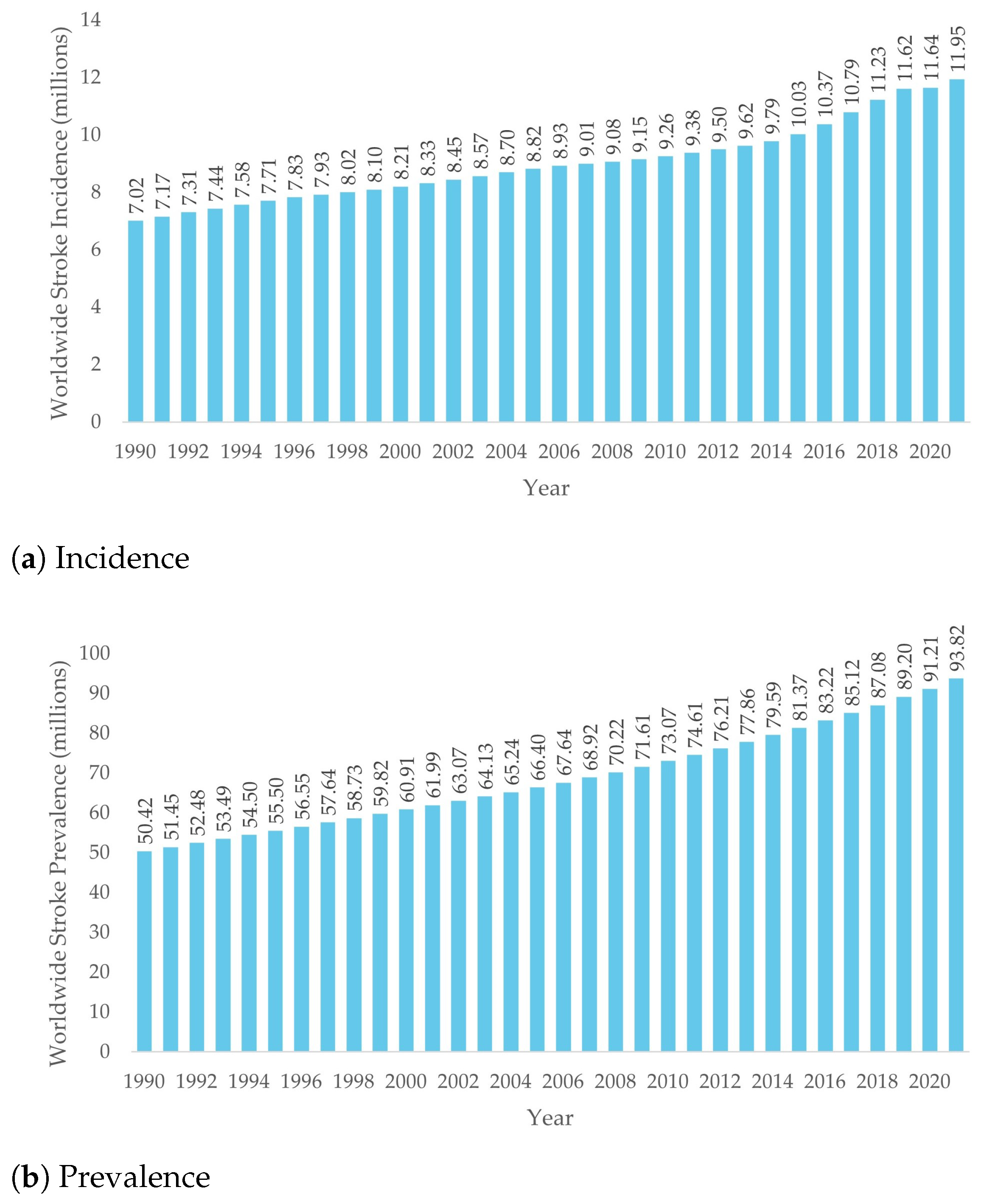
Figure 1. Stroke incidence and prevalence worldwide from 1990 to 2021, in millions, adapted from [8].
Robotics 14 00141 g001
Figure 2. Glenohumeral joint DOF.
Robotics 14 00141 g002
Figure 3. Scapulohumeral rhythm DOF.
Robotics 14 00141 g003
Figure 4. Elbow movements.
Robotics 14 00141 g004
Figure 5. Wrist movements.
Robotics 14 00141 g005
Figure 6. Schematics of exoskeleton-based and end-effector-based therapies, respectively.
Robotics 14 00141 g006
Figure 7. Hierarchy of control philosophies in robot-aided physical therapy.
Robotics 14 00141 g007
Figure 8. Portions of the UL targeted by the reviewed devices.
Robotics 14 00141 g008
Figure 9. Sensors used in the reviewed devices by year.
Robotics 14 00141 g009
Figure 10. Actuators used in the reviewed devices by year.
Robotics 14 00141 g010
Figure 11. Testing status of the reviewed rehabilitation devices.
Robotics 14 00141 g011
Figure 12. Relationship between device complexity and clinical testing advancement. Device complexity was calculated using a composite scoring system where each targeted UL segment (fingers, wrist, elbow, shoulder), each control/sensor type, each assistance type, and each DOF contributes 1 point, and non-electric actuation systems receive an additional point. Testing status is categorized as follows: 0 = not tested, 1 = tested on healthy subjects, 2 = tested on disabled patients, and 3 = commercially available. Polygerinos et al. [159].
Robotics 14 00141 g012
Table 1. Three major types of assistance.
Assistance TypeUser InvolvementRole of RobotTherapy StageGoalReferences
Passive AssistNoneMoves limb entirelyEarly rehabilitationPrevent stiffness, stimulate muscles[69]
Active AssistPartialHelps only if user cannot complete movementIntermediateImprove muscle power and coordination[70]
Active ResistFullApplies resistanceAdvancedIncrease strength and endurance[71]
Table 2. Electric actuator comparison.
Motor TypeAdvantagesDisadvantagesReferences
AC ServomotorsHigh-speed applications
Efficient
Heavy[104,105,106,107]
DC ServomotorsLow-speed, high-torque applicationsLess efficient (compared to AC servomotors)[104,105,106,107]
Brushed DC MotorsSimple
Low-cost
Noisy
Short lifespan
[108]
Brushless DC MotorsQuiet
Efficient
Long lifespan
Expensive[108]
Stepper MotorsAccurate position controlLess efficient for continuous operations[109]
Table 3. Actuation method comparison.
ElectricPneumaticHydraulic
AdvantagesHigh amounts of torque
High precision
Low impedance (with SEAs)
Flexible
Lightweight
Comfortable
High force/weight ratio
Durable
Very high power density
DisadvantagesHeavyweightCompressed air requirementsHeavyweight
Bulky
Prone to leaks
References[102,103][102,103,112][102,103,113]
Table 4. Characteristics of the robotic devices for UL rehabilitation discussed. AA = Active assist, AR = Active resist, PA = Passive assist, and ND = Not disclosed, meaning the authors of this paper did not find the respective information.
Device Name Year Type Target DOFs Control Philosophy and Sensors Actuation Assistance Testing status
Armin III [126]2009ExoskeletonUpper-Limb6Impedance
Position control
ElectricAA
AR
PA
Tested on stroke patients
Armeo®Power [134]2011ExoskeletonUpper-Limb7 (8 with
Manovo®Power
handle)
NDElectricAA
PA
Commercially available
ALEx [137]2012ExoskeletonUpper-Limb6Angular position sensorsElectricAATested on healthy subjects
Polygerinos et al. [159]2015ExoskeletonFingers15Fluidic pressure sensors
Force/Torque sensors
Movement sensors
Position sensors
HydraulicAATested on patient with muscular dystrophy
NESM [143]2016ExoskeletonShoulder
Elbow
4 (plus
8 passive)
Angular sensors
Position sensors
Torque sensors
ElectricAA
PA
ND
eWrist [128]2017ExoskeletonWrist1Admittance
Angle sensors
Force/Torque sensors
sEMG
Velocity sensors
ElectricAATested in healthy subjects
Harmony SHR® [144]2017ExoskeletonShoulder
Elbow
7Impedance
Force/Torque sensors
Movement sensors
Position sensors
ElectricAA
PA
Tested on stroke patients
ROBERT® [170]2018End-effectorUpper-Limb1
(end-effector only)
ND
sEMG
ElectricAA
AR
PA
Commercially Available
Popescu et al. [120]2018ExoskeletonElbow
Wrist
3NDElectricPAND
Chen et al. [156]2019ExoskeletonElbow2Angle sensors
Force/Torque sensors
Impedance
ElectricARND
Copaci et al. [165]2019ExoskeletonElbow2Angle sensors
Force sensors
Position sensors
Pressure sensors
sEMG
Temperature sensors
SMAAAND
MAHI Exo-II [131]2020ExoskeletonElbow
Wrist
4sEMGElectricAA
AR
PA
Tested on patients with SCI
CLEVERarm [141]2021ExoskeletonUpper-Limb8Force/Torque sensors
Movement sensors
Position sensors
ElectricAnti-gravityTested on healthy subjects
Quan Liu et al. [169]2021ExoskeletonElbow
Wrist
3Angle sensors
Force/Torque sensors
PneumaticAAND
u-Rob [142]2021BothUpper-Limb7 (plus
2 passive)
Force sensors
Position sensors
Velocity sensors
ElectricPATested on healthy subjects
Wang and Xu [119]2021End-effectorWrist3sEMGPneumaticNDTested on healthy subjects
Yang Liu et al. [122]2021ExoskeletonElbow1sEMGElectricARTested on healthy subjects
González-Mendoza et al. [124]2022ExoskeletonElbow
Wrist
3Admittance
Force/Torque sensors
sEMG
ElectricAA
PA
ND
ANYexo 2.0 [158]2023ExoskeletonUpper-Limb9Force/Torque sensors
Position sensors
ElectricNDTested on healthy subjects
Zhang et al. [125]2025ExoskeletonElbow
Wrist
2Angle Sensors
Force sensors
ElectricAA
PA
Tested on healthy subjects
Disclaimer/Publisher’s Note: The statements, opinions and data contained in all publications are solely those of the individual author(s) and contributor(s) and not of MDPI and/or the editor(s). MDPI and/or the editor(s) disclaim responsibility for any injury to people or property resulting from any ideas, methods, instructions or products referred to in the content.