Melanin and Neuromelanin in Humans: Insights Across Health, Aging, Diseases, and Unexpected Aspects of Fungal Melanogenesis



Abstract
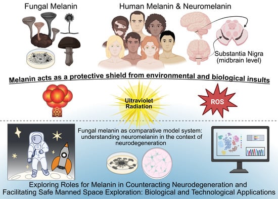
1. Introduction
2. Human Melanin and Neuromelanin
2.1. Melanogenesis
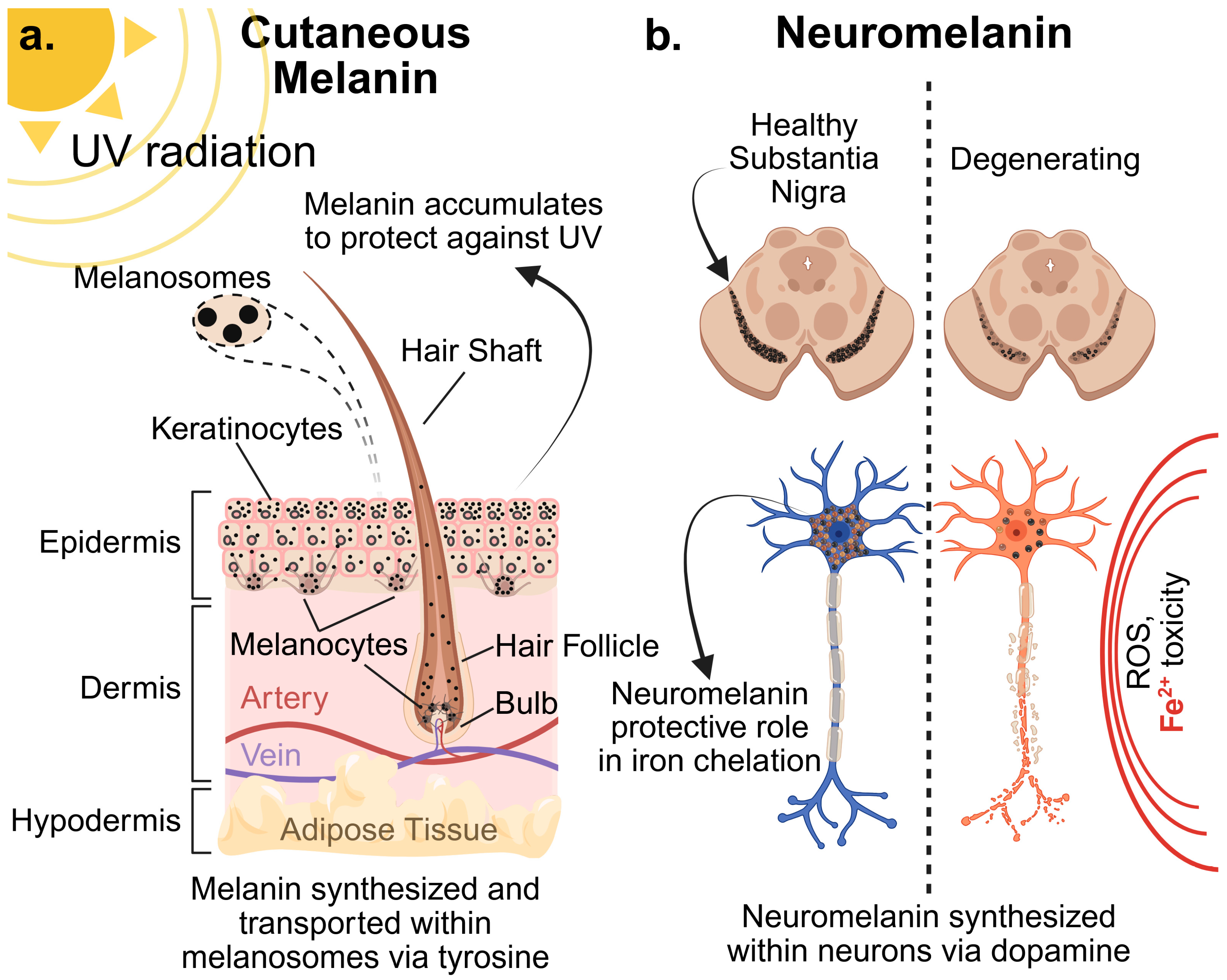
- Tyrosinase (TYR): encodes the rate-limiting enzyme in melanin synthesis. Specifically, TYR catalyzes the hydroxylation of L-tyrosine to L-3,4-dihydroxyphenylalanine (L-DOPA) and the oxidation of L-DOPA to DOPAquinone. Genetic mutations in TYR are the primary cause of oculocutaneous albinism type 1 (OCA1) [12].
- Tyrosinase-Related Protein 1 (TYRP1)/DOPAchrome Tautomerase (DCT): encoded by TYRP1 and DCT, respectively, these enzymes catalyze subsequent steps in the eumelanin pathway. TYRP1 oxidizes 5,6-dihydroxyindole-2-carboxylic acid (DHICA) and stabilizes tyrosinase. DCT catalyzes the tautomerization of DOPAchrome to DHICA. Mutations in TYRP1 cause oculocutaneous albinism type 3 (OCA3), and DCT mutations are linked to certain pigmentation disorders [13].
- Melanocortin 1 Receptor (MC1R): This G protein-coupled receptor, expressed on melanocytes, plays a pivotal role in regulating the type of melanin produced. Upon binding of its ligand, alpha-melanocyte-stimulating hormone (α-MSH), MC1R activates adenylate cyclase, leading to increased intracellular cAMP levels. High cAMP levels promote eumelanin synthesis, while low cAMP levels favor pheomelanin synthesis. Common genetic variants in MC1R are strongly associated with red hair, fair skin, and increased risk of melanoma [14].
- Oculocutaneous Albinism Type 2 (OCA2): Encodes the P protein, a melanosomal transmembrane protein thought to be involved in tyrosine transport into melanosomes or regulation of melanosomal pH. Mutations in OCA2 are the most common cause of OCA2 [15].
- SLC45A2 (Membrane-Associated Transporter Protein, MATP): This gene encodes a transporter protein likely involved in melanosome biogenesis or transport of melanin precursors. Mutations in SLC45A2 cause oculocutaneous albinism type 4 (OCA4) [16].
- Microphthalmia-associated Transcription Factor (MITF): Considered the “master regulator” of melanogenesis, MITF controls the expression of TYR, TYRP1, DCT, and other genes involved in melanocyte development, survival, and melanin synthesis. Genetic variations in MITF can lead to diverse pigmentation and developmental defects [17]. In general, the genetic regulation of cutaneous melanin is tightly controlled by signaling pathways (e.g., Wnt/β-catenin, MAPK/ERK, cAMP/PKA) that converge on MITF, ensuring coordinated production and deposition of melanin within melanosomes.
2.2. Melanin in Human Health and Disease
2.2.1. Skin Cancer
2.2.2. Albinism
2.2.3. Vitiligo
2.3. Neuromelanin Biosynthesis: A Distinct, Age-Dependent Pathway
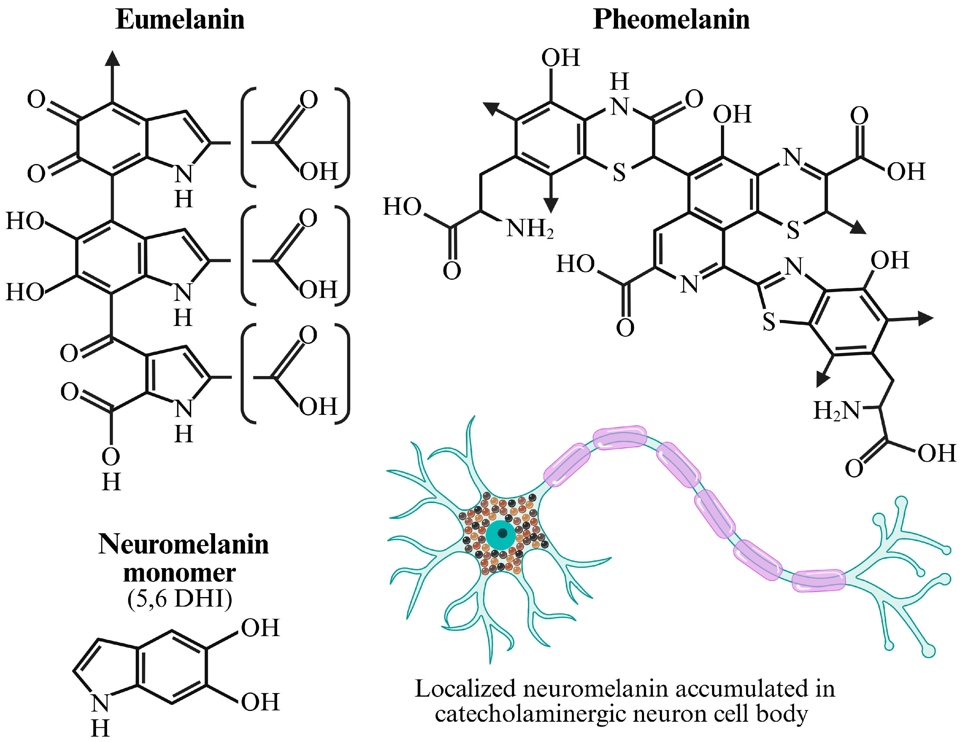
- Precursors. Basically, neuromelanin is derived from dopamine and norepinephrine, which are neurotransmitters synthesized by tyrosine hydroxylase (TH) and dopamine β-hydroxylase (DBH), respectively [31]. Genetic variations in TH and DBH can influence the availability of these precursors, though their direct impact on neuromelanin levels is not fully understood.

- Non-Enzymatic Oxidation. Indeed, the inherent susceptibility of catecholamines to autoxidation in the presence of oxygen and redox-active metals like iron (Fe2+) is central to neuromelanin formation [33]. This process generates ROS and various quinones that polymerize to form neuromelanin.
- Enzymatic Contribution. While not a classical tyrosinase-driven process, some enzymes may still contribute to neuromelanin synthesis. For instance, aldehyde dehydrogenases (ALDHs) detoxify reactive aldehydes generated during catecholamine metabolism. Polymorphisms in ALDH2 have been linked to PD risk and may indirectly affect neuromelanin accumulation by altering the balance of catecholamine metabolites [34]. Furthermore, evidence suggests that lysosomal enzymes and even low levels of non-melanogenic peroxidases within neurons might play a role in shaping neuromelanin aggregates [35].
- Lysosomal Involvement. Neuromelanin granules are enclosed within specialized lysosome-like organelles, often referred to as neuromelanosomes or autolysosomes. Genes involved in lysosomal biogenesis, autophagy, and protein degradation pathways (e.g., GBA, LRRK2, PINK1, PRKN) are critical for neuronal health and are frequently implicated in PD. Dysfunction in these pathways could impair neuromelanin granule turnover or lead to the release of potentially toxic neuromelanin components [36].
- Iron Metabolism. Iron is intimately associated with neuromelanin, and genes regulating iron homeostasis (e.g., FTL for ferritin light chain, SLC11A2 for DMT1, HFE for hemochromatosis gene) are crucial. Dysregulation of iron metabolism, influenced by genetic factors, can exacerbate oxidative stress and influence neuromelanin aggregation and toxicity [37].
2.4. In Vivo and Ex Vivo Neuromelanin Detection Methods
2.4.1. In Vivo Detection Methods:
- MRI. The current gold standard for non-invasive NM detection is neuromelanin-sensitive MRI (NM-MRI). This specialized imaging technique does not visualize the NM pigment directly but rather a key component associated with it: iron. Neuromelanin has a high capacity to chelate, or bind, metals, particularly ferric iron (Fe3+) [7]. This NM-iron complex is paramagnetic, meaning it alters the local magnetic field [38]. This property directly influences the relaxation times of nearby water protons, which is the basis of the MRI signal. Specifically, the paramagnetic NM-iron complex causes a significant shortening of the longitudinal relaxation time, known as the T1 time. MRI sequences that are heavily T1-weighted, such as a two-dimensional fast spin-echo sequence, can capitalize on this effect. In these scans, tissues with a short T1 relaxation time appear bright (hyperintense). Consequently, the SNc and LC, which are rich in NM, are clearly visible as high-intensity regions against the surrounding brain tissue. The clinical utility of this technique is powerful; in patients with Parkinson’s disease, the volume and signal intensity of these bright regions are progressively reduced, correlating strongly with the severity of motor symptoms and the degree of underlying neuron loss [39]. While NM-MRI is an invaluable tool, it is an indirect measure, and the signal can be influenced by changes in iron concentration independent of NM. Therefore, standardized protocols for image acquisition and analysis are critical for reliable and comparable results across studies.
- PET. A more direct and quantitative in vivo method on the horizon is PET. This molecular imaging technique relies on the administration of a radioactive tracer (a radioligand) designed to bind to a specific molecular target in the body. For NM, the goal is to develop a radiotracer that can cross the blood–brain barrier and bind with high specificity and affinity to the pigment itself. Such a tool would provide a direct measure of NM concentration, potentially offering greater sensitivity than the indirect approach of MRI. While a dedicated NM-PET tracer is not yet in clinical use, several compounds have shown promise. For instance, some radioligands initially developed for imaging tau pathology, such as derivatives of phenyl/pyridinyl-isoquinoline, have demonstrated incidental binding to NM in human brain tissue [40]. This has spurred dedicated efforts to synthesize and validate novel tracers specifically for neuromelanin. A successful NM-PET tracer would represent a significant leap forward, enabling more precise quantification of neuronal loss and a more sensitive method for tracking disease progression and the effectiveness of neuroprotective therapies.
2.4.2. Post-Mortem Detection Methods:
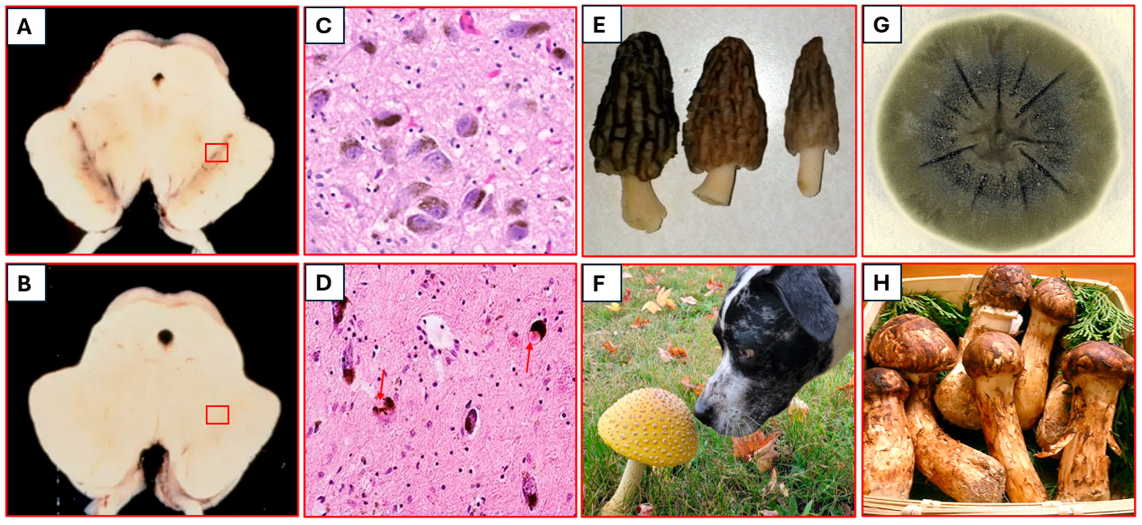
- Histological Staining. The most fundamental post-mortem technique is direct visualization through histological staining. The Fontana-Masson stain is the classic and most specific method for this purpose [41]. This is an argentaffin reaction, where the reducing components within the melanin polymer directly reduce silver nitrate from the staining solution to black, metallic silver. This process stains NM granules a distinct dark brown or black, making them easily identifiable within the cytoplasm of neurons under a light microscope [42]. While other general stains like hematoxylin and eosin (H&E) can also reveal NM as coarse brown granules, the Fontana-Masson method provides superior specificity.
- Immunohistochemistry (IHC) and Immunofluorescence (IF). To understand which specific types of neurons contain NM and are degenerating, researchers use immunohistochemistry (IHC) or immunofluorescence. These techniques employ antibodies to label specific proteins. For example, an antibody against Tyrosine Hydroxylase (TH), the rate-limiting enzyme in dopamine synthesis, can be used to definitively identify dopaminergic neurons. By combining TH staining with the natural visibility of NM (or Masson-Fontana staining), one can confirm that the pigmented cells in the SNc are indeed dopaminergic and can quantify the loss of these specific cells in PD brains.
- Electron Microscopy (EM). For the highest possible resolution, electron microscopy (EM) is used to examine the ultrastructure of the NM granules themselves. EM reveals that NM granules are complex organelles, often containing a lipid core surrounded by a matrix of melanin, proteins, and lipids [43]. This high-magnification view allows researchers to study the morphology and composition of the granules, which may change in disease states, providing critical insights into the underlying cellular pathology of neurodegeneration.
2.5. Neuromelanin in Human Health and Disease
- Transcriptomics. Aged substantia nigra neurons show increased expression of genes related to oxidative stress (e.g., NQO1, HMOX1), inflammation (e.g., complement components, microglial activation markers), and DNA damage response (GADD45A/B) [44]. There is also evidence of reduced expression of genes encoding mitochondrial function and protein quality control pathways, suggesting a decline in cellular resilience [45].
- Proteomics. Proteomic analyses of aged substantia nigra tissue and isolated neuromelanin granules reveal an accumulation of oxidatively damaged proteins, advanced glycation end products (AGEs), and lipofuscin-like aggregates. Metal-binding proteins, particularly ferritin, are also increasingly abundant, consistent with the age-dependent increase in iron within these regions [7].
- Epigenomics. Age-related epigenetic drift, characterized by global hypomethylation and site-specific hypermethylation, is observed in neuromelanin-containing neurons. These changes can alter the expression of genes involved in neuronal function, stress response, and inflammation, potentially priming these neurons for age-related decline and disease susceptibility [46]. For instance, differential methylation of genes involved in dopamine synthesis or synaptic function may contribute to subtle cognitive shifts seen in healthy aging.
2.5.1. Parkinson’s Disease (PD)
- Transcriptomics. Single-cell RNA sequencing (scRNA-seq) has revolutionized our ability to study specific neuronal populations. In PD, scRNA-seq of substantia nigra neurons reveals a selective downregulation of genes critical for dopaminergic identity and function (e.g., TH, SLC6A3 for dopamine transporter, ALDH1A1 for aldehyde dehydrogenase) in vulnerable neuromelanin-rich neurons [48]. Concurrently, there is an upregulation of genes associated with inflammation (HLA-DRB1, CD68), ER stress, and unfolded protein response. Genes related to iron metabolism, such as FTL (ferritin light chain) and SLC11A2 (DMT1), show altered expression, reflecting iron dyshomeostasis in the degenerating substantia nigra [49].
- Proteomics. Proteomic analyses of post-mortem substantia nigra tissue from PD patients consistently show an enrichment of oxidatively modified proteins, lipid peroxidation products, and aggregated α-synuclein. Neuromelanin has been shown to physically interact with and potentially sequester α-synuclein, a key protein in Lewy body pathology [50]. While this might initially be protective, the interaction can also lead to the formation of more toxic, aggregated species when neuromelanin’s buffering capacity is overwhelmed. Lysosomal proteins, essential for neuromelanin granule turnover, also show altered levels, consistent with lysosomal dysfunction being a core component of PD pathogenesis.
- Epigenomics. Epigenetic studies in PD patients reveal widespread DNA methylation changes in the substantia nigra, particularly in promoter regions of genes involved in mitochondrial function, lysosomal pathways, and inflammation. For example, hypermethylation of the SNCA promoter (encoding α-synuclein) can reduce its expression, yet abnormal methylation patterns elsewhere may contribute to disease progression [51]. Histone modification patterns (e.g., acetylation, methylation) are also altered, impacting chromatin accessibility and gene expression in neuromelanin-vulnerable neurons.
- Metabolomics. Metabolomic profiling of CSF and brain tissue in PD indicates dysregulation of catecholamine metabolism. Elevated levels of dopamine oxidation products and decreased levels of protective antioxidants suggest increased oxidative stress directly related to neuromelanin synthesis and breakdown. Changes in iron-related metabolites and lipid profiles also point towards lipid peroxidation, a process exacerbated by iron-laden neuromelanin [52].
- Genetic Intersections with PD Risk Genes. Curiously, many genes linked to familial PD, such as leucine-rich repeat kinase 2 (LRRK2), alpha-synuclein (SNCA), PTEN-induced putative kinase 1 (PINK1), parkin (PRKN), and glucocerebrosidase (GBA), play roles in lysosomal function, mitochondrial quality control, and α-synuclein handling. Mutations in these genes can indirectly impact neuromelanin accumulation and turnover, contributing to the vulnerability of neuromelanin-containing neurons. For example, GBA mutations, which cause Gaucher disease, are a significant risk factor for PD, and GBA enzyme deficiency affects lysosomal function and lipid metabolism, potentially exacerbating neuromelanin-related toxicity [36].
2.5.2. Alzheimer’s Disease (AD)
- Transcriptomics. In early AD, neurons show transcriptional downregulation of genes related to noradrenergic biosynthesis (e.g., DBH, TH) and an upregulation of genes associated with inflammation and stress responses [53]. This suggests that degeneration, beginning even before widespread amyloid/tau pathology, contributes to the cognitive and behavioral symptoms of AD.
- Proteomics. Proteomic studies in AD brains indicate that neuromelanin granules may co-localize with or bind to early aggregates of amyloid-beta (Aβ) and hyperphosphorylated tau [54]. While this binding might initially be a protective sequestration mechanism, it could also contribute to the local neuroinflammation and neuronal death if the neuromelanin-amyloid-β/tau complex becomes toxic.
- Epigenomics. Like PD, epigenomic changes in AD brains, particularly in the hippocampus and cortex, include altered DNA methylation patterns that could influence the expression of genes related to neuronal resilience, inflammation, and AD-specific protein pathology [55].
2.5.3. Multiple System Atrophy (MSA)
2.5.4. Schizophrenia and Other Neuropsychiatric Disorders
2.5.5. Amyotrophic Lateral Sclerosis (ALS)
2.5.6. Neuroinflammation and Immune Responses
- Transcriptomics. Studies show that microglia exposed to synthetic or isolated neuromelanin express pro-inflammatory genes (e.g., IL1β, TNF, NOS2) and downregulate anti-inflammatory markers [60]. This shift in microglial phenotype contributes to chronic neuroinflammation in conditions like PD.
- Proteomics. The proteome of neuromelanin-stimulated microglia reveals an activation of pathways related to antigen presentation (MHC-I/II), phagocytosis, and inflammasome activation. Oxidized proteins and lipids associated with neuromelanin granules can further exacerbate the inflammatory cascade [61].
- Genetic Predisposition. Genetic variants in immune-related genes (e.g., HLA locus, TREM2, LRRK2) can modulate the brain’s inflammatory response to neuromelanin and other DAMPs, influencing disease susceptibility and progression [62].
3. A Special Case: Fungal Melanin as a Comparative Model System (Leveraging Fungal Melanogenesis to Probe Neuromelanin)
3.1. Genetic Pathways of Fungal Melanogenesis
3.1.1. DOPA-Melanin Pathway (Tyrosinase/Laccase Dependent)
3.1.2. Dihydroxynaphthalene (DHN)-Melanin Pathway (Polyketide Synthase Dependent)
- Polyketide Synthases (PKS) (e.g., PKS1 in M. oryzae, ALB1 in A. fumigatus). These are large, multi-domain enzymes that catalyze the initial steps of the pathway, producing 1,3,6,8-tetrahydroxynaphthalene (T4HN) from acetyl-CoA and malonyl-CoA.
- Reductases (e.g., 4HNR for 4-hydroxynaphthalene reductase). Catalyze reduction steps within the pathway.
- Dehydratases (e.g., SCD for scytalone dehydratase). Catalyze dehydration steps.
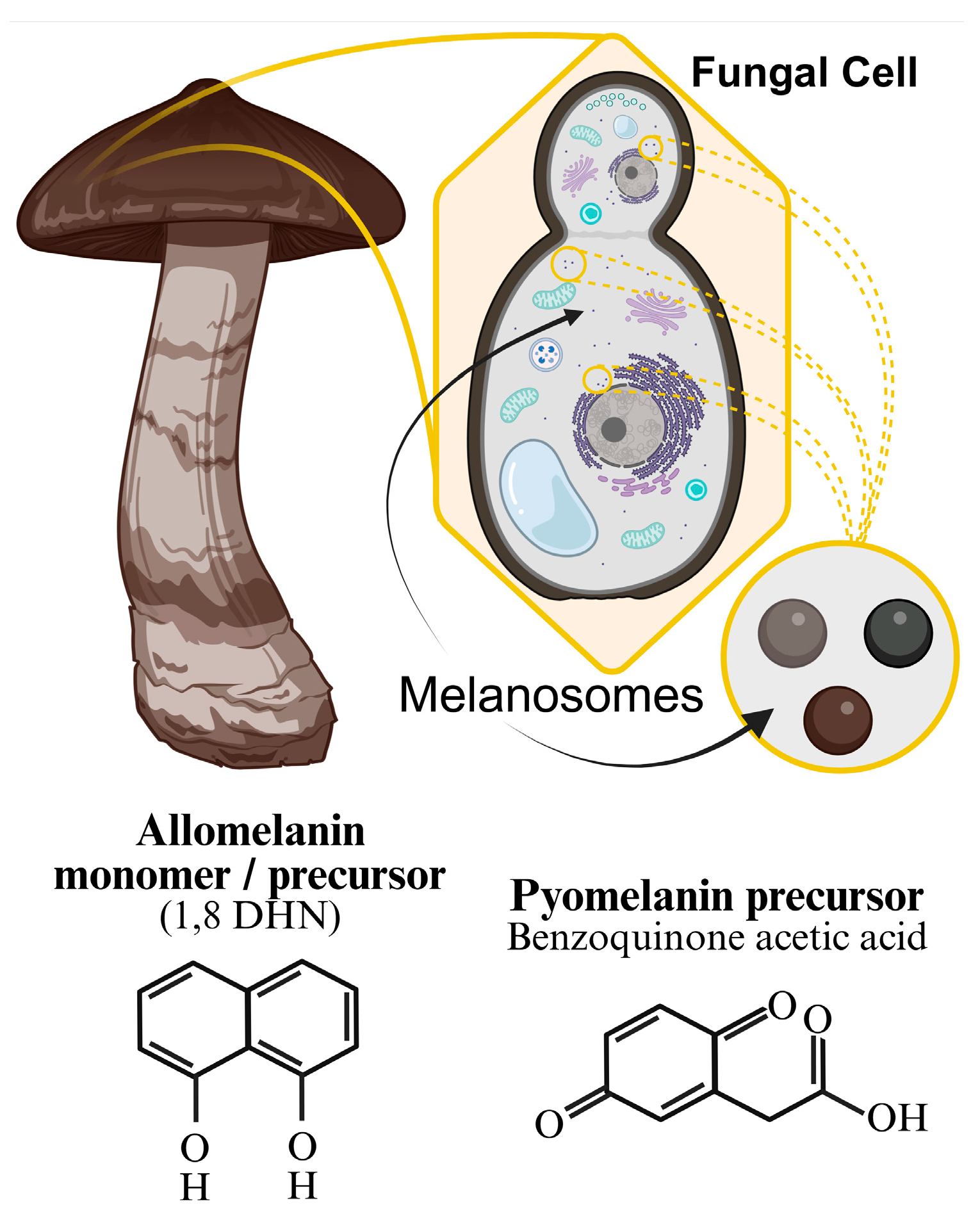
- Laccases (e.g., LAC in A. fumigatus). These enzymes act at the final step, polymerizing DHN precursors (e.g., 1,8-dihydroxynaphthalene, DHN) to form melanin.
3.1.3. Gamma-Glutaminyl-4-Hydroxybenzene (GHB) Melanin Pathway
3.2. Functional Roles and Extremophile Adaptation
3.2.1. Virulence Factor
3.2.2. Stress Resistance
- UV Radiation. Melanin absorbs UV radiation, dissipating the energy as heat, thus protecting cellular components from damage. Genetic pathways for melanin synthesis are often up-regulated in response to UV exposure [79].
- Desiccation. The hydrophobic nature of melanin-containing cell walls helps prevent water loss, enabling survival in arid environments [80].
- Heavy Metals: Like neuromelanin, fungal melanin can chelate heavy metals, detoxifying the environment [81].
3.2.3. Radioprotection and Radiotropism
- Energy Dissipation. Melanin can absorb high-energy photons (gamma rays) and convert their energy into heat, minimizing direct damage to DNA and proteins [79].
- Electron Scavenging. Ionizing radiation produces free radicals and secondary electrons. Melanin’s unique electronic properties allow it to efficiently quench these highly reactive species [84].
- Redox Regulation. Melanin can serve as an electron donor/acceptor, buffering cellular redox states and protecting against oxidative damage induced by radiation [85].
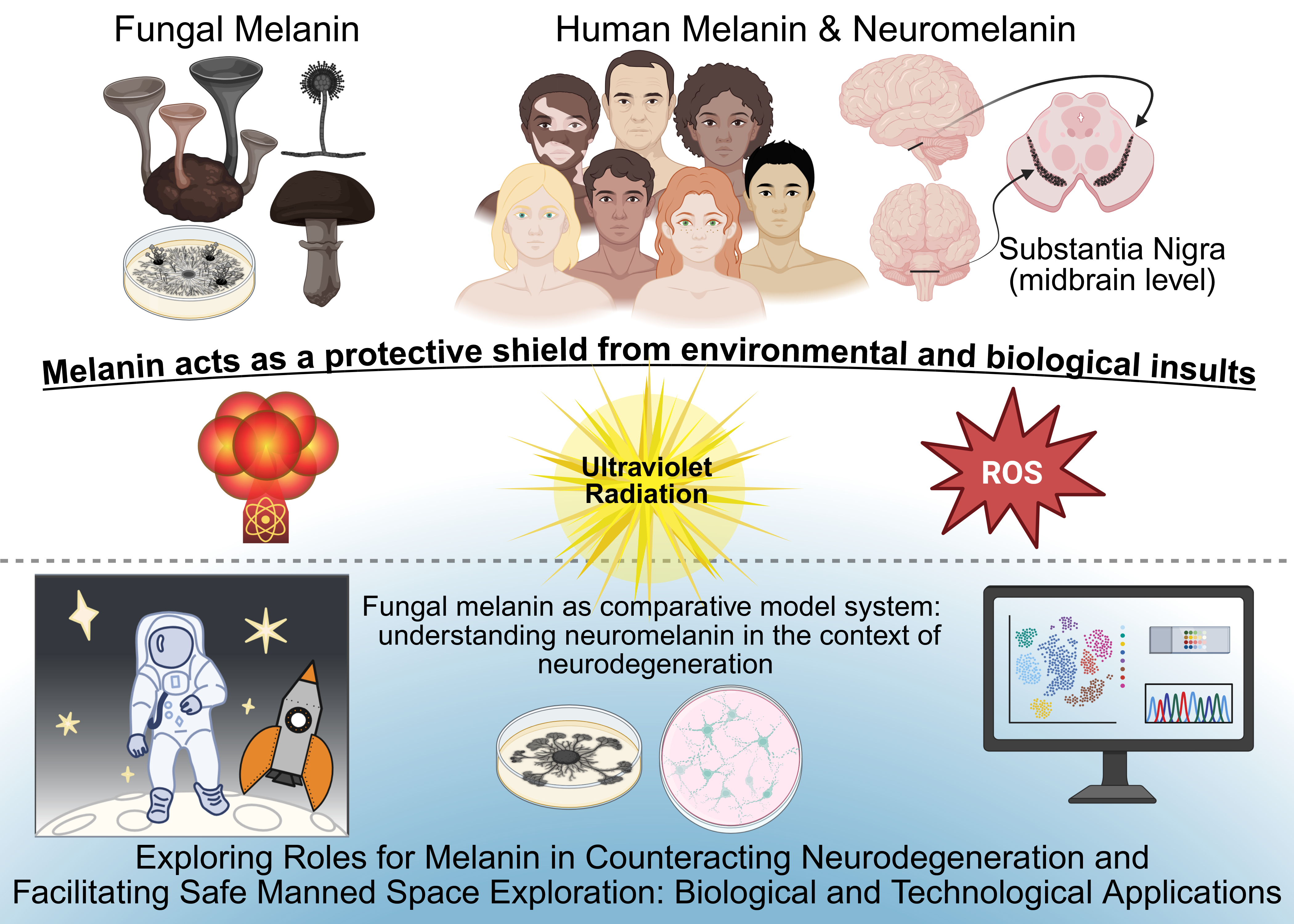
- Radiotropism. Some melanized fungi, notably Cladosporium sphaerospermum isolated from the Chernobyl reactor, exhibit “radiotropism”—growth towards sources of ionizing radiation. This phenomenon suggests that melanin might not only protect but also mediate a form of radiation-induced metabolic activity, potentially utilizing the energy from radiation for growth or cellular processes [84]. The genetic basis for this radiotrophic growth is still under investigation, but it is likely to involve upregulation of melanin synthesis genes and stress response pathways, coupled with unique metabolic adaptations. Crucially, the radiotrophic capacity of highly melanized fungi like Cladosporium sphaerospermum at the Chernobyl exclusion zone and in space [86] to absorb ionizing radiation and convert it through radiosynthesis into chemical energy for biological processes (similar to how plants convert sunlight into energy through photosynthesis) is in stark contrast to other non-melanized fungi like Tricholoma matsutake (actually contains compounds that inhibit melanin production [87]) which have demonstrated a hardy resilience in highly radioactive environments (like post-war Hiroshima and Nagasaki and post-nuclear accident Fukushima in Japan [88]) but which absorb radiation (via radionucleotides from the soil) without converting it, becoming sources of radioactive contamination. This emphasizes the unique role of melanin in these complex adaptive and radioprotective mechanisms, which could be harnessed for human applications (Figure 4). Indeed, frogs at Chernobyl have demonstrated increased melanin production as an adaptive response to radiation, and human populations located in regions with high background radiation have demonstrated enhanced DNA repair capabilities, suggesting that harnessing biological adaptation (and evolutionary survival mechanisms) is possible and necessary for optimizing physiological response to extreme radiation environments [89].
4. Melanin Across Kingdoms
- Polymeric Structure. Both are heterogeneous, amorphous polymers, making their precise chemical structures difficult to fully characterize.
- Dark Pigmentation. Both are dark brown to black, indicative of their extensive conjugated pi-electron systems, which are crucial for light/energy absorption.
- Radical Scavenging. Both are potent free radical scavengers, neutralizing ROS and protecting cells from oxidative damage.
- Metal Binding. Both have a high affinity for binding metal ions, particularly redox-active transition metals like iron and copper. This can be protective (detoxification) or potentially detrimental (catalyzing Fenton reactions in excess).
- Stress Resistance. Both contribute to resistance against various forms of stress, including oxidative, chemical, and potentially radiation stress.
- By contrast, some of the properties seem to diverge. Among the divergent properties, there are:
- Biosynthesis. Human cutaneous melanin relies on tyrosinase; neuromelanin largely on non-enzymatic autoxidation of catecholamines; fungal melanin employs distinct PKS, laccase, or tyrosinase pathways. The genes encoding these enzymatic machineries are unique to each type.
- Localization. Cutaneous melanin in melanocytes/melanosomes, neuromelanin in neuronal autolysosomes, fungal melanin predominantly in melanosomes and the cell wall.
- Age-Dependency. Neuromelanin accumulates with age; cutaneous and fungal melanin are typically synthesized on demand or as part of a developmental program.
- Radiotropism. Explicit radiotrophic growth has only been observed in certain melanized fungi, not in human cells or neurons.
5. Future Directions and Possible Translational Applications
- Spatial Transcriptomics and Proteomics. These technologies will allow for the study of gene and protein expression within specific subregions of the substantia nigra and locus coeruleus, even down to individual neuromelanin-containing neurons, providing unprecedented spatial resolution of molecular changes in aging and disease [100].
- Single-Nuclei Multi-omics. Combining genomics, transcriptomics, and epigenomics from single nuclei can reveal the epigenetic regulatory landscapes that govern gene expression in neuromelanin-rich neurons, identifying novel targets for therapeutic intervention.
- Metabolomics of Neuromelanin Granules. More refined techniques to analyze the metabolic profiles directly from isolated neuromelanin granules could uncover novel biomarkers of oxidative stress or neurodegeneration.
- Non-invasive Imaging of Neuromelanin. Neuromelanin-sensitive MRI sequences (e.g., neuromelanin-MRI) are rapidly evolving as non-invasive tools to visualize and quantify neuromelanin levels in the substantia nigra and in vivo. These techniques can serve as early diagnostic biomarkers for PD, track disease progression, and monitor therapeutic responses. Further refinement and correlation with omics data will enhance their diagnostic and prognostic value [38].
5.1. Harnessing Melanin’s Multifaceted Functionality, Including Therapeutic Strategies Targeting Neuromelanin Pathways
- Iron Chelation. Given the strong association between neuromelanin and iron, iron chelators are being explored as neuroprotective agents in PD to reduce oxidative stress [103].
- Modulation of Autophagy and Lysosomal Function. Strategies aimed at enhancing lysosomal function and autophagy could promote the clearance of damaged neuromelanin granules and α-synuclein aggregates, offering novel therapeutic avenues for PD.
- Melanin-Inspired Biomaterials. The remarkable radioprotective and transducing properties of fungal melanin are inspiring the development of novel biomaterials for radiation shielding [104] and sustainable bioelectronic applications [105]. This could range from melanin-coated surfaces in spacecraft to potential pharmaceutical interventions that enhance endogenous melanin’s protective capacities in astronauts or patients undergoing radiation therapy.
- Targeting Neuroinflammation. Given neuromelanin’s role as a DAMP, therapies aimed at modulating microglial activation and reducing neuroinflammation (e.g., through specific cytokine inhibitors or microglial repolarizing agents) could be beneficial in PD and other neurodegenerative conditions.
- Genetic Risk Factor Integration. Integrating genetic risk factors for PD and AD with neuromelanin-related omics data will help to identify individuals at higher risk for neurodegeneration and allow for personalized preventive or early intervention strategies. Understanding how common genetic variants (e.g., in GBA or LRRK2) impact neuromelanin biology is crucial.
- Comparative Biology for Human Benefit. Continued research into fungal melanin’s radioprotective mechanisms at a genetic and molecular level can provide blueprints for engineering human cells or developing targeted therapies to enhance neuroprotection against cosmic radiation during long-duration space missions, an increasingly critical challenge for space exploration. For instance, Antarctic black fungi (including the highly melanized Cryomyces antarcticus) have demonstrated resistance to simulated Martian conditions, exhibiting biological properties which could prove invaluable to future space exploration [106].
5.2. Fungal Melanin (And Maybe Someday, Neuromelanin) as a Bioavailable Radiation Countermeasure?
5.3. Metabolomic and Microbiomic Perspectives
6. Conclusions and Future Perspectives
Author Contributions
Funding
Institutional Review Board Statement
Informed Consent Statement
Data Availability Statement
Acknowledgments
Conflicts of Interest
References
- Riley, P.A. Melanin. Int. J. Biochem. Cell Biol. 1997, 29, 1235–1239. [Google Scholar] [CrossRef] [Scilit]
- Slominski, A.; Tobin, D.J.; Shibahara, S.; Wortsman, J. Melanin Pigmentation in Mammalian Skin and Its Hormonal Regulation. Physiol. Rev. 2004, 84, 1185–1228. [Google Scholar] [CrossRef] [Scilit]
- Zecca, L.; Tampellini, D.; Gatti, A.; Crippa, R.; Eisner, M.; Sulzer, D.; Ito, S.; Fariello, R.; Gallorini, M. The neuromelanin of human substantia nigra and its interaction with metals. J. Neural Transm. 2002, 109, 663–672. [Google Scholar] [CrossRef] [Scilit]
- Vila, M. Neuromelanin, aging, and neuronal vulnerability in Parkinson’s disease. Mov. Disord. 2019, 34, 1440–1451. [Google Scholar] [CrossRef] [Scilit]
- Cowen, D. The melanoneurons of the human cerebellum (nucleus pigmentosus cerebellaris) and homologues in the monkey. J. Neuropathol. Exp. Neurol. 1986, 45, 205–221. [Google Scholar] [CrossRef] [Scilit]
- Jenner, P. Oxidative stress in Parkinson’s disease. Ann. Neurol. 2003, 53, S26–S36. [Google Scholar] [CrossRef] [Scilit]
- Zucca, F.A.; Segura-Aguilar, J.; Ferrari, E.; Muñoz, P.; Paris, I.; Sulzer, D.; Sarna, T.; Casella, L.; Zecca, L. Interactions of iron, dopamine and neuromelanin pathways in brain aging and Parkinson’s disease. Prog. Neurobiol. 2017, 155, 96–119. [Google Scholar] [CrossRef] [Scilit]
- Carballo-Carbajal, I.; Laguna, A.; Romero-Giménez, J.; Cuadros, T.; Bové, J.; Martinez-Vicente, M.; Parent, A.; Gonzalez-Sepulveda, M.; Peñuelas, N.; Torra, A.; et al. Brain tyrosinase overexpression implicates age-dependent neuromelanin production in Parkinson’s disease pathogenesis. Nat. Commun. 2019, 10, 973. [Google Scholar] [CrossRef] [Scilit] [PubMed]
- Gonzalez-Sepulveda, M.; Compte, J.; Cuadros, T.; Nicolau, A.; Guillard-Sirieix, C.; Peñuelas, N.; Lorente-Picon, M.; Parent, A.; Romero-Giménez, J.; Cladera-Sastre, J.M.; et al. In vivo reduction of age-dependent neuromelanin accumulation mitigates features of Parkinson’s disease. Brain 2023, 146, 1040–1052. [Google Scholar] [PubMed]
- Sulzer, D.; Cassidy, C.; Horga, G.; Kang, U.J.; Fahn, S.; Casella, L.; Pezzoli, G.; Langley, J.; Hu, X.P.; Zucca, F.A.; et al. Neuromelanin detection by magnetic resonance imaging (MRI) and its promise as a biomarker for Parkinson’s disease. npj Parkinsons Dis. 2018, 4, 11. [Google Scholar] [CrossRef] [Scilit] [PubMed]
- Cui, Y.Z.; Man, X.Y. Biology of melanocytes in mammals. Front. Cell Dev. Biol. 2023, 11, 1309557. [Google Scholar] [CrossRef] [Scilit]
- Grønskov, K.; Ek, J.; Brondum-Nielsen, K. Oculocutaneous albinism. Orphanet J. Rare Dis. 2007, 2, 43. [Google Scholar] [CrossRef] [Scilit]
- Box, N.F.; Wyeth, J.R.; O’Gorman, L.E.; Martin, N.G.; Sturm, R.A. Characterization of melanocyte stimulating hormone receptor variant alleles in twins with red hair. Hum. Mol. Genet. 1997, 6, 1891–1897. [Google Scholar]
- Zorina-Lichtenwalter, K.; Lichtenwalter, R.N.; Zaykin, D.V.; Parisien, M.; Gravel, S.; Bortsov, A.; Diatchenko, L. A study in scarlet: MC1R as the main predictor of red hair and exemplar of the flip-flop effect. Hum. Mol. Genet. 2019, 28, 2093–2106. [Google Scholar]
- Brilliant, M.H.; King, R.; Francke, U.; Schuffenhauer, S.; Meitinger, T.; Gardner, J.M.; Durham-Pierre, D.; Nakatsu, Y. The mouse pink-eyed dilution gene: Association with hypopigmentation in Prader-Willi and Angelman syndromes and with human OCA2. Pigment Cell Res. 1994, 7, 398–402. [Google Scholar]
- Inagaki, K.; Suzuki, T.; Ito, S.; Suzuki, N.; Adachi, K.; Okuyama, T.; Nakata, Y.; Shimizu, H.; Matsuura, H.; Oono, T.; et al. Oculocutaneous albinism type 4: Six novel mutations in the membrane-associated transporter protein gene and their phenotypes. Pigment Cell Res. 2006, 19, 451–453. [Google Scholar]
- Levy, C.; Khaled, M.; Fisher, D.E. MITF: Master regulator of melanocyte development and melanoma oncogene. Trends Mol. Med. 2006, 12, 406–414. [Google Scholar] [CrossRef] [Scilit]
- Brenner, M.; Hearing, V.J. The protective role of melanin against UV damage in human skin. Photochem. Photobiol. 2008, 84, 539–549. [Google Scholar] [PubMed]
- Terranova, M.L. Physiological Roles of Eumelanin- and Melanogenesis-Associated Diseases: A Look at the Potentialities of Engineered and Microbial Eumelanin in Clinical Practice. Bioengineering 2024, 11, 756. [Google Scholar] [CrossRef] [Scilit] [PubMed]
- Ito, S.; Wakamatsu, K.; Sarna, T. Photodegradation of Eumelanin and Pheomelanin and Its Pathophysiological Implications. Photochem. Photobiol. 2018, 94, 409–420. [Google Scholar] [PubMed]
- Nasti, T.H.; Timares, L. MC1R, eumelanin and pheomelanin: Their role in determining the susceptibility to skin cancer. Photochem. Photobiol. 2015, 91, 188–200. [Google Scholar] [CrossRef] [Scilit] [PubMed]
- Brunsgaard, E.K.; Wu, Y.P.; Grossman, D. Melanoma in skin of color: Part I. Epidemiology and clinical presentation. J. Am. Acad. Dermatol. 2023, 89, 445–456. [Google Scholar] [CrossRef] [Scilit] [PubMed]
- Marçon, C.R.; Maia, M. Albinism: Epidemiology, genetics, cutaneous characterization, psychosocial factors. An. Bras. Dermatol. 2019, 94, 503–520. [Google Scholar] [CrossRef] [Scilit]
- Galli, J.; Loi, E.; Dusi, L.; Pasini, N.; Rossi, A.; Scaglioni, V. Oculocutaneous albinism: The neurological, behavioral, and neuro-ophthalmological perspective. Eur. J. Pediatr. 2023, 182, 2723–2733. [Google Scholar] [CrossRef] [Scilit]
- Talbert, M.L.; Malicdan, M.C.V.; Introne, W.J. Chediak-Higashi syndrome. Curr. Opin. Hematol. 2023, 30, 144–151. [Google Scholar] [CrossRef] [Scilit]
- Bhambhani, V.; Introne, W.J.; Lungu, C.; Cullinane, A.; Toro, C. Chediak-Higashi syndrome presenting as young-onset levodopa-responsive parkinsonism. Mov. Disord. 2013, 28, 127–129. [Google Scholar] [CrossRef] [Scilit]
- Frisoli, M.L.; Essien, K.; Harris, J.E. Vitiligo: Mechanisms of Pathogenesis and Treatment. Annu. Rev. Immunol. 2020, 38, 621–648. [Google Scholar] [CrossRef] [Scilit]
- Kridin, K.; Ofir, L.; Weinstein, O.; Badarny, S. Clarifying the association between Parkinson’s disease and vitiligo: A population-based large-scale study. Front. Neurol. 2024, 15, 1387404. [Google Scholar] [CrossRef] [Scilit]
- Filimontseva, A.; Fu, Y.; Vila, M.; Halliday, G.M. Neuromelanin and selective neuronal vulnerability to Parkinson’s disease. Trends Neurosci. 2025, 48, 445–459. [Google Scholar] [CrossRef] [Scilit]
- Graham, D.G. Oxidative pathways for catecholamines in the genesis of neuromelanin and the cytotoxic effects of L-DOPA. Mol. Pharmacol. 1978, 14, 633–643. [Google Scholar] [CrossRef] [Scilit] [PubMed]
- Daubner, S.C.; Le, T.; Wang, S. Tyrosine hydroxylase and regulation of dopamine synthesis. Arch. Biochem. Biophys. 2011, 508, 1–12. [Google Scholar] [CrossRef] [Scilit] [PubMed]
- Sansinenea, E.; Ortiz, A. Melanin: A photoprotection for Bacillus thuringiensis based biopesticides. Biotechnol. Lett. 2015, 37, 483–490. [Google Scholar] [CrossRef] [Scilit]
- Fedorow, H.; Tribl, F.; Halliday, G.; Gerlach, M.; Riederer, P.; Double, K.L. Neuromelanin in human dopamine neurons: Comparison with peripheral melanins and relevance to Parkinson’s disease. Prog. Neurobiol. 2005, 75, 109–124. [Google Scholar] [CrossRef] [Scilit]
- Fitzmaurice, A.G.; Rhodes, S.L.; Cockburn, M.; Ritz, B.; Bronstein, J.M. Aldehyde dehydrogenase variation enhances effect of pesticides associated with Parkinson disease. Neurology 2014, 82, 419–426. [Google Scholar] [CrossRef] [Scilit]
- Sulzer, D.; Mosharov, E.; Talloczy, Z.; Zucca, F.A.; Simon, J.D.; Zecca, L. Neuronal pigmented autophagic vacuoles: Lipofuscin, neuromelanin, and ceroid as macroautophagic responses during aging and disease. J. Neurochem. 2008, 106, 24–36. [Google Scholar] [CrossRef] [Scilit] [PubMed]
- Ye, H.; Robak, L.A.; Yu, M.; Cykowski, M.; Shulman, J.M. Genetics and Pathogenesis of Parkinson’s Syndrome. Annu. Rev. Pathol. 2023, 18, 95–121. [Google Scholar] [CrossRef] [Scilit]
- Wise, R.M.; Wagener, A.; Fietzek, U.M.; Klopstock, T.; Mosharov, E.V.; Zucca, F.A.; Sulzer, D.; Zecca, L.; Burbulla, L.F. Interactions of dopamine, iron, and alpha-synuclein linked to dopaminergic neuron vulnerability in Parkinson’s disease and Neurodegeneration with Brain Iron Accumulation disorders. Neurobiol. Dis. 2022, 175, 105920. [Google Scholar] [CrossRef] [Scilit] [PubMed]
- Sasaki, M.; Shibata, E.; Tohyama, K.; Takahashi, J.; Otsuka, K.; Tsuchiya, K.; Takahashi, S.; Ehara, S.; Terayama, Y.; Sakai, A. Neuromelanin magnetic resonance imaging of locus ceruleus and substantia nigra in Parkinson’s disease. Neuroreport 2006, 17, 1215–1218. [Google Scholar] [CrossRef] [Scilit]
- Pérot, J.B.; Ruze, A.; Gaurav, R.; Rebbah, S.; Cadin, C.; Le Troter, A.; Soustelle, L.; Mouton, L.; Valabrègue, R.; Parent, A.; et al. Longitudinal neuromelanin changes in prodromal and early Parkinson’s disease in humans and rat model. Brain Commun. 2025, 7, fcaf204. [Google Scholar] [CrossRef] [Scilit]
- Hansen, A.K.; Knudsen, K.; Lillethorup, T.P.; Landau, A.M.; Parbo, P.; Fedorova, T.; Audrain, H.; Bender, D.; Østergaard, K.; Brooks, D.J.; et al. In vivo imaging of neuromelanin in Parkinson’s disease using 18F-AV-1451 PET. Brain 2016, 139, 2039–2049. [Google Scholar] [CrossRef] [Scilit]
- Bancroft, J.D.; Gamble, M. Theory and Practice of Histological Techniques, 6th ed.; Churchill Livingstone: London, UK, 2008. [Google Scholar]
- Suvarna, K.S.; Layton, C.; Bancroft, J.D. Pigments and minerals. In Bancroft’s Theory and Practice of Histological Techniques, 8th ed.; Elsevier: Amsterdam, The Netherlands, 2019; pp. 239–262. [Google Scholar]
- Biesemeier, A.; Eibl, O.; Eswara, S.; Audinot, J.N.; Wirtz, T.; Pezzoli, G.; Zucca, F.A.; Zecca, L.; Schraermeyer, U. Elemental mapping of Neuromelanin organelles of human Substantia Nigra: Correlative ultrastructural and chemical analysis by analytical transmission electron microscopy and nano-secondary ion mass spectrometry. J. Neurochem. 2016, 138, 339–353. [Google Scholar] [CrossRef] [Scilit]
- Soreq, L.; UK Brain Expression Consortium; North American Brain Expression Consortium; Rose, J.; Soreq, E.; Hardy, J.; Trabzuni, D.; Cookson, M.R.; Smith, C.; Ryten, M.; et al. Major Shifts in Glial Regional Identity Are a Transcriptional Hallmark of Human Brain Aging. Cell Rep. 2017, 18, 557–570. [Google Scholar] [CrossRef] [Scilit]
- Werner, C.J.; Heyny-von Haussen, R.; Mall, G.; Wolf, S. Proteome analysis of human substantia nigra in Parkinson’s disease. Proteome Sci. 2008, 6, 8. [Google Scholar] [CrossRef] [Scilit] [PubMed]
- Wüllner, U.; Kaut, O.; de Boni, L.; Piston, D.; Schmitt, I. DNA methylation in Parkinson’s disease. J. Neurochem. 2016, 139, 108–120. [Google Scholar] [CrossRef] [Scilit]
- Halliday, G.M.; Leverenz, J.B.; Schneider, J.S.; Adler, C.H. The neurobiological basis of cognitive impairment in Parkinson’s disease. Mov. Disord. 2014, 29, 634–650. [Google Scholar] [CrossRef] [Scilit] [PubMed]
- Smajić, S.; Prada-Medina, C.A.; Landoulsi, Z.; Ghelfi, J.; Delcambre, S.; Dietrich, C.; Jarazo, J.; Henck, J.; Balachandran, S.; Pachchek, S.; et al. Single-cell sequencing of human midbrain reveals glial activation and a Parkinson-specific neuronal state. Brain 2022, 145, 964–978. [Google Scholar] [CrossRef] [Scilit] [PubMed]
- Thomas, G.E.C.; Zarkali, A.; Ryten, M.; Shmueli, K.; Gil-Martinez, A.L.; Leyland, L.A.; McColgan, P.; Acosta-Cabronero, J.; Lees, A.J.; Weil, R.S. Regional brain iron and gene expression provide insights into neurodegeneration in Parkinson’s disease. Brain 2021, 144, 1787–1798. [Google Scholar] [CrossRef] [Scilit]
- Lotharius, J.; Brundin, P. Impaired dopamine storage resulting from alpha-synuclein mutations may contribute to the pathogenesis of Parkinson’s disease. Hum. Mol. Genet. 2002, 11, 2395–2407. [Google Scholar] [CrossRef] [Scilit]
- Masliah, E.; Dumaop, W.; Galasko, D.; Desplats, P. Distinctive patterns of DNA methylation associated with Parkinson disease: Identification of concordant epigenetic changes in brain and peripheral blood leukocytes. Epigenetics 2013, 8, 1030–1038. [Google Scholar] [CrossRef] [Scilit]
- LeWitt, P.A.; Li, J.; Lu, M.; Guo, L.; Auinger, P.; Parkinson Study Group–DATATOP Investigators. Metabolomic biomarkers as strong correlates of Parkinson disease progression. Neurology 2017, 88, 862–869. [Google Scholar] [CrossRef] [Scilit]
- Andrés-Benito, P.; Gámez-Valverde, M.; Aso, E.; Ferrer, I. Locus coeruleus at asymptomatic early and middle Braak stages of neurofibrillary tangle pathology. Neuropathol. Appl. Neurobiol. 2020, 46, 687–705. [Google Scholar] [CrossRef] [Scilit] [PubMed]
- Beardmore, R.; Hou, R.; Darekar, A.; Holmes, C.; Boche, D. The Locus Coeruleus in Aging and Alzheimer’s Disease: A Postmortem and Brain Imaging Review. J. Alzheimers Dis. 2021, 83, 5–22. [Google Scholar] [CrossRef] [Scilit]
- Gao, X.; Chen, Q.; Yao, H.; Tan, J.; Liu, Z.; Zou, Z. Epigenetics in Alzheimer’s Disease. Front. Aging Neurosci. 2022, 14, 911635. [Google Scholar] [CrossRef] [Scilit]
- Tanaka, M.T.; Miki, Y.; Kon, T.; Mori, F.; Wakabayashi, K. Pathological and Molecular Insights into the Early Stage of Multiple System Atrophy. Cells 2025, 14, 1966. [Google Scholar] [CrossRef] [Scilit]
- Bogerts, B.; Meertz, E.; Schönfeldt-Bausch, R. Basal ganglia and limbic system pathology in schizophrenia. A Morphometric Study Postmortem Brain. Arch. Gen. Psychiatry 1990, 47, 1052–1062. [Google Scholar]
- Laruelle, M.; Abi-Dargham, A.; Gil, R.; Kegeles, L.; Innis, R. Increased dopamine transmission in schizophrenia: Relationship to illness phases. Biol. Psychiatry 1999, 46, 56–72. [Google Scholar] [CrossRef] [Scilit] [PubMed]
- Shibata, N.; Kataoka, I.; Okamura, Y.; Murakami, K.; Kato, Y.; Yamamoto, T.; Masui, K. Implications for soluble iron accumulation, oxidative stress, and glial glutamate release in motor neuron death associated with sporadic amyotrophic lateral sclerosis. Neuropathology 2025, 45, 177–201. [Google Scholar] [CrossRef] [Scilit] [PubMed]
- Zhang, W.; Phillips, K.; Wielgus, A.R.; Liu, J.; Albertini, A.; Zucca, F.A.; Faust, R.; Qian, S.Y.; Miller, D.S.; Chignell, C.F.; et al. Neuromelanin activates microglia and induces degeneration of dopaminergic neurons: Implications for progression of Parkinson’s disease. Neurotox. Res. 2011, 19, 63–72. [Google Scholar] [CrossRef] [Scilit]
- Viceconte, N.; Burguillos, M.A.; Herrera, A.J.; De Pablos, R.M.; Joseph, B.; Venero, J.L. Neuromelanin activates proinflammatory microglia through a caspase-8-dependent mechanism. J. Neuroinflamm. 2015, 12, 5. [Google Scholar] [CrossRef] [Scilit]
- Zhu, R.; Luo, Y.; Li, S.; Wang, Z. The role of microglial autophagy in Parkinson’s disease. Front. Aging Neurosci. 2022, 14, 1039780. [Google Scholar] [CrossRef] [Scilit]
- Eisenman, H.C.; Casadevall, A. Synthesis and assembly of fungal melanin. Appl. Microbiol. Biotechnol. 2012, 93, 931–940. [Google Scholar] [CrossRef] [Scilit]
- Chhoker, K.; Hausner, G.; Harris, S.D. Regulation of melanin production in fungi. Front. Fungal Biol. 2025, 6, 1621764. [Google Scholar] [CrossRef] [Scilit] [PubMed]
- Zhu, X.; Gibbons, J.; Garcia-Rivera, J.; Casadevall, A.; Williamson, P.R. Laccase of Cryptococcus neoformans is a cell wall-associated virulence factor. Infect. Immun. 2001, 69, 5589–5596. [Google Scholar] [CrossRef] [Scilit] [PubMed]
- Wang, L.; Zhai, B.; Lin, X. The link between morphotype transition and virulence in Cryptococcus neoformans. PLoS Pathog. 2012, 8, e1002765. [Google Scholar] [CrossRef] [Scilit] [PubMed]
- Kwon-Chung, K.J.; Sugui, J.A. Aspergillus fumigatus--what makes the species a ubiquitous human fungal pathogen? PLoS Pathog. 2013, 9, e1003743. [Google Scholar] [CrossRef] [Scilit]
- Qin, Y.; Xia, Y. Melanin in fungi: Advances in structure, biosynthesis, regulation, and metabolic engineering. Microb. Cell Fact. 2024, 23, 334. [Google Scholar] [CrossRef] [Scilit]
- Hegnauer, R.; Hegnauer, M. Chemotaxonomie der Pflanzen; Birkhäuser: Basel, Switzerland, 1989. [Google Scholar]
- Nosanchuk, J.D.; Casadevall, A. The contribution of melanin to microbial pathogenesis. Cell. Microbiol. 2003, 5, 203–223. [Google Scholar] [CrossRef] [Scilit]
- Wu, C.; Jiang, M.L.; Jiang, R.; Pang, T.; Zhang, C.J. The roles of fungus in CNS autoimmune and neurodegeneration disorders. Front. Immunol. 2023, 13, 1077335. [Google Scholar] [CrossRef] [Scilit]
- Chen, Y.; Shi, Z.W.; Strickland, A.B.; Shi, M. Cryptococcus neoformans Infection in the Central Nervous System: The Battle between Host and Pathogen. J. Fungi 2022, 8, 1069. [Google Scholar] [CrossRef] [Scilit]
- Nosanchuk, J.D.; Rosas, A.L.; Lee, S.C.; Casadevall, A. Melanisation of Cryptococcus neoformans in human brain tissue. Lancet 2000, 355, 2049–2050. [Google Scholar] [CrossRef] [Scilit]
- Wu, Y.; Du, S.; Johnson, J.L.; Tung, H.Y.; Landers, C.T.; Liu, Y.; Seman, B.G.; Wheeler, R.T.; Costa-Mattioli, M.; Kheradmand, F.; et al. Microglia and amyloid precursor protein coordinate control of transient Candida cerebritis with memory deficits. Nat. Commun. 2019, 10, 58. [Google Scholar] [CrossRef] [Scilit] [PubMed]
- Wu, Y.; Du, S.; Bimler, L.H.; Mauk, K.E.; Lortal, L.; Kichik, N.; Griffiths, J.S.; Osicka, R.; Song, L.; Polsky, K.; et al. Toll-like receptor 4 and CD11b expressed on microglia coordinate eradication of Candida albicans cerebral mycosis. Cell Rep. 2023, 42, 113240. [Google Scholar]
- Cordero, R.J.; Casadevall, A. Functions of fungal melanin beyond virulence. Fungal Biol. Rev. 2017, 31, 99–112. [Google Scholar] [CrossRef] [Scilit]
- Le Calvez, T.; Burgaud, G.; Mahé, S.; Barbier, G.; Vandenkoornhuyse, P. Fungal diversity in deep-sea hydrothermal ecosystems. Appl. Environ. Microbiol. 2009, 75, 6415–6421. [Google Scholar] [CrossRef] [Scilit]
- Bloomfield, B.J.; Alexander, M. Melanins and resistance of fungi to lysis. J. Bacteriol. 1967, 93, 1276–1280. [Google Scholar] [CrossRef] [Scilit]
- Xie, W.; Dhinojwala, A.; Gianneschi, N.C.; Shawkey, M.D. Interactions of Melanin with Electromagnetic Radiation: From Fundamentals to Applications. Chem. Rev. 2024, 124, 7165–7213. [Google Scholar] [CrossRef] [Scilit]
- King, K.J.; Sinclair, B.J. Water loss in tree weta (Hemideina): Adaptation to the montane environment and a test of the melanisation-desiccation resistance hypothesis. J. Exp. Biol. 2015, 218, 1995–2004. [Google Scholar] [CrossRef] [Scilit]
- Tang, J.; Shen, H.; Lv, W.; Zhang, J.; Fu, J. Edible Fungi Melanin: Recent Advances in Extraction, Characterization, Biological Activity and Applications. J. Fungi 2025, 11, 738. [Google Scholar] [CrossRef] [Scilit] [PubMed]
- El-Naggar, N.E.; Saber, W.I.A. Natural Melanin: Current Trends, and Future Approaches, with Especial Reference to Microbial Source. Polymers 2022, 14, 1339. [Google Scholar] [CrossRef] [Scilit] [PubMed]
- El-Bialy, H.A.; El-Gamal, M.S.; Elsayed, M.A.; Saudi, H.A.; Khalifa, M.A. Microbial melanin physiology under stress conditions and gamma radiation protection studies. Radiat. Phys. Chem. 2019, 162, 178–186. [Google Scholar] [CrossRef] [Scilit]
- Dadachova, E.; Bryan, R.A.; Huang, X.; Moadel, T.; Schweitzer, A.D.; Aisen, P.; Nosanchuk, J.D.; Casadevall, A. Ionizing radiation changes the electronic properties of melanin and enhances the growth of melanized fungi. PLoS ONE 2007, 2, e457. [Google Scholar] [CrossRef] [Scilit]
- Kim, E.; Wang, Z.; Phua, J.W.; Bentley, W.E.; Dadachova, E.; Napolitano, A.; Payne, G.F. Enlisting electrochemistry to reveal melanin’s redox-related properties. Mater. Adv. 2024, 5, 3082. [Google Scholar] [CrossRef] [Scilit]
- Averesch, N.J.H.; Shunk, G.K.; Kern, C. Cultivation of the Dematiaceous Fungus Cladosporium sphaerospermum Aboard the International Space Station and Effects of Ionizing Radiation. Front. Microbiol. 2022, 13, 877625. [Google Scholar] [CrossRef] [Scilit]
- Satooka, H.; Cerda, P.; Kim, H.J.; Wood, W.F.; Kubo, I. Effects of matsutake mushroom scent compounds on tyrosinase and murine B16-F10 melanoma cells. Biochem. Biophys. Res. Commun. 2017, 487, 840–846. [Google Scholar] [CrossRef] [Scilit] [PubMed]
- Orita, M.; Nakashima, K.; Taira, Y.; Fukuda, T.; Fukushima, Y.; Kudo, T.; Endo, Y.; Yamashita, S.; Takamura, N. Radiocesium concentrations in wild mushrooms after the accident at the Fukushima Daiichi Nuclear Power Station: Follow-up study in Kawauchi village. Sci. Rep. 2017, 7, 6744. [Google Scholar] [CrossRef] [Scilit]
- Mortazavi, S.; Rabiee, S.; Fallah, A.; Rashidfar, R.; Seyyedi, Z.; Vafapour, H.; Mortazavi, A.; Makarempour, F.; Kherad, Z.; Abdollahi, M.; et al. Adaptive Responses in High-Radiation Environments: Insights from Chernobyl Wildlife and Ramsar Residents. Dose Response 2025, 23, 15593258251385632. [Google Scholar] [PubMed]
- Solano, F. Melanins: Skin pigments and much more—Types, structural models, biological functions, and formation routes. New J. Sci. 2014, 2014, 498276. [Google Scholar] [CrossRef] [Scilit]
- McNamara, M.E.; Rossi, V.; Slater, T.S.; Rogers, C.S.; Ducrest, A.L.; Dubey, S.; Roulin, A. Decoding the Evolution of Melanin in Vertebrates. Trends Ecol. Evol. 2021, 36, 430–443. [Google Scholar] [CrossRef] [Scilit] [PubMed]
- Hearing, V.J. The expanding role and presence of neuromelanins in the human brain-why gray matter is gray. Pigment Cell Melanoma Res. 2009, 22, 10–11. [Google Scholar] [CrossRef] [Scilit]
- Haining, R.L.; Achat-Mendes, C. Neuromelanin, one of the most overlooked molecules in modern medicine, is not a spectator. Neural Regen. Res. 2017, 12, 372–375. [Google Scholar] [CrossRef] [Scilit]
- Wiesman, A.I.; Madge, V.; Fon, E.A.; Dagher, A.; Collins, D.L.; Baillet, S.; PREVENT-AD Research Group and Quebec Parkinson Network. Associations between neuromelanin depletion and cortical rhythmic activity in Parkinson’s disease. Brain 2025, 148, 875–885. [Google Scholar] [CrossRef] [Scilit]
- Hunter, P. The fungal grid: Fungal communication via electrical signals has inspired the hypothesis of a Wood Wide Web of plants and fungi. EMBO Rep. 2023, 24, e57255. [Google Scholar] [CrossRef] [Scilit]
- Adamatzky, A. Language of fungi derived from their electrical spiking activity. R. Soc. Open Sci. 2022, 9, 211926. [Google Scholar] [CrossRef] [Scilit]
- Buffi, M.; Kelliher, J.M.; Robinson, A.J.; Gonzalez, D.; Cailleau, G.; Macalindong, J.A.; Frau, E.; Schintke, S.; Chain, P.S.G.; Stanley, C.E.; et al. Electrical signaling in fungi: Past and present challenges. FEMS Microbiol. Rev. 2025, 49, fuaf009. [Google Scholar] [CrossRef] [Scilit]
- Jones, R.M.; Reynolds, R.W.; Thurston, A.K.; Barbato, R.A. Fungal Tissue as a Medium for Electrical Signal Transmission: A Baseline Assessment With Melanized Fungus Curvularia Lunata. IEEE J. Electromagn. RF Microw. Med. Biol. 2025, 9, 206–213. [Google Scholar] [CrossRef] [Scilit]
- Casadevall, A.; Cordero, R.J.B.; Bryan, R.; Nosanchuk, J.; Dadachova, E. Melanin, Radiation, and Energy Transduction in Fungi. Microbiol. Spectr. 2017, 5, 509–514. [Google Scholar] [CrossRef] [Scilit] [PubMed]
- Digre, A.; Lindskog, C. The Human Protein Atlas-Spatial localization of the human proteome in health and disease. Protein Sci. 2021, 30, 218–233. [Google Scholar] [CrossRef] [Scilit]
- Suthar, M.; Dufossé, L.; Singh, S.K. The Enigmatic World of Fungal Melanin: A Comprehensive Review. J. Fungi 2023, 9, 891. [Google Scholar] [CrossRef] [Scilit] [PubMed]
- Beeson, W.; Gabriel, K.; Cornelison, C. Fungi as a source of eumelanin: Current understanding and prospects. J. Ind. Microbiol. Biotechnol. 2023, 50, kuad014. [Google Scholar] [CrossRef] [Scilit] [PubMed]
- Devos, D.; Moreau, C.; Devedjian, J.C.; Kluza, J.; Petrault, M.; Laloux, C.; Jonneaux, A.; Ryckewaert, G.; Garçon, G.; Rouaix, N.; et al. Targeting chelatable iron as a therapeutic modality in Parkinson’s disease. Antioxid. Redox Signal. 2014, 21, 195–210. [Google Scholar] [CrossRef] [Scilit]
- Malo, M.E.; Frank, C.; Dadachova, E. Assessing Melanin Capabilities in Radiation Shielding and Radioadaptation. J. Med. Imaging Radiat. Sci. 2019, 50, S2. [Google Scholar] [CrossRef] [Scilit]
- Paulin, J.V.; Bayram, S.; Graeff, C.F.O.; Bufon, C.C.B. Exploring the Charge Transport of a Natural Eumelanin for Sustainable Technologies. ACS Appl. Bio Mater. 2023, 6, 3633–3637. [Google Scholar] [CrossRef] [Scilit] [PubMed]
- Onofri, S.; Barreca, D.; Selbmann, L.; Isola, D.; Rabbow, E.; Horneck, G.; de Vera, J.P.; Hatton, J.; Zucconi, L. Resistance of Antarctic black fungi and cryptoendolithic communities to simulated space and Martian conditions. Stud. Mycol. 2008, 61, 99–109. [Google Scholar] [CrossRef] [Scilit]
- Dadachova, E.; Bryan, R.A.; Howell, R.C.; Schweitzer, A.D.; Aisen, P.; Nosanchuk, J.D.; Casadevall, A. The radioprotective properties of fungal melanin are a function of its chemical composition, stable radical presence and spatial arrangement. Pigment Cell Melanoma Res. 2008, 21, 192–199. [Google Scholar]
- Chen, W.; Zhu, H.; Deng, R.; Qiao, Y.; Ruan, Q.; Zhang, J.; Zhang, Y.; Niu, Y.; Xu, H.; Cao, W. Modified Melanin for Intestinal Radioprotection Via Oral Administration. Cell Biomater. 2025, 1, 100214. [Google Scholar] [CrossRef] [Scilit]
- Huang, Q.; Yang, Y.; Zhu, Y.; Chen, Q.; Zhao, T.; Xiao, Z.; Wang, M.; Song, X.; Jiang, Y.; Yang, Y.; et al. Oral Metal-Free Melanin Nanozymes for Natural and Durable Targeted Treatment of Inflammatory Bowel Disease (IBD). Small 2023, 19, e2207350. [Google Scholar] [PubMed]
- Zhang, D.; Zhong, D.; Ouyang, J.; He, J.; Qi, Y.; Chen, W.; Zhang, X.; Tao, W.; Zhou, M. Microalgae-based oral microcarriers for gut microbiota homeostasis and intestinal protection in cancer radiotherapy. Nat. Commun. 2022, 13, 1413. [Google Scholar] [CrossRef] [Scilit]
- Malo, M.E.; Frank, C.; Khokhoev, E.; Gorbunov, A.; Dontsov, A.; Garg, R.; Dadachova, E. Mitigating effects of sublethal and lethal whole-body gamma irradiation in a mouse model with soluble melanin. J. Radiol. Prot. 2022, 42, 011508. [Google Scholar] [CrossRef] [Scilit] [PubMed]
- Revskaya, E.; Chu, P.; Howell, R.C.; Schweitzer, A.D.; Bryan, R.A.; Harris, M.; Gerfen, G.; Jiang, Z.; Jandl, T.; Kim, K.; et al. Compton scattering by internal shields based on melanin-containing mushrooms provides protection of gastrointestinal tract from ionizing radiation. Cancer Biother. Radiopharm. 2012, 27, 570–576. [Google Scholar] [CrossRef] [Scilit]
- Yang, X.; Tang, C.; Zhao, Q.; Jia, Y.; Qin, Y.; Zhang, J. Melanin: A promising source of functional food ingredient. J. Funct. Foods 2023, 105, 105574. [Google Scholar] [CrossRef] [Scilit]
- Caldas, M.; Santos, A.C.; Veiga, F.; Rebelo, R.; Reis, R.L.; Correlo, V.M. Melanin nanoparticles as a promising tool for biomedical applications—A review. Acta Biomater. 2020, 105, 26–43. [Google Scholar] [CrossRef] [Scilit]
- Liu, Y.; Ai, K.; Ji, X.; Askhatova, D.; Du, R.; Lu, L.; Shi, J. Comprehensive Insights into the Multi-Antioxidative Mechanisms of Melanin Nanoparticles and Their Application to Protect Brain from Injury in Ischemic Stroke. J. Am. Chem. Soc. 2017, 139, 856–862. [Google Scholar] [CrossRef] [Scilit]
- Berg, S.Z.; Berg, J. Melanin: A unifying theory of disease as exemplified by Parkinson’s, Alzheimer’s, and Lewy body dementia. Front. Immunol. 2023, 14, 1228530. [Google Scholar] [CrossRef] [Scilit] [PubMed]
- Gutierrez, M.W.; van Tilburg Bernardes, E.; Ren, E.; Kalbfleisch, K.N.; Day, M.; Lameu, E.L.; Glatthardt, T.; Mercer, E.M.; Sharma, S.; Zhang, H.; et al. Early-life gut mycobiome core species modulate metabolic health in mice. Nat. Commun. 2025, 16, 1467. [Google Scholar] [CrossRef] [Scilit] [PubMed]
- Pérez, J.C. Fungi of the human gut microbiota: Roles and significance. Int. J. Med. Microbiol. 2021, 311, 151490. [Google Scholar] [CrossRef] [Scilit] [PubMed]
- Zhang, F.; Aschenbrenner, D.; Yoo, J.Y.; Zuo, T. The gut mycobiome in health, disease, and clinical applications in association with the gut bacterial microbiome assembly. Lancet Microbe 2022, 3, e969–e983. [Google Scholar] [CrossRef] [Scilit]
- De Souza, H.L.P.; Da Silva, W.Q. Influence of the Cutaneous Microbiome on the Development and Treatment of Melasma: A systematic review. J. Med. Biosci. Res. 2025, 2, 734–746. [Google Scholar] [CrossRef] [Scilit]
- Yang, C.Y.; Guo, Y.; Wu, W.J.; Man, M.Q.; Tu, Y.; He, L. UVB-Induced Secretion of IL-1β Promotes Melanogenesis by Upregulating TYR/TRP-1 Expression In Vitro. Biomed. Res. Int. 2022, 2022, 8230646. [Google Scholar] [CrossRef] [Scilit]
- Tosefsky, K.N.; Zhu, J.; Wang, Y.N.; Lam, J.S.T.; Cammalleri, A.; Appel-Cresswell, S. The Role of Diet in Parkinson’s Disease. J. Park. Dis. 2024, 14, S21–S34. [Google Scholar] [CrossRef] [Scilit]
- Salim, S.; Ahmad, F.; Banu, A.; Mohammad, F. Gut microbiome and Parkinson’s disease: Perspective on pathogenesis and treatment. J. Adv. Res. 2023, 50, 83–105. [Google Scholar] [CrossRef] [Scilit]
- Hamamah, S.; Aghazarian, A.; Nazaryan, A.; Hajnal, A.; Covasa, M. Role of Microbiota-Gut-Brain Axis in Regulating Dopaminergic Signaling. Biomedicines 2022, 10, 436. [Google Scholar] [CrossRef] [Scilit] [PubMed]
- Guo, H.; Chou, W.C.; Lai, Y.; Liang, K.; Tam, J.W.; Brickey, W.J.; Chen, L.; Montgomery, N.D.; Li, X.; Bohannon, L.M.; et al. Multi-omics analyses of radiation survivors identify radioprotective microbes and metabolites. Science 2020, 370, eaay9097. [Google Scholar] [CrossRef] [Scilit] [PubMed]
- Ahmad, S.; Mohammed, M.; Mekala, L.P.; Chintalapati, S.; Chintalapati, V.R. Tryptophan, a non-canonical melanin precursor: New L-tryptophan based melanin production by Rubrivivax benzoatilyticus JA2. Sci. Rep. 2020, 10, 8925. [Google Scholar] [CrossRef] [Scilit]
- Hatch, K.; Horseman, T.S.; Parajuli, B.; Murphy, E.K.; Cole, R.N.; O’Meally, R.N.; Perl, D.P.; Burmeister, D.M.; Iacono, D. Abscopal Brain Proteomic Changes Associated with Microbiome Alterations Induced by Gastrointestinal Acute Radiation Syndrome in Swine. Int. J. Mol. Sci. 2025, 26, 8121. [Google Scholar] [CrossRef] [Scilit] [PubMed]



| Functional Category | Humans (Cutaneous and Neuromelanin) | Fungi (DOPA-Melanin and DHN-Melanin) |
|---|---|---|
| Precursor Molecules | Tyrosine, L-DOPA | Acetyl-CoA, Malonyl-CoA, or L-DOPA |
| Primary Enzymes | Tyrosinase (TYR), Tyrosinase-Related Protein 1 (TYRP1) | Polyketide Synthase (PKS), Laccase (LAC), Fungal Tyrosinases |
| Regulatory Control | MITF (Master Regulator), MC1R | Various Transcription Factors (e.g., MRR1), PKA signaling pathway |
| Primary Function | Pigmentation, UV protection, iron chelation (neuromelanin) | Virulence, protection from radiation, oxidative stress, and host defenses |
| Genes/Pathways | ||
|---|---|---|
| Humans | Fungi | |
| Precursor Synthesis | Tyrosine Hydroxylase (TH): Synthesizes L-DOPA, a precursor for both neuromelanin and cutaneous melanin. | Acetyl-CoA/Malonyl-CoA Synthesis Pathways: Provide the building blocks for DHN-Melanin. |
| Melanin Synthesis Enzymes | Tyrosinase (TYR): The key rate-limiting enzyme for producing skin and hair melanin. | Polyketide Synthase (PKS): Essential for the DHN-melanin pathway (e.g., ALB1 in Aspergillus fumigatus). |
| Tyrosinase-Related Protein 1 (TYRP1) and Dopa-chrome Tautomerase (DCT): Further modify intermediates in the eumelanin (black/brown pigment) pathway. | Laccase (LAC): Key for the DOPA-melanin pathway (e.g., LAC1 in Cryptococcus neoformans). | |
| Fungal Tyrosinases (TYR): Some fungi use tyrosinases, similar in function but different in origin from human TYR. | ||
| Regulation and Transport | MITF: The master transcription factor for melanocyte development and melanin gene expression. | Transcription Factors: Various factors control the expression of melanin genes in response to stress or developmental cues (e.g., MRR1). |
| MC1R: A receptor that regulates the switch between producing black/brown eumelanin and red/yellow pheomelanin. | PKA Pathway: A signaling pathway that regulates melanin production in response to environmental signals. | |
| OCA2, SLC45A2: Transport proteins crucial for the proper function of melanosomes (the melanin-producing organelles). | ||
| Stress Response and Adaptation | Oxidative Stress Response Genes (e.g., SOD2, HMOX1): Neuromelanin is believed to play a role in managing oxidative stress in neurons. | Stress Response Genes: Includes heat shock proteins and antioxidant enzymes that are often co-regulated with melanin. |
| Inflammatory Response Genes: Related to the processes where neuromelanin is involved, such as in neurodegenerative diseases. | DNA Repair Genes: Melanin protects against DNA damage from UV radiation, and these genes work in concert. | |
| Virulence Factor Genes: Melanin is a key virulence factor in many pathogenic fungi, helping them survive host immune attacks. | ||
| Kingdom | Organism Group | Example Species | Cell Types and Primary Location of Melanin |
|---|---|---|---|
| Animalia | Mammals | Humans, Mice | Melanocytes: Specialized cells in the epidermis (skin), hair follicles, and inner ear. They produce melanosomes and transfer them to surrounding keratinocytes. |
| Retinal Pigment Epithelium (RPE) Cells: A layer of pigmented cells in the back of the eye, essential for vision. | |||
| Uveal Melanocytes: Found in the iris and choroid of the eye. | |||
| Neurons: Specific dopamine-producing neurons in the brain’s substantia nigra and locus coeruleus accumulate neuromelanin. | |||
| Birds, Fish, Amphibians, Reptiles | Zebra Finch, Zebrafish, Frogs, Chameleons | Melanophores: Pigment-containing cells in the dermis. Unlike melanocytes, they can actively move pigment granules within the cell to cause rapid changes in skin color for camouflage or signaling. They are a type of chromatophore. | |
| Invertebrates | Fruit Flies, Squid, Octopus | Hemocytes (in insects): Immune cells that produce and deposit melanin around pathogens and wounds in a process called melanization. | |
| Epidermal Cells (in insects): Secrete melanin precursors into the cuticle, where they harden and darken the exoskeleton. | |||
| Chromatophores (in cephalopods): Complex organs containing a sac of melanin pigment that can be rapidly expanded by muscles to change skin color. | |||
| Fungi | Yeasts, Molds, Mushrooms | Cryptococcus neoformans, Aspergillus fumigatus | Cell Wall: Melanin is commonly deposited directly into the fungal cell wall, making it a key structural and protective component of hyphae and yeast cells. |
| Conidia (Spores): The outer layers of asexual spores are often heavily melanized, providing critical protection from UV radiation, heat, and enzymatic degradation. | |||
| Sclerotia: Hardened, dormant masses of mycelium are melanized for long-term survival in harsh environments. | |||
| Plantae | Flowering Plants, Fruits | Sunflower, Banana, Date Palm | Seed Coat: Dark, melanin-like pigments (catechol melanins) are frequently deposited in the cells of the seed coat, offering protection. |
| Damaged Tissues (Various Cell Types): The enzymatic browning of fruits and vegetables is a process of polymerization, creating melanin-like compounds. This occurs in various cells when they are damaged and exposed to oxygen. | |||
| Bark and Heartwood: Cells in these tissues accumulate dark pigments that contribute to durability and defense against microbes. | |||
| Protista/Bacteria | Amoebas, Bacteria | Acanthamoeba castellanii, Streptomyces spp. | Extracellular Matrix/Secretion: Many bacteria synthesize and secrete pyomelanin or eumelanin into their surroundings, often as part of biofilm formation or for protection. |
| Cell Wall/Spore Coat: Melanin can be integrated into the cell wall or the outer coat of cysts and endospores, enhancing resistance to environmental stressors. |
Disclaimer/Publisher’s Note: The statements, opinions and data contained in all publications are solely those of the individual author(s) and contributor(s) and not of MDPI and/or the editor(s). MDPI and/or the editor(s) disclaim responsibility for any injury to people or property resulting from any ideas, methods, instructions or products referred to in the content. |
© 2025 by the authors. Licensee MDPI, Basel, Switzerland. This article is an open access article distributed under the terms and conditions of the Creative Commons Attribution (CC BY) license.
Share and Cite
Hatch, K.; Murphy, E.K.; Cordero, R.J.B.; Iacono, D. Melanin and Neuromelanin in Humans: Insights Across Health, Aging, Diseases, and Unexpected Aspects of Fungal Melanogenesis. Biomolecules 2026, 16, 61. https://doi.org/10.3390/biom16010061
Hatch K, Murphy EK, Cordero RJB, Iacono D. Melanin and Neuromelanin in Humans: Insights Across Health, Aging, Diseases, and Unexpected Aspects of Fungal Melanogenesis. Biomolecules. 2026; 16(1):61. https://doi.org/10.3390/biom16010061
Chicago/Turabian StyleHatch, Kathleen, Erin K. Murphy, Radamés J. B. Cordero, and Diego Iacono. 2026. "Melanin and Neuromelanin in Humans: Insights Across Health, Aging, Diseases, and Unexpected Aspects of Fungal Melanogenesis" Biomolecules 16, no. 1: 61. https://doi.org/10.3390/biom16010061
APA StyleHatch, K., Murphy, E. K., Cordero, R. J. B., & Iacono, D. (2026). Melanin and Neuromelanin in Humans: Insights Across Health, Aging, Diseases, and Unexpected Aspects of Fungal Melanogenesis. Biomolecules, 16(1), 61. https://doi.org/10.3390/biom16010061