Article

Tropomodulin–Tropomyosin Interplay Modulates Interaction Between Cardiac Myosin and Thin Filaments

by
Galina V. Kopylova
1,*,
Anastasia M. Kochurova
1,
Evgeniia A. Beldiia
1,
Andrey V. Slushchev
2,
Victoria V. Nefedova
2,
Natalia S. Ryabkova
3,4,
Ivan A. Katrukha
3,4,
Daria S. Yampolskaya
2,
Alexander M. Matyushenko
2,* and
Daniil V. Shchepkin
1
1
Institute of Immunology and Physiology of the Russian Academy of Sciences, Yekaterinburg 620049, Russia
2
Research Center of Biotechnology of the Russian Academy of Sciences, Moscow 119071, Russia
3
Department of Biochemistry, Faculty of Biology, Lomonosov Moscow State University, Moscow 119991, Russia
4
HyTest Ltd., 20520 Turku, Finland
*
Authors to whom correspondence should be addressed.
Biomolecules 2025, 15(5), 727; https://doi.org/10.3390/biom15050727
Submission received: 13 April 2025 / Revised: 13 May 2025 / Accepted: 14 May 2025 / Published: 16 May 2025
(This article belongs to the Section Cellular Biochemistry)

Abstract

:
Tropomodulin (Tmod) is an actin-binding protein that interacts with tropomyosin and the actin filament at the pointed end. The influence of Tmod on the thin filament activation in the myocardium is not clear. We studied the interactions of Tmod1 and Tmod4 with the cardiac tropomyosin isoforms Tpm1.1 and Tpm1.2 using size-exclusion chromatography, a pull-down assay, and cross-linking with glutaraldehyde. We found that Tmod1 and Tmod4 form complexes with both Tpm1.1 and Tpm1.2, indicating durable interactions between these proteins. The effects of both Tmods on the actin–myosin interaction were studied using an in vitro motility assay. Tmod did not affect the sliding velocity of bare F-actin. Tmod1 slightly dose-dependently decreased the sliding velocity of F-actin–Tpm1.1 filaments and had no effect on the velocity of F-actin–Tpm1.2 filaments. With ventricular myosin, Tmod1 reduced the calcium sensitivity of the sliding velocity of thin filaments containing Tpm1.1 but did not affect it with filaments containing Tpm1.2. With atrial myosin, Tmod1 decreased the calcium sensitivity of the sliding velocities of thin filaments containing both Tpm1.1 and Tpm1.2. We can conclude that Tmod takes part in the regulation of actin–myosin interactions in the myocardium through interactions with Tpm. The effect of Tmod on the activation of thin filaments depends on the protein isoforms.

1. Introduction

It has been shown that the length of a thin filament in the sarcomere of cardiomyocytes significantly affects the mechanics of the left ventricular myocardium [1]. Dysregulation of thin filament length caused by dysfunction of proteins capping the actin filament contributes to heart failure [1,2,3]. In muscle sarcomere, the length of a thin filament is determined by the dynamics of attachment and detachment of actin monomers at the slow-growing (pointed, minus) end, which are regulated by tropomodulin (Tmod) and leiomodin (Lmod) [4,5,6,7,8]. Tmod binds to the minus end of the actin filament and prevents its disassembly and attachment of new actin monomers [9], while Lmod displaces Tmod and promotes attachment of new actin monomers and elongation of the actin filament [5,10]. In striated muscle sarcomere, two Tmod isoforms are present. Tmod1, encoded by the TMOD1 gene, is expressed in both cardiac and skeletal muscles, while Tmod4 (TMOD4 gene) is found only in skeletal muscles [11,12]. Also, in the actin cytoskeleton that is not part of striated muscle sarcomere, Tmod3 (TMOD3 gene) is expressed [12].
Tmod1 is important for maintaining the length of the thin filament and the stability of the cardiomyocyte sarcomere structure. Tmod1 knockout disrupts the development of the heart chambers, with subsequent death of the embryo [13]. Using a culture of cardiomyocytes, it was shown that overexpression of Tmod1 leads to the formation of short thin filaments, while its absence disrupts myofibrillogenesis [14,15].
Experiments on Xenopus showed that Tmod4 is required in myofibrillogenesis in skeletal muscles [16]. In Danio rerio with a knockout of the TMOD4 gene, a significant decrease in the number of myofibrils and impaired muscle force generation were observed [17]. Using a mouse model with TMOD1 gene knockout, it was shown [18,19] that Tmod isoforms directly affect the formation of myosin cross-bridges and skeletal muscle force. Knockout of the TMOD1 gene resulted in the replacement of Tmod1 by Tmod3/4 and a decrease in the mobility of the Tpm strand on the actin filament, reducing the number of cross-bridges in a strongly bound state and the force developed by the fiber [18,19]. It should be noted that in skeletal muscle, unlike the heart, TMOD1 knockout does not affect the length of the thin filament, which means that the effect of Tmod on the regulation of actin–myosin interactions occurs through a change in the mobility of the Tpm strand [19].
The Tmod1 molecule (~40 kDa) can be divided into two parts [20,21,22]. The N-terminal domain of the Tmod1 molecule is predominantly unstructured. According to NMR data, only amino acid (a.a.) residues 24–35 form an α-helix. The N-terminal domain contains three functional sites: two tropomyosin (Tpm)-binding sites (a.a. residues 1–38, TMBS1, and 109–144, TMBS2) and one Tpm-dependent actin-capping site (a.a. residues 48–92, ABS1) [22,23,24,25,26,27,28,29]. The C-terminal part of the molecule is packed into a leucine-rich repeat (LRR) domain-containing second actin-binding site (ABS2) [30,31,32,33]. Using cultures of cardiomyocytes, it has been shown that the LRR domain is required for Tmod1 to bind to the actin filament’s minus end. Deletion of the LRR domain and changes in its structure due to point mutations lead to a complete loss of Tmod1 at the pointed end of the actin filament in the sarcomere of cardiomyocytes [21,34].
Due to the presence of two Tpm-binding sites, the Tmod1 molecule binds two Tpm dimers at the pointed end of the actin filament [27]. The N-terminal part of the Tpm molecule plays an important role in the interactions of Tmods with the minus end of the actin filament [22]. Tropomyosin enhances the binding of Tmod1 to the actin filament by three to four orders of magnitude [9,25,27,35,36].
The question of the influence of Tmod1 on the regulation of actin–myosin interactions in the myocardium remains open. The importance of Tmod for myocardial contraction was demonstrated by the discovery of a mutation that causes dilated and restrictive cardiomyopathy [37]. Here, we studied the effects of Tmod on the actin–myosin interaction in the myocardium using an in vitro motility assay. For the functioning of Tmod in striated muscles, the interaction of Tmod with Tpm is important. It was previously shown that the Tmod1 and Tmod3 isoforms bind Tpm isoforms with different affinities [18,31,38]. In the myocardium, Tpm1.1 (αTpm) and Tpm1.2 (κTpm) are expressed. Tpm 1.2 is the result of alternative splicing of the TPM1 gene [39]. In Tpm1.2 mRNA, exon 2b is replaced by exon 2a, which corresponds to ~ 40 amino acid residues (from a.a. 39 to a.a. 77) in the Tpm molecule [39]. Activation of Tpm1.2-containing thin filaments has been shown to depend on myosin [40].
It can be assumed that the binding of Tmod with the Tpm isoforms in the myocardium specifically modulates the actin–myosin interaction. To test this assumption, we studied the effects of Tmod on the interactions of cardiac myosin with actin using an in vitro motility assay. We assessed the interactions of Tmod with cardiac Tpm isoforms using cross-linking with glutaraldehyde, a pull-down assay, and size-exclusion chromatography. It can be assumed that the regulatory effect of Tmod on actin–myosin interactions depends on Tpm isoforms. We compared the effects of Tmod1 on the interactions of myosin with thin filaments containing Tpm1.1 and Tpm1.2. In addition, we tested whether the effect of Tmod on actin–myosin interactions in the myocardium depends on Tmod isoforms. For this, we used Tmod4, which is normally expressed in skeletal muscles.

2. Materials and Methods

2.1. Experimental Design, Animal Handling, and Ethics Requirements

A public corporation in Kamensk-Uralsky provided a sheep heart. Directive 2010/63/EU of the European Parliament was followed in the treatment of the rats used in the present study. The experimental protocol was approved by The Animal Care and Use Committee of the Institute of Immunology and Physiology. Unless otherwise indicated, Merck & Co. Inc. (Rahway, NJ, USA) was the supplier of all chemicals and reagents.
The institutional vivarium was occupied by 10-week-old male Wistar rats (250–300 g) that were provided free access to food (Delta Feeds LbK 120 S-19, BioPro, Novosibirsk, Russian) and water. An intramuscular injection of 2% Xylazine (1 mL/kg body weight, Alfasan, Woerden, The Netherlands) and Zoletil-100 (0.3 mL/kg body weight, Virbac, Carros, France) was used to anesthetize the rats. Then, heparin (5000 IU/kg, Ellara, Pokrov, Russian) was added, and the rats were euthanized through exsanguination. After removing the hearts, the left ventricles were frozen in portions for myosin isolation and stored at −86 °C.

2.2. Protein Extraction and Purification

Human Tpm1.1 and Tpm1.2 were expressed in E. coli C41(DE3) as previously described [40]. The Tpm1.1 and Tpm1.2 had Ala-Ser N-terminal extensions to mimic the naturally occurring acetylation of native Tpm [41]. A recombinant human cardiac Tn complex composed of TnI, TnT, and TnC was provided by HyTest (Turku, Finland) (Cat.# 8ITCR). Rat Tmod1 was obtained as described by Kostyukova and her co-authors [42,43]. CDSs of human Tmod1 and Tmod4 were obtained from Cloning Facility (Moscow, Russia) in an EV expression vector. All constructs had a 6X N-terminal His-tag. The Tmods were expressed in E. coli C41 (DE3) cells. A night culture was inoculated in 1 L of LB medium in 30 mM HEPES buffer (pH 7.3) with 100 mM NaCl and grew up to an optical density of 0.6 at 37 °C. Then, expression was induced by 0.2 mM IPTG and continued for 4 h at 30 °C. The Tmods were purified on a HisTrap HP column (GE Healthcare, Chicago, IL, USA) using a linear imidazole gradient from 15 to 500 mM. The samples were analyzed using SDS-PAGE electrophoresis. The best fractions were collected, combined, and loaded on a HiLoad 16/600 Superdex 200 pg column for additional purification. Finally, the Tmods were dialyzed against 30 mM HEPES-Na and 100 mM NaCl (pH 7.3) and stored at −80 °C.
The left ventricles and atria of the sheep and rat hearts were subjected to myosin extraction [44]. The atrial myosin from the sheep heart was composed of atrial light chain (LC) and 80% α-myosin heavy chain (MHC) with 20% β-MHC (Figure S1 in the Supplementary Materials). The sheep ventricular myosin entirely comprised β-MHC and ventricular light chain (VC). The rat ventricular myosin contained 90% α-MHC, 10% β-MHC, and ventricular LC. The rat atrial myosin contained 90% α-MHC, 10% β-MHC, and atrial LC. Native thin filaments (NTFs) were extracted from the sheep left ventricles [45]. Standard procedures [46] were utilized to prepare rabbit skeletal muscle actin, which was then polymerized and labeled with TRITC-phalloidin at a 2-fold molecular excess.

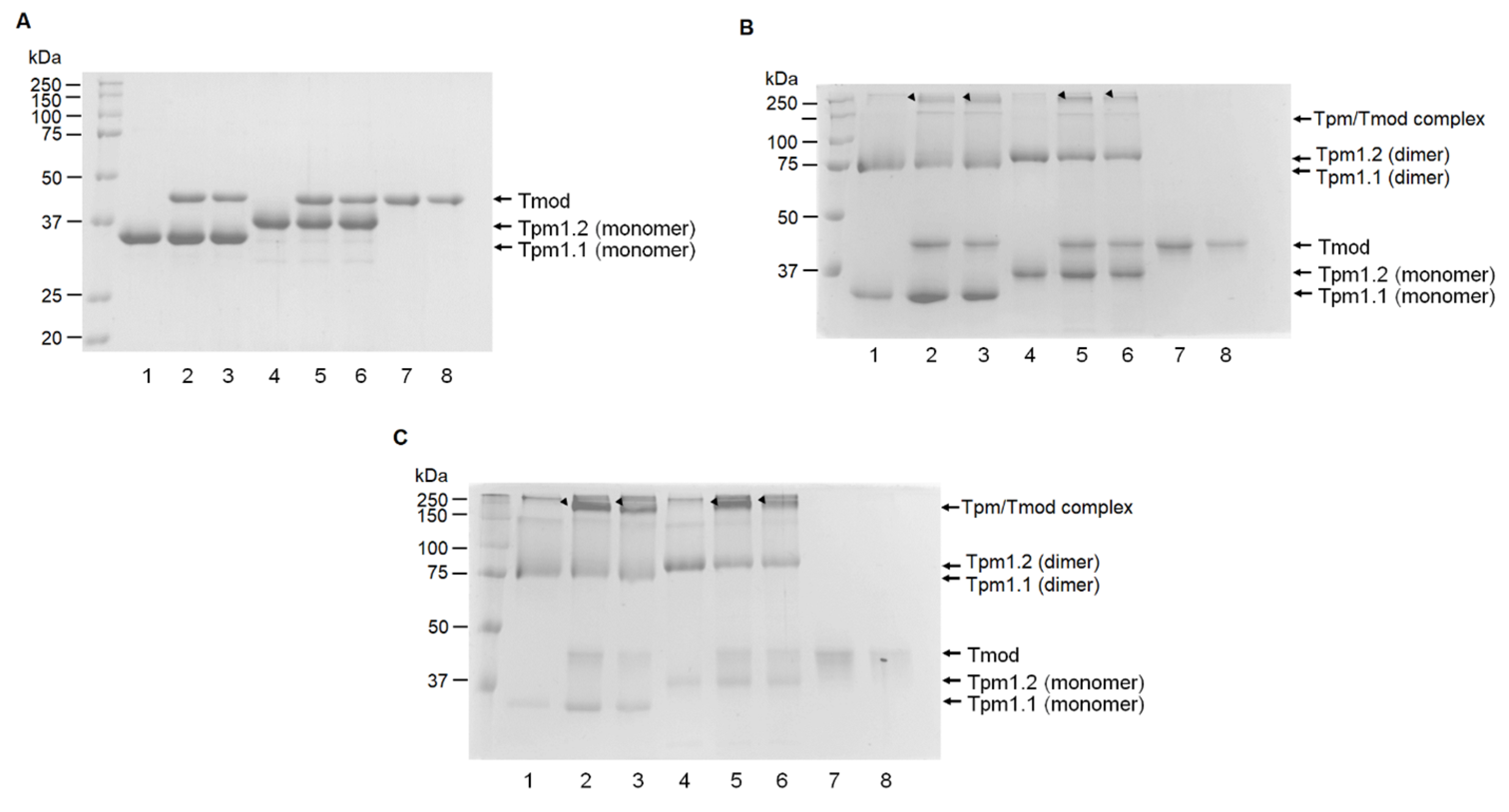
2.3. Chemical Cross-Linking Between Tmod and Tropomyosin

Tpm and Tmod samples were chemically cross-linked using an aqueous solution of glutaraldehyde (TED PELLA, Redding, CA, USA) in a 30 mM HEPES buffer, pH 7.3, with 200 mM NaCl. The concentrations of glutaraldehyde were 0.008% and 0.002%. The Tpm1.1 and Tpm1.2 concentrations were 0.2 mg/mL, and the Tmod1 and Tmod4 concentrations were 0.12 mg/mL. The cross-linking was performed for 15 and 60 min at 30 °C. After that, the samples were analyzed by SDS-PAGE in 12.5% gel.

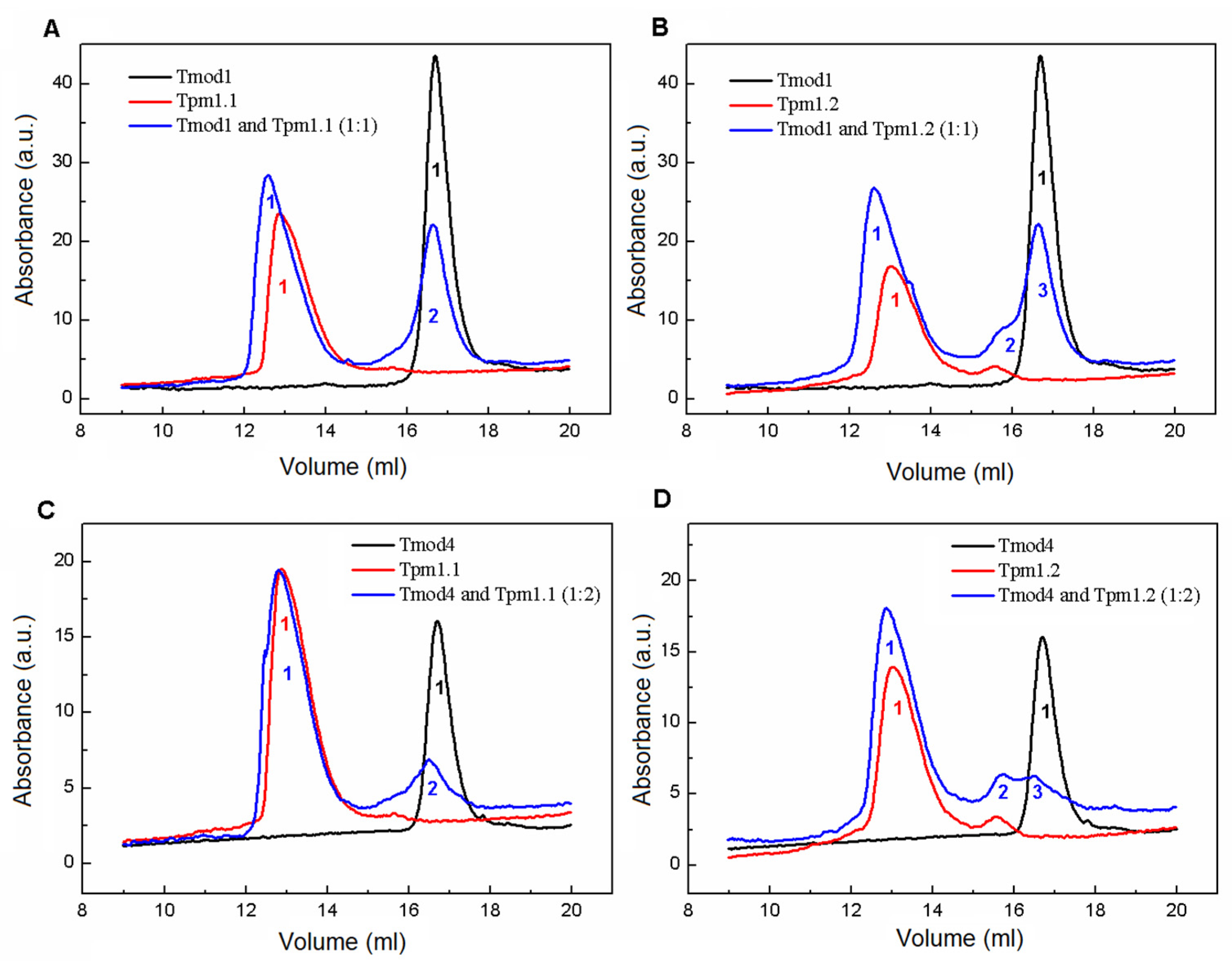
2.4. Analytical Size-Exclusion Chromatography

Size-exclusion chromatography was performed using a Varian ProStar 3250 system (Varian, Belrose, Australia) on a Superose 6 Increase 10/300 GL column (GE Healthcare, Stockholm, Sweden) equilibrated by a 50 mM HEPES-Na buffer (pH 7.3) with 150 mM NaCl and 1 mM DTT. The sample volume was 100 µL with 30 µM Tpm and Tmod1 in all elution profiles with Tmod1 and 26 µM Tpm and 13 µM Tmod4 in all elution profiles with Tmod4. The elution profiles were recorded at 280 nm with a 0.5 mL/min flow rate for all experiments. The following protein standards were used: thyroglobulin (669 kDa), ferritin (440 kDa), aldolase (158 kDa), conalbumin (75 kDa), ovalbumin (43 kDa), and carboanhydrase (29 kDa), with RNAse (13.7 kDa) used for column calibration.

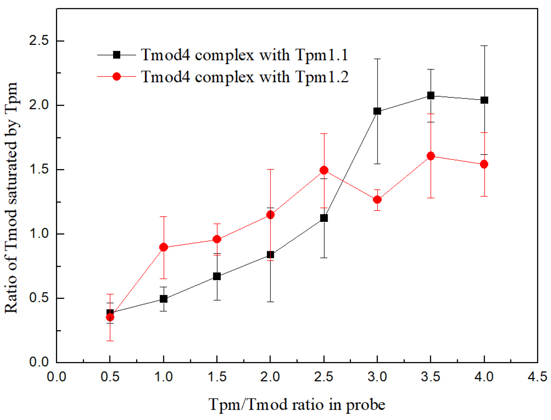
2.5. Pull-Down Assay

Experiments were performed by adding an affinity IMAC Ni-charged resin (Bio-Rad, Hercules, CA, USA) to Tpm/Tmod complexes. Before the experiments, the resin was washed three times by MQ water, then three times by a 30 mM HEPES-Na buffer, pH 7.3. Tmod1 and Tmod4 were incubated for 20 min in a 30 mM HEPES-Na buffer with 200 mM NaCl, pH 7.3, with increasing concentrations of Tpm1.1 and Tpm1.2 to the concentration ratio of 1:4 for Tmod/Tpm. After this, the resin was added to the complexes, and they were mixed and incubated for 5 min. The resin was pelleted by centrifugation and washed by a 30 mM HEPES-Na buffer with 200 mM NaCl, pH 7.3. Then, the resin was pelleted again and washed by 50 mM HEPES-Na, 300 mM NaCl, and 500 mM imidazole (pH 7.3). SDS-PAGE was used to analyze the supernatant probes. Protein bands were analyzed by ImageJ2 software (Scion, Frederick, MD, USA). For each Tpm/Tmod complex, three independent experiments were conducted.

2.6. In Vitro Motility Assay

The in vitro motility assay was carried out in accordance with a previous description [40,47,48]. Myosin (300 µg/mL) in an AB buffer (25 mM KCl, 25 mM imidazole, 4 mM MgCl2, 1 mM EGTA, and 20 mM DTT, pH 7.5) with 0.5 M KCl was loaded into a flow cell. After 2 min, 0.5 mg/mL BSA was added for 1 min. F-actin without labeling was added to an AB buffer with 2 mM ATP and left for 5 min. In order to produce regulated thin filaments, TRITC-phalloidin-labeled F-actin was added to the cell for 5 min. AB buffer was used to wash out the thin filaments that were not bound. The Maxchelator program (http://www.stanford.edu/~cpatton/webmaxc/webmaxcS.html (accessed on 1 November 2018)) was used to calculate the appropriate Ca2+ concentration. Finally, the cell was washed with an AB buffer containing 0.5 mg/mL BSA, an oxygen scavenger system, 20 mM DTT, 2 mM ATP, 0.5% methylcellulose, 100 nM Tpm/Tn, and Ca2+ ions. In each flow cell, ten 30 s image sequences were recorded at 30 °C from different fields containing ~30–50 thin filaments. GMimPro2023 software [49] was used to measure the sliding velocities of the filaments. We measured the sliding velocities of filaments that were at least 2 μm long and moved for at least 10 frames. We found that adding Tmod did not affect the lengths of the F-actin and thin filaments (Figure S2 and Table S1 in the Supplementary Materials).
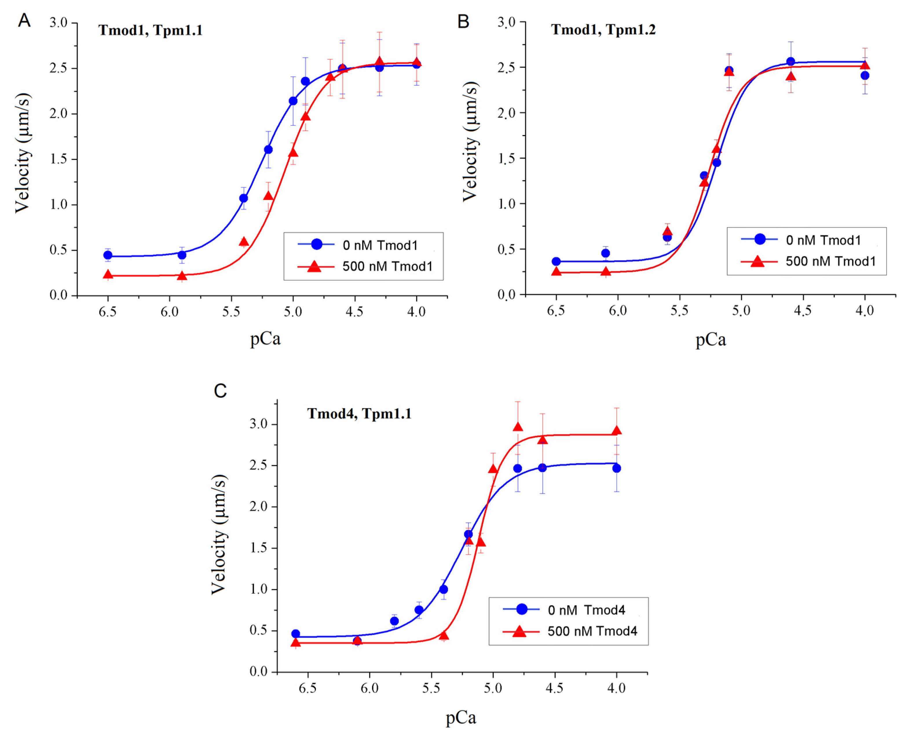
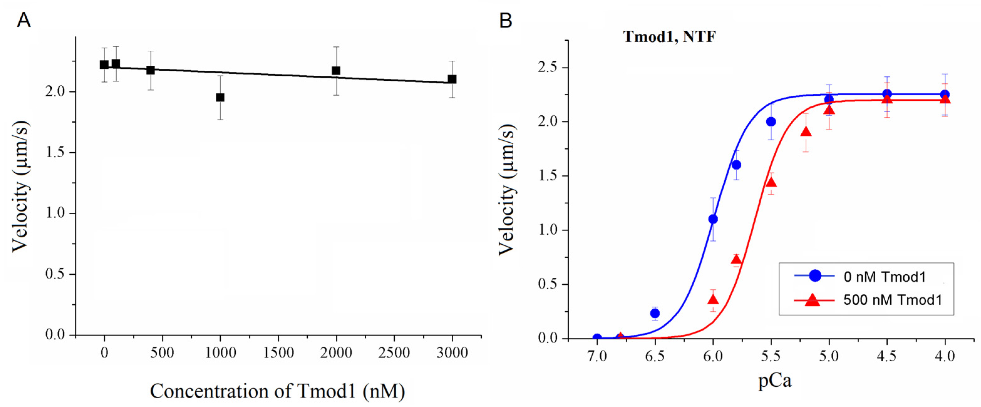
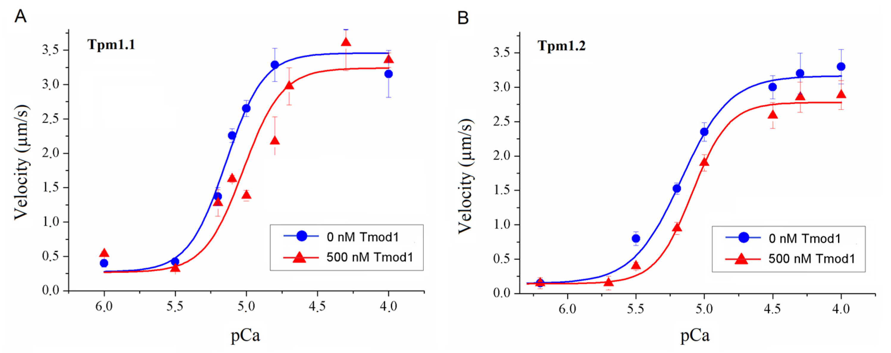
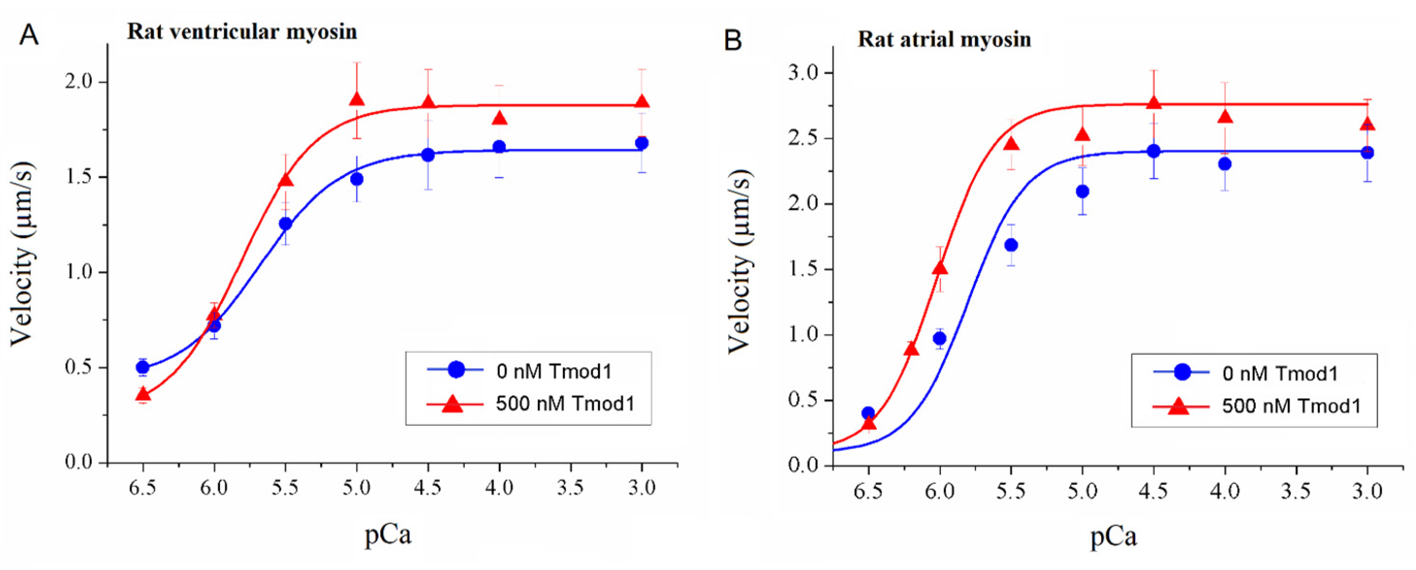
To study the effect of Tmod on the Ca2+ regulation of the actin–myosin interaction, we analyzed the dependence of the sliding velocity of the thin filaments over atrial and ventricular myosin on the Ca2+ concentration. Tmod was added to an AB buffer containing ATP. The means of individual experiments were fitted using the Hill equation: V = Vmax × (1 + 10h(pCa- pCa50))−1, where V and Vmax are the velocity and the maximal velocity at the saturating Ca2+ concentration, respectively; pCa50 (i.e., the Ca2+ sensitivity) is the pCa at which the velocity is half-maximal; and h is the Hill cooperativity coefficient. The parameters of individual experiments were averaged.
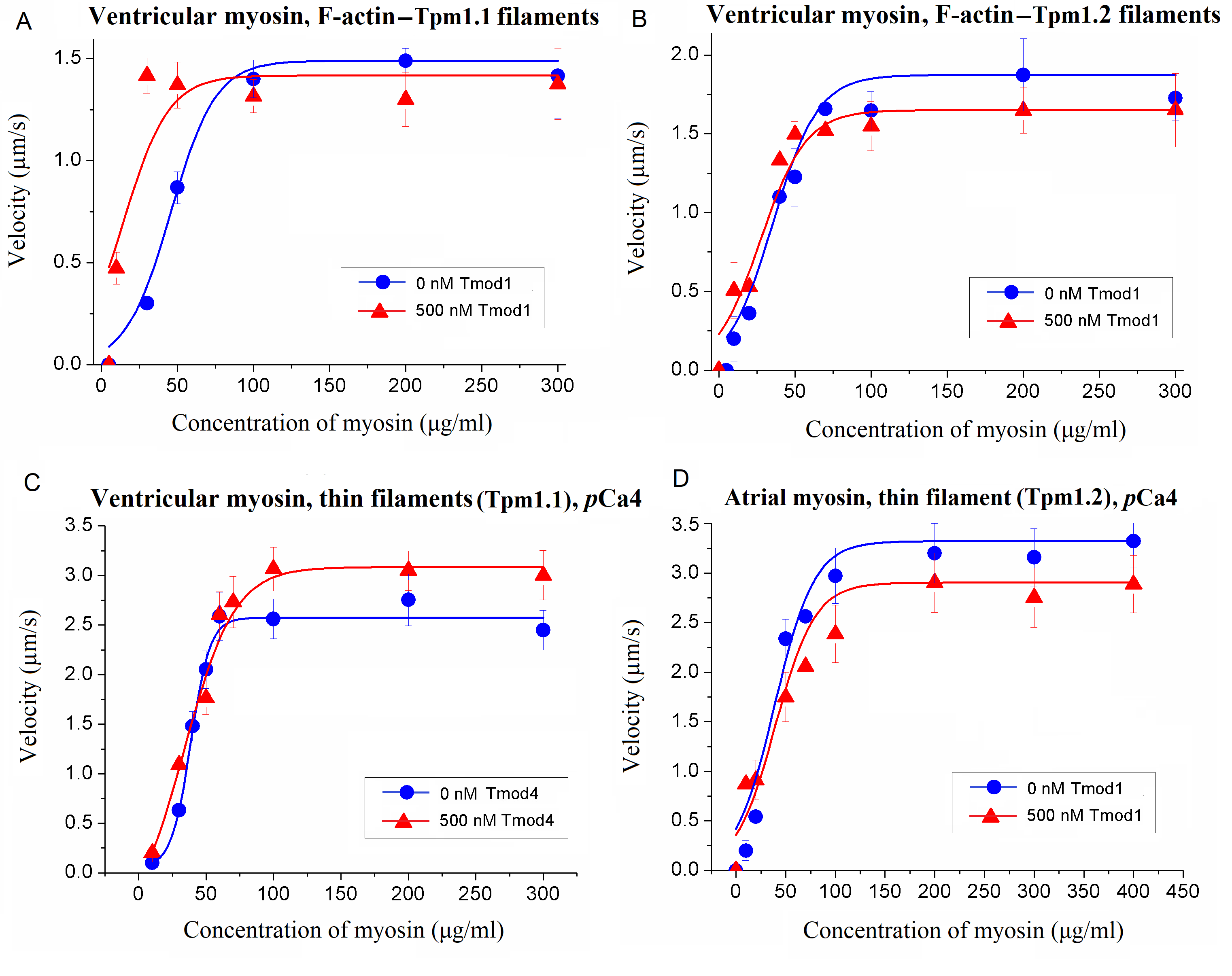
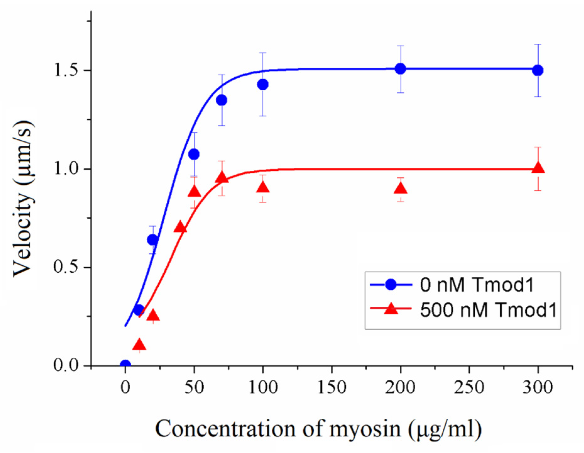
To assess the impact of Tmod on cross-bridge cooperation, the sliding velocities of thin filaments were evaluated in relation to the concentration of myosin added to the flow cell. This dependence was fitted with the modified Hill equation [50] V = Vmax × ch × (c50h + ch)−1, where Vmax is the maximal sliding velocity, C is the myosin concentration, C50 is the concentration required to achieve the half-maximal velocity, and h is the Hill coefficient.
Data analysis was performed using Excel 16 (Microsoft Corp., Redmond, WA, USA) and Origin 8.0 (Origin Lab, Northampton, MA, USA). All values were expressed as means ± SDs after three repetitions of the experiments. The Mann–Whitney U test was used to estimate the statistical significance of the characteristics of the Hill equation. In experiments where the effect of Tmod on the sliding velocity was studied, we measured the velocities of about 100 filaments, and comparisons of the filament sliding velocities at different Tmod concentrations were performed using Student’s t-test.

3. Results

3.1. The Effect of Tmod on the Sliding Velocity of F-Actin–Tpm Filaments on Sheep Myosin in the in Vitro Motility Assay

First, we measured the sliding velocity of F-actin over sheep ventricular myosin at Tmod1 and Tmod4 concentrations from 50 nM to 2000 nM. Tmod1 (Figure 1A) and Tmod4 did not affect the F-actin velocity. To study the effect of Tmod on the interaction of F-actin with Tpm, we analyzed the dependence of the sliding velocity of F-actin–Tpm filaments on the concentration of Tmod1. Tmod1 had different effects on the velocities of F-actin–Tpm filaments containing Tpm1.1 and Tpm1.2. Tmod1 slightly dose-dependently decreased the sliding velocity of F-actin–Tpm1.1 filaments on sheep ventricular and atrial myosins and did not affect the sliding velocity of F-actin–Tpm1.2 filaments (Figure 1B,C).
At the saturating calcium concentration (pCa4) with ventricular myosin, Tmod1 slightly decreased the maximum sliding velocities of thin filaments reconstructed from F-actin, troponin, and Tpm1.1 (Figure 2A) and Tpm1.2 (Figure 2B). With atrial myosin, Tmod1 decreased the maximum sliding velocities of thin filaments containing Tpm1.1 and Tpm1.2 (Figure 2C,D).

3.2. The Effect of Tmod on the Calcium Regulation of the Actin–Myosin Interaction

Ventricular and atrial myosin differ in their functional characteristics [47,48,51,52]. Therefore, in the in vitro motility assay we used ventricular and atrial myosin. We studied the effect of 500 nM Tmod on the Ca2+ regulation of the actin–myosin interaction by analyzing the Ca2+ dependence of the sliding velocities of regulated thin filaments reconstructed from F-actin, Tpm, and troponin on sheep ventricular and atrial myosin in the in vitro motility assay. With ventricular myosin, Tmod1 reduced the Ca2+ sensitivity of the velocity of thin filaments containing Tpm1.1 (Figure 3A and Table 1) and did not affect the Ca2+ dependence of the velocity of thin filaments with Tpm1.2 (Figure 3B and Table 1). Tmod4 decreased the Ca2+ sensitivity of the velocity of thin filaments containing Tpm1.1 but increased both the Hill cooperativity coefficient and the maximum filament velocity on ventricular myosin (Figure 3C and Table 1).
Next, we analyzed the effect of Tmod1 on the Ca2+ regulation of the actin–myosin interaction using myosin and native thin filaments (NTFs) extracted from the left ventricle of a sheep. Tmod1 did not affect the maximal NTF sliding velocity on myosin (Figure 4A) and significantly decreased the Ca2+ sensitivity of the NTF velocity (Figure 4B and Table 1).
Note that the main effect, both in the case of the reconstructed thin filament with recombinant Tpm and in the case of NTFs with natural acetylation of Tpm, is a decrease in the calcium sensitivity of the actin–myosin interaction. According to previous studies [25], the Ala-Ser extension does not significantly affect the interaction of Tpm with Tmod. Our results obtained with NTFs indirectly indicate that the Ala-Ser extension does not affect Tmod’s effects on the actin–myosin interaction.
With sheep atrial myosin, Tmod1 reduced the Ca2+ sensitivity of the velocities of thin filaments with both Tpm1.1 and Tpm1.2 (Figure 5 and Table 2).
Myosins of large and small animals differ in their mechanical and kinetic properties [53,54] and in their effects on the activation of the thin filament [40]. Therefore, we compared the effects of Tmod1 on the calcium regulation of the actin–myosin interaction with sheep and rat myosin. In an experiment with rat myosin, we used rat Tmod1. In contrast to sheep myosin and human Tmod1, rat Tmod1 increased both the maximal thin filament sliding velocities on rat atrial and ventricular myosin and the calcium sensitivity of the actin–myosin interaction (Figure 6 and Table 3).

3.3. Influence of Tmod on Cross-Bridge–Cross-Bridge Cooperativity of Myosin Interaction with Filaments

Cooperative mechanisms play an important role in cardiac muscle contraction, one of which is cross-bridge–cross-bridge (Xb-Xb) cooperativity. To assess the effect of Tmod on Xb-Xb cooperativity, we analyzed the dependence of the F-actin–Tpm filament velocity on the concentration of sheep ventricular myosin loaded into the flow cell. Tmod1 enhanced the Xb-Xb cooperativity of the interaction of myosin with F-actin–Tpm1.1 filaments. Tmod1 decreased the ventricular myosin concentration required to achieve the half-maximal filament velocity (Cmyosin) (Figure 7A and Table 4) but did not affect it with F-actin–Tpm1.2 filaments (Figure 7B and Table 4). At the saturating calcium concentration (pCa 4), Tmod1 and Tmod4 did not affect the Xb-Xb cooperativity of the interactions of thin filaments with ventricular and atrial myosin (Figure 7C,D and Table 4).
Using sheep ventricular myosin, we studied the effect of Tmod1 on the Xb-Xb cooperativity of the actin–myosin interaction at a non-saturating calcium concentration (pCa 5.2). We found that Tmod1 did not affect the myosin concentration at which the sliding velocity of the thin filaments was half-maximal, but it decreased the sliding velocity of the filaments by 30% (Figure 8 and Table 4).

3.4. Chemical Cross-Linking

To study the formation of complexes between Tpm and Tmod isoforms, we applied chemical cross-linking with glutaraldehyde. We found that both Tpm isoforms are able to form complexes with Tmod1 and Tmod4 (Figure 9). The apparent molecular weight of such complexes is around 160–200 kDa.

3.5. Analytical Size-Exclusion Chromatography

To study the interactions of the Tmod and Tpm isoforms, we used analytical size-exclusion chromatography (SEC). First, SEC profiles were obtained for individual proteins of Tmod1, Tmod4, Tpm1.1, and Tpm1.2. The Tmod1 profiles were presented with one symmetrical peak with an elution volume of 16.7 mL, which corresponded to a molecular mass of approximately 40 kDa (Figure 10A,B and Table 5). This fact allows us to conclude that Tmod1 is a monomer. The same data were obtained for Tmod4 (Figure 10C,D and Table 6). More complicated data were obtained for Tpm1.1 (Figure 10A,C) and Tpm1.2 (Figure 10B,D). Elution profiles were presented with single asymmetrical peaks with elution volumes of 12.9 mL for Tpm1.1 and 13.0 mL for Tpm1.2, which corresponded to approximately 600–700 kDa. Two factors could cause such deviations in elution profiles. First, although the SEC experiments were performed in a buffer containing 150 mM NaCl, a Tpm end-to-end interaction leads to cord formation under these experimental conditions. The other factor could be the shape of Tpm, which was far from a globular protein. We are inclined to favor the second option because it is hard to imagine that the formation of a Tpm strand could lead to a population of rods with similar molecular masses.
The elution profile of an equimolar mixture of Tpm1.1 and Tmod1 (Figure 10A) was presented with two major peaks with elution volumes of 12.6 and 16.7 mL (Table 5). The first peak corresponded to the complex of Tpm1.1 and Tmod1, and the second peak corresponded to residual amounts of Tmod1. A two-fold decrease in the second peak indicated that under these experimental conditions, Tmod1 interacted with Tpm1.1 at a ratio close to 1:2. The results obtained for an equimolar mixture of Tpm1.2 and Tmod1 (Figure 10B) were very similar to those for Tpm1.1 and Tmod1. However, a small shoulder appeared in the elution profile near to Tmod1 peak (Figure 10B, peak 2). We suppose that this shoulder corresponded to a complex of Tmod1 with a small amount of Tpm1.2 proteolysis fragments. Tpm1.2 still contained proteolytic fragments that could not be completely removed. They can be seen in Figure 9, lanes 4–6. Other preparations had higher purity, but minor changes in the elution profiles may have been caused by trace amounts of protein impurities. Overall, no differences in the interactions of Tmod1 with Tpm1.1 and Tpm1.2 were found under these experimental conditions.
The elution profile of a mixture of Tmod4 and Tpm1.1 with a concentration ratio of 1:2 was presented with two peaks (Figure 10C) with elution volumes of 12.8 and 16.5 mL (Table 6). These peaks were very close at their maxima to the peaks of free Tpm1.1 and Tmod4. However, a decrease in the absorption value of the Tmod4 peak indicated the formation of a complex between Tmod4 and Tpm1.1. It is noteworthy that the formation of the Tmod4/Tpm1.1 complex did not lead to a shift in the first peak of the elution curve (Figure 10C). The difference in the hydrodynamic radii of the Tpm1.1/Tmod4 and Tpm1.1/Tmod1 complexes may indicate a distinction in the interactions of the Tpm and Tmod isoforms. The elution profile obtained for the Tpm1.2/Tmod4 complex was similar to that of the Tpm1.1/Tmod4 complex (Figure 10D). As for the previous profile obtained for the mixture of Tmod1 and the Tpm1.2 isoform, we assume that the appearance of peak 2 (Figure 10D) corresponds to a complex of Tmod4 with small amounts of Tpm1.2 proteolysis fragments.
Thus, the formation of complexes of both studied Tmod isoforms with cardiac isoforms Tpm1.1 and Tpm1.2 indicates firm interactions between these proteins even without fibrillar actin.

3.6. Formation of Tmod/Tpm Complexes Determined by Pull-Down Assay

To study complex formation between Tmod and different cardiac Tpm isoforms, we used a pull-down assay. We found that the saturation patterns of the Tmod4 complexes with tropomyosin were similar for the two tropomyosin isoforms Tpm1.1 and Tpm1.2 (Figure 11). All curves had growth and a sigmoidal shape, and they reached saturation at Tpm/Tmod ratios of 2.5 for Tpm1.2 and 3 for Tpm1.1 (Figure 11). The deviations and accuracy of this method made these values very close, and they did not differ from each other. Unfortunately, saturation curves for Tmod1 could not be obtained because tropomyosin washed it off the resin.

4. Discussion

Previously, it was found that in skeletal muscle, Tmod not only regulates the length and stability of a thin filament but also affects the movement of the Tpm strand along the actin filament and the formation of myosin cross-bridges [18,19]. The replacement of Tmod1 by Tmod3/4 led to decreases in the mobility of Tpm on the actin filament and in the number of myosin cross-bridges in a strongly bound state. In the heart, a change in the expression of Tmod1 and Lmod2 leads to changes in the length of the thin filament and the structure of the sarcomere [4,5,6], which makes it difficult to study the participation of Tmod1 in the activation of the thin filament in transgenic models. Therefore, to study the functional effects of Tmod on the actin–myosin interaction, we used the in vitro motility assay. In addition, we analyzed the formation of complexes between Tpm and Tmod isoforms with chemical cross-linking by glutaraldehyde, a pull-down assay, and analytical size-exclusion chromatography (SEC). We found that Tmod1 did not affect the sliding velocity of bare F-actin over myosin in the in vitro motility assay. However, the effects of Tmod on actin–myosin interactions were pronounced in the presence of Tpm or the Tpm/Tn complex on the actin filament. In other words, through the interaction of Tmod with Tpm, Tmod influences the interaction of myosin with the actin filament. Tmod’s effects on the actin–myosin interaction depend on the composition of the protein isoforms.

4.1. Isoform-Dependent Effects of Tmod on Actin–Myosin Interaction

We found that the effect of Tmod1 on the sliding velocity of F-actin–Tpm filaments depends on the Tpm isoforms. Tmod1 dose-dependently decreased the sliding of F-actin–Tpm1.1 filaments and had no effect on the velocity of F-actin–Tpm1.2 filaments (Figure 1). The effect of Tmod1 on the calcium regulation of the actin–myosin interaction also depended on the Tpm and myosin isoforms (Figure 3A,B and Figure 4). Tmod1 reduced the calcium sensitivity of thin filaments containing Tpm1.1 with ventricular and atrial myosin. The Tpm1.1 and 1.2 isoforms differ at 27 amino acid residues located between the 39th and 77th residues in the N-terminal part (Figure S3 in the Supplementary Materials) [39]. However, using the example of Tmod2, it was shown that Tmod interacts with 14 amino acid residues located in the N-end region of the Tpm molecule [22]. The difference in Tmod1’s effect on actin–myosin interactions with Tpm1.1 and Tpm1.2 can be explained by a long-range effect caused by the difference in the amino acid sequences of the N-terminal parts of these Tpm isoforms. A similar effect was previously described for myopathic Tpm mutations in the N-terminal part of this molecule [55].
In contrast to the NTF (Figure 4A), the reconstructed thin filament showed a tendency to decrease its sliding velocity with the addition of Tmod1 (Figure 2A). This difference may be explained by post-translational modifications of the NTF proteins, in particular, phosphorylation of Tpm, troponin T, and troponin I.
In addition to the Tmod and Tpm isoforms, the myosin isoform composition should also be considered. Ventricular and atrial myosins differ in the isoform compositions of heavy and light chains and their functional characteristics [47,48,51,52]. This myosin isoform composition affects the cooperativity regulation of the actin–myosin interaction. Fast-cycling atrial myosin activates the thin filament less efficiently than ventricular myosin, thus affecting the calcium regulation of the actin–myosin interaction [47,48]. Here, we found that the effect of Tmod1 on the actin–myosin interaction also depends on myosin isoforms. Tmod1 did not affect the calcium regulation characteristics of ventricular myosin with the thin filament containing Tpm1.2 (Figure 3B and Table 1) but decreased calcium sensitivity with atrial myosin (Figure 4B and Table 2). We previously discovered that atrial myosin activates thin filaments containing Tpm1.2 stronger than ventricular myosin [48]. On the one hand, myosin interacts with tropomyosin [56,57]. The differences in the S1 amino acid sequences of atrial and ventricular myosin can affect this interaction. On the other hand, the atrial and ventricular light chain isoforms also influence the actin–myosin interaction [47,48,52].
Tpm1.2 expression has been shown to be higher in the atria than in the ventricles [58]. We found that Tmod did not affect interactions of ventricular myosin with thin filaments containing Tpm1.2 and influenced interactions of atrial myosin with thin filaments containing Tpm1.2. Therefore, it can be assumed that the interaction of Tmod with Tpm1.2 may contribute to atrial contraction. Further study is needed to confirm this.
It has previously been shown that the Tmod1, Tmod3, and Tmod4 isoforms bind non-muscle Tpm isoforms and striated muscle Tpms with different affinities [18,31,38,59]. In addition, it was found that site 1 and site 2 bind Tpm with different affinities [36], and it can be assumed that this difference may influence the interactions of myosin with the thin filament. Using Tmod4, we tested whether differences in the amino acid sequences of the Tpm-binding sites of Tmod isoforms influence the calcium regulation of the actin–myosin interaction. The homologies of Tpm-binding sites 1 and 2 for Tmod1 and Tmod4 are ~60% and ~80%, respectively (Figure S4 in the Supplementary Materials). Unlike Tmod1, Tmod4 increased the maximum velocity of thin filaments containing Tpm1.1. All these results confirm that the features of the interaction of Tmod with Tpm depend on the isoforms of these proteins. Using SEC and a pull-down assay, we found that Tmod1 and Tmod4 interact in a similar manner with both Tpm1.1 and Tpm1.2 (Table 5 and Table 6 and Figure 11). However, we did not find any significant difference in the complex formation between Tmod and Tpm isoforms. Therefore, we can suppose that the differences in the results can be explained by the peculiarities of the interactions of filaments containing Tpm isoforms with myosin isoforms.
Myosins of large and small animals differ in their functional properties [53,60] and in their effects on the activation of the thin filament [40]. For example, it was shown that the calcium sensitivity of the interaction of mouse ventricular myosin with thin filaments containing Tpm1.2 is lower than that with thin filaments containing Tpm1.1 [39], whereas the calcium sensitivity of the interactions of porcine ventricular myosin with thin filaments containing Tpm1.2 and Tpm1.1 do not differ [40]. Using rat cardiac myosin and rat Tmod1 as an example, we showed that the effect of Tmod on the actin–myosin interaction may depend on the species-specific features of myosin. Unlike human Tmod1 with sheep myosin, rat Tmod1 increased both the maximal sliding velocities of thin filaments over rat ventricular and atrial myosin and the calcium sensitivity of the filament velocity (Figure 5). Different effects of interactions between sarcomere proteins in large and small animals were previously shown for Tpm1.2 [40]. The peculiarities of the functioning of sarcomeric proteins are a mechanism of adaptation to a certain contractile function of the heart of large and small animals..

4.2. The Mechanism of Tmod’s Influence on the Actin–Myosin Interaction

The above results allow us to conclude that Tmod has a direct effect on the actin–myosin interaction in the myocardium and is one of its regulatory components. What is the mechanism of Tmod’s influence on the actin–myosin interaction? Data from previous studies and our work suggest that Tmod affects the mobility of the Tpm cable along the surface of the actin filament. The mobility of the Tpm molecule on the actin filament is necessary for activation of the thin filament. During muscle relaxation, Tpm blocks myosin-binding sites on actin (the blocked state). When calcium enters the cytosol and is bound by troponin C, Tpm begins to move azimuthally along the actin surface, opening myosin-binding sites (the closed state). Attachment of myosin heads promotes propagation of Tpm movement to adjacent actin monomers and complete activation of the thin filament (the open or M state) [61]. Using mouse knockout models and experiments with X-ray diffraction, Ochala et al. [19] demonstrated that the interaction of Tmod with Tpm at the pointed end of the actin filament may affect Tpm movement and that this effect differs between the relaxed and activated states of the thin filament and spreads along its entire length due to a long-range effect. Namely, it affects the movement of the Tpm strand at the beginning of thin filament activation. The presence of such effects during the interaction of Tpm with Tmod was discovered in the work of Moraczewska et al. [55] using the example of myopathic mutation in middle part of the Tpm molecule.
Ochala et al. [19] assumed that to a greater extent, Tmod affects the movement of the Tpm strand along the surface of the actin filament in the initial stages of activation. This assumption was indirectly confirmed by the results of our experiments on the dependence of filament velocity on the myosin concentration. The formation of myosin cross-bridges is important for the activation of the thin filament [61,62,63,64]. We found that Tmod1 enhanced the Xb-Xb cooperativity of the interaction of myosin with F-actin–Tpm filaments (Figure 7A) in the absence of calcium but did not affect it at the saturating calcium concentration (Figure 7C). At a non-saturating calcium concentration, Tmod1 decreased the thin filament velocity (Figure 8), which indicated the number of cross bridges because the filament velocity was determined by the cross-bridge number in the in vitro motility assay. With an increase in the calcium concentration, the influence of Tmod was not significant compared to other processes of activation of the thin filament. Further research is required to explain the molecular mechanisms of Tmod’s participation in the activation of the thin filament.

5. Conclusions

Previously, using X-ray diffraction of single skinned skeletal muscle fibers, it was shown that Tmod affects the movement of the Tpm strand along the actin filament and the formation of myosin cross-bridges [35,36]. Here, with an in vitro motility assay and isolated sarcomeric proteins, we demonstrated that Tmod takes part in the regulation of the actin–myosin interaction in the myocardium and that Tmod’s effect on thin filament activation is determined by the Tpm and myosin isoforms. The specific interaction of the Tmod isoform with actin and tropomyosin may be important for the regulation of the actin–myosin interaction in cardiac and skeletal muscles.

Supplementary Materials

The following supporting information can be downloaded at https://www.mdpi.com/article/10.3390/biom15050727/s1, Figure S1: Gel electrophoresis of heavy chain isoforms of sheep and rat cardiac myosin; Figure S2: The effect of Tmod on filament length. (A) F-actin, thin filaments reconstructed from troponin and Tpm1.1, and native thin filaments (NTFs) on a myosin-coated surface in a flow cell. (B) Filament length; Figure S3: Sequence alignment of Tpm1.1 and Tpm1.2 from Homo sapiens; Figure S4: Alignment of Tpm-binding sites 1 (amino acids 1–39) and 2 (amino acids 109–145) of Tmod1 and Tmod4 from Homo sapiens; Table S1: Effect of Tmod on filament length.

Author Contributions

Conceptualization, G.V.K., A.M.M. and D.V.S.; methodology, G.V.K., A.M.K., V.V.N., A.M.M. and D.V.S.; investigation, G.V.K., A.M.K., E.A.B., N.S.R., I.A.K., D.S.Y., V.V.N., A.V.S., A.M.M. and D.V.S.; formal analysis, G.V.K., A.M.K., E.A.B., V.V.N., A.M.M. and D.V.S.; data curation, G.V.K. and D.V.S.; writing—original draft preparation, G.V.K. and D.V.S.; writing—review and editing, G.V.K., A.M.M. and D.V.S.; visualization, G.V.K. and V.V.N.; supervision, G.V.K. and D.V.S. All authors have read and agreed to the published version of the manuscript.

Funding

This research was funded by the Russian Science Foundation (grant number 22-74-10106 to A.M.M.).

Institutional Review Board Statement

All procedures involving animal care and handling were performed according to the guidelines stated in Directive 2010/63/EU of the European Parliament and were approved by the Animal Care and Use Committee of the Institute of Immunology and Physiology of RAS (protocol code 05/20 from 23 September 2020).

Informed Consent Statement

Not applicable.

Data Availability Statement

All data are presented in this article.

Acknowledgments

The authors express their deep gratitude to professor Nikolai B. Gusev (Lomonosov Moscow State University) for his assistance in carrying out size-exclusion chromatography. This work was performed using equipment at the Shared Research Center of Scientific Equipment of the Institute of Immunology and Physiology of RAS.

Conflicts of Interest

Authors Natalia S. Ryabkova, Ivan A. Katrukha were employed by the company HyTest Ltd. The remaining authors declare that the research was conducted in the absence of any commercial or financial relationships that could be construed as a potential conflict of interest.

Abbreviations

The following abbreviations are used in this manuscript:
LVleft ventricle;
LAleft atria;
SECsize-exclusion chromatography.

References

  1. Mi-Mi, L.; Farman, G.P.; Mayfield, R.M.; Strom, J.; Chu, M.; Pappas, C.T.; Gregorio, C.C. In Vivo Elongation of Thin Filaments Results in Heart Failure. PLoS ONE 2020, 15, e0226138. [Google Scholar] [CrossRef] [PubMed]
  2. Pappas, C.T.; Mayfield, R.M.; Henderson, C.; Jamilpour, N.; Cover, C.; Hernandez, Z.; Hutchinson, K.R.; Chu, M.; Nam, K.-H.; Valdez, J.M.; et al. Knockout of Lmod2 Results in Shorter Thin Filaments Followed by Dilated Cardiomyopathy and Juvenile Lethality. Proc. Natl. Acad. Sci. USA 2015, 112, 13573–13578. [Google Scholar] [CrossRef] [PubMed]
  3. Pappas, C.T.; Farman, G.P.; Mayfield, R.M.; Konhilas, J.P.; Gregorio, C.C. Cardiac-Specific Knockout of Lmod2 Results in a Severe Reduction in Myofilament Force Production and Rapid Cardiac Failure. J. Mol. Cell. Cardiol. 2018, 122, 88–97. [Google Scholar] [CrossRef]
  4. McKeown, C.R.; Nowak, R.B.; Moyer, J.; Sussman, M.A.; Fowler, V.M. Tropomodulin1 Is Required in the Heart but Not the Yolk Sac for Mouse Embryonic Development. Circ. Res. 2008, 103, 1241–1248. [Google Scholar] [CrossRef] [PubMed]
  5. Tsukada, T.; Pappas, C.T.; Moroz, N.; Antin, P.B.; Kostyukova, A.S.; Gregorio, C.C. Leiomodin-2 Is an Antagonist of Tropomodulin-1 at the Pointed End of the Thin Filaments in Cardiac Muscle. J. Cell Sci. 2010, 123, 3136–3145. [Google Scholar] [CrossRef]
  6. Yamashiro, S.; Gokhin, D.S.; Kimura, S.; Nowak, R.B.; Fowler, V.M. Tropomodulins: Pointed-end Capping Proteins That Regulate Actin Filament Architecture in Diverse Cell Types. Cytoskeleton 2012, 69, 337–370. [Google Scholar] [CrossRef]
  7. Gokhin, D.S.; Ochala, J.; Domenighetti, A.A.; Fowler, V.M. Tropomodulin1 Directly Controls Thin Filament Length in Both Wild-Type and Tropomodulin4-Deficient Skeletal Muscle. Development 2015, 142, 4351–4362. [Google Scholar] [CrossRef]
  8. Schultz, L.E.; Colpan, M.; Smith, G.E.; Mayfield, R.M.; Larrinaga, T.M.; Kostyukova, A.S.; Gregorio, C.C. A Nemaline Myopathy–Linked Mutation Inhibits the Actin-Regulatory Functions of Tropomodulin and Leiomodin. Proc. Natl. Acad. Sci. USA 2023, 120, e2315820120. [Google Scholar] [CrossRef]
  9. Weber, A.; Pennise, C.R.; Babcock, G.G.; Fowler, V.M. Tropomodulin Caps the Pointed Ends of Actin Filaments. J. Cell Biol. 1994, 127, 1627–1635. [Google Scholar] [CrossRef]
  10. Tolkatchev, D.; Smith, G.E.; Schultz, L.E.; Colpan, M.; Helms, G.L.; Cort, J.R.; Gregorio, C.C.; Kostyukova, A.S. Leiomodin Creates a Leaky Cap at the Pointed End of Actin-Thin Filaments. PLoS Biol. 2020, 18, e3000848. [Google Scholar] [CrossRef]
  11. Almenar-Queralt, A.; Lee, A.; Conley, C.A.; De Pouplana, L.R.; Fowler, V.M. Identification of a Novel Tropomodulin Isoform, Skeletal Tropomodulin, That Caps Actin Filament Pointed Ends in Fast Skeletal Muscle. J. Biol. Chem. 1999, 274, 28466–28475. [Google Scholar] [CrossRef]
  12. Gokhin, D.S.; Fowler, V.M. Tropomodulin Capping of Actin Filaments in Striated Muscle Development and Physiology. BioMed Res. Int. 2011, 2011, 103069. [Google Scholar] [CrossRef] [PubMed]
  13. Fritz-Six, K.L.; Cox, P.R.; Fischer, R.S.; Xu, B.; Gregorio, C.C.; Zoghbi, H.Y.; Fowler, V.M. Aberrant Myofibril Assembly in Tropomodulin1 Null Mice Leads to Aborted Heart Development and Embryonic Lethality. J. Cell Biol. 2003, 163, 1033–1044. [Google Scholar] [CrossRef]
  14. Sussman, M.A.; Baqué, S.; Uhm, C.-S.; Daniels, M.P.; Price, R.L.; Simpson, D.; Terracio, L.; Kedes, L. Altered Expression of Tropomodulin in Cardiomyocytes Disrupts the Sarcomeric Structure of Myofibrils. Circ. Res. 1998, 82, 94–105. [Google Scholar] [CrossRef] [PubMed]
  15. Sussman, M.A.; Welch, S.; Cambon, N.; Klevitsky, R.; Hewett, T.E.; Price, R.; Witt, S.A.; Kimball, T.R. Myofibril Degeneration Caused by Tropomodulin Overexpression Leads to Dilated Cardiomyopathy in Juvenile Mice. J. Clin. Invest. 1998, 101, 51–61. [Google Scholar] [CrossRef] [PubMed]
  16. Nworu, C.U.; Kraft, R.; Schnurr, D.C.; Gregorio, C.C.; Krieg, P.A. Leiomodin 3 and Tropomodulin 4 Have Overlapping Functions during Skeletal Myofibrillogenesis. J. Cell Sci. 2014, 128, 239–250. [Google Scholar] [CrossRef]
  17. Berger, J.; Tarakci, H.; Berger, S.; Li, M.; Hall, T.E.; Arner, A.; Currie, P.D. Loss of Tropomodulin4 in the Zebrafish Mutant Träge Causes Cytoplasmic Rod Formation and Muscle Weakness Reminiscent of Nemaline Myopathy. Dis. Models Mech. 2014, 7, 1407–1415. [Google Scholar] [CrossRef]
  18. Gokhin, D.S.; Lewis, R.A.; McKeown, C.R.; Nowak, R.B.; Kim, N.E.; Littlefield, R.S.; Lieber, R.L.; Fowler, V.M. Tropomodulin Isoforms Regulate Thin Filament Pointed-End Capping and Skeletal Muscle Physiology. J. Cell Biol. 2010, 189, 95–109. [Google Scholar] [CrossRef]
  19. Ochala, J.; Gokhin, D.S.; Iwamoto, H.; Fowler, V.M. Pointed-end Capping by Tropomodulin Modulates Actomyosin Crossbridge Formation in Skeletal Muscle Fibers. FASEB J. 2014, 28, 408–415. [Google Scholar] [CrossRef]
  20. Krieger, I.; Kostyukova, A.; Yamashita, A.; Nitanai, Y.; Maéda, Y. Crystal Structure of the C-Terminal Half of Tropomodulin and Structural Basis of Actin Filament Pointed-End Capping. Biophys. J. 2002, 83, 2716–2725. [Google Scholar] [CrossRef]
  21. Tsukada, T.; Kotlyanskaya, L.; Huynh, R.; Desai, B.; Novak, S.M.; Kajava, A.V.; Gregorio, C.C.; Kostyukova, A.S. Identification of Residues within Tropomodulin-1 Responsible for Its Localization at the Pointed Ends of the Actin Filaments in Cardiac Myocytes. J. Biol. Chem. 2011, 286, 2194–2204. [Google Scholar] [CrossRef] [PubMed]
  22. Tolkatchev, D.; Kuruba, B.; Smith, G.E.; Swain, K.D.; Smith, K.A.; Moroz, N.; Williams, T.J.; Kostyukova, A.S. Structural Insights into the Tropomodulin Assembly at the Pointed Ends of Actin Filaments. Protein Sci. 2021, 30, 423–437. [Google Scholar] [CrossRef]
  23. Greenfield, N.J.; Fowler, V.M. Tropomyosin Requires an Intact N-Terminal Coiled Coil to Interact with Tropomodulin. Biophys. J. 2002, 82, 2580–2591. [Google Scholar] [CrossRef]
  24. Vera, C.; Sood, A.; Gao, K.-M.; Yee, L.J.; Lin, J.J.-C.; Sung, L.A. Tropomodulin-Binding Site Mapped to Residues 7–14 at the N-Terminal Heptad Repeats of Tropomyosin Isoform 5. Arch. Biochem. Biophys. 2000, 378, 16–24. [Google Scholar] [CrossRef]
  25. Kostyukova, A.S.; Hitchcock-DeGregori, S.E. Effect of the Structure of the N Terminus of Tropomyosin on Tropomodulin Function. J. Biol. Chem. 2004, 279, 5066–5071. [Google Scholar] [CrossRef] [PubMed]
  26. Kostyukova, A.S.; Rapp, B.A.; Choy, A.; Greenfield, N.J.; Hitchcock-DeGregori, S.E. Structural Requirements of Tropomodulin for Tropomyosin Binding and Actin Filament Capping. Biochemistry 2005, 44, 4905–4910. [Google Scholar] [CrossRef] [PubMed]
  27. Kostyukova, A.S.; Choy, A.; Rapp, B.A. Tropomodulin Binds Two Tropomyosins: A Novel Model for Actin Filament Capping. Biochemistry 2006, 45, 12068–12075. [Google Scholar] [CrossRef]
  28. Greenfield, N.J.; Kostyukova, A.S.; Hitchcock-DeGregori, S.E. Structure and Tropomyosin Binding Properties of the N-Terminal Capping Domain of Tropomodulin 1. Biophys. J. 2005, 88, 372–383. [Google Scholar] [CrossRef]
  29. Tolkatchev, D.; Smith, G.E.; Kostyukova, A.S. Role of Intrinsic Disorder in Muscle Sarcomeres. In Progress in Molecular Biology and Translational Science; Elsevier: Amsterdam, The Netherlands, 2019; Volume 166, pp. 311–340. ISBN 978-0-12-816851-6. [Google Scholar]
  30. Gregorio, C.C.; Weber, A.; Bondad, M.; Pennise, C.R.; Fowler, V.M. Requirement of Pointed-End Capping by Tropomodulin to Maintain Actin Filament Length in Embryonic Chick Cardiac Myocytes. Nature 1995, 377, 83–86. [Google Scholar] [CrossRef]
  31. Lewis, R.A.; Yamashiro, S.; Gokhin, D.S.; Fowler, V.M. Functional Effects of Mutations in the Tropomyosin-binding Sites of Tropomodulin1 and Tropomodulin3. Cytoskeleton 2014, 71, 395–411. [Google Scholar] [CrossRef]
  32. Rao, J.N.; Madasu, Y.; Dominguez, R. Mechanism of Actin Filament Pointed-End Capping by Tropomodulin. Science 2014, 345, 463–467. [Google Scholar] [CrossRef]
  33. Fowler, V.M.; Greenfield, N.J.; Moyer, J. Tropomodulin Contains Two Actin Filament Pointed End-Capping Domains. J. Biol. Chem. 2003, 278, 40000–40009. [Google Scholar] [CrossRef]
  34. Moroz, N.A.; Novak, S.M.; Azevedo, R.; Colpan, M.; Uversky, V.N.; Gregorio, C.C.; Kostyukova, A.S. Alteration of Tropomyosin-Binding Properties of Tropomodulin-1 Affects Its Capping Ability and Localization in Skeletal Myocytes. J. Biol. Chem. 2013, 288, 4899–4907. [Google Scholar] [CrossRef] [PubMed]
  35. Weber, A.; Pennise, C.R.; Fowler, V.M. Tropomodulin Increases the Critical Concentration of Barbed End-Capped Actin Filaments by Converting ADP·Pi-Actin to ADP-Actin at All Pointed Filament Ends. J. Biol. Chem. 1999, 274, 34637–34645. [Google Scholar] [CrossRef]
  36. Kostyukova, A.S.; Hitchcock-DeGregori, S.E.; Greenfield, N.J. Molecular Basis of Tropomyosin Binding to Tropomodulin, an Actin-Capping Protein. J. Mol. Biol. 2007, 372, 608–618. [Google Scholar] [CrossRef] [PubMed]
  37. Vasilescu, C.; Colpan, M.; Ojala, T.H.; Manninen, T.; Mutka, A.; Ylänen, K.; Rahkonen, O.; Poutanen, T.; Martelius, L.; Kumari, R.; et al. Recessive TMOD1 Mutation Causes Childhood Cardiomyopathy. Commun. Biol. 2024, 7, 7. [Google Scholar] [CrossRef]
  38. Yamashiro, S.; Gokhin, D.S.; Sui, Z.; Bergeron, S.E.; Rubenstein, P.A.; Fowler, V.M. Differential Actin-Regulatory Activities of Tropomodulin1 and Tropomodulin3 with Diverse Tropomyosin and Actin Isoforms. J. Biol. Chem. 2014, 289, 11616–11629. [Google Scholar] [CrossRef] [PubMed]
  39. Rajan, S.; Jagatheesan, G.; Karam, C.N.; Alves, M.L.; Bodi, I.; Schwartz, A.; Bulcao, C.F.; D’Souza, K.M.; Akhter, S.A.; Boivin, G.P.; et al. Molecular and Functional Characterization of a Novel Cardiac-Specific Human Tropomyosin Isoform. Circulation 2010, 121, 410–418. [Google Scholar] [CrossRef]
  40. Kopylova, G.V.; Kochurova, A.M.; Yampolskaya, D.S.; Nefedova, V.V.; Tsaturyan, A.K.; Koubassova, N.A.; Kleymenov, S.Y.; Levitsky, D.I.; Bershitsky, S.Y.; Matyushenko, A.M.; et al. Structural and Functional Properties of Kappa Tropomyosin. Int. J. Mol. Sci. 2023, 24, 8340. [Google Scholar] [CrossRef]
  41. Monteiro, P.B.; Lataro, R.C.; Ferro, J.A.; Reinach, F.d.C. Functional Alpha-Tropomyosin Produced in Escherichia Coli: A Dipeptide Extension Can Substitute the Amino-Terminal Acetyl Group. J. Biol. Chem. 1994, 269, 10461–10466. [Google Scholar] [CrossRef]
  42. Kostyukova, A.; Maeda, K.; Yamauchi, E.; Krieger, I.; Maéda, Y. Domain Structure of Tropomodulin: Distinct Properties of the N-terminal and C-terminal Halves. Eur. J. Biochem. 2000, 267, 6470–6475. [Google Scholar] [CrossRef]
  43. Bliss, K.T.; Tsukada, T.; Novak, S.M.; Dorovkov, M.V.; Shah, S.P.; Nworu, C.; Kostyukova, A.S.; Gregorio, C.C. Phosphorylation of Tropomodulin1 Contributes to the Regulation of Actin Filament Architecture in Cardiac Muscle. FASEB J. 2014, 28, 3987–3995. [Google Scholar] [CrossRef]
  44. Margossian, S.S.; Lowey, S. [7] Preparation of Myosin and Its Subfragments from Rabbit Skeletal Muscle. In Methods in Enzymology; Elsevier: Amsterdam, The Netherlands, 1982; Volume 85, pp. 55–71. ISBN 978-0-12-181985-9. [Google Scholar]
  45. Spiess, M.; Steinmetz, M.O.; Mandinova, A.; Wolpensinger, B.; Aebi, U.; Atar, D. Isolation, Electron Microscopic Imaging, and 3-D Visualization of Native Cardiac Thin Myofilaments. J. Struct. Biol. 1999, 126, 98–104. [Google Scholar] [CrossRef] [PubMed]
  46. Pardee, J.D.; Aspudich, J. [18] Purification of Muscle Actin. In Methods in Enzymology; Elsevier: Amsterdam, The Netherlands, 1982; Volume 85, pp. 164–181. ISBN 978-0-12-181985-9. [Google Scholar]
  47. Shchepkin, D.V.; Nikitina, L.V.; Bershitsky, S.Y.; Kopylova, G.V. The Isoforms of α-Actin and Myosin Affect the Ca2+ Regulation of the Actin-Myosin Interaction in the Heart. Biochem. Biophys. Res. Commun. 2017, 490, 324–329. [Google Scholar] [CrossRef]
  48. Kopylova, G.V.; Berg, V.Y.; Kochurova, A.M.; Matyushenko, A.M.; Bershitsky, S.Y.; Shchepkin, D.V. The Effects of the Tropomyosin Cardiomyopathy Mutations on the Calcium Regulation of Actin-Myosin Interaction in the Atrium and Ventricle Differ. Biochem. Biophys. Res. Commun. 2022, 588, 29–33. [Google Scholar] [CrossRef] [PubMed]
  49. Mashanov, G.I.; Molloy, J.E. Automatic Detection of Single Fluorophores in Live Cells. Biophys. J. 2007, 92, 2199–2211. [Google Scholar] [CrossRef] [PubMed]
  50. Brunet, N.M.; Chase, P.B.; Mihajlović, G.; Schoffstall, B. Ca2+-Regulatory Function of the Inhibitory Peptide Region of Cardiac Troponin I Is Aided by the C-Terminus of Cardiac Troponin T: Effects of Familial Hypertrophic Cardiomyopathy Mutations cTnI R145G and cTnT R278C, Alone and in Combination, on Filament Sliding. Arch. Biochem. Biophys. 2014, 552–553, 11–20. [Google Scholar] [CrossRef]
  51. Walklate, J.; Ferrantini, C.; Johnson, C.A.; Tesi, C.; Poggesi, C.; Geeves, M.A. Alpha and Beta Myosin Isoforms and Human Atrial and Ventricular Contraction. Cell. Mol. Life Sci. 2021, 78, 7309–7337. [Google Scholar] [CrossRef]
  52. Yamashita, H. Myosin Light Chain Isoforms Modify Force-Generating Ability of Cardiac Myosin by Changing the Kinetics of Actin–Myosin Interaction. Cardiovasc. Res. 2003, 60, 580–588. [Google Scholar] [CrossRef]
  53. Johnson, C.A.; McGreig, J.E.; Jeanfavre, S.T.; Walklate, J.; Vera, C.D.; Farré, M.; Mulvihill, D.P.; Baines, A.J.; Ridout, M.; Leinwand, L.A.; et al. Identification of Sequence Changes in Myosin II That Adjust Muscle Contraction Velocity. PLoS Biol. 2021, 19, e3001248. [Google Scholar] [CrossRef]
  54. Prodanovic, M.; Geeves, M.A.; Poggesi, C.; Regnier, M.; Mijailovich, S.M. Effect of Myosin Isoforms on Cardiac Muscle Twitch of Mice, Rats and Humans. Int. J. Mol. Sci. 2022, 23, 1135. [Google Scholar] [CrossRef] [PubMed]
  55. Moraczewska, J.; Robaszkiewicz, K.; Śliwinska, M.; Czajkowska, M.; Ly, T.; Kostyukova, A.; Wen, H.; Zheng, W. Congenital Myopathy-related Mutations in Tropomyosin Disrupt Regulatory Function through Altered Actin Affinity and Tropomodulin Binding. FEBS J. 2019, 286, 1877–1893. [Google Scholar] [CrossRef] [PubMed]
  56. Doran, M.H.; Pavadai, E.; Rynkiewicz, M.J.; Walklate, J.; Bullitt, E.; Moore, J.R.; Regnier, M.; Geeves, M.A.; Lehman, W. Cryo-EM and Molecular Docking Shows Myosin Loop 4 Contacts Actin and Tropomyosin on Thin Filaments. Biophys. J. 2020, 119, 821–830. [Google Scholar] [CrossRef] [PubMed]
  57. Doran, M.H.; Rynkiewicz, M.J.; Pavadai, E.; Bodt, S.M.L.; Rasicci, D.; Moore, J.R.; Yengo, C.M.; Bullitt, E.; Lehman, W. Myosin Loop-4 Is Critical for Optimal Tropomyosin Repositioning on Actin during Muscle Activation and Relaxation. J. Gen. Physiol. 2023, 155, e202213274. [Google Scholar] [CrossRef]
  58. Peng, Y.; Yu, D.; Gregorich, Z.; Chen, X.; Beyer, A.M.; Gutterman, D.D.; Ge, Y. In-Depth Proteomic Analysis of Human Tropomyosin by Top-down Mass Spectrometry. J. Muscle Res. Cell Motil. 2013, 34, 199–210. [Google Scholar] [CrossRef]
  59. Uversky, V.N.; Shah, S.P.; Gritsyna, Y.; Hitchcock-DeGregori, S.E.; Kostyukova, A.S. Systematic Analysis of Tropomodulin/Tropomyosin Interactions Uncovers Fine-tuned Binding Specificity of Intrinsically Disordered Proteins. J. Mol. Recognit. 2011, 24, 647–655. [Google Scholar] [CrossRef]
  60. Malmqvist, U.P.; Aronshtam, A.; Lowey, S. Cardiac Myosin Isoforms from Different Species Have Unique Enzymatic and Mechanical Properties. Biochemistry 2004, 43, 15058–15065. [Google Scholar] [CrossRef]
  61. McKillop, D.F.; Geeves, M.A. Regulation of the Interaction between Actin and Myosin Subfragment 1: Evidence for Three States of the Thin Filament. Biophys. J. 1993, 65, 693–701. [Google Scholar] [CrossRef]
  62. Longyear, T.; Walcott, S.; Debold, E.P. The Molecular Basis of Thin Filament Activation: From Single Molecule to Muscle. Sci. Rep. 2017, 7, 1822. [Google Scholar] [CrossRef]
  63. Gorga, J.A.; Fishbaugher, D.E.; VanBuren, P. Activation of the Calcium-Regulated Thin Filament by Myosin Strong Binding. Biophys. J. 2003, 85, 2484–2491. [Google Scholar] [CrossRef]
  64. Caremani, M.; Marcello, M.; Morotti, I.; Pertici, I.; Squarci, C.; Reconditi, M.; Bianco, P.; Piazzesi, G.; Lombardi, V.; Linari, M. The Force of the Myosin Motor Sets Cooperativity in Thin Filament Activation of Skeletal Muscles. Commun. Biol. 2022, 5, 1266. [Google Scholar] [CrossRef] [PubMed]
Figure 1. The effect of Tmod1 on the sliding velocities of F-actin and F-actin–Tpm filaments on sheep cardiac myosin in the in vitro motility assay. (A) The dependence of the sliding velocity of F-actin on the concentration of Tmod1 over sheep ventricular myosin. (B,C) The dependence of the sliding velocities of F-actin–Tpm filaments containing Tpm1.1 and Tpm1.2 on the Tmod1 concentration on sheep ventricular (B) and atrial (C) myosin. The experimental data for Tmod1 with F-actin–Tpm1.1 filaments and F-actin–Tpm1.2 filaments are approximated by exponential and linear functions, respectively. The experimental values are presented as means ± SDs.
Biomolecules 15 00727 g001
Figure 2. The effect of Tmod1 on the maximum sliding velocities at pCa4 of thin filaments containing F-actin, Tpm, and Tn on sheep cardiac myosin in the in vitro motility assay. (AD) The dependence of the maximum sliding velocities (pCa4) of thin filaments containing Tpm1.1 (A,C) and Tpm1.2 (B,D) on sheep ventricular (A,B) and atrial (C,D) myosin on the Tmod1 concentration. The experimental data in (AD) are approximated by exponential functions. The experimental values are presented as means ± SDs.
Biomolecules 15 00727 g002
Figure 3. The effect of Tmod1 on the maximum sliding velocities at pCa4 of thin filaments reconstructed from F-actin, troponin, and Tpm on sheep cardiac myosin in the in vitro motility assay. (A,B) The effect of 500 nM Tmod1 on the calcium dependence of the sliding velocities of thin filaments containing Tpm1.1 (A) and Tpm1.2 (B) over sheep ventricular myosin. (C) The effect of 500 nM Tmod4 on the calcium dependence of the sliding velocity of thin filaments containing Tpm1.1 over sheep ventricular myosin. The experimental values are means ± SDs. The calcium dependence of the filament sliding velocity was approximated by the Hill equation; the values of the parameters are given in Table 1.
Biomolecules 15 00727 g003
Figure 4. The effect of Tmod1 on the interaction of sheep ventricular myosin with native thin filaments. (A) The dependence of the maximum sliding velocity (pCa4) of native thin filaments (NTFs) over sheep ventricular myosin on the Tmod1 concentration in the in vitro motility assay. NTFs and myosin were extracted from the sheep left ventricle. The experimental data are presented as means ± SDs and approximated by a linear function. (B) The effect of 500 nM Tmod1 on the calcium dependence of the sliding velocity of NTFs on sheep ventricular myosin in the in vitro motility assay. The experimental values are means ± SDs. The calcium dependence of the filament sliding velocity was approximated by the Hill equation; the values of the parameters are given in Table 1.
Biomolecules 15 00727 g004
Figure 5. The effect of Tmod1 on the calcium dependence of the sliding velocity of thin filaments on sheep atrial myosin in the in vitro motility assay. (A) The effect of 500 nM Tmod1 on the calcium dependence of the sliding velocity of thin filaments containing Tpm1.1 on sheep atrial myosin. (B) The effect of 500 nM Tmod1 on the calcium dependence of the sliding velocity of thin filaments containing Tpm1.2 on sheep atrial myosin. The experimental values are means ± SDs. The calcium dependence of the filament sliding velocity was approximated by the Hill equation, and the values of the parameters are given in Table 2.
Biomolecules 15 00727 g005
Figure 6. The effect of rat Tmod1 on the calcium dependence of the sliding velocity of thin filaments on rat cardiac myosin in the in vitro motility assay. (A) The effect of 500 nM rat Tmod1 on the calcium dependence of the sliding velocity of thin filaments containing Tpm1.1 on rat ventricular myosin. (B) The effect of 500 nM rat Tmod1 on the calcium dependence of the sliding velocity of thin filaments containing Tpm1.1 on rat atrial myosin. The experimental values are means ± SDs. The calcium dependence of the filament sliding velocity was approximated by the Hill equation, and the values of the parameters are given in Table 3.
Biomolecules 15 00727 g006
Figure 7. The effect of Tmod1 on cross-bridge–cross-bridge (Xb-Xb) cooperativity. (A,B) The effect of 500 nM Tmod1 on the Xb-Xb cooperativity of the interactions of sheep ventricular myosin with F-actin–Tpm filaments containing Tpm1.1 (A) and Tpm1.2 (B). (C) The effect of 500 nM Tmod4 on the Xb-Xb cooperativity of the interaction of sheep ventricular myosin with F-actin–Tpm filaments containing Tpm1.1. (D) The effect of 500 nM Tmod1 on the Xb-Xb cooperativity of the interaction of sheep atrial myosin with F-actin–Tpm filaments containing Tpm1.1. The experimental values are presented as means ± SDs. The experimental values are approximated by the Hill equation.
Biomolecules 15 00727 g007
Figure 8. The effect of 500 nM Tmod1 on the cross-bridge–cross-bridge cooperativity of the interaction of sheep ventricular myosin with thin filaments at a non-saturating calcium concentration (pCa5.2). The experimental values are presented as means ± SDs and approximated by the Hill equation.
Biomolecules 15 00727 g008
Figure 9. The chemical cross-linking of Tpm and Tmod isoforms. (A) Samples subjected to cross-linking before adding glutaraldehyde. (B) Chemical cross-linking by 0.002% glutaraldehyde for 15 min. (C) Chemical cross-linking by 0.008% glutaraldehyde for 15 min. Lanes: 1—Tpm1.1; 2—Tpm1.1 with Tmod1; 3—Tpm1.1 with Tmod4; 4—Tpm1.2; 5—Tpm1.2 with Tmod1; 6—Tpm1.2 with Tmod4; 7—Tmod1; 8—Tmod4. Black arrows mark the positions of Tpm/Tmod complexes. Original images can be found in Supplementary Materials.
Biomolecules 15 00727 g009
Figure 10. Analytical size-exclusion chromatography of Tmod1, Tmod4, Tpm1.1, Tpm 1.2, and their mixtures loaded on a Superose 6 Increase 10/300 GL column. (A) The elution profiles of Tmod1, Tpm1.1, and a mixture of Tmod1 and Tpm1.1 at a concentration ratio of 1:1. (B) The elution profiles of Tmod1, Tpm1.2, and a mixture of Tmod1 and Tpm1.2 at a concentration ratio of 1:1. (C) The elution profiles of Tmod4, Tpm1.1, and a mixture of Tmod4 and Tpm1.1 at concentration ratio of 1:2. (D) The elution profiles of Tmod4, Tpm1.2, and a mixture of Tmod4 and Tpm1.2 at a concentration ratio of 1:2. The digits indicate peak numbers; the peak parameters are presented in Table 5 and Table 6.
Biomolecules 15 00727 g010
Figure 11. The interactions of Tmod4 with Tpm1.1 and Tpm1.2 measured by the pull-down assay. This graph shows the dependence of Tmod4 saturation on Tpm1.1 (black line) and Tpm1.2 (red line) added to the sample in different concentrations. The Tmod4 concentration was 6 µM in all samples, and the Tpm concentrations ranged from 3 to 24 µM. The experimental values are presented as means ± SDs.
Biomolecules 15 00727 g011
Table 1. The parameters of the Ca2+ dependence of the sliding velocities of thin filaments over sheep ventricular myosin in the in vitro motility assay.
Tmod IsoformFilamentTmod Concentration (nM)Vmax (µm/s)hpCa50
Tmod1Tpm1.102.6 ± 0.11.2 ± 0.15.25 ± 0.01
5002.6 ± 0.11.5 ± 0.25.06 ± 0.02 *
Tmod402.5 ± 0.11.2 ± 0.15.26 ± 0.01
5002.9 ± 0.1 *2.2 ± 0.2 *5.12 ± 0.01 *
Tmod1Tpm1.202.6 ± 0.11.7 ± 0.15.20 ± 0.01
5002.5 ± 0.11.8 ± 0.15.27 ± 0.02
Tmod1NTF02.3 ± 0.11.7 ± 0.15.99 ± 0.01
5002.2 ± 0.11.8 ± 0.25.64 ± 0.01 *
Vmax is the maximum sliding velocity of thin filaments at a saturating Ca2+ concentration; h is the Hill cooperativity coefficient; and pCa50 is the Ca2+ concentration at which the velocity is half-maximal and the Ca2+ sensitivity. Simbol * indicates differences between the values of the parameters of the Hill equation with and without Tmod (Mann–Whitney U test, p < 0.05).
Table 2. The parameters of the Ca2+ dependence of the sliding velocities of thin filaments on sheep atrial myosin in the in vitro motility assay.
Tpm IsoformTmod1 Concentration (nM)Vmax (µm/s)hpCa50
Tpm1.103.4 ± 0.31.7 ± 0.25.14 ± 0.03
5003.2 ± 0.21.7 ± 0.15.02 ± 0.02 *
Tpm1.203.2 ± 0.21.4 ± 0.25.17 ± 0.01
5002.8 ± 0.11.8 ± 0.25.09 ± 0.03 *
Vmax is the maximum sliding velocity of thin filaments at a saturating Ca2+ concentration; h is the Hill cooperativity coefficient; and pCa50 is the Ca2+ concentration at which the velocity is half-maximal and the Ca2+ sensitivity. Simbol * indicates differences between the values of the parameters of the Hill equation with and without Tmod (Mann–Whitney U test, p < 0.05).
Table 3. The parameters of the Ca2+ dependence of the sliding velocities of thin filaments over rat cardiac myosin in the in vitro motility assay.
MyosinRat Tmod1 Concentration (nM)Vmax (µm/s)hpCa50
LV01.64 ± 0.031.0 ± 0.15.68 ± 0.04
5001.87 ± 0.02 *1.2 ± 0.25.81 ± 0.01 *
LA02.40 ± 0.011.2 ± 0.15.80 ± 0.01
5002.76 ± 0.01 *1.2 ± 0.36.02 ± 0.01 *
LA indicates atrial myosin; LV indicates ventricular myosin; Vmax is the maximum sliding velocity of thin filaments at a saturating Ca2+ concentration; h is the Hill cooperativity coefficient; and pCa50 is the Ca2+ concentration at which the velocity is half-maximal and the Ca2+ sensitivity. Simbol * indicates differences between the values of the parameters of the Hill equation with and without rat Tmod1 (Mann–Whitney U test, p < 0.05).
Table 4. The myosin concentration required to achieve the half-maximal filament velocity (Cmyosin).
MyosinFilamentTmod Concentration (nM)Cmyosin (µg/mL)
Sheep LVF-actin–Tpm1.1 filament0 nM Tmod145.7 ± 5.2
500 nM Tmod115.0 ± 2.2 *
Sheep LVF-actin–Tpm1.2 filament0 nM Tmod135.5 ± 5.1
500 nM Tmod127.4 ± 6.7
Sheep LVpCa 4, thin filament containing Tpm1.10 nM Tmod438.6 ± 4.1
500 nM Tmod433.5 ± 5.2
Sheep LApCa 4, thin filament containing Tpm1.10 nM Tmod156.4 ± 4.3
500 nM Tmod137.9 ± 15.3
Sheep LApCa 4, thin filament containing Tpm1.20 nM Tmod138.7 ± 3.1
500 nM Tmod139.3 ± 5.3
Sheep LVpCa 5.2, thin filament containing Tpm1.10 nM Tmod127.9 ± 2.3
500 nM Tmod133.3 ± 5.1
LA indicates atrial myosin; LV indicates ventricular myosin; and Cmyosin is the myosin concentration required to achieve the half-maximal filament velocity. Simbol * indicates differences between the values of the parameters of the Hill equation with and without rat Tmod (Mann–Whitney U test, p < 0.05).
Table 5. Peak areas and elution volumes obtained from size-exclusion chromatography profiles of Tmod1.
SEC Profile NamePeak Number
Peak 1Peak 2Peak 3
Peak Area (ml * abs)Elution Volume (ml)Peak Area (ml * abs)Elution Volume (ml)Peak Area (ml * abs)Elution Volume (ml)
Tpm1.121.1912.9----
Tpm1.214.8913.0----
Tmod128.9716.7----
Tpm1.1 and Tmod125.8712.615.1816.7--
Tpm1.2 and Tmod125.3712.61.3015.814.4616.7
Table 6. Peak areas and elution volumes obtained from size-exclusion chromatography profiles of Tmod4.
SEC Profile NamePeak Number
Peak 1Peak 2Peak 3
Peak Area (ml * abs)Elution Volume (ml)Peak Area (ml * abs)Elution Volume (ml)Peak Area (ml * abs)Elution Volume (ml)
Tpm1.117.5912.9----
Tpm1.212.3613.0----
Tmod49.4516.7----
Tpm1.1 and Tmod417.6712.83.1516.5--
Tpm1.2 and Tmod417.1012.91.2315.71.6416.5
Disclaimer/Publisher’s Note: The statements, opinions and data contained in all publications are solely those of the individual author(s) and contributor(s) and not of MDPI and/or the editor(s). MDPI and/or the editor(s) disclaim responsibility for any injury to people or property resulting from any ideas, methods, instructions or products referred to in the content.

