Article

Baicalin Relieves Glaesserella parasuis-Triggered Immunosuppression Through Polarization via MIF/CD74 Signaling Pathway in Piglets

1
Wuhan Engineering and Technology Research Center of Animal Disease-Resistant Nutrition, School of Animal Science and Nutritional Engineering, Wuhan Polytechnic University, Wuhan 430023, China
2
Hubei Key Laboratory of Animal Nutrition and Feed Science, School of Animal Science and Nutritional Engineering, Wuhan Polytechnic University, Wuhan 430023, China
*
Author to whom correspondence should be addressed.
These authors contributed equally to the work.
Biomolecules 2025, 15(5), 640; https://doi.org/10.3390/biom15050640
Submission received: 23 February 2025 / Revised: 22 April 2025 / Accepted: 25 April 2025 / Published: 29 April 2025
(This article belongs to the Topic Recent Advances in Veterinary Pharmacology and Toxicology)

Abstract

:
Glaesserella parasuis (G. parasuis) infection is responsible for Glässer’s disease in pigs. G. parasuis could trigger piglet immunosuppression, but the mechanism of inducing immunosuppression by G. parasuis remains unknown. Macrophage migration inhibitory factor (MIF)/CD74 axis has been shown to participate in inflammation response and immunosuppression, but the function of MIF/CD74 during immunosuppression elicited by G. parasuis has not been fully explored. This experiment explored the efficacy of baicalin on immunosuppression elicited by G. parasuis alleviation through regulating polarization via the MIF/CD74 signaling pathway. Our data indicated that baicalin reduced IL-1β, IL-6, IL-8, IL-18, TNF-α, and COX-2 expression, and regulated MIF/CD74 axis expression in the spleen. Immunohistochemistry analysis showed that baicalin enhanced CD74 protein levels in the spleen of piglets induced by G. parasuis. Baicalin regulated the PI3K/Akt/mTOR signaling pathway and RAF/MEK/ERK signaling activation, modified the expression of the autophagy-related proteins Beclin-1, P62, and LC3B, promoted M2 polarization to M1 polarization, and enhanced CD3, CD4, CD8, and TIM3 levels in the spleen of piglets elicited by G. parasuis. Our study reveals the important functions of the MIF/CD74 axis in G. parasuis-induced immunosuppression and may offer a new therapeutic method to control G. parasuis infection.

1. Introduction

Glaesserella parasuis (G. parasuis) is a significant pathogen responsible for Glässer’s disease in pigs [1]. The symptoms caused by G. parasuis are fibrotic polyserositis, arthritis, and meningitis [2]. So far, 15 serovars have been verified by using gel immunodiffusion determination; however, more than 20% of isolates have not been serotyped using this method [3]. Serovars 4, 5, and 13 were considered as the highly virulent strains [4]. G. parasuis is a bacterium that colonizes the upper respiratory tract of pigs, which can spread rapidly within a herd [5]. Once inside the host, G. parasuis has the potential to invade and multiply within the tissues, leading to an acute systemic inflammatory response [6]. This infectious condition poses a considerable threat to swine production worldwide, contributing to substantial economic losses to the pig farms [7]. Prevention and control measures are crucial to mitigate the harmful effects of G. parasuis. However, the diversity of G. parasuis serovars lacks cross-serovar protection, contributing to limiting the effectiveness of vaccines [8]. Thus, it is urgent to find new drugs to prevent and control G. parasuis infection.
Infection of piglets with G. parasuis can result in immunosuppression in piglets [9]; however, the mechanism of immunosuppression elicited by G. parasuis in piglets is still unknown. Leukocyte differentiation antigen 74 (CD74), the MHC II-associated invariant chain, is a type II transmembrane glycoprotein [10]. CD74 promoted the formation of the tumor microenvironment through enhancing S100A8 and S100A9 production in the pancreatic cancer [11]. CD74 aided the accumulation and efficacy of regulatory T cells in tumors [12] and was involved in Staphylococcus aureus-induced dose-dependent immunosuppression [13]. In addition, CD74 was considered the receptor for the macrophage migration inhibitory factor (MIF) [14]. As a receptor for CD74, MIF can regulate the innate and adaptive immune response [15]. MIF has been shown to play a key role in immune system regulation and is involved in autoimmune disorders and tumorigenesis [16]. Furthermore, some studies indicate that MIF contributes to human melanoma myeloid-derived suppressor cell induction and T-cell immunosuppression [17]. Research has reported that MIF/CD74 damaged LECs’ barrier via inhibiting tight junction proteins in an ERK1/2-dependent manner [18]. Interaction between CD74 and MIF enhances the secretion of proinflammatory mediators and promotes unactivated CD4+ T cell infection, leading to viral spreading [19]. Blockade with immunosuppressive MIF/CD74 on macrophages and dendritic cells contributed to the downregulation of immunosuppressive factors from MOs and the upregulation of the capacity of DCs to activate cytotoxic T cells [20]. However, whether MIF/CD74 signaling was related to immunosuppression in piglets induced by G. parasuis has not been evaluated.
Bacterial antibiotic resistance is becoming more widespread and increasingly serious due to excessive utilization of antibiotics in healthcare and agriculture [21]. A Chinese veterinary drug was thought to be a good antibiotic alternative to relieve the negative impacts of infectious diseases [22]. Levamisole, a compound with established immunomodulatory properties, has been used as an immunostimulant and anti-cancer drug in humans and farm animals, making it an effective control in this study [23]. Baicalin, a flavonoid compound, is extracted from the root of Scutellaria baicalensis Georgi [24]. Baicalin possesses anti-inflammatory, antibacterial, antioxidant, and immunomodulatory properties [25]. It has been reported that baicalin suppressed macrophage JNK/SIRT1/p65-triggered adipose tissue inflammation [26] and diminished hepatocyte injury resulting from aflatoxin B1 through TP53-related ferroptosis signaling [27]. In addition, baicalin reduced inflammatory responses to reduce lung cancer by acting on SOCS1/NF-κB/STAT3 axis [28], ameliorated Mycoplasma gallisepticum-elicited inflammatory damage by diminishing ceramide accumulation in DF-1 cells [29] and alleviated lung inflammatory responses and fibrosis elicited by silica through diminishing TLR4/NF-kB pathway in rats [30]. Baicalin also weakened porcine extraintestinal pathogenic Escherichia coli virulence through reducing the LuxS/AI-2 quorum-sensing system [31], and attenuated intestinal inflammatory damage triggered by LPS through diminishing NF-κB and NLRP3 pathways [32]. Baicalin inhibited PANX-1/P2Y6 signaling pathway activation in porcine aortic vascular endothelial cells infected by G. parasuis [33]. Although baicalin has important biological functions, the mechanism by which baicalin can alleviate G. parasuis elicited piglet immunosuppression via MIF/CD74 signaling has not been thoroughly studied.
In this study, the molecular mechanism of MIF/CD74-induced host immunosuppression via polarization in piglet spleen triggered by G. parasuis and whether baicalin could alleviate immunosuppression was investigated. This study deepens our knowledge about the molecular mechanism of inducing immunosuppression and provides new targets to prevent G. parasuis infection.

2. Materials and Methods

2.1. Ethics Statement

The animal study was approved by the Animal Care and Use Committee of Wuhan Polytechnic University, Hubei Province, China (WPU202308003), 2 August 2023. All experimental animals were euthanised at the end of the experiment.

2.2. Bacteria and Drugs

The G. parasuis SH0165 strain, serovar 5, used in this study, is highly virulent. It was isolated from the lung of a commercial pig which had the symptoms of arthritis, fibrinous polyserositis, hemorrhagic pneumonia, and meningitis [34]. The G. parasuis bacteria were grown on TSA or cultured in TSB at 37 °C. Simultaneously, 10 μg/mL of NAD (Guangzhou Saiguo Biotech Co., Ltd., Guangzhou, China) and 10% foetal bovine serum (Sijiqing, Hangzhou, China) were added to the culture.
Baicalin was obtained from Sichuan Xieli Pharmaceutical Co., Ltd. (Pengzhou, Sichuan, China). Levamisole was obtained from Beijing Solarbio Science & Technology Co., Ltd. (Beijing, China).

2.3. Design of the Experiment

The spleen tissue of piglets was obtained from our previous animal experiment [35]. Briefly, sixty 30-day-old naturally farrowed early-weaned piglets were randomly divided into the control group, the infection group, the levamisole group, the 25 mg/kg baicalin group, the 50 mg/kg baicalin group, and the 100 mg/kg baicalin group. All piglets were pretreated with an intramuscular injection of 15 mg/kg body weight (BW) levamisole, 25 mg/kg BW baicalin, 50 mg/kg BW baicalin, or 100 mg/kg BW baicalin, except the control group and the infection group. A total of 2 h later, after the first day of administration, all piglets were intraperitoneally infected with 1 × 10⁸ CFU G. parasuis in 1 mL of TSB except the control group. The piglets from the control group were intraperitoneally injected with an equivalent volume of TSB. After being challenged by G. parasuis for 6 h, the groups were injected with the same drugs, except the control group. Every treatment was administered twice a day for 2 days. All groups were observed for 7 days after G. parasuis infection, and the spleens were collected for analysis.

2.4. RT-PCR

The spleens were collected from all groups for cytokine IL-1β, IL-6, IL-8, IL-10, IL-18, TNF-α, and COX-2 production measurement by RT-PCR [36]. Briefly, the RNAs from the spleens were extracted by using the TRIzol reagent (Invitrogen, Carlsbad, CA, USA). After analysis of the concentration and quality using a Qubit 2.0 fluorometer (Thermo Fisher Scientific, Waltham, MA, USA), the RNA was reverse transcribed to complementary DNA by using a reverse transcriptase (Abclonal, Wuhan, China) and PCR determination was implemented by employing a SYBR Green PCR Kit (Abclonal, Wuhan, China). The transcriptions of each sample were repeated at least three times. GAPDH was employed as the internal control. The relative expression levels of genes were quantified employing the threshold cycle (CT) method. The gene relative expression fold changes were performed employing the 2−ΔΔCT CT formula. The RT-PCR primers used in this study are listed in Table 1.

2.5. Determination of Baicalin on CD3 Expression by Immunofluorescence

Immunofluorescence analysis was employed to assess the function of baicalin and levamisole on the CD3 level in the spleen tissue [37]. Briefly, the spleen tissues were fixed with paraformaldehyde and then treated with 0.5% Triton X-100 (Sangon Biotech, Shanghai, China). Following blocking with 5% BSA at 37 °C for 30 min, the samples were incubated with a primary antibody against CD3 (SouthernBiotech, Birmingham, AL, USA) for 12 h at 4 °C. The nuclei were counterstained with DAPI (Servicebio, Wuhan, China). Afterwards, the tissues were labeled and washed, the fluorescent photographs were obtained using a Nikon Eclipse C1 microscope (Nikon, Tokyo, Japan). Fluorescence intensity and cell surface area were measured by utilizing the Case Viewer software (CaseViewer 3.3, 3DHISTECH, Budapest, Hungary).

2.6. Detection of CD74 Expression by Immunohistochemistry

The CD74 expression level was also determined by using immunohistochemistry analysis [38]. Briefly, spleen tissues were prepared from paraffin-embedded blocks. Then, slices were cut and incubated with an anti-CD74 antibody (Abclonal, Wuhan, China, 1:500, Cat: A24027) for immunohistochemistry (IHC) at 4 °C overnight. CD74 was reacted with goat anti-mouse antibody (1:200) and visualized using diaminobenzidine (DAB) (Servicebio, Wuhan, China). The negative control was only added with the second antibody. Following counterstaining with hematoxylin (Servicebio, Wuhan, China) for 3 min, the microscopic observation was carried out, and the Image 1.52a software was employed to determine the optical density sum of the brown area (Olympus BX43, Tokyo, Japan).

2.7. Western Blotting

The protein levels were estimated by the Western blotting method [39]. Briefly, the tissue proteins were extracted by employing the RIPA Lysis Buffer (Beyotime Biotechnology, Shanghai, China). The protein concentration was determined by employing an Enhanced BCA Protein Assay Kit (Beyotime Biotechnology, Shanghai, China), and then isolated by employing 10% SDS-PAGE and transferred onto a PVDF (polyvinylidene difluoride) membrane. Flowing being blocked by using BSA for 120 min and washed five times using TBST, the membrane was incubated with the primary antibody of MIF (Boster biological technology, Wuhan, China, 1:2000, Cat: BA2058), CD74 (ABclonal, 1:10,000, Cat: A24027), PI3K (ABclonal,1:500, Cat: A4992), p-PI3K (ABclonal, 1:1000, Cat: AP0427), Akt (proteintech, 1:2000, Cat: 10176-2), p-Akt (proteintech, 1:2000, Cat: 66444-1), mTOR (proteintech, 1:5000, Cat: 66888-1), p-mTOR (proteintech, 1:20,000, Cat: 67778-1), RAF (Abmart, Shanghai, China, 1:2000, Cat: T55225), MEK1/2 (ABclonal, 1:6000, Cat: A4868), p-MEK1/2 (ABclonal, 1:10,000, Cat: AP1349), ERK1/2 (ABclonal, 1:5000, Cat: A22447), p-ERK1/2 (ABclonal, 1:2000, Cat: AP0485), P62 (ABclonal, 1:40,000, Cat: A19700), LC3B (ABclonal, 1:4000, Cat: A19665), Beclin-1 (ABclonal, 1:10,000, Cat: A21191), or GAPDH (proteintech, 1:5000, Cat:10494-1-AP) for 12 h at 4 °C, respectively. The membrane was washed with TBST five times and incubated with corresponding HRP Goat Anti-Rabbit IgG (MIF, CD74, PI3K, p-PI3K, Akt, RAF, MEK1/2, p-MEK1/2, ERK1/2, p-ERK1/2, P62, LC3B, Beclin-1, GAPDH) (Abbkine, Wuhan, China, 1:10,000) or HRP Goat Anti-Mouse IgG (H+L) (p-Akt, mTOR, p-mTOR) (ABclonal, 1:5000) at 37 °C for 1 h, and were visualized with an ECL Enhanced Kit (ABclonal, Wuhan, China). The protein levels were quantified in a FluorChem™ FC2 AIC system (Alpha Innotech, Santa Clara, CA, USA) by employing ImageJ software (V1.8.0.112).

2.8. Statistical Analysis

The results are presented as the mean ± standard deviation. Statistical differences were carried out with one-way analysis of variance (ANOVA). ANOVA was used when three or more independent groups were compared. p < 0.05 was considered significantly different. ## p < 0.01 vs. controls; ### p < 0.001 vs. controls; * stands for p < 0.05; ** stands for p < 0.01; *** stands for p < 0.001.

3. Results

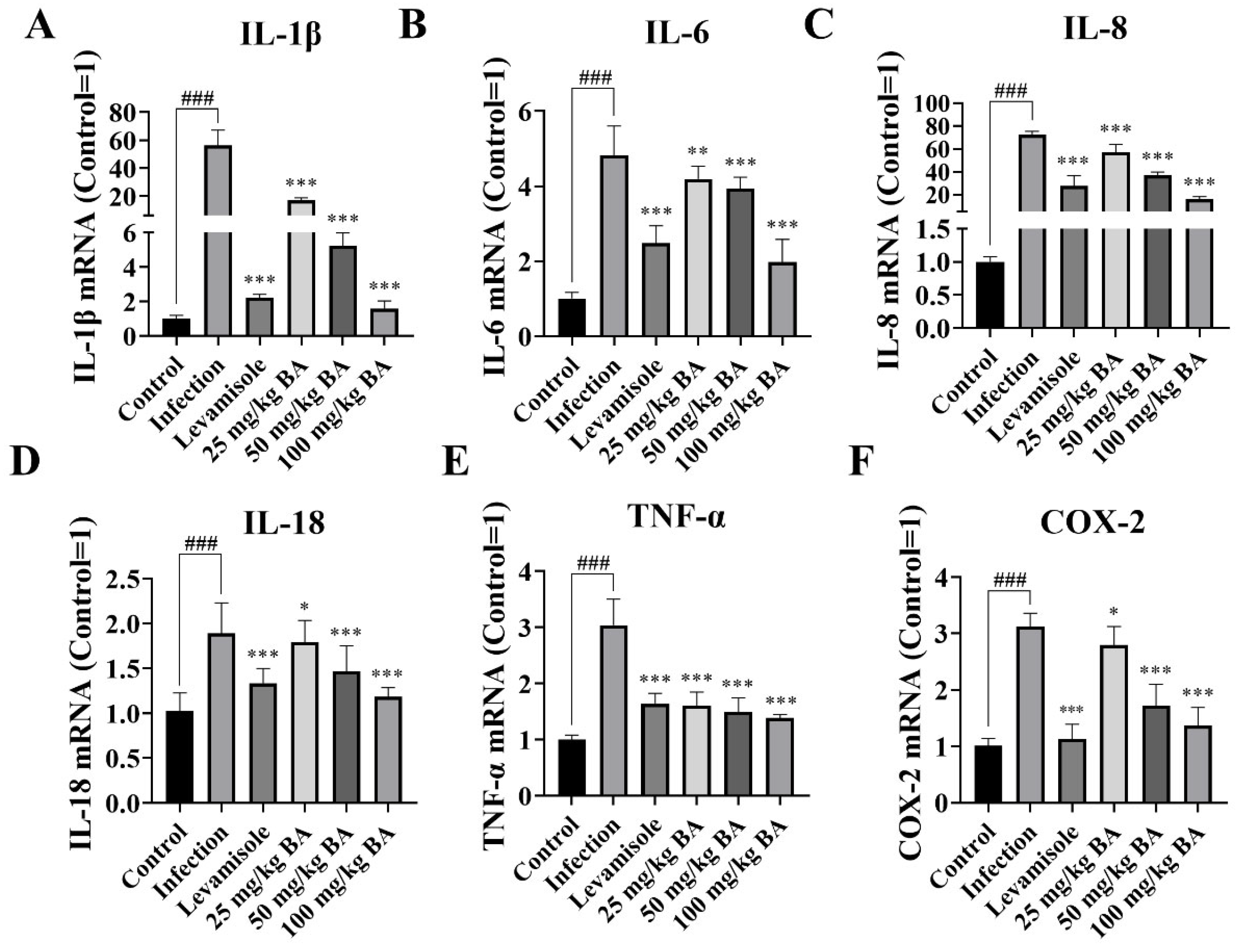
3.1. Baicalin Inhibited IL-1β, IL-6, IL-8, IL-18, TNF-α, and COX-2 Expression in Piglet Spleen Triggered by G. parasuis

Cytokines are multifunctional mediators of cellular communication that are involved in inflammatory responses [40]; thus, we analyzed the effect of baicalin on cytokine production in piglets infected with G. parasuis. The data indicated that IL-1β, IL-6, IL-8, IL-18, TNF-α, and COX-2 mRNA levels in the spleen in the infection group were significantly upregulated (p < 0.001) (Figure 1). Levamisole significantly inhibited IL-1β, IL-6, IL-8, IL-18, TNF-α, and COX-2 mRNA expressions in the spleen compared to the infection group (p < 0.001) (Figure 1). In addition, 25–100 mg/kg baicalin reduced the mRNA expression levels of IL-1β, IL-6, IL-8, IL-18, TNF-α, and COX-2 in the spleen compared to the infection group (p < 0.05) (Figure 1).

3.2. Baicalin Improved MIF/CD74 Axis Levels in Piglet Spleen Elicited by G. parasuis

MIF-CD74 signaling blockade on macrophages reduced the immunosuppressive factors contributing to restoring the antitumor immune response in metastatic melanoma [20]. When the piglets were infected with G. parasuis, the mRNA expression levels of MIF and CD74 in the spleen were significantly reduced (p < 0.001) (Figure 2), while levamisole enhanced the MIF and CD74 mRNA levels in the spleen compared to the infection group (p < 0.01) (Figure 2). Doses of 25–100 mg/kg baicalin also increased the mRNA expression levels of MIF and CD74 in the spleen compared to the infection group (p < 0.05) (Figure 2). The MIF and CD74 protein expression levels in the spleen were determined using the western blotting method. The data indicated that the protein expression levels of MIF and CD74 in the spleen were downregulated in the infection group (p < 0.001), while levamisole and 25–100 mg/kg baicalin could increase CD74 protein level in the spleen compared to the infection group (p < 0.05) (Figure 2). Treatment with 50–100 mg/kg baicalin upregulated MIF protein level in the spleen compared to the infection group (p < 0.001) (Figure 2).
The CD74 protein expression level was also assessed by immunohistochemistry. The data demonstrated that the G. parasuis inhibited the positive protein expression level of CD74 in the spleen compared to the control group, while levamisole and 25–100 mg/kg baicalin promoted the positive protein level of CD74 in the spleen compared to the infection group (p < 0.05) (Figure 2).

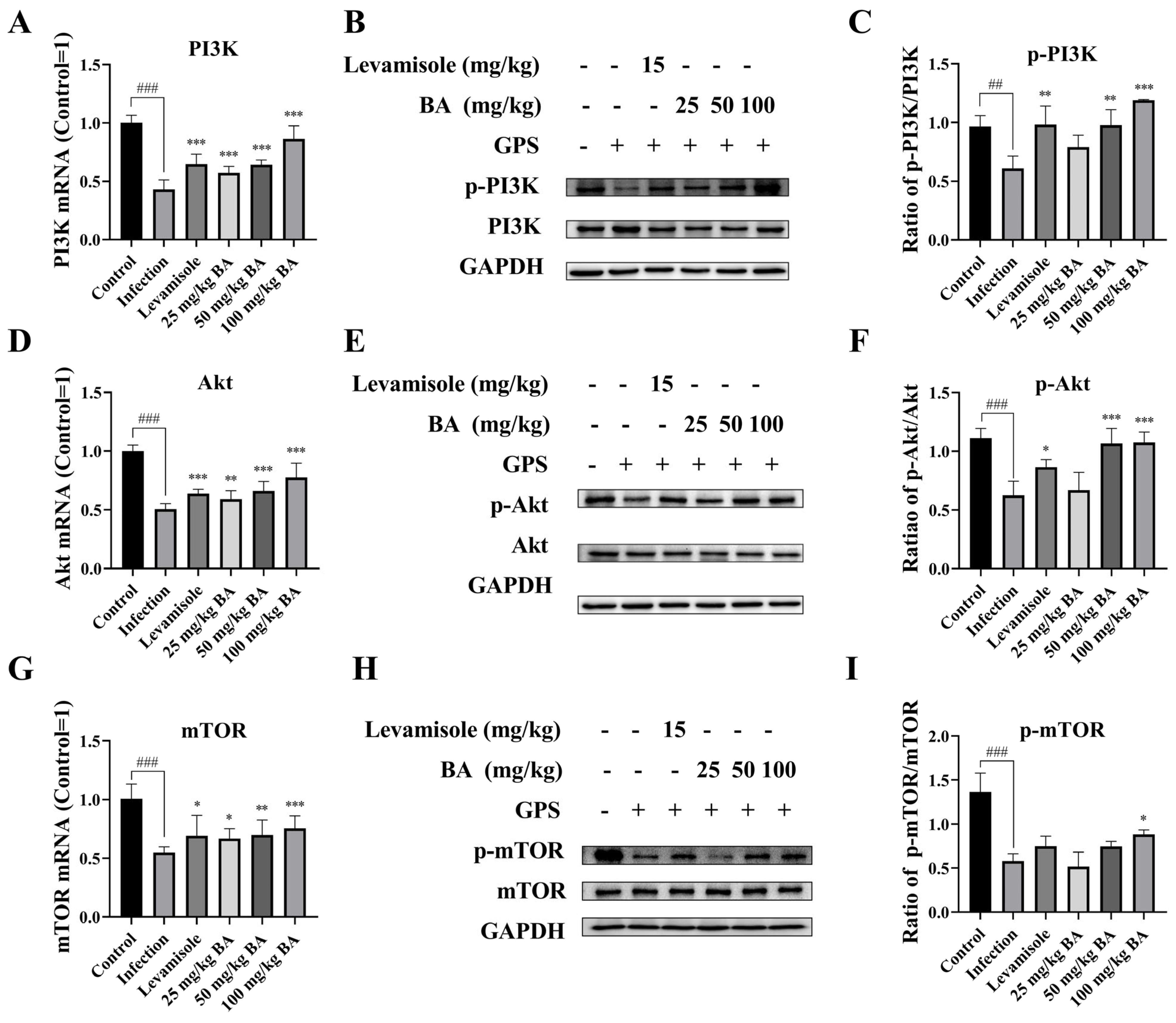
3.3. Baicalin Promoted PI3K/Akt/mTOR Pathway Activation in G. parasuis Triggered Piglet Spleen

Pieces of evidence have shown that a large number of natural products were targeting PI3K/AKT/mTOR-induced autophagy, leading to diminishing tumor growth [41]; thus, we explored the efficacy of baicalin on PI3K/Akt/mTOR signaling activation. The data indicated that the PI3K, Akt, and mTOR mRNA levels were significantly decreased in the spleen when the piglets were challenged by G. parasuis (p < 0.001) (Figure 3). The PI3K, Akt, and mTOR mRNA levels treated with levamisole or baicalin were significantly increased compared to the infection group (p < 0.05) (Figure 3). In addition, p-PI3K, p-Akt, and p-mTOR protein levels were also declined in the infection group compared to the control group (p < 0.01) (Figure 3), while levamisole or 50–100 mg/kg baicalin alleviated the decrease in the protein levels of p-PI3Kand p-Akt (p < 0.05) (Figure 3). Dosages of 100 mg/kg baicalin increased p-mTOR protein levels, in contrast with the infection group (p < 0.05) (Figure 3).

3.4. Baicalin Regulated the RAF/MEK/ERK Signaling Activation in G. parasuis Triggered Piglet Spleen

Previous research reported that MIF activated the CD74-dependent “sustained” activation of RAF/MEK/ERK involved in apoptosis and autophagy [42]. Thus, the RAF, MEK, and ERK levels were evaluated. The data demonstrated that G. parasuis challenge declined the RAF, MEK, and ERK mRNA levels in the spleen (p < 0.001) (Figure 4). Levamisole and 25–100 mg/kg baicalin promoted RAF, MEK, and ERK mRNA levels, in contrast with the infection group (p < 0.01) (Figure 4). The RAF, p-MEK, and p-ERK protein levels were suppressed, in contrast with the control group (p < 0.01) (Figure 4). Levamisole induced MEK and ERK phosphorylation levels in the spleen (p < 0.001) (Figure 4). Furthermore, 50–100 mg/kg baicalin also reversed the effect on RAF and p-ERK protein levels, in contrast with the infection group (p < 0.05) (Figure 4).

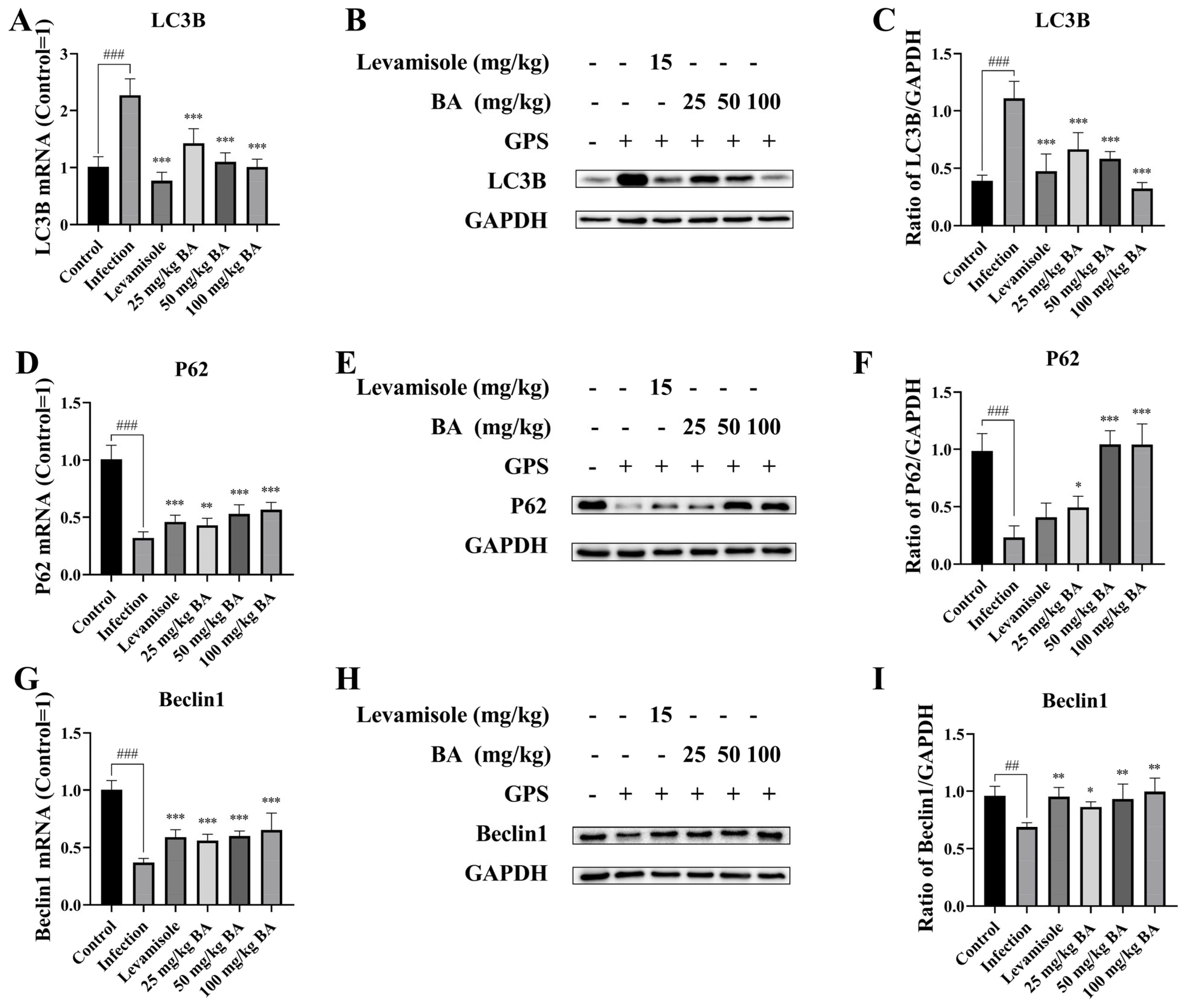
3.5. Baicalin Modified Autophagy-Related Proteins Beclin-1, P62, and LC3B Expression in G. parasuis-Triggered Piglet Spleens

PI3K/Akt/mTOR and RAF/MEK/ERK signaling activation could lead to autophagy [43]. The levels of the autophagy-related proteins Beclin-1, P62, and LC3B in the spleen were determined using the RT-PCR and western blotting methods. The data indicated that at the mRNA and protein level, G. parasuis induced the LC3B expression level and downregulated P62 and Beclin-1 expression levels in the spleen from the infection group, in contrast with the control group (p < 0.001) (Figure 5). Levamisole treatment reduced the LC3B mRNA and protein levels, upregulated Beclin-1 mRNA and protein levels, and increased P62 mRNA expression level in the spleen, in contrast with the infection group (p < 0.01) (Figure 5). Dosages of 25–100 mg/kg baicalin also suppressed the mRNA and protein levels of LC3B and triggered P62, Beclin-1 mRNA, and protein levels in the spleen (p < 0.05) (Figure 5).

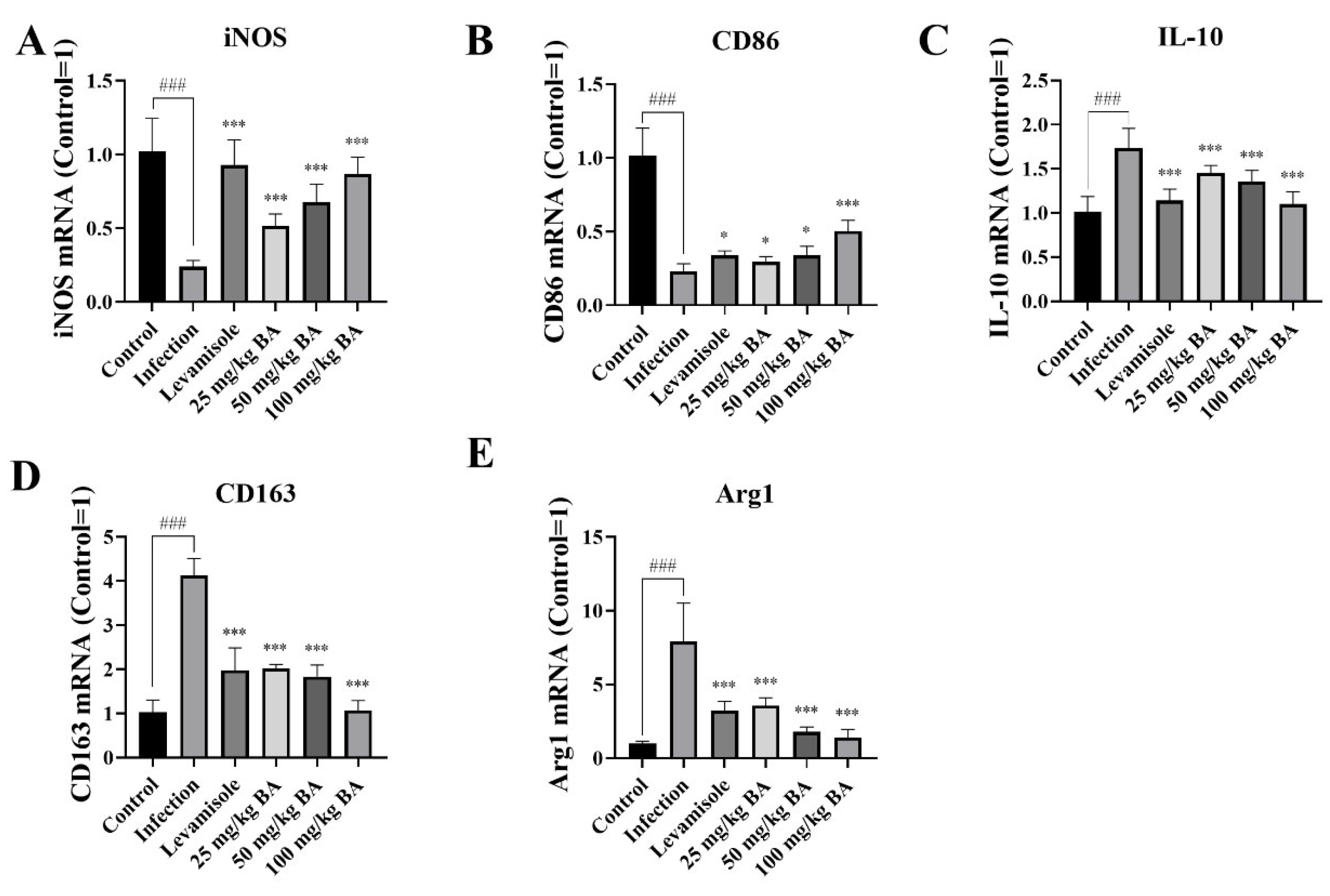
3.6. Baicalin and Levamisole Promoted M2 Polarization to M1 Polarization in G. parasuis-Triggered Piglet Spleens

Targeting MIF-CD74 enhanced microglia M1 polarization in non-small cell lung cancer [44]. To investigate the immunomodulatory effect of the MIF/CD74 axis in the spleen of G. parasuis-infected piglets, the M1 and M2 phenotype markers were detected. The RT-PCR results demonstrated that the G. parasuis remarkably decreased iNOS and CD86 expression (M1 markers) and increased ARG1, IL-10, and CD163 expression (M2 markers) in the spleen (p < 0.05) (Figure 6). Levamisole and 25–100 mg/kg baicalin upregulated the mRNA expression of iNOS and CD86 and attenuated the levels of ARG1, IL-10, and CD163 in the spleen, in contrast with the infection group (p < 0.05) (Figure 6).

3.7. Baicalin Enhanced CD3, CD4, CD8, and TIM3 Levels in G. parasuis-Triggered Piglet Spleens

Previous research reported that tumor-associated M2 macrophages promoted tumor growth and suppressed immune response [45]. The decrease in the number of CD3+, CD4+, and CD8+ T cells is one important reason causing host immunosuppression [46]; thus, we determined the CD3, CD4, CD8, and TIM3 levels in G. parasuis-infected spleen. The results showed that, after G. parasuis challenge, the CD3, CD4, CD8, and TIM3 mRNA levels were decreased, in contrast with the control group (p < 0.001) (Figure 7). When the piglets were pretreated with levamisole or 25–100 mg/kg baicalin, the CD3, CD4, CD8, and TIM3 mRNA levels were increased, in contrast with the infection group (p < 0.05) (Figure 7), suggesting that levamisole and baicalin could modify T cell differentiation.
We also detected CD3 expression using an immunofluorometric assay. The results demonstrated that the mean optical density of CD3 in the infection group was significantly decreased compared to that in the control group (p < 0.001) (Figure 7). Levamisole and 25–100 mg/kg baicalin enhanced the mean optical density of CD3 compared to the control group (p < 0.001) (Figure 7).

4. Discussion

G. parasuis is a Gram-negative bacterium, and it is a common flora in the upper respiratory tract of pigs [47]. Under certain stress conditions, G. parasuis can cause systemic infections characterized by arthritis, meningitis, and polyserositis, known as Glässer’s disease [48]. This disease is considered a major cause of pig deaths, resulting in serious losses to the global pig production [1]. G. parasuis infection can cause piglet immunosuppression; however, the immunosuppression mechanism remains unknown; thus, we used the piglet model to assess the molecular mechanism of immunosuppression caused by G. parasuis.
Our previous studies have shown that the activation of the PD-1/PD-L1 axis triggered by G. parasuis could lead to piglet immunosuppression [9]. In this study, we found that MIF/CD74 axis activation induced by G. parasuis could result in piglet immunosuppression. MIF/CD74 axis, as an immune checkpoint, has been confirmed to promote immunosuppression and evade immune surveillance [49]. MIF-CD74 signaling blockade on macrophages restored the antitumor immune response against metastatic melanoma [20]. Known as a macrophage migration inhibitor, MIF attracted and retained activated immune cells from the periphery to inflammatory tissue [50]. MIF is involved in immune regulation, inflammatory response, and cell proliferation [51]. The biological function of MIF is mainly mediated by its main receptor, CD74 [52]. As a receptor for MIF, CD74 is mainly expressed in macrophages [53]. MIF and the receptor CD74 can be used as a promising target to treat Ewing sarcoma, which is infiltrated by immunosuppressive myeloid populations [54]. CD74 promoted immunosuppressive tumor microenvironment formation in breast cancer in mice; therefore, CD74 might be considered a novel therapeutic target in breast cancer [55]. Therefore, in this study, we investigated the effect of baicalin on MIF/CD74 axis expression in the spleen triggered by G. parasuis. Our results indicated that baicalin regulated MIF/CD74 axis expression in the spleen triggered by G. parasuis, which suggested that MIF and CD74 might serve as new targets to treat G. parasuis infection.
Autophagy is a process by which cells degrade and recycle proteins and organelles to maintain intracellular homeostasis [30]. Autophagy is a key molecular pathway to preserve cellular and organismal homeostasis [56]. Autophagy offers an adaptive function to protect organisms against diverse pathologies, such as infections [57]. LC3B, Beclin-1, and p62 play key functions in the induction of autophagy during the disease state [58]. The autophagy-related protein LC3B accelerates PRMT1 mRNA decay, thereby enhancing autophagy [59]. P62 has been demonstrated to regulate autophagy [60]. This study showed that G. parasuis induced autophagy, and baicalin and levamisole treatment inhibited the autophagy phenomenon. Previous reports showed that MIF promoted cardiac stem cell proliferation and endothelial differentiation via PI3K/Akt/mTOR pathway activation [61]. The PI3K-Akt-mTOR pathway activation promoted necrotic cell death by suppressing autophagy [62]. Scoparone reduced hepatic inflammation and regulated autophagy in nonalcoholic steatohepatitis mice through PI3K/AKT/mTOR pathway regulation in macrophages [63]. Thus, we assessed the effect of baicalin on the PI3K/AKT/mTOR pathway and autophagy activation in the spleen of piglets challenged with G. parasuis. Our data demonstrated that baicalin regulated the PI3K/AKT/mTOR pathway and reduced autophagy in piglets challenged with G. parasuis, which enhances the new function of baicalin in autophagy regulation.
The RAF-MEK-ERK pathway is a characterized MAPK signaling pathway that participates in cell proliferation and survival [64]. Fibroblast GSK-3α promoted fibrosis via the RAF-MEK-ERK pathway in the injured heart [65]. The EIF3H-HAX1 increased RAF-MEK-ERK signaling activity to enhance colorectal cancer progression [66]. In addition, many studies reported that RAF/MEK/ERK signaling is associated with autophagy modification [67]. Geniposide alleviated lipopolysaccharide-induced murine kidney podocyte apoptosis via promoting RAF/MEK/ERK-mediated autophagy [68]. The use of 5-aminolevulinic acid photodynamic therapy diminished HPV viral load through autophagy by modifying RAF/MEK/ERK in HeLa cells [69]. Thus, we inferred that baicalin could modify autophagy through RAF/MEK/ERK pathway regulation, but the specific mechanism needs further research.
Macrophages are usually classified into two categories, which are named M1 macrophages and M2 macrophages [70]. Previous research reported that M1 macrophages and M2 macrophages are associated with inflammation responses [71]. M1 and M2 macrophage polarization imbalance might trigger pathological consequences and lead to diseases [72]. Previous research showed that CD5L promoted M2 macrophage polarization by an autophagy-mediated ID3 increase [73]. Puerarin induced macrophage M2 polarization to display antinon-alcoholic steatohepatitis activity through autophagy activation [74]. Baicalin induces M1 polarization to exert antiviral effects [75]. Thus, we explored the levels of M1 markers (iNOS, CD86) and M2 markers (ARG1, IL-10, CD163). Immunotherapy-activated T cells could recruit and skew late-stage activated M1-like macrophages, critical for cancer immunotherapy [76]. Wnt5a triggered the M2 polarization of tumor-associated macrophages by modifying CaKMII-ERK1/2-STAT3-mediated IL-10 production, leading to the induction of tumor growth and the metastasis of colorectal cancer [77]. The results indicated that M1 was downregulated and M2 was upregulated in the G. parasuis-infected spleen, suggesting that infection with G. parasuis in piglets induces macrophage polarization towards the M2 phenotype, while baicalin could reverse the M2 phenotype to the M1 phenotype.
Previous research reported that nicotinamide phosphoribosyltransferase-driven tumor-associated macrophage M2 polarization results in an immunosuppressive microenvironment in colorectal cancer [78]. Macrophages activated by the histamine receptor H1 could polarize toward the M2-like immunosuppressive phenotype with the dysfunction of T cells [79]. T cells have an important effect in modifying the immune system balance [80]. Sepsis-associated immunosuppression was correlated to a reduction in the number of T cells [81]. CD4+ T cells have important functions in immunosuppression in inflammatory diseases [82]. Metformin improved cancer immunotherapy by remedying tumor-infiltrating CD8 T lymphocytes from the immunosuppression triggered by hypoxia [83]. T-cell exhaustion is a major reason for immunosuppression in the tumor microenvironment [84]. The T cell immunoglobulin domain and mucin domain 3 (TIM3) served as targets for CD8+ T cell exhaustion [7]. Therefore, the expression levels of CD3, CD4, CD8, and TIM3 in G. parasuis-infected piglet spleens were measured, and our results confirmed that infection with G. parasuis in piglets could lead to host immunosuppression, and baicalin could alleviate the immunosuppression in piglets caused by G. parasuis [35].
Taken together, our study showed that G. parasuis infection could promote the MIF/CD74 axis and regulate the PI3K/Akt/mTOR and RAF/MEK/ERK signaling pathways induced autophagy, thereby transforming M1 towards M2 polarization in the spleen, contributing to host immunosuppression, whereas baicalin could reverse this signaling activation in order to reduce host immunosuppression. Our study is the first to report that baicalin could alleviate host immunosuppression by regulating the MIF/CD74 axis-induced polarization of macrophages, which might provide some novel targets to relieve the immunosuppression and inflammation caused by G. parasuis. Our future study will explore the therapeutic potential of baicalin in combination with other treatments.

Supplementary Materials

The following supporting information can be downloaded at: https://www.mdpi.com/article/10.3390/biom15050640/s1. Original Western blot images.

Author Contributions

S.F. conceived and designed the experiments; S.F., Q.D., Y.F., R.L., J.L., Y.S. and S.L. performed the experiments; S.F., Q.D., Y.Q., L.G. and J.H. analyzed the data; S.F. wrote this paper. All authors have read and agreed to the published version of the manuscript.

Funding

This work was supported by the National Natural Science Foundation of China (grant no. 32273067) and Hubei Provincial Natural Science Funding (2023AFB746).

Institutional Review Board Statement

The animal study was approved by the Animal Care and Use Committee of Wuhan Polytechnic University, Hubei Province, China (WPU202308003), approval date 2 August 2023.

Informed Consent Statement

Not applicable.

Data Availability Statement

All relevant data are within the manuscript.

Conflicts of Interest

The authors declare no conflicts of interest.

References

  1. Lin, Y.; Zhu, N.; Liu, J.; Wen, S.; Xu, Y.; Xu, X.; Cai, X. The role of cytolethal distending toxin in Glaesserella parasuis JS0135 strain infection: Cytotoxicity, phagocytic resistance and pathogenicity. Vet. Microbiol. 2024, 295, 110168. [Google Scholar] [CrossRef] [PubMed]
  2. Zhou, Y.; Jiang, D.; Yao, X.; Luo, Y.; Yang, Z.; Ren, M.; Zhang, G.; Yu, Y.; Lu, A.; Wang, Y. Pan-genome wide association study of Glaesserella parasuis highlights genes associated with virulence and biofilm formation. Front. Microbiol. 2023, 14, 1160433. [Google Scholar] [CrossRef]
  3. Wu, C.F.; Hsu, C.Y.; Chou, C.C.; Wang, C.M.; Huang, S.W.; Kuo, H.C. Serotypes, virulence factors and multilocus sequence typing of Glaesserella parasuis from diseased pigs in Taiwan. PeerJ 2023, 11, e15823. [Google Scholar] [CrossRef]
  4. Macedo, N.; Gottschalk, M.; Strutzberg-Minder, K.; Van, C.N.; Zhang, L.; Zou, G.; Zhou, R.; Marostica, T.; Clavijo, M.J.; Tucker, A.; et al. Molecular characterization of Glaesserella parasuis strains isolated from North America, Europe and Asia by serotyping PCR and LS-PCR. Vet. Res. 2021, 52, 68. [Google Scholar] [CrossRef]
  5. Mao, W.; Wang, Z.; Wen, S.; Lin, Y.; Gu, J.; Sun, J.; Wang, H.; Cao, Q.; Xu, Y.; Xu, X.; et al. LRRC8A promotes Glaesserella parasuis cytolethal distending toxin-induced p53-dependent apoptosis in NPTr cells. Virulence 2023, 14, 2287339. [Google Scholar] [CrossRef]
  6. Liu, F.; Gao, Y.; Jiao, J.; Zhang, Y.; Li, J.; Ding, L.; Zhang, L.; Chen, Z.; Song, X.; Yang, G.; et al. Upregulation of TLR4-Dependent ATP Production Is Critical for Glaesserella parasuis LPS-Mediated Inflammation. Cells 2023, 12, 751. [Google Scholar] [CrossRef]
  7. Chen, C.; Zhao, F.; Peng, J.; Zhao, D.; Xu, L.; Li, H.; Ma, S.; Peng, X.; Sheng, X.; Sun, Y.; et al. Soluble Tim-3 serves as a tumor prognostic marker and therapeutic target for CD8+ T cell exhaustion and anti-PD-1 resistance. Cell Rep. Med. 2024, 5, 101686. [Google Scholar] [CrossRef]
  8. Silva, G.F.R.; Moreno, L.Z.; Matajira, C.E.C.; Silva, A.P.S.; Araújo, K.M.; Gomes, V.T.M.; Barbosa, M.R.F.; Sato, M.I.Z.; Moreno, A.M. Serotyping and Antimicrobial Susceptibility Profiling of Glaesserella parasuis Isolated from Diseased Swine in Brazil. Pathogens. 2022, 11, 1443. [Google Scholar] [CrossRef]
  9. Li, J.; Liu, S.; Dong, Q.; Fu, Y.; Sun, Y.; Luo, R.; Tian, X.; Guo, L.; Liu, W.; Qiu, Y.; et al. PD-1/PD-L1 axis induced host immunosuppression via PI3K/Akt/mTOR signalling pathway in piglets infected by Glaesserella parasuis. BMC Vet. Res. 2024, 20, 141. [Google Scholar] [CrossRef]
  10. Tanese, K.; Ogata, D. The role of macrophage migration inhibitory factor family and CD74 in the pathogenesis of melanoma. Exp. Dermatol. 2024, 33, e15122. [Google Scholar] [CrossRef]
  11. Hong, W.C.; Lee, D.E.; Kang, H.W.; Kim, M.J.; Kim, M.; Kim, J.H.; Fang, S.; Kim, H.J.; Park, J.S. CD74 Promotes a Pro-Inflammatory Tumor Microenvironment by Inducing S100A8 and S100A9 Secretion in Pancreatic Cancer. Int. J. Mol. Sci. 2023, 24, 12993. [Google Scholar] [CrossRef] [PubMed]
  12. Bonnin, E.; Rodrigo Riestra, M.; Marziali, F.; Mena Osuna, R.; Denizeau, J.; Maurin, M.; Saez, J.J.; Jouve, M.; Bonté, P.E.; Richer, W.; et al. CD74 supports accumulation and function of regulatory T cells in tumors. Nat. Commun. 2024, 15, 3749. [Google Scholar] [CrossRef] [PubMed]
  13. Rasmussen, G.; Cajander, S.; Bäckman, A.; Källman, J.; Söderquist, B.; Strålin, K. Expression of HLA-DRA and CD74 mRNA in whole blood during the course of complicated and uncomplicated Staphylococcus aureus bacteremia. Microbiol. Immunol. 2017, 61, 442–451. [Google Scholar] [CrossRef] [PubMed]
  14. Li, Q.L.; Tang, J.; Zhao, L.; Ruze, A.; Shan, X.F.; Gao, X.M. The role of CD74 in cardiovascular disease. Front. Cardiovasc. Med. 2022, 9, 1049143. [Google Scholar] [CrossRef]
  15. Merk, M.; Mitchell, R.A.; Endres, S.; Bucala, R. D-dopachrome tautomerase (D-DT or MIF-2): Doubling the MIF cytokine family. Cytokine 2012, 59, 10–17. [Google Scholar] [CrossRef]
  16. Bach, J.P.; Rinn, B.; Meyer, B.; Dodel, R.; Bacher, M. Role of MIF in inflammation and tumorigenesis. Oncology 2008, 75, 127–133. [Google Scholar] [CrossRef]
  17. Yaddanapudi, K.; Rendon, B.E.; Lamont, G.; Kim, E.J.; Al Rayyan, N.; Richie, J.; Albeituni, S.; Waigel, S.; Wise, A.; Mitchell, R.A. MIF Is Necessary for Late-Stage Melanoma Patient MDSC Immune Suppression and Differentiation. Cancer Immunol. Res. 2016, 4, 101–112. [Google Scholar] [CrossRef]
  18. Shu, W.; Wang, Y.; Deji, Z.; Li, C.; Chen, C.; Ding, W.; Du, P.; Wang, X. Infliximab modifies CD74-mediated lymphatic abnormalities and adipose tissue alterations in creeping fat of Crohn’s disease. Inflamm. Res. 2024, 73, 1157–1172. [Google Scholar] [CrossRef]
  19. Trifone, C.; Salido, J.; Ruiz, M.J.; Leng, L.; Quiroga, M.F.; Salomón, H.; Bucala, R.; Ghiglione, Y.; Turk, G. Interaction Between Macrophage Migration Inhibitory Factor and CD74 in Human Immunodeficiency Virus Type I Infected Primary Monocyte-Derived Macrophages Triggers the Production of Proinflammatory Mediators and Enhances Infection of Unactivated CD4+ T Cells. Front. Immunol. 2018, 9, 1494. [Google Scholar] [CrossRef]
  20. Figueiredo, C.R.; Azevedo, R.A.; Mousdell, S.; Resende-Lara, P.T.; Ireland, L.; Santos, A.; Girola, N.; Cunha, R.; Schmid, M.C.; Polonelli, L.; et al. Blockade of MIF-CD74 Signalling on Macrophages and Dendritic Cells Restores the Antitumour Immune Response Against Metastatic Melanoma. Front. Immunol. 2018, 9, 1132. [Google Scholar] [CrossRef]
  21. Tripathy, A.; Sen, P.; Su, B.; Briscoe, W.H. Natural and bioinspired nanostructured bactericidal surfaces. Adv. Colloid Interface Sci. 2017, 248, 85–104. [Google Scholar] [CrossRef] [PubMed]
  22. Abdelnour, S.A.; Ghazanfar, S.; Abdel-Hamid, M.; Abdel-Latif, H.M.R.; Zhang, Z.; Naiel, M.A.E. Therapeutic uses and applications of bovine lactoferrin in aquatic animal medicine: An overview. Vet. Res. Commun. 2023, 47, 1015–1029. [Google Scholar] [CrossRef] [PubMed]
  23. Gholami, M.H.; Rassouli, A.; Mirzaei, S.; Hashemi, F. The potential immunomodulatory effect of levamisole in humans and farm animals. J. Adv. Vet. Anim. Res. 2023, 10, 620–629. [Google Scholar] [CrossRef] [PubMed]
  24. Jin, X.; Liu, M.Y.; Zhang, D.F.; Zhong, X.; Du, K.; Qian, P.; Yao, W.F.; Gao, H.; Wei, M.J. Baicalin mitigates cognitive impairment and protects neurons from microglia-mediated neuroinflammation via suppressing NLRP3 inflammasomes and TLR4/NF-κB signaling pathway. CNS Neurosci. Ther. 2019, 25, 575–590. [Google Scholar] [CrossRef]
  25. Wen, Y.; Wang, Y.; Zhao, C.; Zhao, B.; Wang, J. The Pharmacological Efficacy of Baicalin in Inflammatory Diseases. Int. J. Mol. Sci. 2023, 24, 9317. [Google Scholar] [CrossRef]
  26. Zhu, Z.; Yu, M.; Xu, M.; Ji, X.; Zong, X.; Zhang, Z.; Shang, W.; Zhang, L.; Fang, P. Baicalin suppresses macrophage JNK-mediated adipose tissue inflammation to mitigate insulin resistance in obesity. J. Ethnopharmacol. 2024, 332, 118355. [Google Scholar] [CrossRef]
  27. Zhang, H.J.; Luo, J.Z.; Lan, C.L.; Teng, X.; Ge, B.; Liu, J.Q.; Xie, H.X.; Yang, K.J.; Qin, C.J.; Zhou, X.; et al. Baicalin protects against hepatocyte injury caused by aflatoxin B1 via the TP53-related ferroptosis Pathway. Ecotoxicol. Environ. Saf. 2024, 281, 116661. [Google Scholar] [CrossRef]
  28. Guo, L.; Yue, M.; Ma, C.; Wang, Y.; Hou, J.; Li, H. Baicalin reduces inflammation to inhibit lung cancer via targeting SOCS1/NF-κB/STAT3 axis. Heliyon 2024, 10, e29361. [Google Scholar] [CrossRef]
  29. Chen, X.; Ishfaq, M.; Wang, J. Baicalin ameliorates Mycoplasma gallisepticum-induced inflammatory injury via inhibiting STIM1-regulated ceramide accumulation in DF-1 cells. Poult. Sci. 2023, 102, 102687. [Google Scholar] [CrossRef]
  30. Zhang, Y.; Liu, F.; Jia, Q.; Zheng, L.; Tang, Q.; Sai, L.; Zhang, W.; Du, Z.; Peng, C.; Bo, C.; et al. Baicalin alleviates silica-induced lung inflammation and fibrosis by inhibiting TLR4/NF-?B pathway in rats. Physiol. Res. 2023, 72, 221–233. [Google Scholar] [CrossRef]
  31. Zong, B.; Xiao, Y.; Wang, P.; Liu, W.; Ren, M.; Li, C.; Fu, S.; Zhang, Y.; Qiu, Y. Baicalin Weakens the Virulence of Porcine Extraintestinal Pathogenic Escherichia coli by Inhibiting the LuxS/AI-2 Quorum-Sensing System. Biomolecules 2024, 14, 452. [Google Scholar] [CrossRef] [PubMed]
  32. Lu, Q.; Wang, N.; Wen, D.; Guo, P.; Liu, Y.; Fu, S.; Ye, C.; Wu, Z.; Qiu, Y. Baicalin attenuates lipopolysaccharide-induced intestinal inflammatory injury via suppressing PARP1-mediated NF-κB and NLRP3 signalling pathway. Toxicon 2024, 239, 107612. [Google Scholar] [CrossRef] [PubMed]
  33. Fu, S.; Tian, X.; Peng, C.; Zhang, D.; Zhou, L.; Yuan, Y.; He, J.; Guo, L.; Qiu, Y.; Ye, C.; et al. Baicalin inhibited PANX-1/P2Y6 signaling pathway activation in porcine aortic vascular endothelial cells infected by Glaesserella parasuis. Heliyon 2024, 10, e23632. [Google Scholar] [CrossRef] [PubMed]
  34. Sun, P.; Yang, Y.; Yang, L.; Qian, Y.; Liang, M.; Chen, H.; Zhang, J.; Qiu, Y.; Guo, L.; Fu, S. Quercetin Protects Blood-Brain Barrier Integrity via the PI3K/Akt/Erk Signaling Pathway in a Mouse Model of Meningitis Induced by Glaesserella parasuis. Biomolecules 2024, 14, 696. [Google Scholar] [CrossRef]
  35. Fu, S.; Li, J.; You, J.; Liu, S.; Dong, Q.; Fu, Y.; Luo, R.; Sun, Y.; Tian, X.; Liu, W.; et al. Baicalin attenuates PD-1/PD-L1 axis-induced immunosuppression in piglets challenged with Glaesserella parasuis by inhibiting the PI3K/Akt/mTOR and RAS/MEK/ERK signalling pathways. Vet. Res. 2024, 55, 95. [Google Scholar] [CrossRef]
  36. Liu, Y.; Kim, J.H.; Lim, H.K.; Huang, L.; Choi, W.; Kopalli, S.R.; Lee, S.; Lee, B.H.; Lee, J.H.; Ju, Y.; et al. Millingtonia hortensis L. f. ethanol extract exerts in vivo and in vitro anti-inflammatory activities through inhibition of Syk kinase in NF-κB pathway. J. Ethnopharmacol. 2024, 332, 118386. [Google Scholar] [CrossRef]
  37. Yang, K.; Shan, X.; Songru, Y.; Fu, M.; Zhao, P.; Guo, W.; Xu, M.; Chen, H.; Lu, R.; Zhang, C. Network pharmacology integrated with experimental validation to elucidate the mechanisms of action of the Guizhi-Gancao Decoction in the treatment of phenylephrine-induced cardiac hypertrophy. Pharm. Biol. 2024, 62, 456–471. [Google Scholar] [CrossRef]
  38. Chen, Y.; Zeng, Z.; Chen, J. Role of BACH1 in multiple myeloma. Hematology 2024, 29, 2352687. [Google Scholar] [CrossRef]
  39. Tian, X.; Li, J.; Liu, S.; Dong, Q.; Fu, Y.; Luo, R.; Sun, Y.; Guo, L.; Lu, Q.; Ye, C.; et al. Anemoside B4 attenuates necrotic enteritis of laying hens induced by Clostridium perfringens via inhibiting NF-κB and PI3K/Akt/mTOR signalling pathways. Heliyon 2024, 10, e33161. [Google Scholar] [CrossRef]
  40. Campbell, I.L. Cytokine-mediated inflammation, tumorigenesis, and disease-associated JAK/STAT/SOCS signaling circuits in the CNS. Brain Res. Brain Res. Rev. 2005, 48, 166–177. [Google Scholar] [CrossRef]
  41. Xu, Z.; Han, X.; Ou, D.; Liu, T.; Li, Z.; Jiang, G.; Liu, J.; Zhang, J. Targeting PI3K/AKT/mTOR-mediated autophagy for tumor therapy. Appl. Microbiol. Biotechnol. 2020, 104, 575–587. [Google Scholar] [CrossRef] [PubMed]
  42. Lue, H.; Kapurniotu, A.; Fingerle-Rowson, G.; Roger, T.; Leng, L.; Thiele, M.; Calandra, T.; Bucala, R.; Bernhagen, J. Rapid and transient activation of the ERK MAPK signalling pathway by macrophage migration inhibitory factor (MIF) and dependence on JAB1/CSN5 and Src kinase activity. Cell. Signal. 2006, 18, 688–703. [Google Scholar] [CrossRef] [PubMed]
  43. Yun, Z.S.; Zhihua, S.; Xuelian, T.; Min, X.; Rongjing, H.; Mei, L. Rosmarinic acid activates the Ras/Raf/MEK/ERK signaling pathway to regulate CD8+ T cells and autophagy to clear Chlamydia trachomatis in reproductive tract-infected mice. Mol. Immunol. 2024, 171, 105–114. [Google Scholar] [CrossRef] [PubMed]
  44. Liu, L.; Wang, J.; Wang, Y.; Chen, L.; Peng, L.; Bin, Y.; Ding, P.; Zhang, R.; Tong, F.; Dong, X. Blocking the MIF-CD74 axis augments radiotherapy efficacy for brain metastasis in NSCLC via synergistically promoting microglia M1 polarization. J. Exp. Clin. Cancer Res. CR 2024, 43, 128. [Google Scholar] [CrossRef]
  45. Petrovic, M.; Majchrzak, O.B.; Marecar, R.; Laingoniaina, A.C.; Walker, P.R.; Borchard, G.; Jordan, O.; Tankov, S. Combining antimiR-25 and cGAMP Nanocomplexes Enhances Immune Responses via M2 Macrophage Reprogramming. Int. J. Mol. Sci. 2024, 25, 12787. [Google Scholar] [CrossRef]
  46. Huang, S.; Zhao, Y.; Liao, P.; Wang, J.; Li, Z.; Tan, J.; Zha, X.; Chen, S.; Li, Y.; Zhong, L. Different expression patterns of VISTA concurrent with PD-1, Tim-3, and TIGIT on T cell subsets in peripheral blood and bone marrow from patients with multiple myeloma. Front. Oncol. 2022, 12, 1014904. [Google Scholar] [CrossRef]
  47. Gong, X.; Cui, Q.; Zhang, W.; Shi, Y.; Zhang, P.; Zhang, C.; Hu, G.; Sahin, O.; Wang, L.; Shen, Z.; et al. Genomic insight into the diversity of Glaesserella parasuis isolates from 19 countries. mSphere 2024, 9, e0023124. [Google Scholar] [CrossRef]
  48. Yang, Z.; Zhang, Y.; Zhao, Q.; Du, S.; Huang, X.; Wu, R.; Yan, Q.; Han, X.; Wen, Y.; Cao, S.J. HbpA from Glaesserella parasuis induces an inflammatory response in 3D4/21 cells by activating the MAPK and NF-κB signalling pathways and protects mice against G. parasuis when used as an immunogen. Vet. Res. 2024, 55, 93. [Google Scholar] [CrossRef]
  49. Ghoochani, A.; Schwarz, M.A.; Yakubov, E.; Engelhorn, T.; Doerfler, A.; Buchfelder, M.; Bucala, R.; Savaskan, N.E.; Eyüpoglu, I.Y. MIF-CD74 signaling impedes microglial M1 polarization and facilitates brain tumorigenesis. Oncogene 2016, 35, 6246–6261. [Google Scholar] [CrossRef]
  50. Meza-Romero, R.; Benedek, G.; Leng, L.; Bucala, R.; Vandenbark, A.A. Predicted structure of MIF/CD74 and RTL1000/CD74 complexes. Metab. Brain Dis. 2016, 31, 249–255. [Google Scholar] [CrossRef]
  51. Calandra, T.; Bucala, R. Macrophage Migration Inhibitory Factor (MIF): A Glucocorticoid Counter-Regulator within the Immune System. Crit. Rev. Immunol. 2017, 37, 359–370. [Google Scholar] [CrossRef] [PubMed]
  52. Simons, D.; Grieb, G.; Hristov, M.; Pallua, N.; Weber, C.; Bernhagen, J.; Steffens, G. Hypoxia-induced endothelial secretion of macrophage migration inhibitory factor and role in endothelial progenitor cell recruitment. J. Cell. Mol. Med. 2011, 15, 668–678. [Google Scholar] [CrossRef] [PubMed]
  53. Ezaki, A.; Yano, H.; Pan, C.; Fujiwara, Y.; Anami, T.; Ibe, Y.; Motoshima, T.; Yatsuda, J.; Esumi, S.; Miura, Y.; et al. Potential protumor function of CD74 in clear cell renal cell carcinoma. Hum. Cell 2024, 37, 1535–1543. [Google Scholar] [CrossRef] [PubMed]
  54. He, F.; Xu, J.; Zeng, F.; Wang, B.; Yang, Y.; Xu, J.; Sun, X.; Ren, T.; Tang, X. Integrative analysis of Ewing’s sarcoma reveals that the MIF-CD74 axis is a target for immunotherapy. Cell Commun. Signal. CCS 2025, 23, 23. [Google Scholar] [CrossRef]
  55. Pellegrino, B.; David, K.; Rabani, S.; Lampert, B.; Tran, T.; Doherty, E.; Piecychna, M.; Meza-Romero, R.; Leng, L.; Hershkovitz, D.; et al. CD74 promotes the formation of an immunosuppressive tumor microenvironment in triple-negative breast cancer in mice by inducing the expansion of tolerogenic dendritic cells and regulatory B cells. PLoS Biol. 2024, 22, e3002905. [Google Scholar] [CrossRef]
  56. Klionsky, D.J.; Petroni, G.; Amaravadi, R.K.; Baehrecke, E.H.; Ballabio, A.; Boya, P.; Bravo-San Pedro, J.M.; Cadwell, K.; Cecconi, F.; Choi, A.M.K.; et al. Autophagy in major human diseases. EMBO J. 2021, 40, e108863. [Google Scholar] [CrossRef]
  57. Levine, B.; Kroemer, G. Autophagy in the pathogenesis of disease. Cell 2008, 132, 27–42. [Google Scholar] [CrossRef]
  58. Vishnupriya, S.; Priya Dharshini, L.C.; Sakthivel, K.M.; Rasmi, R.R. Autophagy markers as mediators of lung injury-implication for therapeutic intervention. Life Sci. 2020, 260, 118308. [Google Scholar] [CrossRef]
  59. Hwang, H.J.; Ha, H.; Lee, B.S.; Kim, B.H.; Song, H.K.; Kim, Y.K. LC3B is an RNA-binding protein to trigger rapid mRNA degradation during autophagy. Nat. Commun. 2022, 13, 1436. [Google Scholar] [CrossRef]
  60. Jeong, S.J.; Zhang, X.; Rodriguez-Velez, A.; Evans, T.D.; Razani, B. p62/SQSTM1 and Selective Autophagy in Cardiometabolic Diseases. Antioxid. Redox Signal. 2019, 31, 458–471. [Google Scholar] [CrossRef]
  61. Cui, J.; Zhang, F.; Wang, Y.; Liu, J.; Ming, X.; Hou, J.; Lv, B.; Fang, S.; Yu, B. Macrophage migration inhibitory factor promotes cardiac stem cell proliferation and endothelial differentiation through the activation of the PI3K/Akt/mTOR and AMPK pathways. Int. J. Mol. Med. 2016, 37, 1299–1309. [Google Scholar] [CrossRef] [PubMed]
  62. Wu, Y.T.; Tan, H.L.; Huang, Q.; Ong, C.N.; Shen, H.M. Activation of the PI3K-Akt-mTOR signaling pathway promotes necrotic cell death via suppression of autophagy. Autophagy 2009, 5, 824–834. [Google Scholar] [CrossRef] [PubMed]
  63. Liu, B.; Deng, X.; Jiang, Q.; Li, G.; Zhang, J.; Zhang, N.; Xin, S.; Xu, K. Scoparone improves hepatic inflammation and autophagy in mice with nonalcoholic steatohepatitis by regulating the ROS/P38/Nrf2 axis and PI3K/AKT/mTOR pathway in macrophages. Biomed. Pharmacother. 2020, 125, 109895. [Google Scholar] [CrossRef]
  64. Ullah, R.; Yin, Q.; Snell, A.H.; Wan, L. RAF-MEK-ERK pathway in cancer evolution and treatment. Semin. Cancer Biol. 2022, 85, 123–154. [Google Scholar] [CrossRef]
  65. Umbarkar, P.; Tousif, S.; Singh, A.P.; Anderson, J.C.; Zhang, Q.; Tallquist, M.D.; Woodgett, J.; Lal, H. Fibroblast GSK-3α Promotes Fibrosis via RAF-MEK-ERK Pathway in the Injured Heart. Circ. Res. 2022, 131, 620–636. [Google Scholar] [CrossRef]
  66. Jin, H.; Huang, X.; Pan, Q.; Ma, N.; Xie, X.; Wei, Y.; Yu, F.; Wen, W.; Zhang, B.; Zhang, P.; et al. The EIF3H-HAX1 axis increases RAF-MEK-ERK signaling activity to promote colorectal cancer progression. Nat. Commun. 2024, 15, 2551. [Google Scholar] [CrossRef]
  67. Zhang, L.; Chen, L.; Qi, M.; Yu, F.; Ni, X.; Hong, H.; Xu, H.; Xu, S. Glyphosate induces autophagy in hepatic L8824 cell line through NO-mediated activation of RAS/RAF/MEK/ERK signaling pathway and energy metabolism disorders. Fish Shellfish Immunol. 2023, 137, 108772. [Google Scholar] [CrossRef]
  68. Li, X.; Ma, A.; Liu, K. Geniposide alleviates lipopolysaccharide-caused apoptosis of murine kidney podocytes by activating Ras/Raf/MEK/ERK-mediated cell autophagy. Artif. Cells Nanomed. Biotechnol. 2019, 47, 1524–1532. [Google Scholar] [CrossRef]
  69. Xie, J.; Wang, S.; Li, Z.; Ao, C.; Wang, J.; Wang, L.; Peng, X.; Zeng, K. 5-aminolevulinic acid photodynamic therapy reduces HPV viral load via autophagy and apoptosis by modulating Ras/Raf/MEK/ERK and PI3K/AKT pathways in HeLa cells. J. Photochem. Photobiol. B Biol. 2019, 194, 46–55. [Google Scholar] [CrossRef]
  70. Bardi, G.T.; Smith, M.A.; Hood, J.L. Melanoma exosomes promote mixed M1 and M2 macrophage polarization. Cytokine 2018, 105, 63–72. [Google Scholar] [CrossRef]
  71. Yunna, C.; Mengru, H.; Lei, W.; Weidong, C. Macrophage M1/M2 polarization. Eur. J. Pharmacol. 2020, 877, 173090. [Google Scholar] [CrossRef] [PubMed]
  72. Cutolo, M.; Campitiello, R.; Gotelli, E.; Soldano, S. The Role of M1/M2 Macrophage Polarization in Rheumatoid Arthritis Synovitis. Front. Immunol. 2022, 13, 867260. [Google Scholar] [CrossRef] [PubMed]
  73. Sanjurjo, L.; Aran, G.; Téllez, É.; Amézaga, N.; Armengol, C.; López, D.; Prats, C.; Sarrias, M.R. CD5L Promotes M2 Macrophage Polarization through Autophagy-Mediated Upregulation of ID3. Front. Immunol. 2018, 9, 480. [Google Scholar] [CrossRef]
  74. Fang, X.; Lan, X.; Zhu, M.; He, M.; Sun, M.; Cao, Y.; Zhu, D.; Guo, D.; Luo, H. Puerarin Induces Macrophage M2 Polarization to Exert Antinonalcoholic Steatohepatitis Pharmacological Activity via the Activation of Autophagy. J. Agric. Food Chem. 2024, 72, 7187–7202. [Google Scholar] [CrossRef]
  75. Geng, P.; Zhu, H.; Zhou, W.; Su, C.; Chen, M.; Huang, C.; Xia, C.; Huang, H.; Cao, Y.; Shi, X. Baicalin Inhibits Influenza A Virus Infection via Promotion of M1 Macrophage Polarization. Front. Pharmacol. 2020, 11, 01298. [Google Scholar] [CrossRef]
  76. van Elsas, M.J.; Middelburg, J.; Labrie, C.; Roelands, J.; Schaap, G.; Sluijter, M.; Tonea, R.; Ovcinnikovs, V.; Lloyd, K.; Schuurman, J.; et al. Immunotherapy-activated T cells recruit and skew late-stage activated M1-like macrophages that are critical for therapeutic efficacy. Cancer Cell 2024, 42, 1032–1050.e10. [Google Scholar] [CrossRef]
  77. Liu, Q.; Yang, C.; Wang, S.; Shi, D.; Wei, C.; Song, J.; Lin, X.; Dou, R.; Bai, J.; Xiang, Z.; et al. Wnt5a-induced M2 polarization of tumor-associated macrophages via IL-10 promotes colorectal cancer progression. Cell Commun. Signal. CCS 2020, 18, 51. [Google Scholar] [CrossRef]
  78. Hong, S.M.; Lee, A.Y.; Kim, B.J.; Lee, J.E.; Seon, S.Y.; Ha, Y.J.; Ng, J.T.; Yoon, G.; Lim, S.B.; Morgan, M.J.; et al. NAMPT-Driven M2 Polarization of Tumor-Associated Macrophages Leads to an Immunosuppressive Microenvironment in Colorectal Cancer. Adv. Sci. 2024, 11, e2303177. [Google Scholar] [CrossRef]
  79. Li, H.; Xiao, Y.; Li, Q.; Yao, J.; Yuan, X.; Zhang, Y.; Yin, X.; Saito, Y.; Fan, H.; Li, P.; et al. The allergy mediator histamine confers resistance to immunotherapy in cancer patients via activation of the macrophage histamine receptor H1. Cancer Cell 2022, 40, 36–52.e9. [Google Scholar] [CrossRef]
  80. Yeung, M.Y.; Grimmig, T.; Sayegh, M.H. Costimulation Blockade in Transplantation. Adv. Exp. Med. Biol. 2019, 1189, 267–312. [Google Scholar]
  81. Yuan, Y.; Hua, L.; Zhou, J.; Liu, D.; Ouyang, F.; Chen, X.; Long, S.; Huang, Y.; Liu, X.; Zheng, J.; et al. The effect of artesunate to reverse CLP-induced sepsis immunosuppression mice with secondary infection is tightly related to reducing the apoptosis of T cells via decreasing the inhibiting receptors and activating MAPK/ERK pathway. Int. Immunopharmacol. 2023, 124 Pt A, 110917. [Google Scholar] [CrossRef]
  82. Wang, X.; Zhou, H.; Liu, Q.; Cheng, P.; Zhao, T.; Yang, T.; Zhao, Y.; Sha, W.; Zhao, Y.; Qu, H. Targeting regulatory T cells for cardiovascular diseases. Front. Immunol. 2023, 14, 1126761. [Google Scholar] [CrossRef] [PubMed]
  83. Finisguerra, V.; Dvorakova, T.; Formenti, M.; Van Meerbeeck, P.; Mignion, L.; Gallez, B.; Van den Eynde, B.J. Metformin improves cancer immunotherapy by directly rescuing tumor-infiltrating CD8 T lymphocytes from hypoxia-induced immunosuppression. J. Immunother. Cancer 2023, 11, e005719. [Google Scholar] [CrossRef] [PubMed]
  84. Naoi, Y.; Morinaga, T.; Nagasaki, J.; Ariyasu, R.; Ueda, Y.; Yamashita, K.; Zhou, W.; Kawashima, S.; Kawase, K.; Honobe-Tabuchi, A.; et al. CD106 in Tumor-Specific Exhausted CD8+ T Cells Mediates Immunosuppression by Inhibiting TCR Signaling. Cancer Res. 2024, 84, 2109–2122. [Google Scholar] [CrossRef] [PubMed]
Figure 1. The effect of baicalin on cytokine production in piglet spleen triggered by G. parasuis. The IL-1β (A), IL-6 (B), IL-8 (C), IL-18 (D), TNF-α (E), and COX-2 (F) mRNA levels were determined by RT-PCR. BA: Baicalin; ### p < 0.001 vs. controls; * stands for p < 0.05; ** stands for p < 0.01; *** stands for p < 0.001.
Biomolecules 15 00640 g001
Figure 2. Detection of baicalin on MIF/CD74 axis expression in G. parasuis induced piglet spleen. (A): MIF mRNA expression level using RT-PCR; (B,C): MIF protein expression level using western blot; (D): CD74 mRNA level by RT-PCR; (E,F): CD74 protein expression level by western blot; (G,H): CD74 protein expression level by immunohistochemistry; BA: Baicalin; ### p < 0.001 vs. controls; * stands for p < 0.05; ** stands for p < 0.01; *** stands for p < 0.001. Original Western blot images are provided in the Supplementary Materials.
Biomolecules 15 00640 g002
Figure 3. Measurement of the function of baicalin in PI3K/Akt/mTOR activation in G. parasuis-infected piglet spleen. (A): PI3K mRNA expression level using RT-PCR; (B,C): p-PI3K protein level using western blot; (D): Akt mRNA level using RT-PCR; (E,F): p-Akt protein expression level via western blot; (G): mTOR mRNA expression level via RT-PCR; (H,I): p-mTOR protein expression level via western blot; BA: Baicalin; ## p < 0.01 vs. controls; ### p < 0.001 vs. controls; * stands for p < 0.05; ** stands for p < 0.01; *** stands for p < 0.001. Original Western blot images are provided in the Supplementary Materials.
Biomolecules 15 00640 g003
Figure 4. The efficacy of baicalin on RAF/MEK/ERK activation in G. parasuis-infected piglet spleen. (A): RAF mRNA expression level using RT-PCR; (B,C): RAF protein level using western blot; (D): MEK mRNA level using RT-PCR; (E,F): p-MEK1/2 protein level via western blot; (G): ERK mRNA level via RT-PCR; (H,I): p-ERK1/2 protein expression level via western blot; BA: Baicalin; ## p < 0.01 vs. controls; ### p < 0.001 vs. controls; * stands for p < 0.05; ** stands for p < 0.01; *** stands for p < 0.001. Original Western blot images are provided in the Supplementary Materials.
Biomolecules 15 00640 g004
Figure 5. Assessment of the effect of baicalin on autophagy regulation in G. parasuis-infected piglet spleen. The levels of the autophagy-related proteins Beclin-1, P62, and LC3B were determined using RT-PCR and western blot. (A): LC3B mRNA expression level via RT-PCR; (B,C): LC3B protein expression level via western blot; (D): P62 mRNA expression level via RT-PCR; (E,F): P62 protein expression level via western blot; (G): Beclin-1 mRNA expression level via RT-PCR; (H,I): Beclin-1 protein level via western blot; BA: Baicalin; ## p < 0.01 vs. controls; ### p < 0.001 vs. controls; * stands for p < 0.05; ** stands for p < 0.01; *** stands for p < 0.001. Original Western blot images are provided in the Supplementary Materials.
Biomolecules 15 00640 g005
Figure 6. Determination of the effect of baicalin on polarization change in G. parasuis-infected piglet spleen. The iNOS (A), CD86 (B) (M1 markers), IL-10 (C), CD163 (D), and Arg1 (E) (M2 markers) mRNA levels were determined via RT-PCR. BA: Baicalin; ### p < 0.001 vs. controls; * stands for p < 0.05; *** stands for p < 0.001.
Biomolecules 15 00640 g006
Figure 7. The effect of baicalin on CD3, CD4, CD8, and TIM3 levels in G. parasuis-infected piglet spleen. The CD3 (A), CD4 (B), CD8 (C), and TIM3 (D) levels were determined using RT-PCR. The CD3 protein expression level (E,F) was measured via immunofluorometric assay. BA: Baicalin; ### p < 0.001 vs. controls; * stands for p < 0.05; ** stands for p < 0.01; *** stands for p < 0.001.
Biomolecules 15 00640 g007
Table 1. Primer sequences for qRT-PCR analysis.
Gene Nucleotide Sequence (5′-3′)Tm (°C)Length (bp)
IL-1βForwardTCTGCATGAGCTTTGTGCAAG59.7155
ReverseACAGGGCAGACTCGAATTCAAC60.9
IL-6ForwardCTTCTGGTGATGGCTACTG52.7134
ReverseTTGCCGAGGATGTACTTAA50.0
IL-8ForwardACAGCAGTAACAACAACAAG50.2117
ReverseGACCAGCACAGGAATGAG53.2
IL-10ForwardCGTGGAGGAGGTGAAGAGTG55.4178
ReverseTTAGTAGAGTCGTCATCCTGGAAG55.6
IL-18ForwardAGTAACCATATCTGTGCAGTGT54.0155
ReverseTCTTATCACCATGTCCAGGAAC53.0
TNF-αForwardCGCTCTTCTGCCTACTGCACTTC60.7164
ReverseCTGTCCCTCGGCTTTGACATT57.8
CD74ForwardCTGGGAGTGTACCCGAAGC58.6104
ReverseCGCAGCCAGTTCTCAAAGAG56.6
MIFForwardGTGCGCCCTTTGCAGTCT59.5146
ReverseCCGCGTTCATGTCGTAGTAGTT57.1
COX-2ForwardTGTGAAAGGGAGGAAAGA49.7133
ReverseCTGATGGGTGAAGTGCTG53.5
TIM-3ForwardTCAAGCCTCATCACTTTGG53.7145
ReverseTGACGGAGCAGTAACACTC50.8
PI3KForwardTTGCTACAATCAATCGCCAGGAGAC59.3147
ReverseCTTCCCGTTGTTGCCATCGTTTG59.7
AktForwardGGACGGGCACATCAAGATCACTG60.8126
ReverseTAGTCGTTGTCCTCCAGCACCTC61.2
mTORForwardAGTACCTCCAGGACACCATGAACC60.9108
ReverseCAGACCTCACAGCCACAGAAAGC61.0
GAPDHForwardGGCACAGTCAAGGCGGAGAAC61.9105
ReverseAGCACCAGCATCACCCCATTTG61.0
LC3BForwardAGCCTTCTTCCTGTTAGTG51.3135
ReverseTTCATTCCGAAAGTCTCC48.4
P62ForwardCCCCAATGTGATCTGTGATG53.4126
ReverseTTGCTGTGCTCCTTGTGAAT54.5
Beclin-1ForwardTGAGGATACCCAAGCAAG51.2146
ReverseATGTGGAGAAAGGCAAGA50.4
InosForwardTGGAAGCGGTAACAAAGGA53.7482
ReverseCACGAGGTCAGGAGGGATT56.8
ARG1ForwardGGACCTGTGCTTTGCTGAT55.6122
ReverseTTCCGTTCTTCTTGATTTCTG49.7
RAFForwardCAACACTGATGCTGCTGGTAA55.4113
ReverseCAGATGGCGACTTGGAATG53.8
MEKForwardCGTGAATGAGCCACCTCCC60.7149
ReverseCCACCTCGGACCGTTTGAT60.2
ERKForwardGCTCTTGAAGACGCAGCAC58.6108
ReverseCAGCAGGTTGGAAGGTTTG58.5
CD86ForwardTGTGGGATGGTGTCCTTTG55.0106
ReverseGTTCACTCGCCTTCCTGTT55.2
CD3ForwardGTTTGCTGATGGTGGTGTA52.4144
ReverseTGGGCTCATAGTCTGGATT52.5
CD4ForwardAGCCTCAGTTACCGAGTT52.7138
ReverseATCCTCTTGTCTTCCACTTC51.1
CD8ForwardGTTACATCTCTGGTTACAAGG49.9139
ReverseAAGAAGACGGACATGAAGTT50.7
CD163ForwardTGCTGTAGTCGCTGTTCT55.9117
ReverseACTTTCACCTCCACTCTTC54.0
Disclaimer/Publisher’s Note: The statements, opinions and data contained in all publications are solely those of the individual author(s) and contributor(s) and not of MDPI and/or the editor(s). MDPI and/or the editor(s) disclaim responsibility for any injury to people or property resulting from any ideas, methods, instructions or products referred to in the content.

