Review

Revolutionizing mRNA Vaccines Through Innovative Formulation and Delivery Strategies

1
Department of Microbiology, Gachon University College of Medicine, Incheon 21936, Republic of Korea
2
Lee Gil Ya Cancer and Diabetes Institute, Gachon University, Incheon 21999, Republic of Korea
3
Department of Health Sciences and Technology, GAIHST, Gachon University, Incheon 21999, Republic of Korea
4
Korea mRNA Vaccine Initiative, Gachon University, Seongnam 13120, Republic of Korea
*
Author to whom correspondence should be addressed.
Biomolecules 2025, 15(3), 359; https://doi.org/10.3390/biom15030359
Submission received: 13 January 2025 / Revised: 12 February 2025 / Accepted: 19 February 2025 / Published: 1 March 2025
(This article belongs to the Special Issue Novel Materials for Biomedical Applications: 2nd Edition)

Abstract

:
Modernization of existing methods for the delivery of mRNA is vital in advanced therapeutics. Traditionally, mRNA has faced obstacles of poor stability due to enzymatic degradation. This work examines cutting-edge formulation and emerging techniques for safer delivery of mRNA vaccines. Inspired by the success of lipid nanoparticles (LNP) in delivering mRNA vaccines for COVID-19, a variety of other formulations have been developed to deliver mRNA vaccines for diverse infections. The meritorious features of nanoparticle-based mRNA delivery strategies, including LNP, polymeric, dendrimers, polysaccharide-based, peptide-derived, carbon and metal-based, DNA nanostructures, hybrid, and extracellular vesicles, have been examined. The impact of these delivery platforms on mRNA vaccine delivery efficacy, protection from enzymatic degradation, cellular uptake, controlled release, and immunogenicity has been discussed in detail. Even with significant developments, there are certain limitations to overcome, including toxicity concerns, limited information about immune pathways, the need to maintain a cold chain, and the necessity of optimizing administration methods. Continuous innovation is essential for improving delivery systems for mRNA vaccines. Future research directions have been proposed to address the existing challenges in mRNA delivery and to expand their potential prophylactic and therapeutic application.

1. Introduction

Messenger RNA (mRNA) was discovered in 1961 [1]. In vitro transcription (IVT) of mRNA appeared as a significant milestone to extend the application of mRNA in therapeutics [2]. Advancements in this field were hampered by various challenges, including immunogenicity, instability, and expensive mRNA processing. The development of innovative strategies such as chemical modifications [3], sequence optimization [4,5], and purification of mRNA [6] has significantly impacted vaccine delivery and efficacy. Despite notable advancements in mRNA engineering, obstacles like low cellular uptake, instability, and ineffective delivery prevent it from being used in a wider range of clinical settings. The negative charge of mRNA originates from phosphate moieties, which make it difficult to cross through the anionic cell membrane. Furthermore, due to its intrinsically unstable nature, mRNA is vulnerable to enzymatic breakdown in biological settings, which reduces its therapeutic effect [7]. Precipitation or aggregation of mRNA may reduce its translation, resulting in lower protein production. Moreover, degraded fragments of mRNA may lead to undesired immune response, adversely affecting the efficacy of mRNA vaccines [8]. Because of these limitations, higher dosages of mRNA are needed to achieve the required immune response [8]. The mRNA vaccine induces immune responses through pattern recognition receptors (PRRs), which include Toll-like receptors (TLRs), RIG-I-like receptors (RLRs), and NOD-like receptors (NLRs). These receptors play an important role in activating innate immune responses [9]. The TLRs found in endosomes or cell membranes can detect mRNA; their activation produces type I interferons and pro-inflammatory cytokines [10]. RIG-I detects mRNA in the cytoplasm, and their activation also leads to the production of interferons type I [11]. It is important to recognize that type I interferons play a key role in the immune response induced by mRNA vaccines. Additionally, a cytoplasmic NOD-like receptor activates the RIPK2–NF-κB pathway in response to cellular stress, leading to the secretion of IL-1β and amplifying the inflammatory response [12]. These innate responses promote immune cell recruitment, antigen presentation, and activation of CD8+ T cells, CD4+ T cells, and B cells, which lead to antibody production and long-term immune memory, thereby establishing an adaptive immune response [13,14]. The mRNA needs to be tailored to activate innate immunity sufficiently to trigger adaptive immunity while avoiding over-activation, which may hinder effective translation of mRNA [14]. Hence, comprehension of immune activation of mRNA vaccines is important for designing next-generation mRNA vaccines. Regulating the innate and adaptive immune response is the existing key challenge in mRNA vaccines. Several strategies, such as nucleoside modification, removal of dsRNA contamination, and introducing adjuvants in LNP formulations, are being investigated to address these challenges [15].
Immense efforts are devoted to improving the immunogenicity, stability, and delivery of mRNA vaccines, which could extend their potential for clinical applications [16,17,18,19]. The mRNA vaccine requires a cold chain (−20 °C to −80 °C), which is challenging for resource-constrained settings [20]. Lyophilization as a drying technique has proven to be a compelling approach to storing and transporting mRNA vaccines under nonfreezing conditions without affecting their efficacy [21]. In a recent finding, Alejo et al. demonstrated that lyophilized mRNA-LNPs maintained stability and efficacy for a year in a refrigerated environment. They found that selecting an appropriate buffer solution and optimizing freeze-drying significantly influenced efficacy. Tris buffer has shown better transfection compared to PBS, which was assigned due to the lower leakage of RNA and its minimal influence on particle size [22]. The stability of mRNA vaccines has been mainly improved by developing carrier technologies. Carriers for mRNA delivery are usually composed of nanoparticles, which shield mRNA from degradation and ensure its stable delivery. Interest in nanocarrier-assisted mRNA delivery was sparked by the successful execution of COVID-19 mRNA vaccines (Moderna’s mRNA-1273 and Pfizer/BioNTech’s BNT162b2) [23,24,25,26]. Inspired by LNP, several other nanocarriers have been developed, aiming to prevent the degradation of mRNA and facilitate its smooth delivery [27,28,29,30,31,32]. Moreover, the success of COVID-19 mRNA vaccines has opened new possibilities for adopting this approach to combat other viral infections. It has brought a revolutionary improvement in advanced therapeutics.
This review focuses on recent developments made to deliver mRNA vaccines using various kinds of nanocarriers including LNP, polymeric nanoparticles, dendrimers nanoparticles, polysaccharide nanoparticles, peptide nanoparticles, carbon nanoparticles, metal nanoparticles, DNA nanostructures, hybrid nanoparticles, liposomes and extracellular vesicles. Attention is focused on the application of mRNA vaccines for various infections beyond COVID-19 to demonstrate the universality of mRNA vaccines, highlighting their emerging role in advanced therapeutics. The influence of carriers on enhancing the efficacy, immunogenicity, stability, and cellular uptake of mRNA vaccines has been discussed. The pros and cons of various nanocarriers are highlighted. Furthermore, we have summarized the existing challenges and proposed future research directions to achieve safe and targeted delivery of mRNA vaccines against emerging infectious diseases. This review will facilitate a deeper understanding of nanoparticle-based delivery of mRNA vaccines for emerging infections.

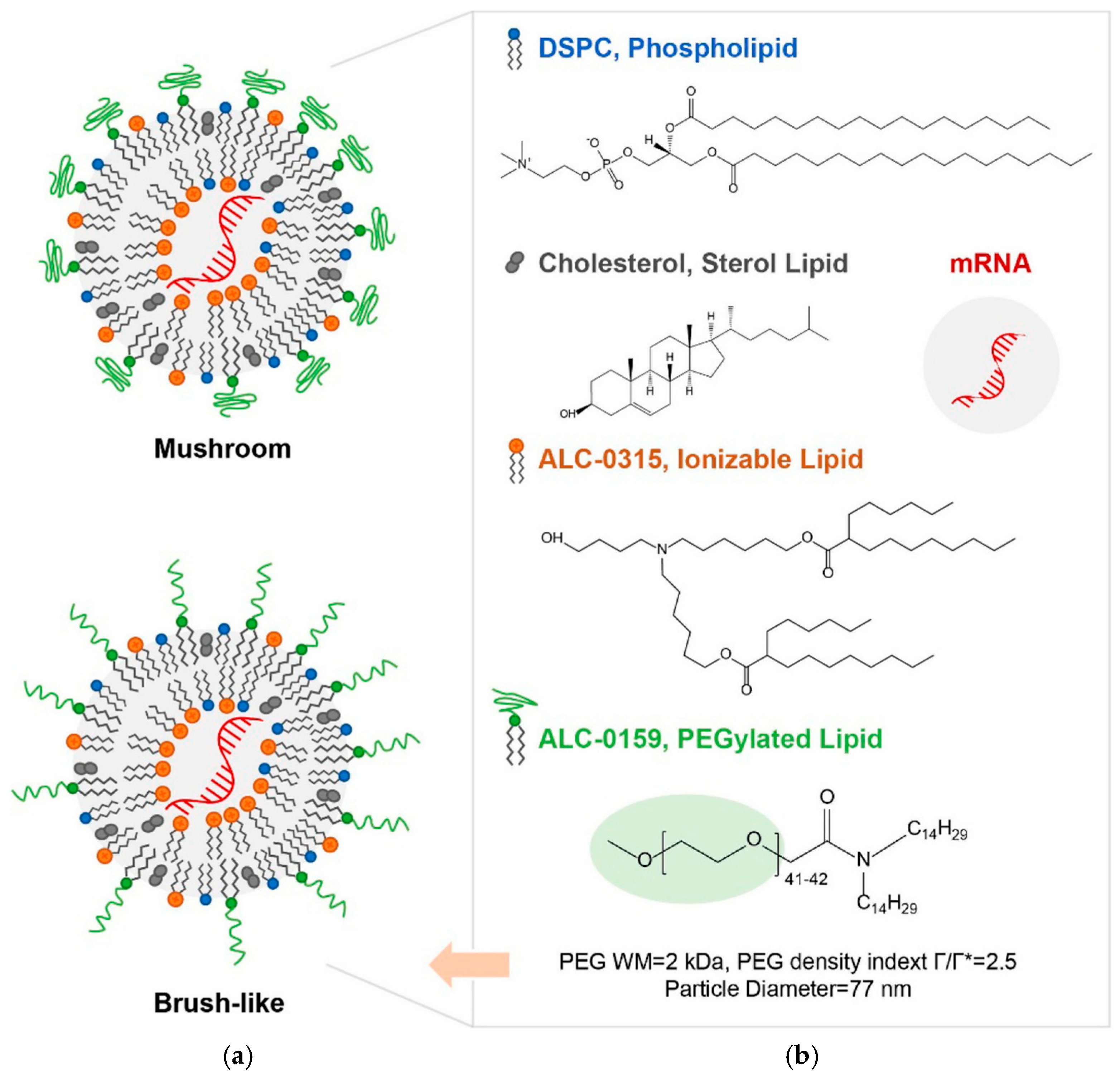
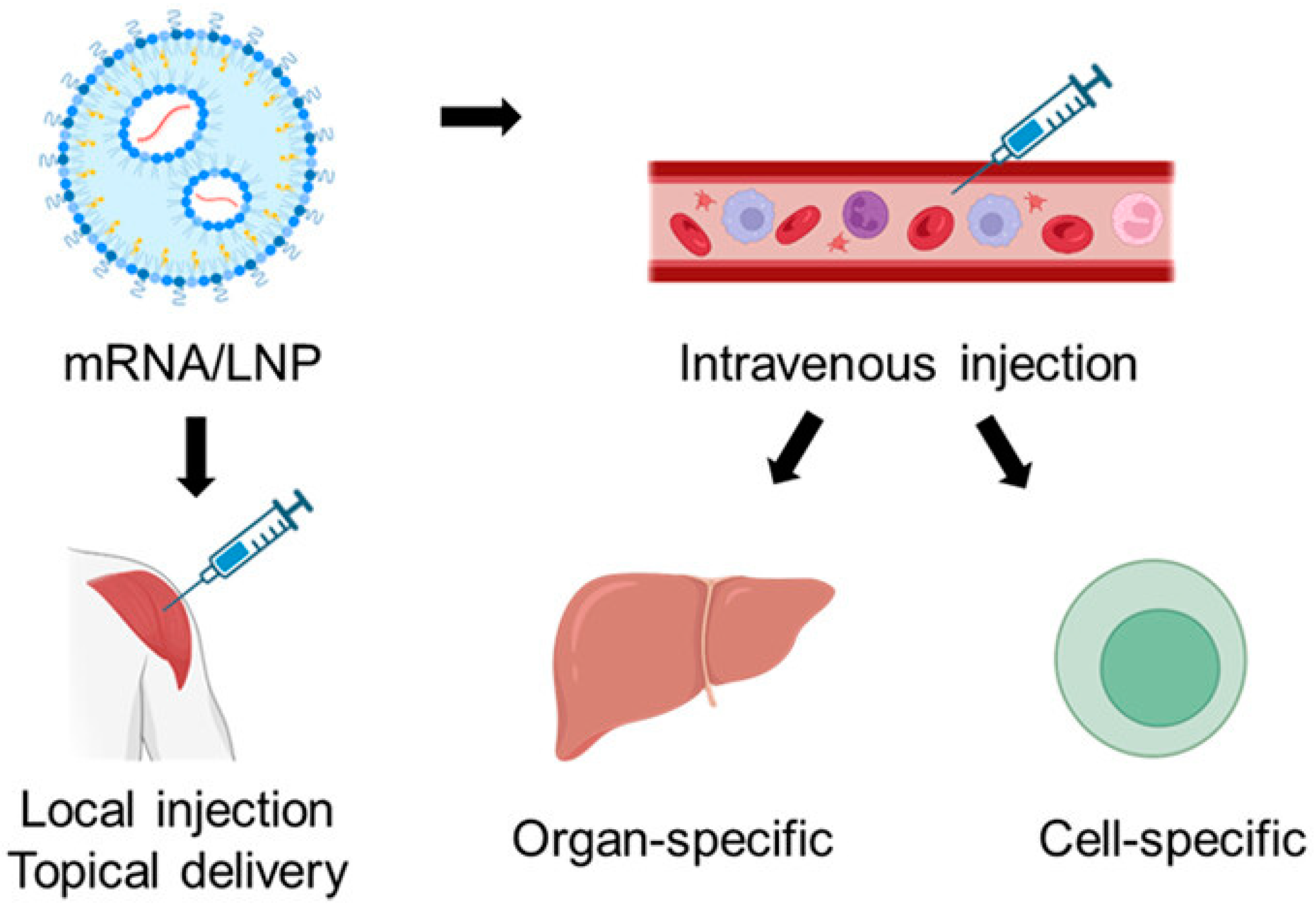
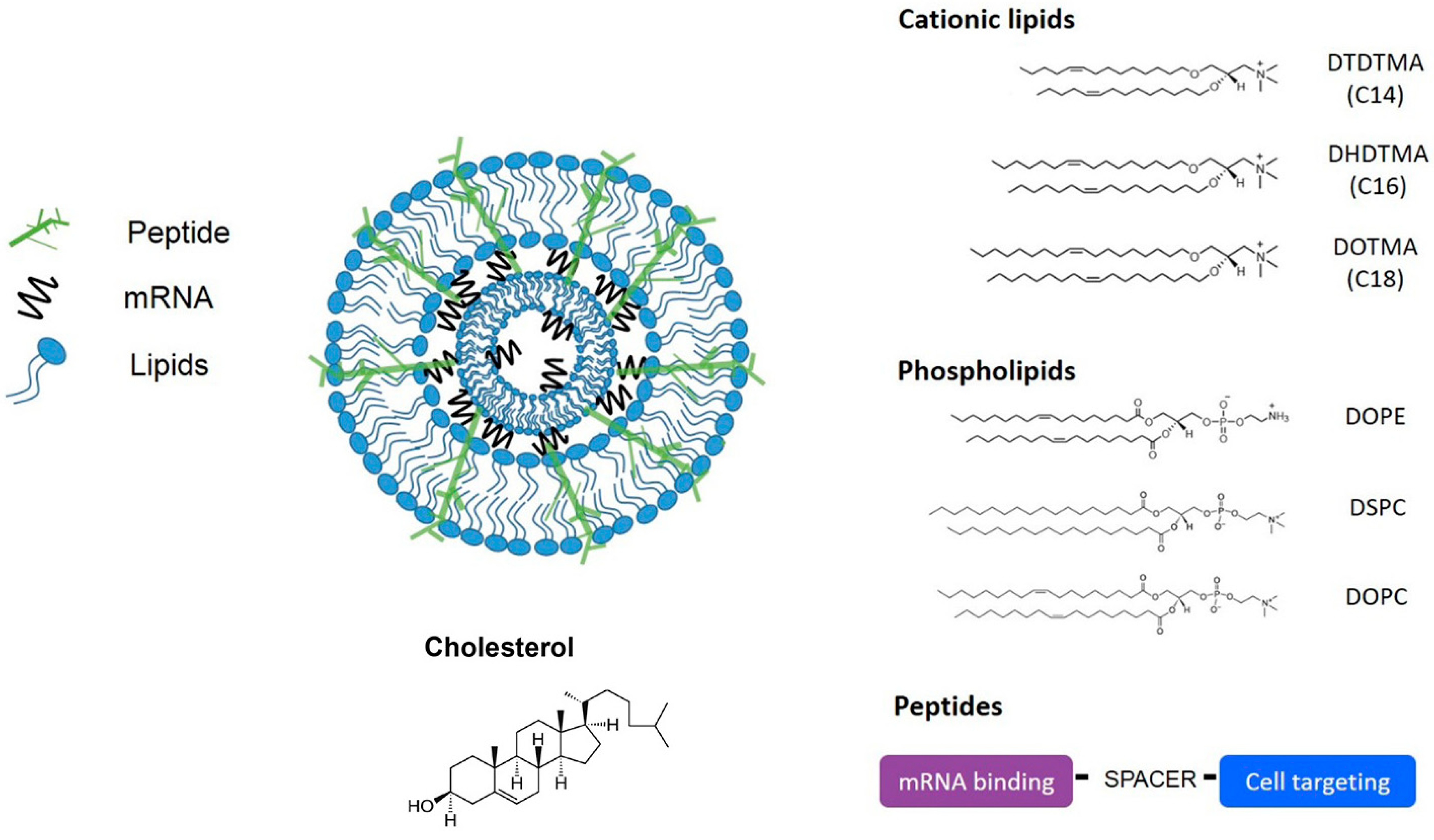
2. Lipid Nanoparticles (LNPs)

LNPs are spherically shaped nanoscale structures. As nanocarriers, they can encapsulate mRNA, protecting it from degradation and releasing it in desired cells or tissues. They have demonstrated smooth delivery of mRNA for infections, cancer, and genetic disorders [33,34,35]. LNPs for clinical applications consist of four components: ionizable cationic lipids, cholesterol, phospholipids, and PEG-lipids [19,36]. The structures of these components are given in Figure 1. LNP promotes cellular uptake of mRNA by endocytosis.
Ionizable LNPs in endosomes can alter their charge, facilitating the release of mRNA, which produces the target protein. This pathway promotes the delivery of mRNA to the targeted cells. Notably, the encapsulation and mRNA delivery capabilities of LNPs are greatly influenced by their size, functionalization, and charge on the surface [19]. Chemical composition can also significantly impact the delivery efficiency and efficacy of mRNA vaccines. Huo et al. have prepared a modified form of ionizable lipid by introducing fluorine in the structure referred to as “F-L319”. The chemical structure of F-L319 is given in Figure 2. Pristine structure without fluorine incorporation was referred to as “L319”. It was observed that the hybrid formulation of F-L319 and L319 with optimized proportions has significantly boosted the delivery of mRNA as compared to individual LNPs [38].
Improvement in the delivery of mRNA by hybrid formulation has been assigned due to improvement in both mRNA loading and cellular intake. This finding highlights the important role of rationalized chemical modification of LNPs in upgrading their activity for mRNA delivery. Han et al. have revealed the integration of LNPs with adjuvant to improve the adjuvanticity of the SARS-CoV-2 mRNA-LNP vaccine [39]. It promoted mRNA delivery and enhanced Toll-like receptor 7/8-agonistic activity. This approach has generated strong T cell and B cell responses, as well as long-lived plasma cell (LLPC) responses (Figure 3).
This study highlights the importance of LNP modification with adjuvant lipidoid to optimize the immune response of mRNA vaccines. This concept has the potential to be extended to other vaccines to optimize immune responses. Hunter et al. highlighted machine learning techniques to achieve enhanced delivery of mRNA vaccine. Advanced cellular and endocytic profiling for intracellular delivery (ACE-ID) is integrated with LNPs for intercellular mRNA delivery [40]. In addition to protecting mRNA vaccines against degradation, LNPs provide the advantage of targeted delivery to specific cells or tissues, avoiding the side effects [41] as shown in Figure 4. Thanks to the diverse advantages of LNPs for the delivery of mRNA vaccines beyond COVID-19, these are being investigated for a diverse range of infections, including viral infections, cancer immunotherapies, metabolic disorders, and genetic disorders [42].

3. Polymeric Nanoparticles

Polymers are macromolecules made by repeating units of monomers. Nanoparticles made from natural and synthetic polymers are an attractive choice for delivering mRNA vaccines. The following classes of polymer nanoparticles have been most applied for mRNA vaccine delivery: esters, dendrimers, polysaccharides, and chitosan. Wilson et al. have highlighted the efficient delivery of self-amplifying mRNA (SAM) by using poly(beta-amino ester)s (PBAEs) as carriers. A significant difference was observed by comparing SAM’s intramuscular (IM) expression with and without polymer formulations. Polymer-assisted formulation has resulted in a 37-fold improvement in SAM expression. This platform has demonstrated superior immunogenicity at lower RNA doses to encode the rabies virus glycoprotein using SAM [43]. The adaptability of polymeric nanoparticles in developing mRNA clinical applications is highlighted by the capacity to tailor their chemical characteristics, which enables extended mRNA stability and biodistribution. Suberi et al. demonstrated inhalable Poly(amine-co-ester) (PACE) polyplexes nanoparticles that effectively deliver mRNA to the lungs. The PACE end was modified with PEG, and it was biodegradable with no harmful toxic effect. This platform was used to prepare mucosal vaccines against SARS-CoV-2, effectively producing cellular and humoral responses in mice [44,45]. It demonstrates the utility of PACE-mRNA-based nontoxic and inhaling complex for lung infection. Patel et al. have exploited PBAEs to design inhalable formulations for mRNA delivery to the lungs. It has consistently produced protein in the lungs without affecting other tissues. No signs of toxicity were observed. It shows the appropriateness of prepared formulations for lung infection [29].
The PBAEs are frequently applied for mRNA delivery. Their cationic characteristics promote electrostatic interaction with mRNA [46]. Meanwhile, a pH-responsive nature facilitates endosomal escape [47]. Other features making them attractive among nanocarriers include their biodegradable nature, non-toxic byproducts, lower cytotoxicity, efficient transfection, and rapid mRNA expression [48,49]. Poly (lactic-co-glycolic acid) (PLGA) is an FDA-approved biodegradable polymer with a long history of drug delivery [50]. However, they have limited interaction with negatively charged mRNA. It makes them less attractive for encapsulating mRNA. Moreover, the transfection efficiency of PLGA could be greatly improved by blending them with cationic polymers such as PEI and PBAE [46,51]. There has been an expansion in the application of polymeric nanoparticle-based mRNA vaccines for inhalable delivery to combat respiratory infections. The nasal administration route has emerged as a useful strategy for addressing threats associated with nasal and respiratory infections [52]. It is a beneficial strategy for addressing lung-related infections, such as asthma, COPD, and cystic fibrosis [53]. Ongun et al. demonstrated localized mRNA delivery to the respiratory tract through the nasal route. This work highlighted the significance of PEG-modified LNP as a nanocarrier in delivering mRNA. Optimized PEG contents were essential to balance colloidal stability and transfection efficiency [54].
Akiva et al. have designed biodegradable polymeric nanocarriers that enable targeted delivery of mRNA to dendritic cells. It has activated antigen-specific CD8+ T cells and shown promising antitumor activity [48]. In addition to PACE polyplexes, polymeric nanoparticles such as chitosan, polylysine, and cationic polymers are investigated. These formulations can facilitate intracellular delivery by forming complexes with mRNA [55,56,57]. Figure 5 shows the structure of polymeric and dendrimer nanoparticles for mRNA delivery. Recent developments in molecularly imprinted polymers (MIP) have shown specificity in interacting with recombinant nucleocapsid protein of SARS-CoV-2 [58]. This finding highlights that polypyrrole (Ppy)-based MIP electrochemical biosensors for detecting the nucleocapsid of SARS-CoV-2 exhibit a high sensitivity of MIPs. The concept of molecular imprinting could be extended to polymeric nanoparticles for mRNA encapsulation and controlled release. It could facilitate the targeted delivery of mRNA to immune cells and enhance the efficacy of the vaccine.

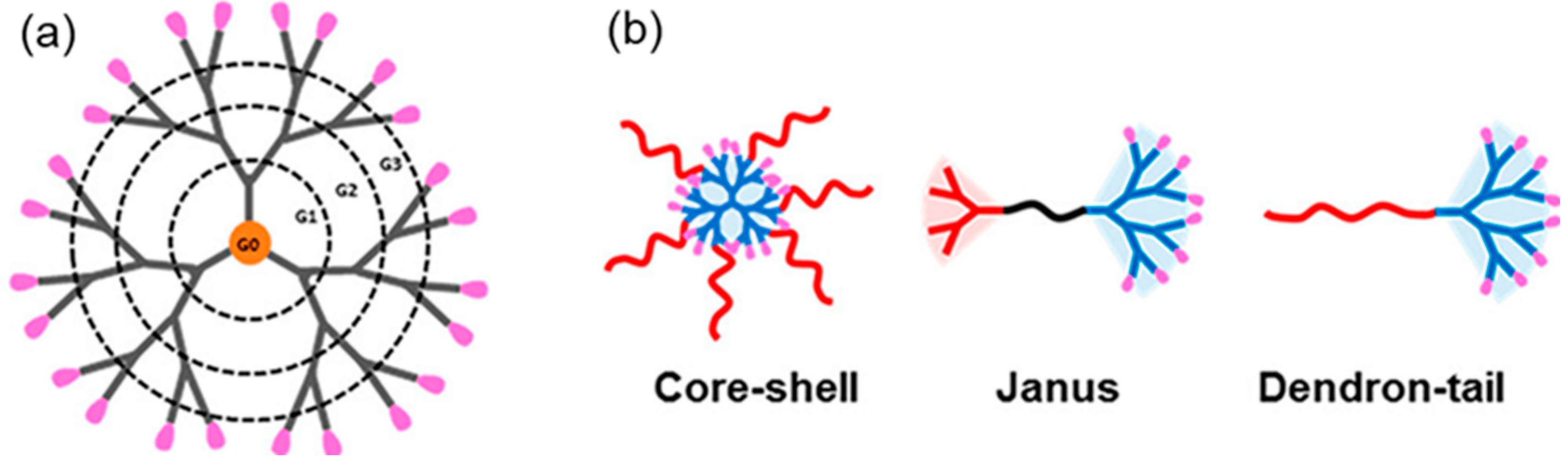
4. Dendrimer Nanoparticles

These offer several advantages in delivering mRNA compared to other nanocarriers. Their distinctive structural characteristics make them a desirable candidate for mRNA delivery. Through efficient encapsulation, they offer enhanced protection to mRNA [60]. Highly branched, three-dimensional structures of dendrimers with tunable surface functionalities enable precise loading and delivery of mRNA. Surface groups at dendrimers facilitate the anchoring of mRNA through the formation of stable complexes. Moreover, observation of biosafety in clinical trials reflects them as a promising candidate to deliver mRNA vaccines [61,62]. Amphiphilic dendrimers are a combination of lipids and dendrimers, and they may provide the benefits of both lipids and polymers. A schematic diagram of dendrimers and amphiphilic dendrimers is shown in Figure 6 [63]. The hydrophilic portion combines RNA, whereas the hydrophobic portion can stabilize RNA. Dendrimers have multiple branching layers, indicated by their generation number. Higher-generation dendrimers have large sizes, higher branches, and more surface charges, leading to increased mRNA encapsulation and improved cellular uptake. In comparison, lower-generation dendrimers have small sizes, fewer branches, reduced surface charges, and limited interaction with mRNA, affecting encapsulation efficiency. However, higher generations may lead to cytotoxicity, necessitating the optimization of the dendrimer branching and charge density for the efficient and safe delivery of the mRNA [61,64]. The toxicity of dendrimers is due to the interaction of cationic groups with cell membranes. The surface modifications of the cationic dendrimers with neutral, anionic, or biodegradable functional groups can reduce their cytotoxicity. Modifying the dendrimers with the biodegradable core, branching units, and functional groups such as polyethers, polyether amines, and polyesters, improves biocompatibility for safety [65,66,67].
Furthermore, the dendrimers are functionalized with specific ligands for targeted delivery of mRNA. For example, dendrimers conjugated with folic acid specifically target folate receptors, which are overexpressed in cancer. Similarly, the attachment of tumor-specific antibodies with dendrimers enables targeted delivery. These cancer-targeting properties are beneficial for improving therapeutic efficacy and reducing off-target effects [68]. Despite these advantages, the complications of manufacturing dendrimers have some challenges that hinder their clinical applications. The synthesis involves multiple steps, leading to batch-to-batch variations and higher costs. Achieving high purity and maintaining mono-dispersity during mass production are essential, as their variation can affect efficacy and safety, specifically for biomedical applications [69].
Commonly adopted dendrimers for mRNA delivery include polyamidoamine (PAMAM), poly-L-lysine, and polypropylenimine [70]. Joubert et al. have demonstrated the chemical modification of PMAM- and lysine-based dendrimers to optimize the loading and delivery of mRNA. They have shown that chemical modifications improved fusogenic characteristics, buffering capacity, or mRNA condensation. Transfection activity depended on chemical composition; amino acids with a moderate basic nature of α-amino were not supportive for transfection [71]. PAMAM with branched configuration and enriched with amino groups facilitates effective anchoring of mRNA [72]. Chahal et al. have demonstrated the application of the modified dendrimers-based platform for mRNA delivery. This synthetic system produced antigens and protected against multiple infections, including Ebola, influenza, and Toxoplasma gondii [73]. Furthermore, Zhang et al. have demonstrated the application of ionizable amphiphilic Janus dendrimers (IAJDs) for the delivery of mRNA to the liver, lungs, and spleen [74]. It has been observed that the hydrophobic behavior of IAJDs has facilitated the targeted delivery of mRNA to desired organs and cells. This work shows the significance of rationalized modification-induced hydrophobicity for tailoring the mRNA delivery capabilities of dendrimers. Dendrimer-based formulations are efficiently absorbed by the numerous cells. They have the potential to be used as delivery formulations for ocular therapeutics, owing to their targeted delivery and enhanced pharmacokinetics [75].

5. Polysaccharide-Based Nanoparticles

Polysaccharide nanoparticles have attracted a lot of interest in delivering mRNA vaccines because of their inherent immunomodulation, biocompatibility, and biodegradable features. Naturally occurring polymers can be modified to generate nanocarriers that can encapsulate mRNA, protecting them from deterioration and promoting cellular uptake [76]. Recent research has shown the capability of polysaccharide-based nanoparticles in delivering mRNA vaccines, where the unique characteristics of these nanoparticles are vital in modulating the immune response [77]. The type of polysaccharide used in nanoparticle formulation, such as chitosan, dextran sulfate, and hyaluronic acid (HA), as well as the inclusion of adjuvant polyinosinic–polycytidylic acid (poly(I:C)), play a role in determining the immune response [78]. The mRNA encapsulation to HA nanocarriers could be challenging due to the anionic nature of HA [79]. Several strategies have been reported to overcome this issue. Djafari et al. demonstrated the HA complexation design with Poly(L-Lysine) as carriers. It has facilitated the encapsulation and delivery of microRNAs, suggesting the potential of this strategy for delivering mRNA [80]. Ryoung et al. have extended the complexation of HA with sulfur-based crosslinkers as suitable nanocarriers to enhance the stability and delivery of mRNA [81]. Myint et al. have demonstrated the fabrication of HA-based nanogels to improve the encapsulation of mRNA. Functional groups of HA can facilitate the formation of nanogels through various strategies such as crosslinking, chemical modification, or polyelectrolyte complexation. Hyaluronic acid has shown excellent colloidal stability and exhibits controlled release [82].
Electrostatic interaction between chitosan and mRNA can prevent the degradation of mRNA. Moreover, its stimuli-responsive nature facilitates the controlled release of encapsulated mRNA [83]. Chitosan, being a biocompatible polymer, offers the advantage of reducing carrier-associated toxicity. For instance, Garcia et al. have demonstrated that adding chitosan-N-arginine (CSA) into DOTAP lipid and DOPE helper lipid has reduced the cytotoxic effects. Besides that, it also improved transfection in HeLa and HEK293T cell lines [84]. Chitosan-based formulations have the potential to be administered through mucosal delivery, which is a needle-free route [85,86,87]. However, certain limitations exist in exploiting chitosan as a nanocarrier to deliver mRNA. These include limited solubility of chitosan in physiological pH, difficulty tailoring uniformity in chitosan-based nanoparticles, and poor mechanical strength, which may cause burst release of mRNA [88]. Other challenges associated with chitosan nanocarriers include scalability, stability, and nanotoxicity. It can be challenging to maintain batch-to-batch uniformity of chitosan nanocarriers [55]. Chemical modifications of chitosan and making a hybrid with crosslinkers can improve the stability and delivery of mRNA [89]. Nonetheless, more investigations are needed to unfold the mechanisms involving polysaccharide-based nanoparticles in mRNA vaccine delivery.

6. Peptide-Derived Nanoparticles

Self-assembled amphipathic peptides can effectively complex and protect mRNA, facilitating cellular uptake and intracellular delivery [90]. One advantage of these nanoparticles is their ability to be customized for specific targeting and enhanced cellular internalization. Peptide-driven mRNA delivery offers unique advantages owing to their diversity [8]. By adjusting the composition of amino acids, diverse functionalities can be incorporated for improved mRNA delivery. These functionalities include enhanced endosomal escape efficiencies, targeted delivery to dendritic cells (DCs), proficient antigen presentation, and targeted lung delivery. For instance, cell-penetrating peptides extended protein expression through stabilizing mRNA intracellularly [91]. The novel cell-penetrating peptides NF424 and NF436 have demonstrated exceptional efficacy in specifically targeting mRNA delivery to spleen tissue [92]. Certain peptide sequences have demonstrated efficacy in promoting binding and uptake by antigen-presenting cells. The arginine-rich peptides have been utilized to formulate mRNA nanocomplexes capable of inducing cytotoxic T cell immune response [93]. Preclinical studies have validated peptide-driven LNPs’ efficacy in delivering mRNA vaccines to the neural retina [94]. Additionally, peptide-based nanocarriers have been investigated to deliver mRNA for tumors and stimulate antigen-specific immune responses against cancer [95]. These findings hold profound significance for the progression of cancer immunotherapy, presenting a promising avenue for activating cytotoxic T cells via mRNA encoding tumor antigens [92]. Figure 7 shows the structure of the lipid–peptide complex. As a biopolymer, albumin has a higher degree of biocompatibility, making it an attractive choice for delivering mRNA. Moreover, the anionic nature of albumin facilitates solubility in water [96]. Several studies have investigated the potential of albumin-based nanoparticles in therapeutics. Cai et al. have exploited albumin as a protein-based nanocarrier for delivering mRNA.
They have modified bovine serum albumin with poly-L-lysine and integrated it with macrophage cell membranes. It has prevented the mRNA from degrading and improved its transfection efficiency [98]. Kida et al. have reported human serum albumin-based nanobubbles as an effective tool to deliver mRNA, bypassing the need for stabilizers. The nanobubble structure was retained during lyophilization [99]. In another study, albumin has been combined with glutaraldehyde as a cross-linker for improving stabilization. However, careful consideration is required to avoid the associated risk of increasing toxicity [100,101]. Endosomal escape is a key limiting factor for peptide-based mRNA delivery systems. Several strategies, such as chemical modification of peptides and the designing of pH-responsive peptides, have been reported to overcome this obstacle.
Zhang et al. have designed poly(glutamic acid) nanoparticles as pH-responsive carriers that can enhance the antigen lysosomal escape. Structural change of α-PGA NPs in acidic conditions promotes membrane fusion and improves cellular immunity [102]. Xu et al. have further investigated PEGylation of peptide nanoparticles for pH-responsive release of mRNA for pulmonary delivery. They observed that the PEG chain of 12 monomers substantially enhanced mRNA delivery compared to the pristine peptide. In contrast, a longer PEG chain has reduced transfection efficiency. It highlights the significance of tailoring chemical modification of peptide nanoparticles for enhancing pH-dependent controlled release of mRNA and its cellular uptake [103]. In a recent finding, Zhang et al. highlighted the significance of introducing peptides to auxiliary lipids for enhancing endosomal escape. They revealed that the synergetic effect of the KHL/DOTAP complex has significantly enhanced endolysosomal escape (about 10-fold) compared to individual components [104].

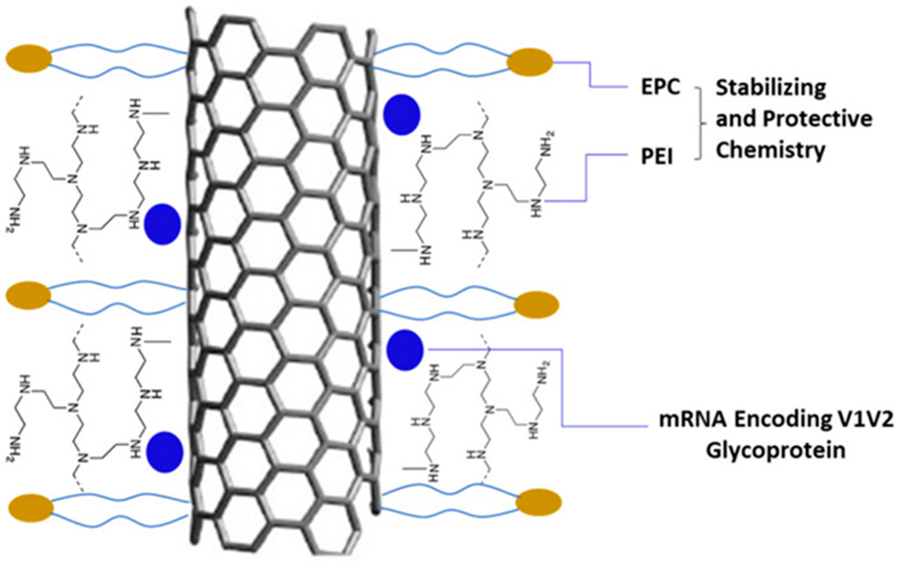
7. Carbon-Based Nanoparticles

Carbon-based nanoparticles have great potential to deliver mRNA vaccines owing to their distinctive characteristics. Carbon nanotubes (CNT) have been studied as a platform (NanoVac) for delivering mRNA encoding the HIV-1 glycoprotein V1V2 region. Surface-modified NanoVac facilitated mRNA loading, cellular entry, and endosomal escape, resulting in efficient transfection. Immunogenicity studies using rabbit and humanized mouse models demonstrated that NanoVac induced robust immune responses for HIV-1 antigen and favorable safety and stability profiles [105]. Furthermore, NanoVac effectively preserved mRNA from degradation and reduced efforts to maintain a cold chain during vaccine deployment [105]. A schematic diagram of CNT for mRNA delivery is given in Figure 8. Recently, by screening different amphiphilic carbon dots, researchers found that O12-Tta-CDs have promoted the delivery of mRNA.
The amphiphilic nature of O12-Tta-CDs, with the hydrophobic alkyl chains and the ionizable carboxylic groups from the thiophene derivative, enabled efficient binding and delivery of mRNA to immune cells. It has actively prevented tumor development by inducing T cell infiltration [106].

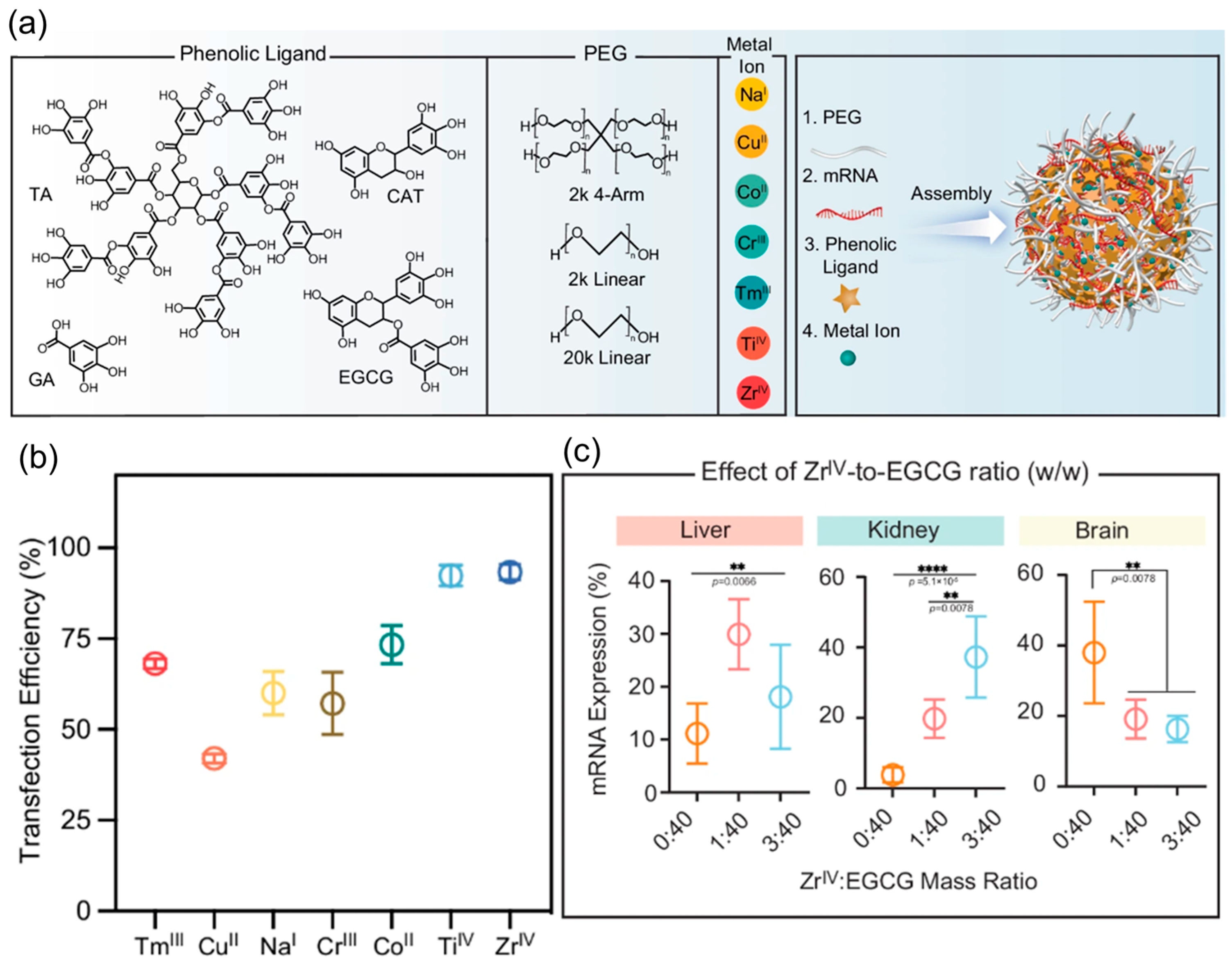
8. Metal-Based Nanoparticles (MNPs)

Recent research highlights the advantages of MNPs in mRNA delivery. For example, dendrimer-coated gold nanoparticles combined with folic acid modification have resulted in better mRNA stability and transgene expression, offering effective protection of mRNA against nucleases and elevated levels of gene expression [107]. As mRNA therapeutics advance, integrating MNP-based delivery systems presents significant potential to address mRNA instability and inefficient cellular uptake challenges, ultimately promoting mRNA vaccine delivery. Gu et al. have investigated the suitability of metal–phenolic networks (MPN) nanoparticles to deliver mRNA for expressing protein in different organs. They have combined poly(ethylene glycol)-polyphenol with a range of metals, as shown in Figure 9. Among these, ZrIV and TiIV have demonstrated higher transfection efficiency. Linear PEG, selected mRNA, epigallocatechin gallate (EGCG), and ZrIV at a mass ratio of 100:1:100:2.5 have shown effective protein expression for the liver, kidney, and brain. The above finding highlights the potential of metal and metal–organic-based nanoparticles for mRNA delivery [108]. Although black phosphorus (BP) exhibits a non-metallic character, BP nanosheets modified with polyethyleneimine have been investigated as a carrier to deliver mRNA. Yang et al. have observed the contribution of BP to facilitate immune activation and antibody production. It highlights the potential of phosphorous-based nanocomposites as vehicles for delivering mRNA [109].

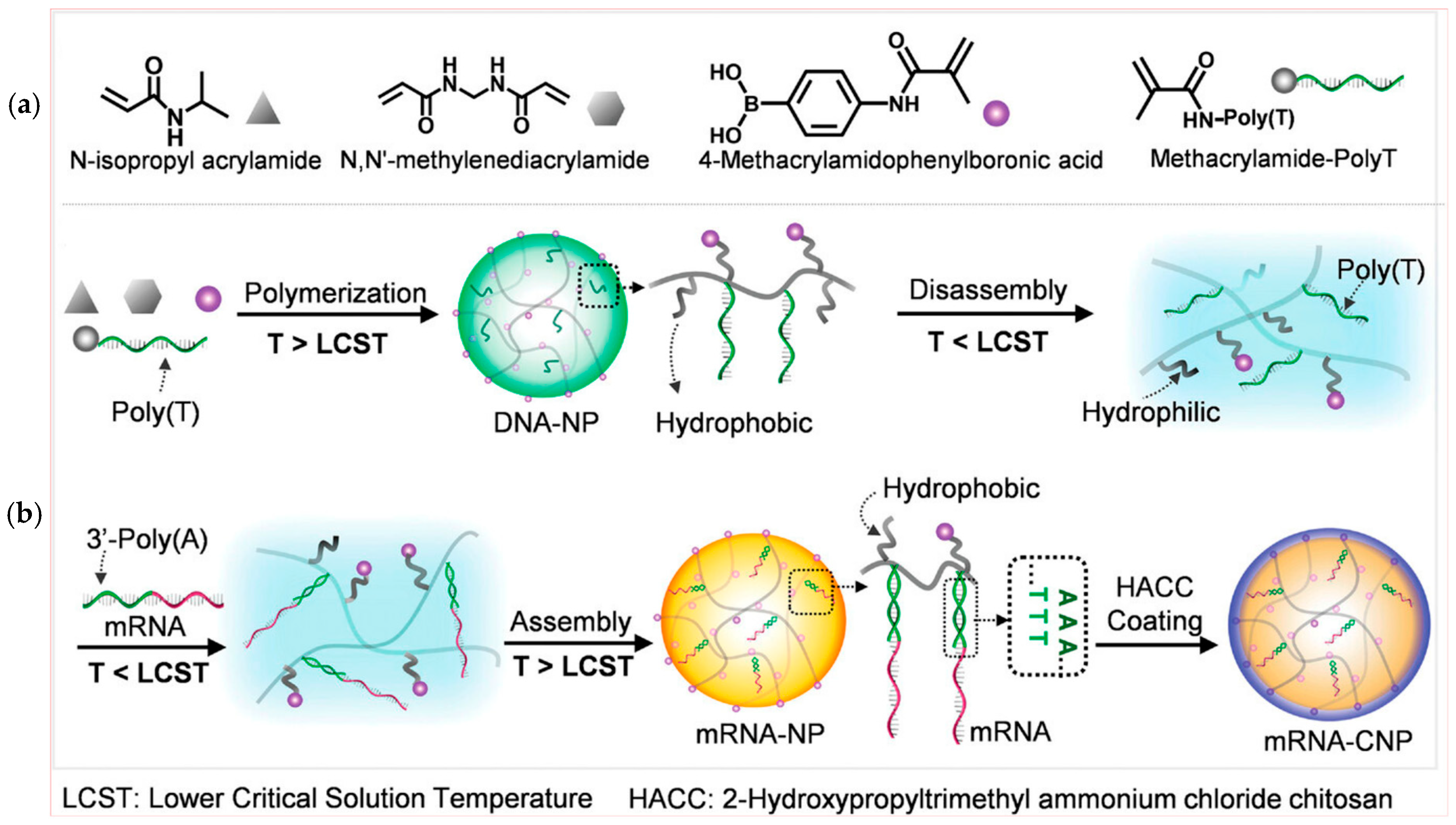
9. DNA Nanostructures

These are crafted from DNA molecules to encapsulate and safeguard mRNA payloads, facilitating their precise delivery to cells. The major benefits of DNA-based carriers are providing precise control of mRNA loading, cellular uptake, and endosomal escape. Furthermore, I-motif and DNA triplet structures are useful for sensing pH, facilitating the controlled release of RNA, specifically in a cellular environment with acidic conditions [110]. Recently, a variety of DNA nanostructures, including DNA tetrahedral nanostructures [111], DNA nano suitcases [112], DNA nano hydrogels [113], and DNA origami [114], have been designed for mRNA delivery. DNA-based hydrogel has effectively released mRNA from cells, which is on par with liposomes but with better biocompatibility [113]. The authors have demonstrated that pH has played a crucial role in delivering mRNA by nano hydrogel. A schematic diagram of nano hydrogel fabrication is presented in Figure 10a. In nano hydrogel X-shaped DNA scaffolds facilitate crosslinking of DNA with mRNA. Under acidic pH conditions (4 to 4.5) in the lysosome, it dissociates and releases mRNA to express the protein. The pH-dependent response of nano hydrogel containing Cy5 is given in Figure 10b, which shows high fluorescence intensity under pH 4.0 to 4.5. It shows the disintegration of nano hydrogel and the release of mRNA. Figure 10c shows quenching and recovery of fluorescence under alkaline and acidic pH. Another study by Li et al. reported the creation of a DNA-based nanomachine that responds to thermal and enzymatic stimuli. The main component of this nanomachine was polythymidine acid (Poly-T) functionalized poly(N-isopropylacrylamide) (DNA-PNIPAM). The core of DNA-PNIPAM was coated with a shell of 2-hydroxypropyltrimethyl ammonium chloride chitosan (HACC) [115]. The HACC shell facilitated cellular uptake and protected mRNA from enzymatic degradation.
It has a lower critical solution temperature (LCST) of about 32 °C in an aqueous environment. It switched to hydrophilic and hydrophobic states below and higher than the LCST, which facilitated the nanomachine to encapsulate mRNA. The molecular design of the nanomachine and the LCST-dependent responsiveness are shown in Figure 11. The charge on the nanomachine was regulated by adjusting the concentration of HACC; without HACC, it showed a negative charge, whereas with 1 and 2 mg/L of HACC, cationic behavior was observed with a zeta potential of +11.72 mV and +25.83 mV, respectively. The average size of the nanomachine observed through TEM analysis was about 154 nm, which is appropriate for cellular uptake. The introduction of a DNA-based nanosystem has addressed the problem of cells that are hard to transfect and have high levels of glutathione (GSH) [116]. To improve mRNA transfection, this approach uses siRNA that promotes ribosome biogenesis. It loads siRNA and mRNA effectively by utilizing sequence-specific molecular recognition and heat responsiveness. Furthermore, intracellular GSH-responsive disassembly promotes siRNA-mediated GSH depletion and optimized mRNA and siRNA release.
The latest finding by Hu et al. has unfolded DNA origami for effective delivery of Smad4 mRNA. Only two circular RNA strands are used as “staples” in this flexible RNA origami structure to bind the mRNA, leaving a large portion in a single-stranded, active form. This adaptable structure permits efficient delivery of mRNA while retaining the ability of ribosomes to recognize and translate it into the cytoplasm. In comparison to other carriers, the researchers showed that this mRNA nanolantern had comparable mRNA delivery efficacy, reduced toxicity, and superior RNase resistance [117]. Rationalized tuning of functional features of DNA nanostructure may improve the stability and delivery of mRNA. The development of these origami-based delivery methods may increase efficacy.

10. Hybrid Nanoparticles

Hybrid nanoparticles provide notable advantages for mRNA delivery by combining the benefits of different materials. Adopting inorganic-organic hybrid nanostructures by incorporating inorganic components like silica or gold nanoparticles into organic matrices like lipids or polymers provides a versatile opportunity [118]. For instance, mesoporous silica-lipid hybrid nanoparticles have been devised for mRNA delivery, showcasing enhanced encapsulation [119]. Lipid–polymer nanoparticles (LPN) as hybrid carriers for mRNA delivery are given in Figure 12 [120]. Zeta potential of LPN with and without loading mRNA with respect to the relative proportion of phospholipid 1,2-dioleoyl-sn-glycero-3-phosphoethanolamine (DOPE) and cationic lipid 1,2-di-O-octadecenyl-3-trimethylammonium propane (DOTMA) are shown in Figure 12a–c. The zeta potential of LPN was observed in the positive range and increased by increasing the DOPE proportion, reaching a maximum at DOPE 70%. It indicated suitability for encapsulating negatively charged mRNA. Increasing DOPE higher than 90% has shown the appearance of a negative charge on LPN, which may not be appropriate for mRNA loading because of lacking electrostatic interactions. This is evident from the trend given in Figure 12d. The LPN with 40% DOPE showed a binding efficiency of around 85.14%, and it decreased drastically to 20.59% as DOPE increased to 100% (Figure 12e).
Hybrid nanoparticles provide mRNA protection and efficient encapsulation via the lipid while imparting stability, controlled release, and potential surface functionalization through the polymer. Li et al. introduced an N1, N3, N5-tris(2-aminoethyl)benzene-1,3,5-tricarboxamide (TT) derived lipid-like nanomaterial (TT3-LLN), which has shown effective mRNA delivery capabilities [121]. Biodegradable and biocompatible polymers are integrated with TT3-LLNs to upgrade delivery. Zhao et al. have introduced hydrophilic polymers, including dextran and HA, and hydrophobic polymers, including poly lactic-co-glycolic acid (PLG) and poly(d,l-lactic acid) (PLA) with TT3 LLN lipid-like nanoparticles [122]. The zeta potential of polymers modified with TT3 LLN was in the positive range except for HA. Furthermore, this finding concluded that hydrophobic polymer modification, especially PLG, has shown better transfection than hydrophilic polymers. Among various formulations, PLGA4-7 LPNs were observed as an optimized formulation for significantly improving the delivery efficiency of mRNA. The size, zeta potential, and encapsulation efficiency of eGFP mRNA-loaded PLGA4-7 LPNs were 287.2 nm, 22.7 mV, and 98.87%, respectively. It demonstrates the potential of hydrophobic modification of TT3-LLNs for efficient delivery of mRNA in human cell lines in advanced biomedical applications.
Recently, Yadava et al. introduced hybrid lipid nanocapsules (hLNCs) composed of kolliphore HS15, labrafac lipophile WL1349, DOPE, a conjugate of oleic acid (OA), and polyethylenimines (PEI) with three different molecular weights (PEI 0.8, 1.8, and 25 kDa). These hLNCs were stable under different pH ranges. In vitro and in vivo analyses have reflected the effective transfection efficiency of mRNA-loaded hLNCs [123]. Zeta potential can significantly affect cellular uptake and transfection capability. The zeta potential of OA-PEI hLNCs was greatly influenced by the molecular weight. A gradual increase in the molecular weight of PEI has resulted in an increased positive charge due to a higher proportion of amine groups. The transfection efficiency trend reflected that lower molecular weight has shown better performance due to their optimized ability to release mRNA. Other hybrid nanoparticles composed of DOTAP–polymer have shown promise for mRNA delivery to treat cancer, infections, and genetic disorders [35].

11. Liposomes and Extracellular Vesicles

In order to improve mRNA stability and immunogenicity for diverse therapeutics, current investigations and endeavors seek to effectively optimize the liposomes and extracellular vesicles (EV) [124]. To overcome the difficulty of inefficient delivery of mRNA, liposomes have become popular as delivery vehicles. Duskunovic et al. have examined the integration of dextran-based nanogel particles, which were modified with oligoarginine peptide (DNPR9), and incorporated into liposomes (LipoDNPR9). These particles improve the effectiveness of mRNA delivery, demonstrating notable enhancements in cellular uptake and transfection rates [125].
The zeta potential of LipoDNPR9 was observed as +34 mV, greater than the +26 mV observed for liposome. Authors have assigned this increase in zeta potential as a potential reason for cellular uptake. Vysochinskaya et al. have investigated the influence of liposome components and lipid-to-mRNA proportion on mRNA transfection by designing cationic liposomes [126]. They found that lipoplexes with an amino-to-phosphate group ratio of 10/1 showed the highest loading and transfection efficiency. The zeta potential of mRNA-eGFP and mRNA-FLuc was observed as >+30 mV, which is crucial for cellular uptake. Pollard et al. reported mRNA encoding HIV-1 antigen Gag, complexed with the cationic lipid DOTAP/DOPE. It induced antigen-specific humoral and cellular immunity [127]. Tane et al. have reported the significance of the modified ethanol injection approach in enhancing mRNA delivery with mRNA lipoplexes. They have evaluated 18 different types of mRNA lipoplex formulations [128]. Authors observed that mRNA lipoplexes LP-DC-1-16/DOPE and LP-DC-1-12/DOPE have shown high protein expression. The zeta potential and size of LP-DC-1-16/DOPE were 28.3 mV and 243.4 nm. Whereas th ezeta potential and size of LP-DC-1-12/DOPE were 35.9 mV and 170 nm. Extracellular vesicles (EVs), like exosomes, showcase promising nanocarriers that can be used to deliver mRNA. They are attractive substitutes for synthetic nanoparticle formulations because of their natural capacity to fuse with target cells and deliver mRNA. The molecular structures of EV are highlighted in Figure 13. Yang et al. have demonstrated the effectiveness of mRNA loading and regulating its release from EVs. The low-density lipoprotein receptor (Ldlr) mRNA was encapsulated into Vs by using MS2 bacteriophage coat protein (CD9-MCP) fusion protein. The average diameter of EVCD9-MCP was observed as 134.1 ± 2.6. Authors have reported enhanced loading and controlled release of mRNA from EVs [129]. Pomatto et al. have applied plant-based Citrus sinensis EVs as carriers to deliver mRNA vaccines for SARS-CoV-2 [130]. The mRNA-loaded EVs have an approximate size of <100 nm, which was retained after lyophilization. Moreover, loaded mRNA has demonstrated elongated stability at ambient conditions for at least 1 year after lyophilization.
Furthermore, Li et al. have highlighted that RNA-binding proteins like L7Ae have been rapidly adsorbed and delivered mRNA antigens when integrated with bacteria-derived outer membrane vesicles (OMVs), resulting in an effective response against tumors [132]. Such developments highlight the potential of EVs as powerful carriers to deliver mRNA and improve its stability.

12. Challenges and Opportunities

Obstacles in existing mRNA delivery include the requirement of the cold chain, targeted delivery of multiple doses, and possible side effects [133]. Confronting these obstacles may enable mRNA vaccines to be more effective and accessible [134]. Selecting a suitable delivery method is essential for the effective administration of mRNA vaccines. The mRNA molecules must cross the lipid bilayer to access the cytoplasm for subsequent translation into functional proteins. Various carriers have been created for mRNA delivery, such as protamine, nanoemulsions, dendrimers, cationic polymers, polysaccharides, and LNPs [135]. Surface modifications of the nanoparticles with ligands can further facilitate the targeted delivery of mRNA [136,137,138]. It may lower the required dosage of mRNA while reducing the side effects to non-targeted cells [139]. However, efforts should be made to optimize mRNA vaccine development by exploring controlled-release and non-invasive administration strategies to improve safety and efficiency [140]. Optimization of nanocarriers for delivery could play a crucial role [141]. Formulation parameters (composition, particulate size, charge, and ligand modification) can significantly impact the delivery of mRNA vaccines. Despite various efforts, the delivery efficiency of LNPs remains suboptimal. A major challenge limiting transfection efficiency is the entrapment of nanoparticles in endosomes following endocytosis. Modifying the surface with a ligand could be beneficial for cellular uptake. Hybrid formulations leverage the benefits of both lipid and polymer structures at once [129,142]. It is evident that the delivery route significantly influences the dissemination of vaccines in organs, expression rate, and vaccine effectiveness.
Recently, noninvasive routes, including oral, nasal, pulmonary, ocular, and transdermal for mRNA delivery, have been explored. These routes provide benefits of self-administration, lower health care costs, minimized risk of blood-borne infections, and have the potential for mass vaccination [143]. While exploring the benefits of oral administration, Abramson et al. have developed robotic pills composed of branched hybrid poly(β-amino ester) mRNA nanoparticles. Oral intake of pills bypassed natural barriers in the gastrointestinal tract and injected mRNA nanoparticles directly into the stomach submucosa [144]. Benetti et al. have highlighted nasal delivery of mRNA. They have adopted chitosan as a carrier to deliver mRNA due to its biocompatible nature and mucoadhesive properties. Preclinical studies showed promising local immune responses without generating systemic antibody responses, revealing its potential for nasopharyngeal immunization [145]. Intramuscular administration is a widely applied approach for COVID-19 vaccines, as it effectively stimulates immune responses. However, an alternate injection route, for instance, intranasal vaccine administration, has the potential to stimulate mucosal immunity. This route is gaining attention as an emerging strategy to deliver mRNA vaccines [146]. Furthermore, the transdermal route is a promising strategy for delivering the vaccines through microneedles. Puigmal et al. have developed Poly(β-amino ester)-based carriers for vaccine delivery. Using this strategy, cell-specific transfection has been observed by integrating Poly(β-amino ester)s with oligopeptide chains. It offers a unique opportunity to exploit peptide-based systems for transdermal delivery of mRNA [147]. Ocular delivery of mRNA is a promising route among non-invasive delivery approaches. Sauvage et al. have exploited the use of polydopamine nanoparticles for delivering mRNA. In this work, irradiation has played an important role in heat generation, facilitating the formation of nanobubbles and intercellular delivery [148].
The nanoparticle-assisted delivery of the mRNA vaccine has made significant improvements in terms of stability and controlled release. However, concerns regarding immunogenicity and over-activation of innate immune responses may cause undesired side effects. Hence, it is important to carefully evaluate the immunogenic impact of nanocarrier-assisted mRNA delivery [149]. Because of compositional complexity, it could be hard to distinguish that the onset of innate immune response is either due to mRNA or LNP [150]. Alameh et al. have highlighted that ionizable lipid nanoparticles are responsible for proinflammatory cytokines [151]. Dimethyldioctadecylammonium bromide (DDAB)-based quaternary ammonium lipids can accelerate innate immune responses. It acts as an immunological adjuvant [35]. Cationic lipid carriers may cause activation of TLR2 and TLR4 proteins and produce proinflammatory cytokines [152]. Hence, there are concerns about applying them as nanocarriers for delivering mRNA. Besides immunogenicity issues linked with lipid nanocarriers, they can also accelerate autoimmune reactions. It is notable that size, charge, administration route, and choice of adjuvants can greatly influence autoimmune reactions [153]. Although the clear mechanism is unknown, clinical findings have indicated autoimmune hepatitis post-COVID-19 vaccination. It has been associated with dominant T cell immunity [154,155,156,157]. Moreover, Chavda et al. have discussed adjuvant-induced autoimmunity and the development of autoimmune diseases by vaccination [157].
The challenge of ultra-cold temperature storage for mRNA vaccines discourages its mass distribution. Current RNA vaccines require a temperature of −20 °C or −80 °C, which hinders global distribution. To address this issue, research is focusing on improving the stability and adaptability of delivery systems. Recently, lyophilized nanostructured lipid carriers (NLCs) have enhanced stability under less stringent storage conditions. It could revolutionize vaccine distribution by enabling stockpiling and rapid deployment in resource-constrained areas [158]. In another finding, Li et al. have applied a mixture of lyoprotectant containing sucrose, trehalose, and mannitol to lyophilize mRNA-LNPs. It has shown stability at 2–8 °C without compromising the immunogenicity [159]. Lamoot et al. have investigated lyophilization of mRNA LNP by introducing 20% w/v of sucrose as cryoprotectants. They observed that mRNA-LNP re-dispersed after lyophilization and retained structural stability. This approach has shown a negligible influence on particle size and zeta potential. In contrast, colloidal stability was damaged without sucrose, affecting transfection efficiency. This study highlights the significance of cryoprotectants during lyophilization [160]. Ai et al. have optimized a lyophilization process with less residual water to achieve the long-term stability of mRNA-LNPs. They have demonstrated that LyomRNA-Omicron enhanced thermostability at 25 °C for up to 6 months [161]. In another study, Wan et al. have shown mannose modification of PEG lipids for circular RNA vaccine delivery. It has sustained stability after lyophilization without negatively affecting immunogenicity and specificity [162]. This finding highlights the significance of mannose modification of nanocarriers in preserving LNPs during the freeze-drying process. Furthermore, innovative approaches such as kinetic modeling have been proposed to evaluate the thermal stability of mRNA vaccines and their deployment to low-resource settings [163].
Clinical translation of an innovative delivery platform faces certain challenges. These include safety and efficacy concerns, complexity in clinical trials, production scalability, and complicated regulatory requirements [164,165]. A fundamental concern in this regard is the risk of the unintended immune response or toxicity induced by nanoparticles. It necessitates extensive investigations to confirm their safety and biocompatibility. Careful design of nanoparticles as delivery tools is essential to avoid mRNA degradation and prevent accumulation in off-targeted sites [154,166]. Clinical trials of nanoparticle-based mRNA delivery face complexity as they need a dual assessment of both mRNA and nanoparticles. Before clinical investigations, comprehensive preclinical evaluations should be thoroughly performed to ensure the safety and efficacy of such formulations [167]. However, due to insufficient information on immunogenicity and toxicity, regulatory approval of nanoparticles for delivery of mRNA could be complicated and time-consuming [168]. Although immense efforts have been made, we are still in the infancy of understanding potential mRNA vaccine delivery systems. In the future, the outcome of the clinical trials would shed light on deeper insight into nanocarrier systems. It can facilitate better administration of various delivery methods of the mRNA vaccine platform [169]. Efforts should focus on increasing the production capacity of carriers and adjuvants. Selecting appropriate routes of administration and elucidating the immune pathways. Improving mRNA delivery systems is key to expanding the potential of mRNA therapeutics.

13. Conclusions

In conclusion, advancements in mRNA vaccine delivery have significantly improved the therapeutic effectiveness. Nanocarriers have made a vital contribution to the safer delivery of mRNA vaccines to targeted cells. It has improved vaccine efficacy while minimizing the off-target side effects. Surface modifications of nanoparticles have been found useful in enhancing cellular uptake of mRNA vaccines. Hyper-branching of dendrimers through large surface areas can promote the loading capability of mRNA. Nanocarriers based on biopolymers such as chitosan, albumin, and DNA are biocompatible in nature and minimize the risk of toxicity. Despite these advancements, several challenges remain to be solved, including optimizing immunogenicity, reducing toxicity, and addressing cold storage requirements. Some nanocarriers may have safety concerns; these may provoke unintended immune responses that lead to inflammation. Ongoing research and development are crucial for overcoming these obstacles and enhancing delivery strategies. Rationalized modification of nanoparticles is indispensable for mRNA delivery to specific cells or tissues. Engineering of stimulus-responsive nanoparticles may ensure the controlled release of mRNA. Furthermore, advancement in biocompatible nanoparticles could be useful to overcome the challenges of undesired immune responses. Enhancing the mRNA payload of nanocarriers could minimize the injectable quantity of nanoparticles. It can significantly reduce the risk of toxicity and side effects associated with nanoparticles. Rigorous preclinical evaluations and clinical trials are needed to ensure the safety of nanoparticle-based mRNA formulations. As the field progresses, improvement in delivery systems holds immense potential to transform mRNA therapeutics.

Author Contributions

K.-J.H. conceptualized and supervised the study; M.F. and T.A. prepared the draft. All authors have read and agreed to the published version of the manuscript.

Funding

This work was financially supported by a grant from the Ministry of Food and Drug Safety (RS-2024-00331833).

Conflicts of Interest

We declare no conflicts of interest concerning the publication.

References

  1. Brenner, S.; Jacob, F.; Meselson, M. An unstable intermediate carrying information from genes to ribosomes for protein synthesis. Nature 1961, 190, 576–581. [Google Scholar] [CrossRef]
  2. Krieg, P.A.; Melton, D.A. Functional messenger RNAs are produced by SP6 in vitro transcription of cloned cDNAs. Nucleic Acids Res. 1984, 12, 7057–7070. [Google Scholar] [CrossRef] [PubMed]
  3. Kariko, K.; Buckstein, M.; Ni, H.; Weissman, D. Suppression of RNA recognition by Toll-like receptors: The impact of nucleoside modification and the evolutionary origin of RNA. Immunity 2005, 23, 165–175. [Google Scholar] [CrossRef]
  4. Lutz, J.; Lazzaro, S.; Habbeddine, M.; Schmidt, K.E.; Baumhof, P.; Mui, B.L.; Tam, Y.K.; Madden, T.D.; Hope, M.J.; Heidenreich, R.; et al. Unmodified mRNA in LNPs constitutes a competitive technology for prophylactic vaccines. NPJ Vaccines 2017, 2, 29. [Google Scholar] [CrossRef] [PubMed]
  5. Thess, A.; Grund, S.; Mui, B.L.; Hope, M.J.; Baumhof, P.; Fotin-Mleczek, M.; Schlake, T. Sequence-engineered mRNA Without Chemical Nucleoside Modifications Enables an Effective Protein Therapy in Large Animals. Mol. Ther. 2015, 23, 1456–1464. [Google Scholar] [CrossRef]
  6. Kariko, K.; Muramatsu, H.; Ludwig, J.; Weissman, D. Generating the optimal mRNA for therapy: HPLC purification eliminates immune activation and improves translation of nucleoside-modified, protein-encoding mRNA. Nucleic Acids Res. 2011, 39, e142. [Google Scholar] [CrossRef] [PubMed]
  7. Cheng, F.; Wang, Y.; Bai, Y.; Liang, Z.; Mao, Q.; Liu, D.; Wu, X.; Xu, M. Research Advances on the Stability of mRNA Vaccines. Viruses 2023, 15, 668. [Google Scholar] [CrossRef]
  8. Wadhwa, A.; Aljabbari, A.; Lokras, A.; Foged, C.; Thakur, A. Opportunities and Challenges in the Delivery of mRNA-based Vaccines. Pharmaceutics 2020, 12, 102. [Google Scholar] [CrossRef]
  9. Wicherska-Pawłowska, K.; Wróbel, T.; Rybka, J. Toll-Like Receptors (TLRs), NOD-Like Receptors (NLRs), and RIG-I-Like Receptors (RLRs) in Innate Immunity. TLRs, NLRs, and RLRs Ligands as Immunotherapeutic Agents for Hematopoietic Diseases. Int. J. Mol. Sci. 2021, 22, 13397. [Google Scholar] [CrossRef]
  10. Kawai, T.; Akira, S. The role of pattern-recognition receptors in innate immunity: Update on Toll-like receptors. Nat. Immunol. 2010, 11, 373–384. [Google Scholar] [CrossRef]
  11. Thoresen, D.; Wang, W.; Galls, D.; Guo, R.; Xu, L.; Pyle, A.M. The molecular mechanism of RIG-I activation and signaling. Immunol. Rev. 2021, 304, 154–168. [Google Scholar] [CrossRef] [PubMed]
  12. Cao, X. Self-regulation and cross-regulation of pattern-recognition receptor signalling in health and disease. Nat. Rev. Immunol. 2016, 16, 35–50. [Google Scholar] [CrossRef]
  13. Iwasaki, A.; Medzhitov, R. Control of adaptive immunity by the innate immune system. Nat. Immunol. 2015, 16, 343–353. [Google Scholar] [CrossRef]
  14. Cao, Q.; Fang, H.; Tian, H. mRNA vaccines contribute to innate and adaptive immunity to enhance immune response in vivo. Biomaterials 2024, 310, 122628. [Google Scholar] [CrossRef] [PubMed]
  15. Matsumura, T.; Takano, T.; Takahashi, Y. Immune responses related to the immunogenicity and reactogenicity of COVID-19 mRNA vaccines. Int. Immunol. 2023, 35, 213–220. [Google Scholar] [CrossRef]
  16. Dowdy, S.F. Overcoming cellular barriers for RNA therapeutics. Nat. Biotechnol. 2017, 35, 222–229. [Google Scholar] [CrossRef]
  17. Sergeeva, O.V.; Koteliansky, V.E.; Zatsepin, T.S. mRNA-Based Therapeutics—Advances and Perspectives. Biochem. (Mosc) 2016, 81, 709–722. [Google Scholar] [CrossRef]
  18. Lin, Y.X.; Wang, Y.; Blake, S.; Yu, M.; Mei, L.; Wang, H.; Shi, J. RNA Nanotechnology-Mediated Cancer Immunotherapy. Theranostics 2020, 10, 281–299. [Google Scholar] [CrossRef] [PubMed]
  19. Reichmuth, A.M.; Oberli, M.A.; Jaklenec, A.; Langer, R.; Blankschtein, D. mRNA vaccine delivery using lipid nanoparticles. Ther. Deliv. 2016, 7, 319–334. [Google Scholar] [CrossRef]
  20. Uddin, M.N.; Roni, M.A. Challenges of Storage and Stability of mRNA-Based COVID-19 Vaccines. Vaccines 2021, 9, 1033. [Google Scholar] [CrossRef]
  21. Muramatsu, H.; Lam, K.; Bajusz, C.; Laczkó, D.; Karikó, K.; Schreiner, P.; Martin, A.; Lutwyche, P.; Heyes, J.; Pardi, N. Lyophilization provides long-term stability for a lipid nanoparticle-formulated, nucleoside-modified mRNA vaccine. Mol. Ther. 2022, 30, 1941–1951. [Google Scholar] [CrossRef] [PubMed]
  22. Teresa, A.; Alfonso, T.-C.; Laura, F.-R.; Andrea, R.; Andrei Mihai, S.; Luna, P.; Victor, J.N.; Juan, M.-O.; Diego de, M. Comprehensive Optimization of a Freeze-Drying Process Achieving Enhanced Long-Term Stability and In Vivo Performance of Lyophilized mRNA-LNPs. Int. J. Mol. Sci. 2024, 25, 10603. [Google Scholar] [CrossRef] [PubMed]
  23. Baden, L.R.; El Sahly, H.M.; Essink, B.; Kotloff, K.; Frey, S.; Novak, R.; Diemert, D.; Spector, S.A.; Rouphael, N.; Creech, C.B.; et al. Efficacy and Safety of the mRNA-1273 SARS-CoV-2 Vaccine. N. Engl. J. Med. 2021, 384, 403–416. [Google Scholar] [CrossRef]
  24. Polack, F.P.; Thomas, S.J.; Kitchin, N.; Absalon, J.; Gurtman, A.; Lockhart, S.; Perez, J.L.; Perez Marc, G.; Moreira, E.D.; Zerbini, C.; et al. Safety and Efficacy of the BNT162b2 mRNA Covid-19 Vaccine. N. Engl. J. Med. 2020, 383, 2603–2615. [Google Scholar] [CrossRef] [PubMed]
  25. Huang, X.; Kon, E.; Han, X.; Zhang, X.; Kong, N.; Mitchell, M.J.; Peer, D.; Tao, W. Nanotechnology-based strategies against SARS-CoV-2 variants. Nat. Nanotechnol. 2022, 17, 1027–1037. [Google Scholar] [CrossRef]
  26. Anderson, E.J.; Rouphael, N.G.; Widge, A.T.; Jackson, L.A.; Roberts, P.C.; Makhene, M.; Chappell, J.D.; Denison, M.R.; Stevens, L.J.; Pruijssers, A.J.; et al. Safety and Immunogenicity of SARS-CoV-2 mRNA-1273 Vaccine in Older Adults. N. Engl. J. Med. 2020, 383, 2427–2438. [Google Scholar] [CrossRef]
  27. Gupta, A.; Andresen, J.L.; Manan, R.S.; Langer, R. Nucleic acid delivery for therapeutic applications. Adv. Drug Deliv. Rev. 2021, 178, 113834. [Google Scholar] [CrossRef]
  28. Granot, Y.; Peer, D. Delivering the right message: Challenges and opportunities in lipid nanoparticles-mediated modified mRNA therapeutics-An innate immune system standpoint. Semin. Immunol. 2017, 34, 68–77. [Google Scholar] [CrossRef]
  29. Patel, A.K.; Kaczmarek, J.C.; Bose, S.; Kauffman, K.J.; Mir, F.; Heartlein, M.W.; DeRosa, F.; Langer, R.; Anderson, D.G. Inhaled Nanoformulated mRNA Polyplexes for Protein Production in Lung Epithelium. Adv. Mater. 2019, 31, e1805116. [Google Scholar] [CrossRef]
  30. Islam, M.A.; Xu, Y.; Tao, W.; Ubellacker, J.M.; Lim, M.; Aum, D.; Lee, G.Y.; Zhou, K.; Zope, H.; Yu, M.; et al. Restoration of tumour-growth suppression in vivo via systemic nanoparticle-mediated delivery of PTEN mRNA. Nat. Biomed. Eng. 2018, 2, 850–864. [Google Scholar] [CrossRef]
  31. Kong, N.; Zhang, R.; Wu, G.; Sui, X.; Wang, J.; Kim, N.Y.; Blake, S.; De, D.; Xie, T.; Cao, Y.; et al. Intravesical delivery of KDM6A-mRNA via mucoadhesive nanoparticles inhibits the metastasis of bladder cancer. Proc. Natl. Acad. Sci. USA 2022, 119, e2112696119. [Google Scholar] [CrossRef] [PubMed]
  32. Kowalski, P.S.; Rudra, A.; Miao, L.; Anderson, D.G. Delivering the Messenger: Advances in Technologies for Therapeutic mRNA Delivery. Mol. Ther. 2019, 27, 710–728. [Google Scholar] [CrossRef]
  33. Hajiaghapour Asr, M.; Dayani, F.; Saedi Segherloo, F.; Kamedi, A.; Neill, A.O.; MacLoughlin, R.; Doroudian, M. Lipid Nanoparticles as Promising Carriers for mRNA Vaccines for Viral Lung Infections. Pharmaceutics 2023, 15, 1127. [Google Scholar] [CrossRef] [PubMed]
  34. Wang, E.Y.; Sarmadi, M.; Ying, B.; Jaklenec, A.; Langer, R. Recent advances in nano- and micro-scale carrier systems for controlled delivery of vaccines. Biomaterials 2023, 303, 122345. [Google Scholar] [CrossRef] [PubMed]
  35. Hou, X.; Zaks, T.; Langer, R.; Dong, Y. Lipid nanoparticles for mRNA delivery. Nat. Rev. Mater. 2021, 6, 1078–1094. [Google Scholar] [CrossRef]
  36. Swetha, K.; Kotla, N.G.; Tunki, L.; Jayaraj, A.; Bhargava, S.K.; Hu, H.; Bonam, S.R.; Kurapati, R. Recent Advances in the Lipid Nanoparticle-Mediated Delivery of mRNA Vaccines. Vaccines 2023, 11, 658. [Google Scholar] [CrossRef]
  37. Wang, M.M.; Wappelhorst, C.N.; Jensen, E.L.; Chi, Y.-C.T.; Rouse, J.C.; Zou, Q. Elucidation of lipid nanoparticle surface structure in mRNA vaccines. Sci. Rep. 2023, 13, 16744. [Google Scholar] [CrossRef]
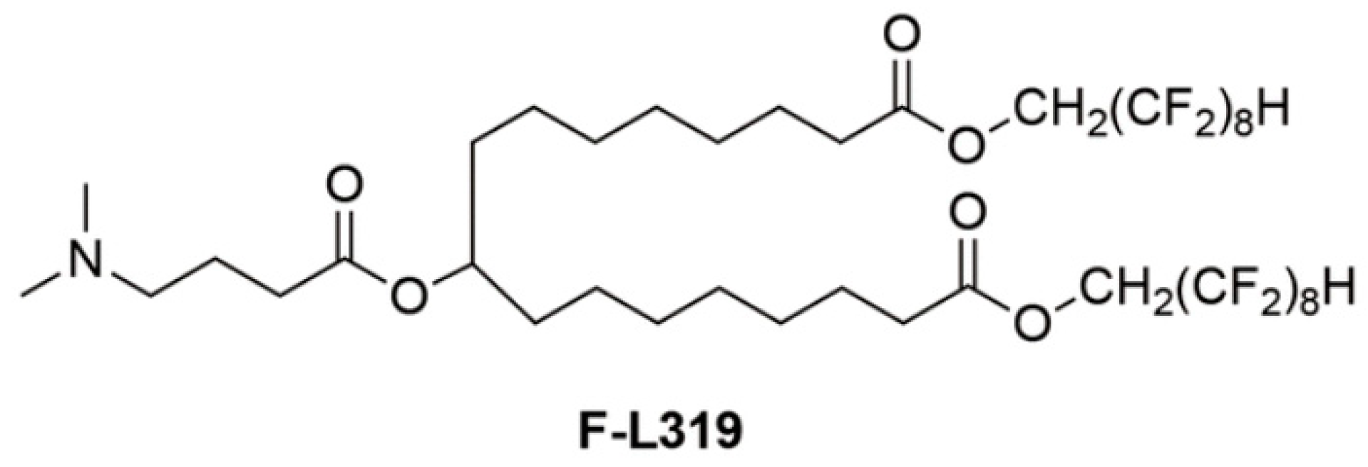
  38. Huo, H.; Cheng, X.; Xu, J.; Lin, J.; Chen, N.; Lu, X. A fluorinated ionizable lipid improves the mRNA delivery efficiency of lipid nanoparticles. J. Mater. Chem. B 2023, 11, 4171–4180. [Google Scholar] [CrossRef]
  39. Han, X.; Alameh, M.G.; Butowska, K.; Knox, J.J.; Lundgreen, K.; Ghattas, M.; Gong, N.; Xue, L.; Xu, Y.; Lavertu, M.; et al. Adjuvant lipidoid-substituted lipid nanoparticles augment the immunogenicity of SARS-CoV-2 mRNA vaccines. Nat. Nanotechnol. 2023, 18, 1105–1114. [Google Scholar] [CrossRef]
  40. Hunter, M.R.; Cui, L.; Porebski, B.T.; Pereira, S.; Sonzini, S.; Odunze, U.; Iyer, P.; Engkvist, O.; Lloyd, R.L.; Peel, S.; et al. Understanding Intracellular Biology to Improve mRNA Delivery by Lipid Nanoparticles. Small Methods 2023, 7, e2201695. [Google Scholar] [CrossRef]
  41. Xu, X.; Xia, T. Recent Advances in Site-Specific Lipid Nanoparticles for mRNA Delivery. ACS Nanosci. Au 2023, 3, 192–203. [Google Scholar] [CrossRef] [PubMed]
  42. Xiao, Y.; Tang, Z.; Huang, X.; Chen, W.; Zhou, J.; Liu, H.; Liu, C.; Kong, N.; Tao, W. Emerging mRNA technologies: Delivery strategies and biomedical applications. Chem. Soc. Rev. 2022, 51, 3828–3845. [Google Scholar] [CrossRef] [PubMed]
  43. Wilson, D.R.; Tzeng, S.Y.; Rui, Y.; Neshat, S.Y.; Conge, M.J.; Luly, K.M.; Wang, E.; Firestone, J.L.; McAuliffe, J.; Maruggi, G.; et al. Biodegradable Polyester Nanoparticle Vaccines Deliver Self-Amplifying mRNA in Mice at Low Doses. Adv Ther (Weinh) 2023, 6, 2200219. [Google Scholar] [CrossRef]
  44. Suberi, A.; Grun, M.K.; Mao, T.; Israelow, B.; Reschke, M.; Grundler, J.; Akhtar, L.; Lee, T.; Shin, K.; Piotrowski-Daspit, A.S.; et al. Inhalable polymer nanoparticles for versatile mRNA delivery and mucosal vaccination. bioRxiv 2022. [Google Scholar] [CrossRef]
  45. Suberi, A.; Grun, M.K.; Mao, T.; Israelow, B.; Reschke, M.; Grundler, J.; Akhtar, L.; Lee, T.; Shin, K.; Piotrowski-Daspit, A.S.; et al. Polymer nanoparticles deliver mRNA to the lung for mucosal vaccination. Sci. Transl. Med. 2023, 15, eabq0603. [Google Scholar] [CrossRef]
  46. Karlsson, J.; Rhodes, K.R.; Green, J.J.; Tzeng, S.Y. Poly(beta-amino ester)s as gene delivery vehicles: Challenges and opportunities. Expert. Opin. Drug Deliv. 2020, 17, 1395–1410. [Google Scholar] [CrossRef] [PubMed]
  47. Rui, Y.; Wilson, D.R.; Tzeng, S.Y.; Yamagata, H.M.; Sudhakar, D.; Conge, M.; Berlinicke, C.A.; Zack, D.J.; Tuesca, A.; Green, J.J. High-throughput and high-content bioassay enables tuning of polyester nanoparticles for cellular uptake, endosomal escape, and systemic in vivo delivery of mRNA. Sci. Adv. 2022, 8, eabk2855. [Google Scholar] [CrossRef]
  48. Ben-Akiva, E.; Karlsson, J.; Hemmati, S.; Yu, H.; Tzeng, S.Y.; Pardoll, D.M.; Green, J.J. Biodegradable lipophilic polymeric mRNA nanoparticles for ligand-free targeting of splenic dendritic cells for cancer vaccination. Proc. Natl. Acad. Sci. USA 2023, 120, e2301606120. [Google Scholar] [CrossRef]
  49. Kavanagh, E.W.; Tzeng, S.Y.; Sharma, N.; Cutting, G.R.; Green, J.J. Ligand-free biodegradable poly(beta-amino ester) nanoparticles for targeted systemic delivery of mRNA to the lungs. Biomaterials 2025, 313, 122753. [Google Scholar] [CrossRef]
  50. Sonawane, S.; Pingale, P.; Amrutkar, S. PLGA: A Wow Smart Biodegradable Polymer in Drug Delivery System. Indian. J. Pharm. Educ. Res. 2023, 57, s189–s197. [Google Scholar] [CrossRef]
  51. Sharifnia, Z.; Bandehpour, M.; Hamishehkar, H.; Mosaffa, N.; Kazemi, B.; Zarghami, N. In-vitro Transcribed mRNA Delivery Using PLGA/PEI Nanoparticles into Human Monocyte-derived Dendritic Cells. Iran. J. Pharm. Res. 2019, 18, 1659–1675. [Google Scholar] [CrossRef] [PubMed]
  52. Ming, Q.; Guangsheng, D.; Xun, S. Recent Advances in the Noninvasive Delivery of mRNA. Acc. Chem. Res. 2021, 10, 493. [Google Scholar] [CrossRef]
  53. Neary, M.T.; Mulder, L.M.; Kowalski, P.S.; MacLoughlin, R.; Crean, A.M.; Ryan, K.B. Nebulised delivery of RNA formulations to the lungs: From aerosol to cytosol. J. Control Release 2024, 366, 812–833. [Google Scholar] [CrossRef] [PubMed]
  54. Melike, O.; Abhijeet, L.; Saahil, B.; Zhenning, S.; Signe Tandrup, S.; Henrik, F.; Rades, T.; Federica, S.; Aneesh, T.; Camilla, F. Lipid nanoparticles for local delivery of mRNA to the respiratory tract: Effect of PEG-lipid content and administration route. Eur. J. Pharm. Biopharm. 2024, 198, 114266. [Google Scholar] [CrossRef]
  55. Karayianni, M.; Sentoukas, T.; Skandalis, A.; Pippa, N.; Pispas, S. Chitosan-Based Nanoparticles for Nucleic Acid Delivery: Technological Aspects, Applications, and Future Perspectives. Pharmaceutics 2023, 15, 1849. [Google Scholar] [CrossRef]
  56. Pilipenko, I.; Korovkina, O.; Gubina, N.; Ekimova, V.; Ishutinova, A.; Korzhikova-Vlakh, E.; Tennikova, T.; Korzhikov-Vlakh, V. Random Copolymers of Lysine and Isoleucine for Efficient mRNA Delivery. Int. J. Mol. Sci. 2022, 23, 5363. [Google Scholar] [CrossRef]
  57. Meyer, R.A.; Hussmann, G.P.; Peterson, N.C.; Santos, J.L.; Tuesca, A.D. A scalable and robust cationic lipid/polymer hybrid nanoparticle platform for mRNA delivery. Int. J. Pharm. 2022, 611, 121314. [Google Scholar] [CrossRef]
  58. Liustrovaite, V.; Ratautaite, V.; Ramanaviciene, A.; Ramanavicius, A. Detection of the SARS-CoV-2 nucleoprotein by electrochemical biosensor based on molecularly imprinted polypyrrole formed on self-assembled monolayer. Biosens. Bioelectron. 2025, 272, 117092. [Google Scholar] [CrossRef]
  59. Paunovska, K.; Loughrey, D.; Dahlman, J.E. Drug delivery systems for RNA therapeutics. Nat. Rev. Genet. 2022, 23, 265–280. [Google Scholar] [CrossRef]
  60. Lim, C.C.; Chia, L.Y.; Kumar, P.V. Dendrimer-based nanocomposites for the production of RNA delivery systems. OpenNano 2023, 13, 100173. [Google Scholar] [CrossRef]
  61. Kisakova, L.A.; Apartsin, E.K.; Nizolenko, L.F.; Karpenko, L.I. Dendrimer-Mediated Delivery of DNA and RNA Vaccines. Pharmaceutics 2023, 15, 1106. [Google Scholar] [CrossRef]
  62. Nanjwade, B.K.; Bechra, H.M.; Derkar, G.K.; Manvi, F.V.; Nanjwade, V.K. Dendrimers: Emerging polymers for drug-delivery systems. Eur. J. Pharm. Sci. 2009, 38, 185–196. [Google Scholar] [CrossRef] [PubMed]
  63. Chen, J.; Zhu, D.; Liu, X.; Peng, L. Amphiphilic Dendrimer Vectors for RNA Delivery: State-of-the-Art and Future Perspective. Acc. Mater. Res. 2022, 3, 484–497. [Google Scholar] [CrossRef] [PubMed]
  64. Janaszewska, A.; Lazniewska, J.; Trzepiński, P.; Marcinkowska, M.; Klajnert-Maculewicz, B. Cytotoxicity of Dendrimers. Biomolecules 2019, 9, 330. [Google Scholar] [CrossRef]
  65. Kumbhar, S.A.; Gorain, B.; Choudhury, H.; Kesharwani, P. Chapter 9—Safety and toxicity issues of dendrimers. In Dendrimer-Based Nanotherapeutics; Kesharwani, P., Ed.; Academic Press: Cambridge, MA, USA, 2021; pp. 143–162. [Google Scholar]
  66. Palmerston Mendes, L.; Pan, J.; Torchilin, V.P. Dendrimers as Nanocarriers for Nucleic Acid and Drug Delivery in Cancer Therapy. Molecules 2017, 22, 1401. [Google Scholar] [CrossRef] [PubMed]
  67. Wang, H.; Huang, Q.; Chang, H.; Xiao, J.; Cheng, Y. Stimuli-responsive dendrimers in drug delivery. Biomater. Sci. 2016, 4, 375–390. [Google Scholar] [CrossRef]
  68. Andreea, M.C.; Alexandru, M.; Alina Simona, Ş.; Anne-Marie, C.; Carmen, C.; Rahela, C.; Alina Gabriela, D. Dendrimers: Advancements and Potential Applications in Cancer Diagnosis and Treatment—An Overview. Pharmaceutics 2023, 15, 1406. [Google Scholar] [CrossRef]
  69. Li, X.; Naeem, A.; Xiao, S.; Hu, L.; Zhang, J.; Zheng, Q. Safety Challenges and Application Strategies for the Use of Dendrimers in Medicine. Pharmaceutics 2022, 14, 1292. [Google Scholar] [CrossRef]
  70. Dzmitruk, V.; Apartsin, E.; Ihnatsyeu-Kachan, A.; Abashkin, V.; Shcharbin, D.; Bryszewska, M. Dendrimers Show Promise for siRNA and microRNA Therapeutics. Pharmaceutics 2018, 10, 126. [Google Scholar] [CrossRef]
  71. Joubert, F.; Munson, M.J.; Sabirsh, A.; England, R.M.; Hemmerling, M.; Alexander, C.; Ashford, M.B. Precise and systematic end group chemistry modifications on PAMAM and poly(l-lysine) dendrimers to improve cytosolic delivery of mRNA. J. Control. Release 2023, 356, 580–594. [Google Scholar] [CrossRef]
  72. Li, X.; Qi, J.; Wang, J.; Hu, W.; Zhou, W.; Wang, Y.; Li, T. Nanoparticle technology for mRNA: Delivery strategy, clinical application and developmental landscape. Theranostics 2024, 14, 738–760. [Google Scholar] [CrossRef] [PubMed]
  73. Chahal, J.S.; Khan, O.F.; Cooper, C.L.; McPartlan, J.S.; Tsosie, J.K.; Tilley, L.D.; Sidik, S.M.; Lourido, S.; Langer, R.; Bavari, S.; et al. Dendrimer-RNA nanoparticles generate protective immunity against lethal Ebola, H1N1 influenza, and Toxoplasma gondii challenges with a single dose. Proc. Natl. Acad. Sci. USA 2016, 113, E4133–E4142. [Google Scholar] [CrossRef]
  74. Zhang, D.; Atochina-Vasserman, E.N.; Lu, J.; Maurya, D.S.; Xiao, Q.; Liu, M.; Adamson, J.; Ona, N.; Reagan, E.K.; Ni, H.; et al. The Unexpected Importance of the Primary Structure of the Hydrophobic Part of One-Component Ionizable Amphiphilic Janus Dendrimers in Targeted mRNA Delivery Activity. J. Am. Chem. Soc. 2022, 144, 4746–4753. [Google Scholar] [CrossRef]
  75. Kalomiraki, M.; Thermos, K.; Chaniotakis, N.A. Dendrimers as tunable vectors of drug delivery systems and biomedical and ocular applications. Int. J. Nanomed. 2016, 11, 1–12. [Google Scholar] [CrossRef]
  76. Cordeiro, A.S.; Alonso, M.J.; de la Fuente, M. Nanoengineering of vaccines using natural polysaccharides. Biotechnol. Adv. 2015, 33, 1279–1293. [Google Scholar] [CrossRef] [PubMed]
  77. Wu, Y.; Liang, X.; Mao, C.; Jiang, Y. The Distinct Properties of Polysaccharide Nanoparticles Tune Immune Responses against mRNA Antigen via Stimulator of Interferon Genes-Mediated Autophagy and Inflammasome. ACS Nano 2023, 17, 21782–21798. [Google Scholar] [CrossRef]
  78. Dacoba, T.G.; Omange, R.W.; Li, H.; Crecente-Campo, J.; Luo, M.; Alonso, M.J. Polysaccharide Nanoparticles Can Efficiently Modulate the Immune Response against an HIV Peptide Antigen. ACS Nano 2019, 13, 4947–4959. [Google Scholar] [CrossRef]
  79. Ganesh, S.; Iyer, A.K.; Morrissey, D.V.; Amiji, M.M. Hyaluronic acid based self-assembling nanosystems for CD44 target mediated siRNA delivery to solid tumors. Biomaterials 2013, 34, 3489–3502. [Google Scholar] [CrossRef]
  80. Jamila, D.; Javier, F.-L.; Hugo, M.S.; Julia, L.; Sergi, R.-C.; Emilia, B.; José Luis, C.-M.; Carlos, L. Study and Preparation of Multifunctional Poly(L-Lysine)@Hyaluronic Acid Nanopolyplexes for the Effective Delivery of Tumor Suppressive MiR-34a into Triple-Negative Breast Cancer Cells. Materials 2020, 13, 5309. [Google Scholar] [CrossRef]
  81. Kim, H.R.; Park, J.H.; Yoon, H.Y. Method for nucleic acid delivery using hyaluronic acid. U.S. Patent 9,744,241, 2014. [Google Scholar]
  82. Su Sundee, M.; Chavee, L.; Sirikool, T.; Supakarn, C.; Jittima Amie, L. Hyaluronic Acid Nanogels: A Promising Platform for Therapeutic and Theranostic Applications. Pharmaceutics 2023, 15, 2671. [Google Scholar] [CrossRef]
  83. Ansari, M.T.; Murteza, S.; Ahsan, M.N.; Hasnain, M.S.; Nayak, A.K. Chapter 15—Chitosan as a responsive biopolymer in drug delivery. In Chitosan in Drug Delivery; Hasnain, M.S., Beg, S., Nayak, A.K., Eds.; Academic Press: Cambridge, MA, USA, 2022; pp. 389–410. [Google Scholar]
  84. Garcia, B.B.; Stefania, D.; Omar, M.; Enrico, M.; Sang, W.H. Efficacy of Chitosan-N-Arginine Chitosomes in mRNA Delivery and Cell Viability Enhancement. ACS Appl. Bio Mater. 2024, 7, 8261–8271. [Google Scholar] [CrossRef] [PubMed]
  85. van der Lubben, I.M.; Verhoef, J.C.; Borchard, G.; Junginger, H.E. Chitosan and its derivatives in mucosal drug and vaccine delivery. Eur. J. Pharm. Sci. 2001, 14, 201–207. [Google Scholar] [CrossRef]
  86. van der Lubben, I.M.; Kersten, G.; Fretz, M.M.; Beuvery, C.; Coos Verhoef, J.; Junginger, H.E. Chitosan microparticles for mucosal vaccination against diphtheria: Oral and nasal efficacy studies in mice. Vaccine 2003, 21, 1400–1408. [Google Scholar] [CrossRef] [PubMed]
  87. Jain, S.; Harde, H.; Indulkar, A.; Agrawal, A.K. Improved stability and immunological potential of tetanus toxoid containing surface engineered bilosomes following oral administration. Nanomedicine 2014, 10, 431–440. [Google Scholar] [CrossRef] [PubMed]
  88. Karthik, S.; Sahithya, M.; Induja, M.; Ashok, B.; Rushil, K.; Srinidhi, G.; Sathiya, K.; Abinaya, S.; Raghav, G.; Selvamurugan, N. Chitosan nanocarriers for non-coding RNA therapeutics: A review. Int. J. Biol. Macromol. 2024, 263, 130361. [Google Scholar] [CrossRef]
  89. Steinle, H.; Ionescu, T.M.; Schenk, S.; Golombek, S.; Kunnakattu, S.J.; Özbek, M.T.; Schlensak, C.; Wendel, H.P.; Avci-Adali, M. Incorporation of Synthetic mRNA in Injectable Chitosan-Alginate Hybrid Hydrogels for Local and Sustained Expression of Exogenous Proteins in Cells. Int. J. Mol. Sci. 2018, 19, 1313. [Google Scholar] [CrossRef]
  90. McCarthy, H.O.; McCaffrey, J.; McCrudden, C.M.; Zholobenko, A.; Ali, A.A.; McBride, J.W.; Massey, A.S.; Pentlavalli, S.; Chen, K.H.; Cole, G.; et al. Development and characterization of self-assembling nanoparticles using a bio-inspired amphipathic peptide for gene delivery. J. Control Release 2014, 189, 141–149. [Google Scholar] [CrossRef]
  91. Lehto, T.; Ezzat, K.; Wood, M.J.A.; El Andaloussi, S. Peptides for nucleic acid delivery. Adv. Drug Deliv. Rev. 2016, 106, 172–182. [Google Scholar] [CrossRef]
  92. Porosk, L.; Hark, H.H.; Arukuusk, P.; Haljasorg, U.; Peterson, P.; Kurrikoff, K. The Development of Cell-Penetrating Peptides for Efficient and Selective In Vivo Expression of mRNA in Spleen Tissue. Pharmaceutics 2023, 15, 952. [Google Scholar] [CrossRef]
  93. Udhayakumar, V.K.; De Beuckelaer, A.; McCaffrey, J.; McCrudden, C.M.; Kirschman, J.L.; Vanover, D.; Van Hoecke, L.; Roose, K.; Deswarte, K.; De Geest, B.G.; et al. Arginine-Rich Peptide-Based mRNA Nanocomplexes Efficiently Instigate Cytotoxic T Cell Immunity Dependent on the Amphipathic Organization of the Peptide. Adv. Heal. Mater. 2017, 6, 1412. [Google Scholar] [CrossRef]
  94. Herrera-Barrera, M.; Ryals, R.C.; Gautam, M.; Jozic, A.; Landry, M.; Korzun, T.; Gupta, M.; Acosta, C.; Stoddard, J.; Reynaga, R.; et al. Peptide-guided lipid nanoparticles deliver mRNA to the neural retina of rodents and nonhuman primates. Sci. Adv. 2023, 9, eadd4623. [Google Scholar] [CrossRef] [PubMed]
  95. Kim, Y.; Kim, H.; Kim, E.H.; Jang, H.; Jang, Y.; Chi, S.G.; Yang, Y.; Kim, S.H. The Potential of Cell-Penetrating Peptides for mRNA Delivery to Cancer Cells. Pharmaceutics 2022, 14, 1271. [Google Scholar] [CrossRef] [PubMed]
  96. Hoogenboezem, E.N.; Duvall, C.L. Harnessing albumin as a carrier for cancer therapies. Adv. Drug Deliv. Rev. 2018, 130, 73–89. [Google Scholar] [CrossRef] [PubMed]
  97. Grant-Serroukh, D.; Hunter, M.R.; Maeshima, R.; Tagalakis, A.D.; Aldossary, A.M.; Allahham, N.; Williams, G.R.; Edbrooke, M.; Desai, A.; Hart, S.L. Lipid-peptide nanocomplexes for mRNA delivery in vitro and in vivo. J. Control. Release 2022, 348, 786–797. [Google Scholar] [CrossRef]
  98. Xinxi, C.; Aftab, U.; Rui, Q.; Junming, C.; Lin, W.; Song, S. Membrane-coated protein nanoparticles for mRNA delivery. J. Drug Deliv. Sci. Technol. 2024, 93, 105427. [Google Scholar] [CrossRef]
  99. Hiroshi, K.; Yutaro, Y.; Loreto, B.F.; Hitomi, E.; Keiji, I.; Katsuro, T. Efficient mRNA Delivery with Lyophilized Human Serum Albumin-Based Nanobubbles. Nanomaterials 2023, 13, 1283. [Google Scholar] [CrossRef]
  100. Hassanin, I.A.; Elzoghby, A.O. Albumin-based nanoparticles: A promising strategy to overcome cancer drug resistance. Cancer Drug Resist. 2020, 3, 930–946. [Google Scholar] [CrossRef]
  101. Rama, P.; Álvaro, S. Albumin Nanostructures for Nucleic Acid Delivery in Cancer: Current Trend, Emerging Issues, and Possible Solutions. Cancers 2021, 13, 3454. [Google Scholar] [CrossRef]
  102. Jiarui, Z.; Yianghao, L.; Li Qian, B.; Gan, G.; Yuxuan, L.; Heyun, S. Ph Responsive Poly(Amino Acid) Nanoparticles As Potent Carrier Adjuvants For Enhancing Cellular Immunity. Macromol. Biosci. 2023, 23, e2200520. [Google Scholar] [CrossRef]
  103. Yingying, X.; Yi-xin, Z.; Chengyan, W.; Xiaoman, H.; Feng, Z. PEGylated pH-responsive peptide-mRNA nano self-assemblies enhance the pulmonary delivery efficiency and safety of aerosolized mRNA. Drug Deliv. 2023, 30, 2219870. [Google Scholar] [CrossRef]
  104. Jiaojiao, Z.; Zixuan, W.; Jiwei, M.; Xuelin, Z.; Rongxin, S.; Yuefei, W.; Wei, Q. Self-Assembly of Peptide-Lipid Nanoparticles for the Efficient Delivery of Nucleic Acids. Langmuir 2023, 39, 7484–7494. [Google Scholar] [CrossRef]
  105. Xu, Y.; Ferguson, T.; Masuda, K.; Siddiqui, M.A.; Smith, K.P.; Vest, O.; Brooks, B.; Zhou, Z.; Obliosca, J.; Kong, X.P.; et al. Short Carbon Nanotube-Based Delivery of mRNA for HIV-1 Vaccines. Biomolecules 2023, 13, 1088. [Google Scholar] [CrossRef] [PubMed]
  106. Chen, P.; He, X.; Hu, Y.; Tian, X.L.; Yu, X.Q.; Zhang, J. Spleen-Targeted mRNA Delivery by Amphiphilic Carbon Dots for Tumor Immunotherapy. ACS Appl. Mater. Interfaces 2023, 15, 19937–19950. [Google Scholar] [CrossRef] [PubMed]
  107. Mbatha, L.S.; Maiyo, F.; Daniels, A.; Singh, M. Dendrimer-Coated Gold Nanoparticles for Efficient Folate-Targeted mRNA Delivery In Vitro. Pharmaceutics 2021, 13, 900. [Google Scholar] [CrossRef] [PubMed]
  108. Yuang, G.; Jingqu, C.; Zhaoran, W.; Chang, L.; Tianzheng, W.; Chan-Jin, K.; Helena, Ď.; Soraia, F.; Darryl, N.J.; Robert De, R.; et al. mRNA delivery enabled by metal–organic nanoparticles. Nat. Commun. 2024, 15, 1–15. [Google Scholar] [CrossRef]
  109. Yang, L.; Wang, T.; Zhang, D.; Huang, X.; Dong, Y.; Gao, W.; Ye, Y.; Ren, K.; Zhao, W.; Qiao, H.; et al. Black Phosphorus Nanosheets Assist Nanoerythrosomes for Efficient mRNA Vaccine Delivery and Immune Activation. Adv. Heal. Mater. 2023, 12, e2300935. [Google Scholar] [CrossRef]
  110. Wu, Y.; Luo, L.; Hao, Z.; Liu, D. DNA-based nanostructures for RNA delivery. Med. Rev. 2024, 4, 207–224. [Google Scholar] [CrossRef]
  111. Lee, H.; Lytton-Jean, A.K.; Chen, Y.; Love, K.T.; Park, A.I.; Karagiannis, E.D.; Sehgal, A.; Querbes, W.; Zurenko, C.S.; Jayaraman, M.; et al. Molecularly self-assembled nucleic acid nanoparticles for targeted in vivo siRNA delivery. Nat. Nanotechnol. 2012, 7, 389–393. [Google Scholar] [CrossRef]
  112. Bujold, K.E.; Hsu, J.C.C.; Sleiman, H.F. Optimized DNA “Nanosuitcases” for Encapsulation and Conditional Release of siRNA. J. Am. Chem. Soc. 2016, 138, 14030–14038. [Google Scholar] [CrossRef]
  113. Fu, X.; Chen, T.; Song, Y.; Feng, C.; Chen, H.; Zhang, Q.; Chen, G.; Zhu, X. mRNA Delivery by a pH-Responsive DNA Nano-Hydrogel. Small 2021, 17, e2101224. [Google Scholar] [CrossRef]
  114. Parsons, M.F.; Allan, M.F.; Li, S.; Shepherd, T.R.; Ratanalert, S.; Zhang, K.; Pullen, K.M.; Chiu, W.; Rouskin, S.; Bathe, M. 3D RNA-scaffolded wireframe origami. Nat. Commun. 2023, 14, 382. [Google Scholar] [CrossRef]
  115. Li, F.; Sun, X.; Yang, J.; Ren, J.; Huang, M.; Wang, S.; Yang, D. A Thermal and Enzymatic Dual-Stimuli Responsive DNA-Based Nanomachine for Controlled mRNA Delivery. Adv. Sci. 2023, 10, e2204905. [Google Scholar] [CrossRef] [PubMed]
  116. Lv, Z.; Huang, M.; Yang, J.; Li, P.; Chang, L.; Tang, Q.; Chen, X.; Wang, S.; Yao, C.; Liu, P.; et al. A Smart DNA-Based Nanosystem Containing Ribosome-Regulating siRNA for Enhanced mRNA Transfection. Adv. Mater. 2023, 35, e2300823. [Google Scholar] [CrossRef]
  117. Hu, M.; Feng, C.; Yuan, Q.; Liu, C.; Ge, B.; Sun, F.; Zhu, X. Lantern-shaped flexible RNA origami for Smad4 mRNA delivery and growth suppression of colorectal cancer. Nat. Commun. 2023, 14, 1307. [Google Scholar] [CrossRef] [PubMed]
  118. Mendes, B.B.; Conniot, J.; Avital, A.; Yao, D.; Jiang, X.; Zhou, X.; Sharf-Pauker, N.; Xiao, Y.; Adir, O.; Liang, H.; et al. Nanodelivery of nucleic acids. Nat. Rev. Methods Primers 2022, 2, 24. [Google Scholar] [CrossRef]
  119. Xu, B.; Li, S.; Shi, R.; Liu, H. Multifunctional mesoporous silica nanoparticles for biomedical applications. Signal Transduct. Target. Ther. 2023, 8, 435. [Google Scholar] [CrossRef] [PubMed]
  120. Kliesch, L.; Delandre, S.; Gabelmann, A.; Koch, M.; Schulze, K.; Guzmán, C.A.; Loretz, B.; Lehr, C.M. Lipid-Polymer Hybrid Nanoparticles for mRNA Delivery to Dendritic Cells: Impact of Lipid Composition on Performance in Different Media. Pharmaceutics 2022, 14, 2675. [Google Scholar] [CrossRef]
  121. Li, B.; Luo, X.; Deng, B.; Wang, J.; McComb, D.W.; Shi, Y.; Gaensler, K.M.; Tan, X.; Dunn, A.L.; Kerlin, B.A.; et al. An Orthogonal Array Optimization of Lipid-like Nanoparticles for mRNA Delivery in Vivo. Nano Lett. 2015, 15, 8099–8107. [Google Scholar] [CrossRef]
  122. Zhao, W.; Zhang, C.; Li, B.; Zhang, X.; Luo, X.; Zeng, C.; Li, W.; Gao, M.; Dong, Y. Lipid Polymer Hybrid Nanomaterials for mRNA Delivery. Cell Mol. Bioeng. 2018, 11, 397–406. [Google Scholar] [CrossRef]
  123. Yadava, S.K.; Reddy, B.P.K.; Prausnitz, M.R.; Cicerone, M.T. Hybrid Lipid Nanocapsules: A Robust Platform for mRNA Delivery. ACS Appl. Mater. Interfaces 2024, 16, 15981–15992. [Google Scholar] [CrossRef]
  124. Martinon, F.; Krishnan, S.; Lenzen, G.; Magné, R.; Gomard, E.; Guillet, J.G.; Lévy, J.P.; Meulien, P. Induction of virus-specific cytotoxic T lymphocytes in vivo by liposome-entrapped mRNA. Eur. J. Immunol. 1993, 23, 1719–1722. [Google Scholar] [CrossRef] [PubMed]
  125. Duskunovic, N.; Im, S.H.; Lee, J.; Chung, H.J. Effective mRNA Delivery by Condensation with Cationic Nanogels Incorporated into Liposomes. Mol. Pharm. 2023, 20, 3088–3099. [Google Scholar] [CrossRef]
  126. Vysochinskaya, V.; Shishlyannikov, S.; Zabrodskaya, Y.; Shmendel, E.; Klotchenko, S.; Dobrovolskaya, O.; Gavrilova, N.; Makarova, D.; Plotnikova, M.; Elpaeva, E.; et al. Influence of Lipid Composition of Cationic Liposomes 2X3-DOPE on mRNA Delivery into Eukaryotic Cells. Pharmaceutics 2022, 15, 8. [Google Scholar] [CrossRef]
  127. Pollard, C.; Rejman, J.; De Haes, W.; Verrier, B.; Van Gulck, E.; Naessens, T.; De Smedt, S.; Bogaert, P.; Grooten, J.; Vanham, G.; et al. Type I IFN counteracts the induction of antigen-specific immune responses by lipid-based delivery of mRNA vaccines. Mol. Ther. 2013, 21, 251–259. [Google Scholar] [CrossRef] [PubMed]
  128. Tang, M.; Sagawa, A.; Inoue, N.; Torii, S.; Tomita, K.; Hattori, Y. Efficient mRNA Delivery with mRNA Lipoplexes Prepared Using a Modified Ethanol Injection Method. Pharmaceutics 2023, 15, 1141. [Google Scholar] [CrossRef]
  129. Yang, Z.; Ji, P.; Li, Z.; Zhang, R.; Wei, M.; Yang, Y.; Yuan, L.; Han, Y.; Yang, G. Improved extracellular vesicle-based mRNA delivery for familial hypercholesterolemia treatment. Theranostics 2023, 13, 3467–3479. [Google Scholar] [CrossRef] [PubMed]
  130. Pomatto, M.A.C.; Gai, C.; Negro, F.; Massari, L.; Deregibus, M.C.; De Rosa, F.G.; Camussi, G. Oral Delivery of mRNA Vaccine by Plant-Derived Extracellular Vesicle Carriers. Cells 2023, 12, 1826. [Google Scholar] [CrossRef]
  131. Jiang, L.; Vader, P.; Schiffelers, R.M. Extracellular vesicles for nucleic acid delivery: Progress and prospects for safe RNA-based gene therapy. Gene Ther. 2017, 24, 157–166. [Google Scholar] [CrossRef]
  132. Li, Y.; Ma, X.; Yue, Y.; Zhang, K.; Cheng, K.; Feng, Q.; Ma, N.; Liang, J.; Zhang, T.; Zhang, L.; et al. Rapid Surface Display of mRNA Antigens by Bacteria-Derived Outer Membrane Vesicles for a Personalized Tumor Vaccine. Adv. Mater. 2022, 34, e2109984. [Google Scholar] [CrossRef]
  133. Verma, A.; Awasthi, A. Innovative Strategies to Enhance mRNA Vaccine Delivery and Effectiveness: Mechanisms and Future Outlook. Curr. Pharm. Des. 2024, 30, 1049–1059. [Google Scholar] [CrossRef]
  134. Al Fayez, N.; Nassar, M.S.; Alshehri, A.A.; Alnefaie, M.K.; Almughem, F.A.; Alshehri, B.Y.; Alawad, A.O.; Tawfik, E.A. Recent Advancement in mRNA Vaccine Development and Applications. Pharmaceutics 2023, 15, 1972. [Google Scholar] [CrossRef] [PubMed]
  135. Lin, L.; Pei, Y.; Li, Z.; Luo, D. Progress and challenges of mRNA vaccines. Interdiscip. Med. 2022, 1, 8. [Google Scholar] [CrossRef]
  136. Geddie, M.L.; Kirpotin, D.B.; Kohli, N.; Kornaga, T.; Boll, B.; Razlog, M.; Drummond, D.C.; Lugovskoy, A.A. Development of disulfide-stabilized Fabs for targeting of antibody-directed nanotherapeutics. MAbs 2022, 14, 2083466. [Google Scholar] [CrossRef]
  137. Huang, J.; Yuen, D.; Mintern, J.D.; Johnston, A.P.R. Opportunities for innovation: Building on the success of lipid nanoparticle vaccines. Curr. Opin. Colloid. Interface Sci. 2021, 55, 101468. [Google Scholar] [CrossRef]
  138. Subramanya, S.; Armant, M.; Salkowitz, J.R.; Nyakeriga, A.M.; Haridas, V.; Hasan, M.; Bansal, A.; Goepfert, P.A.; Wynn, K.K.; Ladell, K.; et al. Enhanced induction of HIV-specific cytotoxic T lymphocytes by dendritic cell-targeted delivery of SOCS-1 siRNA. Mol. Ther. 2010, 18, 2028–2037. [Google Scholar] [CrossRef] [PubMed]
  139. Antimisiaris, S.G.; Marazioti, A.; Kannavou, M.; Natsaridis, E.; Gkartziou, F.; Kogkos, G.; Mourtas, S. Overcoming barriers by local drug delivery with liposomes. Adv. Drug Deliv. Rev. 2021, 174, 53–86. [Google Scholar] [CrossRef]
  140. Wang, Z.; Ma, W.; Fu, X.; Qi, Y.; Zhao, Y.; Zhang, S. Development and applications of mRNA treatment based on lipid nanoparticles. Biotechnol. Adv. 2023, 65, 108130. [Google Scholar] [CrossRef] [PubMed]
  141. Liu, S.; Cheng, Q.; Wei, T.; Yu, X.; Johnson, L.T.; Farbiak, L.; Siegwart, D.J. Membrane-destabilizing ionizable phospholipids for organ-selective mRNA delivery and CRISPR-Cas gene editing. Nat. Mater. 2021, 20, 701–710. [Google Scholar] [CrossRef]
  142. Li, Z.; Zhang, X.Q.; Ho, W.; Bai, X.; Jaijyan, D.K.; Li, F.; Kumar, R.; Kolloli, A.; Subbian, S.; Zhu, H.; et al. Lipid-Polymer Hybrid “Particle-in-Particle” Nanostructure Gene Delivery Platform Explored for Lyophilizable DNA and mRNA COVID-19 Vaccines. Adv. Funct. Mater. 2022, 32, 2204462. [Google Scholar] [CrossRef]
  143. Mangla, B.; Javed, S.; Sultan, M.H.; Ahsan, W.; Aggarwal, G.; Kohli, K. Nanocarriers-Assisted Needle-Free Vaccine Delivery Through Oral and Intranasal Transmucosal Routes: A Novel Therapeutic Conduit. Front. Pharmacol. 2021, 12, 757761. [Google Scholar] [CrossRef]
  144. Abramson, A.; Kirtane, A.R.; Shi, Y.; Zhong, G.; Collins, J.E.; Tamang, S.; Ishida, K.; Hayward, A.; Wainer, J.; Rajesh, N.U.; et al. Oral mRNA delivery using capsule-mediated gastrointestinal tissue injections. Matter 2022, 5, 975–987. [Google Scholar] [CrossRef]
  145. Altay Benetti, A.; Tan, E.Y.Z.; Chang, Z.W.; Bae, K.H.; Thwin, M.T.; Muthuramalingam, R.P.K.; Liao, K.-C.; Wan, Y.; Ng, L.F.P.; Renia, L.; et al. Design and Characterization of a New Formulation for the Delivery of COVID-19-mRNA Vaccine to the Nasal Mucosa. Vaccines 2024, 12, 409. [Google Scholar] [CrossRef] [PubMed]
  146. Chavda, V.P.; Vora, L.K.; Pandya, A.K.; Patravale, V.B. Intranasal vaccines for SARS-CoV-2: From challenges to potential in COVID-19 management. Drug Discov. Today 2021, 26, 2619–2636. [Google Scholar] [CrossRef]
  147. Puigmal, N.; Ramos, V.; Artzi, N.; Borrós, S. Poly(β-amino ester)s-Based Delivery Systems for Targeted Transdermal Vaccination. Pharmaceutics 2023, 15, 1262. [Google Scholar] [CrossRef] [PubMed]
  148. Sauvage, F.; Wels, M.; Nguyen, P.; Roels, D.; Paulus, Y.M.; Braeckmans, K.; De Smedt, S. Photoporation of the ocular surface for enhanced mRNA delivery. Investig. Ophthalmol. Vis. Sci. 2023, 64, 3595. [Google Scholar]
  149. Zhao, R.; Guo, J.; Wu, M. It is time to thoroughly evaluate the risks of mRNA drug and vaccine toxicity. MedComm–Biomater. Appl. 2024, 3, 78. [Google Scholar] [CrossRef]
  150. Bitounis, D.; Jacquinet, E.; Rogers, M.A.; Amiji, M.M. Strategies to reduce the risks of mRNA drug and vaccine toxicity. Nat. Rev. Drug Discov. 2024, 23, 281–300. [Google Scholar] [CrossRef]
  151. Alameh, M.G.; Tombácz, I.; Bettini, E.; Lederer, K.; Sittplangkoon, C.; Wilmore, J.R.; Gaudette, B.T.; Soliman, O.Y.; Pine, M.; Hicks, P.; et al. Lipid nanoparticles enhance the efficacy of mRNA and protein subunit vaccines by inducing robust T follicular helper cell and humoral responses. Immunity 2021, 54, 2877–2892.e2877. [Google Scholar] [CrossRef]
  152. Lee, J.; Woodruff, M.C.; Kim, E.H.; Nam, J.H. Knife’s edge: Balancing immunogenicity and reactogenicity in mRNA vaccines. Exp. Mol. Med. 2023, 55, 1305–1313. [Google Scholar] [CrossRef]
  153. Lee, Y.; Jeong, M.; Park, J.; Jung, H.; Lee, H. Immunogenicity of lipid nanoparticles and its impact on the efficacy of mRNA vaccines and therapeutics. Exp. Mol. Med. 2023, 55, 2085–2096. [Google Scholar] [CrossRef]
  154. Jiaxuan, L.; Yuning, Z.; Yong-Guang, Y.; Tianmeng, S. Advancing mRNA Therapeutics: The Role and Future of Nanoparticle Delivery Systems. Mol. Pharm. 2024, 21, 3743–3763. [Google Scholar] [CrossRef]
  155. Boettler, T.; Csernalabics, B.; Salié, H.; Luxenburger, H.; Wischer, L.; Salimi Alizei, E.; Zoldan, K.; Krimmel, L.; Bronsert, P.; Schwabenland, M.; et al. SARS-CoV-2 vaccination can elicit a CD8 T-cell dominant hepatitis. J. Hepatol. 2022, 77, 653–659. [Google Scholar] [CrossRef]
  156. Bril, F.; Al Diffalha, S.; Dean, M.; Fettig, D.M. Autoimmune hepatitis developing after coronavirus disease 2019 (COVID-19) vaccine: Causality or casualty? J Hepatol 2021, 75, 222–224. [Google Scholar] [CrossRef]
  157. Chavda, V.P.; Gogoi, N.R.; Shah, Y.; Shah, D.; Mazumder, B. Chapter 21—New approaches to vaccines for autoimmunity. In Advanced Vaccination Technologies for Infectious and Chronic Diseases; Chavda, V.P., Vora, L.K., Apostolopoulos, V., Eds.; Academic Press: Cambridge, MA, USA, 2024; pp. 391–414. [Google Scholar]
  158. Gerhardt, A.; Voigt, E.; Archer, M.; Reed, S.; Larson, E.; Van Hoeven, N.; Kramer, R.; Fox, C.; Casper, C. A flexible, thermostable nanostructured lipid carrier platform for RNA vaccine delivery. Mol. Ther. Methods Clin. Dev. 2022, 25, 205–214. [Google Scholar] [CrossRef]
  159. Mingyuan, L.; Lin, J.; Yanbo, X.; Wenlin, M.; Zhihong, Y.; Fufeng, L.; Jie, D.; Ali, Z.; Xue, S.; Wen, S.; et al. Lyophilization process optimization and molecular dynamics simulation of mRNA-LNPs for SARS-CoV-2 vaccine. npj Vaccines 2023, 8, 1–13. [Google Scholar] [CrossRef]
  160. Alexander, L.; Joris, L.; Emily De, L.; Zifu, Z.; Mark, G.; Yong, C.; Thomas De, B.; Bruno, G.D.G. Successful batch and continuous lyophilization of mRNA LNP formulations depend on cryoprotectants and ionizable lipids. Biomater. Sci. 2023, 11, 4327–4334. [Google Scholar] [CrossRef]
  161. Liangxia, A.; Yafei, L.; Li, Z.; Wenrong, Y.; Hao, Z.; Zhaoyu, H.; Jinyu, H.; Weijie, W.; Junmiao, W.; Pan, X.; et al. Lyophilized mRNA-lipid nanoparticle vaccines with long-term stability and high antigenicity against SARS-CoV-2. Cell Discov. 2022, 9, 4327–4334. [Google Scholar] [CrossRef]
  162. Wan, J.; Wang, Z.; Wang, L.; Wu, L.; Zhang, C.; Zhou, M.; Fu, Z.F.; Zhao, L. Circular RNA vaccines with long-term lymph node-targeting delivery stability after lyophilization induce potent and persistent immune responses. Mbio 2023, 9, 1–15. [Google Scholar] [CrossRef]
  163. Kis, Z. Stability Modelling of mRNA Vaccine Quality Based on Temperature Monitoring throughout the Distribution Chain. Pharmaceutics 2022, 14, 430. [Google Scholar] [CrossRef]
  164. Li, M.; Li, Y.; Li, S.; Jia, L.; Wang, H.; Li, M.; Deng, J.; Zhu, A.; Ma, L.; Li, W.; et al. The nano delivery systems and applications of mRNA. Eur. J. Med. Chem. 2022, 227, 113910. [Google Scholar] [CrossRef]
  165. Estapé Senti, M.; García Del Valle, L.; Schiffelers, R.M. mRNA delivery systems for cancer immunotherapy: Lipid nanoparticles and beyond. Adv. Drug Deliv. Rev. 2024, 206, 115190. [Google Scholar] [CrossRef] [PubMed]
  166. Yousefi Adlsadabad, S.; Hanrahan, J.W.; Kakkar, A. mRNA Delivery: Challenges and Advances through Polymeric Soft Nanoparticles. Int. J. Mol. Sci. 2024, 25, 1739. [Google Scholar] [CrossRef] [PubMed]
  167. Itziar, G.-A.; Julen, R.-C.; Marina, B.-M.; Alicia, R.-G.; Ana del, P.-R.; María Ángeles, S. mRNA delivery technologies: Toward clinical translation. Int. Rev. Cell Mol. Biol. 2022, 372, 207–293. [Google Scholar]
  168. Héloïse, R.; Fabienne, D.; Véronique, P.; Robert, L.; Daniel, G.A. Nanoparticle-based drug delivery systems: A commercial and regulatory outlook as the field matures. Expert. Opin. Drug Deliv. 2017, 14, 851–864. [Google Scholar] [CrossRef]
  169. Xu, S.; Yang, K.; Li, R.; Zhang, L. mRNA Vaccine Era-Mechanisms, Drug Platform and Clinical Prospection. Int. J. Mol. Sci. 2020, 21, 6582. [Google Scholar] [CrossRef]
Figure 1. (a) Models of LNP and (b) Structures of LNP components adopted from [37].
Biomolecules 15 00359 g001
Figure 2. The chemical structure of fluorine modified ionizable lipid (F-L319) adopted from [38].
Biomolecules 15 00359 g002
Figure 3. Adjuvant lipidoid-substituted SARS-CoV-2 mRNA-LNP vaccine and its mechanism of elicit immunity adopted from [39]. After injection, mRNA translated inside DCs, antigen is processed and presented, inducing adaptive immune responses.
Biomolecules 15 00359 g003
Figure 4. Targeted delivery of LNPs assisted mRNA vaccines adopted from [41].
Biomolecules 15 00359 g004
Figure 5. Structure of polymeric nanoparticles and dendrimer adopted from [59].
Biomolecules 15 00359 g005
Figure 6. Schematic representation of dendrimer (a) dendrimer showing G0, G1, G2, and G3 generations and (b) amphiphilic dendrimers, red and blue colors representing hydrophobic and hydrophilic portions, respectively, adopted from [63].
Biomolecules 15 00359 g006
Figure 7. Structure of lipid peptide nanocomplex for mRNA vaccine delivery [97].
Biomolecules 15 00359 g007
Figure 8. Surface chemistry of CNT for mRNA delivery adopted from [105].
Biomolecules 15 00359 g008
Figure 9. (a) Schematic diagram for the synthesis of mRNA-MPN NPs through metal–phenolic-mediated assembly of PEG, mRNA, phenolic ligands, and metal ions, (b) transfection efficiency of mRNA-MPN NPs assembled with various metal ions, and (c) mRNA expression in harvested organs using MPN NPs with different ZrIV-to-EGCG mass ratios adopted from [108]. Analysis was carried out using one-way ANOVA or one-way ANOVA with Tukey’s multiple comparisons test. In liver (** p  =  0.0066), kidney (**** p  =  5.1 × 10⁻⁵, ** p  =  0.0078), and brain (** p  =  0.0078).
Biomolecules 15 00359 g009
Figure 10. (a) Schematic representation of DNA nanohydrogel-assisted mRNA delivery and its intracellular pH-responsive release, (b) fluorescence intensity analysis of Xtail + Itail and Xcap + Icap under different pH conditions, and (c) switch cycles of the Xtail + Itail and Xcap + Icap between a of pH 5.0 and 8.0, adopted from [113].
Biomolecules 15 00359 g010
Figure 11. Molecular design and preparation of the nanomachine for mRNA delivery. (a) The monomers and their corresponding legends used in the polymerization for the preparation of DNA-integrated nanoparticles (DNA-NPs) and (b) the synthesis and thermal-responsive phase transition of PNIPAM-based nanostructure adopted from [115].
Biomolecules 15 00359 g011
Figure 12. (a) Composition tuning of DOPE/DOTMA, (b,c) chemical structures of DOPE and DOTMA, (d) composition dependent zeta potential, and (e) corresponding relative fluorescence intensity adopted from [120].
Biomolecules 15 00359 g012
Figure 13. Molecular structure and composition of extracellular vesicles derived from mammalian cells (a) and gram-negative bacteria (b) adopted from [131].
Biomolecules 15 00359 g013
Disclaimer/Publisher’s Note: The statements, opinions and data contained in all publications are solely those of the individual author(s) and contributor(s) and not of MDPI and/or the editor(s). MDPI and/or the editor(s) disclaim responsibility for any injury to people or property resulting from any ideas, methods, instructions or products referred to in the content.

