Review

The Therapeutic Potential of Supersulfides in Oxidative Stress-Related Diseases

1
Department of Environmental Medicine and Molecular Toxicology, Tohoku University Graduate School of Medicine, Sendai 980-8575, Japan
2
Center for Integrated Control, Epidemiology and Molecular Pathophysiology of Infectious Diseases, Akita University, Akita 010-8543, Japan
*
Authors to whom correspondence should be addressed.
Biomolecules 2025, 15(2), 172; https://doi.org/10.3390/biom15020172
Submission received: 23 December 2024 / Revised: 10 January 2025 / Accepted: 20 January 2025 / Published: 23 January 2025

Abstract

:
Oxidation-reduction (redox) reactions are fundamental to sustaining life, with reactive oxygen and nitrogen species playing pivotal roles in cellular signaling and homeostasis. However, excessive oxidative stress disrupts redox balance, contributing to a wide range of diseases, including inflammatory and pulmonary disorders, neurodegeneration, and cancer. Although numerous antioxidant therapies have been developed and tested for oxidative stress-related diseases, their clinical efficacy remains limited. Here, we introduce the emerging concept of ‘supersulfides’, a class of redox molecule species with unique antioxidant and nucleophilic properties, which have recently been recognized as crucial regulators of cellular redox homeostasis. Unlike traditional antioxidants, supersulfides offer novel mechanisms of action that directly target the underlying processes of oxidative stress. This review summarizes current knowledge on supersulfides, highlighting their roles in oxidative stress and associated diseases, as well as the mechanisms underlying oxidative stress-related pathology. The therapeutic potential of synthetic supersulfides for treating oxidative stress-related diseases is also discussed. A comprehensive understanding of the molecular and cellular basis of redox biology can help to guide the development of innovative redox-based therapeutic strategies aimed at preventing and treating diseases associated with disturbed redox regulation.

1. Introduction

Oxidation-reduction (redox) reactions are fundamental to the existence of life across species [1]. Various redox reactions regulate and drive essential processes, such as energy production, biosynthesis, and cellular signaling, through mechanisms collectively referred to as redox regulation [2,3,4,5]. These processes involve the transfer of electrons among reactive species, such as oxygen, nitrogen, sulfur, carbon, selenium, electrophiles, and halogens [6,7,8,9]. In these species, reactive oxygen species (ROS)–comprising radicals (e.g., superoxide (O2)) and non-radical derivatives (e.g., hydrogen peroxide (H2O2))–can engage in complex chemical interactions with other reactive species before interacting with larger biomolecules, such as nucleic acids, proteins, lipids, or carbohydrates [2,10,11] (Figure 1). One such example is the reaction between O2 and the free radical nitric oxide (•NO) generating the reactive nitrogen species (RNS) peroxynitrite (ONOO), a chemical that has potentially harmful effects [12,13,14]. ROS and RNS contribute to oxidative eustress (beneficial stress) at low to intermediate concentrations, a process that is essential for maintaining cellular functions, adaptation, and resilience in response to physiological stimuli including exercise or changes in nutritional status [14,15]. At higher levels, excessive ROS production or an imbalance between oxidant generation and neutralization disrupts redox homeostasis, which can lead to oxidative distress (harmful stress), damaging biological systems and further contributing to diseases ranging from tissue-specific injury to systemic inflammation [14,16,17]. ROS and RNS signaling processes must therefore be maintained in order to avoid oxidative damage and this is regulated by an array of antioxidant systems within the cell [12,18,19].
At the onset of life on Earth approximately 3.8 billion years ago, the environment was devoid of oxygen. Sulfur is believed to have played a critical role in early respiration on Earth and it remains vital to biological systems, as it is incorporated into essential biomolecules, including amino acids, proteins, enzymes, and vitamins [20,21]. The versatile chemistry of sulfur enables compounds containing sulfur to act as effective antioxidants through the stabilization of unpaired electrons [22,23,24]. Advances in mass spectrometry-based metabolomics have stimulated interest in sulfur-containing biomolecules, particularly reactive sulfur species (RSS), leading to a deeper understanding of their biological significance [25,26,27,28,29]. However, researchers have gradually recognized several issues with the use of the abbreviation RSS. Specifically, there is an ongoing debate about the inclusion of hydrogen sulfide (H2S) and disulfides in this category, and the term can easily be confused with that for the perthiyl radical, RSS• [30].
To improve the classification of sulfur-containing molecules, we propose a term, “supersulfides”, to describe species with catenated sulfur moieties, including hydropersulfides (RSSH), hydropolysulfides (RSSnH, n > 1), polysulfides (RSSnR, n > 1), and inorganic persulfides and polysulfides [31] (Figure 2). This review summarizes current knowledge on the chemistry and biology of supersulfides, highlighting their roles in both oxidative eustress and distress. An overview of the mechanisms by which oxidative distress contributes to pathological conditions is also presented, along with a discussion of the emerging therapeutic potentials of natural and synthetic supersulfides in treating oxidative stress-related diseases. An improved understanding of the molecular and cellular basis of supersulfide-driven redox biology can help to guide the development of innovative therapeutic strategies to prevent and treat diseases associated with disturbed redox regulation.

2. Occurrence, Biosynthesis, and Metabolism of Supersulfdes in Organisms

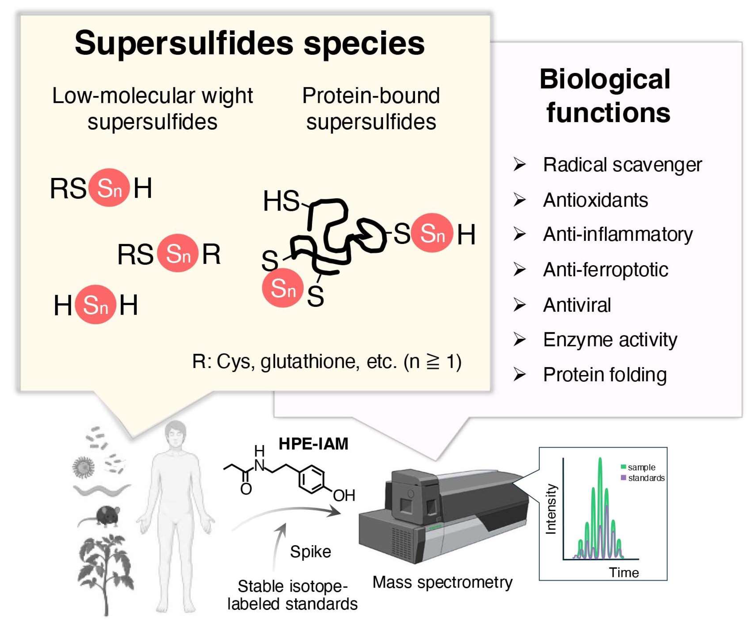
2.1. Occurrence and Quantification
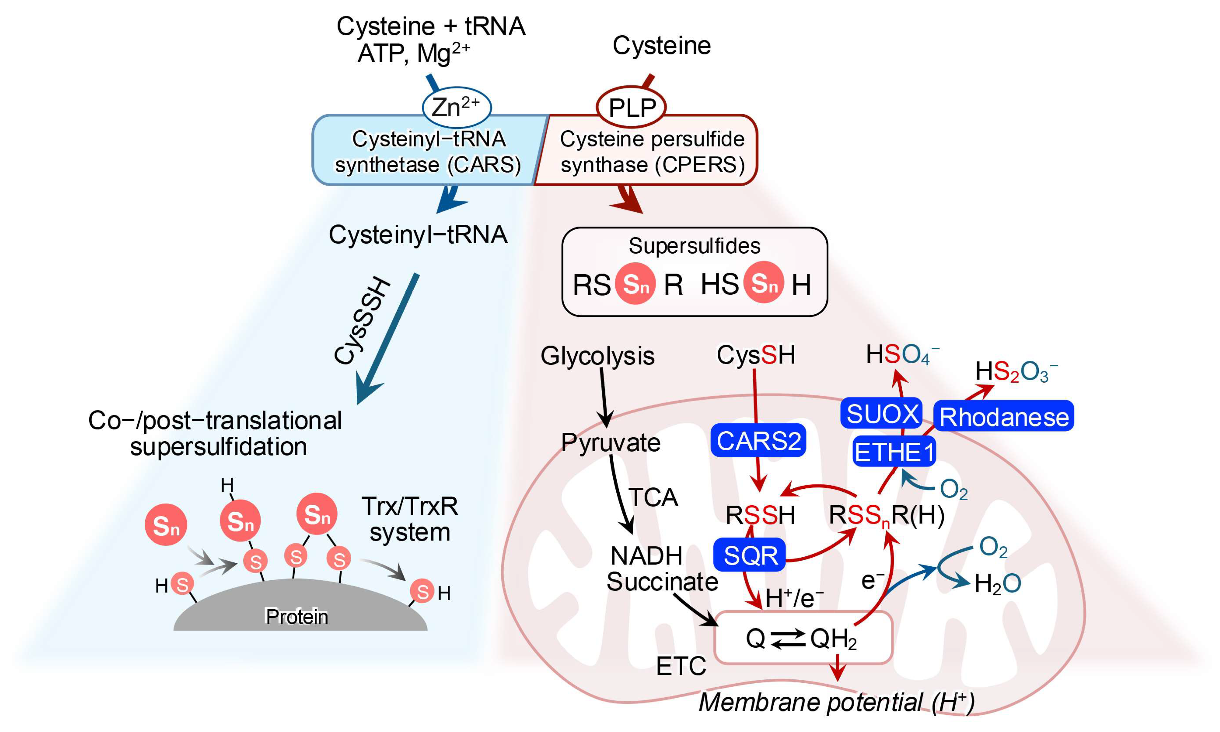
The accurate quantification of supersulfides in biological specimens is the fundamental prerequisite for exploring supersulfide-based redox biology. The approach employed by Akaike and colleagues utilized monobromobimane to alkylate oxidation-labile supersulfides, exploiting their nucleophilic and electrophilic properties [26]. However, due to the hydrolysis equilibrium inherent in polysulfide species, the use of strong electrophilic alkylating agents, including monobromobimane, led to the acceleration of polysulfide degradation through hydrolysis, thereby compromising the precision of quantification [32]. A more suitable method without this limitation was later developed using β-(4-hydroxyphenyl)ethyl iodoacetamide (HPE-IAM) as a supersulfide-trapping agent [27]. In this approach, biological samples are reacted with HPE-IAM to alkylate endogenous supersulfides, and known amounts of isotope-labeled standards are then spiked into the same samples. This is followed by quantification of HPE-IAM-derivatized supersulfide adducts using tandem mass spectrometry in multiple reaction monitoring mode. The peak area ratios between the samples and standards then reveal the concentrations of supersulfides in analytes [33] (Figure 2).
These methods have demonstrated the abundant and ubiquitous distribution of typical low-molecular-weight (LMW) supersulfides, including cysteine (CysSH)-based cysteine persulfide/polysulfide (CysSSH/CysSSnH, n > 1), glutathione persulfide/polysulfide (GSSH/GSSnH, n > 1), cysteine trisulfide (Cys-SSS-Cys) as well as glutathione trisulfide (GSSSG), in both prokaryotic and eukaryotic organisms, including yeast, bacteria, mammals and humans, at submillimolar to millimolar levels [32] (Figure 2). Inorganic supersulfides, such as hydrogen disulfide (H2S2), trisulfide (H2S3), and polymeric sulfur (Sn), have also been reported as potentially existing in living systems [34]. Furthermore, supersulfides have been identified in protein-bound forms through thiol linkages in CysSH residues [29,35,36].

2.2. Biosynthesis

The biosynthesis of supersulfides in all living organisms depends fundamentally on enzymatic mediation. Cystathionine β-synthase (CBS), cystathionine γ-lyase (CSE), and 3-mercaptopyruvate sulfurtransferase (MPST or 3-MST), were previously thought to be the key enzymes responsible for supersulfide generation through the trans-sulfuration pathway [37,38]; however, certain Gram-negative bacteria, such as the Salmonella species, lack CSE, implying the existence of alternative synthetic pathways for supersulfides [39]. A study on mice with a triple knockout of CBS, CSE, and 3-MST supported this notion, revealing that supersulfides are still produced in the absence of these enzymes, further indicating the presence of compensatory or alternative mechanisms [40].
One such mechanism involves cysteinyl-tRNA synthetase (CARS), an enzyme canonically known for catalyzing the synthesis of cysteinyl-tRNA [41]. Two isoforms of this enzyme exist in mammalian cells: cytosolic CARS1 and mitochondrial CARS2, both of which have been identified as novel cysteine persulfide synthase (CPERS), possessing conserved cysteine persulfide-synthesizing activity across species [27,42] (Figure 3). Studies with recombinant CARS have demonstrated that mouse CARS1, human CARS2, and Escherichia coli CARS can produce CysSSH in the presence of the substrate L-cysteine [27]. Furthermore, mass spectrometry-based analyses have shown a significant reduction in supersulfide levels in CARS2 knockout cells. A similar study revealed that heterozygous knockout mice for CARS2 exhibited significantly lower levels of LMW supersulfides compared to wild-type mice [27].
As mentioned above, CARS, like other aminoacyl-tRNA synthetases, is primarily involved in cysteinyl-tRNA production utilizing CysSH. Unexpectedly, CARS also conjugated CysSSH with tRNA in the production of persulfidated cysteinyl-tRNA, enabling the incorporation of CysSSH into nascent polypeptide chains during protein translation [27]. Persulfidated proteins have also been detected in both bacteria and mammalian cells, further corroborating this finding [36,43] (Figure 3).
These studies suggest that CARS serves as a principal supersulfide-synthesizing enzyme which is highly conserved in both prokaryotes and eukaryotes.

2.3. Catabolism

The plasticity of supersulfides gives rise to a complex range of metabolic fates, which remain an active area of investigation. LMW supersulfides are primarily synthesized in mitochondria through CARS2-mediated actions. These supersulfides are subsequently oxidized by sulfide:quinone oxidoreductase (SQR). The oxidized supersulfides participate in sulfur–oxygen hybrid respiration within the electron respiratory chain [27,30,44,45]. Meanwhile, excessive metabolites from supersulfides, such as H2S, are catalyzed by SQR to mitigate sulfide toxicity [46] (Figure 3). H2S oxidation in mitochondria also involves other sulfur oxidation enzymes, including rhodanese and persulfide dioxygenase (ETHE1), which convert H2S to thiosulfate and sulfite. Sulfite oxidase then oxidizes these intermediates to sulfate, with molecular oxygen as an electron acceptor [47] (Figure 3).
On a broader level, protein-bound supersulfides are reduced to regenerate protein thiols, a process that is primarily mediated by the depersulfidation capability of the thioredoxin (Trx) and thioredoxin reductase (TrxR) systems [47,48]. Additionally, the Trx-related protein of 14kDa (TRP14) contributes to depersulfidation, particularly when Trx is engaged heavily in the peroxiredoxin system. The same study also reported that glutathione reductase functions as a depersulfidase [48]. However, it remains unclear whether other members of the redoxin family such as peroxiredoxins and glutaredoxins, which are activated or deactivated through the oxidation and reduction in catalytic thiols, play a role in regulating protein-bound supersulfides.
The wide distribution and tight regulation of supersulfides in various forms provide further evidence of their crucial roles in biological systems.

3. Versatile Physiological Potentials of Supersulfides in Oxidative Distress

Oxidative distress contributes to cellular damage via two major mechanisms. The first involves the overproduction of ROS, particularly H2O2, which serves as a primary factor in both cellular toxicity and damage. The second mechanism is the oxidative modification of macromolecules such as membrane lipids, structural proteins, enzymes, and nucleic acids, causing the disruption of redox signaling pathways [16,49,50]. It should be noted that these mechanisms have been extensively reviewed over the past decades and are beyond the scope of this discussion; readers may refer to related reviews for additional details [1,11,18,51].

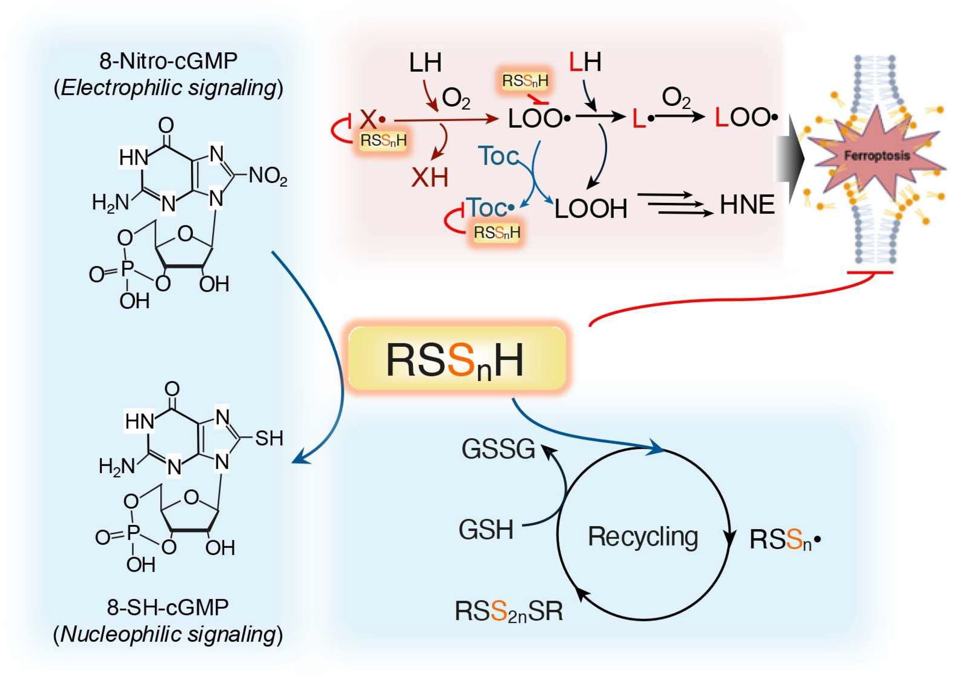
3.1. Supersulfides Regulate Oxidative Distress by Reacting with ROS

Intracellular ROS are major byproducts of the mitochondrial respiratory chain as a result of electron leakage [1,51,52]. Mitochondria are also a major source of supersulfides in mammalian cells, mediated primarily by CARS2, hinting at a potential interaction between ROS and supersulfides. Indeed, supersulfides exhibit superior nucleophilicity and reducing capacity when compared to their corresponding parental thiols [53,54,55]. For example, GSSH, but not its parental thiols GSH or H2S, reduces H2O2 efficiently and protects cells from cell death induced by H2O2 [26]. It was reported by Kunikata et al. that the exogenous addition of GSSSG increased intracellular GSSH levels significantly, thereby suppressing cell death caused by H2O2 exposure [56].
In addition to H2O2 scavenging, supersulfides can also react with free radical species. RSSnH exists in its deprotonated form as alkyl polysulfide anion (RSSn) which readily reacts with radical species to form alkyl polysulfide radicals (RSSn•) [57]. This recombines to yield the polysulfide RSS2nSR, effectively neutralizing free radicals [58]. These polysulfides can then be reduced by cellular thiols, such as GSH, which regenerate active RSSnH and serve as an efficient radical trapping agent [59,60]. A typical model demonstrates the influence of the anti-ferroptotic defense system in mammalian cells, as described below in Section 5.1 [59] (Figure 4).
Another example of the regulation of oxidative distress by supersulfides is revealed in its direct reaction with endogenous electrophiles, including 8-nitroguanosine 3′,5′-cyclic monophosphate (8-nitro-cGMP), an important secondary messenger in redox signaling [26,61,62]. Sawa et al. demonstrated the conversion of 8-nitro-cGMP to 8-mercapt-cGMP, a biologically active metabolite in vivo, by supersulfides [62]. Other endogenous electrophiles, such as nitrated fatty acids, prostaglandin J2 and HNE, exhibit similar reactions, suggesting the key role that supersulfides play in detoxification of electrophiles [63,64,65].
The traditional dogma holds that H2S scavenges ONOO/ONOOH and plays a critical role in vivo [66]; however, no enzyme or pathway that exclusively synthesizes H2S has been identified in mammalian cells. Rather, recent evidence suggests that H2S exists in dynamic equilibrium with supersulfides in biological systems, leading to challenges in isolating the pure effects of H2S. Furthermore, H2S predominantly exists in biological structures in its dissociated form as hydrogen sulfide anion (HS), a molecule which is significantly less reactive than the supersulfide anion RSSn [67]. These results suggest that the role of H2S as a redox signaling molecule may have been overestimated. Both inorganic and organic hydropersulfides (HSSH and RSSH) are major antioxidants that are generated endogenously, highlighting their critical role in cellular defense mechanisms.

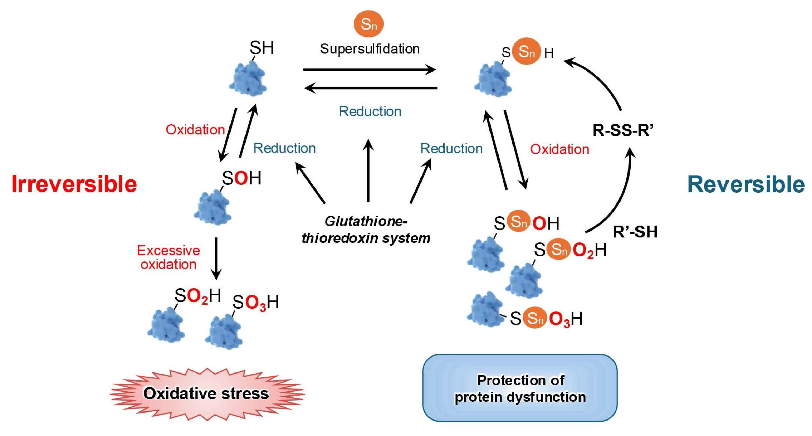
3.2. Supersulfides Regulate Oxidative Distress by Modulating Proteins

Cysteine thiols in proteins are the primary targets for modification by oxidants and electrophilic agents, allowing them to serve as redox switches to control the structure and/or function of redox-regulated proteins [68,69,70]. Advances in quantitative redox proteomic techniques, which are able to differentiate between various thiol modifications, in addition to improvements in the detection via mass spectrometry of low-abundance proteins, have recently stimulated significant research in protein supersulfidation [71,72].
ETHE1, a persulfide dioxygenase, metabolizes GSSH and GSSnH into glutathione with simultaneous oxygen consumption [73]. The Cys247 of ETHE1 reportedly plays a critical role in the formation of supersulfide-bound protein. Polysulfidation at this residue, or its mutation to serine, abolishes persulfide dioxygenase activity [43]. It is, therefore, suggested that ETHE1 is involved in redox regulation through supersulfidation of its cysteine residue. Another example of protein supersulfidation is alcohol dehydrogenase 5 (ADH5), a bifunctional enzyme with both S-nitrosoglutathione reductase activity and formaldehyde dehydrogenase activity [35]. In this case, mutation of Cys174 to serine significantly inhibits ADH5 supersulfidation and abolishes its S-nitrosoglutathione reductase activity, causing an enhancement in NO signaling. This mutation, however, has no effect on the formaldehyde dehydrogenase activity of ADH5 [35].
The effects of persulfidation on protein function are extremely diverse. It may activate or inactivate specific proteins, enabling their adaption under changing conditions [36,74]. During oxidative eustress, oxidation of cysteine thiols by H2O2 leads to protein S-sulfenylation; however, oxidation of sulfenates by H2O2 can produce sulfinates [75]. These products are reversible by sulfiredoxins, but further oxidation by H2O2 irreversibly yields sulfonates, which causes protein function loss, a process typically observed during oxidative distress. The presence of supersulfides enables protein persulfidation; this protects protein thiols from irreversible oxidation by forming perthiosulfenic, perthiosulfinic, and perthiosulfonic derivatives [76]. A more significant aspect of these processes is that the modified proteins can be reduced back to native thiols, restoring protein function [29] (Figure 5).

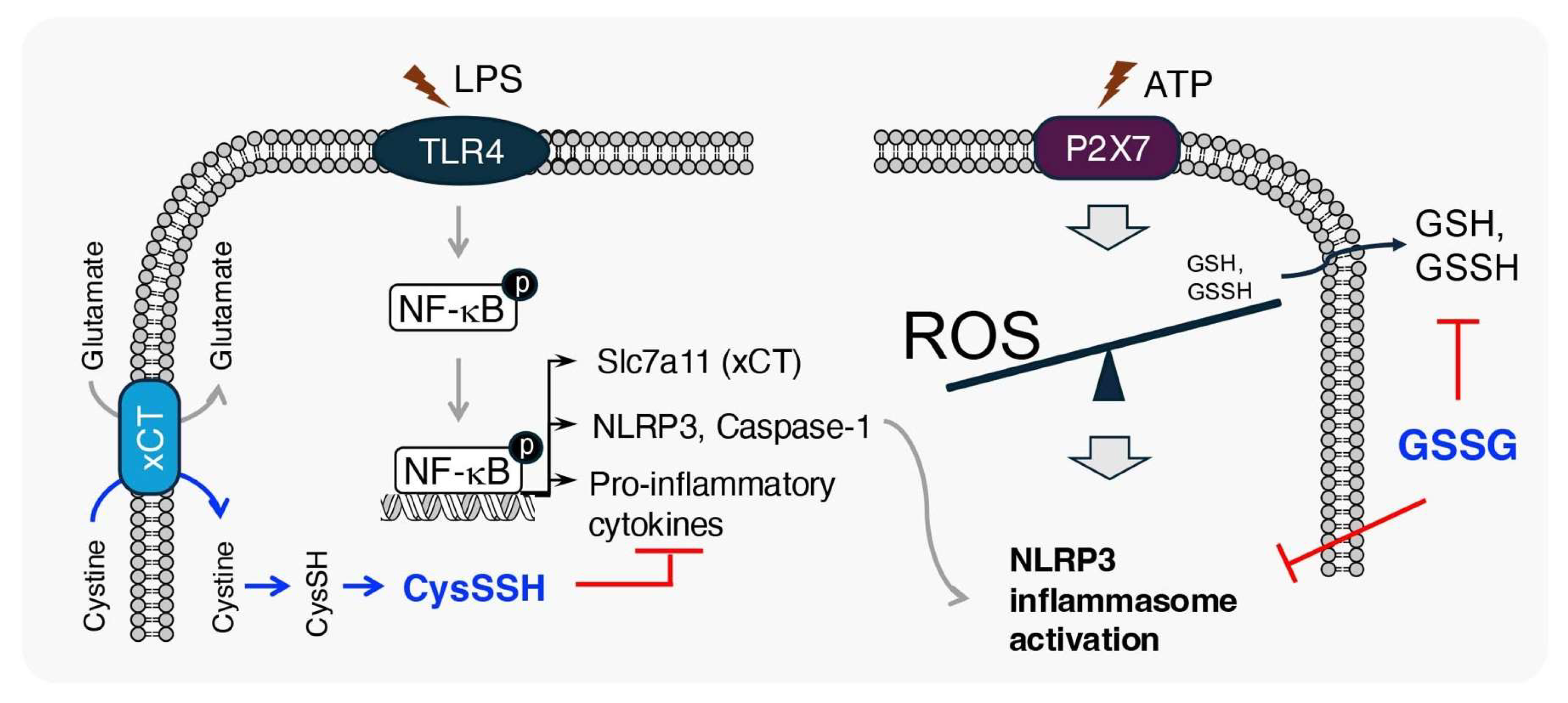
3.3. Anti-Inflammatory Effects of Supersulfides

Numerous studies have extended this research beyond the antioxidative properties of supersulfides to investigate their anti-inflammatory activity [54,77,78,79,80,81,82].
Through a large-scale genome-wide association study, a CARS2 single nucleotide polymorphism was associated with a low-activity phenotype and an increased risk of atherosclerosis. It was also shown that cellular deletion of CARS2 caused a significant increase in the production of inflammatory cytokines in macrophages in response to lipopolysaccharide (LPS), a component of Gram-negative bacteria [81]. Another study revealed, using CARS2 heterozygous knockout mice, that CARS2 plays an anti-inflammatory role under various inflammatory conditions [79]. As CARS2 plays a fundamental role in supersulfide synthesis, these results suggest that supersulfides act as negative regulators in inflammatory responses.
Macrophages, which lack the cystine transporter xCT showed significantly lower levels of supersulfides after LPS stimulation when compared to wild-type macrophages [80]. Under the same conditions, the unavailability of the cystine substrate for supersulfide generation promoted pro-inflammatory gene expression in macrophages. This indicates that supersulfides form a negative feedback loop in the inflammation process [80]. Moreover, it was also shown that under pathological conditions, the NACHT, LRR, and PYD domains-containing protein 3 (NLRP3) inflammasome, a key mediator of acute inflammation, triggers a cytokine storm [83]. Although ROS are believed to be necessary for NLRP3 inflammasome activation, the detailed molecular mechanism of its role remains unclear [84]. Zhang et al. demonstrated that intracellular GSH and its supersulfides, including GSSH and GSSSH, are excreted from macrophages, causing the generation of ROS and subsequent NLRP3 inflammasome activation [78]. The extracellular addition of GSH suppressed the efflux of GSH and its derived supersulfides, causing a significant suppression of NLRP3 inflammasome activation [78]. The anti-inflammatory effects of supersulfides have, therefore, been strongly highlighted by these findings (Figure 6).

4. Limitations of Current Antioxidant Therapeutic Agents

The overproduction of ROS can result in oxidative stress, causing damaging effects and contributing to the pathology of various diseases in humans [85,86,87,88,89,90]. Despite the fact that many synthetic compounds with antioxidant activity have been developed and applied as therapies for oxidative stress-related diseases, their effectiveness has been disappointing [16] (Figure 7). The reasons for this are discussed below.

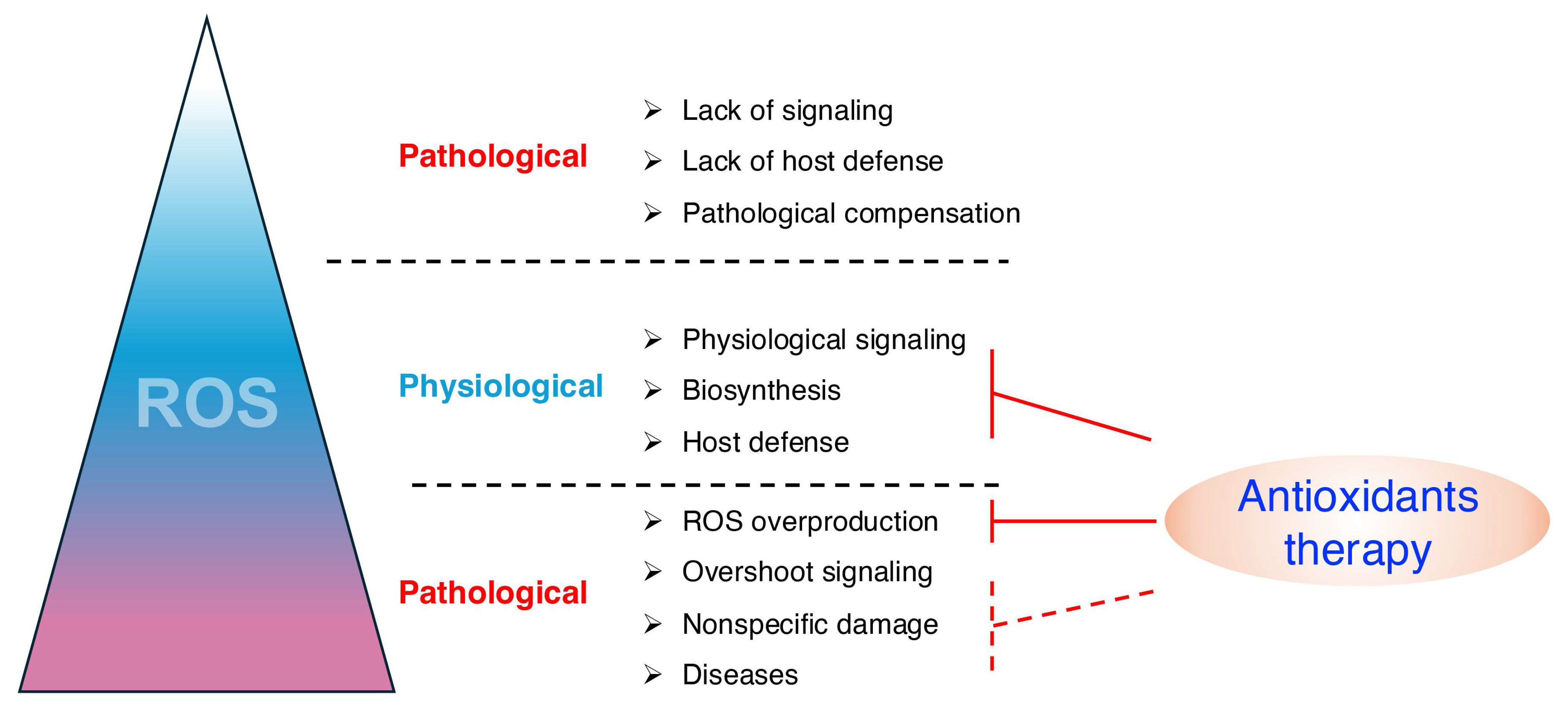
4.1. ROS Play Important Physiological Roles

A significant challenge in the usage of antioxidants is that ROS plays complex biological roles, including essential physiological functions. Scavenging ROS indiscriminately can, in many cases, exacerbate the pathology (Figure 7). For example, in animals, a brief period of cardiac ischemia–reperfusion generates OH•, impairing contractile function during reoxygenation. Although scavenging ROS improves contractile function, it also prevents the adaptation of cardiac function to repeated brief periods of ischemia [91]. Patients with chronic inflammation suffering from severe infections often rely on ROS to help kill pathogens through inducing inflammation [92]; therefore, effective antioxidant therapies may give rise to side effects, including increased susceptibility to infections and disruption of ROS-dependent inflammatory signaling.

4.2. Oxidative Stress Is Not the Primary Mediator of Disease Pathology

While tissue injury often increases ROS production and contributes to further damage, the primary pathological driver may not be oxidative stress, as it occurs in many cases secondary to other initiating factors (Figure 7). Consequently, antioxidant therapies alone may be insufficient to achieve therapeutic efficacy. Instead, combining antioxidants with treatments targeting the primary pathological processes, such as inflammation, metabolic dysregulation, or cellular signaling abnormalities, may offer a more comprehensive approach to disease management. One such example is the intricate link between cytokines and oxidative distress and their influence on physiological and pathological processes [93]. Cytokine production is frequently upregulated during oxidative distress, causing a heightened inflammatory response. The release of pro-inflammatory cytokines such as TNF-α, IL-1β, and IL-6, drive a cytokine storm, and in such cases, antioxidant therapy may alleviate oxidative stress but also fail to address the underlying cytokine storm, limiting its therapeutic efficacy [94].

4.3. Other Limitations

There are still numerous other possible explanations for the ineffectiveness of antioxidant therapies, including: some antioxidants fail to reach the sites of oxidative damage, even if they enter the tissues or organs; they may not adequately reduce oxidative damage despite reaching the correct site [88,95]; paradoxically, excessively high trial doses can promote oxidative stress [96]; the use of single agents may interfere with the uptake or metabolism of other therapeutic agents [97,98]; finally, many antioxidants lack the sufficient reactive capability to neutralize ROS in a timely manner [99] (Figure 7).

5. Supersulfide Donors as Potential Therapeutic Agents for Oxidative Distress-Related Diseases

Both natural and synthetic supersulfide donors, including GSSSG, cysteine trisulfide (CysSSSCys), N-acetylcysteine (NAC) tetrasulfide (NAC-S2), thioglucose tetrasulfide, and inorganic polysulfide sodium salts such as disodium di-, tri-, and tetra-sulfide, can enter inside cells and organs to donate sulfur atoms to intracellular acceptors such as GSH, causing an increase in the supersulfide contents of the cell [100,101] (Figure 8). In this section, several supersulfide donors with therapeutic potential for oxidative stress-related diseases are introduced, along with an exploration of the mechanisms behind their effects.

5.1. Neurodegenerative Diseases

Oxidative stress plays a fundamental role in neurodegenerative diseases such as Alzheimer’s disease and Parkinson’s disease [102]. The initiating causes of these diseases involve complex genetic and environmental factors that vary depending on the condition; nevertheless, common signatures of these diseases include elevated iron levels and neuronal loss, suggesting that ferroptosis is a common neurodegenerative mechanism [103].
Ferroptosis is driven at the cellular level by the iron-catalyzed free radical oxidation of PUFAs in lipids [104]. This is an autocatalytic process in which lipid peroxidation is initiated in the presence of polyunsaturated fatty acids, oxygen, and free radicals. Sustained lipid peroxidation leads to the accumulation of harmful molecules such as lipid peroxides, aldehydes, and their conjugates, ultimately resulting in rupture of the plasma membrane [105,106]. Cells containing oxidizable lipids, therefore, require defense mechanisms to mitigate this process [106]. The enzymatic recycling of small-molecule antioxidants such as α-tocopherol, coenzyme Q10, vitamin K, tetrahydrobiopterin, cystine, and GSH facilitates the reduction in lipid peroxyl radicals to lipid alcohols [106,107,108,109].
RSSnH can act as pluripotent free radical scavengers, acting not only by propagating the lipid peroxyl radical (LOO•) but also by neutralizing the initiating radicals (X•). However, this molecule also reduces the radical forms of other antioxidants, such as tocopherol (Toc•), enhancing the recycling capability [110]. LOO• is generally scavenged via hydrogen atom transfer reactions, in which antioxidant molecules donate hydrogen atoms (H•), reducing LOO• to its inactive form as LOOH. During this process, the antioxidant itself becomes a radical (A•), which is less reactive than LOO•; this can be eliminated to generate non-radical products, and bring a halt to lipid peroxidation. RSSnH is a superior H• donor to RSH, as discussed above in Section 3.1 [110] (Figure 4).
The efficacy of supersulfide donors in inhibiting lipid oxidation was demonstrated in numerous independent studies. Kaneko et al. revealed the inhibition of lipid peroxidation in human plasma via donation from inorganic supersulfides [111]. A similar study employed synthetic organic supersulfide donors to inhibit lipid oxidation in artificial liposomes [112]. Barayue et al. reported that natural supersulfides are able to scavenge lipid radicals to protect cells from ferroptosis [59], highlighting their therapeutic potential for ferroptosis-related diseases. An in vivo study reported the prevention of neurodegeneration in the spinal cord by GSSSG and the recovery of mice from delayed paraplegia after spinal cord ischemia [113]. Additionally, sodium trisulfide, an inorganic polysulfide donor, has been demonstrated to protect midbrain dopaminergic neurons from 1-methyl-4-phenylpyridinium-induced degeneration by ameliorating oxidative stress [114].
These studies are particularly noteworthy given that ferroptosis has recently emerged as an attractive therapeutic target for certain cancers [115]. However, most cancers give rise to upregulated anti-ferroptotic defense systems in order to escape ferroptosis. A promising strategy for cancer therapy could, therefore, involve lowering CARS-mediated supersulfide generation. In addition, glutathione peroxidase 4, as a critical regulator of ferroptosis, facilitates the reduction in lipid hydroperoxides to lipid alcohols [116]. Although the association between supersulfides and GPX4 remains to be elucidated, exploring therapeutic strategies aimed at this enzyme could provide complementary approaches for overcoming anti-ferroptotic defenses in cancer therapy.

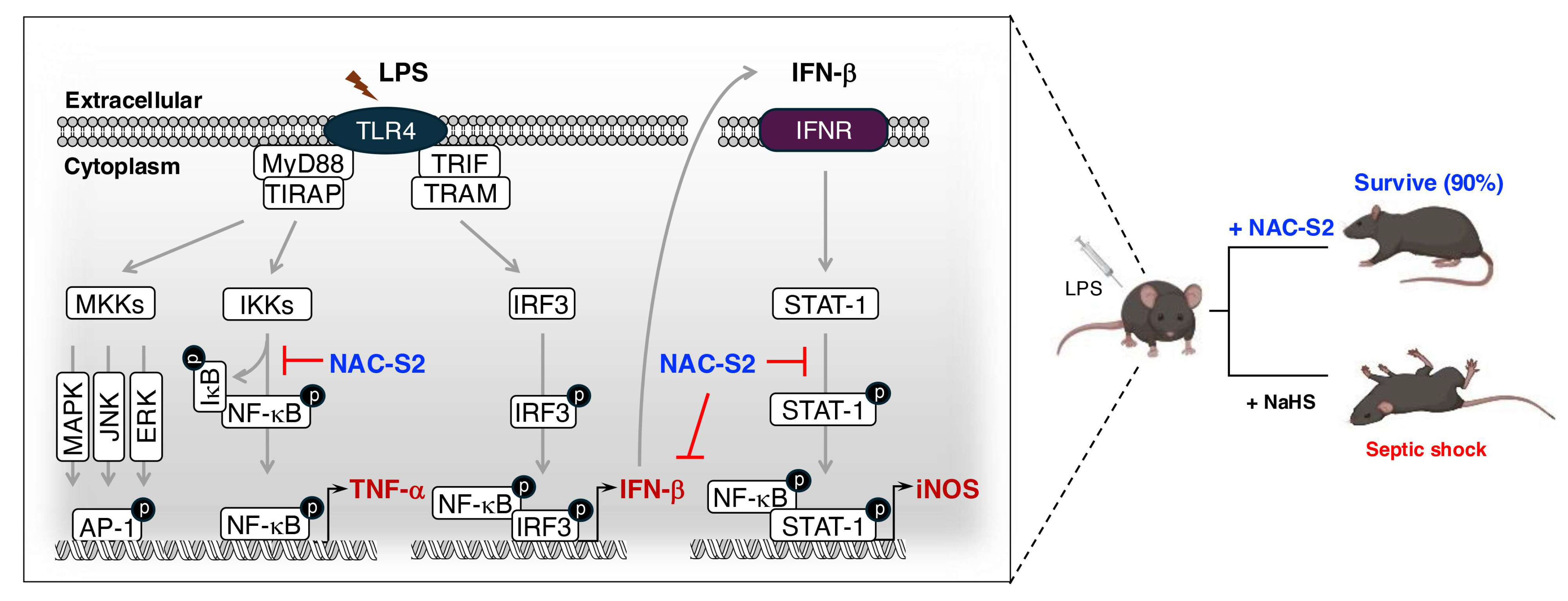
5.2. Systemic Inflammatory Response Syndrome (SIRS)

SIRS is a disorder caused by an exaggerated inflammatory response to infectious pathogens or non-infectious insults, affecting the entire body [117]. The release of oxidants and inflammatory cytokines is provoked by SIRS, and the condition can lead to reversible or irreversible end-organ dysfunction and even death. One such form of SIRS caused by infection is sepsis, which has common features with oxidative stress and inflammation with SIRS caused by non-infectious insults, although sepsis has been more frequently studied.
Toll-like receptor (TLR)-mediated nuclear factor-kappa B (NF-κB) signaling is a key regulatory mechanism in the pathogenesis of sepsis, as it is essential for inducing the expression of inflammatory cytokines and inducible NO synthase (iNOS)-mediated oxidative stress, respectively [118,119] (Figure 9).
The binding of a ligand to a TLR causes adaptor molecules–such as myeloid differentiation primary response 88 (MyD88), Toll/IL-1 receptor (TIR) domain-containing adaptor protein (TIRAP), TIR domain-containing adaptor-inducing interferon-β (TRIF), and TRIF-related adaptor molecule (TRAM)–to bind the TIR domain of the receptor. This interaction phosphorylizes inhibitor kappa B kinases, resulting in the proteasomal degradation of NF-κB inhibitor α (IκBα) [120]. NF-κB then translocate into the nucleus to induce the release of inflammatory cytokines, for example tumor necrosis factor (TNF)-α [121]. During LPS recognition by TLR4, TRIF-mediated signaling activates NF-κB and also triggers the activation of interferon (IFN) regulatory factor 3 (IRF3), which induces IFN-β expression [122]. IFN-β binds to the IFN-α/β receptor on the producing cell itself or on neighboring cells, which causes the amplification of iNOS expression and NO production through the signal transducer and activator of transcription (STAT)-mediated signaling [123] (Figure 9).
Administration of LPS in a mouse model of septic shock decreased the survival rate to 20%. However, when treated with the supersulfide donor NAC-S2, the survival rate increased to 90% and ameliorated inflammatory responses, such as reduced TNF-α production. By contrast, mice treated with sodium hydrogen sulfide exhibited no improvement in survival rate, suggesting a superior anti-inflammatory effect of NAC-S2 over that of H2S [77]. Cell-based analyses demonstrated the suppression of LPS-induced phosphorylation of both IκBα and NF-κB in macrophages treated with NAC-S2. No inhibition of the phosphorylation of p38 mitogen-activated protein (MAP) kinase, c-Jun N-terminal kinase (JNK), or extracellular signal-regulated kinase (ERK) was observed as a result of NAC-S2 treatment, suggesting that supersulfides specifically target NF-κB signaling [77]. Additionally, the expression of iNOS was also inhibited by NAC-S2, which abolishes both IFN-β production and the downstream phosphorylation of STAT-1 [77]. GSSSG, another endogenous supersulfie donor, was shown to suppress LPS-induced inflammatory profiling in both human and mouse epithelial cells [124].
Patients with severe sepsis have exhibited significantly elevated plasma levels of HNE, along with a decrease in levels of antioxidants such as vitamin C, vitamin E, and GSH [125,126,127,128]. Supersulfide donors may therefore exert their therapeutic effects primarily through antioxidative mechanisms rather than the anti-inflammatory effects described above.

5.3. Chronic Obstructive Pulmonary Disease (COPD)

COPD, a chronic lung disease, is a major global health epidemic and the fourth-leading cause of death worldwide, the pathogenesis of which has been linked to oxidative stress [129]. Increased levels of both oxidants and lipid peroxidation products, such as 8-isoprostane, have been detected consistently in the exhaled breath condensate of patients with COPD, along with significantly elevated levels of HNE and its adducts by at least 50% in the airway and alveolar epithelial cells, endothelial cells, and neutrophils of COPD patients [130,131]. Additionally, the urinary level of 8-OHdG, a marker of DNA oxidation, is much higher in patients with COPD [132]. Collectively, these findings reveal the role of oxidative stress in the pathogenesis of COPD, both in the lung and systemically in patients with the disease.
COPD patients experience a progressive limitation of airflow, arising from two primary pathological processes: remodeling and narrowing of the small airways, and destruction of the lung parenchyma, which results in the loss of alveolar attachments due to emphysema [133]. Chronic inflammation in the lung periphery is implicated in these pathological changes, with the intensity of inflammation being correlated with the progression of the disease [89]. The inflammatory responses underlying COPD pathogenesis are both initiated and exacerbated by persistent exposure to oxidants [134]. Studies have indicated that this inflammatory response involves both the innate and adaptive immune systems, characterized by an increase in the number of neutrophils in the airway lumen, along with heightened numbers of macrophages, T lymphocytes, and B lymphocytes in the lung tissue [135] (Figure 10). COPD progression is often not ameliorated by current therapies in many patients, given that they target either free radicals or inflammatory responses independently, as opposed to addressing both simultaneously [136].
Supersulfides have been shown to play an important role in COPD using CARS2 heterozygous knockout mice subjected to viral infection (e.g., SARS-CoV-2) or elastase treatment [79]. The subjects in both models exhibited an increased severity of disease phenotypes compared with wild-type mice. The viral infection model was characterized by a marked increase in 8-OHdG and pro-inflammatory cytokines in CARS2 heterozygous knockout mice compared with wild-type mice. This result implies that the loss of endogenous supersulfides is correlated with oxidative stress and inflammation under COPD conditions [79].
Pathogens such as influenza A virus and SARS-CoV-2 have been treated with GSSSG and inorganic supersulfide donors, in which the supersulfides were shown to block viral entry into cells by directly inactivating viral spike proteins. Viral replication was also inhibited by the presence of supersulfides within cells by targeting key viral proteases in SARS-CoV-2, including the papain-like protease and the 3CL or main protease. Clinically, a reduction in supersulfide levels was observed in lung cells and epithelial lining fluid from patients with COPD [137]. Despite the lack of direct data in this study, the protective effects observed have been attributed to the combined anti-inflammatory and antioxidative properties of supersulfides described above. Thus, the multifaceted mechanisms of supersulfides described herein underscore their protective functions against COPD.

6. Limitations and Future Perspectives of Supersulfide-Based Therapy

While supersulfides hold immense potential in the field of oxidative stress-related diseases, several challenges remain that limit their translational application [138,139].
First, the precise mechanisms governing supersulfide metabolism and their interactions with other redox molecules are not yet fully understood, warranting further mechanistic studies. Additionally, the stability, bioavailability, and pharmacokinetics of supersulfide donors require optimization to ensure therapeutic efficacy while minimizing off-target effects.
Another significant limitation lies in the lack of effective delivery systems that can target supersulfide donors to specific tissues or cellular compartments affected by oxidative stress. Innovative drug delivery technologies, such as nanocarrier systems, may provide a solution to this challenge by enhancing tissue specificity and reducing systemic toxicity. Furthermore, while preclinical studies have demonstrated promising results, comprehensive clinical trials are necessary to validate the safety and efficacy of supersulfide-based therapies in human diseases.
Future research should also focus on identifying disease-specific biomarkers that can aid in patient stratification and therapeutic monitoring, thereby enabling personalized medicine approaches. Another potential area of investigation is the role of Coenzyme A in supersulfide biology. coenzyme A, as a low-molecular-weight thiol, plays a critical role in cellular biosynthesis and energy production. While its involvement in supersulfide biosynthesis and function has been proposed, detailed mechanistic insights and experimental evidence are currently lacking. Future studies should explore this relationship, particularly in the context of redox regulation and metabolic pathways. By addressing these challenges, the therapeutic potential of supersulfides can be fully harnessed, paving the way for innovative treatments targeting oxidative stress-related diseases.

7. Concluding Remarks

In this review, the chemical and biological properties of supersulfides, which are closely related to almost all life-sustaining phenomena, have been outlined. Mounting evidence indicates a dynamic change in supersulfide levels during various oxidative stress-related diseases. This may be explained by the interaction of supersulfides with ROS, RNS or other oxidants, which are excessively produced under these conditions. This review has described thoroughly significant therapeutic potential of supersulfides in addressing both oxidative stress and inflammation. One example of a promising strategy involves the manipulation of supersulfide levels using supersulfide donors; alternatively, inhibiting CARS or other supersulfide-producing enzymes may enable the regulation of supersulfide bioavailability. Given the wide-ranging potential offered by these therapies, deeper analyses of the mechanisms of action, targets, pharmacokinetics, and the potential side effects of supersulfide donors in disease models are essential. Finally, in order to enhance the efficacy of therapeutic agents, it is crucially important to develop drug delivery systems that enable supersulfide donors to target affected areas.

Author Contributions

Writing—original draft preparation, Y.P.; writing—review and editing, T.Z.; figures—Y.P., T.M. and T.Z.; supervision, T.Z. and T.A.; funding acquisition, T.A. All authors have read and agreed to the published version of the manuscript.

Funding

This work was supported by Japan Science and Technology Agency (JST) CREST Grant Number JPMJCR2024 (20348438 to T.A.), Grant-in-Aid for Scientific Research on Innovative Areas(A) “Sulfur biology” (21H05263 and 21H05258 to T.A.), International Leading Research (23K20040 to T.A.), Scientific Research (S) (24H00063 to T.A.), from the Ministry of Education, Culture, Sports, Science and Technology of Japan, and Japan Agency for Medical Research and Development (AMED) to T.A. (JP21zf0127001).

Institutional Review Board Statement

Not applicable.

Informed Consent Statement

Not applicable.

Acknowledgments

We confirm that no AI-based tools were used in the preparation or revision of this article. Some figures were created with BioRender.com.

Conflicts of Interest

The authors declare no competing interests.

References

  1. Sies, H.; Mailloux, R.J.; Jakob, U. Fundamentals of redox regulation in biology. Nat. Rev. Mol. Cell Biol. 2024, 25, 701–719. [Google Scholar] [CrossRef] [PubMed]
  2. Sies, H.; Jones, D.P. Reactive oxygen species (ROS) as pleiotropic physiological signalling agents. Nat. Rev. Mol. Cell Biol. 2020, 21, 363–383. [Google Scholar] [CrossRef]
  3. Panday, A.; Sahoo, M.K.; Osorio, D.; Batra, S. NADPH oxidases: An overview from structure to innate immunity-associated pathologies. Cell Mol. Immunol. 2015, 12, 5–23. [Google Scholar] [CrossRef] [PubMed]
  4. Fang, F.C. Antimicrobial reactive oxygen and nitrogen species: Concepts and controversies. Nat. Rev. Microbiol. 2004, 2, 820–832. [Google Scholar] [CrossRef]
  5. Mishanina, T.V.; Libiad, M.; Banerjee, R. Biogenesis of reactive sulfur species for signaling by hydrogen sulfide oxidation pathways. Nat. Chem. Biol. 2015, 11, 457–464. [Google Scholar] [CrossRef] [PubMed]
  6. Cortese-Krott, M.M.; Koning, A.; Kuhnle, G.G.C.; Nagy, P.; Bianco, C.L.; Pasch, A.; Wink, D.A.; Fukuto, J.M.; Jackson, A.A.; van Goor, H.; et al. The Reactive Species Interactome: Evolutionary Emergence, Biological Significance, and Opportunities for Redox Metabolomics and Personalized Medicine. Antioxid. Redox Signal 2017, 27, 684–712. [Google Scholar] [CrossRef] [PubMed]
  7. Rodriguez, R.; Redman, R. Balancing the generation and elimination of reactive oxygen species. Proc. Natl. Acad. Sci. USA 2005, 102, 3175–3176. [Google Scholar] [CrossRef] [PubMed]
  8. Sharp, R.E.; Chapman, S.K. Mechanisms for regulating electron transfer in multi-centre redox proteins. Biochim. Biophys. Acta 1999, 1432, 143–158. [Google Scholar] [CrossRef] [PubMed]
  9. Kohler, A.; Barrientos, A.; Fontanesi, F.; Ott, M. The functional significance of mitochondrial respiratory chain supercomplexes. EMBO Rep. 2023, 24, e57092. [Google Scholar] [CrossRef] [PubMed]
  10. Lennicke, C.; Cocheme, H.M. Redox metabolism: ROS as specific molecular regulators of cell signaling and function. Mol. Cell 2021, 81, 3691–3707. [Google Scholar] [CrossRef]
  11. Feelisch, M.; Cortese-Krott, M.M.; Santolini, J.; Wootton, S.A.; Jackson, A.A. Systems redox biology in health and disease. EXCLI J. 2022, 21, 623–646. [Google Scholar] [CrossRef] [PubMed]
  12. Lushchak, V.I.; Lushchak, O. Interplay between reactive oxygen and nitrogen species in living organisms. Chem. Biol. Interact. 2021, 349, 109680. [Google Scholar] [CrossRef] [PubMed]
  13. Mullard, A. FDA approves first Friedreich’s ataxia drug. Nat. Rev. Drug Discov. 2023, 22, 258. [Google Scholar] [CrossRef] [PubMed]
  14. Maldonado, E.; Rojas, D.A.; Morales, S.; Miralles, V.; Solari, A. Dual and Opposite Roles of Reactive Oxygen Species (ROS) in Chagas Disease: Beneficial on the Pathogen and Harmful on the Host. Oxid. Med. Cell Longev. 2020, 2020, 8867701. [Google Scholar] [CrossRef]
  15. Sies, H. Hydrogen peroxide as a central redox signaling molecule in physiological oxidative stress: Oxidative eustress. Redox Biol. 2017, 11, 613–619. [Google Scholar] [CrossRef]
  16. Forman, H.J.; Zhang, H. Targeting oxidative stress in disease: Promise and limitations of antioxidant therapy. Nat. Rev. Drug Discov. 2021, 20, 689–709. [Google Scholar] [CrossRef]
  17. Murphy, M.P.; Bayir, H.; Belousov, V.; Chang, C.J.; Davies, K.J.A.; Davies, M.J.; Dick, T.P.; Finkel, T.; Forman, H.J.; Janssen-Heininger, Y.; et al. Guidelines for measuring reactive oxygen species and oxidative damage in cells and in vivo. Nat. Metab. 2022, 4, 651–662. [Google Scholar] [CrossRef]
  18. Halliwell, B. Understanding mechanisms of antioxidant action in health and disease. Nat. Rev. Mol. Cell Biol. 2024, 25, 13–33. [Google Scholar] [CrossRef]
  19. Meng, J.; Lv, Z.; Zhang, Y.; Wang, Y.; Qiao, X.; Sun, C.; Chen, Y.; Guo, M.; Han, W.; Ye, A.; et al. Precision Redox: The Key for Antioxidant Pharmacology. Antioxid. Redox Signal 2021, 34, 1069–1082. [Google Scholar] [CrossRef]
  20. Georgieva, M.N.; Little, C.T.S.; Bailey, R.J.; Ball, A.D.; Glover, A.G. Microbial-tubeworm associations in a 440 million year old hydrothermal vent community. Proc. Biol. Sci. 2018, 285, 20182004. [Google Scholar] [CrossRef]
  21. Atmaca, G. Antioxidant effects of sulfur-containing amino acids. Yonsei Med. J. 2004, 45, 776–788. [Google Scholar] [CrossRef] [PubMed]
  22. Sies, H. Oxidative stress: A concept in redox biology and medicine. Redox Biol. 2015, 4, 180–183. [Google Scholar] [CrossRef] [PubMed]
  23. Mukwevho, E.; Ferreira, Z.; Ayeleso, A. Potential role of sulfur-containing antioxidant systems in highly oxidative environments. Molecules 2014, 19, 19376–19389. [Google Scholar] [CrossRef] [PubMed]
  24. Ulrich, K.; Jakob, U. The role of thiols in antioxidant systems. Free Radic. Biol. Med. 2019, 140, 14–27. [Google Scholar] [CrossRef]
  25. Kasamatsu, S.; Ida, T.; Koga, T.; Asada, K.; Motohashi, H.; Ihara, H.; Akaike, T. High-Precision Sulfur Metabolomics Innovated by a New Specific Probe for Trapping Reactive Sulfur Species. Antioxid. Redox Signal 2021, 34, 1407–1419. [Google Scholar] [CrossRef] [PubMed]
  26. Ida, T.; Sawa, T.; Ihara, H.; Tsuchiya, Y.; Watanabe, Y.; Kumagai, Y.; Suematsu, M.; Motohashi, H.; Fujii, S.; Matsunaga, T.; et al. Reactive cysteine persulfides and S-polythiolation regulate oxidative stress and redox signaling. Proc. Natl. Acad. Sci. USA 2014, 111, 7606–7611. [Google Scholar] [CrossRef] [PubMed]
  27. Akaike, T.; Ida, T.; Wei, F.Y.; Nishida, M.; Kumagai, Y.; Alam, M.M.; Ihara, H.; Sawa, T.; Matsunaga, T.; Kasamatsu, S.; et al. Cysteinyl-tRNA synthetase governs cysteine polysulfidation and mitochondrial bioenergetics. Nat. Commun. 2017, 8, 1177. [Google Scholar] [CrossRef] [PubMed]
  28. Takata, T.; Jung, M.; Matsunaga, T.; Ida, T.; Morita, M.; Motohashi, H.; Shen, X.; Kevil, C.G.; Fukuto, J.M.; Akaike, T. Methods in sulfide and persulfide research. Nitric Oxide 2021, 116, 47–64. [Google Scholar] [CrossRef] [PubMed]
  29. Doka, E.; Ida, T.; Dagnell, M.; Abiko, Y.; Luong, N.C.; Balog, N.; Takata, T.; Espinosa, B.; Nishimura, A.; Cheng, Q.; et al. Control of protein function through oxidation and reduction of persulfidated states. Sci. Adv. 2020, 6, eaax8358. [Google Scholar] [CrossRef]
  30. Barayeu, U.; Sawa, T.; Nishida, M.; Wei, F.Y.; Motohashi, H.; Akaike, T. Supersulfide biology and translational medicine for disease control. Br. J. Pharmacol. 2023. [Google Scholar] [CrossRef]
  31. Akaike, T.; Morita, M.; Ogata, S.; Yoshitake, J.; Jung, M.; Sekine, H.; Motohashi, H.; Barayeu, U.; Matsunaga, T. New aspects of redox signaling mediated by supersulfides in health and disease. Free Radic. Biol. Med. 2024, 222, 539–551. [Google Scholar] [CrossRef] [PubMed]
  32. Ogata, S.; Matsunaga, T.; Jung, M.; Barayeu, U.; Morita, M.; Akaike, T. Persulfide Biosynthesis Conserved Evolutionarily in All Organisms. Antioxid. Redox Signal 2023, 39, 983–999. [Google Scholar] [CrossRef] [PubMed]
  33. Tomaiuolo, M.; Vecchione, G.; Margaglione, M.; Pisanelli, D.; Grandone, E. Stable-isotope dilution LC-ESI-MS/MS techniques for the quantification of total homocysteine in human plasma. J. Chromatogr. B Anal. Technol. Biomed. Life Sci. 2009, 877, 3292–3299. [Google Scholar] [CrossRef] [PubMed]
  34. Wang, T.; Ran, M.; Li, X.; Liu, Y.; Xin, Y.; Liu, H.; Liu, H.; Xia, Y.; Xun, L. The Pathway of Sulfide Oxidation to Octasulfur Globules in the Cytoplasm of Aerobic Bacteria. Appl. Env. Environ. Microbiol. 2022, 88, e0194121. [Google Scholar] [CrossRef] [PubMed]
  35. Kasamatsu, S.; Nishimura, A.; Alam, M.M.; Morita, M.; Shimoda, K.; Matsunaga, T.; Jung, M.; Ogata, S.; Barayeu, U.; Ida, T.; et al. Supersulfide catalysis for nitric oxide and aldehyde metabolism. Sci. Adv. 2023, 9, eadg8631. [Google Scholar] [CrossRef] [PubMed]
  36. Zivanovic, J.; Kouroussis, E.; Kohl, J.B.; Adhikari, B.; Bursac, B.; Schott-Roux, S.; Petrovic, D.; Miljkovic, J.L.; Thomas-Lopez, D.; Jung, Y.; et al. Selective Persulfide Detection Reveals Evolutionarily Conserved Antiaging Effects of S-Sulfhydration. Cell Metab. 2019, 30, 1152–1170.E13. [Google Scholar] [CrossRef]
  37. Pedre, B.; Talwar, D.; Barayeu, U.; Schilling, D.; Luzarowski, M.; Sokolowski, M.; Glatt, S.; Dick, T.P. 3-Mercaptopyruvate sulfur transferase is a protein persulfidase. Nat. Chem. Biol. 2023, 19, 507–517. [Google Scholar] [CrossRef] [PubMed]
  38. Kolluru, G.K.; Shen, X.; Bir, S.C.; Kevil, C.G. Hydrogen sulfide chemical biology: Pathophysiological roles and detection. Nitric Oxide 2013, 35, 5–20. [Google Scholar] [CrossRef] [PubMed]
  39. Khan, S.; Fujii, S.; Matsunaga, T.; Nishimura, A.; Ono, K.; Ida, T.; Ahmed, K.A.; Okamoto, T.; Tsutsuki, H.; Sawa, T.; et al. Reactive Persulfides from Salmonella Typhimurium Downregulate Autophagy-Mediated Innate Immunity in Macrophages by Inhibiting Electrophilic Signaling. Cell Chem. Biol. 2018, 25, 1403–1413.E4. [Google Scholar] [CrossRef] [PubMed]
  40. Zainol Abidin, Q.H.; Ida, T.; Morita, M.; Matsunaga, T.; Nishimura, A.; Jung, M.; Hassan, N.; Takata, T.; Ishii, I.; Kruger, W.; et al. Synthesis of Sulfides and Persulfides Is Not Impeded by Disruption of Three Canonical Enzymes in Sulfur Metabolism. Antioxidants 2023, 12, 868. [Google Scholar] [CrossRef]
  41. Guo, M.; Yang, X.L.; Schimmel, P. New functions of aminoacyl-tRNA synthetases beyond translation. Nat. Rev. Mol. Cell Biol. 2010, 11, 668–674. [Google Scholar] [CrossRef] [PubMed]
  42. Giege, R.; Sissler, M.; Florentz, C. Universal rules and idiosyncratic features in tRNA identity. Nucleic Acids Res. 1998, 26, 5017–5035. [Google Scholar] [CrossRef] [PubMed]
  43. Jung, M.; Kasamatsu, S.; Matsunaga, T.; Akashi, S.; Ono, K.; Nishimura, A.; Morita, M.; Abdul Hamid, H.; Fujii, S.; Kitamura, H.; et al. Protein polysulfidation-dependent persulfide dioxygenase activity of ethylmalonic encephalopathy protein 1. Biochem. Biophys. Res. Commun. 2016, 480, 180–186. [Google Scholar] [CrossRef]
  44. Alam, M.M.; Kishino, A.; Sung, E.; Sekine, H.; Abe, T.; Murakami, S.; Akaike, T.; Motohashi, H. Contribution of NRF2 to sulfur metabolism and mitochondrial activity. Redox Biol. 2023, 60, 102624. [Google Scholar] [CrossRef] [PubMed]
  45. Nishimura, A.; Yoon, S.; Matsunaga, T.; Ida, T.; Jung, M.; Ogata, S.; Morita, M.; Yoshitake, J.; Unno, Y.; Barayeu, U.; et al. Longevity control by supersulfide-mediated mitochondrial respiration and regulation of protein quality. Redox Biol. 2024, 69, 103018. [Google Scholar] [CrossRef] [PubMed]
  46. Marutani, E.; Morita, M.; Hirai, S.; Kai, S.; Grange, R.M.H.; Miyazaki, Y.; Nagashima, F.; Traeger, L.; Magliocca, A.; Ida, T.; et al. Sulfide catabolism ameliorates hypoxic brain injury. Nat. Commun. 2021, 12, 3108. [Google Scholar] [CrossRef]
  47. Filipovic, M.R.; Zivanovic, J.; Alvarez, B.; Banerjee, R. Chemical Biology of H2S Signaling through Persulfidation. Chem. Rev. 2018, 118, 1253–1337. [Google Scholar] [CrossRef] [PubMed]
  48. Doka, E.; Pader, I.; Biro, A.; Johansson, K.; Cheng, Q.; Ballago, K.; Prigge, J.R.; Pastor-Flores, D.; Dick, T.P.; Schmidt, E.E.; et al. A novel persulfide detection method reveals protein persulfide- and polysulfide-reducing functions of thioredoxin and glutathione systems. Sci. Adv. 2016, 2, e1500968. [Google Scholar] [CrossRef]
  49. Valko, M.; Leibfritz, D.; Moncol, J.; Cronin, M.T.; Mazur, M.; Telser, J. Free radicals and antioxidants in normal physiological functions and human disease. Int. J. Biochem. Cell Biol. 2007, 39, 44–84. [Google Scholar] [CrossRef] [PubMed]
  50. Forman, H.J.; Maiorino, M.; Ursini, F. Signaling functions of reactive oxygen species. Biochemistry 2010, 49, 835–842. [Google Scholar] [CrossRef] [PubMed]
  51. Sies, H.; Belousov, V.V.; Chandel, N.S.; Davies, M.J.; Jones, D.P.; Mann, G.E.; Murphy, M.P.; Yamamoto, M.; Winterbourn, C. Defining roles of specific reactive oxygen species (ROS) in cell biology and physiology. Nat. Rev. Mol. Cell Biol. 2022, 23, 499–515. [Google Scholar] [CrossRef] [PubMed]
  52. D’Autreaux, B.; Toledano, M.B. ROS as signalling molecules: Mechanisms that generate specificity in ROS homeostasis. Nat. Rev. Mol. Cell Biol. 2007, 8, 813–824. [Google Scholar] [CrossRef] [PubMed]
  53. Fukuto, J.M.; Ignarro, L.J.; Nagy, P.; Wink, D.A.; Kevil, C.G.; Feelisch, M.; Cortese-Krott, M.M.; Bianco, C.L.; Kumagai, Y.; Hobbs, A.J.; et al. Biological hydropersulfides and related polysulfides—A new concept and perspective in redox biology. FEBS Lett. 2018, 592, 2140–2152. [Google Scholar] [CrossRef] [PubMed]
  54. Yu, B.; Yuan, Z.; Yang, X.; Wang, B. Prodrugs of Persulfides, Sulfur Dioxide, and Carbon Disulfide: Important Tools for Studying Sulfur Signaling at Various Oxidation States. Antioxid. Redox Signal 2020, 33, 1046–1059. [Google Scholar] [CrossRef] [PubMed]
  55. Li, H.; Liu, H.; Chen, Z.; Zhao, R.; Wang, Q.; Ran, M.; Xia, Y.; Hu, X.; Liu, J.; Xian, M.; et al. Using resonance synchronous spectroscopy to characterize the reactivity and electrophilicity of biologically relevant sulfane sulfur. Redox Biol. 2019, 24, 101179. [Google Scholar] [CrossRef] [PubMed]
  56. Kunikata, H.; Ida, T.; Sato, K.; Aizawa, N.; Sawa, T.; Tawarayama, H.; Murayama, N.; Fujii, S.; Akaike, T.; Nakazawa, T. Metabolomic profiling of reactive persulfides and polysulfides in the aqueous and vitreous humors. Sci. Rep. 2017, 7, 41984. [Google Scholar] [CrossRef]
  57. Fukuto, J.M.; Carrington, S.J.; Tantillo, D.J.; Harrison, J.G.; Ignarro, L.J.; Freeman, B.A.; Chen, A.; Wink, D.A. Small molecule signaling agents: The integrated chemistry and biochemistry of nitrogen oxides, oxides of carbon, dioxygen, hydrogen sulfide, and their derived species. Chem. Res. Toxicol. 2012, 25, 769–793. [Google Scholar] [CrossRef] [PubMed]
  58. Everett, S.A.; Wardman, P. Perthiols as antioxidants: Radical-scavenging and prooxidative mechanisms. Methods Enzym. Enzymol. 1995, 251, 55–69. [Google Scholar] [CrossRef]
  59. Barayeu, U.; Schilling, D.; Eid, M.; Xavier da Silva, T.N.; Schlicker, L.; Mitreska, N.; Zapp, C.; Grater, F.; Miller, A.K.; Kappl, R.; et al. Hydropersulfides inhibit lipid peroxidation and ferroptosis by scavenging radicals. Nat. Chem. Biol. 2023, 19, 28–37. [Google Scholar] [CrossRef]
  60. Everett, S.A.; Folkes, L.K.; Wardman, P.; Asmus, K.D. Free-radical repair by a novel perthiol: Reversible hydrogen transfer and perthiyl radical formation. Free Radic. Res. 1994, 20, 387–400. [Google Scholar] [CrossRef]
  61. Ihara, H.; Kasamatsu, S.; Kitamura, A.; Nishimura, A.; Tsutsuki, H.; Ida, T.; Ishizaki, K.; Toyama, T.; Yoshida, E.; Abdul Hamid, H.; et al. Exposure to Electrophiles Impairs Reactive Persulfide-Dependent Redox Signaling in Neuronal Cells. Chem. Res. Toxicol. 2017, 30, 1673–1684. [Google Scholar] [CrossRef]
  62. Sawa, T.; Zaki, M.H.; Okamoto, T.; Akuta, T.; Tokutomi, Y.; Kim-Mitsuyama, S.; Ihara, H.; Kobayashi, A.; Yamamoto, M.; Fujii, S.; et al. Protein S-guanylation by the biological signal 8-nitroguanosine 3′,5′-cyclic monophosphate. Nat. Chem. Biol. 2007, 3, 727–735. [Google Scholar] [CrossRef] [PubMed]
  63. Freeman, B.A.; O’Donnell, V.B.; Schopfer, F.J. The discovery of nitro-fatty acids as products of metabolic and inflammatory reactions and mediators of adaptive cell signaling. Nitric Oxide 2018, 77, 106–111. [Google Scholar] [CrossRef]
  64. Surh, Y.J.; Na, H.K.; Park, J.M.; Lee, H.N.; Kim, W.; Yoon, I.S.; Kim, D.D. 15-Deoxy-Delta12,14-prostaglandin J2, an electrophilic lipid mediator of anti-inflammatory and pro-resolving signaling. Biochem. Pharmacol. 2011, 82, 1335–1351. [Google Scholar] [CrossRef]
  65. Mol, M.; Regazzoni, L.; Altomare, A.; Degani, G.; Carini, M.; Vistoli, G.; Aldini, G. Enzymatic and non-enzymatic detoxification of 4-hydroxynonenal: Methodological aspects and biological consequences. Free Radic. Biol. Med. 2017, 111, 328–344. [Google Scholar] [CrossRef] [PubMed]
  66. Whiteman, M.; Armstrong, J.S.; Chu, S.H.; Jia-Ling, S.; Wong, B.S.; Cheung, N.S.; Halliwell, B.; Moore, P.K. The novel neuromodulator hydrogen sulfide: An endogenous peroxynitrite ’scavenger’? J. Neurochem. 2004, 90, 765–768. [Google Scholar] [CrossRef]
  67. Cuevasanta, E.; Moller, M.N.; Alvarez, B. Biological chemistry of hydrogen sulfide and persulfides. Arch. Biochem. Biophys. 2017, 617, 9–25. [Google Scholar] [CrossRef]
  68. Netto, L.E.; Antunes, F. The Roles of Peroxiredoxin and Thioredoxin in Hydrogen Peroxide Sensing and in Signal Transduction. Mol. Cells 2016, 39, 65–71. [Google Scholar] [CrossRef] [PubMed]
  69. Yamamoto, M.; Kensler, T.W.; Motohashi, H. The KEAP1-NRF2 System: A Thiol-Based Sensor-Effector Apparatus for Maintaining Redox Homeostasis. Physiol. Rev. 2018, 98, 1169–1203. [Google Scholar] [CrossRef] [PubMed]
  70. Delaunay, A.; Pflieger, D.; Barrault, M.B.; Vinh, J.; Toledano, M.B. A thiol peroxidase is an H2O2 receptor and redox-transducer in gene activation. Cell 2002, 111, 471–481. [Google Scholar] [CrossRef] [PubMed]
  71. Vignane, T.; Filipovic, M.R. Emerging Chemical Biology of Protein Persulfidation. Antioxid. Redox Signal 2023, 39, 19–39. [Google Scholar] [CrossRef] [PubMed]
  72. He, B.; Zhang, Z.; Huang, Z.; Duan, X.; Wang, Y.; Cao, J.; Li, L.; He, K.; Nice, E.C.; He, W.; et al. Protein persulfidation: Rewiring the hydrogen sulfide signaling in cell stress response. Biochem. Pharmacol. 2023, 209, 115444. [Google Scholar] [CrossRef] [PubMed]
  73. Pettinati, I.; Brem, J.; McDonough, M.A.; Schofield, C.J. Crystal structure of human persulfide dioxygenase: Structural basis of ethylmalonic encephalopathy. Hum. Mol. Genet. 2015, 24, 2458–2469. [Google Scholar] [CrossRef] [PubMed]
  74. Cuevasanta, E.; Reyes, A.M.; Zeida, A.; Mastrogiovanni, M.; De Armas, M.I.; Radi, R.; Alvarez, B.; Trujillo, M. Kinetics of formation and reactivity of the persulfide in the one-cysteine peroxiredoxin from Mycobacterium tuberculosis. J. Biol. Chem. 2019, 294, 13593–13605. [Google Scholar] [CrossRef]
  75. Garrido Ruiz, D.; Sandoval-Perez, A.; Rangarajan, A.V.; Gunderson, E.L.; Jacobson, M.P. Cysteine Oxidation in Proteins: Structure, Biophysics, and Simulation. Biochemistry 2022, 61, 2165–2176. [Google Scholar] [CrossRef]
  76. Millikin, R.; Bianco, C.L.; White, C.; Saund, S.S.; Henriquez, S.; Sosa, V.; Akaike, T.; Kumagai, Y.; Soeda, S.; Toscano, J.P.; et al. The chemical biology of protein hydropersulfides: Studies of a possible protective function of biological hydropersulfide generation. Free Radic. Biol. Med. 2016, 97, 136–147. [Google Scholar] [CrossRef] [PubMed]
  77. Zhang, T.; Ono, K.; Tsutsuki, H.; Ihara, H.; Islam, W.; Akaike, T.; Sawa, T. Enhanced Cellular Polysulfides Negatively Regulate TLR4 Signaling and Mitigate Lethal Endotoxin Shock. Cell Chem. Biol. 2019, 26, 686–698.E4. [Google Scholar] [CrossRef] [PubMed]
  78. Zhang, T.; Tsutsuki, H.; Islam, W.; Ono, K.; Takeda, K.; Akaike, T.; Sawa, T. ATP exposure stimulates glutathione efflux as a necessary switch for NLRP3 inflammasome activation. Redox Biol. 2021, 41, 101930. [Google Scholar] [CrossRef] [PubMed]
  79. Matsunaga, T.; Sano, H.; Takita, K.; Morita, M.; Yamanaka, S.; Ichikawa, T.; Numakura, T.; Ida, T.; Jung, M.; Ogata, S.; et al. Supersulphides provide airway protection in viral and chronic lung diseases. Nat. Commun. 2023, 14, 4476. [Google Scholar] [CrossRef] [PubMed]
  80. Takeda, H.; Murakami, S.; Liu, Z.; Sawa, T.; Takahashi, M.; Izumi, Y.; Bamba, T.; Sato, H.; Akaike, T.; Sekine, H.; et al. Sulfur metabolic response in macrophage limits excessive inflammatory response by creating a negative feedback loop. Redox Biol. 2023, 65, 102834. [Google Scholar] [CrossRef] [PubMed]
  81. Dang, A.T.; Turner, A.W.; Lau, P.; Mohottalage, D.; Stephanie Fong, Y.K.; Eriksson, P.; Folkersen, L.; Matic, L.; Hedin, U.; Soubeyrand, S.; et al. A novel anti-inflammatory role links the CARS2 locus to protection from coronary artery disease. Atherosclerosis 2022, 348, 8–15. [Google Scholar] [CrossRef] [PubMed]
  82. Forrester, S.J.; Kikuchi, D.S.; Hernandes, M.S.; Xu, Q.; Griendling, K.K. Reactive Oxygen Species in Metabolic and Inflammatory Signaling. Circ. Res. 2018, 122, 877–902. [Google Scholar] [CrossRef]
  83. Mangan, M.S.J.; Olhava, E.J.; Roush, W.R.; Seidel, H.M.; Glick, G.D.; Latz, E. Targeting the NLRP3 inflammasome in inflammatory diseases. Nat. Rev. Drug Discov. 2018, 17, 588–606. [Google Scholar] [CrossRef] [PubMed]
  84. Xu, J.; Nunez, G. The NLRP3 inflammasome: Activation and regulation. Trends Biochem. Sci. 2023, 48, 331–344. [Google Scholar] [CrossRef]
  85. Ursini, F.; Maiorino, M.; Forman, H.J. Redox homeostasis: The Golden Mean of healthy living. Redox Biol. 2016, 8, 205–215. [Google Scholar] [CrossRef] [PubMed]
  86. Evans, J.L.; Goldfine, I.D.; Maddux, B.A.; Grodsky, G.M. Oxidative stress and stress-activated signaling pathways: A unifying hypothesis of type 2 diabetes. Endocr. Rev. 2002, 23, 599–622. [Google Scholar] [CrossRef] [PubMed]
  87. Yang, X.; Li, Y.; Li, Y.; Ren, X.; Zhang, X.; Hu, D.; Gao, Y.; Xing, Y.; Shang, H. Oxidative Stress-Mediated Atherosclerosis: Mechanisms and Therapies. Front. Physiol. 2017, 8, 600. [Google Scholar] [CrossRef] [PubMed]
  88. Butterfield, D.A.; Halliwell, B. Oxidative stress, dysfunctional glucose metabolism and Alzheimer disease. Nat. Rev. Neurosci. 2019, 20, 148–160. [Google Scholar] [CrossRef] [PubMed]
  89. Barnes, P.J. Oxidative stress-based therapeutics in COPD. Redox Biol. 2020, 33, 101544. [Google Scholar] [CrossRef] [PubMed]
  90. Redon, J.; Oliva, M.R.; Tormos, C.; Giner, V.; Chaves, J.; Iradi, A.; Saez, G.T. Antioxidant activities and oxidative stress byproducts in human hypertension. Hypertension 2003, 41, 1096–1101. [Google Scholar] [CrossRef] [PubMed]
  91. Tang, X.L.; Takano, H.; Rizvi, A.; Turrens, J.F.; Qiu, Y.; Wu, W.J.; Zhang, Q.; Bolli, R. Oxidant species trigger late preconditioning against myocardial stunning in conscious rabbits. Am. J. Physiol. Heart Circ. Physiol. 2002, 282, H281–H291. [Google Scholar] [CrossRef] [PubMed]
  92. Hultqvist, M.; Olsson, L.M.; Gelderman, K.A.; Holmdahl, R. The protective role of ROS in autoimmune disease. Trends Immunol. 2009, 30, 201–208. [Google Scholar] [CrossRef] [PubMed]
  93. Checa, J.; Aran, J.M. Reactive Oxygen Species: Drivers of Physiological and Pathological Processes. J. Inflamm. Res. 2020, 13, 1057–1073. [Google Scholar] [CrossRef] [PubMed]
  94. Jena, A.B.; Samal, R.R.; Bhol, N.K.; Duttaroy, A.K. Cellular Red-Ox system in health and disease: The latest update. Biomed. Pharmacother. 2023, 162, 114606. [Google Scholar] [CrossRef] [PubMed]
  95. Pratico, D. Evidence of oxidative stress in Alzheimer’s disease brain and antioxidant therapy: Lights and shadows. Ann. N. Y Acad. Sci. 2008, 1147, 70–78. [Google Scholar] [CrossRef]
  96. Witting, P.K.; Mohr, D.; Stocker, R. Assessment of prooxidant activity of vitamin E in human low-density lipoprotein and plasma. Methods Enzym. Enzymol. 1999, 299, 362–375. [Google Scholar] [CrossRef]
  97. Neuhouser, M.L.; Patterson, R.E.; Thornquist, M.D.; Omenn, G.S.; King, I.B.; Goodman, G.E. Fruits and vegetables are associated with lower lung cancer risk only in the placebo arm of the beta-carotene and retinol efficacy trial (CARET). Cancer Epidemiol. Biomark. Prev. 2003, 12, 350–358. [Google Scholar]
  98. Bohn, T.; de Lera, A.R.; Landrier, J.F.; Ruhl, R. Carotenoid metabolites, their tissue and blood concentrations in humans and further bioactivity via retinoid receptor-mediated signalling. Nutr. Res. Rev. 2023, 36, 498–511. [Google Scholar] [CrossRef] [PubMed]
  99. Halliwell, B. Reflections of an aging free radical. Free Radic. Biol. Med. 2020, 161, 234–245. [Google Scholar] [CrossRef] [PubMed]
  100. Lindahl, S.; Shieh, M.; Zhang, T.; Guo, C.; Robinson, J.R.; Sawa, T.; Xian, M. Thioglucose-derived tetrasulfide, a unique polysulfide model compound. Redox Biol. 2024, 70, 103045. [Google Scholar] [CrossRef]
  101. Tsutsuki, H.; Zhang, T.; Akaike, T.; Sawa, T. Regulation of innate immune and inflammatory responses by supersulfides. Int. Immunol. 2024, 36, 143–154. [Google Scholar] [CrossRef] [PubMed]
  102. Teleanu, D.M.; Niculescu, A.G.; Lungu; Radu, C.I.; Vladacenco, O.; Roza, E.; Costachescu, B.; Grumezescu, A.M.; Teleanu, R.I. An Overview of Oxidative Stress, Neuroinflammation, and Neurodegenerative Diseases. Int. J. Mol. Sci. 2022, 23, 5938. [Google Scholar] [CrossRef]
  103. Berndt, C.; Alborzinia, H.; Amen, V.S.; Ayton, S.; Barayeu, U.; Bartelt, A.; Bayir, H.; Bebber, C.M.; Birsoy, K.; Bottcher, J.P.; et al. Ferroptosis in health and disease. Redox Biol. 2024, 75, 103211. [Google Scholar] [CrossRef] [PubMed]
  104. Dixon, S.J.; Lemberg, K.M.; Lamprecht, M.R.; Skouta, R.; Zaitsev, E.M.; Gleason, C.E.; Patel, D.N.; Bauer, A.J.; Cantley, A.M.; Yang, W.S.; et al. Ferroptosis: An iron-dependent form of nonapoptotic cell death. Cell 2012, 149, 1060–1072. [Google Scholar] [CrossRef] [PubMed]
  105. Hirata, Y.; Cai, R.; Volchuk, A.; Steinberg, B.E.; Saito, Y.; Matsuzawa, A.; Grinstein, S.; Freeman, S.A. Lipid peroxidation increases membrane tension, Piezo1 gating, and cation permeability to execute ferroptosis. Curr. Biol. 2023, 33, 1282–1294.e5. [Google Scholar] [CrossRef] [PubMed]
  106. Conrad, M.; Kagan, V.E.; Bayir, H.; Pagnussat, G.C.; Head, B.; Traber, M.G.; Stockwell, B.R. Regulation of lipid peroxidation and ferroptosis in diverse species. Genes. Dev. 2018, 32, 602–619. [Google Scholar] [CrossRef] [PubMed]
  107. Zilka, O.; Shah, R.; Li, B.; Angeli, J.P.F.; Griesser, M.; Conrad, M.; Pratt, D.A. On the Mechanism of Cytoprotection by Ferrostatin-1 and Liproxstatin-1 and the Role of Lipid Peroxidation in Ferroptotic Cell Death. ACS Cent. Sci. 2017, 3, 232–243. [Google Scholar] [CrossRef] [PubMed]
  108. Angeli, J.P.F.; Schneider, M.; Proneth, B.; Tyurina, Y.Y.; Tyurin, V.A.; Hammond, V.J.; Herbach, N.; Aichler, M.; Walch, A.; Eggenhofer, E.; et al. Inactivation of the ferroptosis regulator Gpx4 triggers acute renal failure in mice. Nat. Cell Biol. 2014, 16, 1180–1191. [Google Scholar] [CrossRef]
  109. Jenkins, N.L.; James, S.A.; Salim, A.; Sumardy, F.; Speed, T.P.; Conrad, M.; Richardson, D.R.; Bush, A.I.; McColl, G. Changes in ferrous iron and glutathione promote ferroptosis and frailty in aging Caenorhabditis elegans. Elife 2020, 9, e56580. [Google Scholar] [CrossRef]
  110. Wu, Z.; Barayeu, U.; Schilling, D.; Dick, T.P.; Pratt, D.A. Emergence of (hydro)persulfides as suppressors of lipid peroxidation and ferroptotic cell death. Curr. Opin. Chem. Biol. 2023, 76, 102353. [Google Scholar] [CrossRef]
  111. Kaneko, T.; Mita, Y.; Nozawa-Kumada, K.; Yazaki, M.; Arisawa, M.; Niki, E.; Noguchi, N.; Saito, Y. Antioxidant action of persulfides and polysulfides against free radical-mediated lipid peroxidation. Free Radic. Res. 2022, 56, 677–690. [Google Scholar] [CrossRef]
  112. Wu, Z.; Khodade, V.S.; Chauvin, J.R.; Rodriguez, D.; Toscano, J.P.; Pratt, D.A. Hydropersulfides Inhibit Lipid Peroxidation and Protect Cells from Ferroptosis. J. Am. Chem. Soc. 2022, 144, 15825–15837. [Google Scholar] [CrossRef]
  113. Kanemaru, E.; Miyazaki, Y.; Marutani, E.; Ezaka, M.; Goto, S.; Ohshima, E.; Bloch, D.B.; Ichinose, F. Intranasal administration of polysulfide prevents neurodegeneration in spinal cord and rescues mice from delayed paraplegia after spinal cord ischemia. Redox Biol. 2023, 60, 102620. [Google Scholar] [CrossRef]
  114. Takahashi, S.; Hisatsune, A.; Kurauchi, Y.; Seki, T.; Katsuki, H. Polysulfide protects midbrain dopaminergic neurons from MPP+-induced degeneration via enhancement of glutathione biosynthesis. J. Pharmacol. Sci. 2018, 137, 47–54. [Google Scholar] [CrossRef]
  115. Lei, G.; Zhuang, L.; Gan, B. The roles of ferroptosis in cancer: Tumor suppression, tumor microenvironment, and therapeutic interventions. Cancer Cell 2024, 42, 513–534. [Google Scholar] [CrossRef] [PubMed]
  116. Stockwell, B.R.; Friedmann Angeli, J.P.; Bayir, H.; Bush, A.I.; Conrad, M.; Dixon, S.J.; Fulda, S.; Gascon, S.; Hatzios, S.K.; Kagan, V.E.; et al. Ferroptosis: A Regulated Cell Death Nexus Linking Metabolism, Redox Biology, and Disease. Cell 2017, 171, 273–285. [Google Scholar] [CrossRef] [PubMed]
  117. Chakraborty, R.K.; Burns, B. Systemic Inflammatory Response Syndrome; StatPearls: Treasure Island, FL, USA, 2024. [Google Scholar]
  118. Kuzmich, N.N.; Sivak, K.V.; Chubarev, V.N.; Porozov, Y.B.; Savateeva-Lyubimova, T.N.; Peri, F. TLR4 Signaling Pathway Modulators as Potential Therapeutics in Inflammation and Sepsis. Vaccines 2017, 5, 34. [Google Scholar] [CrossRef] [PubMed]
  119. Ivashkiv, L.B.; Donlin, L.T. Regulation of type I interferon responses. Nat. Rev. Immunol. 2014, 14, 36–49. [Google Scholar] [CrossRef] [PubMed]
  120. Zhang, T.; Ma, C.; Zhang, Z.; Zhang, H.; Hu, H. NF-kappaB signaling in inflammation and cancer. MedComm 2021, 2, 618–653. [Google Scholar] [CrossRef]
  121. Guo, Q.; Jin, Y.; Chen, X.; Ye, X.; Shen, X.; Lin, M.; Zeng, C.; Zhou, T.; Zhang, J. NF-kappaB in biology and targeted therapy: New insights and translational implications. Signal Transduct. Target. Ther. 2024, 9, 53. [Google Scholar] [CrossRef]
  122. Darnell, J.E., Jr. STATs and gene regulation. Science 1997, 277, 1630–1635. [Google Scholar] [CrossRef] [PubMed]
  123. Mazewski, C.; Perez, R.E.; Fish, E.N.; Platanias, L.C. Type I Interferon (IFN)-Regulated Activation of Canonical and Non-Canonical Signaling Pathways. Front. Immunol. 2020, 11, 606456. [Google Scholar] [CrossRef] [PubMed]
  124. Tawarayama, H.; Umeki, K.; Inoue-Yanagimachi, M.; Takahashi, N.; Hasegawa, H.; Himori, N.; Tsuda, S.; Kunikata, H.; Akaike, T.; Nakazawa, T. Glutathione trisulfide prevents lipopolysaccharide-induced retinal inflammation via inhibition of proinflammatory cytokine production in glial cells. Sci. Rep. 2023, 13, 11513. [Google Scholar] [CrossRef]
  125. Alonso de Vega, J.M.; Diaz, J.; Serrano, E.; Carbonell, L.F. Oxidative stress in critically ill patients with systemic inflammatory response syndrome. Crit. Care Med. 2002, 30, 1782–1786. [Google Scholar] [CrossRef]
  126. Takeda, K.; Shimada, Y.; Amano, M.; Sakai, T.; Okada, T.; Yoshiya, I. Plasma lipid peroxides and alpha-tocopherol in critically ill patients. Crit. Care Med. 1984, 12, 957–959. [Google Scholar] [CrossRef]
  127. Schorah, C.J.; Downing, C.; Piripitsi, A.; Gallivan, L.; Al-Hazaa, A.H.; Sanderson, M.J.; Bodenham, A. Total vitamin C, ascorbic acid, and dehydroascorbic acid concentrations in plasma of critically ill patients. Am. J. Clin. Nutr. 1996, 63, 760–765. [Google Scholar] [CrossRef] [PubMed]
  128. Lyons, J.; Rauh-Pfeiffer, A.; Ming-Yu, Y.; Lu, X.M.; Zurakowski, D.; Curley, M.; Collier, S.; Duggan, C.; Nurko, S.; Thompson, J.; et al. Cysteine metabolism and whole blood glutathione synthesis in septic pediatric patients. Crit. Care Med. 2001, 29, 870–877. [Google Scholar] [CrossRef] [PubMed]
  129. Lozano, R.; Naghavi, M.; Foreman, K.; Lim, S.; Shibuya, K.; Aboyans, V.; Abraham, J.; Adair, T.; Aggarwal, R.; Ahn, S.Y.; et al. Global and regional mortality from 235 causes of death for 20 age groups in 1990 and 2010: A systematic analysis for the Global Burden of Disease Study 2010. Lancet 2012, 380, 2095–2128. [Google Scholar] [CrossRef] [PubMed]
  130. Montuschi, P.; Collins, J.V.; Ciabattoni, G.; Lazzeri, N.; Corradi, M.; Kharitonov, S.A.; Barnes, P.J. Exhaled 8-isoprostane as an in vivo biomarker of lung oxidative stress in patients with COPD and healthy smokers. Am. J. Respir. Crit. Care Med. 2000, 162, 1175–1177. [Google Scholar] [CrossRef] [PubMed]
  131. Rahman, I.; van Schadewijk, A.A.; Crowther, A.J.; Hiemstra, P.S.; Stolk, J.; MacNee, W.; De Boer, W.I. 4-Hydroxy-2-nonenal, a specific lipid peroxidation product, is elevated in lungs of patients with chronic obstructive pulmonary disease. Am. J. Respir. Crit. Care Med. 2002, 166, 490–495. [Google Scholar] [CrossRef] [PubMed]
  132. Igishi, T.; Hitsuda, Y.; Kato, K.; Sako, T.; Burioka, N.; Yasuda, K.; Sano, H.; Shigeoka, Y.; Nakanishi, H.; Shimizu, E. Elevated urinary 8-hydroxydeoxyguanosine, a biomarker of oxidative stress, and lack of association with antioxidant vitamins in chronic obstructive pulmonary disease. Respirology 2003, 8, 455–460. [Google Scholar] [CrossRef]
  133. Dey, S.; Eapen, M.S.; Chia, C.; Gaikwad, A.V.; Wark, P.A.B.; Sohal, S.S. Pathogenesis, clinical features of asthma COPD overlap, and therapeutic modalities. Am. J. Physiol. Lung Cell Mol. Physiol. 2022, 322, L64–L83. [Google Scholar] [CrossRef]
  134. Guo, P.; Li, R.; Piao, T.H.; Wang, C.L.; Wu, X.L.; Cai, H.Y. Pathological Mechanism and Targeted Drugs of COPD. Int. J. Chron. Obs. Pulmon Dis. 2022, 17, 1565–1575. [Google Scholar] [CrossRef] [PubMed]
  135. Brusselle, G.G.; Joos, G.F.; Bracke, K.R. New insights into the immunology of chronic obstructive pulmonary disease. Lancet 2011, 378, 1015–1026. [Google Scholar] [CrossRef]
  136. Uwagboe, I.; Adcock, I.M.; Lo Bello, F.; Caramori, G.; Mumby, S. New drugs under development for COPD. Minerva Med. 2022, 113, 471–496. [Google Scholar] [CrossRef]
  137. Numakura, T.; Sugiura, H.; Akaike, T.; Ida, T.; Fujii, S.; Koarai, A.; Yamada, M.; Onodera, K.; Hashimoto, Y.; Tanaka, R.; et al. Production of reactive persulfide species in chronic obstructive pulmonary disease. Thorax 2017, 72, 1074–1083. [Google Scholar] [CrossRef]
  138. Tabas, I.; Glass, C.K. Anti-inflammatory therapy in chronic disease: Challenges and opportunities. Science 2013, 339, 166–172. [Google Scholar] [CrossRef]
  139. Agrahari, V.; Burnouf, P.A.; Burnouf, T.; Agrahari, V. Nanoformulation properties, characterization, and behavior in complex biological matrices: Challenges and opportunities for brain-targeted drug delivery applications and enhanced translational potential. Adv. Drug Deliv. Rev. 2019, 148, 146–180. [Google Scholar] [CrossRef]
Figure 1. Redox balance and regulation. ROS are generated under the influence of external factors (e.g., UV radiation, pollutants) and internally by enzymes like nicotinamide adenine dinucleotide phosphate (NADPH) oxidases as well as mitochondrial respiration. ROS can modify cellular macromolecules such as proteins, lipids, or nucleic acids. Protein oxidation typically involves cysteine residues (P-SH) forming sulfenyl groups (P-SOH), which are groups that may react further to form disulfides or other modifications. Lipid peroxidation of poly-unsaturated fatty acids (PUFAs) generates lipid radicals and carcinogenic aldehydes like 4-hydroxy-2-nonenal (HNE). Oxidation of nucleic acid bases, particularly guanine to 8-hydroxy-20-deoxyguanosine (8OG), can lead to DNA damage. The body regulates ROS via enzymatic and non-enzymatic mechanisms, involving a diverse range of enzymes and LMW antioxidants (e.g., GSH and vitamins), which neutralize ROS and maintain redox homeostasis. Antioxidants are largely replenished through dietary intake. NRF2, nuclear factor erythroid 2-related factor 2; MPO, myeloperoxidase.
Biomolecules 15 00172 g001
Figure 2. Occurrence and biological functions of supersulfides. Advances in mass spectrometry-based metabolomics have revealed the widespread presence of supersulfides in various organisms, existing as low-molecular-weight and protein-bound forms. Recent studies have uncovered diverse physiological functions of supersulfides, including their roles as radical scavengers, antioxidants, anti-inflammatory agents, anti-ferroptotic mediators, antiviral agents, and regulators of enzyme activity and protein folding.
Biomolecules 15 00172 g002
Figure 3. Dual functions of CARS. CARSs catalyze the production of CysSSH through a pyridoxal phosphate (PLP)-dependent reaction, utilizing a second cysteine molecule as the sulfur donor, independent of ATP and tRNA. This activity contributes to sulfur-oxygen hybrid respiration within mitochondria. Additionally, CARS also catalyzes the formation of tRNA-bound CysSSH adducts, enabling the incorporation of CysSSH into proteins. This process facilitates the generation of protein-bound supersulfides, a crucial step in translation-coupled protein supersulfidation. The dual functions of CARS are integral to cellular sulfur metabolism and redox regulation.
Biomolecules 15 00172 g003
Figure 4. Supersulfides regulate oxidative and electrophilic stress. RSSnH prevent lipid peroxidation by scavenging the initiating radicals (X•) and propagating lipid peroxyl radicals LOO• or antioxidant radicals (e.g., Toc•), forming a key defense system against ferroptosis. Additionally, RSSn• intermediates are recycled by cellular GSH. Supersulfides also rapidly react with electrophilic chemicals, such as 8-nitro-cGMP, effectively mitigating electrophilic stress, as illustrated.
Biomolecules 15 00172 g004
Figure 5. Restoration of protein function by supersulfides under oxidative stress. Protein thiols are initially oxidized to S-sulfenylated protein (Pro-SOH) under oxidative eustress. In the absence of regulatory mechanisms, these proteins can undergo irreversible overoxidation to form Pro-SOnH, leading to loss of function. However, supersulfides facilitating the formation of perthiosulfenic, perthiosulfinic, and perthiosulfonic proteins through supersulfidation, thereby reset the functions of proteins during excessive oxidative distress. The glutathione–thioredoxin system contributes to this process, ensuring redox homeostasis and protein functionality.
Biomolecules 15 00172 g005
Figure 6. Endogenous supersulfides regulate inflammatory responses in macrophages. In activated macrophages, the expression of slc7a11, which encodes the cystine transporter xCT, is significantly upregulated upon LPS challenge. Increased xCT expression enhances cystine uptake, promoting the production of CysSSH and establishing a negative feedback loop to limit excessive inflammation. Concurrently, sensing danger-associated molecular patterns (e.g., ATP) induces the efflux of cellular GSSH, accompanied by ROS accumulation, leading to redox imbalance. This imbalance triggers NLRP3 inflammasome activation, amplifying inflammation. Notably, inhibition of GSSH efflux via exogenous GSSG administration significantly suppresses NLRP3 inflammasome activation, highlighting the regulatory role of supersulfides in inflammation. TLR4, Toll-like receptor 4; NF-κB, nuclear factor-kappa B; P2x7, P2X purinoceptor 7.
Biomolecules 15 00172 g006
Figure 7. Limitation of traditional antioxidant therapy. ROS plays essential roles at moderate levels in maintaining physiological processes, including cell signaling, biosynthesis, and host defense mechanisms. Insufficient ROS levels can lead to impaired antimicrobial defense, hypotension, or loss of otoconia, whereas excessive ROS production is associated with pathological conditions such as cardiovascular disease, neurological disorders, cancers, and chronic inflammation. Traditional antioxidant therapies often fail to strike a balance, either excessively suppressing oxidative stress or neglecting to repair ROS-induced damage, thereby limiting their therapeutic efficacy.
Biomolecules 15 00172 g007
Figure 8. Synthetic supersulfide donors. The scheme illustrates various synthetic supersulfide donors, encompassing both organic and inorganic compounds. These chemical compounds serve as valuable tools for studying the biological roles of supersulfides and their therapeutic potential in pathological conditions.
Biomolecules 15 00172 g008
Figure 9. Therapeutic potential of supersulfides in sepsis. During LPS-induced sepsis, TLR4 signal transduction is initiated via the TIRAP-MyD88 and TRAM-TRIF pathways, leading to the expression of inflammatory mediators, including TNF-α, IFN-β, and iNOS, through the phosphorylation of NF-κB, IRF3, and STAT-1, respectively. Enhancement of cellular supersulfide levels by NAC-S2 potently inhibits the phosphorylation of these proteins, thereby suppressing the downstream inflammatory responses and protecting the mice from septic shock.
Biomolecules 15 00172 g009
Figure 10. Protective roles of GSSSG in COPD. COPD, often triggered by viral infections such as influenza and SARS-CoV-2, is characterized by cytokine and radical storms in the lungs. The interplay between these storms exacerbates severe inflammation and oxidative distress. Endogenously derived or exogenously administrated GSSSG provides multi-faceted protection by inhibiting inflammatory responses, suppressing radical generation, and reducing viral infectivity and replication.
Biomolecules 15 00172 g010
Disclaimer/Publisher’s Note: The statements, opinions and data contained in all publications are solely those of the individual author(s) and contributor(s) and not of MDPI and/or the editor(s). MDPI and/or the editor(s) disclaim responsibility for any injury to people or property resulting from any ideas, methods, instructions or products referred to in the content.

