Article

EC359 Enhances Trametinib Efficacy in Ras/Raf-Driven Ovarian Cancer by Suppressing LIFR Signaling

1
Department of Obstetrics and Gynecology, University of Texas Health San Antonio, San Antonio, TX 78229, USA
2
Evestra, Inc., San Antonio, TX 78245, USA
3
Mays Cancer Canter, University of Texas Health San Antonio, San Antonio, TX 78229, USA
4
Greehey Children’s Cancer Research Institute, University of Texas Health San Antonio, San Antonio, TX 78229, USA
5
Department of Population Health Sciences, University of Texas Health San Antonio, San Antonio, TX 78229, USA
6
Department of Molecular Medicine, University of Texas Health San Antonio, San Antonio, TX 78229, USA
7
Department of Molecular and Translational Medicine, Texas Tech University Health Sciences Center, El Paso, TX 79905, USA
8
Audie L. Murphy Memorial Veterans Hospital, 7400 Merton Minter Boulevard, San Antonio, TX 78229, USA
*
Authors to whom correspondence should be addressed.
Biomolecules 2025, 15(10), 1396; https://doi.org/10.3390/biom15101396
Submission received: 6 August 2025 / Revised: 16 September 2025 / Accepted: 25 September 2025 / Published: 30 September 2025

Abstract

Ovarian cancer (OCa) remains the most lethal gynecologic malignancy in the United States, with low-grade serous and mucinous subtypes frequently driven by KRAS mutations. These mutations activate downstream MAPK and PI3K/AKT signaling pathways, contributing to tumor progression and resistance to therapy. Although the MEK inhibitor trametinib is used to target these pathways, its efficacy is limited in KRAS-mutant OCa due to compensatory activation of the leukemia inhibitory factor (LIF)/LIF receptor (LIFR) axis. In this study, we evaluated the therapeutic potential of combining trametinib with EC359, a selective LIFR inhibitor, in Ras/Raf-driven OCa models. EC359 significantly reduced cell viability, clonogenic survival, and induced cell death via ferroptosis in vitro. Mechanistic studies revealed that EC359 suppressed trametinib-induced activation of LIFR downstream signaling. RNA-seq analysis showed that combination therapy downregulated mitochondrial translation and MYC target genes while upregulating apoptosis-related genes. In vivo, EC359 and trametinib co-treatment significantly reduced tumor growth in xenograft and PDX models without inducing toxicity. Our studies identify LIFR signaling as a critical vulnerability in Ras/Raf-mutant and low grade serous OCa. Further, it provides strong preclinical rationale for EC359 and trametinib combination therapy as a new therapeutic strategy for treating Ras/Raf-driven OCa and low-grade serous OCa.

1. Introduction

Ovarian cancer (OCa) remains the most lethal gynecologic malignancy in the United States [1]. Although most patients initially respond to standard therapies, including cytoreductive surgery and platinum-based chemotherapy, nearly 90% experience recurrence and ultimately develop chemotherapy-resistant disease [2]. Epithelial ovarian cancer (EOC) comprises five histopathological and molecular subtypes: high-grade serous (HGSOC, ~70%), endometrioid (ENOC, ~10%), clear cell (CCOC, ~10%), mucinous (MOC, ~3%), and low-grade serous (LGSOC, <5%) [3,4]. MOC and LGSOC are strongly associated with KRAS mutations, which are present in 33–57% of these subtypes and correlate with poor prognosis and chemoresistance [4].
Aberrant activation of the Ras/Raf signaling cascade including the RAF/MEK/ERK and PI3K/AKT pathways—drives tumor proliferation and survival in Ras/Raf-mutant ovarian cancers. Targeting the RAF–MEK–ERK pathway with MEK inhibitors, enhances growth inhibition and apoptosis in OCa harboring KRAS or BRAF mutations suggesting their dependence on RAS–RAF–MEK–ERK signaling for survival [5]. In clinical trials MEK inhibitor trametinib (NCT02101788) significantly improved progression-free survival (13.0 vs. 7.2 months) in relapsed LGSOC, supporting its therapeutic potential [6]. Despite this knowledge, effective therapies for Ras/Raf-driven OCa remain limited, underscoring the need for new targeted approaches.
Leukemia inhibitory factor (LIF), a pleiotropic cytokine of the interleukin-6 family, signals through a receptor complex composed of LIF receptor (LIFR) and gp130 [7,8]. This axis activates downstream JAK/STAT3, MAPK, AKT, and mTOR pathways, all implicated in OCa progression. Elevated expression of LIF, LIFR correlates with reduced survival in OCa patients and contributes to chemoresistance and tumor microenvironment (TME) modulation [8,9,10].
Emerging evidence from pancreatic cancer models shows that stromal-derived LIF acts in a paracrine manner to promote tumor progression, and that LIFR blockade enhances response to chemotherapy [11]. Notably, LIF expression is upregulated by oncogene KRAS, suggesting a potential link between RAS pathway activation and LIF/LIFR signaling [12]. However, the role of the LIF/LIFR pathway in Ras/Raf-mutant OCa, remains poorly defined.
We previously developed EC359, a small-molecule inhibitor that disrupts LIF/LIFR interaction and inhibits LIFR-driven oncogenic signaling [13]. EC359 has demonstrated preclinical efficacy in triple-negative breast, endometrial, renal, and pancreatic cancers [14,15,16,17,18]. In this study, we investigate the therapeutic potential of EC359 in Ras/Raf-driven OCa. Using multiple preclinical models, we demonstrate that EC359 suppresses LIFR signaling, enhances the efficacy of the MEK inhibitor trametinib, and inhibits tumor growth both in vitro and in vivo. Our findings provide a strong rationale for the further development of LIFR-targeted therapy in Ras/Raf-altered OCa.

2. Materials and Methods

2.1. Cell Culture and Reagents

The OCa cell lines OVCAR8, ES2, OV7, and OV-56 were obtained from the American Type Culture Collection (ATCC, Manassas, VA, USA) and cultured according to ATCC-recommended conditions. All cell lines were routinely tested and confirmed to be free of mycoplasma contamination. Cell line identity was verified by short tandem repeat (STR) DNA profiling. The primary LGSOC cell line OCa-76 was obtained from the Ob/Gyn tissue core under an IRB-approved protocol. OV7 cells harbor KRAS G12D mutation; OV56 cells harbor KRAS G12C mutation; OVCAR8 cells harbor KRAS Pro121His mutation; ES2 harbors a common BRAF p.V600E mutation (Source: cellosaurus.org). OCA-76 represents pathologically confirmed LGSOC based on its expression of markers consistent with serous Müllerian origin (CK7, PAX8, ER, WT1), absence of aberrant p53 expression, a moderate proliferative index (Ki-67~20%), and lack of expression of CK20, GATA3, and CD10. The MEK inhibitor trametinib was purchased from MedChemExpress (Monmouth Junction, NJ, USA). EC359 was developed by Evestra Inc., (San Antonio, TX, USA) and the detailed synthetic protocol has been described in the patent WO 2016/154203 A1.

2.2. Cell Viability, Colony Formation, and Apoptosis Assays

Cell viability was assessed using the MTT assay in control, and EC359-treated, OCa cell lines, following standard MTT assay protocols. The effects of control and treatment groups on colony formation and apoptosis were evaluated using previously established methods, as described in prior studies [19].

2.3. Western Blotting

OCa cells were lysed using RIPA buffer supplemented with protease and phosphatase inhibitors. Protein lysates were quantified and subjected to Western blot analysis using standard protocols. The following primary antibodies were used: Phospho-Akt (S473), Akt, phospho-mTOR (S2448), mTOR, phospho-S6 (S235/236), S6, phospho-STAT3 (Y705), STAT3 (all from Cell Signaling Technology, Beverly, MA, USA), LIF and LIFR (from Santa Cruz Biotechnology, Dallas, TX, USA), and GAPDH and Vinculin (from Sigma-Aldrich, St. Louis, MO, USA).

2.4. RNA Sequencing

RNA was extracted from OVCAR8 cells treated with vehicle, EC359 (150 nM), trametinib (100 nM), or the combination of both agents for 7 h, using the RNeasy Mini Kit (Qiagen, Valencia, CA, USA) according to the manufacturer’s instructions. RNA sequencing and downstream analysis were performed by the UT Health San Antonio Sequencing Core using established protocols as described in our earlier publication [19]. The RNA-seq dataset has been deposited in the Gene Expression Omnibus (GEO) under accession number GSE306834.

2.5. Cell Line-Derived and Patient-Derived Xenograft Studies

All animal experiments were conducted in accordance with protocols approved by the Institutional Animal Care and Use Committee (IACUC) at UT Health San Antonio. Female SCID mice (6–8 weeks old) were obtained from Charles River Laboratories for both cell line-derived xenograft (CDX) and patient-derived xenograft (PDX) studies. For CDX studies, ES2 cells (1 × 10⁵) were subcutaneously injected into the flanks of mice. For PDX studies, OCa-76 tumor cells (1 × 10⁶) were similarly implanted subcutaneously. Once tumors were established, animals were randomized into four treatment groups: vehicle control, EC359 (5 mg/kg, intraperitoneally, 5 days per week), trametinib (0.3 mg/kg, orally, 3 days per week), or a combination of both agents. EC359 dose 5 mg/kg/ip was selected based on dose ranging and toxicity studies that showed that 5 mg/kg has no toxicity [19]. The selected dose is low end of EC359 MTD dose. Similarly, trametinib dose was selected based on published studies 0.3 mg/kg once daily as a well-tolerated, pharmacologically active dose [20]. Group sizes were determined based on prior experimental results and published studies, using an unpaired model with 80% power and a significance level of p = 0.05. Tumor growth was monitored every 3–4 days using caliper measurements, and tumor volume was calculated using the modified ellipsoidal formula: 1/2 × (length × width2). Body weight was also recorded regularly to assess treatment-related toxicity. Toxicity was monitored by weight loss (>20%), signs of acute pain or distress, unable to eat, walk, groom normally, or moribund. We have not observed any of these toxicity signs during study. At the study endpoint, mice were euthanized, and tumors were excised and weighed.

2.6. Statistical Ananlyses

Statistical differences between groups were analyzed using unpaired Student’s t-tests and one-way ANOVA, as appropriate. All analyses were performed using the GraphPad Prism 10 software. Data are presented as means ± standard error (SE). A p-value < 0.05 was considered statistically significant.

3. Results

3.1. EC359 Treatment Reduces Cell Viability, Clonogenic Survival, and Promote Apoptosis in Ras/Raf-Mutant OCa Cells

Among OCa subtypes, KRAS mutations are frequently observed in borderline serous tumors, low-grade serous ovarian cancer (LGSOC), and mucinous ovarian cancer (MOC), with reported mutation frequencies ranging around 33–41%, 35–54%, and 57.1%, respectively. KRAS mutations are associated with poor prognosis and chemoresistance in OCa. Recent studies have also demonstrated that Ras mutations enhance LIF signaling, suggesting a potential therapeutic vulnerability. To investigate the functional relevance of targeting LIFR signaling in Ras/Raf-mutant OCa, we evaluated the efficacy of the LIFR inhibitor EC359. The specificity of EC359 to LIFR has been previously demonstrated in our published work using LIFR knockout (KO) cells [13]. We tested EC359 in three Ras-mutant OCa models (OVCAR8, OV7, OV56), one Raf-mutant model (ES2) and one LGSOC primary cell line (OCA-76). MTT assays revealed that EC359 significantly reduced cell viability in all five models, with IC50 values ranging from 2 to 12 nM (Figure 1A). EC359 also markedly suppressed clonogenic survival (Figure 1B,C). To determine whether reduced viability was associated with cell death, we performed Annexin V assays. The results showed a significant increase in apoptotic cells following EC359 treatment (Figure 1D). Recent studies have shown that the inhibition of LIF signaling can promote ferroptotic cell death [19]. To investigate whether EC359-induced cell death involves ferroptosis, we co-treated OCa cells with Ferrostatin-1 (Fer-1), a selective ferroptosis inhibitor. Co-treatment with Fer-1 effectively rescued cell viability, indicating that ferroptosis contributes to the cytotoxic effects of EC359 (Figure 1E). Consistent with this, Western blot analysis demonstrated downregulation of key ferroptosis regulator GPX4, in EC359-treated cells (Figure 1F). Collectively, these findings indicate that LIFR signaling is essential for the survival and proliferation of Ras/Raf-mutant and low grade OCa cells and that EC359 exerts its antitumor effects through induction of both apoptosis and ferroptosis.

3.2. EC359 Suppresses LIFR Downstream Signaling and Inhibits Tumor Growth in Ras/Raf-Mutant OCa Xenografts

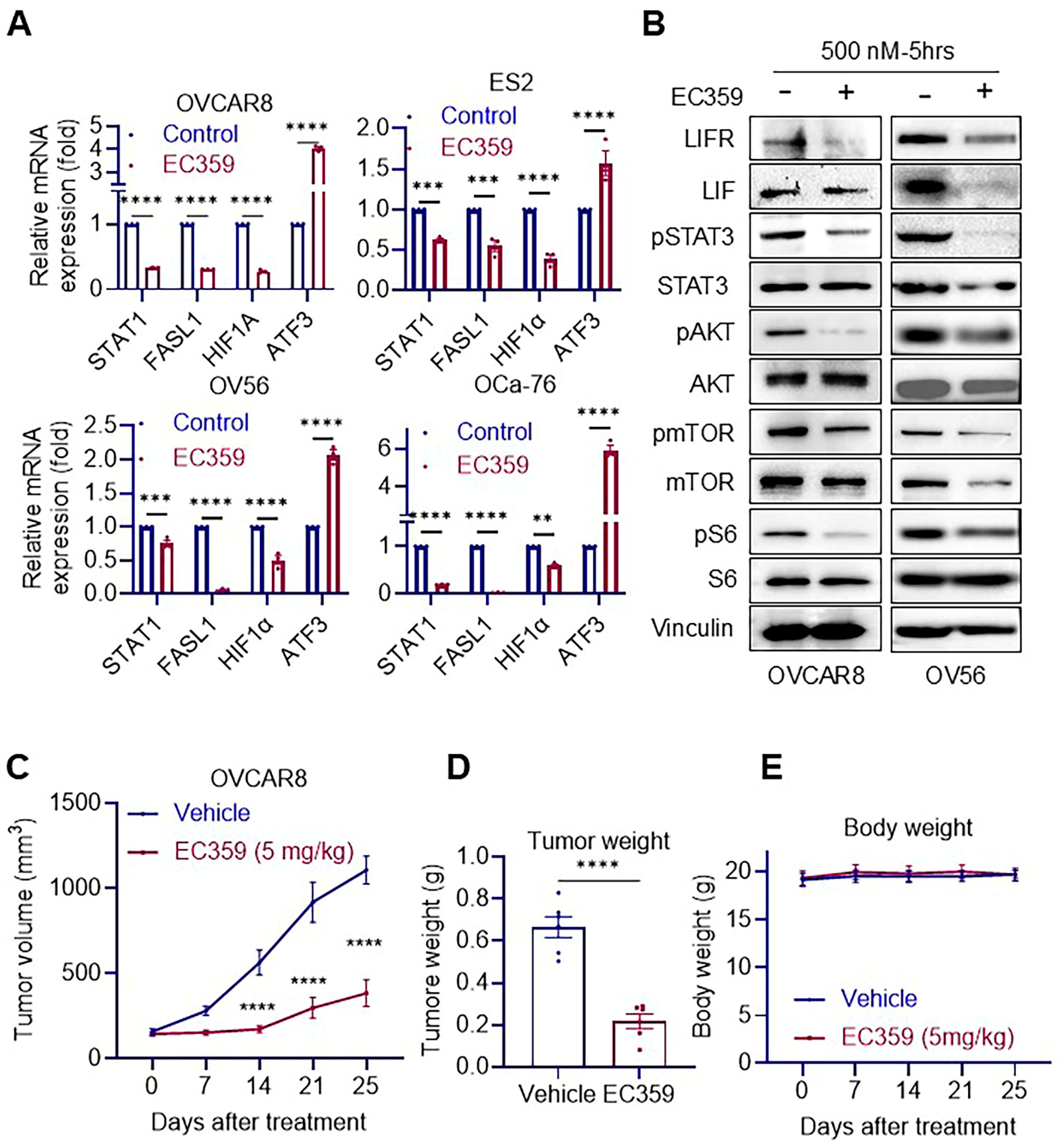
To evaluate the impact of LIFR inhibition, Ras/Raf-mutant OCa cells were treated with EC359 or vehicle control, and analyzed at both the transcriptional and translational levels using RT-qPCR and Western blotting. To assess transcriptional changes, we examined the expression of established LIFR target genes. EC359 treatment significantly downregulated STAT1, FASL1, and HIF1α, while upregulating ATF3, a gene typically repressed by LIFR signaling (Figure 2A). At the protein level, EC359 markedly reduced the phosphorylation of key signaling mediators, including mTOR, STAT3, and S6, confirming effective inhibition of LIFR-mediated signaling pathways (Figure 2B). To investigate the in vivo efficacy of EC359, we established OVCAR8 xenografts in female SCID mice. Mice were randomized to receive either vehicle or EC359 treatment. EC359 significantly suppressed tumor growth and reduced final tumor weights compared to vehicle controls, without affecting overall body weight (Figure 2C–E). Collectively, these findings demonstrate that Ras/Raf-mutant OCa cells depend on LIF/LIFR autocrine signaling for survival and proliferation. EC359 effectively disrupts this pathway, leading to inhibition of downstream signaling and suppression of tumor growth both in vitro and in vivo.

3.3. Trametinib Induces Aberrant Activation of LIFR Signaling in Ras/Raf-Mutant Ovarian Cancer Cells, Which Is Effectively Suppressed by EC359

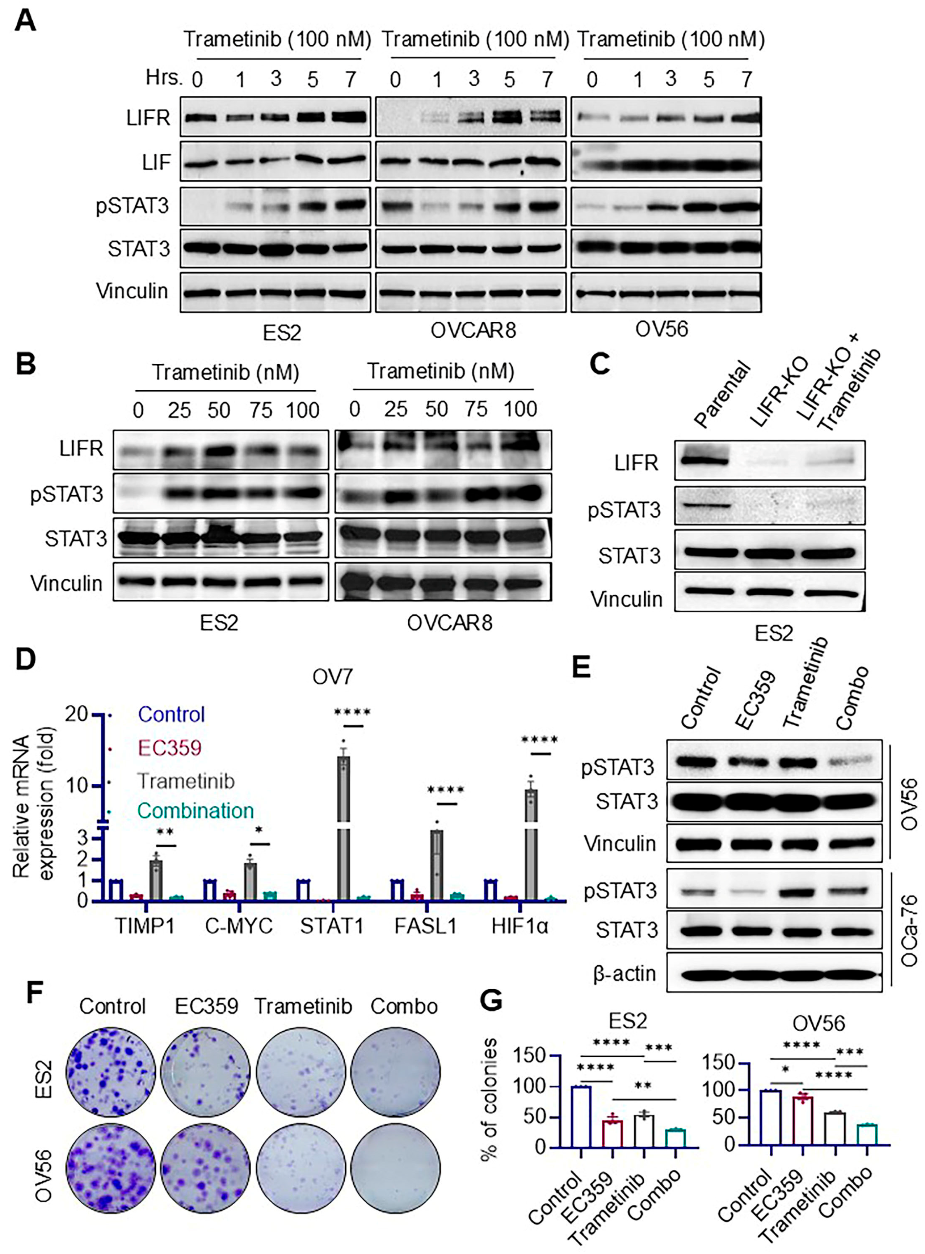
Trametinib, a MEK inhibitor, is known to be effective against tumors harboring Ras and Raf mutations by targeting hyperactive MEK signaling. In this study, we investigated its impact on LIF/LIFR signaling in Ras/Raf-mutant OCa cells. Western blot analyses revealed that trametinib treatment led to a time- and dose-dependent increase in LIFR expression and phosphorylation of STAT3 across multiple OCa cell lines (Figure 3A,B). This trametinib-induced activation of STAT3 was abolished in LIFR knockout (LIFR-KO) cells, confirming that LIFR is essential for mediating this response (Figure 3C). To determine whether inhibition of LIFR could counteract this feedback activation, we examined the effect of EC359, a small molecule LIFR inhibitor. RT-qPCR analysis in OV7 cells demonstrated that EC359 downregulated LIFR downstream target genes, indicating effective transcriptional suppression of LIFR signaling (Figure 3D). Western blot analysis in OV56 and OCa-76 cells further showed that EC359 blocked trametinib-induced upregulation of LIFR and pSTAT3, confirming that it attenuates LIFR-mediated signaling at the protein level (Figure 3E). Functionally, colony formation assays revealed that the combination of EC359 with trametinib significantly reduced the clonogenic survival of Ras/Raf-mutant OCa cells compared to trametinib alone (Figure 3F,G). The observed synergy between trametinib and EC359 is mechanistically linked to the aberrant activation of LIFR signaling specifically in Ras/Raf-mutated contexts. To address whether this mechanism extends to wild-type Ras OCa models (SKOV3, OVCAR3), we tested LIFR activation in those cells and found no evidence of LIFR activation (Supplementary Figure S1). These findings suggest that trametinib paradoxically induces LIFR signaling in Ras/Raf-mutant OCa cells, potentially contributing to adaptive resistance, and that co-treatment with EC359 effectively suppresses this compensatory pathway, enhancing anti-tumor efficacy.

3.4. Combination Therapy with Trametinib and EC359 Modulates Transcriptional Programs Associated with Apoptosis and Ribosome Biogenesis

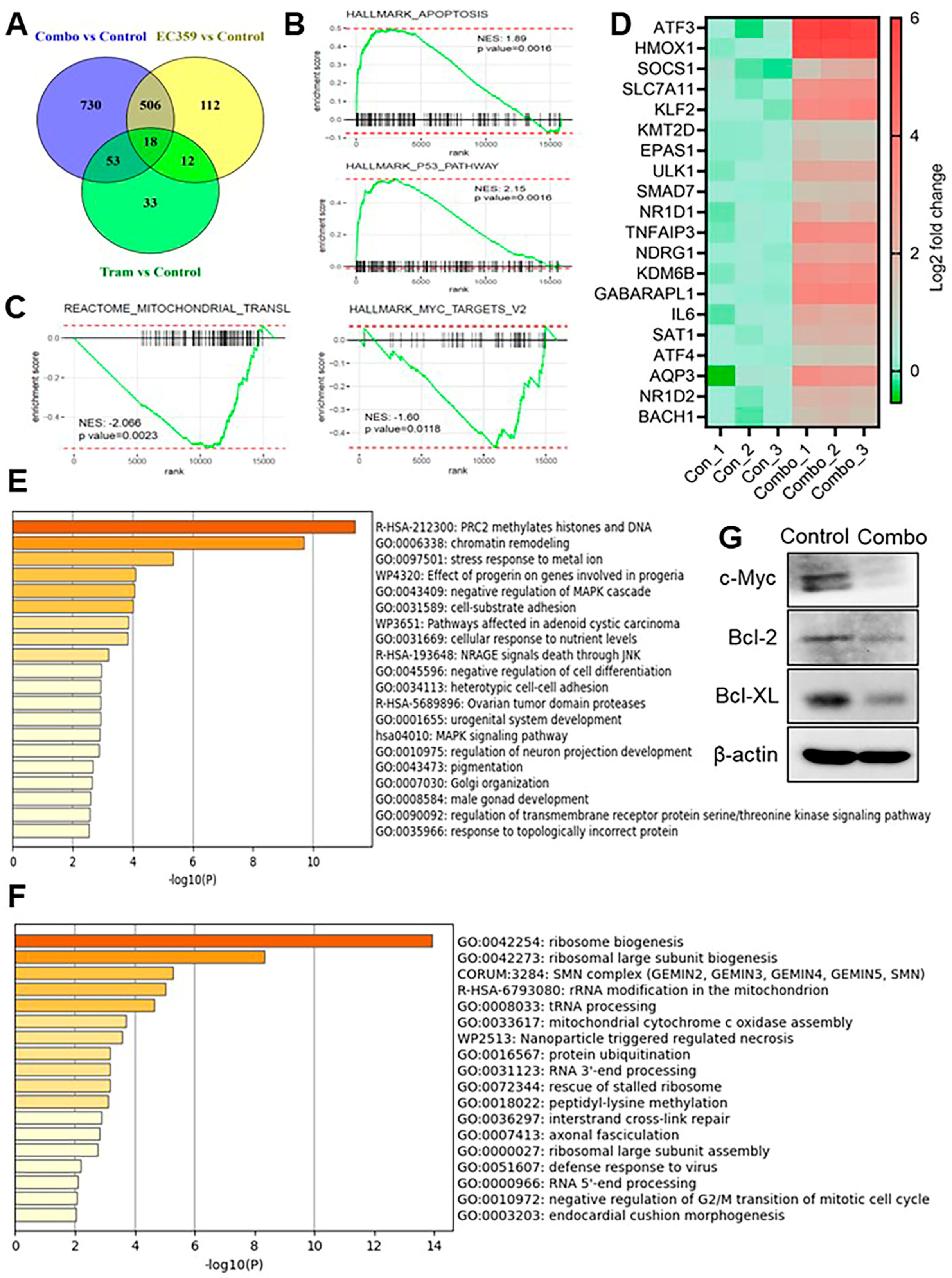
To better understand the molecular mechanisms underlying the enhanced anti-tumor effects of EC359 and trametinib combination therapy, we performed RNA sequencing on OVCAR8 cells treated with vehicle, EC359, trametinib, or the combination of both agents. Differential gene expression analysis revealed a distinct transcriptional profile in the combination-treated group, with 1307 genes showing ≥2-fold change compared to control. A Venn diagram illustrates the overlap of differentially expressed genes (DEGs) across treatment conditions, with a substantial number of unique DEGs in the combination group (Figure 4A). Gene Set Enrichment Analysis (GSEA) demonstrated that combination therapy significantly upregulated genes involved in apoptotic signaling, while concurrently downregulating genes associated with mitochondrial translation and MYC targets, two pathways critical for cellular proliferation and survival (Figure 4B,C). Additionally, RNA-seq analysis confirmed the upregulation of ferroptosis-inducing genes in the combination group when compared to control group indicating a breakdown in ferroptosis defense mechanisms that ultimately contributes to ferroptotic cell death (Figure 4D). To further explore the biological processes uniquely affected by the combination treatment, we performed pathway enrichment analysis using Metascape. Genes uniquely upregulated in the combination group were significantly enriched in pathways related to epigenetic regulation, including PRC2-mediated histone and DNA methylation, chromatin remodeling, and cellular stress response to metal ions, suggesting activation of tumor-suppressive and regulatory processes (Figure 4E). Conversely, genes uniquely downregulated in the combination group were enriched in ribosome biogenesis, ribosomal large subunit assembly, rRNA modification, and RNA 3′-end processing—pathways indicative of suppressed translational activity and protein synthesis (Figure 4F). RNA-seq findings were validated by Western blot analysis, which showed reduced levels of c-Myc and anti-apoptotic proteins following combination treatment (Figure 4G).
Collectively, these findings demonstrate that EC359 and trametinib combination therapy reprograms Ras/Raf-mutant OCa cells by activating pro-apoptotic and stress response pathways while simultaneously repressing biosynthetic and proliferative programs, contributing to the observed anti-tumor efficacy.

3.5. Combination Therapy of EC359 and Trametinib Reduces Tumor Growth in Ras/Raf Mutant OCa Xenografts

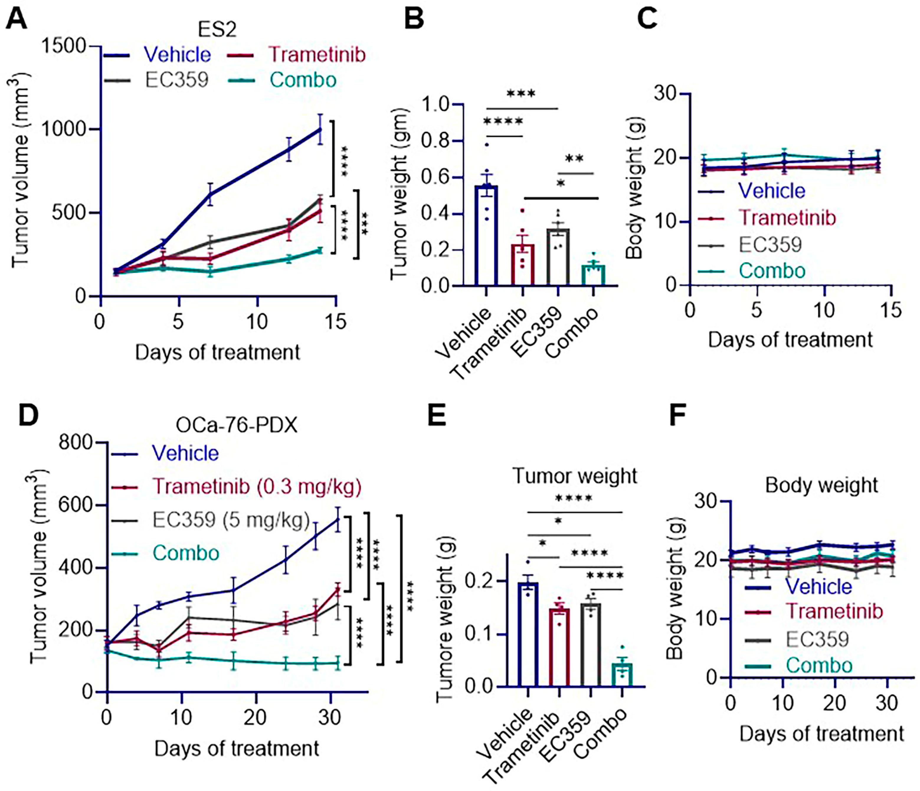
To assess the in vivo efficacy of EC359 and trametinib combination therapy in Ras/Raf-mutant OCa, we utilized two xenograft models. In the ES2 CDX model harboring Ras mutations, SCID mice were randomized into treatment groups (n = 6 tumors/group) and received intraperitoneal EC359, oral trametinib, or their combination. The combination therapy significantly suppressed tumor growth, as evidenced by reduced tumor volume and final tumor weight compared to either single-agent treatment or control (Figure 5A,B). Body weight remained consistent across all treatment groups, indicating good tolerability (Figure 5C). To further validate these findings, we employed the OCa-76 PDX model, which represent LGSOC. Similar to the ES2 model, combination therapy markedly reduced tumor volume and tumor weight compared to monotherapies or control (Figure 5D,E), with no significant changes in body weight observed (Figure 5F). Together, these results demonstrate that co-targeting LIFR and MEK pathways using EC359 and trametinib provides superior anti-tumor efficacy in both xenograft and PDX models, without inducing observable toxicity, supporting its potential as a promising therapeutic strategy.

4. Discussion

Ras and Raf mutations are frequently observed in specific subtypes of OCa, including LGSOC, borderline serous tumors, and MOC [5]. These mutations are associated with poor prognosis and resistance to conventional therapies, largely due to persistent activation of the MAPK signaling pathway [21]. Recent studies in pancreatic cancer have also implicated Ras mutations in the upregulation of LIF signaling, which further promotes tumor cell survival and therapy resistance [11]. Our study identifies LIFR signaling as a critical vulnerability in Ras/Raf-mutant OCa, and LGSOC and demonstrates the therapeutic potential of targeting this pathway using the LIFR inhibitor EC359, both as a monotherapy and in combination with the MEK inhibitor trametinib.
LGSOC and other Ras/Raf-driven ovarian cancers pose a significant therapeutic challenge due to their intrinsic resistance to chemotherapy and limited treatment options. Our group previously developed and characterized EC359, a selective small-molecule inhibitor that binds to LIFR and blocks ligand interaction [22]. The specificity and efficacy of EC359 have been validated in several cancer types, including Oca [19], endometrial [14,15], pancreatic [16,17], and renal cancers [18]. In this study, we show that EC359 significantly reduced tumor growth in Ras-mutant OVCAR8 xenografts without affecting body weight, signifies its therapeutic potential and tolerability in vivo.
The LIF/LIFR axis has been implicated in several hallmarks of cancer, including enhanced proliferation, immune evasion, chemoresistance, and poor patient survival. Autocrine or paracrine LIF/LIFR signaling loops have been described in various malignancies [23,24,25]. For instance, LIF acts in a paracrine manner in Ras/Raf-mutant pancreatic cancer to support tumor progression [11], and to promote stemness and invasion in pancreatic cancer [22]. In our study, we demonstrate that Ras- and Raf-mutant OCa cells express both LIF and LIFR, supporting the presence of an autocrine signaling loop. Pharmacologic inhibition of LIFR with EC359 significantly impaired cell viability and clonogenic survival across multiple Ras- and Raf-mutant OCa models.
EC359-induced cytotoxicity was associated with ferroptosis, a regulated form of cell death driven by iron-dependent lipid peroxidation. EC359 treatment increased lipid peroxidation in OCa cells as observed by BODIPY-C11 staining [18]. Further, EC359 treatment is promoted mitochondrial morphological changes characteristic of ferroptosis, as observed by electron microscopy [19]. Mechanistically, EC359 treatment suppressed phosphorylation of key downstream effectors of LIFR signaling, including mTOR, STAT3, and S6, indicating effective blockade of oncogenic signaling. In our studies using Ras/Raf mutant cancer cells, co-treatment with the ferroptosis inhibitor Ferrostatin-1 rescued EC359-induced cell death, and Western blot analysis confirmed downregulation of the ferroptosis regulator GPX4. These findings are consistent with recent reports that LIFR inhibition promotes ferroptosis [18,19] and suggest that LIFR signaling supports redox homeostasis in Ras/Raf-mutant OCa cells.
Published studies showed that trametinib resistance occur via multiple mechanisms including epigenetic adaptation, compensatory activation of PI3K/AKT or RTKs, and reactivation of the MAPK pathway [26,27,28]. Interestingly, in our studies we observed that trametinib treatment paradoxically upregulated LIF and LIFR expressions, potentially contributing to adaptive resistance. This aligns with previous studies showing feedback activation of survival pathways following MEK inhibition. Importantly, co-treatment with EC359 abrogated trametinib-induced LIFR activation, resulting in enhanced suppression of clonogenic survival and downstream signaling. Transcriptomic analysis further revealed that the EC359 + trametinib combination downregulated genes associated with the MYC and mitochondrial translation pathways. Further, combination therapy upregulated apoptosis-related genes, indicating a shift from a proliferative to a pro-death transcriptional program. These molecular changes were consistent with the observed in vivo efficacy of the combination therapy in both Ras- and Raf-mutant xenograft models, where it significantly reduced tumor volume and weight without inducing toxicity.
Our data suggest a favorable therapeutic window for combination therapy, as the doses of EC359 and trametinib used in this study were well tolerated and showed no signs of systemic toxicity. Our data suggest that the tumor-selective activity of this combination therapy arises from the presence of oncogenic Ras/Raf mutations in cancer cells, which drive constitutive activation of MEK signaling, and tumor cells with Ras/Raf mutations exhibit autocrine activation of the LIF/LIFR pathway, further sensitizing them to EC359. In contrast, normal cells lack these oncogenic drivers and therefore are less susceptible to combination therapy. Conducting detailed toxicity studies is beyond the scope of this study, and these will be examined in future studies.
Our research findings have important clinical implications for Ras/Raf-mutant OCa, which are resistant to standard chemotherapy and associated with poor prognosis. By demonstrating that EC359 potently reduces viability and clonogenic survival and induces both apoptosis and ferroptosis in Ras/Raf-mutant and low grade OCa cells, this work identifies LIFR signaling as a critical therapeutic vulnerability. Further, the ability of EC359 to overcome trametinib-induced compensatory LIFR activation also adds to its translation potential. Collectively, these results support a EC359/trametinib combination therapy in Ras/Raf-driven OCa and LGSOC.

5. Conclusions

In summary, our findings underscore the therapeutic potential of dual targeting the LIFR and MEK pathways in Ras/Raf-mutant OCa. EC359 effectively disrupts LIFR-mediated survival signaling and enhances the antitumor efficacy of trametinib by counteracting its compensatory upregulation of the LIF/LIFR axis. This combination strategy presents a promising approach to overcoming resistance mechanisms and improving therapeutic outcomes in patients with Ras/Raf-driven OCa.

6. Patents

Evestra Inc. San Antonio has a patent on EC359.

Supplementary Materials

The following supporting information can be downloaded at: https://www.mdpi.com/article/10.3390/biom15101396/s1.

Author Contributions

Conceptualization, Y.A.L., R.K.V., H.B.N. and S.V.; methodology, W.C.A., D.M.P., B.S. (Baskaran Subramani), B.E., M.M., P.R. and S.V.; Bio-informatic analysis, Y.C., Z.L. and S.V.; validation, R.K.V. and S.V.; formal analysis, W.C.A., D.M.P., B.S. (Baskaran Subramani), B.E., M.M. and P.R.; investigation, R.K.V. and S.V.; resources, B.S. (Bindu Santhamma), H.B.N., E.R.K. and Y.A.L.; data curation, W.C.A., D.M.P., B.S. (Baskaran Subramani), B.E., M.M., P.R., R.K.V. and S.V.; writing—original draft preparation, Y.A.L., R.K.V. and S.V.; writing—review and editing, Y.A.L., R.K.V., H.B.N., E.R.K. and S.V.; supervision, Y.A.L., R.K.V., S.V. and H.B.N.; funding acquisition, Y.A.L., H.B.N., R.K.V. and S.V. All authors have read and agreed to the published version of the manuscript.

Funding

This research was supported by MCC Pilot grant (Y.A.L.); NIH R01 CA267893 (S.V., R.V.), NIH R01 CA266970-01A1 (S.V., R.K.V., H.B.N.), VA I01 BX006280-01 (R.K.V.), NCI F99 CA284284-01 (B.E), AstraZeneca Young Investigator award (P.R) and NIH NCI R50 CA265339 (Z.L.). RNA-seq data were generated in the Genome Sequencing Facility at UT Health San Antonio, which is supported by NIH-NCI P30 CA054174 (Cancer Center at UT Health San Antonio), NIH Shared Instrumentation Grant 1S10 OD030311-01, and the CPRIT Core Facility Award RP220662.

Institutional Review Board Statement

This study used primary LGSOC cell line obtained from the Ob Gyn tissue core. The core obtained these materials in accordance with the approved IRB methodology. The study was carried out in compliance with the Declaration of Helsinki and received approval from the Institutional Review Board of UT Health San Antonio (HSC20190695N, 3 March 2019). The animal study protocol received approval from the Office of the Institutional Animal Care and Use Committee (Protocol number: 20220037AR, 5 September 2022) for research with animals.

Informed Consent Statement

Not applicable.

Data Availability Statement

All data generated for this study is included within this article. RNA-seq data have been deposited in the GEO database with GEO accession number GSE306834.

Conflicts of Interest

Bindu Santhamma is current employee of Evestra Inc. Ratna K. Vadlamudi serves as a co-founder of ETIRA Rx company. All other remaining authors declare no competing interests.

Abbreviations

The following abbreviations are used in this manuscript:
AKTProtein Kinase B
ANOVAAnalysis of Variance
ATCCAmerican Type Culture Collection
ATF3Activating Transcription Factor 3
BRAFB-Raf Proto-Oncogene, Serine/Threonine Kinase
EOCEpithelial Ovarian Cancer
ENOCEndometrioid Ovarian Cancer
FASL1Fas Ligand 1
GEOGene Expression Omnibus
GPX4Glutathione Peroxidase 4
HGSOCHigh-Grade Serous Ovarian Cancer
HIF1αHypoxia-Inducible Factor 1-alpha
IC50Half-Maximal Inhibitory Concentration
IPIntraperitoneal
JAKJanus Kinase
KRASKirsten Rat Sarcoma Viral Oncogene Homolog
LGSOCLow-Grade Serous Ovarian Cancer
LIFLeukemia Inhibitory Factor
LIFRLeukemia Inhibitory Factor Receptor
MEKMitogen-Activated Protein Kinase Kinase
MOCMucinous Ovarian Cancer
mTORMechanistic Target of Rapamycin
MTT3-(4,5-Dimethylthiazol-2-yl)-2,5-Diphenyltetrazolium Bromide
OCaOvarian Cancer
OCCCOvarian Clear Cell Carcinoma
PI3KPhosphoinositide 3-Kinase
RNA-seqRNA Sequencing
RT-qPCRReverse Transcription Quantitative PolymeRase Chain Reaction
SCIDSevere Combined Immunodeficiency
SEStandard Error
STAT3Signal Transducer and Activator of Transcription 3
STRShort Tandem Repeat
TMETumor Microenvironment

References

  1. Bowtell, D.D.; Bohm, S.; Ahmed, A.A.; Aspuria, P.J.; Bast, R.C., Jr.; Beral, V.; Berek, J.S.; Birrer, M.J.; Blagden, S.; Bookman, M.A.; et al. Rethinking ovarian cancer II: Reducing mortality from high-grade serous ovarian cancer. Nat. Rev. Cancer 2015, 15, 668–679. [Google Scholar] [CrossRef]
  2. Bast, R.C., Jr.; Matulonis, U.A.; Sood, A.K.; Ahmed, A.A.; Amobi, A.E.; Balkwill, F.R.; Wielgos-Bonvallet, M.; Bowtell, D.D.L.; Brenton, J.D.; Brugge, J.S.; et al. Critical questions in ovarian cancer research and treatment: Report of an American Association for Cancer Research Special Conference. Cancer 2019, 125, 1963–1972. [Google Scholar] [CrossRef]
  3. Prat, J. Ovarian carcinomas: Five distinct diseases with different origins, genetic alterations, and clinicopathological features. Virchows Arch. 2012, 460, 237–249. [Google Scholar] [CrossRef] [PubMed]
  4. Peng, Y.; Yang, Q. Targeting KRAS in gynecological malignancies. FASEB J. 2024, 38, e70089. [Google Scholar] [CrossRef] [PubMed]
  5. Nakayama, N.; Nakayama, K.; Yeasmin, S.; Ishibashi, M.; Katagiri, A.; Iida, K.; Fukumoto, M.; Miyazaki, K. KRAS or BRAF mutation status is a useful predictor of sensitivity to MEK inhibition in ovarian cancer. Br. J. Cancer 2008, 99, 2020–2028. [Google Scholar] [CrossRef]
  6. Gershenson, D.M.; Miller, A.; Brady, W.E.; Paul, J.; Carty, K.; Rodgers, W.; Millan, D.; Coleman, R.L.; Moore, K.N.; Banerjee, S.; et al. Trametinib versus standard of care in patients with recurrent low-grade serous ovarian cancer (GOG 281/LOGS): An international, randomised, open-label, multicentre, phase 2/3 trial. Lancet 2022, 399, 541–553. [Google Scholar] [CrossRef]
  7. Nicola, N.A.; Babon, J.J. Leukemia inhibitory factor (LIF). Cytokine Growth Factor Rev. 2015, 26, 533–544. [Google Scholar] [CrossRef]
  8. Stahl, N.; Boulton, T.G.; Farruggella, T.; Ip, N.Y.; Davis, S.; Witthuhn, B.A.; Quelle, F.W.; Silvennoinen, O.; Barbieri, G.; Pellegrini, S.; et al. Association and activation of Jak-Tyk kinases by CNTF-LIF-OSM-IL-6 beta receptor components. Science 1994, 263, 92–95. [Google Scholar] [CrossRef]
  9. Li, X.; Yang, Q.; Yu, H.; Wu, L.; Zhao, Y.; Zhang, C.; Yue, X.; Liu, Z.; Wu, H.; Haffty, B.G.; et al. LIF promotes tumorigenesis and metastasis of breast cancer through the AKT-mTOR pathway. Oncotarget 2014, 5, 788–801. [Google Scholar] [CrossRef] [PubMed]
  10. Liu, S.C.; Tsang, N.M.; Chiang, W.C.; Chang, K.P.; Hsueh, C.; Liang, Y.; Juang, J.L.; Chow, K.P.; Chang, Y.S. Leukemia inhibitory factor promotes nasopharyngeal carcinoma progression and radioresistance. J. Clin. Investig. 2013, 123, 5269–5283. [Google Scholar] [CrossRef]
  11. Shi, Y.; Gao, W.; Lytle, N.K.; Huang, P.; Yuan, X.; Dann, A.M.; Ridinger-Saison, M.; DelGiorno, K.E.; Antal, C.E.; Liang, G.; et al. Targeting LIF-mediated paracrine interaction for pancreatic cancer therapy and monitoring. Nature 2019, 569, 131–135. [Google Scholar] [CrossRef]
  12. Wang, M.T.; Fer, N.; Galeas, J.; Collisson, E.A.; Kim, S.E.; Sharib, J.; McCormick, F. Blockade of leukemia inhibitory factor as a therapeutic approach to KRAS driven pancreatic cancer. Nat. Commun. 2019, 10, 3055. [Google Scholar] [CrossRef]
  13. Viswanadhapalli, S.; Luo, Y.; Sareddy, G.R.; Santhamma, B.; Zhou, M.; Li, M.; Ma, S.; Sonavane, R.; Pratap, U.P.; Altwegg, K.A.; et al. EC359: A First-in-Class Small-Molecule Inhibitor for Targeting Oncogenic LIFR Signaling in Triple-Negative Breast Cancer. Mol. Cancer Ther. 2019, 18, 1341–1354. [Google Scholar] [CrossRef]
  14. Tang, W.; Ramasamy, K.; Pillai, S.M.A.; Santhamma, B.; Konda, S.; Venkata, P.P.; Blankenship, L.; Liu, J.; Liu, Z.; Altwegg, K.A.; et al. LIF/LIFR oncogenic signaling is a novel therapeutic target in endometrial cancer. Cell Death Discov. 2021, 7, 216. [Google Scholar] [CrossRef]
  15. Blankenship, L.; Pratap, U.P.; Yang, X.; Liu, Z.; Altwegg, K.A.; Santhamma, B.; Ramasamy, K.; Konda, S.; Chen, Y.; Lai, Z.; et al. Inhibition of LIFR Blocks Adiposity-Driven Endometrioid Endometrial Cancer Growth. Cancers 2022, 14, 5400. [Google Scholar] [CrossRef] [PubMed]
  16. Hall, B.R.; Cannon, A.; Thompson, C.; Santhamma, B.; Chavez-Riveros, A.; Bhatia, R.; Nair, H.B.; Nickisch, K.; Batra, S.K.; Kumar, S. Utilizing cell line-derived organoids to evaluate the efficacy of a novel LIFR-inhibitor, EC359 in targeting pancreatic tumor stroma. Genes. Cancer 2019, 10, 1–10. [Google Scholar] [CrossRef] [PubMed]
  17. Di Giorgio, C.; Lupia, A.; Marchiano, S.; Bordoni, M.; Bellini, R.; Massa, C.; Urbani, G.; Roselli, R.; Moraca, F.; Sepe, V.; et al. Repositioning Mifepristone as a Leukaemia Inhibitory Factor Receptor Antagonist for the Treatment of Pancreatic Adenocarcinoma. Cells 2022, 11, 3482. [Google Scholar] [CrossRef]
  18. Feng, C.-Z.; Li, N.-Z.; Hu, X.-B.; Xie, Y.-Y.; Huang, Q.-H.; Zhang, J.; Chen, Z.; Chen, S.-J.; Wang, F.; Sun, X.-J. The LIFR-targeting small molecules EC330/EC359 are potent ferroptosis inducers. Genes Dis. 2023, 10, 735–738. [Google Scholar] [CrossRef]
  19. Ebrahimi, B.; Viswanadhapalli, S.; Pratap, U.P.; Rahul, G.; Yang, X.; Venkata, P.P.; Drel, V.; Santhamma, B.; Konda, S.; Li, X.; et al. Pharmacological inhibition of the LIF/LIFR autocrine loop reveals vulnerability of ovarian cancer cells to ferroptosis. NPJ Precis. Oncol. 2024, 8, 118. [Google Scholar] [CrossRef]
  20. Walters, D.M.; Lindberg, J.M.; Adair, S.J.; Newhook, T.E.; Cowan, C.R.; Stokes, J.B.; Borgman, C.A.; Stelow, E.B.; Lowrey, B.T.; Chopivsky, M.E.; et al. Inhibition of the growth of patient-derived pancreatic cancer xenografts with the MEK inhibitor trametinib is augmented by combined treatment with the epidermal growth factor receptor/HER2 inhibitor lapatinib. Neoplasia 2013, 15, 143–155. [Google Scholar] [CrossRef]
  21. Malpica, A.; Wong, K.K. The molecular pathology of ovarian serous borderline tumors. Ann. Oncol. 2016, 27 (Suppl. 1), i16–i19. [Google Scholar] [CrossRef] [PubMed]
  22. Sasaki, N.; Hirano, K.; Shichi, Y.; Gomi, F.; Yoshimura, H.; Matsushita, A.; Toyoda, M.; Ishiwata, T. Gp130-Mediated STAT3 Activation Contributes to the Aggressiveness of Pancreatic Cancer through H19 Long Non-Coding RNA Expression. Cancers 2022, 14, 2055. [Google Scholar] [CrossRef]
  23. Hanahan, D. Hallmarks of Cancer: New Dimensions. Cancer Discov. 2022, 12, 31–46. [Google Scholar] [CrossRef]
  24. Jorgensen, M.M.; de la Puente, P. Leukemia Inhibitory Factor: An Important Cytokine in Pathologies and Cancer. Biomolecules 2022, 12, 217. [Google Scholar] [CrossRef]
  25. Zhang, C.; Liu, J.; Wang, J.; Hu, W.; Feng, Z. The emerging role of leukemia inhibitory factor in cancer and therapy. Pharmacol. Ther. 2021, 221, 107754. [Google Scholar] [CrossRef] [PubMed]
  26. Kun, E.; Tsang, Y.T.M.; Ng, C.W.; Gershenson, D.M.; Wong, K.K. MEK inhibitor resistance mechanisms and recent developments in combination trials. Cancer Treat. Rev. 2021, 92, 102137. [Google Scholar] [CrossRef]
  27. Kakadia, S.; Yarlagadda, N.; Awad, R.; Kundranda, M.; Niu, J.; Naraev, B.; Mina, L.; Dragovich, T.; Gimbel, M.; Mahmoud, F. Mechanisms of resistance to BRAF and MEK inhibitors and clinical update of US Food and Drug Administration-approved targeted therapy in advanced melanoma. OncoTargets Ther. 2018, 11, 7095–7107. [Google Scholar] [CrossRef] [PubMed]
  28. Sanchez, J.N.; Wang, T.; Cohen, M.S. BRAF and MEK Inhibitors: Use and Resistance in BRAF-Mutated Cancers. Drugs 2018, 78, 549–566. [Google Scholar] [CrossRef]
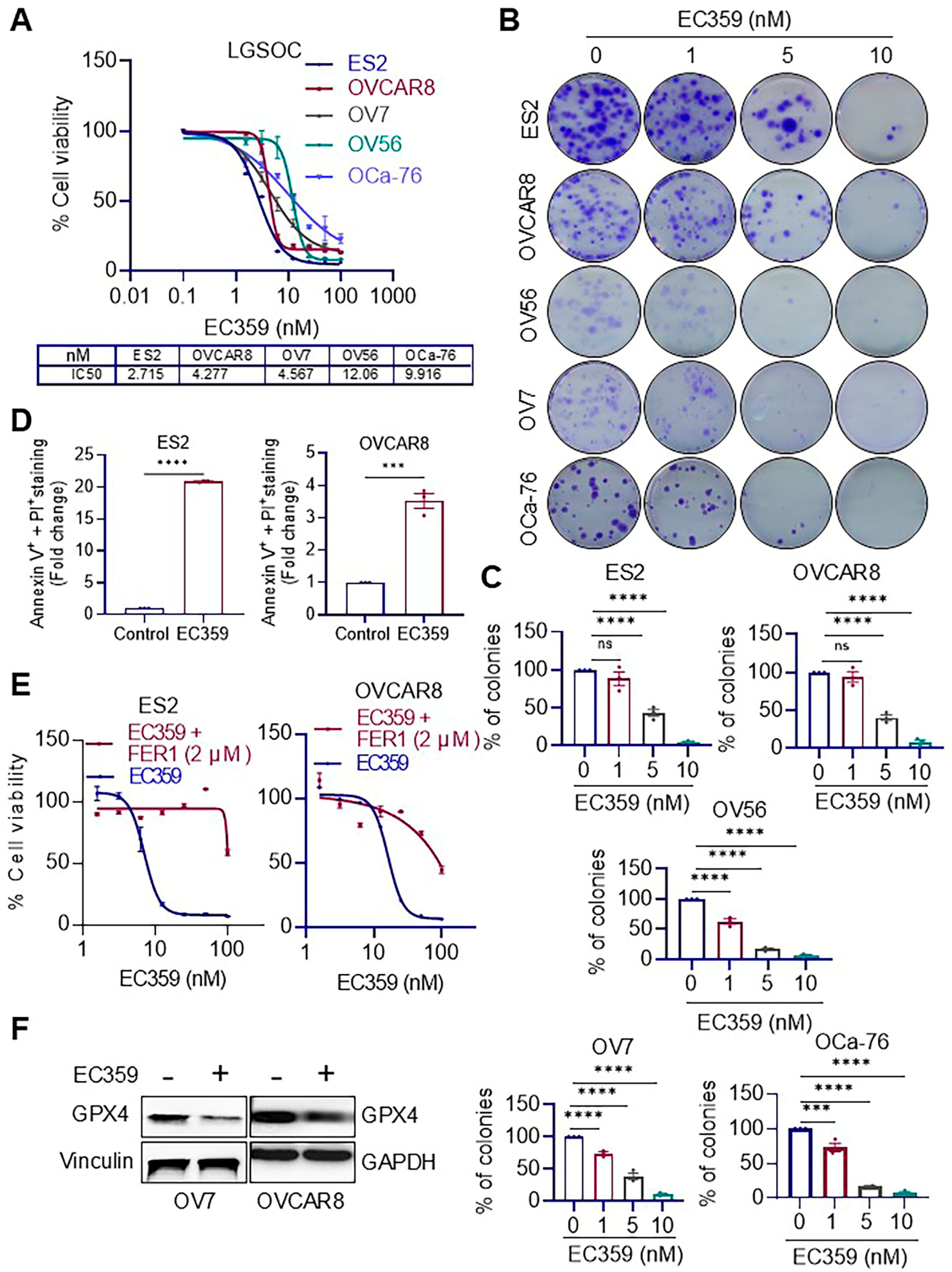
Figure 1. EC359 inhibits cell viability and induces ferroptosis in OCa models. (A) MTT assays demonstrate a dose-dependent decrease in cell viability following EC359 treatment across five OCa models (OVCAR8, OV7, OV56, OCa-76, and ES2). (B,C) EC359 markedly suppresses clonogenic survival in representative cell lines. Representative colony images (B) and corresponding quantification (C) are shown. (D) Annexin V assays reveal a significant increase in apoptotic cell populations upon EC359 (100 nM-48 h) treatment. (E) Co-treatment with the ferroptosis inhibitor ferrostatin-1 (Fer-1) rescues EC359-induced loss of cell viability. (F) Western blot analysis shows downregulation of the key ferroptosis regulator GPX4 in EC359 (100 nM-7 h) treated cells. Dots represent number of replicates. Data presented as mean ± SEM; significance was determined by Student’ t-test; one-way ANOVA; *** p < 0.001; **** p < 0.0001; ns, not significant.
Biomolecules 15 01396 g001
Figure 2. EC359 inhibits LIFR downstream signaling and reduces tumor growth in vivo. (A) RT-qPCR analysis showing reduced expression of LIFR downstream target genes in Ras/Raf mutant cell lines following EC359 (100 nM-7 h) treatment compared to control. (B) Western blot analysis demonstrating decreased levels of phosphorylated STAT3 and other LIFR pathway effectors in EC359-treated cell lines. (C) Tumor volume measurements over time show significant reduction in tumor growth in EC359-treated mice compared to vehicle-treated controls bearing OVCAR8 xenografts. (D) Final tumor weight at the end of the treatment period was significantly lower in the EC359 group compared to vehicle. (E) Body weight of mice treated with EC359, or vehicle remained unchanged throughout the study, indicating lack of overt systemic toxicity. Data presented as mean ± SEM; significance was determined by two-way ANOVA; ** p < 0.01; *** p < 0.001; **** p < 0.0001.
Biomolecules 15 01396 g002
Figure 3. EC359 attenuates trametinib-induced LIFR signaling and enhances therapeutic efficacy in OCa cells. (A,B) Western blot analysis of multiple OCa cell lines shows a time- (A) and dose- (B) dependent increase in LIFR and phospho-STAT3 levels following trametinib treatment. (C) Western blot analysis of LIFR knockout (LIFR-KO) cells treated with trametinib (100 nM-7 h) reveals no induction of phospho-STAT3 in the absence of LIFR, confirming its requirement for trametinib-induced STAT3 activation. (D) RT-qPCR analysis in OV7 cells shows that EC359 suppresses LIFR downstream target gene expression, supporting its inhibitory effect on LIFR signaling. (E) Western blot analysis in OV56 and OCa-76 cells demonstrates that EC359 attenuates trametinib-induced upregulation of LIFR and pSTAT3, confirming its role in blocking LIFR-mediated signaling. (F,G) Colony formation assays reveal that combination treatment with EC359 (ES2-1 nM; OV56-5 nM) and trametinib (ES2-5 nM; OV56-10 nM) significantly reduces colony-forming ability compared to trametinib alone. Representative images (F) and quantification of colony formation (G) are shown. Data presented as mean ± SEM; significance was determined by two-way ANOVA; * p < 0.05; ** p < 0.01; *** p < 0.001; **** p < 0.0001.
Biomolecules 15 01396 g003
Figure 4. RNA-seq analysis reveals modulation of apoptotic and ribosomal biogenesis pathways in OVCAR8 cells following combination treatment with EC359 and trametinib. (A) Venn diagram showing the overlap of differentially expressed genes (DEGs) in OVCAR8 cells treated with EC359, trametinib, or their combination compared to control. (B,C) Gene Set Enrichment Analysis (GSEA) shows the top significantly enriched pathways in the combination-treated group, including upregulation of apoptotic signaling (B) and downregulation of mitochondrial translation and MYC target genes (C). (D) Heatmap show activation of several ferroptosis-related genes following combination treatment (E) Bar plot displaying the top enriched pathways (ranked by −log10(P)) from genes uniquely upregulated in the combination group, as identified by Metascape. Notable enriched pathways include PRC2-mediated histone and DNA methylation, chromatin remodeling, and stress response to metal ion indicating activation of key tumor-suppressive and regulatory processes. (F) Bar plot showing the top enriched pathways (ranked by −log10(P)) derived from genes uniquely downregulated in the combination group. These include ribosome biogenesis, ribosomal large subunit biogenesis, rRNA modification, and RNA processing. (G) Western blot showing the levels of c-MYC protein levels, and BCL-2 family proteins following combination treatment. Significant genes with fold change > 2 and adjusted p value < 0.05 were used for interpreting the biological pathways using GSEA. Fold change values in heatmap were calculated by normalization of FPKM values to those for control.
Biomolecules 15 01396 g004
Figure 5. EC359 enhances the antitumor efficacy of trametinib in OCa xenograft and PDX models. (A,B) In the ES2 xenograft model, combination therapy with EC359 and trametinib led to a significant reduction in tumor volume (A) and final tumor weight (B) compared to control and single-agent treatments (n = 6 tumors). (C) Body weight remained stable across all treatment groups, indicating good tolerability. (D,E) In the OCa-76 PDX model, combination therapy significantly decreased tumor volume (D) and tumor weight (E) relative to control or monotherapies (n = 4 tumors). (F) No significant changes in body weight were observed among treatment groups, confirming treatment safety in vivo. Data presented as mean ± SEM; significance was determined by two-way ANOVA; * p < 0.05; ** p < 0.01; *** p < 0.001; **** p < 0.0001.
Biomolecules 15 01396 g005
Disclaimer/Publisher’s Note: The statements, opinions and data contained in all publications are solely those of the individual author(s) and contributor(s) and not of MDPI and/or the editor(s). MDPI and/or the editor(s) disclaim responsibility for any injury to people or property resulting from any ideas, methods, instructions or products referred to in the content.