Article

Probing the Influence of Novel Organometallic Copper(II) Complexes on Spinach PSII Photochemistry Using OJIP Fluorescence Transient Measurements

by
Sergei K. Zharmukhamedov
1,*,
Mehriban S. Shabanova
2,
Irada M. Huseynova
2,
Mehmet Sayım Karacan
3,
Nurcan Karacan
3,
Hande Akar
3,
Vladimir D. Kreslavski
1,
Hesham F. Alharby
4,
Barry D. Bruce
5 and
Suleyman I. Allakhverdiev
1,6,7,*
1
Institute of Basic Biological Problems, FRC PSCBR RAS, 142290 Pushchino, Russia
2
Bionanotechnology Laboratory, Institute of Molecular Biology and Biotechnology, Azerbaijan National Academy of Sciences, AZ1143 Baku, Azerbaijan
3
Department of Chemistry, Science Faculty, Gazi University, Teknikokullar, Ankara 06500, Turkey
4
Department of Biological Sciences, Faculty of Science, King Abdulaziz University, Jeddah 21589, Saudi Arabia
5
Departments of Biochemistry & Cellular and Molecular Biology, Chemical and Biomolecular Engineering and Microbiology, University of Tennessee, Knoxville, TN 37996, USA
6
K.A. Timiryazev Institute of Plant Physiology, Russian Academy of Sciences, Botanicheskaya Street 35, 127276 Moscow, Russia
7
Faculty of Engineering and Natural Sciences, Bahcesehir University, Istanbul 34349, Turkey
*
Authors to whom correspondence should be addressed.
Biomolecules 2023, 13(7), 1058; https://doi.org/10.3390/biom13071058
Submission received: 22 February 2023 / Revised: 26 June 2023 / Accepted: 27 June 2023 / Published: 29 June 2023

Abstract

:
Modern agricultural cultivation relies heavily on genetically modified plants that survive after exposure to herbicides that kill weeds. Despite this biotechnology, there is a growing need for new sustainable, environmentally friendly, and biodegradable herbicides. We developed a novel [CuL2]Br2 complex (L = bis{4H-1,3,5-triazino[2,1-b]benzothiazole-2-amine,4-(2-imidazole) that is active on PSII by inhibiting photosynthetic oxygen evolution on the micromolar level. [CuL2]Br2 reduces the FV of PSII fluorescence. Artificial electron donors do not rescind the effect of [CuL2]Br2. The inhibitory mechanism of [CuL2]Br2 remains unclear. To explore this mechanism, we investigated the effect of [CuL2]Br2 in the presence/absence of the well-studied inhibitor DCMU on PSII-containing membranes by OJIP Chl fluorescence transient measurements. [CuL2]Br2 has two effects on Chl fluorescence transients: (1) a substantial decrease of the Chl fluorescence intensity throughout the entire kinetics, and (2) an auxiliary “diuron-like” effect. The initial decrease dominates and is observed both with and without DCMU. In contrast, the “diuron-like” effect is small and is observed only without DCMU. We propose that [CuL2]Br2 has two binding sites for PSII with different affinities. At the high-affinity site, [CuL2]Br2 produces effects similar to PSII reaction center inhibition, while at the low-affinity site, [CuL2]Br2 produces effects identical to those of DCMU. These results are compared with other PSII-specific classes of herbicides.

Graphical Abstract

1. Introduction

Currently, new approaches in agricultural economics are being intensively developed and put into practice, which selectively endow economically significant (genetically modified) crops with survival when treated with modern chemical agents that effectively suppress the growth and development of unwanted plant species [1]. The other side of these approaches (no less significant than the creation of genetically modified plant species) are studies aimed at identifying and studying the mechanism(s) of action of the widest possible range and the largest number of different chemical agents—potential prototypes of new, more effective, more environmentally friendly, more bioremediable, etc. of compounds for suppressing the growth of weeds [2]. In addition, these detectable chemical agents may turn out to be new highly specific tools of scientific knowledge, acting as inhibitors, acceptors, or donors of an electron with special properties [3]. Many well-studied inhibitors of photosynthetic electron transfer, electron donors, and acceptors are already widely used in scientific research, for example, to isolate the desired site of the electron transport chain of photosynthesis [4,5] and even for practical purposes—for example, in modified natural systems for generating molecular hydrogen [5,6].
Photosynthesis, the only process of generating biomass on the planet from carbon dioxide and water due to the energy of solar radiation, is carried out by the photosynthetic apparatus (PA) of phototrophs, including higher plants. One of the main PA pigment–protein–lipid complexes is the PSII complex, which splits water into high-energy electrons and protons, and evolving oxygen. The structural and functional organization of PA, including PSII, has been considered in detail in numerous reviews [7]. Among all the complexes that make up PA, PSII, and, especially, the oxygen-evolving complex PSII, which oxidizes water, suffer most from the action of various stress factors, including chemical agents [8]. Suppressing the activity of PA, primarily its most vulnerable part, PSII, is the most effective way to block the growth of a plant. This approach underlies the action of the majority of herbicides, inhibitors of photosynthesis [5].
Among the studies of various chemical inhibitors of various kinds of biological activities, including the photosynthetic activity of the components of the electron transport chain of photosynthesis (but mainly PSII), studies are developing organic complexes [9,10,11,12], as well as complexes of organic ligands with semimetals (Sb, As, etc.) [13] and transition metals (Fe, Pb, Co, Ni, Cr, Zn) [3,14,15], including organometallic complexes based on copper (Cu(II)) [16,17].
Many of these metals and semimetals in their free form have a very high ability to enter into various reactions, but exhibit low solubility in hydrophobic media and, as a result, may not achieve their intended targets as plant growth inhibitors. This also applies to copper cations, which are efficiently bound by organic buffer solutions [18], e.g., diphenylcarbazide, an exogenous electron donor widely used in photosynthesis studies [19,20], that chemically interact in solution with sodium ascorbate [21], hydroxylamine [22], dithionite [23], quinones [24], ferricyanide [25].
The several effects of a new organometallic complex based on Cu(II) ions with organic ligand (L = bis{4H-1,3,5-triazino [2,1-b]benzothiazole-2-amine,4-(2-imidazole)}copper(II) bromide complex)—[CuL2]Br2 on photochemical activity in PSII-containing membranes have recently been described [26]. To relieve the reader of the need to refer to our previous publication, we present the structure of the ligand (A) and the Cu(II)-complex (B) here again in Figure 1.
It has been shown that [CuL2]Br2: (1) is not an artificial electron acceptor for PSII; (2) inhibits photosynthetic electron transfer measured as photoinduced oxygen evolution; (3) in steady-state measurements, diminished FM values of the PSII chlorophyll fluorescence yield occur exclusively at the expense of FV values, which do not recover by adding artificial electron donors. (4) This new compound presumably has no significant effect on the native state of PSII proteins nor on the interaction of PSI with PSII with efficacy, which is no more than 5–10% of the DCMU effect [26]. Based on obtained results, it was proposed that main effect on the PSII photochemical activity is probably due to the interaction of the inhibitory agent with reaction center (RC) leading to some conformational changes in its structure. Further studies using other methods are needed to obtain a more accurate answer.
Registration of the PSII fast chlorophyll fluorescence induction (OJIP-kinetic) and its comprehensive analysis (JIP-test) is a quick, non-invasive, non-destructive, reliable, sensitive, informative, and convenient research method with which information can be readily obtained concerning the state and functioning of practically every component of the donor and acceptor sides of the PSII RC as well as about all intermediates of the PA in intact samples under stress impacts, including effects of various inhibitors [27,28]. If several sites of inhibitor action are detected by JIP-testing of PSII, one may determine which of these sites of action (and/or effects) of the inhibitor are of a primary or secondary nature. It may be revealed by differences in the dependence of changes in the magnitude of different peaks of OJIP kinetics on the concentration of the added inhibitor, and/or by identifying additional peaks after appropriate normalization procedures and the subtraction of control kinetics OJIP from the OJIP-kinetics in the presence of the inhibitor [27,28].
Here, we used the JIP test to elucidate inhibitory impacts of [CuL2]Br2 on PSII-containing membranes in more detail. Furthermore, we studied the effects of [CuL2]Br2 on PSII-containing membranes in the presence of DCMU, a known inhibitor of electron transfer on the acceptor side of PSII, blocking the oxidation of the reduced primary electron acceptor QA (QA) by the plastoquinone molecules from the membrane pool [4,29,30]. Investigation of the effects of [CuL2]Br2 in the presence of DCMU is a constructive experimental approach, since it allows using DCMU to exclude the possible influence of the remaining (in the PSII-membranes after their isolation) molecules of plastoquinone electron acceptors (PQ-9), approximately two molecules of PQ-9 per PSII RC [31,32]. On the other hand, if it turns out that the effects of [CuL2]Br2 and DCMU manifest themselves dependently or independently of each other, then these data will make it possible to more clearly judge the possible site of action and/or binding of the new Cu(II)-complex, as was shown, for example, in a study evaluating the site of action and/or binding of perfluoroisopropyldinitrobenzene derivatives, inhibitors of the K-15 type [33], and a protein synthesis inhibitor, chloramphenicol [34].

2. Materials and Methods

2.1. Isolation of PSII-Containing Membranes

The oxygen-evolving PSII-containing membranes were isolated from the leaves of the greenhouse spinach (Spinacia oleracea L.), according to [35], with a little modification as in [36]. These PSII-containing membranes contain about 200 chlorophyll molecules per RC (per one molecule of photoactive pheophytin) [37] and, when illuminated with red light (λ ≥ 650 nm) of saturating intensity, they release oxygen in the presence of two artificial electron acceptors (0.1 mM 2,5-dichloro-p-benzoquinone and 1 mM K3Fe(CN)6) at a rate of 450–500 µmol O2 (mg Chl)−1 h−1. PSII-containing membranes suspended in medium (A) (50 mM MES-NaOH (pH 6.5), 300 mM sucrose, 15 mM NaCl) were stored at −80 °C. The concentration of total chlorophyll in PSII-containing membranes was determined by extraction with 96% (v/v) ethanol [38].

2.2. Fast Induction Kinetics of Chlorophyll Fluorescence

Fast induction kinetics of chlorophyll fluorescence associated with photoreduction of the PSII primary electron acceptor, plastoquinone QA, were recorded using a MULTI-COLOR-PAM fluorimeter (Heinz Walz GmbH, Pfullingen, Germany) in a quartz cuvette (optical path length, 1 cm), at room temperature and constant stirring, after adaptation in the dark for at least 15 min. The final concentration of PSII-containing membranes in terms of chlorophyll was 4 µg mL−1. The conditions for measuring fast induction curves of chlorophyll fluorescence using this fluorimeter are described in detail in [39]. Each kinetic result represents are average of 5 independent experiments. The measurements were carried out as follows. A volume of the initial solution of PSII-containing membranes was prepared in medium containing: 50 mM MES–NaOH (pH 6.5), 300 mM sucrose, 15 mM NaCl, and then either an inhibitor solution or the same volume of solvent (in which this inhibitory agent was prepared) was added to an aliquot taken from this volume. This guaranteed the same chlorophyll concentration in all measurements. Based on the chlorophyll fluorescence fast induction curves, a number of fluorescence parameters of PSII chlorophyll were determined and/or calculated.

2.3. Spectrophotometric Measurements

The absorption spectra of the [CuL2]Br2 complex were recorded in a standard quartz cell (Hellma, Müllheim, Germany) with an optical path length of 10 mm on a two-beam Shimadzu spectrophotometer, model UV-1800 (Shimadzu UV-1800, Shimadzu Europa GmbH, Duisburg, Germany) in the wavelength range 200–700 nm (optical slit width 2 nm, write speed 2 nm s−1) at room temperature, in measurement medium used for OJIP kinetics. The concentration of the [CuL2]Br2 complex was 0.1 mM and corresponded to the maximum concentration used in all experiments.

2.4. Solutions of Inhibitory Agents

Stock solutions of (3-(3,4-dichlorophenyl)-1,1-dimethylurea, DCMU,) and [CuL2]Br2 and subsequent dilute solutions were prepared in dimethyl sulfoxide (DMSO). In all measurements, the final concentration of DMSO did not exceed 1%. In separate experiments, we have shown that DMSO at this concentration has no effect on either the intensity or the shape of the OJIP-kinetic.

3. Results

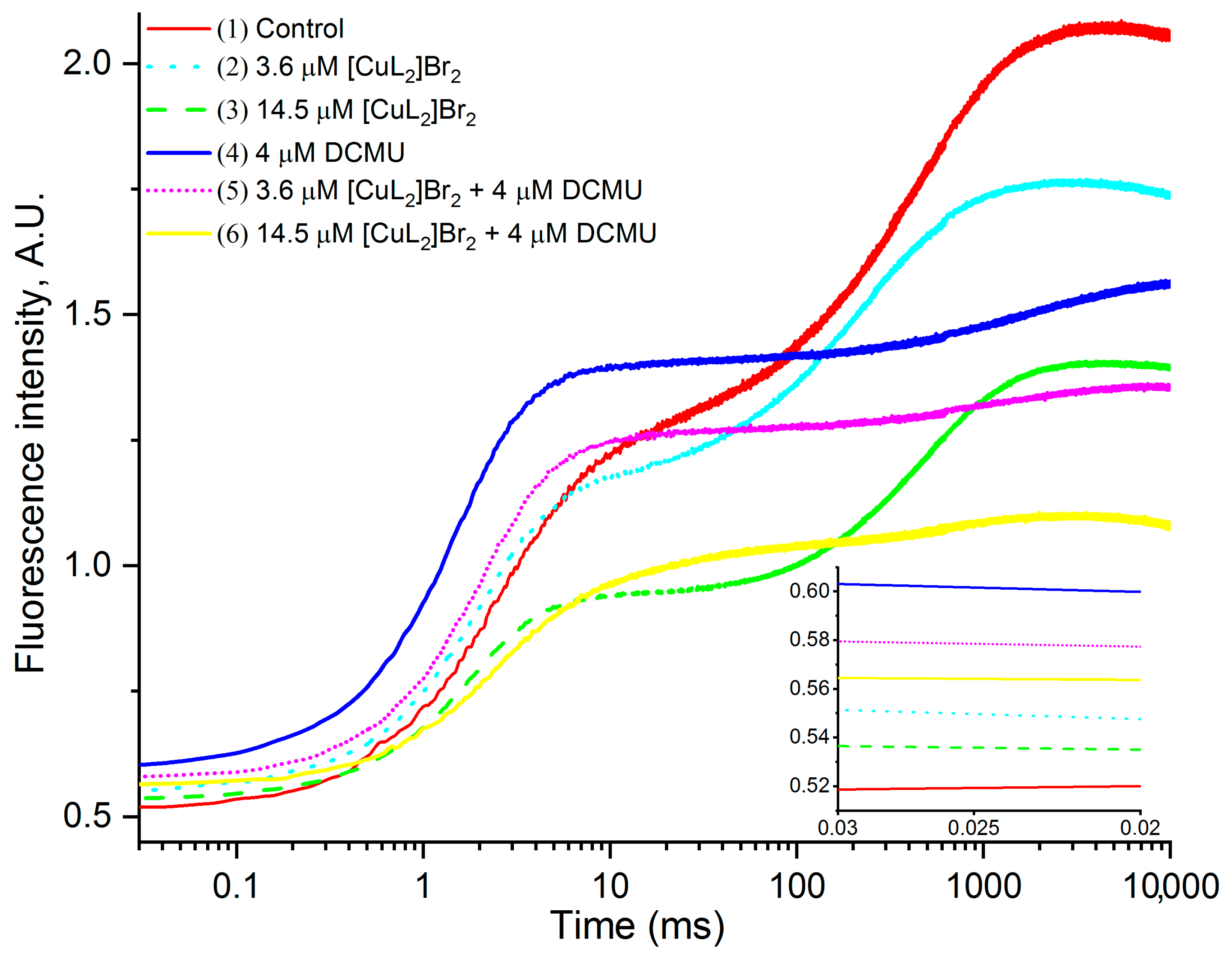
3.1. Original OJIP Kinetics

Figure 2 shows the original OJIP kinetics measured on PSII-containing membranes in the absence of other additives (control—kinetic 1) or in the presence of: 3.6 μM [CuL2]Br2 (kinetic 2); 14.5 μM [CuL2]Br2 (kinetic 3); 4 μM DCMU (kinetic 4); 3.6 μM [CuL2]Br2 + 4 μM DCMU (kinetic 5); 14.5 μM [CuL2]Br2 + 4 μM DCMU (kinetic 6). The FV/FM ratio is a generally accepted, widely used measure characterizing the quantum yield of the primary photochemical reaction of PSII [28]. Based on the analysis of the original kinetics presented in Figure 2, it can be seen that all the studied agents and their combinations cause significant decreases in the Chl “a” fluorescence intensity, especially noticeable at the FM level leading to a decrease in the variable fluorescence (FV). Furthermore, 3.6 μM [CuL2]Br2 (kinetics 2); 14.5 μM [CuL2]Br2 (kinetic 3); and 4 μM DCMU (kinetic 4) also induced insignificant increases in the F0 level (inset to Figure 2). In the presence of DCMU, such increases of F0 by [CuL2]Br2 are not evident. It is important to especially note that the decrease in the FM value caused by both [CuL2]Br2 concentrations is at list in ten times more significant than the increase in the F0 value induced by [CuL2]Br2 without DCMU. An increase in the F0 level in the presence of DCMU has been repeatedly noted earlier on leaves [40], thylakoids [41], and PSII-containing membranes [42,43,44]. Both types of these changes (FM and F0 levels) lead to a decrease in FV/FM ratio.

3.2. Original OJIP Kinetics Normalized Relative to F0 (F0.02ms)

In order to make it easier to analyze and more clearly represent the possible changes caused by the agents added to the control (in the absence of other additives); in comparison with the control, normalization is carried out to the initial level of fluorescence F0, as a rule, by the value of F20µs or F50µs measured at 20 µs or 50 µs, respectively [28], but sometimes by the F0 value measured at time t = 0 [45,46]. In recent years, normalization to F0.05ms has been favored, although normalization to F0.02ms is acceptable and still quite common [47,48]. In addition, it is shown that the possible errors in the calculation of the parameters of the JIP test in the case when F50µs is used as F0 is higher than for F20µs and Ft→0 [45].
The original OJIP-kinetics normalized relative to F0 are presented as Ft − F0 versus time in Figure 3 (where F0 is the fluorescence at time 0.02 ms; Ft is the fluorescence at time t). The analysis of the presented kinetics shows the following main properties of the obtained kinetics and their changes caused by the studied agents and their combinations.
Kinetics measured in the absence of additions (control) are completely identical to those recorded on PSII-containing membranes [42,43,44,49]. There is no peak I in the kinetic (plateau JI), the main feature characterizing the kinetics of fast chlorophyll fluorescence induction measured on PSII-containing membranes [42,43,44,49] and therefore the kinetics will be designated below as OJP kinetics [42]. The absence of peak I (plateau JI) in the OJP kinetics of PSII-containing membranes has been substantiated previously [42].
In the presence of both studied concentrations (3.6 μM and 14.5 μM) of [CuL2]Br2, a significant simultaneous almost synchronous decrease in the chlorophyll fluorescence intensity (F) is observed along the entire length of the OJP kinetics. The decrease also includes the FJ level (2–3 ms), and it is in greater extent in the presence of 14.5 μM [CuL2]Br2. The chlorophyll fluorescence decrease is especially significant at the FM level—in the presence of 3.6 μM and 14.5 μM [CuL2]Br2 by 22% and 45%, respectively, kinetics 2 and 3, Table 1 compared with the control (kinetic 1). The FM decrease is especially pronounced at 14.5 μM [CuL2]Br2 (kinetic 3). Let us designate these decreases in F (including FJ and FM) as described above as the “effect of [CuL2]Br2”.
Thus, these experimental data suggest that out of the total number of PSII-containing membranes, 22% and 45%, PSII-containing membranes (respectively, in the presence of 3.6 μM and 14.5 μM [CuL2]Br2) are no longer capable of photochemical reduction of the corresponding components of the acceptor side of PSII. This effect is a consequence of a certain suppressive effect of [CuL2]Br2 on the components providing either charge separation or the source of electrons from the components of the donor side of PSII, and onward can be excluded from further consideration because they no longer produce JIP kinetics due to the action of [CuL2]Br2. Therefore, the remainder of the total number of PSII-containing membranes that retained photochemical activity in the presence of 3.6 μM and 14.5 μM [CuL2]Br2, respectively, should be considered, namely 78% and 55%. And using these data, it will be possible to find out by what mechanisms [CuL2]Br2 disrupts the functioning of PSII and in what sequence these mechanisms function.
In addition, FM is reduced in the presence of 4 μM DCMU and especially in the presence of its combinations with both concentrations of [CuL2]Br2 (Figure 3, Table 1). Moreover, in the case of a combination of 14.5 μM [CuL2]Br2 + 4 μM DCMU, an almost synchronous decrease in the chlorophyll fluorescence intensity (F) occurs along the entire length of the OJP kinetics, which are similar to described above.
In the presence of DCMU (without [CuL2]Br2), changes in the OJP kinetics characteristic of DCMU are observed (the so-called “DCMU effect”)—namely, an increase in the FJ peak to the so-called FM peak (kinetic 4), the intensity of which is less than the FM peak of control. The effects of DCMU have been repeatedly shown and explained previously by other authors [41,42,43,44,50]. In the presence of DCMU, all the amount of QA present in the sample is restored, which is expressed in an increase of the J peak to the highest possible level. At the same time, there is a decrease in the FM value to a value that is 62% from the control FM. This decrease is due to the quenching of F by oxidized PQ-9 molecules [41,42,43,44,50]. A further decrease in the FM intensity by above reason seems unlikely, since in a preliminary experiment, we showed that 4 μM DCMU inhibited the oxidation of all reduced QA molecules at the concentration of PSII-containing membranes we used.
Of particular interest and significance are the changes in OJP kinetics that occur in the presence of simultaneously both DCMU and [CuL2]Br2 (kinetics 5 and 6). Both kinetics are similar to the kinetics recorded in the presence of only DCMU (“DCMU effect” (kinetic 4)), but at the same time, the intensity of chlorophyll fluorescence decreases even more significantly over the entire OJP kinetics (“[CuL2]Br2) effect”). This decrease is especially evident in the case of 14.5 μM [CuL2]Br2)+ 4 μM DCMU (kinetic 6). The intensity of F at the FM level decreases in the case of these combinations of inhibitors (3.6 μM [CuL2]Br2) + 4 μM DCMU) and (14.5 μM [CuL2]Br2) + 4 μM DCMU), by 50% and 66%, respectively, relative to the control FM.
In this case, in the presence of both combinations of DCMU with 3.6 μM and 14.5 μM [CuL2]Br2 (similar to situation without DCMU described above), there is for further research only part from the total number of PSII-containing membranes that retained photochemical activity, namely 50% and 34% in this case relative to FM in the presence of 4 μM DCMU alone. In such case, in the presence of both combinations of DCMU with 3.6 μM and or 14.5 μM [CuL2]Br2, the remaining parts of the total number of PSII-membranes that retained photochemical activity, namely 50% and 34%, should be further considered.
Thus, DCMU induces a “DCMU effect” regardless of the presence of [CuL2]Br2. At the same time, [CuL2]Br2 effectively suppresses the FM value both in the absence and in the presence of DCMU.
In the presence of DCMU, it is important to correctly estimate how much [CuL2]Br2 reduces the FM value. Since a further decrease due to quenching of F by oxidized PQ-9 molecules remaining in PSII-membrane is unlikely, since oxidation of all available QA molecules is blocked by DCMU, then the observed decrease caused by both concentrations of [CuL2]Br2 in the presence of DCMU is based on another reason, and the percentage of decrease in FM in this case should be calculated by taking as 100% the value of FM measured in the presence of 4 μM DCMU. In this case, a further FM reduction due to quenching of F by oxidized PQ-9 molecules remaining in PSII-containing membranes is unlikely, since oxidation of all available QA molecules is blocked by DCMU. Consequently, the observed FM reduction caused by both concentrations of [CuL2]Br2 in the presence of DCMU is based on another reason. The percentage of decreased FM reduction in this case should be calculated by taking as 100% the value of FM measured in the presence of 4 μM DCMU. These calculated data are shown in Table 1 in parentheses and highlighted by asterisks. Comparing these data, we can see the following: in the absence of DCMU, both concentrations of [CuL2]Br2 suppress the FM value by 22% and 45%, respectively, and in the presence of DCMU, by 19% and 44%, respectively. These values are fairly well comparable.
The revealed coincidence of the values of FM decrease by [CuL2]Br2 in the presence of DCMU and without DCMU suggests that in both cases [CuL2]Br2 inhibits the activity of PSII-containing membranes by the same mechanism.

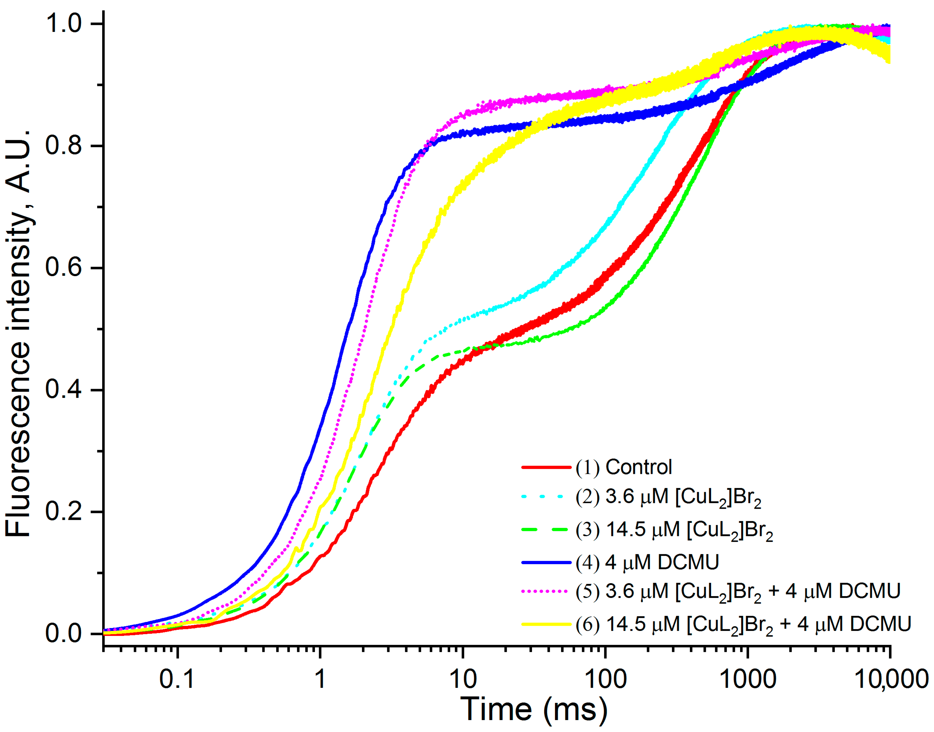
3.3. OJIP Kinetics Normalized Relative to F20µs and FM

Many stresses, including high or low temperature stress; high light intensities; UV-B; inhibitors of PSII photochemical activity, etc., affect the photoinduced redox state of QA, and this is reflected in the form of changes in the intensity of the FJ peak of OJIP kinetics and/or time to J-peak [40,41,51,52,53].
In Figure 3, it is not easy to understand how the intensity F changes at the level of peak J for almost every kinetic compared to the control, with the exception of kinetics 4 (4 μM DCMU) and 5 (3.6 μM [CuL2]Br2 + 4 μM DCMU) in which an increase in FJ intensity is clearly shown. Normalization of the original OJP kinetics simultaneously relative to the value of F0 and the value of FM makes it possible to reveal in more detail possible changes, including intermediate peaks, in the case of PSII-containing membranes—peak J. It was of interest to clarify more clearly how [CuL2]Br2 affects the properties of the J peak in the absence and the presence of DCMU.
Figure 4 shows the original OJP kinetics normalized relative to F0.02ms and to FM. After such normalization, it became obvious that, in addition to the simultaneous decrease in the chlorophyll fluorescence intensity over the entire OJP kinetics (slightly at the FJ level (2–3 ms) and especially pronounced at the FM level), which was clearly pronounced after normalization original OJP kinetics relative only to F0.02ms, now there are significant changes in OJP kinetics compared with the control in the presence of both concentrations of [CuL2]Br2, as well as their combinations with DCMU, which in this case became especially pronounced in the region of peak J (Figure 4).
From the data presented in Figure 4, it is evident that: (1) without DCMU in the presence of 3.6 μM [CuL2]Br2 (kinetic 2), the intensity of the J peak increases compared to the control (kinetic 1), but at a higher concentration of [CuL2]Br2 (14.5 μM) (kinetic 3), this effect, which is expressed in an increase in the J peak, already becomes significantly less; (2) in the case of a combination of 3.6 μM [CuL2]Br2 and 4 μM DCMU (kinetic 5), the J peak becomes a little bit higher compared to 4 μM DCMU (kinetic 4), however, at a higher concentration of [CuL2]Br2 (14.5 μM) in this combination inhibitors (kinetic 6), a significant decrease in the J peak is already observed.
Thus, in both above cases (namely in the absence and in the presence of DCMU), the differently directed effect on the F intensity of the J peak of these two concentrations of [CuL2]Br2 (3.6 μM and 14.5 μM) is clearly visible. It should emphasize that in the presence of DCMU the difference in the above effects between these concentrations is much greater. Despite the fact that after this normalization it is possible to identify additional changes in the OJP kinetics, nevertheless, in this case, these changes are not yet clearly expressed, and it is not possible to quantify the degree of these changes.

3.4. Comparison [CuL2]Br2 and DCMU Effects

Peak I is known to be absent in PSII-containing membranes [34,42,43,44,49]. The IP phase is directly related to PSI activity, while JI phase parallels the reduction of PQ pool [27,28].
Since there is no I peak in PSII-containing membranes, in order to more conveniently analyze and visualize possible changes at the level of the J peak, which are induced by the studied inhibitory agents and their combinations, we first double normalized the original kinetics relative to both the F0 level (F0.02ms) and to the level of finding the peak I (30ms), i.e., to the level F30ms, according to the formula:
V0I = (Ft − F0)/(FI − F0),
in our case
V0I = (Ft − F0.02ms)/(F30ms − F0.02ms)
The resulting kinetics V0I = (Ft − F0.02ms)/(F30ms − F0.02ms) are shown in Figure 5A. Next, we subtracted the kinetic obtained in the absence of any additions (control) from the kinetics obtained in the presence of inhibitory agents, for each of the studied inhibitory agents and their combinations. The obtained difference kinetics W0I = V0I experiment − V0I control are shown in Figure 5B.
It is known that DCMU blocks electron transfer from the reduced primary PSII electron acceptor, plastoquinone QA, into the membrane pool of plastoquinones (PQ-9), competing with the PSII secondary electron acceptor, plastoquinone QB for the binding site on the so-called QB herbicide-binding site of the D1 protein. Therefore, in the presence of DCMU, the so-called “diuron effect” is observed, which is expressed on the original OJIP kinetics as a significant increase in fluorescence intensity at a level of 2–3 ms (peak J) compared to the control [40,41,51,52,53]. This can be especially clearly seen in the difference OJP-kinetics obtained by subtracting from OJP-kinetics measured in the presence of DCMU, the kinetics obtained in the absence of any additions (control) [52,53].
Preliminarily, for the conditions of our measurements (the concentration of PSII-containing membranes, expressed as the concentration of chlorophyll contained in them, is 4 μg mL−1), we found that the concentration of DCMU used by us (4 μM) causes practically maximal “diuron effect” on the chlorophyll fluorescence of PSII.
In addition, the use of higher concentrations of DCMU may be accompanied by the effects of DCMU on other sites of the PSII electron transport chain, as described earlier [54,55,56,57]. In order to quantify the “diuron effect” of other studied inhibitory agents or their combined use with DCMU, we evaluated the FJ values for the other difference OJP-kinetics (W0I = V0I exp − V0I control) presented in Figure 5B in % from that with DCMU. The magnitude of the “diuron effect” FJ measured in the presence of 4 μM DCMU, which indicates amount of reduced QA (QA), we took as 100%. And the effects of other supplements were evaluated in % relative to this effect of DCMU. The data obtained are presented in Table 2.

3.4.1. Effects of [CuL2]Br2 in the Absence of DCMU

From the data presented in Table 2, it can be seen that in the absence of DCMU, low concentrations (3.6 μM) of [CuL2]Br2 cause a “diuron effect” of approximately 38% of that caused by DCMU. With an increase in the [CuL2]Br2 concentration to 14.5 µM, the “diuron effect” increases and is already 71% of the “diuron effect” caused by DCMU.

3.4.2. Effects of [CuL2]Br2 in the Presence of DCMU

A completely different effect of the [CuL2]Br2 complex on the photochemical activity of PSII is observed when [CuL2]Br2 complex is added in the presence of DCMU. In this case, both concentrations (3.6 μM and 14.5 μM) of [CuL2]Br2 significantly reduced the “diuron effect” of DCMU from 100%, respectively, to 59% and to about 3%.
Thus, from the data presented in Table 2, it is obvious that in the absence of DCMU, the amount of reduced QA increases with increasing concentration of the [CuL2]Br2 complex. However, in the presence of DCMU, on the contrary, the amount of reduced QA decreases significantly with an increase in the concentration of the [CuL2]Br2.
We evaluated the potency of these effects of [CuL2]Br2, namely (1) the effect of increasing the amount of reduced QA in the absence of DCMU and (2) the effect of decreasing the amount of reduced QA in the presence of DCMU on the concentration of the [CuL2]Br2 complex from the slope of the corresponding fitted curves. It turned out that the second mechanism of action of the [CuL2]Br2 complex, which manifests itself in a decrease in the amount of reduced QA in the presence of DCMU, is about two times more effective than the first one, the accumulation of the amount of reduced QA in the absence of DCMU.
Based on the comparison of the positions of the J peaks on the time scale, it can be roughly assumed that in the presence of 4 μM DCMU, the time to reach the maximum value of the fluorescence intensity of the J peak (FJ) on the difference kinetics WOI = VOI exp − VOI control (Figure 5B), which characterizes the rate of QA reduction with increasing concentration of the [CuL2]Br2 complex, also increases—as can be seen when comparing the difference kinetics for (3.6 μM [CuL2]Br2 + 4 μM DCMU) and (14.5 μM [CuL2]Br2 + 4 μM DCMU). Moreover, this property of the [CuL2]Br2 complex to slow down the rate of photoinduced QA reduction even increases with an increase in its concentration with DCMU. This is evident when comparing the different kinetics of (3.6 µM [CuL2]Br2) and (3.6 µM [CuL2]Br2 + 4 µM DCMU), as well as (14.5 µM [CuL2]Br2) and (14.5 µM [CuL2]Br2 + 4 µM DCMU).
However, there is a more reliable way to quantify the rate of photoinduced QA reduction.

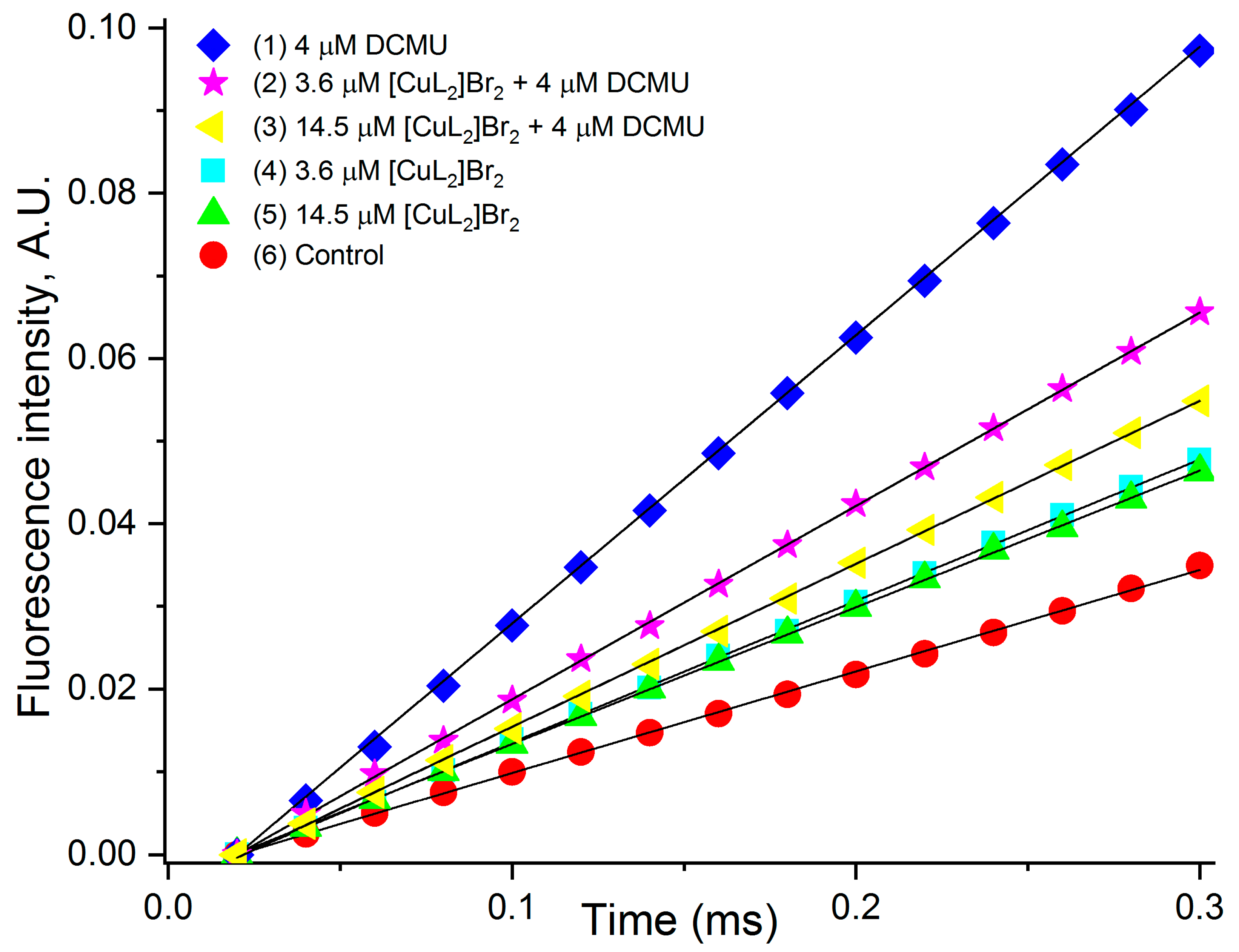
3.5. Estimation of the Rate of Photoinduced Reduction of QA

Graphical or computational determination of the initial slope (M0) of the JIP kinetics makes it possible to estimate the rate of photoinduced reduction of QA and its changes as a result of various influences [53,58,59,60,61,62]. We have used both of these approaches.
The graphical data presented in Figure 6 allow you to see much more clearly what changes are induced by the studied agents in PSII photochemical reactions; the graphical approach is also used by other researchers [53,58,60,62]. In addition, we calculated the values of M0 using the corresponding formula M0 = 4 (F300 − F0)/(FM − F0). The results of the calculations are presented in the Table 3.
It should be noted that the values (M0) determined on the basis of the data presented in Figure 6 almost coincide with those obtained as a result of calculations. However, since the “first” ones were determined as a result of approximating real values, the data obtained in the calculations should be considered more accurate.
From the data presented in the form of kinetics in Figure 6 and the corresponding values of M0 in the Table 3, it follows that in all variants in the presence of the studied agents (both concentrations of [CuL2]Br2 without DCMU, 4 μM DCMU, both combinations of [CuL2]Br2 with DCMU), the rate of accumulation of reduced QA (QA), compared to the control is above (Figure 6 and Table 3).
If we evaluate the rate of accumulation of reduced QA (QA), compared with DCMU, then in the absence of DCMU, both concentrations of [CuL2]Br2 increase the rate of accumulation of reduced QA (QA), compared with the control, as well as in the presence of DCMU alone, however, with a significantly lower efficiency compared to DCMU (46% and 32% of that of DCMU) (kinetics 4 and 5). Interestingly, in the presence of a lower concentration of [CuL2]Br2 (3.6 µM), this effect is greater (46%) compared with a higher concentration (14.5 µM) of this agent (32%), i.e., without DCMU, the ability to cause an increase in the rate of QA reduction decreases with increasing concentration of [CuL2]Br2.
In the presence of DCMU, [CuL2]Br2 also reduces the rate of accumulation of reduced QA (QA), respectively, to 69% and 56%, relative to DCMU (Table 3 of kinetics 2 and 3), and this effect of [CuL2]Br2 also increases with increasing concentration [CuL2]Br2.
We evaluated the effectiveness of the impact of [CuL2]Br2 on the rate of QA reduction in the absence and presence of DCMU by the slope of the approximated lines plotted using the corresponding experimental data from Table 3. It turned out that both in the absence of DCMU and with DCMU, the rate of QA reduction with increasing concentration of [CuL2]Br2 goes down. However, in the presence of DCMU, the rate of QA reduction with increasing concentration of [CuL2]Br2 decreases approximately three times faster than in the absence of DCMU.

3.6. Absorption Spectrum of [CuL2]Br2

It could be assumed that the observed simultaneous synchronous decrease in the intensity of the fast chlorophyll fluorescence induction curves and almost all its peaks in the presence of [CuL2]Br2, which enlarges with an increase in the concentration of this organometallic complex added to the measuring medium, could be due to (1) a decrease in the intensity of the measuring and/or acting light due to the screening effect—absorption of light quanta by [CuL2]Br2 or (2) a decrease in the intensity of fluorescence emitted by chlorophyll molecules due to its absorption by the molecules of the [CuL2]Br2—the effect of chlorophyll fluorescence reabsorption. To test this assumption, we studied the absorption spectrum of the [CuL2]Br2 complex. As shown in Figure 7, the [CuL2]Br2 complex has no absorption bands either in the region of wavelengths of both types of light or in the region of emission wavelengths of chlorophyll fluorescence. Therefore, the above assumption is erroneous.

4. Discussion

4.1. Main Inhibitory Impact of [CuL2]Br2 on OJP Transients

The strongest and, therefore, undoubtedly, the main effect of the studied complex [CuL2]Br2 on the photochemical activity of PSII-containing membranes is a total synchronous decrease in the F intensity along the entire JIP kinetics (Figure 2 and Figure 3, kinetics 2, 3 as well as Table 1). [CuL2]Br2 causes some changes in PSII when it becomes no longer capable of photoinduced QA reduction. Already at a concentration of 3.6 µM, [CuL2]Br2 is able to completely exclude 22% PSII-containing membranes from the total number of photochemically active PSII-containing membranes, but when at a concentration of 14.5 µM [CuL2]Br2 totally disables already 45% PSII-containing membranes (in the absence of DCMU). It is important to note that [CuL2]Br2 also demonstrates this effect on PSII in the presence of DCMU, with an efficiency that is well comparable to that estimated in the absence of DCMU (Figure 2 and Figure 3 (kinetics 5, 6) and Table 1, data highlighted in green). Based on these data, we can make an experimentally substantiated conclusion that the main effect of [CuL2]Br2 on PSII does not depend on DCMU. Fairly well-comparable values of FM reduction by both concentrations of [CuL2]Br2 in the absence and presence of DCMU (Figure 3, Table 1) suggest that in both cases, PSII inhibition by the [CuL2]Br2 complex may be based on the same mechanism of action. This in turn suggests that [CuL2]Br2 does not need to bind to the DCMU binding site to exert this effect. The fact that [CuL2]Br2 suppresses FM regardless of the presence of diuron suggests that the site of action and/or binding of [CuL2]Br2 on PSII is prior to the site of action and/or binding of diuron. Similarly, based on the obtained data about the independent manifestation of the effects of diuron and chloramphenicol on the OJIP kinetics of PSII-containing membranes, an experimentally substantiated conclusion was made that the site of action of chloramphenicol in PSII is located before the site of action of diuron [34].
What are the reasons for the revealed total synchronous decrease in the intensity F over the entire kinetics of JIP induced by [CuL2]Br2?
The corresponding decrease in the intensity F may be due to electron acceptance if this Cu(II)-complex acts as artificial electron acceptor. A similar decrease in fluorescence intensity is observed in the presence of known PSII electron acceptors, such as DCBQ [43,63]. The similar effect was observed in the case of chloramphenicol capable of effectively oxidizing pheophytin [34]. However, we have previously shown that [CuL2]Br2 is not an artificial electron acceptor, because it does not support photosynthetic oxygen evolution [26].
A significant simultaneous almost synchronous decrease in the fluorescence intensity of chlorophyll along the entire length of the OJP kinetics, especially at the FM level, increasing with increasing concentration of [CuL2]Br2, which we designated as the “[CuL2]Br2 effect”, may be the result of a violation of the donor side or the reaction PSII center itself. Similar changes in OJIP kinetics were observed when the PSII donor side becomes non-functional [42,44]. However, artificial electron donors do not eliminate the inhibitory action of [CuL2]Br2 [26].
Based on experimental data obtained earlier [26], now we propose that [CuL2]Br2 probably acts directly on the reaction center of PSII, and it is concerning to its main impact. It was shown that single-walled carbon nanotubes (SWCNT) at concentration of 300 mg/L influence the fast chlorophyll fluorescence induction curve [64]. The effect is very similar to that of the [CuL2]Br2. In the presence of 300 mg/L SWCNT, a total decrease in fluorescence intensity occurs along the entire length of the OJIP kinetics, which is especially pronounced at the FM level. It was shown that this effect of SWCNT is due to SWCNT inactivation of PSII RC [64]. Furthermore, earlier it was shown that Cu(II) aqua-ions act at the level of reaction centers of PSII [65].

4.2. Impact of [CuL2]Br2 on J and 0 Peaks

Realistically, the % of the main effect of [CuL2]Br2 on JIP-transients is probably induced rather by lower concentrations of [CuL2]Br2 discussed above. This assumption is based on the fact that the remaining part of the PSII-containing membranes in the presence of the indicated concentrations changed their properties if we compare the OJP kinetics in the presence of [CuL2]Br2 with the control OJP kinetics. It is more convenient to consider these auxiliary (not main) [CuL2]Br2 effects to separate variants in the presence and the absence of DCMU.

4.2.1. Impact of [CuL2]Br2 on J and 0 Peaks in the Absence DCMU

[CuL2]Br2 causes a slight increase in F0 levels (Figure 2 and inset kinetics 2 and 3) like it is usually induced by DCMU and agents with similar inhibitory mechanism—stopping electron transfer from reduced QA onto the next mediator of electron transport chain. This is not observed in the presence of DCMU.
[CuL2]Br2 causes a slight increase in FJ levels (Figure 4 and Figure 5B). It is interesting that in these figures, at the first glance, there is opposite dependence of the [CuL2]Br2 effects on the FJ intensity. On Figure 4, at low [CuL2]Br2 concentration (3.6 μM), increasing of the FJ intensity seems more expressed than at 14.5 μM). Whereas on Figure 5B, the dependence is opposite.
In fact, a correct picture of the [CuL2]Br2 influence on the FJ level can be obtained by subtracting the control OJP kinetic doubly normalized relative to F0 and FM from those in the presence of [CuL2]Br2, i.e., kinetics 2 and 4 shown on Figure 5B. This figure shows that the auxiliary effect of [CuL2]Br2 on the difference kinetics is similar to the effect of diuron, i.e., an increase in the amount of reduced QA. This “diuron-like effect” increases with increasing concentration of [CuL2]Br2. Thus, in the absence of DCMU, both of the above results, namely, an increase in the level of F0 and FJ, suggest that in the absence of DCMU, an auxiliary (not main) effect of [CuL2]Br2 is that [CuL2]Br2 acts like a DCMU, but with less efficiency than DCMU.

4.2.2. Impact of [CuL2]Br2 on J and 0 Peaks in the Presence DCMU

Figure 4 shows that in the presence of DCMU, both concentrations of [CuL2]Br2 seem to cause an increase in the J level comparable to that induced by DCMU (kinetics 5 and 6). However in fact, it must be taken into account that this increase in J is a joint effect of DCMU and [CuL2]Br2. The real picture of the influence of both [CuL2]Br2 concentrations on the magnitude of the J peak in the presence of DCMU is clear only on the difference kinetics (Figure 5B of kinetics 3 and 5). Figure 5B shows that both [CuL2]Br2 concentrations in fact decrease the DCMU effect, and the decrease is more at higher [CuL2]Br2 concentration (respectively, to 59% and to about 3% relative to 100% DCMU effect) (Table 2).
It could be assumed that [CuL2]Br2 can displace DCMU from its binding site, and this results in the decrease in the effect of DCMU. However, in this case, kinetics 3 and 5 would not be observed, but kinetics 2 and 4, because in the absence of DCMU, [CuL2]Br2 causes such auxiliary effects (the so-called “diuron effect”). However, this is in fact not the case. This means that in the presence of DCMU, [CuL2]Br2 causes some changes in PSII when it is no longer capable of photoinduced QA reduction even in the presence of DCMU, an effect similar to the main effect of [CuL2]Br2.

4.3. The Rate of Photoinduced Reduction of QA

The fact that in the absence of DCMU simultaneously with the main effect [CuL2]Br2 appears to have a much less effective auxiliary effect is also evidenced by the fact that the M0 values reflecting the rate of photoinduced QA reduction in the presence of this complex are higher compared to the control (Figure 6 kinetics 4 and 5, Table 3). That higher M0 values are the result of an auxiliary effect of [CuL2]Br2 is evidenced by the fact that the ability to cause an increase in the rate of QA reduction decreases with increasing concentration of [CuL2]Br2. This trend is also observed in the presence of DCMU. The evidence that in the presence of DCMU, the rate of QA reduction with increasing concentration of [CuL2]Br2 decreases approximately three times faster than in the absence of DCMU also strongly suggests that in this case the ancillary effect of [CuL2]Br2 becomes the main one.
It could be assumed that the revealed decrease in the F intensity of OJP kinetics caused by [CuL2]Br2 is the result of the physical or functional separation of the antenna from the RC [66]. In this case, an increase in the F0 level can serve as a fairly reliable indication of this effect [66]. In our case, in the absence of DCMU, [CuL2]Br2 does not cause any increase in the F0 level, and it is obvious that its effect on PSII is not associated with the separation of the antenna from the RC.
It could be assumed that the decrease in the intensity of the chlorophyll fluorescence through whole OJP kinetic (but not F0 level) in the presence of our exogenous agent is the result of quenching of the chlorophyll fluorescence. It has been previously shown in thylakoids that DCBQ can act as an artificial electron acceptor and as a chlorophyll fluorescence quencher [63]. In this case, in addition to a decrease in the chlorophyll fluorescence intensity over the entire OJP kinetics, as a result of electron acceptance and an increase in the photochemistry rate, a decrease in the F0 level is also observed as a result of F antenna quenching. Moreover, it is important that the decrease in F0 caused by DCBQ is observed even in the presence of DCMU [63]. In our studies in the presence of DCMU, [CuL2]Br2 does not cause any decrease in the F0 value and, therefore, it is not a chlorophyll fluorescence quencher (Figure 2 and Figure 3 kinetics 5, 6).
The fact that the [CuL2]Br2 complex has no absorption bands either in the region of wavelengths of both types of light, or in the region of chlorophyll fluorescence emission wavelengths (Figure 7) gives grounds to suggest that a decrease in the chlorophyll fluorescence intensity of PSII-containing membranes in the presence of the [CuL2]Br2 complex could be due to its screening effect on the measuring and/or acting light. Another reason—the reabsorption of chlorophyll fluorescence by molecules of the [CuL2]Br2 could be excluded.

5. Conclusions

Based on the results obtained, we can assume:
(1) The main (dominating in terms of the degree of inhibition of PSII activity) effect of [CuL2]Br2 on PSII is probably associated with inhibition of the activity of the PSII RC. (2) The manifestation of auxiliary effects of [CuL2]Br2 on PSII is determined by the presence of DCMU. In the absence of DCMU, i.e., when the DCMU binding site is free, some part of [CuL2]Br2 that is involved in the induction of an auxiliary effect on PSII causes a “diuron-like” effect—an increase in the level of F0 and FJ, i.e., blocks electron transfer from the reduced QA into the electron transport chain, but with less efficiency compared to DCMU. However, in the presence of DCMU, i.e., when the DCMU binding site is occupied by DCMU, this part of [CuL2]Br2, which is involved in the induction of the auxiliary effect on PSII, participates in the induction of the main effect, i.e., a total decrease in the intensity F over the entire OJP kinetics. Thus, it can be assumed that in PSII-containing membranes, there are two binding sites for [CuL2]Br2 with different affinities for [CuL2]Br2. At the high affinity site, [CuL2]Br2 produces effects similar inhibition of the PSII RC activity, while at the low affinity site, [CuL2]Br2 produces effects similar to those of DCMU. The data obtained can be useful in the development of promising herbicides for use in agricultural economics.

Author Contributions

S.K.Z., V.D.K., H.F.A. and S.I.A. conceived and designed the research; M.S.K. and N.K., synthesized complexes; S.K.Z., V.D.K. and M.S.S., performed the experiments; S.K.Z., I.M.H., V.D.K., H.F.A., B.D.B. and S.I.A. wrote the manuscript. H.A. investigation and data curation. All authors contributed to data evaluation and interpretation. All authors have read and agreed to the published version of the manuscript.

Funding

This work was funded by the Russian Science Foundation (19-14-00118). Graphical determination of the initial slope (M0) of the JIP kinetics presented on Figure 6 was carried out under the state contract of the Ministry of Science and Higher Education of the Russian Federation (theme No. 122050400128-1).

Institutional Review Board Statement

Not applicable.

Informed Consent Statement

Not applicable.

Data Availability Statement

The data presented in this study are available on request from the corresponding authors.

Conflicts of Interest

The authors declare no conflict of interest.

References

  1. Ahmad, N.; Mukhtar, Z. Genetic Manipulations in Crops: Challenges and Opportunities. Genomics 2017, 109, 494–505. [Google Scholar] [CrossRef]
  2. Schütte, G.; Eckerstorfer, M.; Rastelli, V.; Reichenbecher, W.; Restrepo-Vassalli, S.; Ruohonen-Lehto, M.; Saucy, A.G.W.; Mertens, M. Herbicide Resistance and Biodiversity: Agronomic and Environmental Aspects of Genetically Modified Herbicide-Resistant Plants. Environ. Sci. Eur. 2017, 29, 5. [Google Scholar] [CrossRef] [PubMed] [Green Version]
  3. Karki, L.; Lakshmi, K.V.; Szalai, V.A.; Brudvig, G.W. Low-Temperature Turnover Control of Photosystem II Using Novel Metal- Containing Redox-Active Herbicides. J. Am. Chem. Soc. 2000, 122, 5180–5188. [Google Scholar] [CrossRef]
  4. Trebst, A. Inhibitors in the Functional Dissection of the Photosynthetic Electron Transport System. Photosynth. Res. 2007, 92, 217–224. [Google Scholar] [CrossRef] [PubMed]
  5. Zharmukhamedov, S.K.; Allakhverdiev, S.I. Chemical Inhibitors of Photosystem II. Russ. J. Plant Physiol. 2021, 68, 212–227. [Google Scholar] [CrossRef]
  6. Ran, C.; Yu, X.; Jin, M.; Zhang, W. Role of Carbonyl Cyanide M-Chlorophenylhydrazone in Enhancing Photobiological Hydrogen Production by Marine Green Alga Platymonas Subcordiformis. Biotechnol. Prog. 2006, 22, 438–443. [Google Scholar] [CrossRef]
  7. Nelson, N.; Junge, W. Structure and Energy Transfer in Photosystems of Oxygenic Photosynthesis. Annu. Rev. Biochem. 2015, 84, 659–683. [Google Scholar] [CrossRef]
  8. Vass, I. Molecular Mechanisms of Photodamage in the Photosystem II Complex. Biochim. Biophys. Acta Bioenerg. 2012, 1817, 209–217. [Google Scholar] [CrossRef] [Green Version]
  9. Semelkova, L.; Konecna, K.; Paterova, P.; Kubicek, V.; Kunes, J.; Novakova, L.; Marek, J.; Naesens, L.; Pesko, M.; Kralova, K.; et al. Synthesis and Biological Evaluation of N-Alkyl-3-(Alkylamino)-Pyrazine-2-Carboxamides. Molecules 2015, 20, 8687–8711. [Google Scholar] [CrossRef]
  10. Jampilek, J.; Kralova, K.; Pesko, M.; Kos, J. Ring-Substituted 8-Hydroxyquinoline-2-Carboxanilides as Photosystem II Inhibitors. Bioorg. Med. Chem. Lett. 2016, 26, 3862–3865. [Google Scholar] [CrossRef]
  11. Gonec, T.; Kralova, K.; Pesko, M.; Jampilek, J. Antimycobacterial N-Alkoxyphenylhydroxynaphthalenecarboxamides Affecting Photosystem II. Bioorg. Med. Chem. Lett. 2017, 27, 1881–1885. [Google Scholar] [CrossRef] [PubMed]
  12. Gonec, T.; Kos, J.; Pesko, M.; Dohanosova, J.; Oravec, M.; Liptaj, T.; Kralova, K.; Jampilek, J. Halogenated 1-Hydroxynaphthalene-2-Carboxanilides Affecting Photosynthetic Electron Transport in Photosystem II. Molecules 2017, 22, 1709. [Google Scholar] [CrossRef] [PubMed] [Green Version]
  13. Karacan, M.S.; Rodionova, M.V.; Tunç, T.; Venedik, K.B.; Mamaş, S.; Shitov, A.V.; Zharmukhamedov, S.K.; Klimov, V.V.; Karacan, N.; Allakhverdiev, S.I. Characterization of Nineteen Antimony(III) Complexes as Potent Inhibitors of Photosystem II, Carbonic Anhydrase, and Glutathione Reductase. Photosynth. Res. 2016, 130, 167–182. [Google Scholar] [CrossRef] [PubMed]
  14. Sersen, F.; Kralova, K.P.M.C.M. Electrophysiological Variability in the SH-SY5Y Cellular Line. Gen. Physiol. Biophys. 2014, 31, 375–382. [Google Scholar] [CrossRef] [Green Version]
  15. Karacan, M.S.; Zharmukhamedov, S.K.; Mamaş, S.; Kupriyanova, E.V.; Shitov, A.V.; Klimov, V.V.; Özbek, N.; Özmen, Ü.; Gündüzalp, A.; Schmitt, F.J.; et al. Screening of Novel Chemical Compounds as Possible Inhibitors of Carbonic Anhydrase and Photosynthetic Activity of Photosystem II. J. Photochem. Photobiol. B Biol. 2014, 137, 156–167. [Google Scholar] [CrossRef]
  16. Sersen, F.; Kral’Ova, K.; Bumbalova, A.; Svajlenova, O. The Effect of Cu(II) Ions Bound with Tridentate Schiff Base Ligands upon the Photosynthetic Apparatus. J. Plant Physiol. 1997, 151, 299–305. [Google Scholar] [CrossRef]
  17. Rodionova, M.V.; Zharmukhamedov, S.K.; Karacan, M.S.; Venedik, K.B.; Shitov, A.V.; Tunç, T.; Mamaş, S.; Kreslavski, V.D.; Karacan, N.; Klimov, V.V.; et al. Evaluation of New Cu(II) Complexes as a Novel Class of Inhibitors against Plant Carbonic Anhydrase, Glutathione Reductase, and Photosynthetic Activity in Photosystem II. Photosynth. Res. 2017, 133, 139–153. [Google Scholar] [CrossRef]
  18. Good, N.E.; Winget, G.D.; Winter, W.; Conolly, N.T.; Izawa, S.; Singh, R.M.M. Hydrogen Ion Buffers for Biological Research. Biochemistry 1966, 5, 467–477. [Google Scholar] [CrossRef]
  19. Balt, S.; Van Dalen, E. The Reactions of Diphenylcarbazide and Diphenylcarbazone with Cations. Anal. Chim. Acta 1963, 29, 466–471. [Google Scholar] [CrossRef]
  20. Crespo, G.A.; Andrade, F.J.; Iñón, F.A.; Tudino, M.B. Kinetic Method for the Determination of Trace Amounts of Copper(II) in Water Matrices by Its Catalytic Effect on the Oxidation of 1,5-Diphenylcarbazide. Anal. Chim. Acta 2005, 539, 317–325. [Google Scholar] [CrossRef]
  21. Weissberger, A.; Lu Valle, J.E. Oxidation Processes. XVII. The Autoxidation of Ascorbic Acid in the Presence of Copper. J. Am. Chem. Soc. 1944, 66, 700–705. [Google Scholar] [CrossRef]
  22. Anderson, J.H. The Copper-Catalysed Oxidation of Hydroxylamine. Analyst 1964, 89, 357–362. [Google Scholar] [CrossRef]
  23. Keller, R.N.; Wrcoff, H.D. Copper(I) Chloride. In Inorganic Syntheses; Fernelius, W.C., Ed.; McGraw-Hill Book Company, Inc.: New York, NY, USA, 1946; pp. 1–4. ISBN 9780470131619. [Google Scholar]
  24. Yuan, X.; Pham, A.N.; Miller, C.J.; Waite, T.D. Copper-Catalyzed Hydroquinone Oxidation and Associated Redox Cycling of Copper under Conditions Typical of Natural Saline Waters. Environ. Sci. Technol. 2013, 47, 8355–8364. [Google Scholar] [CrossRef] [PubMed]
  25. López-Cueto, L.; Casado-Riobó, J.A. Catalytic Effect of Copper on the Hexacyanoferrate(III)-Cyanide Redox Reaction-I. The Uncatalysed and Catalysed Reactions. Talanta 1979, 26, 127–132. [Google Scholar] [CrossRef] [PubMed]
  26. Zharmukhamedov, S.K.; Shabanova, M.S.; Rodionova, M.V.; Huseynova, I.M.; Karacan, M.S.; Karacan, N.; Aşık, K.B.; Kreslavski, V.D.; Alwasel, S.; Allakhverdiev, S.I. Effects of Novel Photosynthetic Inhibitor [CuL2]Br2 Complex on Photosystem II Activity in Spinach. Cells 2022, 11, 2680. [Google Scholar] [CrossRef]
  27. Kalaji, H.M.; Jajoo, A.; Oukarroum, A.; Brestic, M.; Zivcak, M.; Samborska, I.A.; Cetner, M.D.; Łukasik, I.; Goltsev, V.; Ladle, R.J.; et al. The Use of Chlorophyll Fluorescence Kinetics Analysis to Study the Performance of Photosynthetic Machinery in Plants. In Emerging Technologies and Management of Crop Stress Tolerance; Elsevier: Amsterdam, The Netherlands, 2014; pp. 347–384. ISBN 978-0-12-800875-1. [Google Scholar]
  28. Kalaji, M.H.; Goltsev, V.N.; Zuk-Golaszewska, K.; Zivcak, M.; Brestic, M. Chlorophyll Fluorescence, Understanding Crop Performance; CRC Press: Boca Raton, FL, USA, 2017; ISBN 978-1-4987-6449-0. [Google Scholar]
  29. Velthuys, B.R. Electron-Dependent Competition between Plastoquinone and Inhibitors for Binding to Photosystem II. FEBS Lett. 1981, 126, 277–281. [Google Scholar] [CrossRef] [Green Version]
  30. Wraight, C.A. Oxidation-Reduction Physical Chemistry of the Acceptor Quinone Complex in Bacterial Photosynthetic Reaction Centers: Evidence for a New Model of Herbicide Activity. Isr. J. Chem. 1981, 21, 348–354. [Google Scholar] [CrossRef]
  31. Umena, Y.; Kawakami, K.; Shen, J.-R.; Kamiya, N. Crystal Structure of Oxygen-Evolving Photosystem II at a Resolution of 1.9 Å. Nature 2011, 473, 55–60. [Google Scholar] [CrossRef]
  32. Suga, M.; Akita, F.; Hirata, K.; Ueno, G.; Murakami, H.; Nakajima, Y.; Shimizu, T.; Yamashita, K.; Yamamoto, M.; Ago, H.; et al. Native Structure of Photosystem II at 1.95 Å Resolution Viewed by Femtosecond X-Ray Pulses. Nature 2015, 517, 99–103. [Google Scholar] [CrossRef]
  33. Zharmukhamedov, S.K.; Klimov, V.V.; Allakhverdiev, S.I. The Absence of Competition for the Binding Site between Diuron and Novel Electron Transfer Inhibitors in Photosystem 2—Derivatives of Perfluoroisopropyldinitrobenzene. Biochem. Mosc. 1995, 60, 723–728. [Google Scholar]
  34. Rehman, A.U.; Kodru, S.; Vass, I. Chloramphenicol Mediates Superoxide Production in Photosystem II and Enhances Its Photodamage in Isolated Membrane Particles. Front. Plant Sci. 2016, 7, 479. [Google Scholar] [CrossRef] [Green Version]
  35. Berthold, D.A.; Babcock, G.T.; Yocum, C.F. A Highly Resolved, Oxygen-Evolving Photosystem II Preparation from Spinach Thylakoid Membranes. FEBS Lett. 1981, 134, 231–234. [Google Scholar] [CrossRef] [Green Version]
  36. Ford, R.C.; Evans, M.C.W. Isolation of a Photosystem 2 Preparation from Higher Plants with Highly Enriched Oxygen Evolution Activity. FEBS Lett. 1983, 160, 159–164. [Google Scholar] [CrossRef] [Green Version]
  37. Klimov, V.V.; Allakhverdiev, S.I.; Shuvalov, V.A.; Krasnovsky, A.A. Effect of Extraction and Re-Addition of Manganese on Light Reactions of Photosystem-II Preparations. FEBS Lett. 1982, 148, 307–312. [Google Scholar] [CrossRef] [Green Version]
  38. Arnon, D.I. Copper Enzymes in Isolated Chloroplasts. Polyphenoloxidase in Beta Vulgaris. Plant Physiol. 1949, 24, 1–15. [Google Scholar] [CrossRef] [Green Version]
  39. Yanykin, D.; Sundyreva, M.; Khorobrykh, A.; Semenova, G.; Savchenko, T. Functional Characterization of the Corticular Photosynthetic Apparatus in Grapevine. Biochim. Biophys. acta. Bioenerg. 2020, 1861, 148260. [Google Scholar] [CrossRef]
  40. Tóth, S.Z.; Schansker, G.; Strasser, R.J. In Intact Leaves, the Maximum Fluorescence Level (FM) Is Independent of the Redox State of the Plastoquinone Pool: A DCMU-Inhibition Study. Biochim. Biophys. Acta Bioenerg. 2005, 1708, 275–282. [Google Scholar] [CrossRef] [PubMed] [Green Version]
  41. Hiraki, M.; Van Rensen, J.J.S.; Vredenberg, W.J.; Wakabayashi, K. Characterization of the Alterations of the Chlorophyll a Fluorescence Induction Curve after Addition of Photosystem II Inhibiting Herbicides. Photosynth. Res. 2003, 78, 35–46. [Google Scholar] [CrossRef]
  42. Pospíšil, P.; Dau, H. Chlorophyll Fluorescence Transients of Photosystem II Membrane Particles as a Tool for Studying Photosynthetic Oxygen Evolution. Photosynth. Res. 2000, 65, 41–52. [Google Scholar] [CrossRef]
  43. Heredia, P.; Rivas, J.D. Las Fluorescence Induction of Photosystem II Membranes Shows the Steps till Reduction and Protonation of the Quinone Pool. J. Plant Physiol. 2003, 160, 1499–1506. [Google Scholar] [CrossRef]
  44. Lovyagina, E.R.; Semin, B.K. Mechanism of Inhibition and Decoupling of Oxygen Evolution from Electron Transfer in Photosystem II by Fluoride, Ammonia and Acetate. J. Photochem. Photobiol. B Biol. 2016, 158, 145–153. [Google Scholar] [CrossRef] [PubMed]
  45. Padhi, B.; Chauhan, G.; Kandoi, D.; Stirbet, A.; Tripathy, B.C.; Govindjee, G. A Comparison of Chlorophyll Fluorescence Transient Measurements, Using Handy Pea and Fluorpen Fluorometers. Photosynthetica 2021, 59, 399–408. [Google Scholar] [CrossRef]
  46. Rastogi, A.; Kovar, M.; He, X.; Zivcak, M.; Kataria, S.; Kalaji, H.M.; Skalicky, M.; Ibrahimova, U.F.; Hussain, S.; Mbarki, S.; et al. JIP-Test as a Tool to Identify Salinity Tolerance in Sweet Sorghum Genotypes. Photosynthetica 2020, 58, 518–528. [Google Scholar] [CrossRef] [Green Version]
  47. Viljevac Vuletić, M.; Mihaljević, I.; Tomaš, V.; Horvat, D.; Zdunić, Z.; Vuković, D. Physiological Response to Short-Term Heat Stress in the Leaves of Traditional and Modern Plum (Prunus Domestica L.) Cultivars. Horticulturae 2022, 8, 72. [Google Scholar] [CrossRef]
  48. Liu, Y.; Hao, C.; Wang, G.; Li, Q.; Shao, A. Exogenously Spermidine Alleviates Damage from Drought Stress in the Photosystem II of Tall Fescue. Plant, Soil Environ. 2021, 67, 558–566. [Google Scholar] [CrossRef]
  49. Pospíšil, P.; Dau, H. Valinomycin Sensitivity Proves That Light-Induced Thylakoid Voltages Result in Millisecond Phase of Chlorophyll Fluorescence Transients. Biochim. Biophys. Acta Bioenerg. 2002, 1554, 94–100. [Google Scholar] [CrossRef] [Green Version]
  50. Bukhov, N.G.; Egorova, E.A.; Govindachary, S.; Carpentier, R. Changes in Polyphasic Chlorophyll a Fluorescence Induction Curve upon Inhibition of Donor or Acceptor Side of Photosystem II in Isolated Thylakoids. Biochim. Biophys. Acta Bioenerg. 2004, 1657, 121–130. [Google Scholar] [CrossRef] [Green Version]
  51. Martinazzo, E.G.; Perboni, A.T.; Bacarin, M.A. The Effect of Inhibitors on Photosynthetic Electron Transport Chain in Canola Leaf Discs. Acta Sci. Biol. Sci. 2015, 37, 159–167. [Google Scholar] [CrossRef] [Green Version]
  52. De Carvalho, A.C.; Salvador, J.P.; Pereira, T.d.M.; Ferreira, P.H.A.; Lira, J.C.S.; Veiga, T.A.M. Fluorescence of Chlorophyll for Discovering Inhibitors of Photosynthesis in Plant Extracts. Am. J. Plant Sci. 2016, 7, 1545–1554. [Google Scholar] [CrossRef] [Green Version]
  53. Guo, Y.; Lu, Y.; Goltsev, V.; Strasser, R.J.; Kalaji, H.M.; Wang, H.; Wang, X.; Chen, S.; Qiang, S. Comparative Effect of Tenuazonic Acid, Diuron, Bentazone, Dibromothymoquinone and Methyl Viologen on the Kinetics of Chl a Fluorescence Rise OJIP and the MR820 Signal. Plant Physiol. Biochem. 2020, 156, 39–48. [Google Scholar] [CrossRef]
  54. Klimov, V.V.; Shuvalov, V.A.; Heber, U. Photoreduction of Pheophytin as a Result of Electron Donation from the Water-Splitting System to Photosystem-II Reaction Centers. Biochim. Biophys. Acta Bioenerg. 1985, 809, 345–350. [Google Scholar] [CrossRef]
  55. Klimov, V.V.; Allakhverdiev, S.I.; Ladygin, V.G. Photoreduction of Pheophytin in Photosystem II of the Whole Cells of Green Algae and Cyanobacteria. Photosynth. Res. 1986, 10, 355–363. [Google Scholar] [CrossRef] [PubMed]
  56. Hsu, B.D.; Lee, J.Y. Toxic Effects of Copper on Photosystem II of Spinach Chloroplasts. Plant Physiol. 1988, 87, 116–119. [Google Scholar] [CrossRef] [Green Version]
  57. Kamachi, H.; Tamura, N.; Inoué, H. Putative Second Binding Site of DCMU on the Oxidizing Side of Photosystem II in Photosystem II Membranes Depleted of Functional Mn. Plant Cell Physiol. 1992, 33, 437–443. [Google Scholar] [CrossRef]
  58. Srivastava, A.; Strasser, R.J. Govindjee Polyphasic Rise of Chlorophyll a Fluorescence in Herbicide-Resistant D1 Mutants of Chlamydomonas Reinardtii. Photosynth. Res. 1995, 43, 131–141. [Google Scholar] [CrossRef] [PubMed]
  59. Strasser, R.J.; Srivastava, A. Govindjee Polyphasic Chlorophyll a Fluorescence Transient in Plants and Cyanobacteria. Photochem. Photobiol. 1995, 61, 32–42. [Google Scholar] [CrossRef]
  60. Haldimann, P.; Strasser, R.J. Effects of Anaerobiosis as Probed by the Polyphasic Chlorophyll a Fluorescence Rise Kinetic in Pea (Pisum Sativum L.). Photosynth. Res. 1999, 62, 67–83. [Google Scholar] [CrossRef] [Green Version]
  61. Strasser, R.J.; Tsimilli-Michael, M.; Srivastava, A. Analysis of the Chlorophyll a Fluorescence Transient. In Chlorophyll A Fluorescence: A Signature of Photosynthesis; Springer: New York, NY, USA, 2004; pp. 321–362. [Google Scholar]
  62. Tsimilli-Michael, M. Special Issue in Honour of Prof. Reto J. Strasser-Revisiting JIP-Test: An Educative Review on Concepts, Assumptions, Approximations, Definitions and Terminology. Photosynthetica 2020, 58, 275–292. [Google Scholar] [CrossRef] [Green Version]
  63. Srivastava, A.; Strasser, R.J. Govindjee Differential Effects of Dimethylbenzoquinone and Dichlorobenzoquinone on Chlorophyll Fluorescence Transient in Spinach Thylakoids. J. Photochem. Photobiol. B Biol. 1995, 31, 163–169. [Google Scholar] [CrossRef]
  64. Petrova, N.; Paunov, M.; Petrov, P.; Velikova, V.; Goltsev, V.; Krumova, S. Polymer-Modified Single-Walled Carbon Nanotubes Affect Photosystem II Photochemistry, Intersystem Electron Transport Carriers and Photosystem I End Acceptors in Pea Plants. Molecules 2021, 26, 5958. [Google Scholar] [CrossRef]
  65. Yruela, I.; Pueyo, J.J.; Alonso, P.J.; Picorel, R. Photoinhibition of Photosystem II from Higher Plants: Effect of Copper Inhibition. J. Biol. Chem. 1996, 271, 27408–27415. [Google Scholar] [CrossRef] [PubMed] [Green Version]
  66. Srivastava, A.; Guissé, B.; Greppin, H.; Strasser, R.J. Regulation of Antenna Structure and Electron Transport in Photosystem II of Pisum Sativum under Elevated Temperature Probed by the Fast Polyphasic Chlorophyll a Fluorescence Transient: OKJIP. Biochim. Biophys. Acta Bioenerg. 1997, 1320, 95–106. [Google Scholar] [CrossRef] [Green Version]
Figure 1. Ligand (L), 4H-1,3,5-triazino [2,1-b]benzothiazole-2-amine,4-(2-imidazole) (A), structure of [Cu(II)L2]Br2 complex (B), optimized structure of cationic copper(II) complex (C) [26]. Structure of the ligand (A) and the Cu(II)-complex (B) are given in Figure 1. The Cu(II)-complex is a [CuL2]2+ mononuclear cationic complex, with two bromide counterions to achieve neutrality, based on MS spectrum corresponding to [CuL2]2 cation, a 1:2 electrolyte matching molar conductivity measurement, and elemental analysis values. The neutral bidentate ligand is bound to a copper(II) atom with an imidazole nitrogen atom and benzothiazol nitrogen atom. Geometrical optimization calculation with DFT/B3LYP/6-31G(d,p) method showed that it has distorted tetrahedral geometries around Cu(II) atom.
Biomolecules 13 01058 g001
Figure 2. Original OJIP kinetics without any normalization, measured on PSII-containing membranes in the absence of other additions (control—kinetic 1) or in the presence of: 3.6 μM [CuL2]Br2 (kinetic 2); 14.5 μM [CuL2]Br2 (kinetic 3); 4 μM DCMU (kinetic 4); 3.6 μM [CuL2]Br2 + 4 μM DCMU (kinetic 5); 14.5 μM [CuL2]Br2 + 4 μM DCMU (kinetic 6). For clarity, the inset shows the initial positions of each kinetics.
Biomolecules 13 01058 g002
Figure 3. OJIP kinetics normalized relative to F0.02ms, measured on PSII-containing membranes in the absence of other additives (control—kinetic 1) or in the presence of: 3.6 μM [CuL2]Br2 (kinetic 2); 14.5 μM [CuL2]Br2 (kinetic 3); 4 μM DCMU (kinetic 4); 3.6 μM [CuL2]Br2 + 4 μM DCMU (kinetic 5); 14.5 μM [CuL2]Br2 + 4 μM DCMU (kinetic 6).
Biomolecules 13 01058 g003
Figure 4. OJP kinetics normalized relative to F0.02ms and FM, measured on PSII-containing membranes in the absence of other additives (control—kinetic 1) or in the presence of: 3.6 μM [CuL2]Br2 (kinetic 2); 14.5 μM [CuL2]Br2 (kinetic 3); 4 μM DCMU (kinetic 4); 3.6 μM [CuL2]Br2 + 4 μM DCMU (kinetic 5); 14.5 μM [CuL2]Br2 + 4 μM DCMU (kinetic 6).
Biomolecules 13 01058 g004
Figure 5. (A) OJP kinetics double normalized relative to F0.02ms and F30ms, measured on PSII-containing membranes (VOI = (Ft − F0.02ms)/(F30ms − F0.02ms)) in the absence of other additives (control—kinetic 1) or in the presence of: 3.6 μM [CuL2]Br2 (kinetic 2); 14.5 μM [CuL2]Br2 (kinetic 3); 4 μM DCMU (kinetic 4); 3.6 μM [CuL2]Br2 + 4 μM DCMU (kinetic 5); 14.5 μM [CuL2]Br2 + 4 μM DCMU (kinetic 6). (B) The difference OJP-kinetics (WOI = VOI exp − VOI cont) plotted in the 0.02–30 ms time range, and obtained by subtraction of the control OJP-transients (in the absence of any inhibitors) from the OJP-transients of the PSII-membranes in the presence of the both concentrations of [CuL2]Br2 and their combinations with DCMU.
Biomolecules 13 01058 g005aBiomolecules 13 01058 g005b
Figure 6. Initial sections of OJP kinetics normalized relative to F0 (F0.02ms) and maximum fluorescence FM, Vt = (Ft − F0)/(FM − F0) = f(t), on a linear time scale from 0.02 ms to 0.3 ms in the absence and presence of the inhibitors indicated in the figure or their combinations. The black color shows the straight lines obtained by fitting the initial portion of each kinetic used to determine the M0 values.
Biomolecules 13 01058 g006
Figure 7. Absorption spectrum of [CuL2]Br2 at a concentration of 0.1 mM in the range of 200–700 nm, in the medium for measuring OJIP-kinetics at room temperature.
Biomolecules 13 01058 g007
Table 1. Maximal recorded fluorescence intensities, at the peak P of OJP kinetics (FM values) expressed as % of control as well as FM decreases as % of control determined for both concentrations of [CuL2]Br2 and for their combination with DCMU.
VariantsFM, % of
Control
FM Decreases,
% of Control
Control1000
3.6 µM [CuL2]Br27822
14.5 µM [CuL2]Br25545
4 µM DCMU62 (100)38 (0)
3.6 µM [CuL2]Br2 + 4 µM DCMU50 (81)50 (19)
14.5 µM [CuL2]Br2 + 4 µM DCMU34 (56)66 (44)
Table 2. Fluorescence intensity at the J-step (2 ms) of the difference OJP-kinetics (WOI = VOI exp − VOI cont)—the FJ values (so-called “diuron effect”) of the both concentrations of [CuL2]Br2 and their combinations with DCMU expressed as % of FJ with DCMU.
VariantsFJ, % of FJ with DCMU
4 µM DCMU100
3.6 µM M [CuL2]Br237.9 ± 0.3
14.5 µM M [CuL2]Br271.3 ± 0.3
3.6 µM [CuL2]Br2 + 4 µM DCMU59.0 ± 0.3
14.5 µM [CuL2]Br2 +4 µM DCMU2.9 ± 0.3
Table 3. Calculated values of M0—initial slope of the fluorescence transient normalized on the maximal variable fluorescence FV expressed as % of control and % of DCMU.
VariantsM0, % of
Control
M0, % of
DCMU
4 µM DCMU290100
3.6 µM [CuL2]Br2 + 4 µM DCMU19969
14.5 µM [CuL2]Br2 + 4 µM DCMU16156
3.6 µM [CuL2]Br214650
14.5 µM [CuL2]Br213246
Control10035
Disclaimer/Publisher’s Note: The statements, opinions and data contained in all publications are solely those of the individual author(s) and contributor(s) and not of MDPI and/or the editor(s). MDPI and/or the editor(s) disclaim responsibility for any injury to people or property resulting from any ideas, methods, instructions or products referred to in the content.

