Review

Critical Roles of METTL3 in Translation Regulation of Cancer

1
Department of Thoracic Surgery, Union Hospital, Tongji Medical College, Huazhong University of Science and Technology, Wuhan 430022, China
2
Department of Thoracic Surgery, Ruijin Hospital, Shanghai Jiao Tong University School of Medicine, Shanghai 200000, China
*
Author to whom correspondence should be addressed.
These authors contributed equally to this work.
Biomolecules 2023, 13(2), 243; https://doi.org/10.3390/biom13020243
Submission received: 11 December 2022 / Revised: 22 January 2023 / Accepted: 24 January 2023 / Published: 27 January 2023
(This article belongs to the Topic Novel Discoveries in Oncology)

Abstract

:
Aberrant translation, a characteristic feature of cancer, is regulated by the complex and sophisticated RNA binding proteins (RBPs) in the canonical translation machinery. N6-methyladenosine (m6A) modifications are the most abundant internal modifications in mRNAs mediated by methyltransferase-like 3 (METTL3). METTL3 is commonly aberrantly expressed in different tumors and affects the mRNA translation of many oncogenes or dysregulated tumor suppressor genes in a variety of ways. In this review, we discuss the critical roles of METTL3 in translation regulation and how METTL3 and m6A reader proteins in collaboration with RBPs within the canonical translation machinery promote aberrant translation in tumorigenesis, providing an overview of recent efforts aiming to ‘translate’ these results to the clinic.

1. Introduction

N6-methyladenosine (m6A) modifications in RNA were first identified in the 1870s [1]. The enzyme that catalyzes the formation of m6A is known as m6A “writer”, methyltransferase-like 3 (METTL3), which is the only catalytic subunit of the methyltransferase complex and can synthesize almost all m6A modifications in mRNAs [2]. As research progressed, it was gradually realized that m6A modifications are an essential regulatory modality in biological development, affecting cell differentiation and other physiological processes. Since the rise of m6A high-throughput sequencing methods in 2012 [3,4], more and more studies have found that the epitranscriptome plays a key role in regulating the fate and function of mRNAs in cells.
m6A is a selective modification enriched in specific mRNAs [5]. Some mRNAs contain only a single m6A site, but some contain 20 or even more m6A sites [4]. Overall, about 50–80% of mammalian mRNAs may m6A sites be absent [4,6,7,8]. Under physiological conditions, m6A is enriched in the 3’ untranslated region (3’UTR) and near the stop codons of the transcripts [3]. Analysis of mRNAs enriched in m6A modifications showed enrichment of developmental regulation and cell fate-related genes [9]. In contrast, transcripts of some highly stable “housekeeping” genes, including ribosomal proteins, showed a de-enrichment of m6A [9]. However, in pathological situations, some m6A sites may be regulated in a disease-specific manner. In various cellular stresses, the investigators also observed changes in m6A levels in the 5’UTR as well [10]. Thus, the role of m6A modifications in different diseases and the role they play remains to be elucidated.
In recent years, the role of m6A modifications in cancer has received increasing attention as epigenomics and oncology studies continue to progress. It has been found that m6A modifications in tumors can regulate the stability, splicing, nuclear translocation, and translation efficiency of various mRNAs [11,12], which in turn leads to a complex series of molecular events. m6A is added to RNA by the m6A writer-complex which includes METTL3, METTL14, WTAP, VIRMA, RBM15/15B, ZC3H13, and CBLL1 [13,14]. m6A readers include YTHDF1/2/3, YTHDC1/2, IGF2BP1/2/3, HNRNPC/G/A2B1 [13]. They act as RNA binding proteins to exert their effect on the RNA life cycle subsequently. m6A erasers, ALKBH5 and FTO, are demethylases that can remove m6A from RNA. As the key catalytic subunit forming m6A modification, there is increasing evidence in recent years that the m6A writer METTL3 is significantly aberrantly expressed in tumors and can play a key role as an oncogene in most cases, leading to different phenotypic changes in tumors, resulting in proliferation, invasion, metastasis, and drug resistance. For example, in bladder cancer, METTL3 is significantly overexpressed and is associated with proliferation, invasion, and tumorigenic capacity in in vivo, and METTL3 promotes tumor progression through m6A modification on AFF4 and NF-κB mRNA, which in turn activates MYC transcription [15]. In hepatoblastoma, abnormally high expression of METTL3 leads to a significant increase in m6A levels in the tumor, and m6A is enriched not only near the mRNA stop codon but also in the coding sequence (CDS) region. The elevated stability of CTNNB1 due to its m6A modification leads to significant activation of the Wnt/β-catenin signaling pathway, which in turn promotes malignant proliferation of tumors [16]. In esophageal cancer, METTL3 is also significantly overexpressed and can lead to mRNA degradation by upregulating the m6A level of APC mRNA and recruiting the m6A “reader” protein YTHDF2. The reduced expression of APC leads to abnormal activation of the Wnt/β-catenin signaling pathway, thus promoting the glycolytic process and malignant cell proliferation in tumors [17]. Interestingly, sometimes METTL3 also acts as a tumor suppressor. Cui et al. [18] reported that knocking down METTL3 altered m6A enrichment on ADAM19 and promoted the malignancy of glioblastoma stem cells. Wu et al. revealed that METTL3 mediated m6A modification of FBXW7 and suppressed the development of lung adenocarcinoma subsequently [19]. Thus, METTL3 regulates the fate of these RNAs through m6A modification at key transcripts, which in turn affects the development of many cancers, including hematologic malignancies and solid tumors.

2. Aberrant Translation in Cancer

In the life cycle of mRNA, translation, the process of protein synthesis, is the most energy-consuming step in the entire cell [20], and this step plays a key role in the regulation of gene expression. With the rapid development of high-throughput sequencing technologies in recent years, mathematical modeling and multi-omics analysis have revealed that the magnitude of translational regulation in cells exceeds the sum of transcription, mRNA degradation, and protein degradation [21]. Components of the translational machinery integrate almost all oncogenic signals [22], and dysregulation of the translational process is considered one of the hallmarks of tumors and is associated with abnormal proliferation, angiogenesis, differentiation, and immune response [23,24]. Aberrant mRNA translation is a common feature of tumors, in which the process cannot be separated from the involvement of RNA-binding proteins (RBPs) in canonical translation machinery, including eukaryotic initiation factor (eIF) and elongation factor (eEF) (Figure 1). Their signals are aberrantly amplified in tumors [25,26].

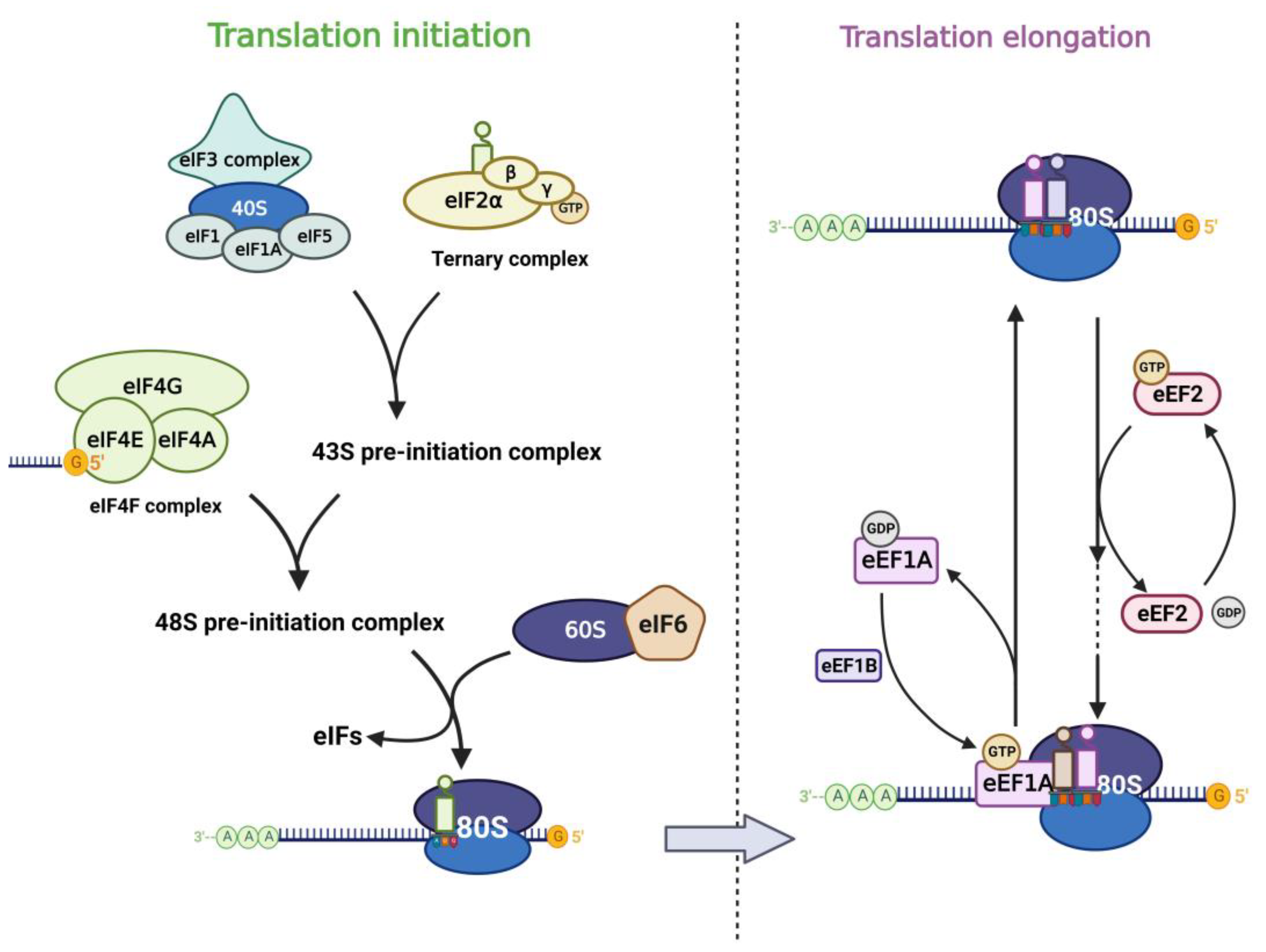
2.1. Translation Factors in Eukaryotic Translation

Translation of mRNA in eukaryotic cells includes cap-dependent and cap-independent translation, in which the eIF4F complex plays an important role. eIF4F contains three components (eIF4E, eIF4G, eIF4A), of which eIF4E is the cap-binding subunit of the eIF4F complex and is required for cap-dependent translation of all nuclear-encoded mRNAs [27]. In addition, eIF4E can also stimulate the RNA unwinding enzyme activity of eIF4A independently of its cap-binding function and thus promotes translation [28]. eIF4E interacts with eIF4G and binds the m7G cap structure of mRNA, which in turn promotes translation. eIF3 plays a central role in the translation initiation of classical cap-dependent translation and cap-independent translation [29,30,31]. Different subunits of eIF3 confer different functions to the eIF3 core complex. Besides, other eukaryotic initiation factors also play important role in the translation process. Translation initiation is generally regulated by the 43S pre-initiation complex (43S PIC), which consists of eIF1, eIF1A, eIF3, eIF5, and the ternary complex (TC) [22]. The TC is formed by eIF2 (containing α, β, γ subunits), tRNA, and GTP. When eIF2α is phosphorylated under stress, the TC formation is inhibited and the global translation is downregulated subsequently [32,33]. eIF6 was first reported to participate in the biogenesis of the 60S ribosomal subunit in the nucleus as an anti-association factor [34,35]. However, Gandin et al. [36] found that eIF6 is rate-limiting for efficient translation initiation. In the cytoplasm of mammalian cells, the phosphorylation of eIF6 on Ser235 leads to its release from the 60S, which promotes the formation of a translation-competent 80S ribosome [37]. The translation elongation process is carried out by the ribosome with the assistance of eEFs. Among them, eEF1A is an important component of the translational apparatus as it interacts with tRNA [26]. eEF2 possesses an RNA binding site that interacts with tRNAs and promotes conformational changes, thus allowing the latter to interact with the coding region of mRNAs, mediating the translocation of peptide chains in extension to the P-site of the ribosome [38].

2.2. Dysregulation of Translation Factors in Cancer

Most of the eIF4E-sensitive mRNAs have a long and highly structured 5’UTR region [39], and these mRNAs encode many proteins associated with cell proliferation and tumor progression, including MYC, VEGF, cyclin, and others [40]. Many studies have reported that overexpression of eIF4E is associated with poor prognosis in cancer patients and can lead to tumor vascularization and invasion [41]. In recent years, various subunits of eIF3 have been found to have altered expression in malignant tumors, affecting translation of oncogenic mRNAs. eIF3a expression level were first found to be elevated in breast cancer tissues compared to paired normal breast tissues by Bachmann et al. [42], and eIF3a might play an important role in regulating translation of specific mRNAs encoding α-microtubulin, RRM2, and proteins associated with the cell cycle [43]. In virus-induced murine mammary tumors, the eIF3e gene was identified as a common insertion site and suggested that production of truncated eIF3e could lead to malignant transformation of mammary epithelial cells [44]. In prostate cancer, elevation of eIF3h positively correlates with tumor stages. The expression level of eIF3h is higher in metastatic prostate cancer than in primary prostate cancer, and eIF3h may play an important regulatory role in the translation of specific mRNAs [45]. Therefore, aberrant overexpression of eIF3h may contribute to tumor development by upregulating the translation of important mRNAs associated with cell proliferation [46]. Other translation factors are also dysregulated in cancer. eIF1 expression is downregulated in pancreatic ductal adenocarcinoma [47]. eIF1A is essential for cell proliferation and the cell cycle in cancer [48]. Interestingly, although eIF2α phosphorylation leads to reduced global translation, the translation of a restricted subset of mRNAs is enhanced, which facilitates glycolysis and cell invasion in cancer [49,50]. eIF5 is overexpressed in colorectal cancer and hepatocellular carcinoma and predicts poor prognosis [22,51]. eIF6 is reported to be markedly upregulated in hepatocellular carcinoma, colorectal cancer, and gallbladder cancer, which lead to tumor progression via mTOR and AKT-related signaling pathways [52,53,54,55]. Targeting eIF6-mediated translation blunts lipid accumulation and oncogenic transformation in the liver [56]. eEF1A plays an important and well-defined role in cancer development and progression [26,57], and eEF1A is aberrantly highly expressed in a variety of tumors and suggests a poor prognosis [58,59]. eEF2 also plays an important role in promoting the progression of tumors such as breast cancer [38,60,61]. Taken together, these translation factors affect the translation initiation and elongation process of multiple mRNAs in different types of cancer.
As a key enzyme regulating mRNA fate, the regulation of METTL3 on the translation process in tumors cannot be ignored. In recent years, many studies have reported that METTL3 could lead to changes in the expression of target genes through the regulation of mRNA translation process, which in turn caused tumor progression. The importance of RBPs in canonical translation machinery in the aberrant translation of m6A-modified mRNAs was also mentioned in many studies [7,62]. Therefore, the following is intended to introduce the various mechanisms involved in the translational regulation of mRNA by METTL3 in cancer and the interconnection between various m6A reading proteins and canonical translation machinery in these processes, so as to provide new ideas and possibilities targeting METTL3-mediated translation regulation for cancer treatment.

3. METTL3 Functions as a Translation Regulator in Cancer

The life cycle of m6A-modified mRNA begins with the transcriptional process, and m6A modifications are mainly mediated by METTL3 in the nucleus. In general, when mRNA is transported to the cytoplasm, specific m6A reader proteins bind to m6A, which in turn affects the translation of mRNA [63]. Many studies in recent years have found that METTL3 could be directly or indirectly involved in the translational regulation of mRNAs through a variety of mechanisms, as summarized in Table 1.

3.1. METTL3 Mediates the Binding of Other m6A Reader Proteins to Promote Translation through m6A Modification

Wang et al. reported in 2015 that after m6A modification of the 3’UTR and stop codon regions of mRNA by METTL3 in the nucleus, mRNA translocated to the cytoplasm, where the m6A reader YTHDF1 selectively recognized the m6A sites. Eukaryotic translation initiation factors were recruited by YTHDF1 subsequently, which in turn promoted ribosome loading and assembling on target mRNA, and advanced cap-dependent or cap-independent translation initiation [7,64].
Subsequently, a large number of studies have emerged to support and enrich this theory. Song et al. discovered that in colorectal cancer, METTL3 catalyzed m6A modification in 3’UTR of HSF1 mRNA and protein expression of HSF1 was significantly downregulated after knockdown of YTHDF1, demonstrating that METTL3-mediated m6A could promote translation through the m6A reader YTHDF1 [65]. In endometrial cancer, reduced METTL3 expression leads to a decrease in m6A modification on PHLPP2 mRNA, resulting in attenuated YTHDF1-mediated translation of PHLPP2, which in turn caused de-repression of the AKT pathway and promotes tumor progression [66]. In melanoma, knockdown of METTL3 in bone marrow cells results in the lack of m6A modification on SPRED2, which in turn disrupts YTHDF1-mediated mRNA translation, leading to enhanced activation of NF-κB and STAT3 via the ERK pathway, contributing to tumor progression [67]. In gastric cancer, METTL3-mediated SPHK2 m6A modification followed by YTHDF1 facilitates translation initiation through interacting with eIF3a, which in turn upregulates the translation efficiency and promotes tumor progression [68]. In lung adenocarcinoma, m6A modification promotes YTHDF1-mediated translation of ENO1 and SLC7A11, thereby enhancing tumor glycolysis and ferroptosis [69,70]. In ocular melanoma, down-regulated METTL3-mediated m6A modification leads to attenuated YTHDF1-mediated translation of tumor suppressor HINT2, which promotes tumor progression [71]. In hepatocellular carcinoma, researchers discovered that METTL3 could catalyze m6A modification in CDS and 3’UTR of SNAI1 mRNA, and YTHDF1 tended to bind to m6A in CDS of SNAI1 to mediate translation. Researchers then treated cells with rapamycin and found that YTHDF1 mediated the cap-independent translation of SNAI1 and that YTHDF1 could synergize with eEF2 to promote translation extension of SNAI1 mRNA [72]. In gastrointestinal stromal tumors, METTL3 recruits YTHDF1 through m6A modification of MRP1 mRNA in 5’UTR and promotes translation extension by eEF1, which in turn leads to intracellular translocation of MRP1 to imatinib and promotes drug resistance [73]. In cervical and liver cancer, METTL3 mediates m6A modification of PDK4 in 5’UTR, and subsequently YTHDF1 synergizes with eEF2 to promote the translation of PDK4, which in turn enhances the glycolytic process in tumors [74]. In breast cancer, METTL3 mediates the m6A modification of KRT7 in CDS, followed by enhanced translation elongation with the involvement of YTHDF1/eEF1 [75].
In addition to YTHDF1, METTL3 can also regulate translation of mRNAs through YTHDF3. Researchers identified a certain overlap between YTHDF3 and YTHDF1-bound proteins in the cytoplasm, and it was found that YTHDF3 and YTHDF1 could simultaneously interact with eIF4A, which in turn accelerated the translation process. It was suggested that the m6A reader YTHDF3 could enhance YTHDF1-mediated translation after METTL3-mediated m6A modification in some mRNAs [64]. This mechanism was validated in the subsequent studies: METTL3 could promote translation initiation complex formation through m6A modification of YAP mRNA in lung cancer by recruiting YTHDF1/3 as well as eIF3b, which in turn improved the translation efficiency and stability of YAP mRNA [76], resulting in tumor metastasis. In bladder cancer, after m6A modification of ITGA6, YTHDF1 cooperated with YTHDF3 to promote the translation of ITGA6 and mediated tumor progression [77].
METTL3 also enhances mRNA translation through other m6A readers. Liu et al. found that METTL3 mediated m6A modification in 3’UTR of BMI1 in oral cancer [78]. Overexpression of METTL3 increased the binding of BMI1 mRNA to polysomes without altering the stability of BMI1 mRNA and the rate of protein degradation, suggesting an enhancement of the translation process. Subsequently, to investigate through which m6A reader protein METTL3 promotes translation, authors knocked down IGF2BP1, IGF2BP2, IGF2BP3, and YTHDF1 and found that BMI1 mRNA expression was not altered, while knockdown of IGF2BP1 downregulated BMI1 protein level. The above experiments illustrated that METTL3 could also promote the translation of mRNAs such as BMI1 through m6A readers other than YTHDF1/3, such as IGF2BP1. In esophageal cancer, METTL3 mediates m6A modification in 3’UTR of TNFR1 mRNA [79]. ATXN2 acts as a novel RNA binding protein and enhances the translation of m6A-modified TNFR1 mRNA.

3.2. METTL3 Enters the Cytoplasm to Facilitate the Translation Process

In oncology studies, researchers found that after m6A modification in the 3’ UTR of a large subset of mRNAs at sites close to the stop codon, METTL3 itself could tether to the mRNA as an m6A reader in the cytoplasm. Subsequently, METTL3 formed as a “bridge” between the 3’ UTR and the 5’cap-binding proteins of mRNA, which supported an mRNA looping mechanism for ribosome recycling and translational control. In addition, the researchers observed the close proximity of METTL3 and individual polyribosomes with cap-binding proteins such as eIF4E by electron microscopy, and found that METTL3 and eIF3h had direct physical and functional interactions, thus promoting the translation of a large number of oncogenic mRNAs including BRD4 [62,80].
Several studies subsequently reported evidence for the direct involvement of METTL3 in translation regulation in the cytoplasm. Song et al. found that METTL3 deletion in colorectal cancer significantly reduced the level of HSF1 mRNA in the polyribosome fractions and increased its level in the non-translating ribosome fractions. m6A-modified HSF1 resulted in direct tethering of METTL3 to HSF1 mRNA in the cytoplasm to facilitate the translation process [65]. In addition, interestingly, miR455-3p could also inhibit the translation of HSF1 mRNA by interacting with the m6A site of HSF1 located in 3’UTR, preventing METTL3-mediated m6A modification as well as the direct binding. In cervical cancer, researchers found that METTL3 was mainly localized in the cytoplasm. Knockdown of YTHDF1 did not affect protein expression of AXL, but protein expression of AXL was significantly upregulated after overexpression of both wild-type or catalytic mutant METTL3, suggesting that METTL3 was directly involved in the translation process of AXL in the cytoplasm [81]. In chronic myeloid leukemia (CML), investigators found the presence of METTL3 in the cytoplasm, then they verified that METTL3 knockdown led to a reduction in global translation efficiency in CML cells and showed a critical role for METTL3 in maintaining ribosome levels and translational potential [82]. Subsequent knockdown of METTL3 resulted in a significant decrease in m6A levels of genes involved in ribosome biogenesis and translation such PES1. After overexpression of wild-type and mutant METTL3 in cells, it was found that the protein levels of PES1 were both significantly increased, while the mRNA levels were unchanged, and both wild-type and catalytic mutant METTL3 were found to bind to 3’UTR of PES1 mRNA in the cytoplasm. Thus, this study revealed that METTL3 could promote the production of PES1 protein by directly binding to m6A-modified PES1 in the cytoplasm, which in turn upregulated the translation efficiency of its mRNA, ultimately allowing for enhanced ribosome synthesis and translation processes of other oncogenic mRNAs. In bladder cancer, investigators found that m6A levels in the 3’UTR of CDCP1 mRNA were increased during malignant transformation [83,84]. Mechanistically, after METTL3 mediated the m6A modification of CDCP1, METTL3 cooperated with YTHDF1 to bind to the 3’UTR m6A site of CPCP1 and thus promoted translation. Overexpression of METTL3 had no effect on the expression level and stability of CPCP1 mRNA, but upregulated the protein level of CDCP1 without changing the protein degradation rate, and significantly upregulated the polysome-bound CPCP1 mRNA. The catalytic mutant METTL3 can also promote the translation of m6A-modified CDCP1 mRNA, although with weaker activity compared with the wild type METTL3.
In 2022, Wei et al. [85] revealed a novel m6A-independent mechanism for METTL3 to regulate translation in gastric cancer progression. Cytoplasm-anchored METTL3 can promote the looping of some non-m6A-modified oncogenic mRNAs by interacting with PABPC1 and eIF4F complex. This study assigned a new function to cytoplasmic distributed METTL3 and expanded the ways in which METTL3 facilitates translation. We look forward to further studies to provide more evidence for this important and interesting finding.

3.3. Promoter-Bound METTL3 Enhances Translation

In addition to METTL3’s ability to upregulate translation efficiency through the binding of other reading proteins or binding to m6A-modified mRNAs directly, researchers have identified a mechanism by which METTL3 promotes m6A-dependent translation regulation through binding to the promoter of target genes. They found that in acute myeloid leukemia (AML), METTL3 could be localized to the transcription initiation site of target genes in chromatin independently of METTL14 which was essential for m6A modification [86]. Since the majority of target genes had the CAATT-box binding protein CEBPZ at the transcription initiation site, METTL3 could induce m6A modifications within the CDS region of related transcripts such as SP1 and SP2 mRNAs after their transcription by interacting with the CEBPZ protein and binding to these transcription initiation sites on chromosomes. Researchers found that the transcripts of METTL3-bound target genes were enriched in [GAG]n sequences that could cause ribosomal arrest during translation. When these sequences were modified by METTL3-mediated m6A methylation, ribosomal stalling was lifted, thereby contributing to enhanced translation. As transcription factors, SP1 and SP2 proteins played an important role in promoting AML progression. The transcriptional activity of SP1 and SP2 was unaffected by METTL3 deletion, but due to the lack of m6A modification on the transcripts, the transcripts were shifted to low molecular weight polyribosomes, resulting in reduced translational efficiency and less protein production, ultimately leading to reduced malignancy of AML cells.

3.4. Other Possible Pathways

Protein translation usually begins with the recruitment of the 43S pre-initiation complex to the 5’ cap structure of the mRNA via the cap-binding complex. However, some transcripts can be translated in a cap-independent manner through certain mechanisms. A study found that a single 5’UTR m6A could directly bind eIF3, which in turn allowed it to recruit the 43S complex to initiate translation without the involvement of the cap-binding protein eIF4E. The inhibition of adenosine methylation also selectively reduced the translation efficiency of the 5’UTR m6A-modified mRNA. This study revealed that cells under different stresses induced a redistribution of m6A modifications at the transcriptome level and could generate a translation pattern resulting from a 5’UTR m6A modification that bypassed the involvement of the eIF4F complex with eIF3 as a novel m6A reading protein [87]. Meanwhile, a similar mechanism was reported: in mammalian cells, the asymmetric distribution of m6A along mRNA resulted in relatively little methylation in the 5’UTR. However, in the heat shock stress response, certain adenosines in the 5’UTR of the newly transcribed mRNA were preferentially methylated, and increased m6A modification in this region promoted cap-independent translation initiation, revealing a novel mechanism of translation regulation under stress [88]. Therefore, it is worthwhile to further explore whether this mechanism of mRNA translation with eIF3 directly as an m6A reader involved in 5’UTR m6A modification exists in cancer.
In summary, METTL3 is involved in translation regulation in a variety of ways in cancer, which can be summarized as follows (see Figure 2):
(1)
METTL3 modifies m6A in target mRNAs and then recruits canonical translation machinery through classical or novel m6A readers YTHDF1/YTHDF3/IGF2BP1/ATXN2 to promote translation.
(2)
After m6A modification of target mRNAs, METTL3 directly binds m6A sites in the cytoplasm and recruits canonical translation machinery to promote translation.
(3)
METTL3 interacts with CEBPZ to bind the promoter of target genes and enhances the translation of the associated mRNAs by relieving ribosomal arrest through m6A modification.

4. Conclusions and Perspectives

In recent years, more and more studies have reported that METTL3-mediated m6A modification of mRNAs affected various processes in the mRNA life cycle through multiple intermolecular interactions, which in turn mediated various phenotypic changes in cancer. In this review, we focused on the involvement of METTL3 in the regulation of mRNA translation. First, we briefly introduced the aberrant translation in cancer and its relationship with canonical translation machinery. Then, we introduced in detail the different roles of METTL3 in the aberrant translation process of m6A-modified mRNA. With the gradual clarification of the mechanism of aberrant translation regulation in cancer, targeting the METTL3-mediated translation process has become a possibility. Since METTL3 interacts with multiple translation-related RBPs in the process of translation promotion, the use of small molecule inhibitors targeting canonical translation machinery for cancer treatment seems possible. Small molecule inhibitors targeting eIF4A and eIF4E have been introduced for a long time. For example, 4EGI-1, an inhibitor of eIF4E- eIF4G interaction [89], can decrease eIF4E-sensitive mRNA translation level and demonstrate a favorable antitumor effect. However, advancing to clinical application has been delayed [89,90]. The inhibitors against the binding of eIF6 to the 60S were also identified and might have dose- and cell-specific effects [91]. We cannot help but wonder whether targeting key molecules upstream of the translation machinery in cancer translation regulation could produce a broader spectrum and better efficacy of inhibitory effects on aberrant translation.
Encouragingly, the first launch of STM2457, a small molecule inhibitor targeting METTL3, was published in 2021, and its promising antitumor effect was validated in AML [92]. Moreover, Du et al. [93] used virtual screening of 1042 natural products and identified quercetin as a qualified METTL3 small molecule inhibitor. Moroz et al. [94] developed a METTL3 inhibitor UZH1a using a structure-based drug discovery approach. Lee et al. [95] reported eltrombopag as an allosteric inhibitor of the METTL3-14 complex. Since the determinants of successful clinical application of small molecule inhibitors include confirmation of the compound-mechanism hypothesis, compound-target action and pharmacodynamic activity [96], if the likelihood of successful drug development is to be maximized, a high standard of screening for inhibitors targeting translational regulation is required in preclinical studies. Therefore, whether drugs such as STM2457 can target METTL3 and thereby inhibit aberrant translation in other types of cancer such as lung cancer remains to be explored and validated through numerous studies. Combining small molecule inhibitors of METTL3 and canonical translation machinery inhibitors to overcome intra-tumor heterogeneity and inhibit the oncogenic signal of aberrant translation integration in cancer cells is also promising. Since the initial success of therapeutic approaches targeting abnormal translation in cancer has been achieved, we believe that in the near future, these drugs will move from the laboratory to the clinic and achieve breakthroughs in anti-cancer therapy.

Author Contributions

Conceptualization, W.M.; investigation, W.M. and H.X.; writing—original draft preparation, P.M.; writing—review and editing, J.C., Y.W. and R.Z.; visualization, J.C., Y.W. and R.Z.; supervision, Y.L., W.M. and H.X. contributed equally to this work. All authors have read and agreed to the published version of the manuscript.

Funding

This work was supported by the National Natural Science Foundation of China (No. 82072593), and Department of Science and Technology of Hubei Province (No. 2020BCB027).

Institutional Review Board Statement

Not applicable.

Informed Consent Statement

Not applicable.

Data Availability Statement

The data that support the findings of this study are available from the corresponding author upon reasonable request.

Conflicts of Interest

The authors declare that they have no conflicts of interest.

References

  1. Desrosiers, R.; Friderici, K.; Rottman, F. Identification of methylated nucleosides in messenger RNA from Novikoff hepatoma cells. Proc. Natl. Acad. Sci. USA 1974, 71, 3971–3975. [Google Scholar] [CrossRef] [PubMed] [Green Version]
  2. Bokar, J.A.; Shambaugh, M.E.; Polayes, D.; Matera, A.G.; Rottman, F.M. Purification and cDNA cloning of the AdoMet-binding subunit of the human mRNA (N6-adenosine)-methyltransferase. RNA 1997, 3, 1233–1247. [Google Scholar] [PubMed]
  3. Meyer, K.D.; Saletore, Y.; Zumbo, P.; Elemento, O.; Mason, C.E.; Jaffrey, S.R. Comprehensive analysis of mRNA methylation reveals enrichment in 3’ UTRs and near stop codons. Cell 2012, 149, 1635–1646. [Google Scholar] [CrossRef] [PubMed] [Green Version]
  4. Dominissini, D.; Moshitch-Moshkovitz, S.; Schwartz, S.; Salmon-Divon, M. Topology of the human and mouse m6A RNA methylomes revealed by m6A-seq. Nature 2012, 485, 201–206. [Google Scholar] [CrossRef] [PubMed]
  5. Linder, B.; Grozhik, A.; Olarerin-George, A.; Meydan, C. Single-nucleotide-resolution mapping of m6A and m6Am throughout the transcriptome. Nat. Methods 2015, 12, 767–772. [Google Scholar] [CrossRef]
  6. Perry, R.P.; Scherrer, K. The methylated constituents of globin mRNA. FEBS Lett. 1975, 57, 73–78. [Google Scholar] [CrossRef] [Green Version]
  7. Wang, X.; Zhao, B.S.; Roundtree, I.A. N(6)-methyladenosine Modulates Messenger RNA Translation Efficiency. Cell 2015, 161, 1388–1399. [Google Scholar] [CrossRef] [Green Version]
  8. Tang, Y.; Chen, K.; Song, B.; Ma, J.; Wu, X.; Xu, Q.; Wei, Z.; Su, J.; Liu, G.; Rong, R.; et al. m6A-Atlas: A comprehensive knowledgebase for unraveling the N6-methyladenosine (m6A) epitranscriptome. Nucleic Acids Res. 2021, 49, D134–D143. [Google Scholar] [CrossRef]
  9. Geula, S.; Moshitch-Moshkovitz, S.; Dominissini, D.; Mansour, A.A.; Kol, N.; Salmon-Divon, M.; Hershkovitz, V.; Peer, E.; Mor, N.; Manor, Y.S.; et al. Stem cells. m6A mRNA methylation facilitates resolution of naïve pluripotency toward differentiation. Science 2015, 347, 1002–1006. [Google Scholar] [CrossRef]
  10. Yoon, K.-J.; Ringeling, F.R.; Vissers, C.; Jacob, F.; Pokrass, M.; Jimenez-Cyrus, D.; Su, Y.; Kim, N.-S.; Zhu, Y.; Zheng, L.; et al. Temporal Control of Mammalian Cortical Neurogenesis by m6A Methylation. Cell 2017, 171, 877–889.e17. [Google Scholar]
  11. Livneh, I.; Moshitch-Moshkovitz, S.; Amariglio, N.; Rechavi, G.; Dominissini, D. The m 6 A epitranscriptome: Transcriptome plasticity in brain development and function. Nat. Rev. Neurosci. 2020, 21, 36–51. [Google Scholar] [PubMed]
  12. Zaccara, S.; Ries, R.J.; Jaffrey, S.R. Reading, writing and erasing mRNA methylation. Nat. Rev. Mol. Cell Biol. 2019, 20, 608–624. [Google Scholar]
  13. Deng, L.J.; Deng, W.Q.; Fan, S.R.; Chen, M.F.; Qi, M.; Lyu, W.Y.; Qi, Q.; Tiwari, A.K.; Chen, J.X.; Zhang, D.M.; et al. m6A modification: Recent advances, anticancer targeted drug discovery and beyond. Mol. Cancer 2022, 21, 52. [Google Scholar] [PubMed]
  14. Oerum, S.; Meynier, V.; Catala, M.; Tisné, C. A comprehensive review of m6A/m6Am RNA methyltransferase structures. Nucleic Acids Res. 2021, 49, 7239–7255. [Google Scholar] [CrossRef] [PubMed]
  15. Cheng, M.; Sheng, L.; Gao, Q.; Xiong, Q.; Zhang, H.; Wu, M.; Liang, Y.; Zhu, F.; Zhang, Y.; Zhang, X.; et al. The m6A methyltransferase METTL3 promotes bladder cancer progression via AFF4/NF-κB/MYC signaling network. Oncogene 2019, 38, 3667–3680. [Google Scholar] [CrossRef]
  16. Liu, L.; Wang, J.; Sun, G.; Wu, Q.; Ma, J.; Zhang, X.; Huang, N.; Bian, Z.; Gu, S.; Xu, M.; et al. m6A mRNA methylation regulates CTNNB1 to promote the proliferation of hepatoblastoma. Mol. Cancer 2019, 18, 188. [Google Scholar] [CrossRef] [PubMed] [Green Version]
  17. Wang, W.; Shao, F.; Yang, X.; Wang, J.; Zhu, R.; Yang, Y.; Zhao, G.; Guo, D.; Sun, Y.; Wang, J.; et al. METTL3 promotes tumour development by decreasing APC expression mediated by APC mRNA N6-methyladenosine-dependent YTHDF binding. Nat. Commun. 2021, 12, 3803. [Google Scholar] [CrossRef]
  18. Cui, Q.; Shi, H.; Ye, P.; Li, L.; Qu, Q.; Sun, G.; Sun, G.; Lu, Z.; Huang, Y.; Yang, C.-G.; et al. m(6)A RNA Methylation Regulates the Self-Renewal and Tumorigenesis of Glioblastoma Stem Cells. Cell Rep. 2017, 18, 2622–2634. [Google Scholar] [CrossRef]
  19. Wu, Y.; Chang, N.; Zhang, Y.; Zhang, X.; Xu, L.; Che, Y.; Qiao, T.; Bin Wu, B.; Zhou, Y.; Jiang, J.; et al. METTL3-mediated m(6)A mRNA modification of FBXW7 suppresses lung adenocarcinoma. J. Exp. Clin. Cancer Res. 2021, 40, 90. [Google Scholar] [CrossRef]
  20. Buttgereit, F.; Brand, M.D. A hierarchy of ATP-consuming processes in mammalian cells. Biochem. J. 1995, 312, 163–167. [Google Scholar] [CrossRef]
  21. Schwanhäusser, B.; Busse, D.; Li, N.; Dittmar, G.; Schuchhardt, J.; Wolf, J.; Chen, W.; Selbach, M. Global quantification of mammalian gene expression control. Nature 2011, 473, 337–342. [Google Scholar] [CrossRef] [PubMed] [Green Version]
  22. Bhat, M.; Robichaud, N.; Hulea, L.; Sonenberg, N.; Pelletier, J.; Topisirovic, I. Targeting the translation machinery in cancer. Nat. Rev. Drug Discov. 2015, 14, 261–278. [Google Scholar] [CrossRef] [PubMed]
  23. Colina, R.; Costa-Mattioli, M.; Dowling, R.J.O.; Jaramillo, M.; Tai, L.-H.; Breitbach, C.J.; Martineau, Y.; Larsson, O.; Rong, L.; Svitkin, Y.V.; et al. Translational control of the innate immune response through IRF-7. Nature 2008, 452, 323–328. [Google Scholar] [CrossRef]
  24. Larsson, O.; Perlman, D.M.; Fan, D.; Reilly, C.S.; Peterson, M.; Dahlgren, C.; Liang, Z.; Li, S.; Polunovsky, V.A.; Wahlestedt, C.; et al. Apoptosis resistance downstream of eIF4E: Posttranscriptional activation of an anti-apoptotic transcript carrying a consensus hairpin structure. Nucleic Acids Res. 2006, 34, 4375–4386. [Google Scholar] [CrossRef] [Green Version]
  25. Ruggero, D. Translational control in cancer etiology. Cold Spring Harb. Perspect. Biol. 2013, 5, a012336. [Google Scholar] [CrossRef]
  26. Rubio, A.; Garland, G.D.; Sfakianos, A. Aberrant protein synthesis and cancer development: The role of canonical eukaryotic initiation, elongation and termination factors in tumorigenesis. Semin. Cancer Biol. 2022, 86, 151–165. [Google Scholar] [CrossRef] [PubMed]
  27. De Benedetti, A.; Graff, J.R. eIF-4E expression and its role in malignancies and metastases. Oncogene 2004, 23, 3189–3199. [Google Scholar] [CrossRef] [Green Version]
  28. Feoktistova, K.; Tuvshintogs, E.; Do, A.; Fraser, C.S. Human eIF4E promotes mRNA restructuring by stimulating eIF4A helicase activity. Proc. Natl. Acad. Sci. USA 2013, 110, 13339–13344. [Google Scholar] [CrossRef] [Green Version]
  29. Merrick, W.C.; Pavitt, G.D. Protein Synthesis Initiation in Eukaryotic Cells. Cold Spring Harb. Perspect. Biol. 2018, 10, a033092. [Google Scholar] [CrossRef]
  30. Kozak, M. New ways of initiating translation in eukaryotes? Mol. Cell Biol. 2001, 21, 1899–1907. [Google Scholar] [CrossRef] [PubMed] [Green Version]
  31. Bhardwaj, U.; Powell, P.; Goss, D.J. Eukaryotic initiation factor (eIF) 3 mediates Barley Yellow Dwarf Viral mRNA 3’-5’ UTR interactions and 40S ribosomal subunit binding to facilitate cap-independent translation. Nucleic Acids Res. 2019, 47, 6225–6235. [Google Scholar] [CrossRef] [PubMed] [Green Version]
  32. Hinnebusch, A.G. The scanning mechanism of eukaryotic translation initiation. Annu. Rev. Biochem. 2014, 83, 779–812. [Google Scholar] [CrossRef] [PubMed]
  33. Rios-Fuller, T.J.; Mahe, M.; Walters, B.; Abbadi, D.; Pérez-Baos, S. Translation Regulation by eIF2α Phosphorylation and mTORC1 Signaling Pathways in Non-Communicable Diseases (NCDs). Int. J. Mol. Sci. 2020, 21, 5301. [Google Scholar] [CrossRef] [PubMed]
  34. Sanvito, F.; Piatti, S.; Villa, A.; Bossi, M.; Lucchini, G.; Marchisio, P.C.; Biffo, S. The beta4 integrin interactor p27(BBP/eIF6) is an essential nuclear matrix protein involved in 60S ribosomal subunit assembly. J. Cell Biol. 1999, 144, 823–837. [Google Scholar] [CrossRef] [Green Version]
  35. Basu, U.; Si, K.; Warner, J.R.; Maitra, U. The Saccharomyces cerevisiae TIF6 gene encoding translation initiation factor 6 is required for 60S ribosomal subunit biogenesis. Mol. Cell Biol. 2001, 21, 1453–1462. [Google Scholar] [CrossRef] [Green Version]
  36. Gandin, V.; Miluzio, A.; Barbieri, A.M.; Beugnet, A.; Kiyokawa, H.; Marchisio, P.C.; Biffo, S. Eukaryotic initiation factor 6 is rate-limiting in translation, growth and transformation. Nature 2008, 455, 684–688. [Google Scholar] [CrossRef] [Green Version]
  37. Miluzio, A.; Beugnet, A.; Volta, V.; Biffo, S. Eukaryotic initiation factor 6 mediates a continuum between 60S ribosome biogenesis and translation. EMBO Rep. 2009, 10, 459–465. [Google Scholar] [CrossRef] [PubMed] [Green Version]
  38. Proud, C.G. Regulation and roles of elongation factor 2 kinase. Biochem. Soc. Trans. 2015, 43, 328–332. [Google Scholar] [CrossRef]
  39. Robichaud, N.; Sonenberg, N.; Ruggero, D.; Schneider, R.J. Translational Control in Cancer. Cold Spring Harb. Perspect. Biol. 2019, 11, a032896. [Google Scholar] [CrossRef] [Green Version]
  40. Kevil, C.G.; De Benedetti, A.; Payne, D.K.; Coe, L.L.; Laroux, F.S.; Alexander, J.S. Translational regulation of vascular permeability factor by eukaryotic initiation factor 4E: Implications for tumor angiogenesis. Int. J. Cancer 1996, 65, 785–790. [Google Scholar] [CrossRef]
  41. Nathan, C.-A.; Carter, P.; Liu, L.; Li, B.D.; Abreo, F.; Tudor, A.; Zimmer, S.G.; De Benedetti, A. Elevated expression of eIF4E and FGF-2 isoforms during vascularization of breast carcinomas. Oncogene 1997, 15, 1087–1094. [Google Scholar] [CrossRef] [PubMed] [Green Version]
  42. Bachmann, F.; Bänziger, R.; Burger, M.M. Cloning of a novel protein overexpressed in human mammary carcinoma. Cancer Res. 1997, 57, 988–994. [Google Scholar] [PubMed]
  43. Dong, Z.; Zhang, J.T. EIF3 p170, a mediator of mimosine effect on protein synthesis and cell cycle progression. Mol. Biol. Cell 2003, 14, 3942–3951. [Google Scholar] [CrossRef] [PubMed] [Green Version]
  44. Mayeur, G.L.; Hershey, J.W. Malignant transformation by the eukaryotic translation initiation factor 3 subunit p48 (eIF3e). FEBS Lett. 2002, 514, 49–54. [Google Scholar] [CrossRef] [PubMed] [Green Version]
  45. Nupponen, N.N.; Porkka, K.; Kakkola, L.; Tanner, M.; Persson, K.; Borg, A.; Isola, J.; Visakorpi, T. Amplification and overexpression of p40 subunit of eukaryotic translation initiation factor 3 in breast and prostate cancer. Am. J. Pathol. 1999, 154, 1777–1783. [Google Scholar] [CrossRef] [Green Version]
  46. Saramäki, O.; Willi, N.; Bratt, O.; Gasser, T.C.; Koivisto, P.; Nupponen, N.N.; Bubendorf, L.; Visakorpi, T. Amplification of EIF3S3 gene is associated with advanced stage in prostate cancer. Am. J. Pathol. 2001, 159, 2089–2094. [Google Scholar] [CrossRef] [Green Version]
  47. Golob-Schwarzl, N.; Puchas, P.; Gogg-Kamerer, M.; Weichert, W.; Göppert, B.; Haybaeck, J. New Pancreatic Cancer Biomarkers eIF1, eIF2D, eIF3C and eIF6 Play a Major Role in Translational Control in Ductal Adenocarcinoma. Anticancer Res. 2020, 40, 3109–3118. [Google Scholar] [CrossRef]
  48. Sehrawat, U.; Koning, F.; Ashkenazi, S.; Stelzer, G.; Leshkowitz, D.; Dikstein, R. Cancer-Associated Eukaryotic Translation Initiation Factor 1A Mutants Impair Rps3 and Rps10 Binding and Enhance Scanning of Cell Cycle Genes. Mol. Cell Biol. 2019, 39, e00441-18. [Google Scholar] [CrossRef] [PubMed] [Green Version]
  49. García-Jiménez, C.; Goding, C.R. Starvation and Pseudo-Starvation as Drivers of Cancer Metastasis through Translation Reprogramming. Cell Metab. 2019, 29, 254–267. [Google Scholar] [CrossRef] [Green Version]
  50. Li, Z.; Ge, Y.; Dong, J.; Wang, H.; Zhao, T.; Wang, X.; Liu, J.; Gao, S.; Shi, L.; Yang, S.; et al. BZW1 Facilitates Glycolysis and Promotes Tumor Growth in Pancreatic Ductal Adenocarcinoma Through Potentiating eIF2α Phosphorylation. Gastroenterology 2022, 162, 1256–1271.e14. [Google Scholar] [CrossRef]
  51. Golob-Schwarzl, N.; Krassnig, S.; Toeglhofer, A.M.; Park, Y.N.; Gogg-Kamerer, M.; Vierlinger, K.; Schröder, F.; Rhee, H.; Schicho, R.; Fickert, P.; et al. New liver cancer biomarkers: PI3K/AKT/mTOR pathway members and eukaryotic translation initiation factors. Eur. J. Cancer 2017, 83, 56–70. [Google Scholar] [CrossRef] [PubMed]
  52. Sun, L.; Liu, S.; Wang, X.; Zheng, X.; Chen, Y.; Shen, H. eIF6 promotes the malignant progression of human hepatocellular carcinoma via the mTOR signaling pathway. J. Transl. Med. 2021, 19, 216. [Google Scholar] [CrossRef] [PubMed]
  53. Scagliola, A.; Miluzio, A.; Mori, G.; Ricciardi, S.; Oliveto, S.; Manfrini, N.; Biffo, S. Inhibition of eIF6 Activity Reduces Hepatocellular Carcinoma Growth: An In Vivo and In Vitro Study. Int. J. Mol. Sci. 2022, 23, 7720. [Google Scholar] [CrossRef] [PubMed]
  54. Lin, J.; Yu, X.; Xie, L.; Wang, P.; Li, T.; Xiao, Y.; Zhou, J.; Peng, S.; Huang, J.; Luo, Y.; et al. eIF6 Promotes Colorectal Cancer Proliferation and Invasion by Regulating AKT-Related Signaling Pathways. J. Biomed. Nanotechnol. 2019, 15, 1556–1567. [Google Scholar] [CrossRef]
  55. Golob-Schwarzl, N.; Wodlej, C.; Kleinegger, F.; Gogg-Kamerer, M.; Birkl-Toeglhofer, A.M.; Petzold, J.; Aigelsreiter, A.; Thalhammer, M.; Park, Y.N.; Haybaeck, J. Eukaryotic translation initiation factor 6 overexpression plays a major role in the translational control of gallbladder cancer. J. Cancer Res. Clin. Oncol. 2019, 145, 2699–2711. [Google Scholar] [CrossRef] [Green Version]
  56. Scagliola, A.; Miluzio, A.; Ventura, G.; Oliveto, S.; Cordiglieri, C. Targeting of eIF6-driven translation induces a metabolic rewiring that reduces NAFLD and the consequent evolution to hepatocellular carcinoma. Nat. Commun. 2021, 12, 4878. [Google Scholar] [CrossRef]
  57. Abbas, W.; Kumar, A.; Herbein, G. The eEF1A Proteins: At the Crossroads of Oncogenesis, Apoptosis, and Viral Infections. Front. Oncol. 2015, 7, 75. [Google Scholar] [CrossRef] [Green Version]
  58. Lee, S.; Francoeur, A.; Liu, E.W.S. Tissue-specific expression in mammalian brain, heart, and muscle of S1, a member of the elongation factor-1 alpha gene family. J. Biol. Chem. 1992, 267, 24064–24068. [Google Scholar] [CrossRef]
  59. Tomlinson, V.A.; Newbery, H.J.; Wray, N.R.; Jackson, J.; Larionov, A.; Miller, W.R.; Dixon, J.M. Translation elongation factor eEF1A2 is a potential oncoprotein that is overexpressed in two-thirds of breast tumours. BMC Cancer 2005, 12, 113. [Google Scholar] [CrossRef] [Green Version]
  60. Liu, R.; Proud, C.G. Eukaryotic elongation factor 2 kinase as a drug target in cancer, and in cardiovascular and neurodegenerative diseases. Acta Pharmacol. Sin. 2016, 37, 285–294. [Google Scholar] [CrossRef] [Green Version]
  61. Castello, A.; Fischer, B.; Eichelbaum, K.; Horos, R.; Beckmann, B.M.; Strein, C.; Davey, N.E.; Humphreys, D.T.; Preiss, T.; Steinmetz, L.M.; et al. Insights into RNA biology from an atlas of mammalian mRNA-binding proteins. Cell 2012, 8, 1393–1406. [Google Scholar] [CrossRef] [PubMed] [Green Version]
  62. Choe, J.; Lin, S.; Zhang, W.; Liu, Q.; Wang, L.; Ramirez-Moya, J.; Du, P.; Kim, W.; Tang, S.; Sliz, P.; et al. mRNA circularization by METTL3-eIF3h enhances translation and promotes oncogenesis. Nature 2018, 561, 556–560. [Google Scholar] [CrossRef] [PubMed]
  63. Meyer, K.D.; Jaffrey, S.R. Rethinking m6A Readers, Writers, and Erasers. Annu. Rev. Cell Dev. Biol. 2017, 33, 319–342. [Google Scholar] [CrossRef] [PubMed] [Green Version]
  64. Shi, H.; Wang, X.; Lu, Z.; Zhao, B.S.; Ma, H.; Hsu, P.J.; Liu, C.; He, C. YTHDF3 facilitates translation and decay of N6-methyladenosine-modified RNA. Cell Res. 2017, 27, 315–328. [Google Scholar] [CrossRef] [PubMed] [Green Version]
  65. Song, P.; Feng, L.; Li, J.; Dai, D.; Zhu, L.; Wang, C.; Li, J.; Li, L.; Zhou, Q.; Shi, R.; et al. β-catenin represses miR455-3p to stimulate m6A modification of HSF1 mRNA and promote its translation in colorectal cancer. Mol. Cancer 2020, 19, 129. [Google Scholar] [CrossRef] [PubMed]
  66. Liu, J.; Eckert, M.A.; Harada, B.T.; Liu, S.-M.; Lu, Z.; Yu, K.; Tienda, S.M.; Chryplewicz, A.; Zhu, A.C.; Yang, Y.; et al. m6A mRNA methylation regulates AKT activity to promote the proliferation and tumorigenicity of endometrial cancer. Nat. Cell Biol. 2018, 20, 1074–1083. [Google Scholar] [CrossRef] [PubMed]
  67. Yin, H.; Zhang, X.; Yang, P.; Zhang, X.; Peng, Y.; Li, D.; Yu, Y.; Wu, Y.; Wang, Y.; Zhang, J.; et al. RNA m6A methylation orchestrates cancer growth and metastasis via macrophage reprogramming. Nat. Commun. 2021, 12, 1394. [Google Scholar] [CrossRef]
  68. Huo, F.-C.; Zhu, Z.-M.; Zhu, W.-T.; Du, Q.-Y.; Liang, J.; Mou, J. METTL3-mediated m6A methylation of SPHK2 promotes gastric cancer progression by targeting KLF2. Oncogene 2021, 40, 2968–2981. [Google Scholar] [CrossRef]
  69. Ma, L.; Xue, X.; Zhang, X.; Yu, K.; Xu, X.; Tian, X.; Miao, Y.; Meng, F.; Liu, X.; Guo, S.; et al. The essential roles of m6A RNA modification to stimulate ENO1-dependent glycolysis and tumorigenesis in lung adenocarcinoma. J. Exp. Clin. Cancer Res. 2022, 41, 36. [Google Scholar] [CrossRef]
  70. Xu, Y.; Lv, D.; Yan, C.; Su, H. METTL3 promotes lung adenocarcinoma tumor growth and inhibits ferroptosis by stabilizing SLC7A11 m 6 A modification. Cancer Cell Int. 2022, 22, 11. [Google Scholar] [CrossRef]
  71. Jia, R.; Chai, P.; Wang, S.; Sun, B. m6A modification suppresses ocular melanoma through modulating HINT2 mRNA translation. Mol. Cancer 2019, 18, 161. [Google Scholar] [CrossRef] [Green Version]
  72. Lin, X.; Chai, G.; Wu, Y.; Li, J.; Chen, F.; Liu, J.; Luo, G.; Tauler, J.; Du, J.; Lin, S.; et al. RNA m6A methylation regulates the epithelial mesenchymal transition of cancer cells and translation of Snail. Nat. Commun. 2019, 10, 2065. [Google Scholar] [CrossRef] [PubMed]
  73. Xu, K.; Zhang, Q.; Chen, M.; Li, B.; Wang, N.; Li, C.; Gao, Z.; Zhang, D.; Yang, L.; Xu, Z.; et al. N6-methyladenosine modification regulates imatinib resistance of gastrointestinal stromal tumor by enhancing the expression of multidrug transporter MRP1. Cancer Lett. 2022, 530, 85–99. [Google Scholar] [CrossRef] [PubMed]
  74. Li, Z.; Peng, Y.; Li, J. N6-methyladenosine regulates glycolysis of cancer cells through PDK4. Nat. Commun. 2020, 11, 2578. [Google Scholar] [CrossRef]
  75. Chen, F.; Chen, Z.; Guan, T. N6-methyladenosine regulates mRNA stability and translation efficiency of KRT7 to promote breast cancer lung metastasis. Cancer Res. 2021, 81, 2847–2860. [Google Scholar] [CrossRef]
  76. Jin, D.; Guo, J.; Wu, Y.; Du, J.; Yang, L.; Wang, X.; Di, W.; Hu, B.; An, J.; Kong, L.; et al. m(6)A mRNA methylation initiated by METTL3 directly promotes YAP translation and increases YAP activity by regulating the MALAT1-miR-1914-3p-YAP axis to induce NSCLC drug resistance and metastasis. J. Hematol. Oncol. 2019, 12, 135. [Google Scholar] [CrossRef] [PubMed] [Green Version]
  77. Jin, H.; Ying, X.; Que, B. N6-methyladenosine modification of ITGA6mRNA promotes the development and progression of bladder cancer. EBioMedicine 2019, 47, 195–207. [Google Scholar] [CrossRef] [Green Version]
  78. Liu, L.; Wu, Y.; Li, Q.; Liang, J.; He, Q. METTL3 Promotes Tumorigenesis and Metastasis through BMI1 m6A Methylation in Oral Squamous Cell Carcinoma. Mol. Ther. 2020, 28, 2177–2190. [Google Scholar] [CrossRef]
  79. Li, R.; Zeng, L.; Zhao, H.; Deng, J.; Pan, L.; Zhang, S.; Wu, G.; Ye, Y.; Zhang, J.; Su, J.; et al. ATXN2-mediated translation of TNFR1 promotes esophageal squamous cell carcinoma via m(6)A-dependent manner. Mol. Ther. 2022, 30, 1089–1103. [Google Scholar] [CrossRef]
  80. Lin, S.; Choe, J.; Du, P.; Triboulet, R.; Gregory, R.I. The m(6)A Methyltransferase METTL3 Promotes Translation in Human Cancer Cells. Mol. Cell 2016, 62, 335–345. [Google Scholar] [CrossRef] [Green Version]
  81. Hua, W.; Zhao, Y.; Jin, X.; Yu, D.; He, J.; Xie, D.; Duan, P. METTL3 promotes ovarian carcinoma growth and invasion through the regulation of AXL translation and epithelial to mesenchymal transition. Gynecol. Oncol. 2018, 151, 356–365. [Google Scholar] [CrossRef]
  82. Ianniello, Z.; Sorci, M.; Ginistrelli, L.C.; Iaiza, A.; Marchioni, M.; Tito, C.; Capuano, E.; Masciarelli, S.; Ottone, T.; Attrotto, C.; et al. New insight into the catalytic -dependent and -independent roles of METTL3 in sustaining aberrant translation in chronic myeloid leukemia. Cell Death Dis. 2021, 12, 870. [Google Scholar] [CrossRef]
  83. Yang, F.; Jin, H.; Que, B.; Chao, Y.; Zhang, H.; Ying, X.; Zhou, Z.; Yuan, Z.; Su, J.; Wu, B.; et al. Dynamic m6A mRNA methylation reveals the role of METTL3-m6A-CDCP1 signaling axis in chemical carcinogenesis. Oncogene 2019, 38, 4755–4772. [Google Scholar] [CrossRef] [PubMed]
  84. Ying, X.; Jiang, X.; Zhang, H.; Liu, B. Programmable N6-methyladenosine modification of CDCP1 mRNA by RCas9-methyltransferase like 3 conjugates promotes bladder cancer development. Mol. Cancer 2020, 19, 169. [Google Scholar] [CrossRef] [PubMed]
  85. Wei, X.; Huo, Y.; Pi, J.; Gao, Y.; Rao, S.; He, M.; Wei, Q.; Song, P.; Chen, Y.; Lu, D.; et al. METTL3 preferentially enhances non-m(6)A translation of epigenetic factors and promotes tumourigenesis. Nat. Cell Biol. 2022, 24, 1278–1290. [Google Scholar] [CrossRef]
  86. Barbieri, I.; Tzelepis, K.; Pandolfini, L.; Shi, J.; Millán-Zambrano, G.; Robson, S.C.; Aspris, D.; Migliori, V.; Bannister, A.J.; Han, N.; et al. Promoter-bound METTL3 maintains myeloid leukaemia by m6A-dependent translation control. Nature 2017, 552, 126–131. [Google Scholar] [CrossRef] [PubMed]
  87. Meyer, K.D.; Patil, D.P.; Zhou, J.; Zinoviev, A.; Skabkin, M.A.; Elemento, O.; Pestova, T.V.; Qian, S.-B.; Jaffrey, S.R. 5’ UTR m(6)A Promotes Cap-Independent Translation. Cell 2015, 163, 999–1010. [Google Scholar] [CrossRef] [Green Version]
  88. Zhou, J.; Wan, J.; Gao, X.; Zhang, X.; Jaffrey, S.R.; Qian, S.-B. Dynamic m(6)A mRNA methylation directs translational control of heat shock response. Nature 2015, 526, 591–594. [Google Scholar] [CrossRef] [Green Version]
  89. Moerke, N.J.; Aktas, H.; Chen, H.; Cantel, S.; Reibarkh, M.Y.; Fahmy, A.; Gross, J.D.; Degterev, A.; Yuan, J.; Chorev, M.; et al. Small-molecule inhibition of the interaction between the translation initiation factors eIF4E and eIF4G. Cell 2007, 128, 257–267. [Google Scholar] [CrossRef]
  90. Bordeleau, M.-E.; Mori, A.; Oberer, M.; Lindqvist, L.; Chard, L.S.; Higa, T.; Belsham, G.J.; Wagner, G.; Tanaka, J.; Pelletier, J. Functional characterization of IRESes by an inhibitor of the RNA helicase eIF4A. Nat. Chem. Biol. 2006, 2, 213–220. [Google Scholar] [CrossRef]
  91. Pesce, E.; Miluzio, A.; Turcano, L.; Minici, C.; Cirino, D.; Calamita, P.; Manfrini, N.; Oliveto, S.; Ricciardi, S.; Grifantini, R.; et al. Discovery and Preliminary Characterization of Translational Modulators that Impair the Binding of eIF6 to 60S Ribosomal Subunits. Cells 2020, 9, 172. [Google Scholar] [CrossRef] [PubMed] [Green Version]
  92. Yankova, E.; Blackaby, W.; Albertella, M. Small molecule inhibition of METTL3 as a strategy against myeloid leukaemia. Nature 2021, 593, 597–601. [Google Scholar] [CrossRef] [PubMed]
  93. Du, Y.; Yuan, Y.; Xu, L.; Zhao, F.; Wang, W.; Xu, Y.; Tian, X. Discovery of METTL3 Small Molecule Inhibitors by Virtual Screening of Natural Products. Front. Pharmacol. 2022, 13, 878135. [Google Scholar] [CrossRef] [PubMed]
  94. Moroz-Omori, E.V.; Huang, D.; Bedi, R.K.; Cheriyamkunnel, S.J.; Bochenkova, E.; Dolbois, A.; Rzeczkowski, M.D.; Li, Y.; Wiedmer, L.; Caflisch, A. METTL3 Inhibitors for Epitranscriptomic Modulation of Cellular Processes. ChemMedChem 2021, 16, 3035–3043. [Google Scholar] [CrossRef] [PubMed]
  95. Lee, J.H.; Choi, N. Eltrombopag as an Allosteric Inhibitor of the METTL3-14 Complex Affecting the m(6)A Methylation of RNA in Acute Myeloid Leukemia Cells. Pharmaceuticals 2022, 15, 440. [Google Scholar] [CrossRef]
  96. Cook, D.; Brown, D.; Alexander, R.; March, R.; Morgan, P.; Satterthwaite, G.; Pangalos, M.N. Lessons learned from the fate of AstraZeneca’s drug pipeline: A five-dimensional framework. Nat. Rev. Drug Discov. 2014, 13, 419–431. [Google Scholar] [CrossRef]
Figure 1. Translation factors participate in the mRNA translation process. The left panel shows the translation initiation process: eIF2 subunits combine with initiator methionyl tRNA and GTP to form the ternary complex (TC). TC associates with the 40S ribosomal subunit complex which consists of eIF3, eIF1, eIF1A, and eIF5 to form the 43S pre-initiation complex (43S PIC). Then 43S PIC is recruited to the mRNA template by combining to eIF4F complex and they form the 48S pre-initiation complex (48S PIC). The phosphorylation of eIF6 allows the 60S ribosomal subunit to join the 40S subunit, which leads to the formation of the translation-competent 80S ribosome and marks the end of translation initiation. The right panel shows the translation elongation cycle: eEF1A-GTP helps to deliver the aa-tRNA to the Aminoacyl site in ribosome and eEF1B recycles the released eEF1A-GDP subsequently. eEF2-GTP mediates the translocation of the elongating peptide to the Peptidyl site of the ribosome.
Biomolecules 13 00243 g001
Figure 2. Molecular mechanisms underlying translation regulation of METTL3 on mRNAs in cancers. METTL3 methylates target mRNA transcripts in the nucleus and enhances its translation in the following ways: 1. METTL3 methylates target mRNA transcripts and recruits canonical translation machinery through classical reader proteins YTHDF1/YTHDF3/IGF2BP1 or novel reader protein ATXN2 to enhance translation. 2. METTL3 directly binds to 3’UTR of methylated mRNA in the cytoplasm and recruits canonical translation machinery to promote translation. 3. METTL3 methylates target mRNA transcripts and eIF3 binds to 5’UTR m6A to enhance translation in a cap-independent manner, but this mechanism has not been reported in cancer cells. 4. METTL3 interacts with CEBPZ to bind the promoter of target genes and relieves ribosome stalling through m6A modification to enhance translation.
Biomolecules 13 00243 g002
Table 1. Mechanisms of METTL3 involved in translation regulation through m6A modification in human cancers.
PMIDCancer TypesReader Proteins *TargetsFunctionm6A SitesRBPs of Canonical Translational Machinery
31061416Liver cancerYTHDF1SNAI1Epithelial-mesenchymal transitionCDSeEF2
35032557Gastrointestinal stromal tumorYTHDF1MRP1Drug resistance5′UTReEF1
32444598Cervical and liver cancerYTHDF1PDK4Glycolysis5′UTReEF2
33795252Breast cancerYTHDF1KRT7MetastasisCDSeEF1
33654093MelanomaYTHDF1SPRED2Tumor growth and metastasisCDS, 3′UTR
30154548Endometrial cancerYTHDF1PHLPP2Proliferation and tumorigenicity
33758320Gastric cancerYTHDF1SPHK2Progression eIF3a
35078505Lung adenocarcinomaYTHDF1ENO1Glycolysis and tumorigenesis eIF3e
31722709Ocular melanomaYTHDF1HINT2Progression3’UTR
34996469Lung adenocarcinomaYTHDF1SLC7A11Progression
33618740NSCLCYTHDF1, YTHDF3YAPDrug resistance and metastasis3’UTReIF3b
31409574Bladder cancerYTHDF1, YTHDF3ITGA6Progression3’UTR
32621798Oral squamous cell carcinomaYTHDF1,
IGF2BP1
BMI1Tumorigenesis and metastasis3’UTR
32838807Colorectal cancerMETTL3,
YTHDF1
HSF1Progression3’UTR
30796352Bladder cancerMETTL3, YTHDF1CDCP1Chemical carcinogenesis3’UTR
27117702Lung cancerMETTL3EGFR, TAZProgression3’UTReIF4E, eIF3
30232453Lung cancerMETTL3BRD4Tumorigenesis3’UTReIF3h, eIF4E
34561421Chronic myeloid leukemiaMETTL3PES1Proliferation and drug resistance3’UTR
30249526Ovarian carcinomaMETTL3AXLEpithelial-mesenchymal transition
34995801Esophageal cancerATXN2TNFR1Progression and tumorigenesis3’UTR
29186125Acute myeloid leukemia SP1,SP2ProliferationCDS
34631715Kidney cancer ABCD1Progression5’UTR
33676554Lung adenocarcinoma FBXW7Apoptosis and
proliferation
CDS
34530048Melanoma EGFRDrug resistance3’UTR
33217448Colorectal cancer GLUT1Progression3’UTR
28920958Acute myeloid leukemia c-MYC, BCL-2, PTENProliferation3’UTR
33267838Bladder cancer CDCP1Progression3’UTR
31454538Breast cancer BCL-2Progression
* The reader proteins here refer to the classical or novel m6A readers as well as METTL3, which can also act as an m6A reader directly in some cases.
Disclaimer/Publisher’s Note: The statements, opinions and data contained in all publications are solely those of the individual author(s) and contributor(s) and not of MDPI and/or the editor(s). MDPI and/or the editor(s) disclaim responsibility for any injury to people or property resulting from any ideas, methods, instructions or products referred to in the content.

