Review

Acquisition and Exaptation of Endogenous Retroviruses in Mammalian Placenta

1
Genome Editing Innovation Center, Hiroshima University, Higashi-Hiroshima, Hiroshima 739-0046, Japan
2
Graduate School of Integrated Sciences for Life, Hiroshima University, Higashi-Hiroshima, Hiroshima 739-8526, Japan
Biomolecules 2023, 13(10), 1482; https://doi.org/10.3390/biom13101482
Submission received: 15 August 2023 / Revised: 2 October 2023 / Accepted: 3 October 2023 / Published: 4 October 2023

Abstract

:
Endogenous retroviruses (ERVs) are retrovirus-like sequences that were previously integrated into the host genome. Although most ERVs are inactivated by mutations, deletions, or epigenetic regulation, some remain transcriptionally active and impact host physiology. Several ERV-encoded proteins, such as Syncytins and Suppressyn, contribute to placenta acquisition, a crucial adaptation in mammals that protects the fetus from external threats and other risks while enabling the maternal supply of oxygen, nutrients, and antibodies. In primates, Syncytin-1 and Syncytin-2 facilitate cell–cell fusion for placental formation. Suppressyn is the first ERV-derived protein that inhibits cell fusion by binding to ASCT2, the receptor for Syncytin-1. Furthermore, Syncytin-2 likely inserted into the genome of the common ancestor of Anthropoidea, whereas Syncytin-1 and Suppressyn likely inserted into the ancestor of catarrhines; however, they were inactivated in some lineages, suggesting that multiple exaptation events had occurred. This review discusses the role of ERV-encoded proteins, particularly Syncytins and Suppressyn, in placental development and function, focusing on the integration of ERVs into the host genome and their contribution to the genetic mechanisms underlying placentogenesis. This review provides valuable insights into the molecular and genetic aspects of placentation, potentially shedding light on broader evolutionary and physiological processes in mammals.

1. Introduction

Retroviruses possess RNA as their genome. During their life cycle, they convert genomic RNA to DNA using a reverse transcriptase and incorporate genomic DNA into the host genome using an integrase. When retroviruses are integrated into the germline genome DNA, they form an inherited proviral sequence called an endogenous retrovirus (ERV) [1,2,3]. Although most ERVs are inactivated by mutations, deletions, and epigenetic regulations (DNA methylation and histone modification), some retain transcriptional activity and play a role in host physiological functions, such as placentation, embryogenesis, and muscle cell differentiation [1,2,3], as well as in immune tolerance and resistance to infection by exogenous virus via a receptor interference mechanism [4,5,6,7,8].
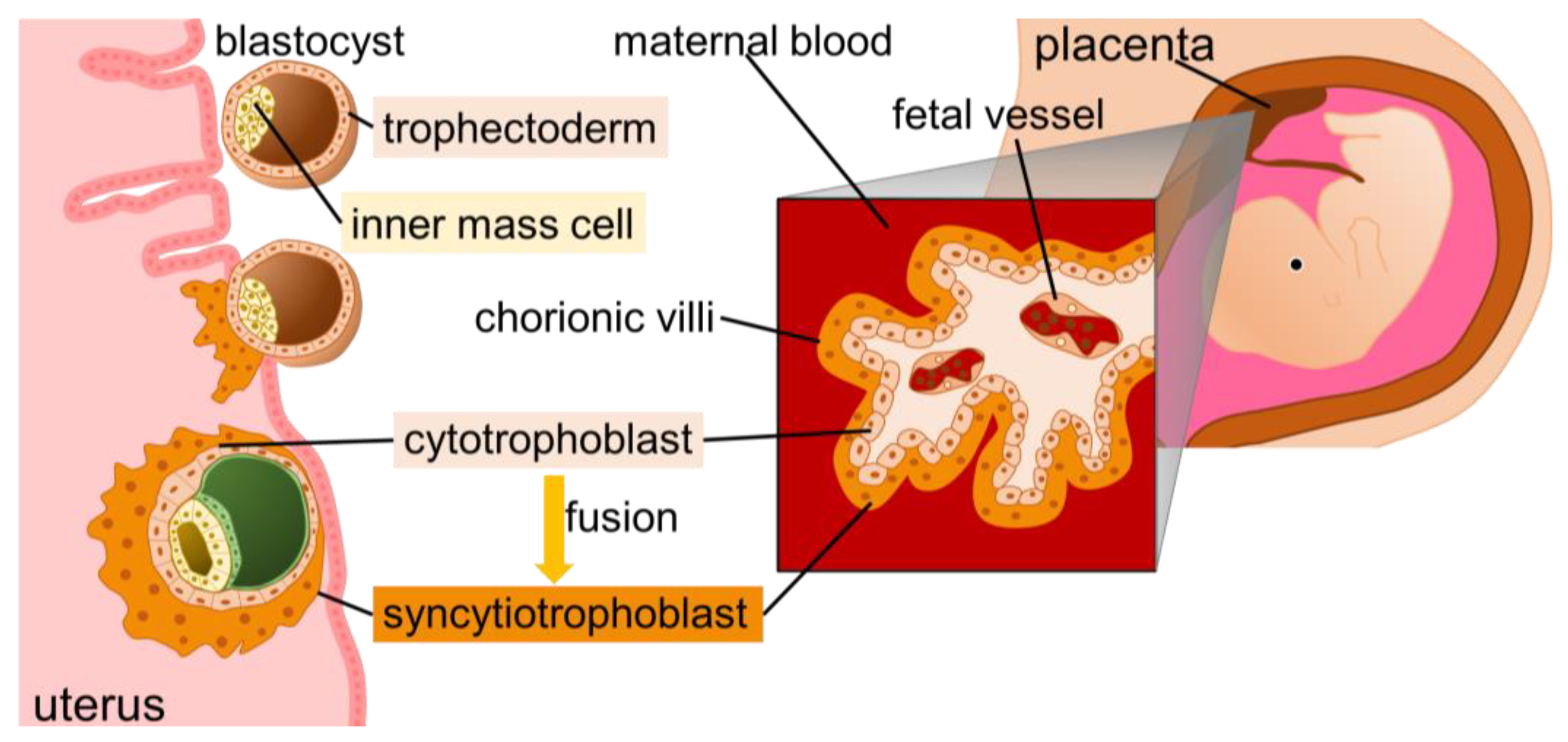
The placenta, which comprises the endometrium from the mother and the trophoblast from the fetus, supports fetal life. After fertilization, the fertilized egg repeatedly divides, eventually becoming a blastocyst, which attaches itself to the uterine wall. The outer layer of the blastocyst called the trophectoderm ultimately forms the placenta. In humans, starting at embryonic day 6, mononuclear cytotrophoblasts fuse to form syncytiotrophoblasts [9]. The syncytiotrophoblasts interact and exchange substances with maternal blood, secrete placental lactogen and chorionic gonadotropin, and participate in immune tolerance. The morphology and structure of the placenta vary among animal species [10], and ERVs and retrotransposons are expected to be implicated in this diversity [10,11,12,13]. Syncytin proteins are encoded by modified env genes of ERVs. The Syncytin family, identified in various animals, was linked to placental dysfunction observed in preeclampsia, intrauterine growth retardation (IGR), and Down syndrome [14,15,16,17,18,19]. In this review, we aimed to consolidate the existing knowledge on the evolution of placental morphogenesis and physiology.

2. Evolutionary History of the Mammalian Placenta and Retrotransposons

2.1. Evolutionary History of the Mammalian Placenta

Mammals are divided into three major groups: monotremes, marsupials, and eutherians. Monotremes, which are the most primitive mammals, are ovoviviparous, although they still nourish their offspring with milk from the mother [20]. Most marsupials have a placenta that develops from the yolk sac; however, this placenta is relatively inefficient, resulting in a short gestation period (between 8 and 42 days) [21], leading to their offspring being born immature and raised in the marsupium [22,23]. Although not mammals, some fishes, reptiles, and amphibians have yolk sac placentas. Among them, hammerhead sharks have a long gestation period of approximately one year [24]. Eutherians additionally possess a chorioallantoic placenta [25]. In this review, we specifically discussed chorioallantoic placentas.
Approximately 250 million years ago (MYA), in the early Mesozoic era (Triassic), the oldest mammal, Adelobasileus cromptoni, diverged from reptiles [26]. Adelobasileus is believed to have been an oviparous species, living a slender existence amidst thriving dinosaurs [26]. Today, mammals inhabit diverse habitats worldwide [27]. Their survival and success can be attributed to the presence of placentas within the female body, which facilitates offspring development. Conversely, dinosaur and reptile mothers lay their eggs externally and provide minimal parental care, relying on laying numerous eggs for survival [28]. However, some mammals have evolved to possess placentas, allowing the mother to nourish her unborn child in her womb while continuing with her life activities. This adaptation offers protection from external factors, including weather threats and other exogenous risks, enabling the mother to invest more in a smaller number of offspring while ensuring their reliable growth [29].
In 2011, a fossil of a small animal resembling a shrew, measuring approximately 10 cm in length, was discovered in Liaoning, China. This animal lived 160 MYA (Jurassic period) and was named “Juramaia,” meaning Jurassic mother, owing to its possession of a placenta [30]. When the placenta is formed, part of the oxygen the mother takes in is provided to the child, with the placenta consuming 40% of the oxygen supplied to the fetoplacental unit [31]. During the Triassic era, the Earth was very dry, with sparse vegetation and an oxygen concentration of approximately 11% [32,33]. However, during the Jurassic period, the Earth warmed, vegetation flourished, and oxygen levels rose to 20% [32,33]. It was precisely during this period that mammals acquired placentas.

2.2. Placenta Acquisition and Retrotransposons

Retrotransposons, transposons, and repetitive sequences comprise approximately 40% of the eukaryotic genome [34]. Transposons relocate their genome using a cut-and-paste mechanism. In contrast, retrotransposons duplicate their genome: the retrotransposon RNA is reverse-transcribed, and the resulting DNA is inserted into another part of the genome. Moreover, retrotransposons contribute to the acquisition of the mammalian placenta. Monotremes diverged at 166 MYA [35]; then, paternally expressed gene 10/sushi-ichi retrotransposon homolog 1 (PEG10/SIRH1) was inserted into the common ancestor of marsupials and eutherians. Subsequently, marsupials diverged at 148 MYA, and PEG11/SIRH2 (retrotransposon-like 1, [Rtl1]) was inserted into the common ancestor of eutherians [36,37,38]. Both PEG10 and PEG11/RTL1 encode proteins that are highly homologous to group-specific antigen (GAG) and polymerase (POL) proteins of the sushi-ichi retrotransposon of the pufferfish genome; sushi-ichi retrotransposons belong to the gypsy retrotransposon family [39,40]. PEG10 knockout mice lack labyrinthine and trabecular chorionic layers and experience early embryonic lethality [41]. The Gag protein of PEG10 forms virus-like particles that bind to cellular RNAs, including Hbegf (Heparin-binding EGF-like growth factor), playing an important role in placental formation [42]. PEG10 might bind to and stabilize these cellular RNAs and was suggested to be involved in placentation [42]. Furthermore, mice deficient in the protease activity of PEG10 exhibit defects in placental fetal capillaries, resulting in perinatal lethality due to poor placental and fetal growth after mid-life [43]. The biochemical function of the Pol protein of PEG10 remains unknown; however, its protease domain is highly conserved in all Eutherians, suggesting that the protease activity plays an important role [43]. PEG11 is expressed in the endothelial cells of placental fetal capillaries, and PEG11 knockout mice experience embryonic lethality due to growth delay caused by disruption of the fetal capillary network [44,45]. Recently, Sirh7/Ldoc1 was also discovered as a retrotransposon-derived placenta-associated gene with a high homology to the GAG of the sushi-ichi retrotransposon. In Sirh7/Ldoc1 knockout mice, trophoblast differentiation and maturation are abnormal, and the expression and timing of hormones such as placental progesterone and placental lactogen from trophoblast giant cells are altered, resulting in abnormal pregnancy maintenance and delayed parturition [46]. Sirh7, as well as PEG11, was inserted into the common ancestor of eutherians [46]. These findings suggest that PEG10 is essential for primitive placenta formation and that PEG11 and Sirh7 promote the construction of eutherian-specific placental structures [36,41,46,47]. The biochemical functions of PEG11 and Sirh7 are also unknown and require future study.

3. Classification of the Mammalian Placenta

The placenta comprises trophoblastic cells from the fetus and uterine cells from the mother. Trophoblastic cells constitute the chorionic villi, which are divided into four types according to their distribution: diffuse, cotyledonary, zonary, and discoid placentas [10] (Figure 1). A diffuse placenta comprises chorionic villi that are scattered diffusely over almost the entire placental surface. A cotyledonary placenta is characterized by numerous ridges called caruncles in the endometrium; the chorionic villi invade to form circular patches called cotyledons. A zonary placenta takes the form of a belt around the fetus. Primates have discoid placentas, which appear as a round disk in a part of the uterus. Although primate placentas occupy a smaller area of the fetus than other placentas, the bond between the mother and fetus is typically the strongest.
Placenta classification is based on how deeply the trophoblast cells penetrate the endometrium of the uterus; a phenomenon termed “invasiveness”. Arranged from the most invasive to least invasive, placentas are hemochorial, endotheliochorial, syndesmochorial, and epitheliochorial [48] (Figure 1). Although exceptions exist, the diffuse, cotyledonary, zonary, and discoid placentas roughly correspond to the epitheliochorial, syndesmochorial, endotheliochorial, and hemochorial placentas, respectively [11]. Apes have a hemochorial placenta, in which cells on the placental side penetrate deeply into the endometrium, and the endometrium forms a decidua [11]. The placental villous epithelium is in direct contact with maternal blood, allowing the efficient exchange of maternal IgG antibodies, oxygen, and other substances [49]. Although the risk of miscarriage is relatively low with a hemochorial placenta, it takes longer for the placenta to detach during parturition, leading to more blood loss during this process [50]. Apes are social, and non-mothers in their community often participate in child-rearing, enabling them to assist each other during challenging births or when the afterbirth is damaging.
Physiological and morphological evolution, which are not consistent with the molecular phylogenetic tree (often referred to as convergent evolution), are also caused by the horizontal and inter-species/individual transmission of ERVs or retrotransposon copies via virion-producing copies from ERV families or other “carrier” viruses [51,52,53,54,55]. To date, multiple ERVs involved in placental diversification have been identified.

4. ERVs and Placenta

ERVs are retrovirus-like sequences that were previously integrated into the host germline genome. ERVs are passed on to offspring by vertical transmission via Mendelian inheritance [56] and spread between species and individuals by horizontal transmission [54]. ERVs were identified in all eukaryotes, constituting approximately 10% of the genome [34]. Most ERVs are inactivated by mutations, deletions, or epigenetic regulation; however, some retain their transcriptional activity and contribute to host physiology [3]. Several ERVs were identified to function in the placenta [11].

4.1. Syncytins

Cell–cell fusion is the fusion of cell membranes and the mixing of the cytoplasm to form multinucleated cells [57], which is essential for reproduction, development, and homeostasis in multicellular organisms [58,59,60,61,62]. The fusion of sperm and oocytes requires fertilization; during skeletal muscle development, myoblasts fuse and become multinucleated. In human and mouse placentas, mononuclear cytotrophoblast cells fuse continuously with overlying syncytiotrophoblast cells to maintain their function and exchange materials [63]. The ERV-derived Syncytin family is a cell–cell fusion factor with an important physiological role [60,64,65]. Moreover, retroviruses form syncytia [66,67,68]. Cell fusion is inhibited by antibodies against SU, a viral surface protein, suggesting that SU is essential for cell–cell fusion [69].
The fertilized egg undergoes repeated cell division and becomes a blastocyst, implanting itself into the uterine wall. The inner cell mass of the blastocyst forms the fetus, and the trophectoderm forms the placenta (Figure 2). The placenta, which comprises the endometrium derived from the mother and trophoblastic cells derived from the fetus, supports the life activities of the fetus. The placenta physically supports the fetus in the uterus and secretes hormones to maintain normal pregnancy. Syncytiotrophoblasts are located on the outermost side of the fetus and are formed by the fusion and multinucleation of cytotrophoblasts. Syncytiotrophoblasts interact and exchange substances with the maternal blood, secrete placental lactogen and chorionic gonadotropin, and are involved in immune tolerance [9,70].
Syncytins have functional roles in the placenta and are encoded by various ERV families. In humans, Syncytin-1 is encoded by the HERV-W env gene located on chromosome 7q21.2 [71,72]. Syncytin-1 mRNA is expressed in syncytiotrophoblasts, exhibiting stable expression levels throughout the gestation period [73]. Syncytin-2 is derived from the env gene of HERV-FRD and is located on chromosome 6p24.1 of the human genome [74]. Syncytin-2 is expressed in cytotrophoblasts, exhibiting decreased expression levels toward late gestation [73,75]. The Syncytin-1 receptor, alanine, serine, cysteine transporter 2 (ASCT2) is expressed in cytotrophoblasts [76,77,78,79], whereas the Syncytin-2 receptor, major facilitator superfamily domain containing 2 (MFSD2), is expressed in syncytiotrophoblasts [80]. Syncytin-1 and -2 comprise 538 amino acids, including surface (SU) and transmembrane (TM) subunits, and both possess cell–cell fusion ability [71,74]. Syncytin-2 has immunosuppressive activity, whereas Syncytin-1 does not [81]. During pregnancy, the mother harbors the fetus, "the stranger," which carries antigens of paternal origin. During egg laying, the mother is not exposed to antigens of paternal origin; however, placental acquisition necessitates the evolution of immunosuppressive mechanisms. The differences between Syncytin-1 and -2 regarding localization, expression level transition, and immunosuppressive activity suggest that they were acquired for different purposes. Syncytin-1 and -2 are found in primates, whereas Syncytin-A and B are found in mice and Syncytin-Rum1 in cattle [82,83,84]. Syncytin-A and -B were identified via an in silico search of the mouse genome [82]. The mouse placenta consists of three layers: trophoblast giant cells composed of cytoprophoblasts and syncytiotrophoblast layer-I and -II (ST-I and ST-II) composed of syncytiotrophoblasts. Syncytin-A and -B are expressed in ST-I and ST-II, respectively [85]. Syncytin-A KO mice are unable to form ST-I, and the fetuses die between embryonic days 11.5 and 13.5 [83]. Syncytin-B KO mice fail to form ST-II, but the fetuses survive, showing only developmental delay and a reduced number of offspring [86]. Syncytin-B fusion ability was confirmed in only canine MDCK cells. Syncytin-B has an immunosuppressive capacity and may have other functions than cell fusion [81]. Syncytin-Mab1 was also found in viviparous reptiles (Mabuya skinks) [87]. All syncytins have different origins, suggesting that retroviruses infected each animal lineage independently and acquired similar functions by chance. Furthermore, Syncytin was associated with cell–cell fusion during osteoclastogenesis [64] and cancer development [88,89,90].

4.2. Suppressyn

The two main routes of retrovirus entry into host cells are direct membrane fusion and endocytosis [91]. When the envelope protein of the retrovirus binds to a specific receptor on the cell surface, the envelope protein undergoes a conformational change, and direct membrane fusion occurs on the plasma membrane. In endocytosis, membrane fusion occurs in early endosomes after transport into the cytoplasm. EIAV, JSRV, and other retroviruses enter the host cells via endocytosis [92,93]. Receptor interference occurs when retroviruses using the same molecule as a receptor antagonize each other via competitive binding to the receptor. The endogenous avian leukemia virus (ALV) in chickens and the endogenous Jaagsiekte sheep retrovirus (JSRV) (enJSRV) in sheep function as antiviral factors against exogenous ALV and JSRV, respectively, via receptor interference [4,5,6]. The retroviral envelope protein comprises two subunits: SU, which contains a receptor-binding site, and TM. Many ERVs functioning as infection defense factors, such as enJSRV, inhibit infection via exogenous retroviruses by expressing the SU region rather than the full length of the env gene and by binding to the receptor [7]. Thus, ERV-derived antiviral factors were identified in various animal species, except in humans, for a considerable time. However, a human ERV-derived infection defense factor was recently discovered [8].
Suppressyn, the first ERV-derived protein shown to inhibit cell–cell fusion, is derived from the env gene of the HERV-H family of viruses (ERVH48-1) and is located on chromosome 21q22.3 of the human genome [94]. Northern blot analysis revealed that Suppressyn mRNA is expressed specifically in the placenta [75]. Single-cell RNA sequencing data from the placenta at multiple developmental stages showed that Suppressyn is expressed in cytotrophoblasts, extravillous trophoblasts, and syncytiotrophoblasts [8,12]. Immunostaining in the first trimester (at 7 weeks) showed that Suppressyn is expressed in cytotrophoblasts (especially in the progenitor and intermediate cytotrophoblasts) and extravillous trophoblasts but is limited in syncytiotrophoblasts [79]. Immunostaining in the second trimester (at 21 weeks) showed that Suppressyn is expressed in syncytiotrophoblasts and also in cytotrophoblasts [8]. Immunostaining in the third trimester showed that Suppressyn is expressed in syncytiotrohpblasts and extravillous trophoblasts [8,94]. Furthermore, immunostaining of blastocysts showed that Suppressyn is expressed in the trophectoderm and some Octamer-binding transcription factor 4-positive cells [8]. Suppressyn consists of SU sites with a stop codon preceding the SU-TM cleavage site. Suppressyn uses ASCT2 as a receptor to inhibit the Syncytin-1-mediated fusion of cytotrophoblasts by binding to ASCT2 [79,94]. Suppressyn is detected in cultured human trophoblast cells and placenta tissue samples in both intracellular and secreted forms, both of which bind directly to ASCT2 [94]. Both of them bind directly to ASCT2, suggesting that suppressyn might inhibit Syncytin-1-mediated cell–cell fusion via autocrine and paracrine mechanisms [94]. Intracellularly, Suppressyn alters the glycosylation of ASCT2 [79,94], resulting in changes in the ligand binding capacity [95] and protein stability [96] of ASCT2. However, the glycosylation changes in ASCT2 do not affect the cell fusion mediated by Syncytin-1 [94]. ASCT2 is an amino acid transporter that transports glutamine, alanine, serine, cysteine, and threonine in a Na+ ion-dependent manner [97] and is expressed in tissues throughout the body. Particularly, glutamine is essential in the placenta, and inhibiting its uptake leads to impaired embryonic development [98]. ASCT2 functions as a receptor for the feline ERV RD-114 virus, baboon ERVs, and simian retroviruses, among others. These viruses which use ASCT2 as a receptor are referred to as the RD114-and-D-type-retrovirus (RDR) interference group [99,100,101,102]. Recently, Suppressyn was found to function as an antiviral agent against RDR infections [8].

4.3. Other Placenta-Associated ERVs

In the human placenta, in addition to Syncytin-1 and -2, other ERV env-derived proteins are expressed, including ERVV-1, ERVV-2, ERV3-1 (also known as ERVR), and ERVMER34-1 (also known as HEMO). ERVV-1, ERVV-2, and ERVV3-1 are expressed in syncytiotrophoblasts throughout gestation [12,103,104]. Human ERVVV-1 and -2 should not have fusion activity [105,106]. However, ERVV-2 in old world monkeys (OWMs) and marmosets (a species of new world monkeys [NWMs]) shows fusion activity [106]. Thus, ERVV-2 is also called mac-syncytin-3 [106]. ERV3-1 has no fusion activity owing to a conserved premature stop codon but has a functional immunosuppressive domain [81,107,108]. ERVMER34-1 is expressed in cytotrophoblasts and extravillous trophoblasts in the first gestation and is detected in the blood of pregnant females [109]. ERVMER34-1 has no fusion activity, and its function is unknown; however, it localizes in a manner similar to ERVH48-1, suggesting that it may function as an inhibitor of cell fusion [12].

4.4. ERV Abnormalities and Placenta-Associated Diseases

In preeclampsia, which is one of the most severe complications during pregnancy, there is reported dysregulation of Syncytin-1 and -2 expression [14,110,111], leading to incomplete cell–cell fusion and abnormal differentiation from cytotrophoblasts to syncytiotrophoblasts [15,16]. IUGR, also known as fetal growth restriction, is a condition characterized by poor fetal development in the uterus during pregnancy. Syncytin-1 mRNA and protein expression levels are lower in the IUGR placenta than in normal placentas, resulting in abnormal cell fusion and increased apoptosis [17,18]. In addition, cell–cell fusion is disrupted in the placenta of trisomy 21 (Down syndrome) owing to the abnormal expression of Syncytin-2, resulting in dysplasia of the syncytiotrophoblast [112,113]. In trisomy 21 placenta, the number of Suppressyn-positive cytotrophoblasts increased, and the expression level of Suppressyn per cytotrophoblast also increased [19]. The expression level of Suppressyn in the serum is elevated in trisomy 21, resulting in excessive suppression of placental formation and immature placenta [19].

5. Acquisition and Exaptation of ERVs in Primates

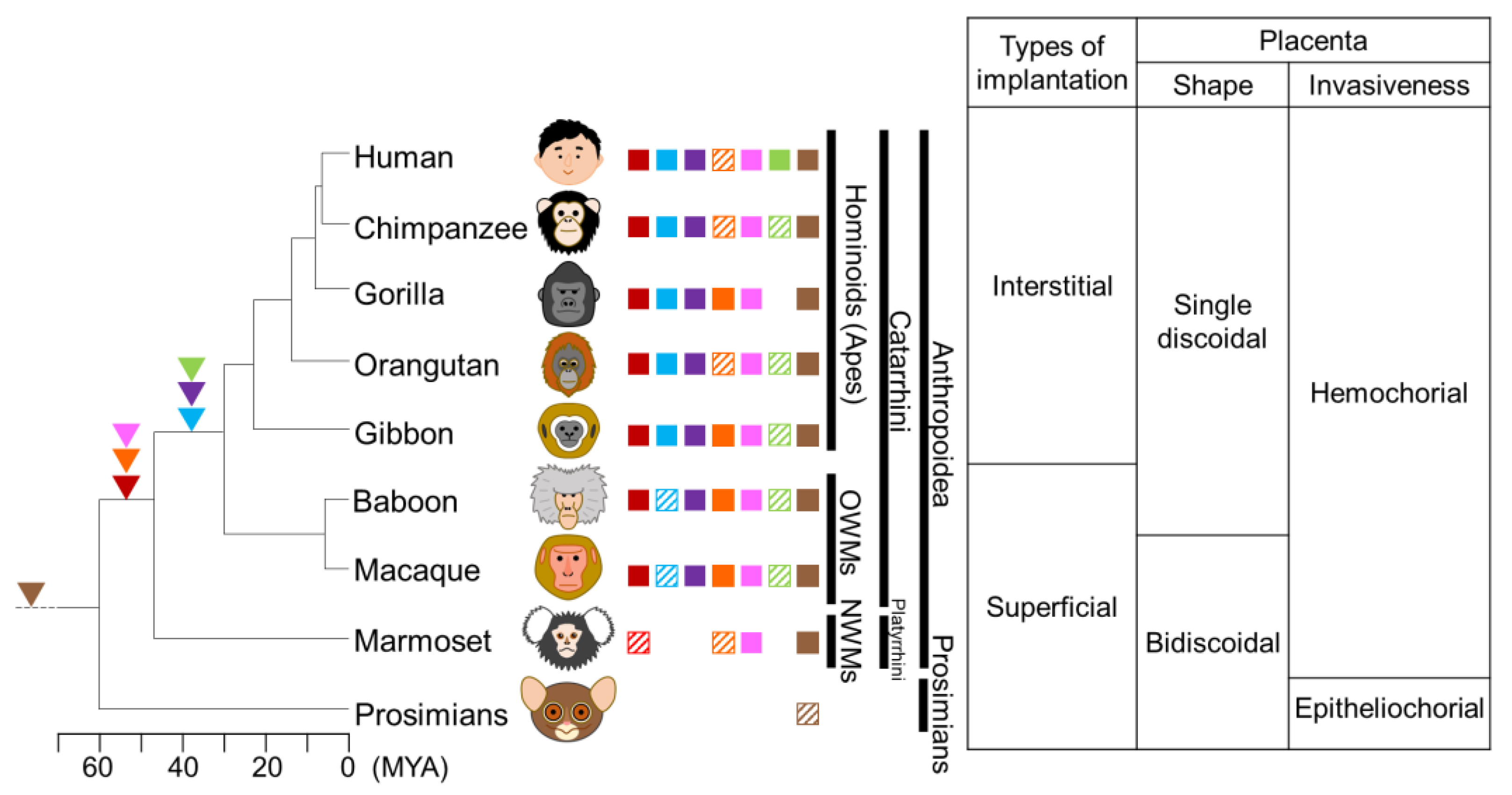
The presence of retained ERV-FRD in apes, OWMs, and NWMs suggests that ERV-FRD infected and acquired these common ancestors over 40 MYA [106,114]. However, ERV-FRD Envelope proteins of NWMs have lost or have weakened cell–cell fusion ability [74,115] (Figure 3). Notably, NWMs have hemochorionic placentas and form syncytiotrophoblasts, which contradicts the dysfunction of ERV-FRD Envelope proteins in this species. This suggests that NWMs might possess another Syncytin-like factor. ERVV-1 and ERVV-2 are retained in apes, OWMs, and NWMs, suggesting that they were inserted into the genome of the common ancestor of Anthropoidea at the same time as ERV-FRD [116]. ERVV-2 may have originally had cell fusion activity, but it is speculated that this activity was lost in some apes and NWMs, suggesting that ERVV-2 may have been replaced by Syncytin-2 or may have acquired some other function, such as immunosuppression [106]. The ancestors of NWMs are believed to have migrated from Africa to South America approximately 34 MYA [117]; therefore, it is plausible that another Syncytin-like factor, such as ERVV-2, was acquired in these populations.
The retention of Syncytin-1 orthologs in apes and OWMs suggests that ERV-W infected the common ancestor of catarrhines after 40 MYA, following the divergence of catarrhines and platyrrhines. However, the open reading frame is disrupted by stop codons and frameshifts in OWMs [118], suggesting that exaptation, which refers to acquiring features or functions that are different from the original, occurred in the common ancestor of apes. Syncytiotrophoblasts are incapable of proliferating and retaining cellular activity when fused with cytotrophoblasts [63] (Supplementary Materials Figure S1). Moreover, Synytin-1 is expressed throughout gestation [73], suggesting that Syncytin-1 function is important for long-term placental maintenance. Around 24 MYA, apes and OWMs diverged, with larger apes prospering. Gestational length and body size are positively correlated [119]. Therefore, it is speculated that the acquisition of ERV-W enabled the maintenance of syncytiotrophoblastic trophoblasts, prolonged the gestation period, and increased ape size. The gestation period of great apes is 210 to 290 days, whereas that of OWMs is 150 days.
The presence of retained Suppressyn orthologs in apes and most OWMs suggests that ERVH48-1 was inserted into the common ancestor of catarrhines after 40 MYA [8,120]. This indicates that ERVH48-1 and ERV-W were simultaneously acquired. Both Syncytin-1 and Suppressyn are expressed during pregnancy but have different localization and expression dynamics. Syncytin-1 is expressed in syncytiotrophoblasts, and its expression level gradually increases during the differentiation process of primary cultured trophoblast cells [73,79,121]. Suppressyn is expressed in cytotrophoblast cells and EVT, and its expression level gradually decreases during the in vitro differentiation process [79]. The expression pattern of Suppressyn is more similar to that of ASCT2 than that of Syncytin-1 [79]. Although Suppressyn is known to have an antagonistic role against Syncytin-1, it was speculated that Suppressyn was acquired for other purposes. ERVH48-1 was also inactivated in some OWMs via frameshifting [8]. The ancestral sequence of Suppressyn retains its antiviral activity against RDR but is speculated to have lost its function in some lineages of OWMs. Suppressyn inhibits cell–cell fusion via Syncytin-1; however, it also inhibits infection via other retroviruses that use ASCT2 as a receptor [8]. The acquisition of the Suppressyn function is likely linked to the epidemic spread and conservation of some retroviruses that utilize ASCT2 as an antiviral factor. ASCT2, the receptor for Syncytin-1 and Suppressyn, contains eight transmembrane domains, and Region C at the C-terminus of extracellular loop 2 is identified as the host-range-determining region [95]. In mice and rats, insertions and deletions (indels) in Region C render ASCT2 non-functional as a receptor for RDR. These indels may have resulted in the acquisition of resistance to pathogenic viruses that utilize ASCT2 [122]. Comparing ASCT2 sequences in primates, a high degree of homology was observed among apes, OWMs, and NWM; however, some prosimians exhibited indels in Region C (Figure 4). ERVs utilizing ASCT2, including Syncytin-1 and Suppressyn, were not found in these species, suggesting that they may have acquired resistance to RDR via indels in Region C of ASCT2, as in mice and rats. Pig-tailed macaques also exhibit a large deletion in Region C; however, their genomes contain ERV-W and ERVH48-1 insertions, suggesting that this deletion occurred after the insertion of these viruses. It is speculated that Syncytin-1 and Suppressyn were acquired at around the same time, but a contradiction arises. Suppressyn inhibits cell–cell fusion by binding to ASCT2, the receptor for Syncytin-1. If retroviruses that use the same receptor cause receptor interference, it may be impossible for them to endogenize at the same time. The endogenization mechanism of retroviruses remains unclear. If the details of the acquisition process of Syncytin-1 and Suppressyn are revealed, it will be helpful to elucidate the endogenization mechanism.
ERV3-1 is conserved in apes except gorillas and in OWMs; it is thought to have entered the genome of the common ancestor of catarrhines after 40 MYA and was inactivated in gorillas [123].
ERVMER34-1 was retained in Laurasian and Eurasian theropods, but not in Afrotheria or Xenarthrans, suggesting that it entered the genome of Boreoeutheria at 100–120 MYA [109,124]. The ORF encoding the full-length protein was conserved in all simians and cats [109], suggesting that it may have some function.

6. Conclusions and Future Perspectives

The acquisition of the placenta is a crucial adaptation in mammals, which enables them to carry their developing fetuses within their bodies. The placenta protects the fetus from external threats and other risks. However, it also exposes the mother to other antigens and the risk of hemorrhage. Several retrotransposons and ERVs contributed to placental acquisition. For example, Syncytins were identified in various animal species as representative ERVs that contribute to placental formation. Syncytins have different origins in each animal species, likely arising from independent infection by other viruses; however, they acquired similar functions [11]. In primates, Suppressyn antagonizes Syncytin-1-mediated cell–cell fusion [94]. Syncytin-2 is thought to have infected and was inserted into the common ancestor of Anthropoidea, and Syncytin-1 and Suppressyn into the common ancestor of catarrhines. However, both were inactivated in some lineages, suggesting that multiple exaptation events had occurred. Syncytins and Suppressyn are species- or lineage-specific, posing challenges for in vivo analysis in mice, thereby impeding comprehensive analyses. Nevertheless, recent progress in research has enabled the isolation of trophoblast stem cells from human blastocysts and placentas in early pregnancy [125], and techniques were developed to induce the differentiation of trophoblast cells [126,127,128,129,130] and blastocyst-like tissue [131] from human pluripotent stem cells. These techniques will serve as valuable tools to further understand the function of ERVs in the placenta. The study of retrotransposons and ERVs, particularly Syncytins and Suppressyn, in placental development and function provides valuable insights into the genetic mechanisms underlying placental formation. Furthermore, understanding ERVs functioning in the placenta has potential implications for the management of placental-associated diseases such as preeclampsia, IUGR, and Down syndrome in humans.

Supplementary Materials

The following supporting information can be downloaded at https://www.mdpi.com/article/10.3390/biom13101482/s1. Figure S1: Schematic representation of trophoblast differentiation. Cytotrophoblasts differentiate and increase their number of intracellular organelles, mRNA, and proteins. Differentiated cytotrophoblasts fuse with overlying syncytiotrophoblasts. Eventually, the intracellular organelles are degraded, and the nuclei of late apoptotic syncytiotrophoblasts accumulate in the syncytial knots and are released into the maternal bloodstream. Arrows indicate the routes of cellular changes during trophoblast differentiation. CT, Cytotrophoblast; ST, Syncytiotrophoblast.

Author Contributions

Conceptualization, Writing—Original Draft Preparation, Writing—Review and Editing, S.S. All authors have read and agreed to the published version of the manuscript.

Funding

This research was funded by JSPS KAKENHI [grant number: JP22K06043].

Institutional Review Board Statement

Not applicable.

Informed Consent Statement

Not applicable.

Data Availability Statement

Not applicable.

Acknowledgments

I am grateful to Takayuki Miyazawa from Kyoto University, Kyoto, Japan, for providing valuable guidance. The manuscript went through professional English language editing.

Conflicts of Interest

The author declares no conflict of interest. The sponsors had no role in the design, execution, interpretation, or writing of the study.

References

  1. Stoye, J.P. Studies of Endogenous Retroviruses Reveal a Continuing Evolutionary Saga. Nat. Rev. Microbiol. 2012, 10, 395–406. [Google Scholar] [CrossRef] [PubMed]
  2. Johnson, W.E. Origins and Evolutionary Consequences of Ancient Endogenous Retroviruses. Nat. Rev. Microbiol. 2019, 17, 355–370. [Google Scholar] [CrossRef] [PubMed]
  3. Gifford, R.; Tristem, M. The Evolution, Distribution and Diversity of Endogenous Retroviruses. Virus Genes 2003, 26, 291–315. [Google Scholar] [CrossRef] [PubMed]
  4. Robinson, H.L.; Astrin, S.M.; Senior, A.M.; Salazar, F.H. Host Susceptibility to Endogenous Viruses: Defective, Glycoprotein-Expressing Proviruses Interfere with Infections. J. Virol. 1981, 40, 745–751. [Google Scholar] [CrossRef] [PubMed]
  5. Varela, M.; Spencer, T.E.; Palmarini, M.; Arnaud, F. Friendly Viruses: The Special Relationship between Endogenous Retroviruses and Their Host. Ann. N. Y. Acad. Sci. 2009, 1178, 157–172. [Google Scholar] [CrossRef] [PubMed]
  6. Arnaud, F.; Caporale, M.; Varela, M.; Biek, R.; Chessa, B.; Alberti, A.; Golder, M.; Mura, M.; Zhang, Y.P.; Yu, L.; et al. A Paradigm for Virus-Host Coevolution: Sequential Counter-Adaptations between Endogenous and Exogenous Retroviruses. PLoS Pathog. 2007, 3, e170. [Google Scholar] [CrossRef] [PubMed]
  7. Malfavon-Borja, R.; Feschotte, C. Fighting Fire with Fire: Endogenous Retrovirus Envelopes as Restriction Factors. J. Virol. 2015, 89, 4047–4050. [Google Scholar] [CrossRef] [PubMed]
  8. Frank, J.A.; Singh, M.; Cullen, H.B.; Kirou, R.A.; Benkaddour-Boumzaouad, M.; Cortes, J.L.; Garcia Pérez, J.; Coyne, C.B.; Feschotte, C. Evolution and Antiviral Activity of a Human Protein of Retroviral Origin. Science 2022, 378, 422–428. [Google Scholar] [CrossRef]
  9. Bischof, P.; Irminger-Finger, I. The Human Cytotrophoblastic Cell, a Mononuclear Chameleon. Int. J. Biochem. Cell Biol. 2005, 37, 1–16. [Google Scholar] [CrossRef]
  10. Wooding, P.F.P.; Burton, G. Comparative Placentation: Structures, Functions and Evolution; Springer: Berlin/Heidelberg, Germany, 2008. [Google Scholar]
  11. Imakawa, K.; Nakagawa, S.; Miyazawa, T. Baton Pass Hypothesis: Successive Incorporation of Unconserved Endogenous Retroviral Genes for Placentation During Mammalian Evolution. Genes Cells 2015, 20, 771–788. [Google Scholar] [CrossRef]
  12. Roberts, R.M.; Ezashi, T.; Schulz, L.C.; Sugimoto, J.; Schust, D.J.; Khan, T.; Zhou, J. Syncytins Expressed in Human Placental Trophoblast. Placenta 2021, 113, 8–14. [Google Scholar] [CrossRef] [PubMed]
  13. Imakawa, K.; Kusama, K.; Kaneko-Ishino, T.; Nakagawa, S.; Kitao, K.; Miyazawa, T.; Ishino, F. Endogenous Retroviruses and Placental Evolution, Development, and Diversity. Cells 2022, 11, 2458. [Google Scholar] [CrossRef] [PubMed]
  14. Vargas, A.; Toufaily, C.; LeBellego, F.; Rassart, É.; Lafond, J.; Barbeau, B. Reduced Expression of Both Syncytin 1 and Syncytin 2 Correlates with Severity of Preeclampsia. Reprod. Sci. 2011, 18, 1085–1091. [Google Scholar] [CrossRef] [PubMed]
  15. Chen, C.P.; Wang, K.G.; Chen, C.Y.; Yu, C.; Chuang, H.C.; Chen, H. Altered Placental Syncytin and Its Receptor ASCT2 Expression in Placental Development and Pre-eclampsia. BJOG 2006, 113, 152–158. [Google Scholar] [CrossRef] [PubMed]
  16. Gauster, M.; Moser, G.; Orendi, K.; Huppertz, B. Factors Involved in Regulating Trophoblast Fusion: Potential Role in the Development of Preeclampsia. Placenta 2009, 30 (Suppl. A), S49–S54. [Google Scholar] [CrossRef] [PubMed]
  17. Ruebner, M.; Langbein, M.; Strissel, P.L.; Henke, C.; Schmidt, D.; Goecke, T.W.; Faschingbauer, F.; Schild, R.L.; Beckmann, M.W.; Strick, R. Regulation of the Human Endogenous Retroviral Syncytin-1 and Cell-Cell Fusion by the Nuclear Hormone Receptors PPARγ/RXRα in Placentogenesis. J. Cell. Biochem. 2012, 113, 2383–2396. [Google Scholar] [CrossRef] [PubMed]
  18. Langbein, M.; Strick, R.; Strissel, P.L.; Vogt, N.; Parsch, H.; Beckmann, M.W.; Schild, R.L. Impaired Cytotrophoblast Cell-Cell Fusion Is Associated with Reduced Syncytin and Increased Apoptosis in Patients with Placental Dysfunction. Mol. Reprod. Dev. 2008, 75, 175–183. [Google Scholar] [CrossRef] [PubMed]
  19. Sugimoto, J.; Schust, D.J.; Yamazaki, T.; Kudo, Y. Involvement of the HERV-Derived Cell-Fusion Inhibitor, Suppressyn, in the Fusion Defects Characteristic of the Trisomy 21 Placenta. Sci. Rep. 2022, 12, 10552. [Google Scholar] [CrossRef]
  20. Abbot, P.; Rokas, A. Mammalian Pregnancy. Curr. Biol. 2017, 27, R127–R128. [Google Scholar] [CrossRef]
  21. Karlen, S.J.; Krubitzer, L. Marsupial Neocortex. In Encyclopedia of Neuroscience; Squire, L.R., Ed.; Academic Press: Cambridge, MA, USA, 2009; pp. 671–679. [Google Scholar]
  22. Tyndale-Biscoe, H.; Renfree, M. Reproductive Physiology of Marsupials; Cambridge University Press: Cambridge, UK, 1987. [Google Scholar]
  23. Renfree, M.B. Review: Marsupials: Placental Mammals with a Difference. Placenta 2010, 31 (Suppl. A), S21–S26. [Google Scholar] [CrossRef]
  24. Soma, H.; Murai, N.; Tanaka, K.; Oguro, T.; Kokuba, H.; Yoshihama, I.; Fujita, K.; Mineo, S.; Toda, M.; Uchida, S.; et al. Review: Exploration of Placentation from Human Beings to Ocean-Living Species. Placenta 2013, 34 (Suppl. A), S17–S23. [Google Scholar] [CrossRef] [PubMed]
  25. Langer, P. The Phases of Maternal Investment in Eutherian Mammals. Zoology 2008, 111, 148–162. [Google Scholar] [CrossRef] [PubMed]
  26. Lucas, S.G.; Luo, Z.X. Adelobasileus from the Upper Triassic of West Texas: The Oldest Mammal. J. Vertebr. Paleontol. 1993, 13, 309–334. [Google Scholar] [CrossRef]
  27. Cole, F.R.; Reeder, D.M.; Wilson, D.E. A Synopsis of Distribution Patterns and the Conservation of Mammal Species. J. Mammal. 1994, 75, 266–276. [Google Scholar] [CrossRef]
  28. Thulborn, R. The Discovery of Dinosaur Eggs. Mod. Geol. 1991, 16, 113–126. [Google Scholar]
  29. Recknagel, H.; Elmer, K.R. Differential Reproductive Investment in Co-occurring Oviparous and Viviparous Common Lizards (Zootoca vivipara) and Implications for Life-History Trade-Offs with Viviparity. Oecologia 2019, 190, 85–98. [Google Scholar] [CrossRef] [PubMed]
  30. Luo, Z.X.; Yuan, C.X.; Meng, Q.J.; Ji, Q. A Jurassic Eutherian Mammal and Divergence of Marsupials and Placentals. Nature 2011, 476, 442–445. [Google Scholar] [CrossRef] [PubMed]
  31. Carter, A.M. Placental Oxygen Consumption. Part I: In Vivo Studies–A Review. Placenta 2000, 21 (Suppl. A), S31–S37. [Google Scholar] [CrossRef]
  32. Berner, R.A.; Vandenbrooks, J.M.; Ward, P.D. Evolution. Oxygen and Evolution. Science 2007, 316, 557–558. [Google Scholar] [CrossRef]
  33. Falkowski, P.G.; Katz, M.E.; Milligan, A.J.; Fennel, K.; Cramer, B.S.; Aubry, M.P.; Berner, R.A.; Novacek, M.J.; Zapol, W.M. The Rise of Oxygen over the past 205 Million Years and the Evolution of Large Placental Mammals. Science 2005, 309, 2202–2204. [Google Scholar] [CrossRef]
  34. Gregory, T.R. Synergy between Sequence and Size in Large-Scale Genomics. Nat. Rev. Genet. 2005, 6, 699–708. [Google Scholar] [CrossRef] [PubMed]
  35. Bininda-Emonds, O.R.; Cardillo, M.; Jones, K.E.; MacPhee, R.D.; Beck, R.M.; Grenyer, R.; Price, S.A.; Vos, R.A.; Gittleman, J.L.; Purvis, A. The Delayed Rise of Present-Day Mammals. Nature 2007, 446, 507–512. [Google Scholar] [CrossRef] [PubMed]
  36. Suzuki, S.; Ono, R.; Narita, T.; Pask, A.J.; Shaw, G.; Wang, C.; Kohda, T.; Alsop, A.E.; Marshall Graves, J.A.; Kohara, Y.; et al. Retrotransposon Silencing by DNA Methylation Can Drive Mammalian Genomic Imprinting. PLoS Genet. 2007, 3, e55. [Google Scholar] [CrossRef] [PubMed]
  37. Warren, W.C.; Hillier, L.W.; Marshall Graves, J.A.; Birney, E.; Ponting, C.P.; Grützner, F.; Belov, K.; Miller, W.; Clarke, L.; Chinwalla, A.T.; et al. Genome Analysis of the Platypus Reveals Unique Signatures of Evolution. Nature 2008, 453, 175–183. [Google Scholar] [CrossRef] [PubMed]
  38. Kaneko-Ishino, T.; Ishino, F. Mammalian-Specific Genomic Functions: Newly Acquired Traits Generated by Genomic Imprinting and LTR Retrotransposon-Derived Genes in Mammals. Proc. Jpn. Acad. Ser. B Phys. Biol. Sci. 2015, 91, 511–538. [Google Scholar] [CrossRef] [PubMed]
  39. Kim, A.; Terzian, C.; Santamaria, P.; Pélisson, A.; Purd’homme, N.; Bucheton, A. Retroviruses in Invertebrates: The Gypsy Retrotransposon Is Apparently an Infectious Retrovirus of Drosophila melanogaster. Proc. Natl. Acad. Sci. USA 1994, 91, 1285–1289. [Google Scholar] [CrossRef] [PubMed]
  40. Song, S.U.; Gerasimova, T.; Kurkulos, M.; Boeke, J.D.; Corces, V.G. An env-Like Protein Encoded by a Drosophila Retroelement: Evidence that Gypsy Is an Infectious Retrovirus. Genes Dev. 1994, 8, 2046–2057. [Google Scholar] [CrossRef] [PubMed]
  41. Ono, R.; Nakamura, K.; Inoue, K.; Naruse, M.; Usami, T.; Wakisaka-Saito, N.; Hino, T.; Suzuki-Migishima, R.; Ogonuki, N.; Miki, H.; et al. Deletion of Peg10, an Imprinted Gene Acquired from a Retrotransposon, Causes Early Embryonic Lethality. Nat. Genet. 2006, 38, 101–106. [Google Scholar] [CrossRef]
  42. Abed, M.; Verschueren, E.; Budayeva, H.; Liu, P.; Kirkpatrick, D.S.; Reja, R.; Kummerfeld, S.K.; Webster, J.D.; Gierke, S.; Reichelt, M.; et al. The Gag Protein PEG10 Binds to RNA and Regulates Trophoblast Stem Cell Lineage Specification. PLoS ONE 2019, 14, e0214110. [Google Scholar] [CrossRef]
  43. Shiura, H.; Ono, R.; Tachibana, S.; Kohda, T.; Kaneko-Ishino, T.; Ishino, F. PEG10 Viral Aspartic Protease Domain Is Essential for the Maintenance of Fetal Capillary Structure in the Mouse Placenta. Development 2021, 148, dev199564. [Google Scholar] [CrossRef]
  44. Sekita, Y.; Wagatsuma, H.; Nakamura, K.; Ono, R.; Kagami, M.; Wakisaka, N.; Hino, T.; Suzuki-Migishima, R.; Kohda, T.; Ogura, A.; et al. Role of Retrotransposon-Derived Imprinted Gene, Rtl1, in the Feto-Maternal Interface of Mouse Placenta. Nat. Genet. 2008, 40, 243–248. [Google Scholar] [CrossRef]
  45. Kitazawa, M.; Tamura, M.; Kaneko-Ishino, T.; Ishino, F. Severe Damage to the Placental Fetal Capillary Network Causes Mid- to Late Fetal Lethality and Reduction in Placental Size in Peg11/Rtl1 KO Mice. Genes Cells 2017, 22, 174–188. [Google Scholar] [CrossRef] [PubMed]
  46. Naruse, M.; Ono, R.; Irie, M.; Nakamura, K.; Furuse, T.; Hino, T.; Oda, K.; Kashimura, M.; Yamada, I.; Wakana, S.; et al. Sirh7/Ldoc1 Knockout Mice Exhibit Placental P4 Overproduction and Delayed Parturition. Development 2014, 141, 4763–4771. [Google Scholar] [CrossRef] [PubMed]
  47. Ono, R.; Kobayashi, S.; Wagatsuma, H.; Aisaka, K.; Kohda, T.; Kaneko-Ishino, T.; Ishino, F. A Retrotransposon-Derived Gene, PEG10, Is a Novel Imprinted Gene Located on Human Chromosome 7q21. Genomics 2001, 73, 232–237. [Google Scholar] [CrossRef] [PubMed]
  48. Enders, A.C.; Blankenship, T.N.; Conley, A.J.; Jones, C.J. Structure of the Midterm Placenta of the Spotted Hyena, Crocuta crocuta, with Emphasis on the Diverse Hemophagous Regions. Cells Tissues Organs 2006, 183, 141–155. [Google Scholar] [CrossRef] [PubMed]
  49. Malek, A. Role of IgG Antibodies in Association with Placental Function and Immunologic Diseases in Human Pregnancy. Expert Rev. Clin. Immunol. 2013, 9, 235–249. [Google Scholar] [CrossRef] [PubMed]
  50. Enders, A.C.; Carter, A.M. What Can Comparative Studies of Placental Structure Tell Us?—A Review. Placenta 2004, 25 (Suppl. A), S3–S9. [Google Scholar] [CrossRef] [PubMed]
  51. Zhang, H.H.; Feschotte, C.; Han, M.J.; Zhang, Z. Recurrent Horizontal Transfers of Chapaev Transposons in Diverse Invertebrate and Vertebrate Animals. Genome Biol. Evol. 2014, 6, 1375–1386. [Google Scholar] [CrossRef]
  52. Piskurek, O.; Jackson, D.J. Transposable Elements: From DNA Parasites to Architects of Metazoan Evolution. Genes 2012, 3, 409–422. [Google Scholar] [CrossRef]
  53. Gilbert, C.; Schaack, S.; Pace, J.K., 2nd; Brindley, P.J.; Feschotte, C. A Role for Host-Parasite Interactions in the Horizontal Transfer of Transposons Across Phyla. Nature 2010, 464, 1347–1350. [Google Scholar] [CrossRef]
  54. Belshaw, R.; Katzourakis, A.; Paces, J.; Burt, A.; Tristem, M. High Copy Number in Human Endogenous Retrovirus Families Is Associated with Copying Mechanisms in Addition to Reinfection. Mol. Biol. Evol. 2005, 22, 814–817. [Google Scholar] [CrossRef] [PubMed]
  55. Isfort, R.; Jones, D.; Kost, R.; Witter, R.; Kung, H.J. Retrovirus Insertion into Herpesvirus In Vitro and In Vivo. Proc. Natl. Acad. Sci. USA 1992, 89, 991–995. [Google Scholar] [CrossRef] [PubMed]
  56. Weiss, R.A. The Discovery of Endogenous Retroviruses. Retrovirology 2006, 3, 67. [Google Scholar] [CrossRef] [PubMed]
  57. Aguilar, P.S.; Baylies, M.K.; Fleissner, A.; Helming, L.; Inoue, N.; Podbilewicz, B.; Wang, H.; Wong, M. Genetic Basis of Cell-Cell Fusion Mechanisms. Trends Genet. 2013, 29, 427–437. [Google Scholar] [CrossRef] [PubMed]
  58. Chen, E.H.; Olson, E.N. Unveiling the Mechanisms of Cell-Cell Fusion. Science 2005, 308, 369–373. [Google Scholar] [CrossRef] [PubMed]
  59. Ogle, B.M.; Cascalho, M.; Platt, J.L. Biological Implications of Cell Fusion. Nat. Rev. Mol. Cell Biol. 2005, 6, 567–575. [Google Scholar] [CrossRef] [PubMed]
  60. Oren-Suissa, M.; Podbilewicz, B. Cell Fusion During Development. Trends Cell Biol. 2007, 17, 537–546. [Google Scholar] [CrossRef]
  61. Hernández, J.M.; Podbilewicz, B. The Hallmarks of Cell-Cell Fusion. Development 2017, 144, 4481–4495. [Google Scholar] [CrossRef]
  62. Brukman, N.G.; Uygur, B.; Podbilewicz, B.; Chernomordik, L.V. How Cells Fuse. J. Cell Biol. 2019, 218, 1436–1451. [Google Scholar] [CrossRef]
  63. Pötgens, A.J.; Schmitz, U.; Bose, P.; Versmold, A.; Kaufmann, P.; Frank, H.G. Mechanisms of Syncytial Fusion: A Review. Placenta 2002, 23 (Suppl. A), S107–S113. [Google Scholar] [CrossRef]
  64. Søe, K.; Andersen, T.L.; Hobolt-Pedersen, A.S.; Bjerregaard, B.; Larsson, L.I.; Delaissé, J.M. Involvement of Human Endogenous Retroviral syncytin-1 in Human Osteoclast Fusion. Bone 2011, 48, 837–846. [Google Scholar] [CrossRef] [PubMed]
  65. Larsson, L.I.; Bjerregaard, B.; Talts, J.F. Cell Fusions in Mammals. Histochem. Cell Biol. 2008, 129, 551–561. [Google Scholar] [CrossRef]
  66. Nagy, K.; Clapham, P.; Cheingsong-Popov, R.; Weiss, R.A. Human T-Cell Leukemia Virus Type I: Induction of Syncytia and Inhibition by Patients’ Sera. Int. J. Cancer 1983, 32, 321–328. [Google Scholar] [CrossRef] [PubMed]
  67. Portis, J.L.; McAtee, F.J.; Evans, L.H. Infectious Entry of Murine Retroviruses into Mouse Cells: Evidence of a Postadsorption Step Inhibited by Acidic pH. J. Virol. 1985, 55, 806–812. [Google Scholar] [CrossRef] [PubMed]
  68. Lifson, J.D.; Feinberg, M.B.; Reyes, G.R.; Rabin, L.; Banapour, B.; Chakrabarti, S.; Moss, B.; Wong-Staal, F.; Steimer, K.S.; Engleman, E.G. Induction of CD4-Dependent Cell Fusion by the HTLV-III/LAV Envelope Glycoprotein. Nature 1986, 323, 725–728. [Google Scholar] [CrossRef] [PubMed]
  69. Zarling, D.A.; Keshet, I. Fusion Activity of Virions of Murine Leukemia Virus. Virology 1979, 95, 185–196. [Google Scholar] [CrossRef] [PubMed]
  70. Moffett, A.; Loke, C. Immunology of Placentation in Eutherian Mammals. Nat. Rev. Immunol. 2006, 6, 584–594. [Google Scholar] [CrossRef] [PubMed]
  71. Mi, S.; Lee, X.; Li, X.; Veldman, G.M.; Finnerty, H.; Racie, L.; LaVallie, E.; Tang, X.Y.; Edouard, P.; Howes, S.; et al. Syncytin Is a Captive Retroviral Envelope Protein Involved in Human Placental Morphogenesis. Nature 2000, 403, 785–789. [Google Scholar] [CrossRef]
  72. Frendo, J.L.; Olivier, D.; Cheynet, V.; Blond, J.L.; Bouton, O.; Vidaud, M.; Rabreau, M.; Evain-Brion, D.; Mallet, F. Direct Involvement of HERV-W Env Glycoprotein in Human Trophoblast Cell Fusion and Differentiation. Mol. Cell. Biol. 2003, 23, 3566–3574. [Google Scholar] [CrossRef]
  73. Kudaka, W.; Oda, T.; Jinno, Y.; Yoshimi, N.; Aoki, Y. Cellular Localization of Placenta-Specific Human Endogenous Retrovirus (HERV) Transcripts and Their Possible Implication in Pregnancy-Induced Hypertension. Placenta 2008, 29, 282–289. [Google Scholar] [CrossRef]
  74. Blaise, S.; de Parseval, N.; Bénit, L.; Heidmann, T. Genomewide Screening for Fusogenic Human Endogenous Retrovirus Envelopes Identifies Syncytin 2, a Gene Conserved on Primate Evolution. Proc. Natl. Acad. Sci. USA 2003, 100, 13013–13018. [Google Scholar] [CrossRef] [PubMed]
  75. Okahara, G.; Matsubara, S.; Oda, T.; Sugimoto, J.; Jinno, Y.; Kanaya, F. Expression Analyses of Human Endogenous Retroviruses (HERVs): Tissue-Specific and Developmental Stage-Dependent Expression of HERVs. Genomics 2004, 84, 982–990. [Google Scholar] [CrossRef] [PubMed]
  76. Blond, J.L.; Lavillette, D.; Cheynet, V.; Bouton, O.; Oriol, G.; Chapel-Fernandes, S.; Mandrand, B.; Mallet, F.; Cosset, F.L. An Envelope Glycoprotein of the Human Endogenous Retrovirus HERV-W Is Expressed in the Human Placenta and Fuses Cells Expressing the Type D Mammalian Retrovirus Receptor. J. Virol. 2000, 74, 3321–3329. [Google Scholar] [CrossRef] [PubMed]
  77. Kudo, Y.; Boyd, C.A. Changes in Expression and Function of Syncytin and Its Receptor, Amino Acid Transport System B(0) (ASCT2), in Human Placental Choriocarcinoma BeWo Cells During Syncytialization. Placenta 2002, 23, 536–541. [Google Scholar] [CrossRef] [PubMed]
  78. Hayward, M.D.; Pötgens, A.J.; Drewlo, S.; Kaufmann, P.; Rasko, J.E. Distribution of Human Endogenous Retrovirus Type W Receptor in Normal Human Villous Placenta. Pathology 2007, 39, 406–412. [Google Scholar] [CrossRef] [PubMed]
  79. Sugimoto, J.; Schust, D.J.; Kinjo, T.; Aoki, Y.; Jinno, Y.; Kudo, Y. Suppressyn Localization and Dynamic Expression Patterns in Primary Human Tissues Support a Physiologic Role in Human Placentation. Sci. Rep. 2019, 9, 19502. [Google Scholar] [CrossRef] [PubMed]
  80. Esnault, C.; Priet, S.; Ribet, D.; Vernochet, C.; Bruls, T.; Lavialle, C.; Weissenbach, J.; Heidmann, T. A Placenta-Specific Receptor for the Fusogenic, Endogenous Retrovirus-Derived, Human syncytin-2. Proc. Natl. Acad. Sci. USA 2008, 105, 17532–17537. [Google Scholar] [CrossRef] [PubMed]
  81. Mangeney, M.; Renard, M.; Schlecht-Louf, G.; Bouallaga, I.; Heidmann, O.; Letzelter, C.; Richaud, A.; Ducos, B.; Heidmann, T. Placental Syncytins: Genetic Disjunction between the Fusogenic and Immunosuppressive Activity of Retroviral Envelope Proteins. Proc. Natl. Acad. Sci. USA 2007, 104, 20534–20539. [Google Scholar] [CrossRef]
  82. Dupressoir, A.; Marceau, G.; Vernochet, C.; Bénit, L.; Kanellopoulos, C.; Sapin, V.; Heidmann, T. Syncytin-A and syncytin-B, Two Fusogenic Placenta-Specific Murine Envelope Genes of Retroviral Origin Conserved in Muridae. Proc. Natl. Acad. Sci. USA 2005, 102, 725–730. [Google Scholar] [CrossRef]
  83. Dupressoir, A.; Vernochet, C.; Bawa, O.; Harper, F.; Pierron, G.; Opolon, P.; Heidmann, T. Syncytin-A Knockout Mice Demonstrate the Critical Role in Placentation of a Fusogenic, Endogenous Retrovirus-Derived, Envelope Gene. Proc. Natl. Acad. Sci. USA 2009, 106, 12127–12132. [Google Scholar] [CrossRef]
  84. Cornelis, G.; Heidmann, O.; Degrelle, S.A.; Vernochet, C.; Lavialle, C.; Letzelter, C.; Bernard-Stoecklin, S.; Hassanin, A.; Mulot, B.; Guillomot, M.; et al. Captured Retroviral Envelope Syncytin Gene Associated with the Unique Placental Structure of Higher Ruminants. Proc. Natl. Acad. Sci. USA 2013, 110, E828–E837. [Google Scholar] [CrossRef] [PubMed]
  85. Rawn, S.M.; Cross, J.C. The Evolution, Regulation, and Function of Placenta-Specific Genes. Annu. Rev. Cell Dev. Biol. 2008, 24, 159–181. [Google Scholar] [CrossRef] [PubMed]
  86. Dupressoir, A.; Vernochet, C.; Harper, F.; Guégan, J.; Dessen, P.; Pierron, G.; Heidmann, T. A Pair of Co-Opted Retroviral Envelope Syncytin Genes Is Required for Formation of the Two-Layered Murine Placental Syncytiotrophoblast. Proc. Natl. Acad. Sci. USA 2011, 108, E1164–E1173. [Google Scholar] [CrossRef] [PubMed]
  87. Cornelis, G.; Funk, M.; Vernochet, C.; Leal, F.; Tarazona, O.A.; Meurice, G.; Heidmann, O.; Dupressoir, A.; Miralles, A.; Ramirez-Pinilla, M.P.; et al. An Endogenous Retroviral Envelope Syncytin and Its Cognate Receptor Identified in the Viviparous Placental Mabuya Lizard. Proc. Natl. Acad. Sci. USA 2017, 114, E10991–E11000. [Google Scholar] [CrossRef] [PubMed]
  88. Pawelek, J.M.; Chakraborty, A.K. Fusion of Tumour Cells with Bone Marrow-Derived Cells: A Unifying Explanation for Metastasis. Nat. Rev. Cancer 2008, 8, 377–386. [Google Scholar] [CrossRef] [PubMed]
  89. Lu, X.; Kang, Y. Cell Fusion as a Hidden Force in Tumor Progression. Cancer Res. 2009, 69, 8536–8539. [Google Scholar] [CrossRef] [PubMed]
  90. Duelli, D.; Lazebnik, Y. Cell-to-Cell Fusion as a Link between Viruses and Cancer. Nat. Rev. Cancer 2007, 7, 968–976. [Google Scholar] [CrossRef]
  91. Dimitrov, D.S. Virus Entry: Molecular Mechanisms and Biomedical Applications. Nat. Rev. Microbiol. 2004, 2, 109–122. [Google Scholar] [CrossRef]
  92. Brindley, M.A.; Maury, W. Equine Infectious Anemia Virus Entry Occurs Through Clathrin-Mediated Endocytosis. J. Virol. 2008, 82, 1628–1637. [Google Scholar] [CrossRef]
  93. Bertrand, P.; Côté, M.; Zheng, Y.M.; Albritton, L.M.; Liu, S.L. Jaagsiekte Sheep Retrovirus Utilizes a PH-Dependent Endocytosis Pathway for Entry. J. Virol. 2008, 82, 2555–2559. [Google Scholar] [CrossRef]
  94. Sugimoto, J.; Sugimoto, M.; Bernstein, H.; Jinno, Y.; Schust, D. A Novel Human Endogenous Retroviral Protein Inhibits Cell-Cell Fusion. Sci. Rep. 2013, 3, 1462. [Google Scholar] [CrossRef] [PubMed]
  95. Marin, M.; Lavillette, D.; Kelly, S.M.; Kabat, D. N-Linked Glycosylation and Sequence Changes in a Critical Negative Control Region of the ASCT1 and ASCT2 Neutral Amino Acid Transporters Determine Their Retroviral Receptor Functions. J. Virol. 2003, 77, 2936–2945. [Google Scholar] [CrossRef]
  96. Console, L.; Scalise, M.; Salerno, S.; Scanga, R.; Giudice, D.; De Bartolo, L.; Tonazzi, A.; Indiveri, C. N-Glycosylation Is Crucial for Trafficking and Stability of SLC3A2 (CD98). Sci. Rep. 2022, 12, 14570. [Google Scholar] [CrossRef] [PubMed]
  97. Utsunomiya-Tate, N.; Endou, H.; Kanai, Y. Cloning and Functional Characterization of a System ASC-Like Na+-Dependent Neutral Amino Acid Transporter. J. Biol. Chem. 1996, 271, 14883–14890. [Google Scholar] [CrossRef] [PubMed]
  98. McIntyre, K.R.; Vincent, K.M.M.; Hayward, C.E.; Li, X.; Sibley, C.P.; Desforges, M.; Greenwood, S.L.; Dilworth, M.R. Human Placental Uptake of Glutamine and Glutamate Is Reduced in Fetal Growth Restriction. Sci. Rep. 2020, 10, 16197. [Google Scholar] [CrossRef] [PubMed]
  99. Rasko, J.E.; Battini, J.L.; Gottschalk, R.J.; Mazo, I.; Miller, A.D. The RD114/Simian Type D Retrovirus Receptor Is a Neutral Amino Acid Transporter. Proc. Natl. Acad. Sci. USA 1999, 96, 2129–2134. [Google Scholar] [CrossRef] [PubMed]
  100. Tailor, C.S.; Nouri, A.; Zhao, Y.; Takeuchi, Y.; Kabat, D. A Sodium-Dependent Neutral-Amino-Acid Transporter Mediates Infections of Feline and Baboon Endogenous Retroviruses and Simian Type D Retroviruses. J. Virol. 1999, 73, 4470–4474. [Google Scholar] [CrossRef]
  101. Sinha, A.; Johnson, W.E. Retroviruses of the RDR Superinfection Interference Group: Ancient Origins and Broad Host Distribution of a Promiscuous Env Gene. Curr. Opin. Virol. 2017, 25, 105–112. [Google Scholar] [CrossRef]
  102. Scalise, M.; Pochini, L.; Console, L.; Losso, M.A.; Indiveri, C. The Human SLC1A5 (ASCT2) Amino Acid Transporter: From Function to Structure and Role in Cell Biology. Front. Cell Dev. Biol. 2018, 6, 96. [Google Scholar] [CrossRef]
  103. Boyd, M.T.; Bax, C.M.; Bax, B.E.; Bloxam, D.L.; Weiss, R.A. The Human Endogenous Retrovirus ERV-3 Is Upregulated in Differentiating Placental Trophoblast Cells. Virology 1993, 196, 905–909. [Google Scholar] [CrossRef]
  104. West, R.C.; Ming, H.; Logsdon, D.M.; Sun, J.; Rajput, S.K.; Kile, R.A.; Schoolcraft, W.B.; Roberts, R.M.; Krisher, R.L.; Jiang, Z.; et al. Dynamics of Trophoblast Differentiation in Periimplantation-Stage Human Embryos. Proc. Natl. Acad. Sci. USA 2019, 116, 22635–22644. [Google Scholar] [CrossRef] [PubMed]
  105. Blaise, S.; de Parseval, N.; Heidmann, T. Functional Characterization of Two Newly Identified Human Endogenous Retrovirus Coding Envelope Genes. Retrovirology 2005, 2, 19. [Google Scholar] [CrossRef] [PubMed]
  106. Esnault, C.; Cornelis, G.; Heidmann, O.; Heidmann, T. Differential Evolutionary Fate of an Ancestral Primate Endogenous Retrovirus Envelope Gene, the EnvV Syncytin, Captured for a Function in Placentation. PLoS Genet. 2013, 9, e1003400. [Google Scholar] [CrossRef] [PubMed]
  107. Venables, P.J.; Brookes, S.M.; Griffiths, D.; Weiss, R.A.; Boyd, M.T. Abundance of an Endogenous Retroviral Envelope Protein in Placental Trophoblasts Suggests a Biological Function. Virology 1995, 211, 589–592. [Google Scholar] [CrossRef] [PubMed]
  108. Matsuda, T.; Sasaki, M.; Kato, H.; Yamada, H.; Cohen, M.; Barrett, J.C.; Oshimura, M.; Wake, N. Human Chromosome 7 Carries a Putative Tumor Suppressor Gene(S) Involved in Choriocarcinoma. Oncogene 1997, 15, 2773–2781. [Google Scholar] [CrossRef] [PubMed]
  109. Heidmann, O.; Béguin, A.; Paternina, J.; Berthier, R.; Deloger, M.; Bawa, O.; Heidmann, T. HEMO, an Ancestral Endogenous Retroviral Envelope Protein Shed in the Blood of Pregnant Women and Expressed in Pluripotent Stem Cells and Tumors. Proc. Natl. Acad. Sci. USA 2017, 114, E6642–E6651. [Google Scholar] [CrossRef] [PubMed]
  110. Lee, X.; Keith, J.C., Jr.; Stumm, N.; Moutsatsos, I.; McCoy, J.M.; Crum, C.P.; Genest, D.; Chin, D.; Ehrenfels, C.; Pijnenborg, R.; et al. Downregulation of Placental Syncytin Expression and Abnormal Protein Localization in Pre-eclampsia. Placenta 2001, 22, 808–812. [Google Scholar] [CrossRef] [PubMed]
  111. Knerr, I.; Beinder, E.; Rascher, W. Syncytin, a Novel Human Endogenous Retroviral Gene in Human Placenta: Evidence for Its Dysregulation in Preeclampsia and HELLP Syndrome. Am. J. Obstet. Gynecol. 2002, 186, 210–213. [Google Scholar] [CrossRef]
  112. Malassiné, A.; Frendo, J.L.; Blaise, S.; Handschuh, K.; Gerbaud, P.; Tsatsaris, V.; Heidmann, T.; Evain-Brion, D. Human Endogenous Retrovirus-FRD Envelope Protein (Syncytin 2) Expression in Normal and Trisomy 21-Affected Placenta. Retrovirology 2008, 5, 6. [Google Scholar] [CrossRef]
  113. Malassiné, A.; Frendo, J.L.; Evain-Brion, D. Trisomy 21- Affected Placentas Highlight Prerequisite Factors for Human Trophoblast Fusion and Differentiation. Int. J. Dev. Biol. 2010, 54, 475–482. [Google Scholar] [CrossRef]
  114. Chang, C.; Chen, P.T.; Chang, G.D.; Huang, C.J.; Chen, H. Functional Characterization of the Placental Fusogenic Membrane Protein Syncytin. Biol. Reprod. 2004, 71, 1956–1962. [Google Scholar] [CrossRef] [PubMed]
  115. Shoji, H.; Kitao, K.; Miyazawa, T.; Nakagawa, S. Potentially Reduced Fusogenicity of syncytin-2 in New World Monkeys. FEBS Open Bio 2023, 13, 459–467. [Google Scholar] [CrossRef] [PubMed]
  116. Kjeldbjerg, A.L.; Villesen, P.; Aagaard, L.; Pedersen, F.S. Gene Conversion and Purifying Selection of a Placenta-Specific ERV-V Envelope Gene During Simian Evolution. BMC Evol. Biol. 2008, 8, 266. [Google Scholar] [CrossRef] [PubMed]
  117. Fleagle, J.G. Primate Adaptation and Evolution, 3rd ed.; Academic Press: Cambridge, MA, USA, 2013. [Google Scholar]
  118. Mallet, F.; Bouton, O.; Prudhomme, S.; Cheynet, V.; Oriol, G.; Bonnaud, B.; Lucotte, G.; Duret, L.; Mandrand, B. The Endogenous Retroviral Locus ERVWE1 Is a Bona Fide Gene Involved in Hominoid Placental Physiology. Proc. Natl. Acad. Sci. USA 2004, 101, 1731–1736. [Google Scholar] [CrossRef] [PubMed]
  119. Martin, R.D.; MacLarnon, A.M. Gestation Period, Neonatal Size and Maternal Investment in Placental Mammals. Nature 1985, 313, 220–223. [Google Scholar] [CrossRef]
  120. Perelman, P.; Johnson, W.E.; Roos, C.; Seuánez, H.N.; Horvath, J.E.; Moreira, M.A.; Kessing, B.; Pontius, J.; Roelke, M.; Rumpler, Y.; et al. A Molecular Phylogeny of Living Primates. PLoS Genet. 2011, 7, e1001342. [Google Scholar] [CrossRef]
  121. Muir, A.; Lever, A.M.L.; Moffett, A. Human Endogenous Retrovirus-W Envelope (Syncytin) Is Expressed in Both Villous and Extravillous Trophoblast Populations. J. Gen. Virol. 2006, 87, 2067–2071. [Google Scholar] [CrossRef] [PubMed]
  122. Miyaho, R.N.; Nakagawa, S.; Hashimoto-Gotoh, A.; Nakaya, Y.; Shimode, S.; Sakaguchi, S.; Yoshikawa, R.; Takahashi, M.U.; Miyazawa, T. Susceptibility of Domestic Animals to a Pseudotype Virus Bearing RD-114 Virus Envelope Protein. Gene 2015, 567, 189–195. [Google Scholar] [CrossRef]
  123. Hervé, C.A.; Forrest, G.; Löwer, R.; Griffiths, D.J.; Venables, P.J. Conservation and Loss of the ERV3 Open Reading Frame in Primates. Genomics 2004, 83, 940–943. [Google Scholar] [CrossRef]
  124. Meredith, R.W.; Janečka, J.E.; Gatesy, J.; Ryder, O.A.; Fisher, C.A.; Teeling, E.C.; Goodbla, A.; Eizirik, E.; Simão, T.L.; Stadler, T.; et al. Impacts of the Cretaceous Terrestrial Revolution and KPg Extinction on Mammal Diversification. Science 2011, 334, 521–524. [Google Scholar] [CrossRef]
  125. Okae, H.; Toh, H.; Sato, T.; Hiura, H.; Takahashi, S.; Shirane, K.; Kabayama, Y.; Suyama, M.; Sasaki, H.; Arima, T. Derivation of Human Trophoblast Stem Cells. Cell Stem Cell 2018, 22, 50–63.e6. [Google Scholar] [CrossRef]
  126. Dong, C.; Beltcheva, M.; Gontarz, P.; Zhang, B.; Popli, P.; Fischer, L.A.; Khan, S.A.; Park, K.M.; Yoon, E.J.; Xing, X.; et al. Derivation of Trophoblast Stem Cells from Naïve Human Pluripotent Stem Cells. eLife 2020, 9, 52504. [Google Scholar] [CrossRef] [PubMed]
  127. Wei, Y.; Wang, T.; Ma, L.; Zhang, Y.; Zhao, Y.; Lye, K.; Xiao, L.; Chen, C.; Wang, Z.; Ma, Y.; et al. Efficient Derivation of Human Trophoblast Stem Cells from Primed Pluripotent Stem Cells. Sci. Adv. 2021, 7, abf4416. [Google Scholar] [CrossRef] [PubMed]
  128. Io, S.; Kabata, M.; Iemura, Y.; Semi, K.; Morone, N.; Minagawa, A.; Wang, B.; Okamoto, I.; Nakamura, T.; Kojima, Y.; et al. Capturing Human Trophoblast Development with Naive Pluripotent Stem Cells In Vitro. Cell Stem Cell 2021, 28, 1023–1039.e13. [Google Scholar] [CrossRef] [PubMed]
  129. Tan, J.P.; Liu, X.; Polo, J.M. Establishment of Human Induced Trophoblast Stem Cells via Reprogramming of Fibroblasts. Nat. Protoc. 2022, 17, 2739–2759. [Google Scholar] [CrossRef]
  130. Jang, Y.J.; Kim, M.; Lee, B.K.; Kim, J. Induction of Human Trophoblast Stem-Like Cells from Primed Pluripotent Stem Cells. Proc. Natl. Acad. Sci. USA 2022, 119, e2115709119. [Google Scholar] [CrossRef] [PubMed]
  131. Yu, L.; Wei, Y.; Duan, J.; Schmitz, D.A.; Sakurai, M.; Wang, L.; Wang, K.; Zhao, S.; Hon, G.C.; Wu, J. Blastocyst-Like Structures Generated from Human Pluripotent Stem Cells. Nature 2021, 591, 620–626. [Google Scholar] [CrossRef]
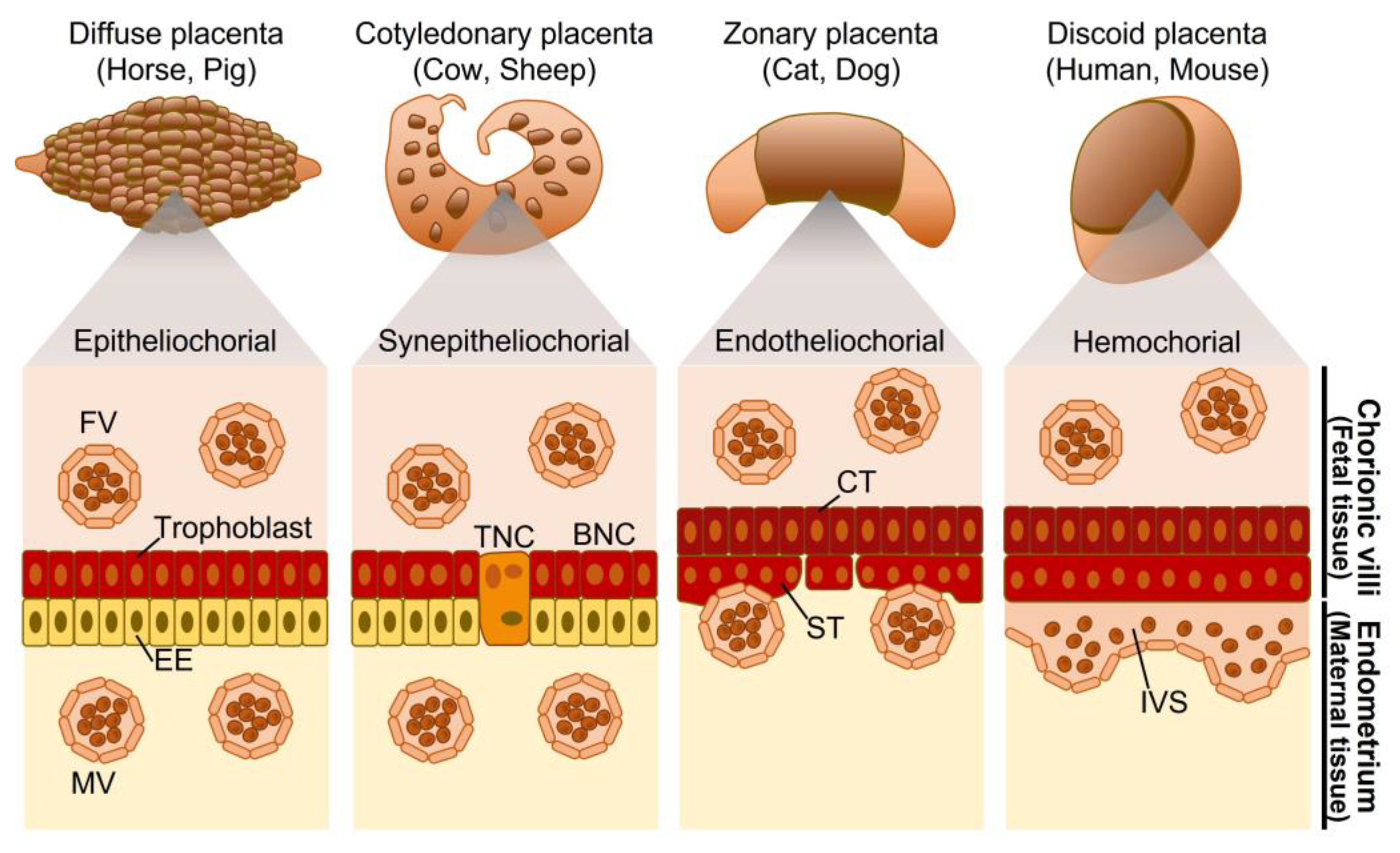
Figure 1. Placental classification based on the distribution of chorionic villi and tissue layer. FV, fetal blood vessel; MV, maternal blood vessel; EE, endometrial epithelium; BNC, binucleate cell; TNC, trinucleate cell; CT, cytotrophoblast; ST, syncytiotrophoblast; IVS, intervillous space.
Biomolecules 13 01482 g001
Figure 2. The process of placenta formation. Blastocysts are implanted in the endometrium, and trophectoderm differentiate into cytotrophoblasts. The cytotrophoblasts then fuse to form syncytiotrophoblasts, which are in direct contact with maternal blood.
Biomolecules 13 01482 g002
Figure 3. Phylogenetic tree of primates and status of placenta-associated ERVs with the type of implantation, shape, and invasiveness of placenta. Arrowheads represent the date of insertion of the indicated retroviral env genes into the genome of the primate ancestors. The filled squares indicate the conserved open reading frame of the env genes of ERVs, whereas the hatched squares indicate that the coding sequence was prematurely interrupted by mutations, insertions, deletions, and frameshifting. Red, blue, violet, orange, pink, green, and brown represent ERV-FRD, ERV-W, ERVH48-1, ERVV-1, ERVV-2, ERV3-1, and ERVMER34-1, respectively. MYA, million years ago; OWMs, Old World Monkeys; NWMs, New World Monkeys.
Biomolecules 13 01482 g003
Figure 4. Multiple alignments of the amino acid sequences of region C of ASCT2 in Primates. The first and last numbers in the alignment indicate the start and end sites of each sequence, respectively. Amino acid sequences of alanine, serine, and cysteine transporter 2 (ASCT2) were obtained from the GenBank database with the following accession numbers: human (NM_005628.3), Bonobo (XM_003817486.4), Chimpanzee (XM_016936300.2), Gorilla (XM_019015032.2), Orangutan (XM_002829451.5), White-cheeked gibbon (XM_030807697.1), Black Snub-nosed Monkey (XM_017874300.1), Golden Snub-nosed Monkey (XM_030913729.1), Olive Baboon (XM_031659126.1), Gelada (XM_025365811.1), Drill (XM_011969657.1), Sooty Mangabey (XM_012080639.1), Rhesus Macaque (XM_015124323.2), Crab-eating Macaque (XM_045379941.1), Pig-tailed macaque (XM_011736052.2), Green Monkey (XM_037991882.1), Marmoset (XM_035284840.1), Coquerel’s sifaka (XM_012653384.1), Ring-tailed lemur (XM_045531338.1), Gray mouse lemur (XM_012761502.2), Slow loris (XM_053604173.1), and Small-eared galago (XM_012811197.2). Asterisks indicate conserved nucleotides between all species. Periods indicate sites with more than 50% matches.
Biomolecules 13 01482 g004
Disclaimer/Publisher’s Note: The statements, opinions and data contained in all publications are solely those of the individual author(s) and contributor(s) and not of MDPI and/or the editor(s). MDPI and/or the editor(s) disclaim responsibility for any injury to people or property resulting from any ideas, methods, instructions or products referred to in the content.