Article

The Resistome of ESKAPEE Pathogens in Untreated and Treated Wastewater: A Polish Case Study

1
Department of Water Protection Engineering and Environmental Microbiology, Faculty of Geoengineering, University of Warmia and Mazury in Olsztyn, 10-720 Olsztyn, Poland
2
Department of Environmental Biotechnology, Faculty of Geoengineering, University of Warmia and Mazury in Olsztyn, 10-709 Olsztyn, Poland
3
Environmental Microbiology Unit, Institute for Ecology of Industrial Areas, 40-844 Katowice, Poland
*
Author to whom correspondence should be addressed.
Biomolecules 2022, 12(8), 1160; https://doi.org/10.3390/biom12081160
Submission received: 7 July 2022 / Revised: 11 August 2022 / Accepted: 19 August 2022 / Published: 21 August 2022
(This article belongs to the Special Issue State-of-the-Art in Antibiotics Resistance)

Abstract

:
The aim of this study was to quantify ESKAPEE bacteria, genes encoding resistance to antibiotics targeting this group of pathogens, as well as integrase genes in municipal wastewater and river water. Environmental DNA was extracted from the collected samples and used in deep sequencing with the Illumina TruSeq kit. The abundance of bacterial genera and species belonging to the ESKAPEE group, 400 ARGs associated with this microbial group, and three classes of integrase genes were determined. A taxonomic analysis revealed that Acinetobacter was the dominant bacterial genus, whereas Acinetobacter baumannii and Escherichia coli were the dominant bacterial species. The analyzed samples were characterized by the highest concentrations of the following ARGs: blaGES, blaOXA-58, blaTEM, qnrB, and qnrS. Acinetobacter baumannii, E. coli, and genes encoding resistance to β-lactams (blaVEB-1, blaIMP-1, blaGES, blaOXA-58, blaCTX-M, and blaTEM) and fluoroquinolones (qnrS) were detected in samples of river water collected downstream from the wastewater discharge point. The correlation analysis revealed a strong relationship between A. baumannii (bacterial species regarded as an emerging human pathogen) and genes encoding resistance to all tested groups of antimicrobials. The transmission of the studied bacteria (in particular A. baumannii) and ARGs to the aquatic environment poses a public health risk.

1. Introduction

Antibiotics play a fundamental role in the treatment of bacterial infections. However, the spread of antibiotic resistance in bacterial communities has led to a steady decline in the efficacy of antimicrobials [1,2]. In recent decades, antibiotic resistance has increased at an alarming rate, and experts have warned that humanity is approaching a post-antibiotic era [3,4]. Until recently, healthcare facilities have been regarded as the main sources of antibiotic resistance, but research has shown that animal husbandry, aquaculture, and urban environments also contribute to the spread of drug resistance. Wastewater treatment plants (WWTPs) pose a particular threat because they discharge antibiotic-resistant bacteria (ARB), antibiotic resistance genes (ARGs), and residual concentrations of antibiotics into downstream aquatic and terrestrial environments [5,6,7].
ESKAPEE (Enterococcus faecium, Staphylococcus aureus, Klebsiella pneumoniae, Acinetobacter baumannii, Pseudomonas aeruginosa, Enterobacter spp., and Escherichia coli) bacteria are increasingly associated with antibiotic resistance, which poses a serious threat to public health [8,9]. These bacteria harbor genes that encode resistance to the most popular classes of antibiotics, including vancomycin, methicillin, broad-spectrum β-lactams, carbapenems, fluoroquinolones, and aminoglycosides [10,11,12]. In 2019, the European Center for Disease Prevention and Control (ECDC) published a report on antibiotic resistance in ESKAPEE pathogens in the European Union [13]. According to the literature, an increase in the percentage of vancomycin-resistant enterococci (VRE) harboring the vanA gene is of particular concern [14]. Staphylococcus aureus, the second alarming pathogen on the list, can be represented by two genotypes: methicillin-resistant S. aureus (MRSA) harboring the mecA gene, and vancomycin-resistant S. aureus (VRSA) harboring the vanA gene [15,16]. In recent years, Klebsiella pneumoniae has also acquired a variety of β-lactamase enzymes (including blaNDM and blaKPC genes), and this pathogen is now capable of breaking down the chemical structure of penicillins, cephalosporins, as well as carbapenems, which are regarded as drugs of last resort [17,18]. Acinetobacter baumannii is an emerging human pathogen that can easily acquire and transfer ARGs to other bacteria [19]. This pathogen harbors Ambler class B and D genes that confer resistance to carbapenems, and it poses a significant problem in the medical sector [20,21,22]. The ESKAPEE group also contains Pseudomonas aeruginosa, Enterobacter spp., and E. coli pathogens that harbor extended-spectrum β-lactamase (ESBL) genes, including subtypes of blaCTX-M, blaSHV, and blaTEM genes, as well as carbapenem resistance genes, and fluoroquinolone resistance genes from the qnr group [23].
In 2017, the World Health Organization (WHO) published a list of antibiotic-resistant priority pathogens that pose a serious threat to human health, and all ESKAPEE bacteria were placed on that list. Research studies conducted in recent years have demonstrated that ESKAPEE pathogens are transmitted not only in healthcare facilities, but also in environments subjected to anthropogenic and agricultural pressure, including untreated and treated wastewater [24]. In Poland, the spread of ESKAPEE bacteria has been investigated mainly in the hospital sector and in clinical isolates [25,26,27,28,29,30], whereas far fewer studies have examined the prevalence of these pathogens in municipal wastewater and in the natural environment, including river water [31,32].
In view of the above, this metagenomic study was undertaken to quantify ESKAPEE bacteria, genes encoding resistance to antibiotics targeting this microbial group, as well as integron-integrase genes in municipal wastewater and river water. Global surveillance of these pathogens and ARGs has emerged as a critical research problem in an era of growing antimicrobial resistance.

2. Materials and Methods

2.1. Research Site and Sampling

Environmental samples were collected from a WWTP located in the region of Warmia and Mazury in northeastern Poland (Europe). The analyzed plant is equipped with mechanical and biological wastewater treatment systems. The plant processes wastewater generated in the city of Olsztyn (regional capital) and four surrounding municipalities, and it has an average daily processing capacity of 35,000 m3. The plant also receives wastewater from four hospitals, which accounts for 2% of the treated wastewater. Detailed information about the examined WWTP was previously provided by Hubeny et al. [33].
Samples of untreated wastewater (UWW), wastewater exiting from a primary clarifier (PC), wastewater from an activated sludge (AS) bioreactor, wastewater from a multifunctional reactor (MR), treated wastewater (TWW), and river water collected upstream (URW) and downstream (DRW) from the wastewater discharge point were collected in June and November 2018, and in March 2019 (Figure S1). The sampling sites have been described in detail by Rolbiecki et al. [34].
Samples of wastewater and river water were collected in sterile 500 mL and 1000 mL bottles (SIMAX). Wastewater was sampled in triplicate at hourly intervals for 24 h to obtain representative samples, and the collected hourly samples were pooled into a composite sample. River water samples were also collected in triplicate and pooled into a composite sample. The samples were transported to the laboratory at a temperature of 4 °C.

2.2. DNA Extraction and Sequencing

The samples were passed through polycarbonate membrane filters (porosity, 0.2 μm) (Millipore, Merck, Darmstadt, Germany) with the use of a vacuum pump (Millipore, Merck, Darmstadt, Germany). The filters were placed in sterile 10 mL Falcon tubes (Eppendorf, Hamburg, Germany), and 5 mL of 1X PBS (Invitrogen, Thermo Fisher Scientific, USA) solution was added. The samples were shaken for 5 h at 50 rpm at room temperature (20–22 °C) using a Grant-Bio PTR-60 rotator (Grant Instruments, Shepreth, UK). The resulting suspension was transferred to sterile 2-mL microcentrifuge tubes (Eppendorf, Hamburg, Germany) and centrifuged (Centrifuge 5415R, Eppendorf, Hamburg, Germany) for 5 min at 4300 g at a temperature of 4 °C. The supernatant was discarded, and the obtained pellet was used for DNA isolation. Environmental DNA was extracted from wastewater and river water samples with the Power Water kit (MoBio Laboratories Inc., Carlsbad, CA, USA) according to the manufacturer’s instructions. The volumes of the analyzed samples were: UWW, 40 mL; PC, 40 mL; AS, 20 mL; MR, 40 mL; TWW, 200 mL; URW, 400 mL; and DRW, 400 mL. Samples were quantified via the PicoGreen assay using a VICTOR3 fluorometric plate reader, and their quality and quantity were assessed. Illumina TruSeq DNA PCR-Free libraries were prepared manually based on the manufacturer’s protocol (TruSeq DNA PCR-Free Sample Preparation Guide, Part #15036187 Rev. D; Illumina, San Diego, CA, USA). The libraries were tested using LightCycle qPCR. Library size distribution was assessed using the Agilent Technologies 2100 Bioanalyzer with a DNA 1000 chip, and the libraries were sequenced in the NovaSeq 6000 system in an S4 flow cell lane with 2 × 150 bp configuration.

2.3. Bioinformatic Analysis

Wastewater and river water samples collected over three seasons were subjected to metagenomic analysis. FASTQ sequences were quality-filtered, and adaptor sequences were trimmed with Trimmomatic v.0.39 [35] with default parameters. Paired-end sequences were merged using PANDAseq [36]. The reads that were orphans due to quality trimming were excluded from further analyses. The quality of the resulting data sequences was checked using the FASTX-Toolkit 0.0.14–5 (http://hannonlab.cshl.edu/fastx_toolkit/) (accessed on 4 March 2022) with the FASTQ_quality_filter tool. Data were filtered, and reads with less than 80% of bases and a minimum quality score of q = 20 were eliminated. Using seqtk (https://github.com/lh3/seqtk) (accessed on 4 March 2022), FASTQ files were converted to FASTA files for BLAST alignment. Subsequently, sequences were analyzed by BLASTx-type search against the Comprehensive Antibiotic Resistance Database (CARD) [37] (https://card.mcmaster.ca; October 2019 3.0.7) (accessed on 4 March 2022). Samples were parsed and filtered to collect the top hits for each sample, and positive-hit reads with E values of ≤1 × 10⁻¹⁰ [38], amino acid identities of ≥90% [39], and amino acid alignment lengths of ≥25 were considered [40,41,42,43].
The integron database was created by downloading DNA sequences from INTEGRALL (https://integrall.bio.ua.pt) (accessed on 4 March 2022). Integron sequences were identified by aligning the sequences against database sequences using BLASTn with an E value of ≤1 × 10⁻¹⁰ [44]. If a BLASTn hit for the alignment against the INTEGRALL database had a ≥90% nucleotide read identity for ≥100 bp, the sequence was annotated as a resistance gene. Sequencing results were recorded in FASTQ files and uploaded to the MetaGenome Rapid Annotation Subsystems Technology (MG-RAST) server for analysis. Each file underwent quality control, which involved quality filtering (removing sequences with five or more ambiguous base pairs) and length filtering (removing sequences with lengths at least two standard deviations from the mean). The identified rRNA sequences were clustered with the UCLUST algorithm [45]. Based on the Greengenes reference database, a representative sequence of each cluster was used for taxonomic identification. Metagenomic data were deposited in the NCBI Sequence Read Archive (SRA) under accession number SRP286056. The results obtained for the samples collected over the three seasons were averaged for further analyses.
The co-occurrence patterns of microbial communities and resistomes were determined by calculating all pairwise Spearman’s rank correlation coefficients (p-value < 0.01) [46]. The resulting correlation matrices were translated into an associated network using Gephi 0.9.1 [47].

3. Results and Discussion

A resistome analysis was conducted on 15 wastewater samples collected during subsequent stages of treatment, as well as six samples of river water collected upstream and downstream from the wastewater discharge point. Illumina deep sequencing was used to identify ARGs, mobile genetic elements (MGEs), and the taxonomic structure of pathogens in wastewater and river water samples. More than 290 Gb of raw data was generated from 16 samples, and the number of reads ranged from 75.5 million to 201.9 million. Detailed information is presented in Table S1. The abundance of bacterial genera and species belonging to the ESKAPEE group, 400 ARGs associated with this microbial group, and three classes of integrase genes were determined in metagenomic analyses. The results were averaged and are expressed in terms of gene copies per 1 million reads (ppm).

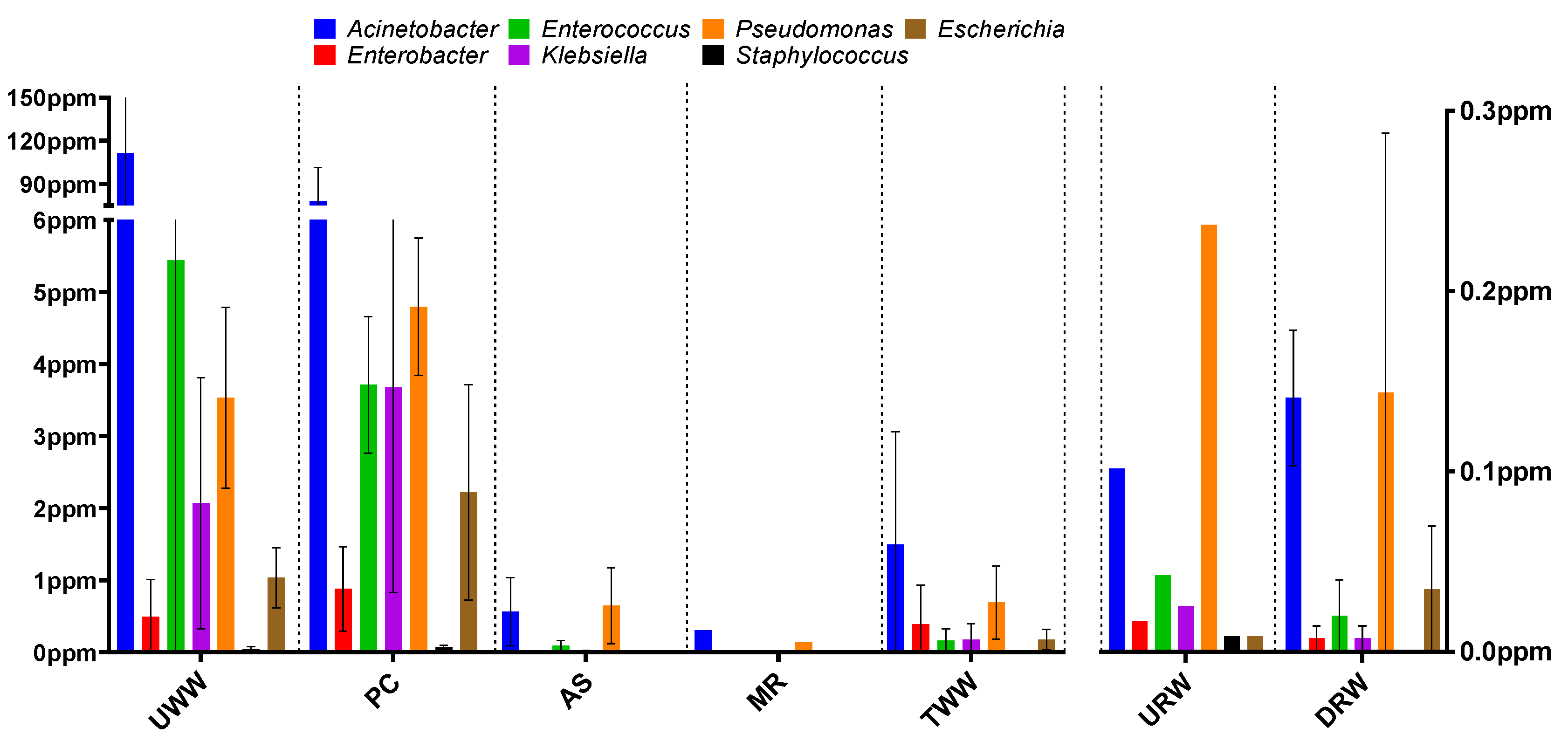
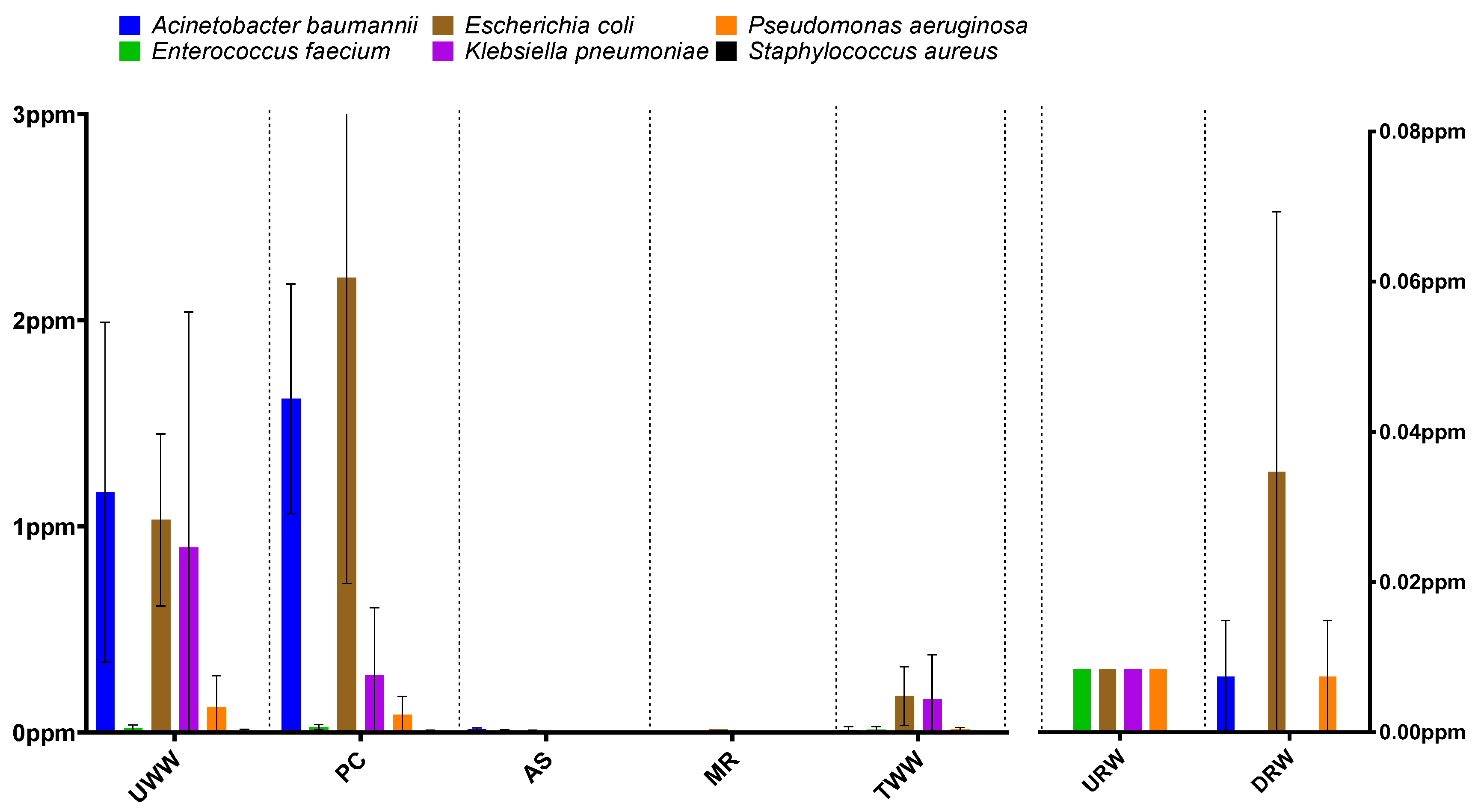
3.1. Taxonomic Structure Analysis

In all analyzed samples, the structure of the microbial communities was determined by assigning reads to taxonomic ranks. Special attention was paid to ESKAPEE pathogens, and the analysis was based on information about bacterial genera and species belonging to this group (Tables S2 and S3). The samples collected in the first two stages of wastewater treatment (UWW and PC) were characterized by a predominance of bacteria belonging to the genus Acinetobacter (111.5 ppm in UWW, 78 ppm in PC), and the abundance of the remaining bacterial genera (Enterococcus, Pseudomonas, Klebsiella, Escherichia, Enterobacter, and Staphylococcus) was determined in the range of 0–5.4 ppm (Figure 1).
ESKAPEE pathogens belong to the phyla Firmicutes and Proteobacteria, which are the most prevalent bacterial groups in wastewater. The predominant bacterial families are Enterococcaceae (including Enterococcus spp.) of the phylum Firmicutes, as well as Enterobacteriaceae (including Klebsiella spp. and Escherichia spp.) and Moraxellaceae (including Acinetobacter spp.) of the phylum Proteobacteria [48].
An analysis of six ESKAPEE group species (Acinetobacter baumannii, Enterococcus faecium, Escherichia coli, Klebsiella pneumoniae, Pseudomonas aeruginosa, and Staphylococcus aureus) confirmed that A. baumannii (1.16 ppm in UWW; 1.62 ppm in PC) and E. coli (1.16 ppm in UWW; 1.62 ppm in PC) were the dominant pathogens. In successive stages of wastewater treatment (samples AS and MR), a significant reduction was observed in the abundance of ESKAPEE genera (0.002–0.64 ppm and 0–0.3 ppm, respectively) and species (0–0.014 ppm and 0–0.015 ppm, respectively).
Similar results have been reported by other authors. Numberger et al. [48] analyzed the abundance of the three most prevalent bacterial phyla (Proteobacteria, Bacteroidetes, and Firmicutes) in samples of UWW and TWW, and found that bacteria of the family Moraxellaceae were more prevalent than bacteria of the family Enterobacteriaceae. Furthermore, Acinetobacter was the dominant bacterial genus in a taxonomic structure analysis conducted by McLellan et al. [49].
The activated sludge process in the WWTP equipped with a biological and mechanical wastewater treatment system reduced the abundance of ESKAPEE genera and species by 80% on average (UWW vs. TWW) (Table S4). In other studies, biological and mechanical wastewater treatment systems led to a similar reduction in microbial levels (80–99%) [33,50]. According to the literature, the effectiveness of wastewater treatment can be significantly affected by the type of wastewater inflow and fluctuations in a plant’s performance [51,52,53]. There is evidence to indicate that the activated sludge process can both decrease and increase the size of the bacterial population in wastewater. The results obtained for bacterial genera and species indicate that their abundance decreased drastically during successive stages of biological treatment. This is not surprising because the biological treatment process is expected to reduce microbial abundance by up to 99.99%. However, an analysis of treated effluent samples revealed an increase in microbial abundance. The mechanisms underlying this phenomenon have not been thoroughly described in the literature. Nonetheless, it appears that it could be caused by the immobilization of heavy metals or antibiotics (factors exerting selective pressure) in sewage sludge. Another reason could be the formation of biofilm in successive stages of biological treatment. All processes responsible for changes in bacterial abundance during wastewater treatment have not yet been fully elucidated, but an analysis of the pattern of these changes may shed light on the activity of microbial communities.
An analysis of bacterial genera and species in river water sampled downstream from the wastewater discharge point revealed an increase in the counts of two genera and their associated species. The abundance of A. baumannii increased from 0.000 ppm in URW to 0.007 ppm in DRW, whereas E. coli counts increased from 0.008 ppm to 0.035 ppm (Figure 2). The release of treated effluent into the river had no effect on the abundance of the remaining bacterial species. According to the literature, WWTPs are potential sources of both E. coli [54,55] and A. baumannii [56] in river water.
A comparison of microbial abundance at the level of genus and species in samples collected in different seasons and at different sites did not reveal any clear trends. However, a comparison of the results obtained during the entire treatment process shows that the average abundance of the studied taxonomic groups was highest in November, followed by June and March (Tables S5 and S6). The above trend was also observed in an earlier study by Hubeny et al. [57], where the number of Acinetobacter sp. strains isolated from wastewater and river water was highest in fall and lowest in winter.

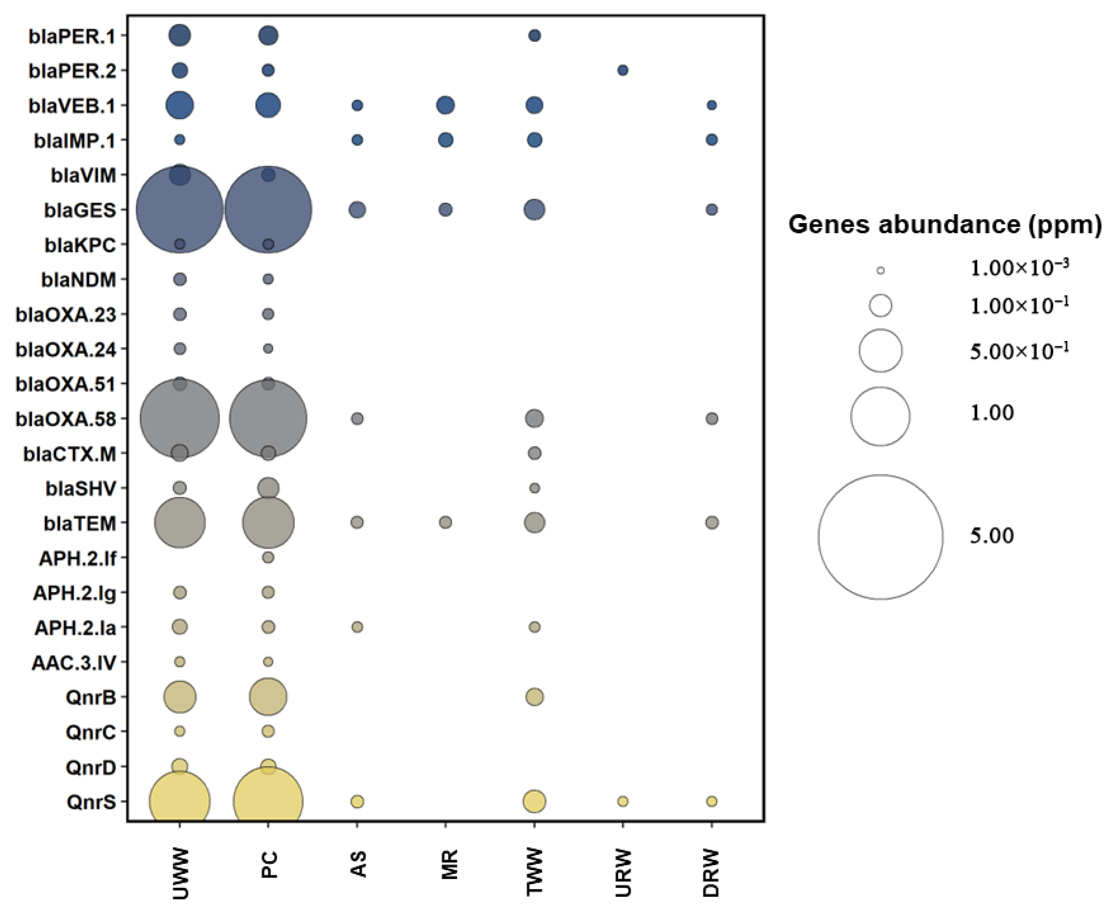
3.2. Prevalence of Antibiotic Resistance Determinants

The prevalence of ARGs and class 1, 2, and 3 integron-integrase genes was determined with the use of CARD and INTEGRALL databases. Four hundred ARGs (including subtypes), including blaPER, blaVEB, blaIMP, blaVIM, blaGES, blaKPC, blaNDM, blaOXA, blaCTX-M, blaSHV, and blaTEM (encoding resistance to β-lactams); qnrB, qnrC, qnrD, and qnrS (encoding resistance to fluoroquinolones); aph(2’’)-If, aph(2’’)-Ig, aph(2’’)-Ia, and aac(3)-IV (encoding resistance to aminoglycosides); as well as intI1, intI2, and intI3 integrase genes were selected for analysis. The selected genes confer resistance to the antibiotic groups listed in the ECDC report on antimicrobial resistance in ESKAPEE pathogens. Detailed information about the analyzed gene types and subtypes is presented in the Supplementary Materials (Table S7).
The examined ARGs were most prevalent in UWW (0.009–2.36 ppm) and PC (0.005–2.34 ppm) (Figure 3). In the work of Karkman et al. [58], ARGs were also most prevalent in samples collected in the initial stages of wastewater treatment. In the present study, the following genes were predominant in UWW and PC: blaGES (2.36 and 2.34 ppm, respectively), blaOXA-58 (1.82 and 1.93 ppm, respectively), blaTEM (0.72 and 0.76 ppm, respectively), qnrB (0.25 and 0.36 ppm, respectively), and qnrS (1.08 and 1.45 ppm, respectively) (Table S8). These genes were also frequently identified in wastewater in other studies. Wang et al. [59] found that blaTEM, blaOXA, and qnrS were highly prevalent in municipal water in Europe, USA, and Canada. Pärnänen et al. [60] investigated the prevalence of ARGs in Europe and reported that blaGES and blaOXA were commonly detected in influent and effluent wastewater. In the current study, genes encoding resistance to β-lactams and quinolones were most prevalent in UWW and PC. According to the ECDC report for 2020, β-lactams and quinolones were widely used in the Polish medical sector [61]. Research has demonstrated that the use of specific antibiotics in healthcare facilities is frequently correlated with an increase in the prevalence of ARGs in wastewater [62]. A decrease in the abundance of the tested genes was observed when TWW was compared with DRW. Tang et al. [63] also noted a decrease in the abundance of ARGs when comparing TWW with river water sampled downstream from the wastewater discharge point. This phenomenon can be explained by the fact that wastewater reaching the river can be diluted more than 250 times. Natural self-purification of water bodies also contributes to decreasing the abundance of ARGs in river water samples. The absence of the blaPER-2 gene in DRW can be explained in a similar way.
The heat map revealed a significant decrease in ARG abundance in TWW. However, the prevalence of ARGs in TWW increased relative to the samples collected from the activated sludge reactor (AS) and the multifunctional reactor (MR) (Figure 3); however, the observed differences were not statistically significant (Table S9). In a study by Zieliński et al. [64], the abundance of ARGs encoding resistance to β-lactams and quinolones was also higher in TWW than in the AS. During subsequent stages of biological treatment, the abundance of ARGs decreases due to reduced nutrient availability [65]. In turn, an increase in the prevalence of antibiotic resistance determinants in TWW may be associated with the formation of biofilm by bacteria harboring those genes [64]. Despite the observed increase in ARG abundance, more than 90% of the examined genes were removed during the biological treatment process (Table S10). In other studies, the prevalence of ARGs was also reduced in TWW [66,67,68].
An analysis of integrase genes (MGEs) revealed that intI1 was more prevalent in wastewater samples (78.17–2911.29 ppm) than intI2 (0.37–34.41 ppm) and intI3 (0.15–10.03 ppm) (Figure 4 and Table S11). Similar results were noted in our previous study [33], where intI1 was the most abundant integrase gene (166.59–2883.91 ppm), whereas intI2 (0.02–29.87 ppm) and intI3 (0.09–2.46 ppm) were determined in smaller quantities. The concentrations of integrase genes followed a similar pattern in other studies [69,70].
The present study confirms that the analyzed WWTP contributed to the contamination of river water with ARGs and all analyzed integrons. The developed heat maps indicate that the abundance of blaVEB-1, blaIMP-1, blaGES, blaOXA-58, blaTEM, qnrS, and class 1, 2, and 3 integron-integrase genes was higher in river water sampled downstream than upstream from the wastewater discharge point (Table S12). Sabri et al. [71] reported similar results in samples of river water collected downstream from the wastewater discharge point. In the cited study, ARG concentrations were also higher in sampling sites located downstream than upstream. The concentrations of ARGs and class 1 integron-integrase genes were more than two orders of magnitude higher in samples collected from downstream sites. Czekalski et al. [72] and Osińska et al. [73] also reported an increase in the abundance of antibiotic resistance determinants in river water receiving effluents from WWTPs. In the present study, effluents from four hospitals accounted for 2% of the wastewater processed by the analyzed WWTP. According to Hocqeut et al. [74] and Paulus et al. [75], hospitals play a major role in the transmission of ARB and ARGs. The polluting effects of hospital wastewater could be exacerbated by the fact that the use of last-resort drugs has increased in recent years, and hospital effluents could have a different ARG profile than other types of wastewater. This observation indicates that growing levels of antibiotic resistance and the spread of ARGs in the environment can be largely attributed to human activity [76].
Aquatic environments such as wastewater and river water are affected by physicochemical factors that contribute to changes in these environments [77]. Zieliński et al. [78] focused on the epidemiological threat posed by antibiotic resistance determinants to WWTP employees and the environment. The cited authors studied the same samples of wastewater, sewage sludge, and river water as those analyzed in the current study, and described the influence of physicochemical parameters on the prevalence and diversity of ARGs. Based on their data, it can be concluded that total organic carbon (TOC), chemical oxygen demand (COD), and biological oxygen demand (BOD) are strongly correlated with the concentrations of different groups of ARGs, in particular, those conferring resistance to β-lactams. According to the literature, lower TOC levels contribute to an increase in the diversity and abundance of ARGs. Environmental physicochemical parameters could have a direct effect on the diversity and the abundance of ARGs in water bodies.
A comparative analysis of ESKAPEE abundance in different seasons did not reveal any specific trends of changes in their concentrations in samples collected during subsequent stages of the treatment process. A comparison of overall ARG abundance data between seasons revealed only negligible differences (Tables S13 and S14).
The extent to which ARB and ARGs present in the natural environment affect human health continues to attract increasing interest from researchers. Bouki et al. [79] reported an increase in the number of environmental strains harboring ARGs that are most often detected in clinically relevant bacteria, which implies that many bacteria have developed resistance to one or more antimicrobials. Ultimately, these bacteria can be transferred from the environment to humans.

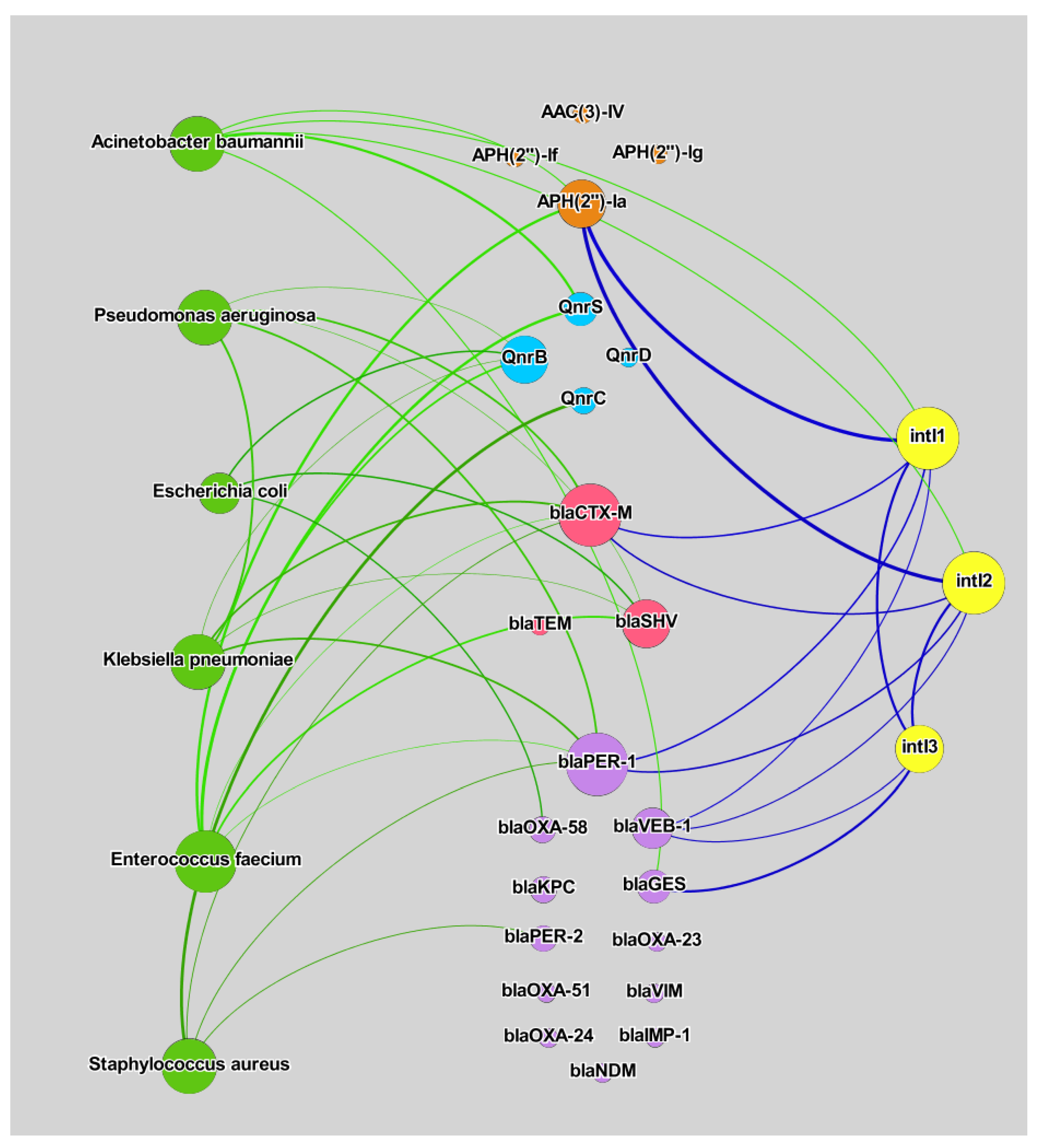
3.3. Correlation Analysis

The examined bacterial species of the ESKAPEE group, ARGs, and integrase genes were subjected to a correlation analysis. The number and strength of the correlations between the studied variables were determined in a network analysis (Figure 5). Three types of correlations were investigated: (1) bacterial species vs. ARGs, (2) bacterial species vs. integrase genes, and (3) integrase genes vs. ARGs. In all analyses, the values of the correlation coefficient ranged from 0.875 to 0.964 (Table S15). The results of Spearman’s rank-order correlation tests are presented in the Supplementary Materials. Not all of the examined ARGs were bound by significant correlations with ESKAPEE species or integrase genes. However, a correlation analysis investigates the relationships between quantitative variables; therefore, the absence of significant relationships could be due to the fact that some genes occur rarely in a given environment [80].
In the group of the analyzed bacterial species, A. baumannii and E. faecium were bound by the highest number of significant correlations with ARGs. The presence of these species was associated with the prevalence of genes encoding resistance to β-lactams, aminoglycosides, and fluoroquinolones. A network analysis conducted by Alexander et al. [81] revealed significant correlations between A. baumannii and enterococcus counts and the abundance of genes encoding resistance to β-lactams in wastewater samples. Strong correlations between microbial counts and the above ARGs could suggest that: (1) the presence of these bacteria in the group of opportunistic ESKAPEE pathogens is associated with hospital infection outbreaks, and (2) extensive use of β-lactams and fluoroquinolones in the medical sector contributes to antibiotic resistance in the environment [82,83,84].
Integrons are MGEs that can harbor ARGs [85]. Integrons and other MGEs (such as transposons and plasmids) have a high potential for transferring ARGs [86]. The role of integrons as potential hosts for ARGs has been analyzed in the literature [87,88]. In the current study, integrase genes were also bound by significant correlations with ARGs. The intI3 gene was strongly correlated with blaGES and blaVEB-1, as well as with intI1 and intI2 genes. Both intI1 and intI2 were associated with APH(2’’)-Ia, blaCTX-M, blaPER-1, and blaVEB-1 genes, as well as A. baumannii counts. Acinetobacter baumannii was the only ESKAPEE species that was correlated with integrase genes. According to the literature, the genome of A. baumannii harbors both class 1 [89,90,91] and class 2 integrons [92,93,94]. Class I integrons can carry 40 ARGs associated with aminoglycosides, β-lactams, chloramphenicol, macrolides, and sulfonamides [91]. The absence of a direct correlation between A. baumannii and intI3 could be attributed to the fact that this integron is very rarely found in the genome of the above bacterial species [95,96]. In recent years, Acinetobacter spp. (including A. baumannii) have been identified as a major cause of nosocomial infections, and these emerging human pathogens have impressive genetic capabilities for acquiring and disseminating antibiotic resistance. Acinetobacter baumannii strains isolated from hospitals and the natural environment were positively correlated with ARG abundance [97]. The results of the present study and the findings of other authors suggest that the spread of ESKAPEE pathogens, in particular A. baumannii, in river water can increase the prevalence of antibiotic resistance determinants (integrons) and expand the resistome of environmental bacteria.

4. Conclusions

This study investigated the prevalence of opportunistic pathogens from the ESKAPEE group and antibiotic resistance determinants in wastewater and river water samples. The examined pathogens and genes determining resistance to clinically important antimicrobials were identified in the studied samples. These results indicate that WWTPs play an important role in the dissemination of critical ARGs and ESKAPEE bacteria in the natural environment. Despite the fact that the abundance of the analyzed bacteria was significantly reduced by the wastewater treatment process, river water sampled downstream from the wastewater discharge point was polluted with A. baumannii and E. coli, which are important pathogens with highly mobile resistomes. A correlation analysis revealed that integrase genes were strongly correlated only with A. baumannii, which has been identified as an emerging human pathogen. The presence of these bacteria in the aquatic environment poses a serious threat to public health. To the best of our knowledge, this is the first study to investigate the transmission of ESKAPEE pathogens and ARGs from the wastewater matrix to the natural environment in central–eastern Europe. Further research is needed to elucidate the evolution of antibiotic resistance mechanisms in clinically important ESKAPEE bacteria in the natural environment.

Supplementary Materials

The following supporting information can be downloaded at: https://www.mdpi.com/article/10.3390/biom12081160/s1. Table S1: Total read base and total read count of raw data in the analyzed samples; Table S2: Abundance of ESKAPEE group members (genus level) in wastewater and river water samples (ppm). Averaged results from three seasons; Table S3: Abundance of ESKAPEE group members (species level) in wastewater and river water samples (ppm). Averaged results from three seasons; Table S4: Calculated reduction the abundance of ESKAPEE group members (genus and species levels) after the wastewater treatment process. Averaged results from three seasons; Table S5. Abundance of ESKAPEE group members (genus level) in wastewater and river water samples (ppm) collected over three seasons; Table S6. Abundance of ESKAPEE group members (species level) in wastewater and river water samples (ppm) collected over three seasons; Table S7: Selected subtypes and/or types of antibiotic resistance genes in a metagenomic analysis; Table S8: Abundance of the analyzed antibiotic resistance genes associated with ESKAPEE bacteria in wastewater and river water samples (ppm). Averaged results from three seasons; Table S9: Statistical analysis—Mann–Whitney U test; Table S10: Calculated reduction in the abundance of the analyzed antibiotic resistance genes after the wastewater treatment process. Averaged results from three seasons (ppm); Table S11: Abundance of the analyzed integron genes in wastewater and river water samples (ppm). Averaged results from three seasons; Table S12: Calculated pollution of the analyzed samples with antibiotic resistance genes after the wastewater treatment process. Averaged results from three seasons (ppm); Table S13: Abundance of the analyzed antibiotic resistance genes associated with ESKAPEE bacteria in wastewater and river water samples (ppm) collected over three seasons; Table S14: Abundance of the analyzed integron genes in wastewater and river water samples (ppm) collected over three seasons; Table S15: Spearman’s rank correlation analysis of experimental variables (p < 0.01); Figure S1: Diagram of the analyzed WWTP. Sampling sites are marked in red.

Author Contributions

J.H.: Conceptualization, Methodology, Validation, Formal analysis, Investigation, Data curation, Writing—original draft, Visualization. M.H.: Conceptualization, Methodology, Validation, Resources, Supervision, Project administration, Funding acquisition. S.C.: Conceptualization, Methodology, Validation. G.P.: Conceptualization, Validation, Project administration, Funding acquisition. E.K.: Conceptualization, Review. All authors have read and agreed to the published version of the manuscript.

Funding

This research was founded by grants No. 2017/M/NZ9/00071 and 2017/27/B/NZ9/00267 from the Polish National Science Centre.

Institutional Review Board Statement

Not applicable.

Informed Consent Statement

Not applicable.

Data Availability Statement

Metagenomic data were deposited in the NCBI Sequence Read Archive (SRA) under accession number SRP286056.

Acknowledgments

Jakub Hubeny has received a scholarship from the Interdisciplinary Doctoral Program in Bioeconomy (POWR.03.02.00-00-I034/16 00) financed by the European Social Fund.

Conflicts of Interest

The authors declare no conflict of interest.

References

  1. Nesme, J.; Simonet, P. The soil resistome: A critical review on antibiotic resistance origins, ecology and dissemination potential in telluric bacteria. Environ. Microbiol. 2015, 17, 913–930. [Google Scholar] [CrossRef] [PubMed]
  2. Timothy, J.F. Antibiotic resistance in Staphylococcus aureus. Curr. Status Future Prospect. FEMS Microbiol. Rev. 2017, 41, 430–449. [Google Scholar]
  3. Heymann, D.L. The World Health Report 2007: A Safer Future: Global Public Health Security in the 21st Century; World Health Organization: Geneva, Switzerland, 2007. [Google Scholar]
  4. Marano, R.B.M.; Fernandes, T.; Manaia, C.M.; Nunes, O.; Morrison, D.; Berendonk, T.U.; Kreuzinger, N.; Tenson, T.; Corno, G.; Fatta-Kassinos, D.; et al. A global multinational survey of cefotaxime-resistant coliforms in urban wastewater treatment plants. Environ. Int. 2020, 144, 106035. [Google Scholar] [CrossRef] [PubMed]
  5. Ventola, C.L. The antibiotic resistance crisis: Part 1: Causes and threats. Pharm. Ther. 2015, 40, 277–283. [Google Scholar]
  6. Berendonk, T.U.; Manaia, C.M.; Merlin, C.; Fatta-Kassinos, D.; Cytryn, E.; Walsh, F.; Bürgmann, H.; Sørum, H.; Norström, M.; Pons, M.N.; et al. Tackling antibiotic resistance: The environmental framework. Nat. Rev. Microbiol. 2015, 13, 310–317. [Google Scholar] [CrossRef]
  7. Tripathi, V.; Cytryn, E. Impact of anthropogenic activities on the dissemination of antibiotic resistance across ecological boundaries. Essays Biochem. 2017, 61, 11–21. [Google Scholar] [CrossRef] [Green Version]
  8. Santajit, S.; Indrawattana, N. Mechanisms of Antimicrobial Resistance in ESKAPE Pathogens. BioMed Res. Int. 2016, 2016, 1–8. [Google Scholar] [CrossRef] [Green Version]
  9. Savin, M.; Bierbaum, G.; Hammerl, J.A.; Heinemann, C.; Parcina, M.; Sib, E.; Voigt, A.; Kreyenschmidt, J. Antibiotic-resistant bacteria and antimicrobial residues in wastewater and process water from German pig slaughterhouses and their receiving municipal wastewater treatment plants. Sci. Total Environ. 2020, 727, 138788. [Google Scholar] [CrossRef]
  10. Grochowalska, A.; Kozioł-Montewka, M.; Oliynyk, O.; Krasij, N. Ventilator-associated pneumonia (VAP) caused by Acinetobacter baumannii in view of the microbial properties of the ESKAPE group in neighbouring countries—Poland and Ukraine. J. Pre-Clin. Clin. Res. 2017, 11, 111–115. [Google Scholar] [CrossRef] [Green Version]
  11. Behroozian, S.; Svensson, S.L.; Davies, J. Kisameet Clay Exhibits Potent Antibacterial Activity against the ESKAPE Pathogens. MBio 2016, 7, e01842-15. [Google Scholar] [CrossRef] [Green Version]
  12. Bales, P.M.; Renke, E.M.; May, S.L.; Shen, Y.; Nelson, D.C. Purification and Characterization of Biofilm-Associated EPS Exopolysaccharides from ESKAPE Organisms and Other Pathogens. PLoS ONE 2013, 8, e67950. [Google Scholar] [CrossRef] [PubMed] [Green Version]
  13. ECDC. Antimicrobial resistance in the EU/EEA (EARS-Net). Antimicrob. Resist. EU/EEA 2020, 174, 341. [Google Scholar]
  14. Smith, A. Baterial resistance to antibiotics. In Hugo and Russell’s Pharmaceutical Microbiology; Wiley-Blackwell: Hoboken, NJ, USA, 2004; pp. 220–232. ISBN 0-632-06467-6. [Google Scholar]
  15. Indrawattana, N.; Sungkhachat, O.; Sookrung, N.; Chongsa-nguan, M.; Tungtrongchitr, A.; Voravuthikunchai, S.P.; Kong-ngoen, T.; Kurazono, H.; Chaicumpa, W. Staphylococcus aureus Clinical Isolates: Antibiotic Susceptibility, Molecular Characteristics, and Ability to Form Biofilm. BioMed. Res. Int. 2013, 2013, 1–11. [Google Scholar] [CrossRef] [Green Version]
  16. Appelbaum, P.C. Reduced glycopeptide susceptibility in methicillin-resistant Staphylococcus aureus (MRSA). Int. J. Antimicrob. Agents 2007, 30, 398–408. [Google Scholar] [CrossRef]
  17. Yong, D.; Toleman, M.A.; Giske, C.G.; Cho, H.S.; Sundman, K.; Lee, K.; Walsh, T.R. Characterization of a New Metallo-β-Lactamase Gene, bla NDM-1, and a Novel Erythromycin Esterase Gene Carried on a Unique Genetic Structure in Klebsiella pneumoniae Sequence Type 14 from India. Antimicrob. Agents Chemother. 2009, 53, 5046–5054. [Google Scholar] [CrossRef] [PubMed] [Green Version]
  18. Kumarasamy, K.K.; Toleman, M.A.; Walsh, T.R.; Bagaria, J.; Butt, F.; Balakrishnan, R.; Chaudhary, U.; Doumith, M.; Giske, C.G.; Irfan, S.; et al. Emergence of a new antibiotic resistance mechanism in India, Pakistan, and the UK: A molecular, biological, and epidemiological study. Lancet Infect. Dis. 2010, 10, 597–602. [Google Scholar] [CrossRef]
  19. Bonnin, R.A.; Poirel, L.; Nordmann, P. New Delhi metallo-β-lactamase-producing Acinetobacter baumannii: A novel paradigm for spreading antibiotic resistance genes. Future Microbiol. 2014, 9, 33–41. [Google Scholar] [CrossRef] [PubMed] [Green Version]
  20. Boucher, H.W.; Talbot, G.H.; Bradley, J.S.; Edwards, J.E.; Gilbert, D.; Rice, L.B.; Scheld, M.; Spellberg, B.; Bartlett, J. Bad bugs, no drugs: No ESKAPE! An update from the Infectious Diseases Society of America. Clin. Infect. Dis. 2009, 48, 1–12. [Google Scholar] [CrossRef] [PubMed] [Green Version]
  21. Vila, J.; Martí, S.; Sánchez-Céspedes, J. Porins, efflux pumps and multidrug resistance in Acinetobacter baumannii. J. Antimicrob. Chemother. 2007, 59, 1210–1215. [Google Scholar] [CrossRef] [Green Version]
  22. Vrancianu, C.O.; Pelcaru, C.F.; Alistar, A.; Gheorghe, I.; Marutescu, L.; Popa, M.; Czobor, I.; Gradisteanu, G.; Dobre, E.G.; Chifiriuc, M.C. Escaping from ESKAPE. Clinical significance and antibiotic resistance mechanisms in acinetobacter baumannii: A review. Biointerface Res. Appl. Chem. 2021, 11, 8190–8203. [Google Scholar] [CrossRef]
  23. Castanheira, M.; Deshpande, L.M.; Mathai, D.; Bell, J.M.; Jones, R.N.; Mendes, R.E. Early Dissemination of NDM-1- and OXA-181-Producing Enterobacteriaceae in Indian Hospitals: Report from the SENTRY Antimicrobial Surveillance Program, 2006–2007. Antimicrob. Agents Chemother. 2011, 55, 1274–1278. [Google Scholar] [CrossRef] [PubMed] [Green Version]
  24. Pendleton, J.N.; Gorman, S.P.; Gilmore, B.F. Clinical relevance of the ESKAPE pathogens. Expert Rev. Anti. Infect. Ther. 2013, 11, 297–308. [Google Scholar] [CrossRef] [PubMed]
  25. Laudy, A.E.; Róg, P.; Smolinska-Król, K.; Ćmiel, M.; Søoczynska, A.; Patzer, J.; Dzierzanowska, D.; Wolinowska, R.; Starosciak, B.; Tyski, S. Prevalence of ESBL-producing Pseudomonas aeruginosa isolates in Warsaw, Poland, detected by various phenotypic and genotypic methods. PLoS ONE 2017, 12, e0180121. [Google Scholar] [CrossRef] [Green Version]
  26. Kot, B.; Wierzchowska, K.; Piechota, M.; Gruzewska, A. Antimicrobial Resistance Patterns in Methicillin-Resistant Staphylococcus aureus from Patients Hospitalized during 2015-2017 in Hospitals in Poland. Med. Princ. Pract. 2020, 29, 61–68. [Google Scholar] [CrossRef] [PubMed]
  27. Litwin, A.; Fedorowicz, O.; Duszynska, W. Characteristics of microbial factors of healthcare-associated infections including multidrug-resistant pathogens and antibiotic consumption at the university intensive care unit in Poland in the years 2011–2018. Int. J. Environ. Res. Public Health 2020, 17, 6943. [Google Scholar] [CrossRef]
  28. Zając-Spychała, O.; Wachowiak, J.; Frączkiewicz, J.; Salamonowicz, M.; Kałwak, K.; Gorczyńska, E.; Chybicka, A.; Czyżewski, K.; Dziedzic, M.; Wysocki, M.; et al. Multidrug-resistant bacterial infections in children undergoing haematopoietic stem cell transplantation over a 6-year period: Analysis of the Polish Pediatric Group for Hematopoietic Stem Cell Transplantation. J. Appl. Microbiol. 2020, 128, 292–300. [Google Scholar] [CrossRef] [Green Version]
  29. Zając-Spychała, O.; Wachowiak, J.; Pieczonka, A.; Siewiera, K.; Frączkiewicz, J.; Kałwak, K.; Gorczyńska, E.; Chybicka, A.; Czyżewski, K.; Jachna-Sawicka, K.; et al. Bacterial infections in pediatric hematopoietic stem cell transplantation recipients: Incidence, epidemiology, and spectrum of pathogens: Report of the Polish Pediatric Group for Hematopoietic Stem Cell Transplantation. Transpl. Infect. Dis. 2016, 18, 690–698. [Google Scholar] [CrossRef]
  30. Talaga-Ćwiertnia, K.; Krzyściak, P.; Bulanda, M. Do bacteria isolated from ICU patients ESKAPE antibiotic treatment?—In vitro susceptibility of the Enterobacteriaceae family to tigecycline. Anaesthesiol. Intensive Ther. 2017, 49, 210–214. [Google Scholar] [CrossRef] [Green Version]
  31. Gotkowska-Płachta, A. The Prevalence of Virulent and Multidrug-Resistant Enterococci in River Water and in Treated and Untreated Municipal and Hospital Wastewater. Int. J. Environ. Res. Public Health 2021, 18, 563. [Google Scholar] [CrossRef]
  32. Rybak, B.; Wawrzyniak, N.; Wolska, L.; Potrykus, M. Escherichia coli and Serratia fonticola ESBLs as a potential source of antibiotics resistance dissemination in the Tricity water reservoirs. Acta Biochim. Pol. 2021, 68, 437–448. [Google Scholar] [CrossRef]
  33. Hubeny, J.; Ciesielski, S.; Harnisz, M.; Korzeniewska, E.; Dulski, T.; Jałowiecki, Ł.; Płaza, G. Impact of Hospital Wastewater on the Occurrence and Diversity of Beta-Lactamase Genes During Wastewater Treatment with an Emphasis on Carbapenemase Genes: A Metagenomic Approach. Front. Environ. Sci. 2021, 9, 1–12. [Google Scholar] [CrossRef]
  34. Rolbiecki, D.; Harnisz, M.; Korzeniewska, E.; Buta, M.; Hubeny, J.; Zieliński, W. Detection of carbapenemase-producing, hypervirulent Klebsiella spp. in wastewater and their potential transmission to river water and WWTP employees. Int. J. Hyg. Environ. Health 2021, 237, 113831. [Google Scholar] [CrossRef] [PubMed]
  35. Bolger, A.M.; Lohse, M.; Usadel, B. Trimmomatic: A flexible trimmer for Illumina sequence data. Bioinformatics 2014, 30, 2114–2120. [Google Scholar] [CrossRef] [PubMed] [Green Version]
  36. Masella, A.P.; Bartram, A.K.; Truszkowski, J.M.; Brown, D.G.; Neufeld, J.D. PANDAseq: Paired-end assembler for illumina sequences. BMC Bioinform. 2012, 13, 31. [Google Scholar] [CrossRef] [Green Version]
  37. McArthur, A.G.; Waglechner, N.; Nizam, F.; Yan, A.; Azad, M.A.; Baylay, A.J.; Bhullar, K.; Canova, M.J.; De Pascale, G.; Ejim, L.; et al. The Comprehensive Antibiotic Resistance Database. Antimicrob. Agents Chemother. 2013, 57, 3348–3357. [Google Scholar] [CrossRef] [Green Version]
  38. Escudeiro, P.; Pothier, J.; Dionisio, F.; Nogueira, T. Antibiotic Resistance Gene Diversity and Virulence Gene Diversity Are Correlated in Human Gut and Environmental Microbiomes. mSphere 2019, 4, e00135-19. [Google Scholar] [CrossRef] [Green Version]
  39. Casaburi, G.; Duar, R.M.; Vance, D.P.; Mitchell, R.; Contreras, L.; Frese, S.A.; Smilowitz, J.T.; Underwood, M.A. Early-life gut microbiome modulation reduces the abundance of antibiotic-resistant bacteria. Antimicrob. Resist. Infect. Control 2019, 8, 131. [Google Scholar] [CrossRef]
  40. Kristiansson, E.; Fick, J.; Janzon, A.; Grabic, R.; Rutgersson, C.; Weijdegård, B.; Söderström, H.; Larsson, D.G.J. Pyrosequencing of Antibiotic-Contaminated River Sediments Reveals High Levels of Resistance and Gene Transfer Elements. PLoS ONE 2011, 6, e17038. [Google Scholar] [CrossRef]
  41. Zhang, T.; Zhang, X.-X.; Ye, L. Plasmid Metagenome Reveals High Levels of Antibiotic Resistance Genes and Mobile Genetic Elements in Activated Sludge. PLoS ONE 2011, 6, e26041. [Google Scholar] [CrossRef] [Green Version]
  42. Yang, Y.; Li, B.; Ju, F.; Zhang, T. Exploring Variation of Antibiotic Resistance Genes in Activated Sludge over a Four-Year Period through a Metagenomic Approach. Environ. Sci. Technol. 2013, 47, 10197–10205. [Google Scholar] [CrossRef]
  43. Su, J.-Q.; An, X.-L.; Li, B.; Chen, Q.-L.; Gillings, M.R.; Chen, H.; Zhang, T.; Zhu, Y.-G. Metagenomics of urban sewage identifies an extensively shared antibiotic resistome in China. Microbiome 2017, 5, 84. [Google Scholar] [CrossRef] [PubMed]
  44. Tian, Z.; Zhang, Y.; Yu, B.; Yang, M. Changes of resistome, mobilome and potential hosts of antibiotic resistance genes during the transformation of anaerobic digestion from mesophilic to thermophilic. Water Res. 2016, 98, 261–269. [Google Scholar] [CrossRef] [PubMed]
  45. Edgar, R.C. Search and clustering orders of magnitude faster than BLAST. Bioinformatics 2010, 26, 2460–2461. [Google Scholar] [CrossRef] [PubMed] [Green Version]
  46. Barberán, A.; Bates, S.T.; Casamayor, E.O.; Fierer, N. Using network analysis to explore co-occurrence patterns in soil microbial communities. ISME J. 2012, 6, 343–351. [Google Scholar] [CrossRef] [PubMed] [Green Version]
  47. Bastian, M.; Heymann, S.; Jacomy, M. Gephi: An Open Source Software for Exploring and Manipulating Networks. In Proceedings of the Third International AAAI Conference on Weblogs and Social Media, San Jose, CA, USA, 17–20 May 2009; pp. 361–362. [Google Scholar] [CrossRef] [Green Version]
  48. Numberger, D.; Ganzert, L.; Zoccarato, L.; Mühldorfer, K.; Sauer, S.; Grossart, H.-P.; Greenwood, A.D. Characterization of bacterial communities in wastewater with enhanced taxonomic resolution by full-length 16S rRNA sequencing. Sci. Rep. 2019, 9, 9673. [Google Scholar] [CrossRef] [Green Version]
  49. McLellan, S.L.; Huse, S.M.; Mueller-Spitz, S.R.; Andreishcheva, E.N.; Sogin, M.L. Diversity and population structure of sewage-derived microorganisms in wastewater treatment plant influent. Environ. Microbiol. 2010, 12, 378–392. [Google Scholar] [CrossRef] [Green Version]
  50. Jäger, T.; Hembach, N.; Elpers, C.; Wieland, A.; Alexander, J.; Hiller, C.; Krauter, G.; Schwartz, T. Reduction of Antibiotic Resistant Bacteria During Conventional and Advanced Wastewater Treatment, and the Disseminated Loads Released to the Environment. Front. Microbiol. 2018, 9, 2599. [Google Scholar] [CrossRef] [Green Version]
  51. Rafraf, I.D.; Lekunberri, I.; Sànchez-Melsió, A.; Aouni, M.; Borrego, C.M.; Balcázar, J.L. Abundance of antibiotic resistance genes in five municipal wastewater treatment plants in the Monastir Governorate, Tunisia. Environ. Pollut. 2016, 219, 353–358. [Google Scholar] [CrossRef]
  52. Oliveira, M.; Serrano, I.; Van Harten, S.; Bessa, L.J.; Bernardo, F.; da Costa, P.M. Fecal contamination of wastewater treatment plants in Portugal. Environ. Sci. Pollut. Res. 2016, 23, 14671–14675. [Google Scholar] [CrossRef]
  53. Niestępski, S.; Harnisz, M.; Ciesielski, S.; Korzeniewska, E.; Osińska, A. Environmental fate of Bacteroidetes, with particular emphasis on Bacteroides fragilis group bacteria and their specific antibiotic resistance genes, in activated sludge wastewater treatment plants. J. Hazard. Mater. 2020, 394, 122544. [Google Scholar] [CrossRef]
  54. Osińska, A.; Korzeniewska, E.; Harnisz, M.; Niestępski, S. The prevalence and characterization of antibiotic-resistant and virulent Escherichia coli strains in the municipal wastewater system and their environmental fate. Sci. Total Environ. 2017, 577, 367–375. [Google Scholar] [CrossRef] [PubMed]
  55. Bessa, L.J.; Barbosa-Vasconcelos, A.; Mendes, Â.; Vaz-Pires, P.; Martins da Costa, P. High prevalence of multidrug-resistant Escherichia coli and Enterococcus spp. in river water, upstream and downstream of a wastewater treatment plant. J. Water Health 2014, 12, 426–435. [Google Scholar] [CrossRef] [PubMed]
  56. Maravić, A.; Skočibušić, M.; Fredotović, Ž.; Šamanić, I.; Cvjetan, S.; Knezović, M.; Puizina, J. Urban riverine environment is a source of multidrug-resistant and ESBL-producing clinically important Acinetobacter spp. Environ. Sci. Pollut. Res. 2016, 23, 3525–3535. [Google Scholar] [CrossRef] [PubMed]
  57. Hubeny, J.; Korzeniewska, E.; Buta-Hubeny, M.; Zieliński, W.; Rolbiecki, D.; Harnisz, M. Characterization of carbapenem resistance in environmental samples and Acinetobacter spp. isolates from wastewater and river water in Poland. Sci. Total Environ. 2022, 822, 153437. [Google Scholar] [CrossRef] [PubMed]
  58. Karkman, A.; Johnson, T.A.; Lyra, C.; Stedtfeld, R.D.; Tamminen, M.; Tiedje, J.M.; Virta, M. High-throughput quantification of antibiotic resistance genes from an urban wastewater treatment plant. FEMS Microbiol. Ecol. 2016, 92, fiw014. [Google Scholar] [CrossRef] [Green Version]
  59. Wang, J.; Chu, L.; Wojnárovits, L.; Takács, E. Occurrence and fate of antibiotics, antibiotic resistant genes (ARGs) and antibiotic resistant bacteria (ARB) in municipal wastewater treatment plant: An overview. Sci. Total Environ. 2020, 744, 140997. [Google Scholar] [CrossRef]
  60. Pärnänen, K.M.M.; Narciso-Da-Rocha, C.; Kneis, D.; Berendonk, T.U.; Cacace, D.; Do, T.T.; Elpers, C.; Fatta-Kassinos, D.; Henriques, I.; Jaeger, T.; et al. Antibiotic resistance in European wastewater treatment plants mirrors the pattern of clinical antibiotic resistance prevalence. Sci. Adv. 2019, 5, eaau9124. [Google Scholar] [CrossRef] [Green Version]
  61. ECDC. Antimicrobial consumption in the EU/EEA (ESAC-Net). Eur. Cent. Dis. Prev. Control 2021, 11–14. [Google Scholar]
  62. Duarte, D.J.; Oldenkamp, R.; Ragas, A.M.J. Modelling environmental antibiotic-resistance gene abundance: A meta-analysis. Sci. Total Environ. 2019, 659, 335–341. [Google Scholar] [CrossRef] [Green Version]
  63. Tang, J.; Bu, Y.; Zhang, X.-X.; Huang, K.; He, X.; Ye, L.; Shan, Z.; Ren, H. Metagenomic analysis of bacterial community composition and antibiotic resistance genes in a wastewater treatment plant and its receiving surface water. Ecotoxicol. Environ. Saf. 2016, 132, 260–269. [Google Scholar] [CrossRef]
  64. Zieliński, W.; Hubeny, J.; Buta-Hubeny, M.; Rolbiecki, D.; Harnisz, M.; Paukszto, Ł.; Korzeniewska, E. Metagenomics analysis of probable transmission of determinants of antibiotic resistance from wastewater to the environment—A case study. Sci. Total Environ. 2022, 827, 154354. [Google Scholar] [CrossRef] [PubMed]
  65. Bondarczuk, K.; Piotrowska-Seget, Z. Microbial diversity and antibiotic resistance in a final effluent-receiving lake. Sci. Total Environ. 2019, 650, 2951–2961. [Google Scholar] [CrossRef] [PubMed]
  66. Kumar, M.; Ram, B.; Sewwandi, H.; Honda, R.; Chaminda, T. Treatment enhances the prevalence of antibiotic-resistant bacteria and antibiotic resistance genes in the wastewater of Sri Lanka, and India. Environ. Res. 2020, 183. [Google Scholar] [CrossRef]
  67. Koczura, R.; Mokracka, J.; Jabłońska, L.; Gozdecka, E.; Kubek, M.; Kaznowski, A. Antimicrobial resistance of integron-harboring Escherichia coli isolates from clinical samples, wastewater treatment plant and river water. Sci. Total Environ. 2012, 414, 680–685. [Google Scholar] [CrossRef] [PubMed]
  68. Korzeniewska, E.; Harnisz, M. Relationship between modification of activated sludge wastewater treatment and changes in antibiotic resistance of bacteria. Sci. Total Environ. 2018, 639, 304–315. [Google Scholar] [CrossRef] [PubMed]
  69. An, X.-L.; Chen, Q.-L.; Zhu, D.; Zhu, Y.-G.; Gillings, M.R.; Su, J.-Q. Impact of Wastewater Treatment on the Prevalence of Integrons and the Genetic Diversity of Integron Gene Cassettes. Appl. Environ. Microbiol. 2018, 84, e02766-17. [Google Scholar] [CrossRef] [Green Version]
  70. Wang, M.; Shen, W.; Yan, L.; Wang, X.H.; Xu, H. Stepwise impact of urban wastewater treatment on the bacterial community structure, antibiotic contents, and prevalence of antimicrobial resistance. Environ. Pollut. 2017, 231, 1578–1585. [Google Scholar] [CrossRef]
  71. Sabri, N.A.; Schmitt, H.; Van Der Zaan, B.; Gerritsen, H.W.; Zuidema, T.; Rijnaarts, H.H.M.; Langenhoff, A.A.M. Prevalence of antibiotics and antibiotic resistance genes in a wastewater effluent-receiving river in the Netherlands. J. Environ. Chem. Eng. 2020, 8, 102245. [Google Scholar] [CrossRef]
  72. Czekalski, N.; Berthold, T.; Caucci, S.; Egli, A.; Bürgmann, H. Increased Levels of Multiresistant Bacteria and Resistance Genes after Wastewater Treatment and Their Dissemination into Lake Geneva, Switzerland. Front. Microbiol. 2012, 3, 106. [Google Scholar] [CrossRef] [Green Version]
  73. Osińska, A.; Korzeniewska, E.; Harnisz, M.; Felis, E.; Bajkacz, S.; Jachimowicz, P.; Niestępski, S.; Konopka, I. Small-scale wastewater treatment plants as a source of the dissemination of antibiotic resistance genes in the aquatic environment. J. Hazard. Mater. 2020, 381, 121221. [Google Scholar] [CrossRef]
  74. Hocquet, D.; Muller, A.; Bertrand, X. What happens in hospitals does not stay in hospitals: Antibiotic-resistant bacteria in hospital wastewater systems. J. Hosp. Infect. 2016, 93, 395–402. [Google Scholar] [CrossRef] [PubMed]
  75. Paulus, G.K.; Hornstra, L.M.; Alygizakis, N.; Slobodnik, J.; Thomaidis, N.; Medema, G. The impact of on-site hospital wastewater treatment on the downstream communal wastewater system in terms of antibiotics and antibiotic resistance genes. Int. J. Hyg. Environ. Health 2019, 222, 635–644. [Google Scholar] [CrossRef] [PubMed]
  76. Zhang, Q.-Q.; Ying, G.-G.; Pan, C.-G.; Liu, Y.-S.; Zhao, J.-L. Comprehensive Evaluation of Antibiotics Emission and Fate in the River Basins of China: Source Analysis, Multimedia Modeling, and Linkage to Bacterial Resistance. Environ. Sci. Technol. 2015, 49, 6772–6782. [Google Scholar] [CrossRef] [PubMed]
  77. Roberto, A.A.; Van Gray, J.B.; Engohang-Ndong, J.; Leff, L.G. Distribution and co-occurrence of antibiotic and metal resistance genes in biofilms of an anthropogenically impacted stream. Sci. Total Environ. 2019, 688, 437–449. [Google Scholar] [CrossRef]
  78. Zieliński, W.; Korzeniewska, E.; Harnisz, M.; Drzymała, J.; Felis, E.; Bajkacz, S. Wastewater treatment plants as a reservoir of integrase and antibiotic resistance genes—An epidemiological threat to workers and environment. Environ. Int. 2021, 156, 106641. [Google Scholar] [CrossRef]
  79. Bouki, C.; Venieri, D.; Diamadopoulos, E. Detection and fate of antibiotic resistant bacteria in wastewater treatment plants: A review. Ecotoxicol. Environ. Saf. 2013, 91, 1–9. [Google Scholar] [CrossRef]
  80. Hutinel, M.; Larsson, D.G.J.; Flach, C.-F. Antibiotic resistance genes of emerging concern in municipal and hospital wastewater from a major Swedish city. Sci. Total Environ. 2022, 812, 151433. [Google Scholar] [CrossRef]
  81. Alexander, J.; Hembach, N.; Schwartz, T. Evaluation of antibiotic resistance dissemination by wastewater treatment plant effluents with different catchment areas in Germany. Sci. Rep. 2020, 10, 8952. [Google Scholar] [CrossRef]
  82. Sinel, C.; Cacaci, M.; Meignen, P.; Guérin, F.; Davies, B.W.; Sanguinetti, M.; Giard, J.-C.; Cattoir, V. Subinhibitory Concentrations of Ciprofloxacin Enhance Antimicrobial Resistance and Pathogenicity of Enterococcus faecium. Antimicrob. Agents Chemother. 2017, 61, e02763-16. [Google Scholar] [CrossRef] [Green Version]
  83. Sung, J.Y. Molecular Characterization and Antimicrobial Susceptibility of Biofilm-forming Acinetobacter baumannii Clinical Isolates from Daejeon, Korea. Korean J. Clin. Lab. Sci. 2018, 50, 100–109. [Google Scholar] [CrossRef] [Green Version]
  84. Asadollahi, P.; Akbari, M.; Soroush, S.; Taherikalani, M.; Asadollahi, K.; Sayehmiri, K.; Maleki, A.; Maleki, M.H.; Karimi, P.; Emaneini, M. Antimicrobial resistance patterns and their encoding genes among Acinetobacter baumannii strains isolated from burned patients. Burns 2012, 38, 1198–1203. [Google Scholar] [CrossRef] [PubMed]
  85. Ma, L.; Li, A.-D.; Yin, X.-L.; Zhang, T. The Prevalence of Integrons as the Carrier of Antibiotic Resistance Genes in Natural and Man-Made Environments. Environ. Sci. Technol. 2017, 51, 5721–5728. [Google Scholar] [CrossRef] [PubMed]
  86. Partridge, S.R.; Kwong, S.M.; Firth, N.; Jensen, S.O. Mobile Genetic Elements Associated with Antimicrobial Resistance. Clin. Microbiol. Rev. 2018, 31, e00088-17. [Google Scholar] [CrossRef] [PubMed] [Green Version]
  87. Berglund, B. Environmental dissemination of antibiotic resistance genes and correlation to anthropogenic contamination with antibiotics. Infect. Ecol. Epidemiol. 2015, 5, 28564. [Google Scholar] [CrossRef] [Green Version]
  88. Deng, Y.; Bao, X.; Ji, L.; Chen, L.; Liu, J.; Miao, J.; Chen, D.; Bian, H.; Li, Y.; Yu, G. Resistance integrons: Class 1, 2 and 3 integrons. Ann. Clin. Microbiol. Antimicrob. 2015, 14, 1–11. [Google Scholar] [CrossRef] [Green Version]
  89. Huang, L.-Y.; Chen, T.-L.; Lu, P.-L.; Tsai, C.-A.; Cho, W.-L.; Chang, F.-Y.; Fung, C.-P.; Siu, L.K. Dissemination of multidrug-resistant, class 1 integron-carrying Acinetobacter baumannii isolates in Taiwan. Clin. Microbiol. Infect. 2008, 14, 1010–1019. [Google Scholar] [CrossRef] [Green Version]
  90. Amin, M.; Navidifar, T.; Shooshtari, F.S.; Goodarzi, H. Association of the genes encoding metallo-β-lactamase with the presence of integrons among multidrug-resistant clinical isolates of acinetobacter baumannii. Infect. Drug Resist. 2019, 12, 1171–1180. [Google Scholar] [CrossRef] [Green Version]
  91. Chen, J.; Li, H.; Yang, J.; Zhan, R.; Chen, A.; Yan, Y. Prevalence and Characterization of Integrons in Multidrug Resistant Acinetobacter baumannii in Eastern China: A Multiple-Hospital Study. Int. J. Environ. Res. Public Health 2015, 12, 10093–10105. [Google Scholar] [CrossRef] [Green Version]
  92. Ramírez, M.S.; Quiroga, C.; Centrón, D. Novel Rearrangement of a Class 2 Integron in Two Non-Epidemiologically Related Isolates of Acinetobacter baumannii. Antimicrob. Agents Chemother. 2005, 49, 5179–5181. [Google Scholar] [CrossRef] [Green Version]
  93. Martins, N.; Picão, R.C.; Adams-Sapper, S.; Riley, L.W.; Moreira, B.M. Association of Class 1 and 2 Integrons with Multidrug-Resistant Acinetobacter baumannii International Clones and Acinetobacter nosocomialis Isolates. Antimicrob. Agents Chemother. 2015, 59, 698–701. [Google Scholar] [CrossRef] [Green Version]
  94. Al-Kadmy, I.M.S.; Ibrahim, S.A.; Al-Saryi, N.; Aziz, S.N.; Besinis, A.; Hetta, H.F. Prevalence of Genes Involved in Colistin Resistance in Acinetobacter baumannii: First Report from Iraq. Microb. Drug Resist. 2020, 26, 616–622. [Google Scholar] [CrossRef] [PubMed]
  95. Alam, M.Z. Molecular Characterization of Integrons and Their Association with Antibiotic Resistance in Acinetobacter baumannii Isolated from Hospitals in Jeddah. Appl. Biochem. Microbiol. 2021, 57, S64–S70. [Google Scholar] [CrossRef]
  96. Halaji, M.; Rezaei, A.; Zalipoor, M.; Faghri, J. Investigation of Class I, II, and III Integrons Among Acinetobacter Baumannii Isolates from Hospitalized Patients in Isfahan, Iran. Oman Med. J. 2018, 33, 37–42. [Google Scholar] [CrossRef]
  97. Shaheli, M.; Baseri Salehi, M.; Bahador, N. The influence of integrons on multidrug resistant acinetobacter spp. Isolated from environment and clinical samples. Trop. Biomed. 2018, 35, 354–364. [Google Scholar] [PubMed]
Figure 1. Abundance of ESKAPEE group members in the analyzed samples (genus level). The distribution of data (standard deviation) is marked by whiskers in the box plot. Sampling sites: UWW—untreated wastewater, PC—wastewater exiting from the primary clarifier, AS—wastewater from the activated sludge tank, MR—wastewater from the multifunctional reactor, TWW—treated wastewater, URW—river water collected upstream from the wastewater discharge point, DRW—river water collected downstream from the wastewater discharge point. Data regarding UWW, PC, AS, MR, and TWW correspond to the left axis, and data regarding URW and DRW correspond to the right axis.
Biomolecules 12 01160 g001
Figure 2. Abundance of ESKAPEE group members in the analyzed samples (species level). The distribution of data (standard deviation) is marked by whiskers in the box plot. Sampling sites: UWW—untreated wastewater, PC—wastewater exiting from the primary clarifier, AS—wastewater from the activated sludge tank, MR—wastewater from the multifunctional reactor, TWW—treated wastewater, URW—river water collected upstream from the wastewater discharge point, DRW—river water collected downstream from the wastewater discharge point. Data regarding UWW, PC, AS, MR, and TWW correspond to the left axis, and data regarding URW and DRW correspond to the right axis.
Biomolecules 12 01160 g002
Figure 3. Distribution of antibiotic resistance genes (ppm) in subsequent stages of wastewater treatment and in river water. Sampling sites: UWW—untreated wastewater, PC—wastewater exiting from the primary clarifier, AS—wastewater from the activated sludge tank, MR—wastewater from the multifunctional reactor, TWW—treated wastewater, URW—river water collected upstream from the wastewater discharge point, DRW—river water collected downstream from the wastewater discharge point.
Biomolecules 12 01160 g003
Figure 4. Distribution of integron-integrase genes (ppm) in subsequent stages of wastewater treatment and in river water. Sampling sites: UWW—untreated wastewater, PC—wastewater exiting from the primary clarifier, AS—wastewater from the activated sludge reactor, MR—wastewater from the multifunctional reactor, TWW—treated wastewater, URW—river water collected upstream from the wastewater discharge point, DRW—river water collected downstream from the wastewater discharge point.
Biomolecules 12 01160 g004
Figure 5. Network analysis of the correlations between variables in wastewater and river water samples. Three types of correlations were evaluated: (1) bacterial species vs. ARGs, (2) integrase genes vs. ARGs, and (3) bacterial species vs. integrase genes. All correlations (edges) in the analysis are statistically significant.
Biomolecules 12 01160 g005
Publisher’s Note: MDPI stays neutral with regard to jurisdictional claims in published maps and institutional affiliations.

