Article

Alpha Lipoic-Acid Potentiates Ex Vivo Expansion of Human Steady-State Peripheral Blood Hematopoietic Primitive Cells

by
Christelle Debeissat
1,2,3,
Maryse Avalon
1,2,3,
Mathilde Huart
1,2,3,
Pascale Duchez
1,2,3,
Laura Rodriguez
1,2,3,
Marija Vlaski-Lafarge
1,2,3,
Zoran Ivanovic
1,2,3 and
Philippe Brunet de la Grange
1,2,3,*
1
Etablissement Français du Sang Nouvelle Aquitaine, Place Amélie Raba Léon, CS22010, CEDEX, 33075 Bordeaux, France
2
Inserm Bordeaux UMR 1035, 33000 Bordeaux, France
3
Department of Biological and Medical Sciences, Campus Carreire, University of Bordeaux, 33000 Bordeaux, France
*
Author to whom correspondence should be addressed.
Biomolecules 2022, 12(3), 431; https://doi.org/10.3390/biom12030431
Submission received: 11 February 2022 / Revised: 2 March 2022 / Accepted: 6 March 2022 / Published: 11 March 2022

Abstract

:
Steady state peripheral blood (SSPB) contains hematopoietic stem and progenitor cells (HSPCs) presenting characteristics of real hematopoietic stem cells, and thus represents an interesting alternative cell supply for hematopoietic cell transplantation. Development of ex vivo expansion strategies could overcome the low HSPC numbers usually rescued from SSPB. We investigated the effect of alpha lipoic acid (ALA) on ex vivo culture of SSPB CD34 positive (CD34pos) cells on primitive cell expansion, cell cycle, and oxidative metabolism as estimated by determining the ROS and GSH content. ALA increased the ex vivo expansion of total CD34pos cells and of phenotypically defined CD34pos HSPCs subpopulations that retained in vivo repopulating capacity, concomitantly to a decreased expansion of differentiating cells. ALA did not modify cell cycle progression nor the proliferation of ex vivo expanded CD34pos cells, and coherently did not affect the ROS level. On the contrary, ALA decreased the proliferation and disturbed cell cycle progression of cells reaching a differentiated status, a phenomenon that seems to be associated with a drop in ROS level. Nonetheless, ALA affected the redox status of hematopoietic primitive cells, as it reproducibly increased GSH content. In conclusion, ALA represents an interesting molecule for the improvement of ex vivo expansion strategies and further clinical application in hematopoietic cell transplantation (HCT).

1. Introduction

Adult hematopoietic cell transplantation (HCT) currently represents a curative therapy for a large panel of hematological and non-hematological disorders. Nowadays, hematopoietic cells used for HCT are predominantly obtained from mobilized peripheral blood (mPB), as this source allows for a fast, complete, and long-term hematopoietic recovery, combined with a secure and well-tolerated conditioning process for the donor during cell explant. Consequently, bone marrow (BM) and cord blood (CB) represent alternative hematopoietic stem cell (HSC) sources used when mPB is not adapted. In clinic, the assessment of the quality of cell populations to be transplanted is based on their content of CD34pos cell population, which contain the majority of hematopoietic progenitors (HPs) and HSCs. While the vast majority of human HSCs resides in BM, a small proportion are normally released in steady state peripheral blood (SSPB). In the 1980s, a clinical study demonstrated the ability of SSPB to repopulate patients receiving myeloablative conditioning in an autologous context [1], but the low number of HSCs contained in SSPB implied the pooling of cells from several leukaphereses to achieve hematopoietic engraftment. The development of a mobilization regimen with hematopoietic growth factors at the end of the 1980s [2,3,4] stopped clinical investigations on SSPB in HCT, and progressively replaced conventional BM transplantation during the 1990s to become the reference method in auto-HCT. Nonetheless, despite the clinical efficiency of mPB in HCT and the global good tolerance of the mobilization regimen for the donor, PB mobilization can induce severe adverse effects in rare cases and nearly all donors experience inconveniences such as bone pain, headache, and nausea [5]. Therefore, as SSPB HSCs possess functional characteristics of real HSCs [6,7,8,9,10], they represent an easy and interesting source of HSCs for HCT, provided reliable ex vivo procedures to amplify HSC to numbers sufficient for an effective engraftment are developed. The ex vivo expansion of HSC without the loss of a marrow repopulating ability and long-term stem cell potential is a challenging question. Numerous studies have focused on CB ex vivo expansion strategies for transplantation [11], but due to biological discrepancies between the two sources of HSCs, one cannot assume that the conditions proven effective for CB will also be effective for SSPB. Thus, SSPB-specific studies are required.
Regulation of oxidative and energy metabolism has been demonstrated to be essential for HSCs homeostasis: HSC mainly relies on glycolysis for energy generation and presents a low mitochondrial activity, which is essential for stem cell maintenance [12,13]. The combination of a weakly oxygenated BM microenvironment [14] and faintly active mitochondria favors a low ROS content in HSCs, which supports their maintenance through cell cycle regulation [15,16,17,18]. In addition, Nrf-2, a major sensor/regulator of the cellular oxidative metabolism, is involved in the maintenance of HSC function [19], emphasizing the importance of energy metabolism in HSC biology. In line with this, several studies have demonstrated the capacity of exogenous antioxidant molecules to enhance the maintenance of HSC during ex vivo culture [20,21].
Alpha lipoic acid (ALA) is a naturally occurring molecule, synthesized in mammals directly and covalently bound to mitochondrial multi-enzymatic complexes implied in metabolism regulation. ALA is essential for the stabilization and biological activity regulation of mitochondrial enzymatic complexes involved in the tricarboxylic acid cycle, and thereby participates in cellular energy control. Exogenous ALA will be reduced in the cell and thereby acts as a potent antioxidant, directly scavenging free radicals or modulating different metabolic pathways. The in vitro supply of ALA in various models and cell types has been shown to protect cells against the toxicity of several chemical compounds that induce oxidative stress either through the Nrf2 pathway [22,23,24] and/or increased glutathione regeneration [25,26]. Moreover, ALA was recently shown to increase the hematopoietic reconstitution potential after ex vivo culture of the total CB cells [27]. ALA thus represents a good candidate to support the maintenance of the stem cell potential in the ex vivo culture of SSPB.
In this study, we assessed the effects of ALA on the ex vivo expansion of SSPB primitive hematopoietic cells. We observed that ALA treatment allowed for an increase in the total numbers of CD34pos cells accompanied by the maintenance or even the tendency to increase phenotypically and functionally defined HSCs during the ex vivo culture, but it also increased the primitive hematopoietic progenitor content. We explored the oxidative metabolism to further understand the mechanisms implied in this phenomenon and showed that, despite no mitochondrial or cytoplasmic ROS level modification in the primitive CD34pos population, the presence of ALA in ex vivo culture induced a trend towards intracellular GSH increase, consequently affecting the physiological oxidative metabolism of primitive hematopoietic cells.

2. Materials and Methods

2.1. CD34pos Isolation from Steady State Peripheral Blood

SSPB mononuclear cells (MNCs) were retrieved from leukoreduction filters (LRF; LEUCOFLEX, Macopharma, Tourcoing, France) with an extraction buffer consisting in 0.2% glucose and 0.9% NaCl solution supplemented with 8.3% ACD-A (Ref. ABS0734EU, Macopharma) and 25% human plasma, loaded on Ficoll and centrifuged (HERAEUS Multifuge X3R, Thermo Scientific, Illkirch, France) 23 min at 930× g. MNC were recovered and washed first with sucrose for 15 min at 1200× g and then twice with selection buffer-PBS −1.4% albumin-2 mM EDTA. The cells were incubated for 5 min in Pulmozyme and labeled with “EasySepTM Human CD34 positive kit II” (Stem Cell Technologies, Grenoble, France): the cells were incubated 10 min at +4 °C with “EasySepTM human CD34 positive selection cocktail” (30 µL for each LRF), and for an additional 10 min at +4 °C with “EasySepTM dextran rapid spheresTM 50100” (20 µL for each LRF). The cells were then washed with a selection buffer and loaded on an LS column inserted in the MidiMACS separator (Miltenyi Biotec, Paris, France). Magnetically retained CD34pos cells were then flushed with Stem SpanTM SFEM II (Stem Cell Technologies), counted with trypan blue exclusion dye, and processed for experiments.

2.2. Cell Culture

CD34pos cells were cultured at 105 cells/mL in Stem SpanTM SFEM II supplemented with 100 ng/mL rhSCF (Peprotech, Neuilly-sur-Seine, France), 100 ng/mL rh Flt-3L (Peprotech), 20 ng/mL rhTPO (Peprotech), and 10 ng/mL rhG-CSF (Neupogen; AMGEN, Boulogne Billancourt, France). The cells were diluted in a fresh medium at days 3 and 6 to maintain the cell concentration under 1.5 × 10⁶ cells/mL, and were analyzed at the time points specified in the Results section.

2.3. Immunophenotyping

The phenotype of primitive hematopoietic cells was evaluated at different time points during the ex vivo expansion based on the cell membrane expression of specific antigens. The cells were harvested from the culture, centrifuged (HERAEUS Multifuge X3R, Thermo Scientific, Illkirch, France) at 300× g for 5 min, and labeled 15 min at room temperature (RT) with BV480 anti-human CD45RA (BD Biosciences, Le Pont de Claix, France), APC-Alexa Fluor 750 anti-human CD34 (Beckman Coulter, Villepinte, France), PE anti-human CD133 (BD Biosciences), and 7AAD (Exbio-Clinisciences, Nanterre, France). The cells were washed with PBS, centrifuged for 5 min at 300× g and analyzed by flow cytometry with a FACS Canto II (BD Biosciences). The FACS data were processed with FlowJoTM software version 10.8.0 (BD Biosciences).

2.4. Apoptosis

Apoptosis was evaluated during the expansion culture based on extra-membrane detection of phosphatidylserine with AnnexinV-FITC kit (IM3546, Beckman Coulter, Villepinte, France), according to the manufacturer’s instructions. Three hundred thousand cells were washed with PBS, resuspended in 100 µL of the binding buffer provided in the kit, and incubated for 15 min on ice with 1 µL AnnexinV-FITC and 10 µL PI. After incubation, the cells were maintained on ice, diluted with 300 µL binding buffer, and immediately analyzed by flow cytometry with a FACS Canto II. FACS data were processed with FlowJoTM software version 10.8.0 (BD Biosciences).

2.5. Mice

NSG mice were obtained from the Jackson laboratory and bred in-house. Female NSG mice from 8 to 12 weeks old were used for all of the experiments.

2.6. SCID-Repopulating Cells (SRC) Limiting Dilution Assay

The mice were conditioned by two intra-peritoneal injections of 20 mg/kg busulfan (Busilvex®; Pierre Fabre, Boulogne, France), at 24 h intervals. One to seven days after the second injection of busulfan, the mice were injected by the intravenous route with the cell progeny of 10,000, 5000, or 2500 initially—time 0-seeded CD34pos. Twelve weeks after transplantation, the mice were sacrificed and the femoral BM was harvested from the femurs. The total BM cells were labeled after 15 min at RT with BV711 BV421 anti-murine CD45, FITC anti-human CD45, PE anti-human CD19, and APC anti-human CD33. Human vs. mouse chimerism was analyzed by flow cytometry with FACS Canto II.
The SRC frequency was determined using extreme limiting dilution analysis (ELDA) webtool accessible at http://bioinf.wehi.edu.au/software/elda/ (accessed on 26 January 2022) [28].

2.7. Proliferation Analysis

Cell proliferation was evaluated by Carboxy Fluorescein Succinimidyl Ester (CFSE) labeling. For this purpose, CD34pos cells were labeled at time 0 immediately after their sorting from LRF. The CD34pos cells were washed twice with PBS, resuspended at 1.10 × 10⁶ cells/mL in PBS, and incubated with 1 µM CFSE (ThermoFisher Scientific, Illkirch, France) for 10 min at 37 °C. The cells were then washed twice with PBS (300× g, 5 min) and a third time with Stem SpanTM SFEM II. The CFSE labeled cells were incubated overnight at +4 °C before use, to allow for the excess unconjugated reagent to leave the cells. The level of fluorescence was evaluated by flow cytometry with FACS Canto II at time 0 and during the first 5 days of culture. FACS data were processed with FlowJoTM software version 10.8.0.

2.8. Cell Cycle Analysis

The progression of cultured cells through the cell cycle phases—G0, G1, S, and G2/M—was evaluated thanks to Ki67 and 7AAD labeling. For this purpose, 1.10 × 10⁶ cells were centrifuged (HERAEUS Multifuge X3R, Thermo Scientific, Illkirch, France) at 300× g for 5 min, resuspended in PBS, and incubated with BV421 anti-human CD34 for 15 min at RT. The cells were then washed twice with PBS, resuspended with 100 µL PBS, and incubated for 30 min at RT with 900 µL of fixation buffer—formaldehyde 0.4%, saponin 0.03%, and HEPES 1 mM. Cells were next washed at 300× g during 5 min and resuspended with washing buffer—PBS, FBS 2%, saponin 0.03%—and incubated with Alexa Fluor 647 anti-human Ki67 and 7AAD for 30 min at RT. Finally, the cells were washed with PBS at 300× g for 5 min and fluorescence collected with a FACS Canto II. FACS data were processed with FlowJoTM software version 10.8.0.

2.9. Mitochondrial and Cytoplasmic ROS Level Analysis

The ROS levels in the mitochondria and cytoplasm were evaluated by labeling with the MitoSOX red mitochondrial superoxide indicator (ThermoFisher Scientific, Illkirch, France) and the CellROXTM deep red reagent (ThermoFisher Scientific), respectively.
For the mitochondrial ROS evaluation, 1.10 × 10⁵ cells were washed with HBSS, resuspended at 1.106 cells/mL in HBSS-5 µM MitoSOX, and incubated 10 min at 37 °C. Cells were washed three times with HBSS, resuspended in PBS, incubated with BV421 anti-human CD34 and APC anti-human CD133 for 15 min at RT. The cells were then washed with PBS and the fluorescence data immediately collected with FACS Canto II.
For the cytoplasmic ROS evaluation, 1.105 cells were incubated 30 min at 37 °C in 500 µL Stem SpanTM SFEM II-5 µM CellROX. After incubation, the cells were washed three times with PBS, resuspended in PBS, and incubated with BV421 anti-human CD34 and PE anti-human CD133 for 15 min at RT. After incubation, the cells were washed with PBS and the fluorescence data were immediately collected with FACS Canto II.
FACS data were processed with FlowJoTM software version 10.8.0.

2.10. GSH and GSSG Determination

The reduced (GSH) and oxidized (GSSG) forms of glutathione were determined by luminescence assay using a GSH/GSSG-GloTM Assay (Promega, Charbonnières-les-Bains, France), according to the manufacturer’s instructions. The cells were harvested at different time-points of culture, and 5000 CD45RAnegCD34posCD133pos, CD45RAneg CD34posCD133neg, or CD45RApos CD34posCD133pos cells were FACS-sorted with a FACS Melody (BD Biosciences), centrifuged (HERAEUS Multifuge X3R, Thermo Scientific, Illkirch, France), and resuspended in 25 µL HBSS before loading on a 96-well luminometer-compatible plate. Twenty-five microliters of total glutathione lysis reagent (for total glutathione measurement) or oxidized glutathione lysis reagent (for oxidized glutathione measurement) were added to each well, and the plate was kept under shaking for 5 min at RT. After incubation, 50 µL of the luciferin generation reagent were added and incubated once more for 30 min at RT. Finally, 100 µL of the luciferin detection reagent were added and incubated for 15 min, and the luminescence was recorded with a GloMax luminometer (Promega, Charbonnières-les-Bains, France) with a light signal integration time of 1 s. Each biological sample was run in three technical replicate wells. For analysis, the GSH level was the result of total GSH—oxidized GSSG.

2.11. Statistical Analysis

Statistical analysis was performed under R environment with RStudio software version 2021.09.0+351. In vitro data are presented as arithmetic mean ± SEM, and Wilcoxon or Friedman tests were performed to evaluate the statistical significance. In vivo data are presented as median for each group of mice, and the Kruskal–Wallis test followed by Dunn post-hoc test were performed to evaluate statistical significance.

3. Results

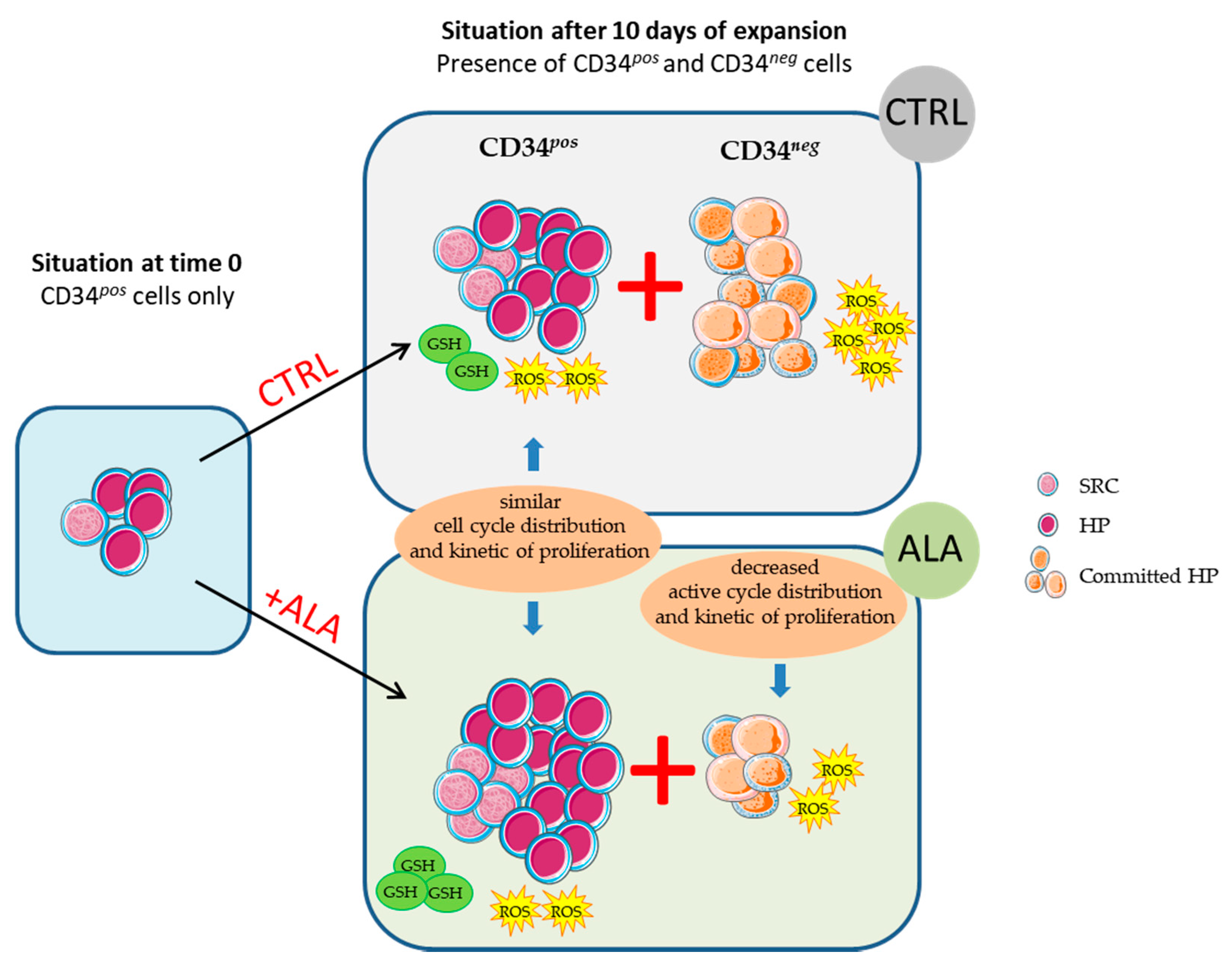
3.1. ALA Allows Expansion of Phenotypically-Defined Primitive Cells

We preliminary tested the effect of 30 to 250 µM ALA concentrations on the ex vivo expansion of SSPB CD34pos cells over 10 days. We observed a dose-dependent decrease in the total cell expansion in the presence of ALA (Figure 1a). This event was not due to apoptosis as we observed only a slight induction of apoptosis even at the highest ALA dose of 250 µM (9.1 ± 1.4% AnnexinV+/PIneg and 4.5 ± 0.8% AnnexinV+/PI+) compared to the control condition (4.9 ± 1.5% AnnexinV+/PIneg and 2.2 ± 0.3% AnnexinV+/PI+) (Figure 1b).
Interestingly, focusing on the criteria used to qualify hematopoietic graft in the context of cell therapy, i.e., CD34pos cells, we demonstrated a significant increased expansion of this primitive CD34pos population, reaching a peak after 10 days for 50 µM, 100 µM, and 250 µM ALA with 55%, 66%, and 52%, respectively, increased expansion compared to the control condition (Figure 2).
The following analyses were conducted at 100 µM ALA, which provided the highest CD34pos cell expansion. We next performed a deeper phenotypic analysis combining CD45RA, CD133, and CD34, which allowed for detecting the HSC/multipotent progenitors (HSC/MPP; CD34pos, CD45RAneg, and CD133pos), lymphoid-primed multipotent progenitors (LMP; CD34pos, CD45RApos, and CD133pos), and erythro-myeloid progenitors (EMP; CD34pos, CD45RAneg, and CD133low) [29]. The expansion of HSC/MMPs peaked at D7 of the ex vivo culture (Figure 2a), while the expansion of EMP (Figure 2b) and LMP (Figure 2c) gradually increased until D10. ALA induced a significant increase in the expansion of these three hematopoietic lineages compared to the control condition (Figure 3a–c), confirming the ALA capacity to ex vivo preserve hematopoietic cell primitiveness.

3.2. ALA Increases SRC Frequency after 10 Days of Culture

We next analyzed the effects of ALA on cells retaining the ability to reconstitute hematopoiesis in vivo, i.e., on SRC. The frequency of SRC was evaluated through injection of the total progeny of 10,000, 5000, and 2500 time 0-SSPB CD34pos cells after 10 days of culture. As shown in Figure 4, we first of all confirmed previous data [8] showing that ex vivo culture of SSPB CD34pos cells increased the SSPB repopulation potential, as we observed a significantly higher hCD45/mCD45 ratio in the mice infused with D10 control-expanded cells compared to the freshly isolated cells (Figure 4a). In this context, ALA-treated cultures exhibited a trend toward an increased repopulation potential compared to the control condition-expanded cells. Notably, the progeny of 10,000 time 0 cells provided a hCD45/mCD45 median chimerism of 0.808% vs. 0.352% for the ALA and control conditions, respectively (Figure 4a). We then performed a limiting dilution assay through the injection of different cell doses to mice (Figure 4b), and estimated SRC frequencies thanks to the ELDA webtool in each condition (Table 1). In this context, we obtained SRC frequencies of 1/19,390 for time 0 SSPB CD34pos cells, 1/3796 for D10 control-expanded cells, and 1/2485 for D10 ALA-expanded cells. Thus, the ex vivo expansion of SSPB CD34pos cells in the presence of ALA tends to improve the expansion of cells retaining a hematopoietic reconstitution potential compared to the control condition.

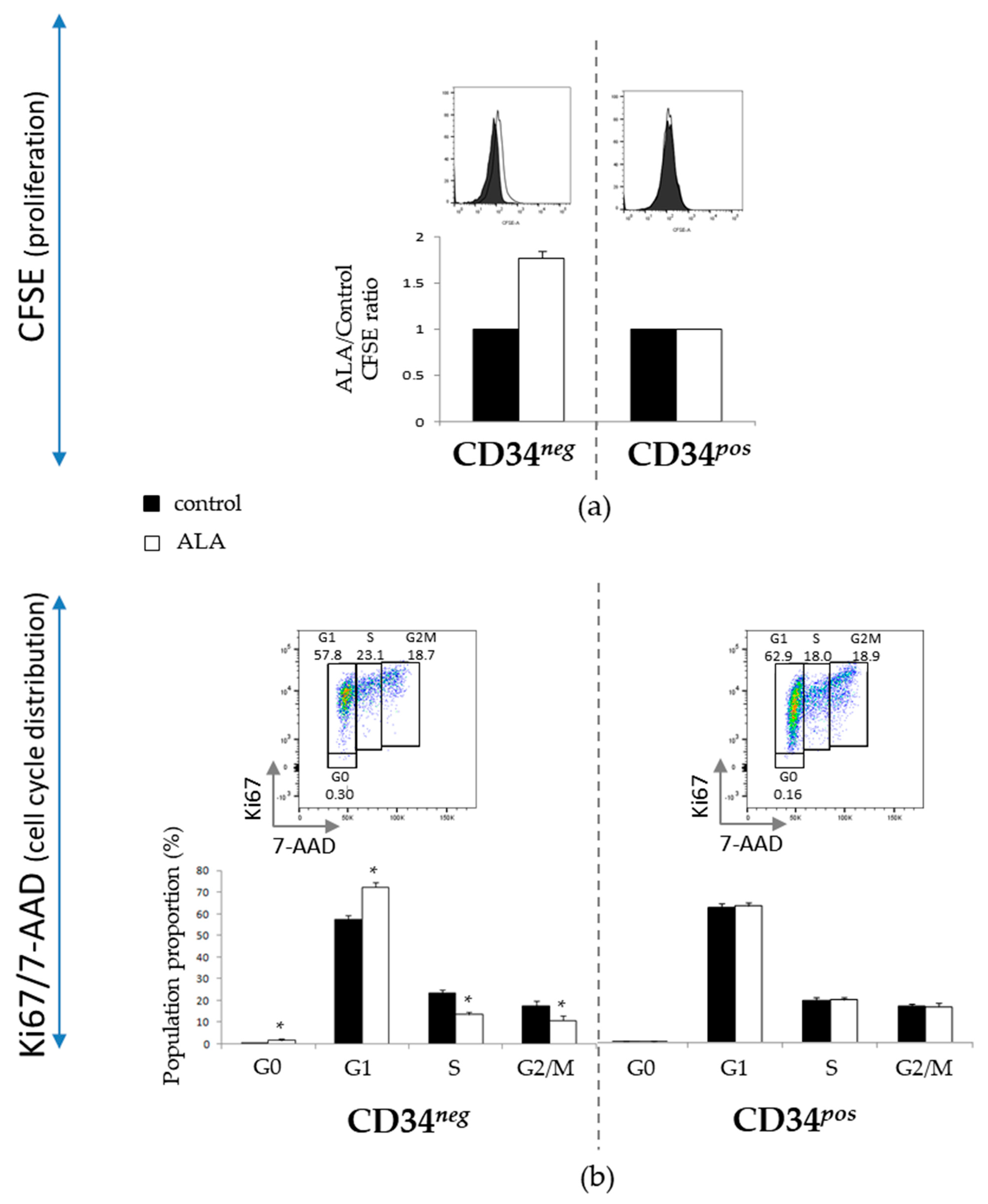
3.3. ALA Modifies Proliferation and Cell Cycle Distribution of CD34neg Population but Not of CD34pos

To gain insight into the cellular mechanisms implied in primitive hematopoietic cell preservation by ALA, we analyzed the proliferation kinetic (number of cell divisions) and cell cycle distribution by CFSE and Ki67/7AAD labeling, respectively. We observed that after 5 days of culture, ALA induced a decreased proliferation kinetic in the CD34neg population (Figure 5a) associated with an increased proportion of cells in G0 and G1 phases at the expense of proliferative S and G2/M phases (Figure 5b). In contrast, ALA did not modify CD34pos cells’ proliferation kinetic (Figure 5a) or cell cycle distribution (Figure 5b). The total cell decrease, previously reported in Figure 1, in the presence of ALA therefore reflects the decreased expansion of the more differentiated CD34neg population, without modification of the CD34pos population proliferation’s kinetic (Figure 5a), in spite of the increased number of CD34pos cells.

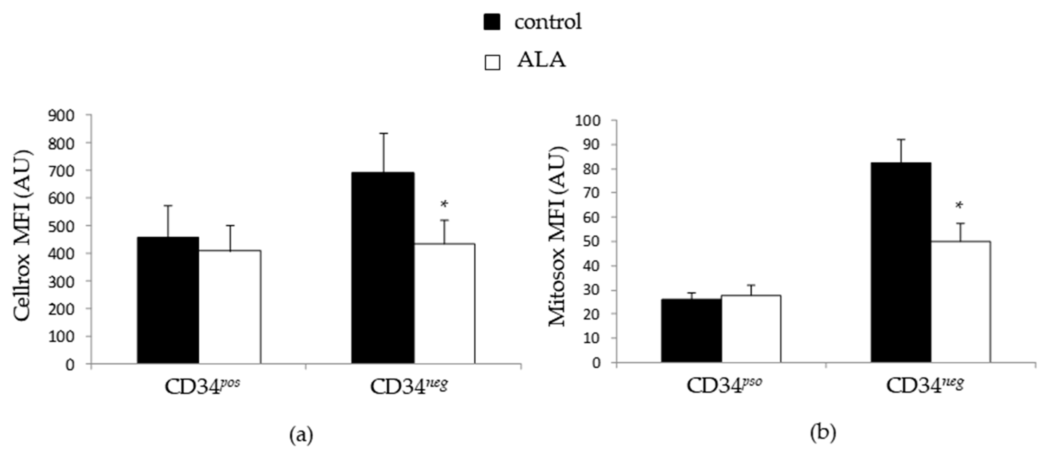
3.4. ALA Doest Not Modify Intracellularros Status in CD34pos Cells but Increases Their GSH Content

Due to the antioxidant nature of ALA and the importance of oxidative metabolism in HSC biology, we analyzed the mitochondrial and cytoplasmic ROS in the course of incubation. We observed that both the mitochondrial and cytoplasmic ROS levels were higher in the CD34neg population compared to the CD34pos one. Interestingly, the ROS analysis after 5 days of culture did not reveal any difference for cytoplasmic (Figure 6a) or mitochondrial ROS levels (Figure 6b) between the control and ALA-treated conditions in the CD34pos population, while ALA induced a marked reduction of both cytoplasmic and mitochondrial ROS levels in the CD34neg population. Consequently, the significantly higher CD34pos cell expansion that we observed in the presence of ALA seemed to be unrelated to the direct intracellular ROS-scavenging effect of ALA.
In numerous studies [24,25,30], ALA was shown to modulate the oxidative metabolism through an increase in the intracellular GSH level. To gain insight into how ALA could be involved in the redox metabolism during the ex vivo expansion of CD34pos hematopoietic cells, we analyzed the GSH content at D5 in the three primitive populations. We observed in HSC/MMP, LMP, and EMP, previously defined as a non-statistically significant but reproducible increased level of GSH (Figure 7a) in the presence of ALA.

4. Discussion

SSPB represents an interesting source of HSCs for HCT, as it is easily accessible and safe for donors. However, one of the limitations of its use is the low CD34pos cell number collected in a single apheresis. This implies developing ex vivo protocols allowing for both the expansion of hematopoietic progenitors that support rapid (but transient) hematopoietic recovery in recipients, and the maintenance/increase of HSCs ensuring a long-term reconstitution potential. Therefore, successful grafting is directly linked to the quality of the transplanted cell population, which is evaluated in clinic through the analysis of its CD34pos content.
In this study, we report the beneficial effect of ALA to significantly increase the expansion of SSPB CD34pos cells in ex vivo culture. The main results of the study are summarized in Figure 8.
ALA effects were notably observed in the most primitive CD34pos CD45RAneg CD133lowHSC/MPP population, as well as in CD34pos CD45RAneg CD133pos EMP and CD34pos CD45RApos CD133pos LMP committed progenitors. The SRC assay analysis was in line with these results and showed that ALA not only increased CD34pos cells, but also concomitantly preserved, and even extended, the engraftment potential after 10 days of ex vivo culture. Interestingly, our results showed that in SSPB, the effect of ALA is higher on MPP than on SRC, highlighting the potential interest in the use of ALA during cell preparation so as to achieve rapid recovery in patients thanks to increased HP numbers, without losing the potential of long-term recovery ensured by SRC. These data supplement previous work on human pluripotent stem cells (hPSCs) and cord blood cells, which showed that ALA was able to favor the regeneration of hematopoietic stem/progenitor cells derived from hPSCs, and better maintain the in vivo reconstitution potential of cord blood cells after ex vivo culture [27]. In view of deciphering the mechanism involved in such an effect, we then observed that ALA decreased the proliferation of the differentiated CD34neg population that accumulated in the G0/G1 phases at the expense of the S and G2M phase. The antiproliferative effect of exogenous ALA in ex vivo culture has been previously highlighted in numerous studies [31,32,33], and is often associated with a decrease in ROS content [27,34,35]. This is consistent with the need for a physiological increase in ROS for cell cycle progression from the G1 to S phase, already described [36]. Here, ALA did not modify the cell cycle distribution or cell division number in the CD34pos population, sustaining that ALA did not stimulate the proliferation kinetic of primitive cells but rather affected the nature of cell division and consequently oriented cell fate decision towards self-renewal through pathways that need to be dissected.
Considering the importance of redox status in proliferation/quiescence and differentiation/self-renewal balances of hematopoietic cells [37,38], we next investigated the cytoplasmic and mitochondrial ROS levels in the course of the culture. Our data showed that ALA differentially affected hematopoietic cells according to their differentiation state. We observed indeed an antioxidant effect of ALA on the CD34neg population, with a decrease of both mitochondrial and cytoplasmic ROS levels, while ALA did not affect ROS status in the more primitive CD34pos population. These observations were consistent with the aforementioned effect of ALA on cell proliferation. Indeed, the diminution of ROS observed in the presence of ALA in the CD34neg population was in line with its decreased proliferation through cell cycle reduction, while the unmodified proliferation of primitive hematopoietic CD34pos cells matched with the equal ROS content observed between the control and ALA condition. In addition, the lower ROS content in CD34pos compared to the CD34neg population was coherent with the low metabolic activity observed in the most primitive hematopoietic cells, while differentiation induced metabolic changes, including an increase of oxidative phosphorylation accompanied by a rise of ROS level [39]. Taken together, we observed that ALA slowed down the expansion of SSPB differentiating hematopoietic cells through ROS attenuation and favored primitive hematopoietic expansion in ex vivo culture in a ROS-independent mechanism. The differential effects of ALA on ROS content depending on hematopoietic differentiation state is of particular interest and needs further investigations.
While ALA can display its antioxidant effect through direct ROS scavenging, the recycling of the intracellular antioxidant, in particular GSH, is also reported [24,25,30]. To address in better detail the question of the mode of action of ALA in our expansion model, we determined the intracellular GSH content in the course of the culture and showed that ALA reproducibly increased GSH level in all CD34pos subpopulations, namely HSC/MPP, LMP, and EMP. The GSH-GPx system was shown to be essential for HSC preservation through a reduction of peroxydation [40], a fact that could explain the increased primitive hematopoietic cell expansion observed in our study. Several pathways were highlighted to induce GSH rise, including Nrf2 activation [24,25] and the increased intracellular availability of the reduced form of cysteine [30]. The mechanisms implied in GSH rise observed in our study remain to be elucidated.
Overall, further experiments are needed to improve the understanding of the ability of ALA to favor HSC/MPP self-renewal and therefore to expand primitive SSPB hematopoietic cells ex vivo. However, ALA represents an indisputable interesting molecule to increase the CD34pos cell content in hematopoietic grafts. Therefore, ALA could be used for therapeutic purposes in the ex vivo culture of SSPB primitive cells, potentially in combination with other described molecules, in the context of HCT.

Author Contributions

Conceptualization, P.B.d.l.G. and C.D.; methodology, C.D. and P.B.d.l.G.; investigation, C.D., M.A., P.B.d.l.G., M.H., L.R. and P.D.; data curation, C.D.; writing—original draft preparation, C.D.; writing—review and editing, P.B.d.l.G., C.D., M.V.-L. and Z.I.; supervision, P.B.d.l.G.; project administration, P.B.d.l.G.; funding acquisition, P.B.d.l.G. All authors have read and agreed to the published version of the manuscript.

Funding

This project was supported by the annual research budget of Etablissement Français du Sang-Nouvelle Aquitaine (EFS-NVAQ). All the expenses regarding the study were authorized by financial manager of EFS-NVAQ.

Institutional Review Board Statement

The use of human cells for the purpose of scientific research in our laboratory was approved by the Ministry of Education and Research of the French Republic in compliance with the regulatory procedures that apply (DC 2013-1998, DC 2018-3143, and DC 2019-3713). The animal experiments presented in this paper were authorized by the Ethical Committee of Gironde (CEEA50, 2017/03/30), France (ref. APAFlS#3945-2016020211179892 v4). All animal experiments were performed in agreement with French regulation—licenses granted to C.D., Z.I., and P.B.d.l.G. through nos. 33 07 010, B33 11 005, and A33 12 054, respectively.

Informed Consent Statement

Informed consent was obtained from all subjects whose cells were involved in the study, in compliance with the regulatory procedures that apply and the Ministry of Education authorizations (DC 2018-3143 and DC 2019-3713).

Data Availability Statement

All data are conserved at the internal server of EFS-NVAQ Bordeaux and are available upon request.

Acknowledgments

We are grateful to Jean-François Simoes for technical support by providing us leukoreduction filters. Cell sorting was performed in the TBMCore platform in Bordeaux University, and we thank Atika Zouine and Vincent Pitard for technical assistance at the Flow cytometry facility, CNRS UMS 3427, INSERM US 005, University of Bordeaux, F-33000 Bordeaux, France. We thank Benoit Rousseau and all the staff of the Animalerie A2, Service Commun des Animaleries, University of Bordeaux, F-33000 Bordeaux, France, for animal housing.

Conflicts of Interest

The authors declare no conflict of interest.

References

  1. Kessinger, A.; Armitage, J.O.; Landmark, J.D.; Weisenburger, D.D. Reconstitution of Human Hematopoietic Function with Autologous Cryopreserved Circulating Stem Cells. Exp. Hematol. 1986, 14, 192–196. [Google Scholar] [PubMed]
  2. Duhrsen, U.; Villeval, J.; Boyd, J.; Kannourakis, G.; Morstyn, G.; Metcalf, D. Effects of Recombinant Human Granulocyte Colony-Stimulating Factor on Hematopoietic Progenitor Cells in Cancer Patients. Blood 1988, 72, 2074–2081. [Google Scholar] [CrossRef] [PubMed] [Green Version]
  3. Socinski, M.; Elias, A.; Schnipper, L.; Cannistra, S.; Antman, K.; Griffin, J. Granulocyte-Macrophage Colony Stimulating Factor Expands the Circulating Haemopoietic Progenitor Cell Compartment in Man. Lancet 1988, 331, 1194–1198. [Google Scholar] [CrossRef]
  4. Haas, R.; Ho, A.D.; Bredthauer, U.; Cayeux, S.; Egerer, G.; Knauf, W.; Hunstein, W. Successful Autologous Transplantation of Blood Stem Cells Mobilized with Recombinant Human Granulocyte-Macrophage Colony-Stimulating Factor. Exp. Hematol. 1990, 18, 94–98. [Google Scholar] [PubMed]
  5. Pulsipher, M.A.; Chitphakdithai, P.; Miller, J.P.; Logan, B.R.; King, R.J.; Rizzo, J.D.; Leitman, S.F.; Anderlini, P.; Haagenson, M.D.; Kurian, S.; et al. Adverse Events among 2408 Unrelated Donors of Peripheral Blood Stem Cells: Results of a Prospective Trial from the National Marrow Donor Program. Blood 2009, 113, 3604–3611. [Google Scholar] [CrossRef] [PubMed]
  6. Udomsakdi, C.; Lansdorp, P.M.; Hogge, D.E.; Reid, D.S.; Eaves, A.C.; Eaves, C.J. Characterization of Primitive Hematopoietic Cells in Normal Human Peripheral Blood. Blood 1992, 80, 2513–2521. [Google Scholar] [CrossRef] [Green Version]
  7. Hirayama, F.; Yamaguchi, M.; Yano, M.; Yasui, K.; Horie, Y.; Matsumoto, K.; Nagao, N.; Ikebuchi, K.; Azuma, H.; Ikeda, H.; et al. Spontaneous and Rapid Reexpression of Functional CXCR4 by Human Steady-State Peripheral Blood CD34+ Cells. Int. J. Hematol. 2003, 78, 48–55. [Google Scholar] [CrossRef]
  8. Brunet de la Grange, P.; Vlaski, M.; Duchez, P.; Chevaleyre, J.; Lapostolle, V.; Boiron, J.-M.; Praloran, V.; Ivanovic, Z. Long-Term Repopulating Hematopoietic Stem Cells and “Side Population” in Human Steady State Peripheral Blood. Stem Cell Res. 2013, 11, 625–633. [Google Scholar] [CrossRef] [Green Version]
  9. Lapostolle, V.; Chevaleyre, J.; Duchez, P.; Rodriguez, L.; Vlaski-Lafarge, M.; Sandvig, I.; Brunet de la Grange, P.; Ivanovic, Z. Repopulating Hematopoietic Stem Cells from Steady-State Blood before and after Ex Vivo Culture Are Enriched in the CD34+ CD133+ CXCR4 low Fraction. Haematologica 2018, 103, 1604–1615. [Google Scholar] [CrossRef] [Green Version]
  10. Bourdieu, A.; Avalon, M.; Lapostolle, V.; Ismail, S.; Mombled, M.; Debeissat, C.; Guérinet, M.; Duchez, P.; Chevaleyre, J.; Vlaski-Lafarge, M.; et al. Steady State Peripheral Blood Provides Cells with Functional and Metabolic Characteristics of Real Hematopoietic Stem Cells. J. Cell. Physiol. 2018, 233, 338–349. [Google Scholar] [CrossRef]
  11. Zimran, E.; Papa, L.; Hoffman, R. Ex Vivo Expansion of Hematopoietic Stem Cells: Finally Transitioning from the Lab to the Clinic. Blood Rev. 2021, 50, 100853. [Google Scholar] [CrossRef] [PubMed]
  12. Simsek, T.; Kocabas, F.; Zheng, J.; DeBerardinis, R.J.; Mahmoud, A.I.; Olson, E.N.; Schneider, J.W.; Zhang, C.C.; Sadek, H.A. The Distinct Metabolic Profile of Hematopoietic Stem Cells Reflects Their Location in a Hypoxic Niche. Cell Stem Cell 2010, 7, 380–390. [Google Scholar] [CrossRef] [PubMed] [Green Version]
  13. Vannini, N.; Girotra, M.; Naveiras, O.; Nikitin, G.; Campos, V.; Giger, S.; Roch, A.; Auwerx, J.; Lutolf, M.P. Specification of Haematopoietic Stem Cell Fate via Modulation of Mitochondrial Activity. Nat. Commun. 2016, 7, 13125. [Google Scholar] [CrossRef] [PubMed]
  14. Spencer, J.A.; Ferraro, F.; Roussakis, E.; Klein, A.; Wu, J.; Runnels, J.M.; Zaher, W.; Mortensen, L.J.; Alt, C.; Turcotte, R.; et al. Direct Measurement of Local Oxygen Concentration in the Bone Marrow of Live Animals. Nature 2014, 508, 269–273. [Google Scholar] [CrossRef] [Green Version]
  15. Ito, K.; Hirao, A.; Arai, F.; Matsuoka, S.; Takubo, K.; Hamaguchi, I.; Nomiyama, K.; Hosokawa, K.; Sakurada, K.; Nakagata, N.; et al. Regulation of Oxidative Stress by ATM Is Required for Self-Renewal of Haematopoietic Stem Cells. Nature 2004, 431, 997–1002. [Google Scholar] [CrossRef]
  16. Ito, K.; Hirao, A.; Arai, F.; Takubo, K.; Matsuoka, S.; Miyamoto, K.; Ohmura, M.; Naka, K.; Hosokawa, K.; Ikeda, Y.; et al. Reactive Oxygen Species Act through P38 MAPK to Limit the Lifespan of Hematopoietic Stem Cells. Nat. Med. 2006, 12, 446–451. [Google Scholar] [CrossRef]
  17. Tothova, Z.; Kollipara, R.; Huntly, B.J.; Lee, B.H.; Castrillon, D.H.; Cullen, D.E.; McDowell, E.P.; Lazo-Kallanian, S.; Williams, I.R.; Sears, C.; et al. FoxOs Are Critical Mediators of Hematopoietic Stem Cell Resistance to Physiologic Oxidative Stress. Cell 2007, 128, 325–339. [Google Scholar] [CrossRef] [Green Version]
  18. Chen, C.; Liu, Y.; Liu, R.; Ikenoue, T.; Guan, K.-L.; Liu, Y.; Zheng, P. TSC–MTOR Maintains Quiescence and Function of Hematopoietic Stem Cells by Repressing Mitochondrial Biogenesis and Reactive Oxygen Species. J. Exp. Med. 2008, 205, 2397–2408. [Google Scholar] [CrossRef] [Green Version]
  19. Tsai, J.J.; Dudakov, J.A.; Takahashi, K.; Shieh, J.-H.; Velardi, E.; Holland, A.M.; Singer, N.V.; West, M.L.; Smith, O.M.; Young, L.F.; et al. Nrf2 Regulates Haematopoietic Stem Cell Function. Nat. Cell Biol. 2013, 15, 309–316. [Google Scholar] [CrossRef]
  20. Gupta, R.; Karpatkin, S.; Basch, R.S. Hematopoiesis and Stem Cell Renewal in Long-Term Bone Marrow Cultures Containing Catalase. Blood 2006, 107, 1837–1846. [Google Scholar] [CrossRef] [Green Version]
  21. Li, Y.; He, M.; Zhang, W.; Yang, M.; Ding, Y.; Xu, S.; Gu, J.; Li, Y.; Yin, J.; Gao, Y. Antioxidant Small Molecule Compound Chrysin Promotes the Self-Renewal of Hematopoietic Stem Cells. Front. Pharmacol. 2020, 11, 399. [Google Scholar] [CrossRef] [PubMed]
  22. Fayez, A.M.; Zakaria, S.; Moustafa, D. Alpha Lipoic Acid Exerts Antioxidant Effect via Nrf2/HO-1 Pathway Activation and Suppresses Hepatic Stellate Cells Activation Induced by Methotrexate in Rats. Biomed. Pharmacother. 2018, 105, 428–433. [Google Scholar] [CrossRef] [PubMed]
  23. Lee, J.; Jung, S.-Y.; Yang, K.-J.; Kim, Y.; Lee, D.; Lee, M.H.; Kim, D.-K. α-Lipoic Acid Prevents against Cisplatin Cytotoxicity via Activation of the NRF2/HO-1 Antioxidant Pathway. PLoS ONE 2019, 14, e0226769. [Google Scholar] [CrossRef] [PubMed]
  24. Gu, L.; Li, S.; Bai, J.; Zhang, Q.; Han, Z. A-Lipoic Acid Protects against Microcystin-LR Induced Hepatotoxicity through Regeneration of Glutathione via Activation of Nrf2. Environ. Toxicol. 2020, 35, 738–746. [Google Scholar] [CrossRef] [PubMed]
  25. Shi, C.; Zhou, X.; Zhang, J.; Wang, J.; Xie, H.; Wu, Z. α-Lipoic Acid Protects against the Cytotoxicity and Oxidative Stress Induced by Cadmium in HepG2 Cells through Regeneration of Glutathione by Glutathione Reductase via Nrf2/ARE Signaling Pathway. Environ. Toxicol. Pharmacol. 2016, 45, 274–281. [Google Scholar] [CrossRef]
  26. Song, G.; Liu, Z.; Wang, L.; Shi, R.; Chu, C.; Xiang, M.; Tian, Q.; Liu, X. Protective Effects of Lipoic Acid against Acrylamide-Induced Neurotoxicity: Involvement of Mitochondrial Energy Metabolism and Autophagy. Food Funct. 2017, 8, 4657–4667. [Google Scholar] [CrossRef]
  27. Dong, Y.; Bai, J.; Zhang, Y.; Zhou, Y.; Pan, X.; Li, X.; Zhou, Q.; Chen, Y.; Lai, M.; Mao, B.; et al. Alpha Lipoic Acid Promotes Development of Hematopoietic Progenitors Derived from Human Embryonic Stem Cells by Antagonizing ROS Signals. J. Leukoc. Biol. 2020, 108, 1711–1725. [Google Scholar] [CrossRef]
  28. Hu, Y.; Smyth, G.K. ELDA: Extreme Limiting Dilution Analysis for Comparing Depleted and Enriched Populations in Stem Cell and Other Assays. J. Immunol. Methods 2009, 347, 70–78. [Google Scholar] [CrossRef]
  29. Radtke, S.; Görgens, A.; Kordelas, L.; Schmidt, M.; Kimmig, K.R.; Köninger, A.; Horn, P.A.; Giebel, B. CD133 Allows Elaborated Discrimination and Quantification of Haematopoietic Progenitor Subsets in Human Haematopoietic Stem Cell Transplants. Br. J. Haematol. 2015, 169, 868–878. [Google Scholar] [CrossRef]
  30. Han, D.; Handelman, G.; Marcocci, L.; Sen, C.K.; Roy, S.; Kobuchi, H.; Tritschler, H.J.; Flohé, L.; Packer, L. Lipoic Acid Increases de Novo Synthesis of Cellular Glutathione by Improving Cystine Utilization. BioFactors 1997, 6, 321–338. [Google Scholar] [CrossRef]
  31. van de Mark, K.; Chen, J.S.; Steliou, K.; Perrine, S.P.; Faller, D.V. α-Lipoic Acid Induces P27Kip-Dependent Cell Cycle Arrest in Non-Transformed Cell Lines and Apoptosis in Tumor Cell Lines. J. Cell. Physiol. 2003, 194, 325–340. [Google Scholar] [CrossRef] [PubMed]
  32. Yang, L.; Wen, Y.; Lv, G.; Lin, Y.; Tang, J.; Lu, J.; Zhang, M.; Liu, W.; Sun, X. α-Lipoic Acid Inhibits Human Lung Cancer Cell Proliferation through Grb2-Mediated EGFR Downregulation. Biochem. Biophys. Res. Commun. 2017, 494, 325–331. [Google Scholar] [CrossRef] [PubMed]
  33. Kim, K.-H.; Lee, B.; Kim, Y.-R.; Kim, M.-A.; Ryu, N.; Jung, D.J.; Kim, U.-K.; Baek, J.-I.; Lee, K.-Y. Evaluating Protective and Therapeutic Effects of Alpha-Lipoic Acid on Cisplatin-Induced Ototoxicity. Cell Death Dis. 2018, 9, 827. [Google Scholar] [CrossRef] [PubMed]
  34. Baeeri, M.; Bahadar, H.; Rahimifard, M.; Navaei-Nigjeh, M.; Khorasani, R.; Rezvanfar, M.A.; Gholami, M.; Abdollahi, M. α-Lipoic Acid Prevents Senescence, Cell Cycle Arrest, and Inflammatory Cues in Fibroblasts by Inhibiting Oxidative Stress. Pharmacol. Res. 2019, 141, 214–223. [Google Scholar] [CrossRef]
  35. Dozio, E.; Ruscica, M.; Passafaro, L.; Dogliotti, G.; Steffani, L.; Pagani, A.; Demartini, G.; Esposti, D.; Fraschini, F.; Magni, P. The Natural Antioxidant Alpha-Lipoic Acid Induces P27Kip1-Dependent Cell Cycle Arrest and Apoptosis in MCF-7 Human Breast Cancer Cells. Eur. J. Pharmacol. 2010, 641, 29–34. [Google Scholar] [CrossRef]
  36. Havens, C.G.; Ho, A.; Yoshioka, N.; Dowdy, S.F. Regulation of Late G1 /S Phase Transition and APCCdh1 by Reactive Oxygen Species. Mol. Cell. Biol. 2006, 26, 4701–4711. [Google Scholar] [CrossRef] [Green Version]
  37. Bigarella, C.L.; Liang, R.; Ghaffari, S. Stem Cells and the Impact of ROS Signaling. Development 2014, 141, 4206–4218. [Google Scholar] [CrossRef] [Green Version]
  38. Papa, L.; Djedaini, M.; Hoffman, R. Mitochondrial Role in Stemness and Differentiation of Hematopoietic Stem Cells. Stem Cells Int. 2019, 2019, 4067162. [Google Scholar] [CrossRef] [Green Version]
  39. Tan, D.Q.; Suda, T. Reactive Oxygen Species and Mitochondrial Homeostasis as Regulators of Stem Cell Fate and Function. Antioxid. Redox Signal. 2018, 29, 149–168. [Google Scholar] [CrossRef]
  40. Fan, J.; Cai, H.; Yang, S.; Yan, L.; Tan, W. Comparison between the Effects of Normoxia and Hypoxia on Antioxidant Enzymes and Glutathione Redox State in Ex Vivo Culture of CD34+ Cells. Comp. Biochem. Physiol. Part B Biochem. Mol. Biol. 2008, 151, 153–158. [Google Scholar] [CrossRef]
Figure 1. ALA decreases the total cell expansion of SSPB CD34pos cells over 10 days of ex vivo culture. Ex vivo culture of SSPB CD34pos were performed in the presence of 0, 30, 50, 100, or 250 µM ALA over 10 days. (a) Total cell growth of SSPB CD34pos was evaluated from D6 to D10 by trypan blue exclusion, and expressed as fold expansion relative to time 0 (n = 4–6). (b) Apoptosis analyzed by flow cytometry after AnnV/PI labeling at D5 (n = 4). * p < 0.05.
Biomolecules 12 00431 g001
Figure 2. ALA specifically increases the expansion of the CD34pos compartment during the ex vivo culture of SSPB CD34pos cells. SSPB CD34pos cells were cultured in the presence of 0, 30, 50, 100, or 250 µM ALA. The CD34pos cell content is evaluated from D6 to D10 by flow cytometry after CD34 labeling, and is expressed as fold expansion relative to time 0 (n = 4–6). * p < 0.05.
Biomolecules 12 00431 g002
Figure 3. ALA increases the expansion of phenotypically defined primitive hematopoietic populations during ex vivo culture. SSPB CD34pos cells cultured for 10 days in the presence of 0 or 100 µM ALA. Analysis of (a) HSC/MPP, (b) EMP, and (c) LMP populations assessed at D6, D7, D8, D9, and D10 through a combination of CD45RA, CD34, and CD133 labeling and flow cytometry analysis, and expressed as fold expansion relative to time 0 (n = 6). * p < 0.05.
Biomolecules 12 00431 g003
Figure 4. ALA favors the expansion of cells retaining a hematopoietic reconstitution potential. Freshly isolated time 0 or 10 day-expanded 10,000 SSPB CD34pos cells in the absence or presence of 100 µM ALA injected in busulfan-conditioned mice and BM cells analyzed 12 weeks after injection by flow cytometry after mCD45/hCD45 co-labeling. (a) Mouse vs. human chimerism; * p < 0.05, *** p < 0.0005. (b) Percentages of negative mice plotted as a function of the cell dose injected.
Biomolecules 12 00431 g004
Figure 5. ALA does not modify the proliferation or cell cycle repartition of ex vivo expanded SSPB CD34pos cells (n = 5). (a) Analysis of proliferation in CD34pos and CD34neg populations. SSPB CD34pos cells labeled with CFSE at time 0 and cultured for 5 days in the absence or the presence of 100 µM ALA. The proliferation of expanded CD34neg and CD34pos cells evaluated by flow cytometry after CD34 labeling (n = 3); upper panel: representative experiment for CD34neg and CD34pos populations. (b) Cell cycle performed by flow cytometry after CD34/Ki67/7AAD co-labeling (n = 5); upper panels: representative experiments. * p < 0.05.
Biomolecules 12 00431 g005
Figure 6. ALA does not modify the mitochondrial and cytoplasmic ROS level in the CD34pos-expanded population. SSPB CD34pos were cultured for 5 days in the absence or the presence of 100 µM ALA, and analyzed for (a) cytoplasmic ROS content by flow cytometry following CellROX labeling (n = 5) or (b) mitochondrial ROS content by flow cytometry following MitoSOX labeling (n = 4). * p < 0.05.
Biomolecules 12 00431 g006
Figure 7. ALA increases the GSH content in HSC/MPP, LMP, and EMP populations. SSPB CD34pos cultured for 5 days in the absence or the presence of 100 µM ALA, and FACS-sorted for each CD34pos cell phenotype and analyzed for the GSH content.
Biomolecules 12 00431 g007
Figure 8. Overall representation of the main results of the study.
Biomolecules 12 00431 g008
Table 1. ALA increases the SRC frequencies of ex vivo expanded SSPB CD34pos. Freshly isolated or the progeny of 10 days-expanded 10,000, 5000, and 2500 SSPB CD34pos cells, in the absence or the presensence of 100 µM ALA, were injected in busulfan-conditioned mice. BM hCD45 chimerism analyzed 12 weeks after injection by flow cytometry, after mCD45/hCD45 co-labeling and SRC frequencies calculated with ELDA webtool.
SRC Frequency (1/…)Pairwise Test (ELDA Webtool)
GroupLowerEstimateUpperGroup 1Group 2ChisqDFPr (>Chisq)
Time 064,24726,73611,126ControlTime 016.614.51 × 10−5
Control664337962169ALATime 023.211.44 × 10−6
ALA459624851343ALAControl0.97210.324
Publisher’s Note: MDPI stays neutral with regard to jurisdictional claims in published maps and institutional affiliations.

