Review

Endoplasmic Reticulum Stress in Chronic Obstructive Pulmonary Disease: Mechanisms and Future Perspectives

Department of Respiratory Medicine, Beijing Friendship Hospital, Capital Medical University, Beijing 100050, China
*
Authors to whom correspondence should be addressed.
Contributed equally and should be considered co-first authors.
Biomolecules 2022, 12(11), 1637; https://doi.org/10.3390/biom12111637
Submission received: 12 October 2022 / Revised: 31 October 2022 / Accepted: 1 November 2022 / Published: 4 November 2022
(This article belongs to the Topic Cell Signaling Pathways)

Abstract

:
The endoplasmic reticulum (ER) is an integral organelle for maintaining protein homeostasis. Multiple factors can disrupt protein folding in the lumen of the ER, triggering ER stress and activating the unfolded protein response (UPR), which interrelates with various damage mechanisms, such as inflammation, apoptosis, and autophagy. Numerous studies have linked ER stress and UPR to the progression of chronic obstructive pulmonary disease (COPD). This review focuses on the mechanisms of other cellular processes triggered by UPR and summarizes drug intervention strategies targeting the UPR pathway in COPD to explore new therapeutic approaches and preventive measures for COPD.

Graphical Abstract

1. Introduction

Eukaryotic cells depend primarily on the endoplasmic reticulum (ER) for the biosynthesis, folding, and modification of membrane-bound and secreted proteins in eukaryotic cells. Generally, cells can maintain a dynamic equilibrium between protein biosynthesis and folding, a process known as ER homeostasis [1]. ER stress is generated by an abnormal accumulation of misfolded proteins in the ER, which occurs when various factors disrupt the ER homeostasis. These physiological and pathological factors include oxidative stress, nutrient deprivation, ischemia, hypoxia, glucose deprivation, viral infections, and a loss of calcium homeostasis. The cellular processes that defend against ER stress and cope with the accumulation of unfolded and misfolded proteins are collectively termed unfolded protein response (UPR) [2,3]. By boosting the folding capacity of ER proteins and enlarging the ER compartment size, UPR activation reverses the process of ER stress. Moreover, UPR activation promotes ER-associated protein degradation to eliminate misfolded proteins and restore ER homeostasis [4,5]. If these protective processes fail to restore proteostasis, prolonged or severe UPR activation leads to ER functional damage and initiates proapoptotic pathways leading to cell death [6,7].
A surge of evidence has emerged linking the pathology of various diseases to ER stress and UPR [8,9]. In recent years, an increasing number of studies have concentrated on the involvement of ER stress in the pathophysiology of chronic obstructive pulmonary disease (COPD). Numerous reports have denoted that ER stress is one of the main reasons for the early development of COPD and the leading cause of apoptosis of alveolar epithelial cells [10,11,12]. Pharmacological agents that reduce ER stress or inhibit specific signaling molecules in the UPR pathways have demonstrated some therapeutic advantages in animal models of COPD [6]. In this review, we highlight the link between the regulatory mechanisms of the three branches of UPR and COPD and summarize recent pharmacological intervention strategies targeting the UPR pathway in COPD. In addition, we discuss the potential links between UPR and the pathogenesis of, and treatment strategies for, COPD.

2. The Function of the ER

The ER is a continuous membrane system composed of tubules and sheets that extend from the nuclear membrane throughout the cell [13]. The ER is required for the synthesis, folding, and transport of secretory and membrane proteins, as well as lipid and steroid synthesis, carbohydrate metabolism, and calcium storage [14].
Approximately 30% of proteins are synthesized in the ER [15], and ER membrane-associated ribosomes translate and transfer proteins into the lumen of the ER and fold proteins into their unique three-dimensional structures [16]. The proteins destined for secretion must be appropriately folded and undergo various post-translational modifications with the aid of chaperones and folding enzymes. These modifications involve glycosylation, disulfide bond formation, and oligomerization [14]. Moreover, the ER has a significantly greater calcium content and a higher oxidizing redox potential than the cytosol [17,18], which facilitates proper folding, modification, and assembly of secreted proteins. However, even with several proteins and complexes specializing in adequate folding, some proteins still do not acquire the correct structural and functional forms and are misfolded. A portion of incompletely folded or misfolded proteins can be refolded by binding to calnexin after glycosylation modification, while unmodifiable unfolded proteins can be transported back to the cytoplasm through the ER-associated degradation (ERAD) pathway. Subsequent ubiquitination and degradation by the 26S proteasome ensure that abnormal peptides do not inadvertently enter the secretory pathway and affect cell function [19,20].

3. UPR Signaling

Protein folding in the ER can be influenced by a variety of stresses, and ER stress can occur when the amount of protein that has to be folded exceeds the ER’s ability to fold it [21]. To ensure proper protein folding and maintenance of the ER function, ER stress triggers a series of cellular signaling pathways, called the UPR, to reprogram gene transcription, mRNA translation, and protein modifications to eliminate unfolded or misfolded proteins and restore protein homeostasis [22,23].
In mammalian cells, the UPR is primarily initiated by three ER transmembrane protein sensors, namely inositol-requiring enzyme 1 (IRE1) [24], protein kinase-like ER kinase (PERK) [25], and activating transcription factor 6 (ATF6) [26]. Under homeostatic conditions, the three ER stress sensors are combined with a chaperon, binding immunoglobulin protein (BiP; also known as glucose-regulated protein 78; GRP78) [27,28]. However, when misfolded proteins accumulate in the ER, it causes GRP78 to dissociate from IRE1, PERK, and ATF6 due to the high affinity of GRP78 for the exposed hydrophobic structural domains of the misfolded proteins [29,30]. The three sensors activate their downstream signaling cascades, slowing protein translation, eventually improving the folding capacity, and restoring ER homeostasis (Figure 1). Overall, the transcriptional program of the UPR acts as a complex signaling network that implements an adaptive program through multiple signaling cascades to restore ER homeostasis.

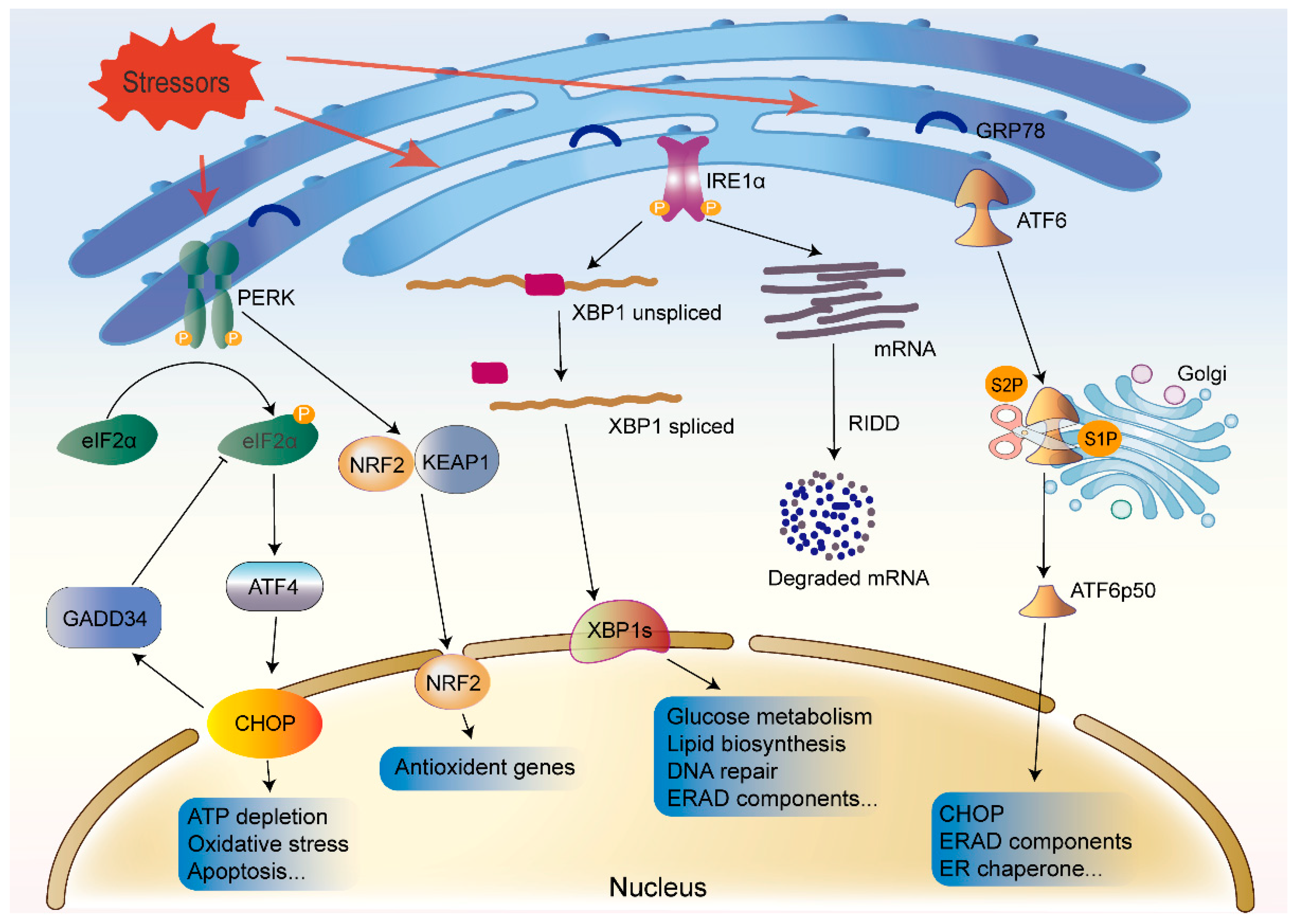
3.1. UPR Pathway Proteins

3.1.1. IRE1

IRE1 is a type I transmembrane protein with two enzymatic activities, a serine/threonine kinase and an endoribonuclease, in its cytosolic domain [31]. In humans, IRE1 exists as two homologs, IRE1α and IRE1β [32]. In the UPR signaling pathway, IRE1α is a critical sensor involved in cell fate regulation. IRE1α is widely expressed, and IRE1α knockout mice exhibit embryonic lethality [33]; meanwhile, IRE1β is abundant only in intestinal and pulmonary epithelial and mucosal epithelial tissues, and IRE1β knockout mice survive [32,34]. GRP78 dissociation triggers IRE1 oligomerization in the ER membrane, facilitating trans-autophosphorylation of the IRE1 and activation of its cytoplasmic kinase domain [35,36]. Selective cleavage of X-box binding protein 1 (XBP1) mRNA by the activated IRE1 results in the expression of a functionally active spliced isoform (XBP1s) of the transcription factor XBP1 [15,37,38]. The XBP1s then translocates to the nucleus and drives the expression of genes involved in protein trafficking, folding, and the degradation of misfolded proteins to relieve ER stress and restore homeostasis [39]. XBP1s is also involved in glucose metabolism [40], lipid biosynthesis [41], redox metabolism [42], and insulin signaling [43]. It affects cell survival, differentiation, and development [35,44]. Moreover, XBP1s with transactivation domain modulates the expression of downstream target genes by combining with specific sites, such as hypoxia-response element reporter and G protein subunit alpha 12 [45,46].
In addition, IRE1 cleaves and downregulates mRNAs and microRNAs (miRNAs) with its RNase domain through a process known as regulated IRE1-dependent decay (RIDD) [47,48,49,50]. RIDD decreases the load of nascent proteins entering the ER and is essential for maintaining ER homeostasis and cell survival. Interestingly, the RIDD activity gradually increases during periods of heightened intensity or a long duration of ER stress. By contrast, ER stress-induced XBP1 mRNA splicing is independent of the intensity and duration of ER stress [51]. Under long-term persistent ER stress, XBP1 mRNA splicing is reduced, and RIDD activity is increased, aggravating uncontrolled cell death [48,51].

3.1.2. PERK

PERK, a type I transmembrane protein kinase in the ER membrane, is activated through autophosphorylation and homodimerization after dissociating from GRP78 [25]. The activated PERK phosphorylates the downstream eukaryotic initiation factor 2α (eIF2α), which induces an overall downregulation of translation and inhibits protein synthesis [25]. Paradoxically, eIF2α phosphorylation elevates the translation of specific mRNAs with upstream open reading frames, such as transcription factor 4 (ATF4) [52]. ATF4 is a stress-induced transcription factor that induces apoptosis by initiating the transcription of C/EBP homologous protein (CHOP) under sustained stress conditions [53]. Furthermore, eIF2α phosphorylation is engaged in the translation of other ER stress-related proteins, such as those involved in growth stagnation. Growth arrest and DNA damage-inducible gene 34 (GADD34), which is positively regulated by phosphorylated eIF2α, is additionally transcriptionally induced by ATF4 and CHOP [54]. GADD34 is a modulatory subunit of the protein phosphatase 1C complex, which interacts to dephosphorylate eIF2α, thereby forming a negative feedback loop that restores protein synthesis [49]. Overall, PERK activation lessens the protein load in the ER, and, if the mechanisms involved are unable to restore ER homeostasis, PERK initiates cell death.
In addition, PERK phosphorylates nuclear factor erythroid 2-related factor 2 (NRF2) dissociates from Kelch-like ECH-associated protein 1 (KEAP1) and migrates into the nucleus. The translocation of NRF2 into the nucleus increases the production of antioxidant proteins, such as haem oxygenase 1 (HO-1), which aid in protein collapsing and restore ER homeostasis [32,55].

3.1.3. ATF6

ATF6, a type II transmembrane protein, has two configurations, ATF6α and ATF6β, which show some functional redundancy [50,56]. Thus, ATF6α or ATF6β knockout mice can survive; however, embryonic lethality occurs when both the ATF6α and ATF6β are deleted [57]. ATF6α plays a dominant role in ER stress. Upon ER stress, GRP78 dissociates from the ATF6α, exposing the Golgi localization signal of ATF6α, which leads to the translocation of the ATF6α to the Golgi apparatus. In the Golgi, the full-length form of ATF6, ATF6α (p90), is sequentially cleaved by site-1 protease (S1P) and site-2 protease (S2P), liberating the cytoplasmic ATF6α (p50) segment [58]. The activated ATF6α (p50) fragment subsequently binds to cis-acting ER stress response elements in the nucleus, thereby promoting the expression of genes encoding proteins with functions to increase the ER capacity, including GRP78, GRP94, CHOP, and ERAD components, to restore protein folding homeostasis. Among these genes, CHOP depends on ATF6α rather than ATF4 alone for the transcription under ER stress. ATF6α can also induce increased expression of XBP1 and forms a heterodimer with XBP1 to reduce ER stress and increase the levels of genes required for ERAD [50,59].

3.2. The UPR and Other Cellular Processes
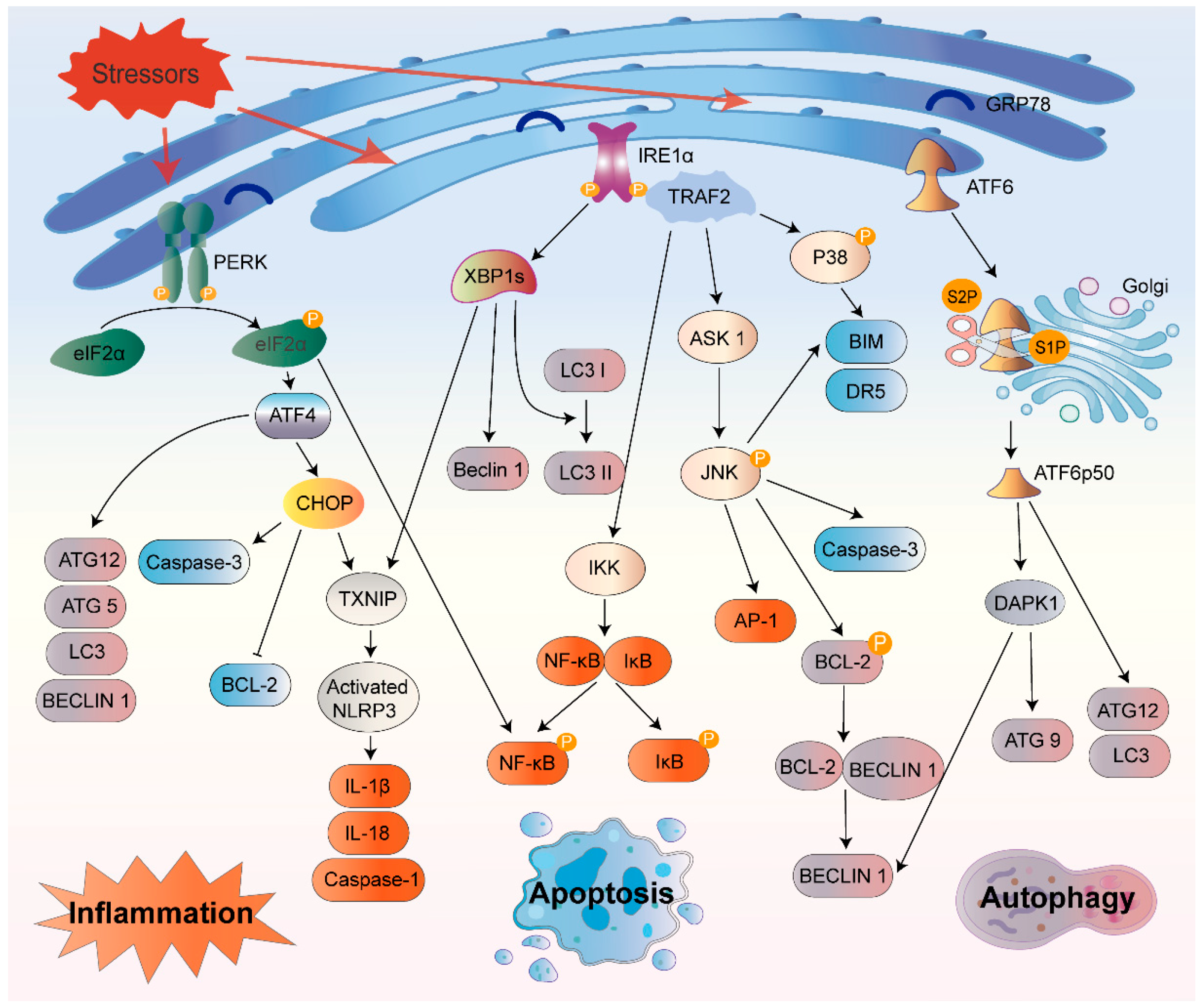
In response to prolonged ER stress, the UPR can start inflammatory responses, autophagy, ERAD, and apoptosis (Figure 2). However, if adaptive responses cannot restore protein folding homeostasis, UPR signaling eventually morphs into a terminal UPR that promotes cell death [18].

3.2.1. Inflammation

Li and colleagues found that the onset of the UPR was adequate to prompt low degrees of inflammatory cytokine production, even without any obvious, irresistible stimuli [60]. Moreover, all three sensors of the UPR participate in the stimulation of inflammatory processes under ER stress. As a central transcriptional regulator, NF-κB is involved in multiple proinflammatory pathways. Normally, NF-κB forms a complex with the inhibitor ΙκΒ, which can translocate to the nucleus and initiate gene transcription. However, under ER stress, activated IRE1α binds with TNF receptor-associated factor 2 (TRAF2) to form a complex, which further activates apoptosis signal-regulating kinase 1 (ASK1) and IκB kinase (IKK). Subsequently, the IκB is phosphorylated and degraded, which eventually drives NF-κB translocation and triggers cytokine expression [61]. The IRE1α–TRAF2 complex can also activate c-Jun N-terminal kinase (JNK), which consequently phosphorylates and initiates the bZIP transcription factor activator protein 1 (AP-1) [62]. The activated PERK–eIF2α arm can restrain the translation of IκB and initiate the NF-κB pathway, prompting NF-κB nuclear translocation [63]. CHOP is activated within the PERK branch and enhances NF-κB signaling via transcriptional repression of the negative regulator peroxisome proliferator-activated receptor [64].
In addition to the induction of classical proinflammatory cytokines, there is mounting evidence that ER stress exacerbates the inflammasome-induced inflammatory cascade. The IRE1α/XBP1 pathway has been found to initiate NOD-, LRR-, and pyrin domain-containing protein 3 (NLRP3) inflammasome-mediated inflammation. In particular, XBP1 can activate the NLRP3 inflammasome, convert inactive caspase-1 to an active form, and promote IL-1β secretion to the extracellular space [65]. Active caspase-1 could trigger pyroptosis, a specific form of inflammatory cell death. An overexpression of CHOP activates the NLRP3 inflammasome and leads to pyroptosis [66]. The inhibition of ER stress could reduce the caspase-1 activity and rescue the cell from pyroptotic death [66]. Additional evidence indicates that the mechanism by which ER stress leads to inflammatory activation of NLRP3 is, at least in part, through thioredoxin-interacting protein (TXNIP), which is strongly induced downstream of IRE1α and/or PERK [23,67]. Interestingly, the UPR also interacts with the cytoplasmic peptidoglycan receptors NOD1 and NOD2 to induce the secretion of IL-6 [68].

3.2.2. ERAD

Under ER stress, the UPR components activate two major protein degradation pathways: the ubiquitin–proteasome system (UPS) via ERAD and lysosome-mediated protein degradation via autophagy. ERAD is the process by which unfolded proteins are transported from the ER to the cytoplasm and degraded by the UPS [69]. Ubiquitination is essential for successfully targeting substrates for degradation. E3 ubiquitin ligase is a core component of the ERAD machinery, which connects multiple ER lumen and cytoplasmic junction proteins to translocate misfolded substrates into the cytoplasm for efficient degradation in the proteasome [20].
In mammalian cells, more than a dozen E3 ubiquitin–ligase enzymes are involved in ERAD. By ubiquitinating IRE1α, the E3 ligase, a mitochondrial ubiquitin ligase, can restrain ER stress-induced apoptosis [70]. Moreover, the E3 ligase carboxy-terminus of the HSC70-interacting protein (CHIP) induces IRE1α ubiquitination at Lys545 and Lys828 and optionally affects IRE1α phosphorylation and TRAF2 binding/JNK activation, thereby regulating ER stress-induced apoptosis and senescence [71]. In addition, the E3 ligase hydroxymethyglutaryl reductase degradation protein 1 (HRD1) is regulated by IRE1α and ATF6 [72]. A recent study has indicated that HRD1 could maintain T regulatory cell stability and function by inhibiting the IRE1α/p38-mediated ER stress response [73]. The phosphorylation of PERK and eIF2α is also crucial to the ERAD process. PERK activation can promote the phosphorylation of the E3 ligases MARCH5, MULAN, and Parkin, thereby increasing the degradation of related substrates [74]. HRD1 can promote eIF2α ubiquitination and proteasomal degradation to protect cells from apoptosis [75]. Furthermore, when ATF6α forms a heterodimer with XBP1s, the complex directs the expression of several ERAD machinery components [57,76]. ATF6 can regulate ERAD gene expression, which is essential for maintaining protein homeostasis [77].

3.2.3. Autophagy

Autophagy is involved in many physiological processes and is essential for maintaining metabolic homeostasis. When misfolded or unfolded proteins accumulate beyond the ER capacity, autophagy can be induced as a secondary reaction to degrade the accumulated proteins, thereby alleviating ER stress. All three branches of the UPR can differentially regulate autophagy during ER stress. The IRE1α–TRAF2–ASK1 complex activates JNK, which leads to the phosphorylation of B-cell lymphoma-2 (BCL-2). This leads to the dissociation of BECLIN-1 from BCL-2, and the activation of the phosphatidylinositol 3-kinase (PI3K) complex promotes autophagy [78]. Moreover, JNK can directly regulate the expression of BECLIN-1 [79]. Notably, the autophagy-related protein (ATG) 12 can bind to and inhibit the BCL-2 protein, thereby promoting cell death [80]. Furthermore, the IRE1α/XBP1s axis increases the conversion of LC3 I to LC3 II in epithelial cells, leading to autophagy [81]. In addition, XBP1s triggers an autophagic signaling pathway through the transcriptional regulation of BECLIN-1 [82]. The PERK/eIF2α/ATF4 pathway appears to be the most critical pathway for inducing autophagy-related gene expression. It has been shown that PERK drives the expression of more than 10 autophagy genes, such as ATG12, ATG5, and BECLIN-1, to mediate autophagy through ATF4 [83]. Hypoxia-induced ER stress can lead to the upregulation of microtubule-associated protein-1 light chain-3β and ATG5 [84]. PERK/eIF2α/ATF4 signaling can also inhibit the mechanistic target of rapamycin (mTOR) complex I-activated autophagy by upregulating sestrin-2 [85]. Additionally, the depletion of ATF6 can decrease the transcription of ATG3 and BECLIN-1 [86] and inhibit autophagy via the mTOR pathway [87]. Another study has found that LC3 and ATG12 were transcriptionally upregulated through the ATF6 pathway [88]. ER stress can also trigger autophagy through ATF6/death-associated protein kinase (DAPK1)-mediated ATG9 transport and BECLIN-1 phosphorylation [89].
In summary, UPR and autophagy can be considered two programs for cellular homeostasis that work either independently or synergize to protect cells from various stresses. The induction of autophagy often acts as a protective mechanism. However, the overactivation of autophagy may be detrimental to cell survival. If misfolded proteins cannot be completely removed by autophagy and the UPS, they cause excessive ER stress, shifting the cell from survival to death mode.

3.2.4. Cellular Senescence

Cellular senescence is an irreversible cell cycle arrest and increased secretion of inflammatory factors caused by various types of cellular stress, such as ER stress, mitochondrial dysfunction, and oxidative damage [90]. Studies have indicated that ER stress represses cyclin D1 and cyclin B1 expression through eIF2α phosphorylation and subsequent GADD45α induction, resulting in cell cycle arrest in G1/S and G2/M phases [91,92]. In addition, UPR-inducers induced senescence through the ATF6-cyclooxygenase 2 (COX2)/prostaglandin E2 (PGE2) axis; specifically, the silencing of the ATF6α and IRE1α decreased the expression of the COX2/PGE2 [93]. Dysfunctional autophagy has been reported to induce premature cellular senescence [94]. The evidence suggests that activation of ATF4 and CHOP, the molecular components of the ER stress pathway, can lead to cellular senescence through the induction of autophagy [95]. The inhibition of ER stress and autophagy could depress the expression of senescence markers and alleviate the senescence-associated secretory phenotype [90,96]. This evidence demonstrates that ER stress and autophagy in tandem play a crucial role in promoting cellular senescence.

3.2.5. Apoptosis

When ER stress cannot be reversed, cellular functions deteriorate, frequently resulting in cell death. IRE1 is a critical molecule in the UPR signaling pathways. The activation of IRE1 also derepresses the translation of caspase-2 mRNA by degrading a specific miRNA, thus stimulating the mitochondrial apoptotic pathway [97]. In addition to inducing transcription of inflammatory factors, the IRE1α/TRAF2/ASK1 pathway causes the activation of downstream JNK and p38 mitogen-activated protein kinases (MAPKs), which promotes the apoptosis [98,99]. The ASK1−/− mice model displays reduced JNK activation and apoptosis under ER stress [100]. Activated JNK can promote the expression of apoptosis-related genes, such as caspase-3, and then initiate the death receptor or mitochondrial pathway to induce apoptosis [101]. BCL-2 and BCL2-like11 (BIM), respectively, are apoptosis-related substrates of JNK that are suppressed and activated by JNK phosphorylation [102,103]. In addition, p38 MAPK phosphorylation activates the transcription factor CHOP, which increases the expression of BIM and death receptor 5 (DR5) while decreasing that of BCL-2 to promote apoptosis [104]. Moreover, PERK overactivation can upregulate the CHOP/GADD153 transcription factor, which inhibits the antiapoptotic protein BCL-2 and enhances the expression of associated proapoptotic proteins [105]. A recent study has shown that the knockdown of CHOP and ATF4 by RNA interference can reduce the level of apoptosis in response to ER stress [106]. The transcription of CHOP is also regulated by ATF6 [107].
ER stress can also induce apoptosis through a caspase-dependent pathway. Caspase-12 is located in the outer membrane of the ER and is a critical molecule that mediates ER stress-related apoptosis. Similar to other caspases, caspase-12 exists in the form of an inactive zymogen. ER stress causes caspase-12 activation, and activated caspase-12 cleaves and activates caspase-9; in turn, activated caspase-9 activates caspase-3 and other caspases, ultimately leading to apoptosis [108]. A recent study has shown that ER stress-mediated apoptosis could be alleviated by inhibiting the IRE1α/XBP1s/caspase-12 pathway [109].

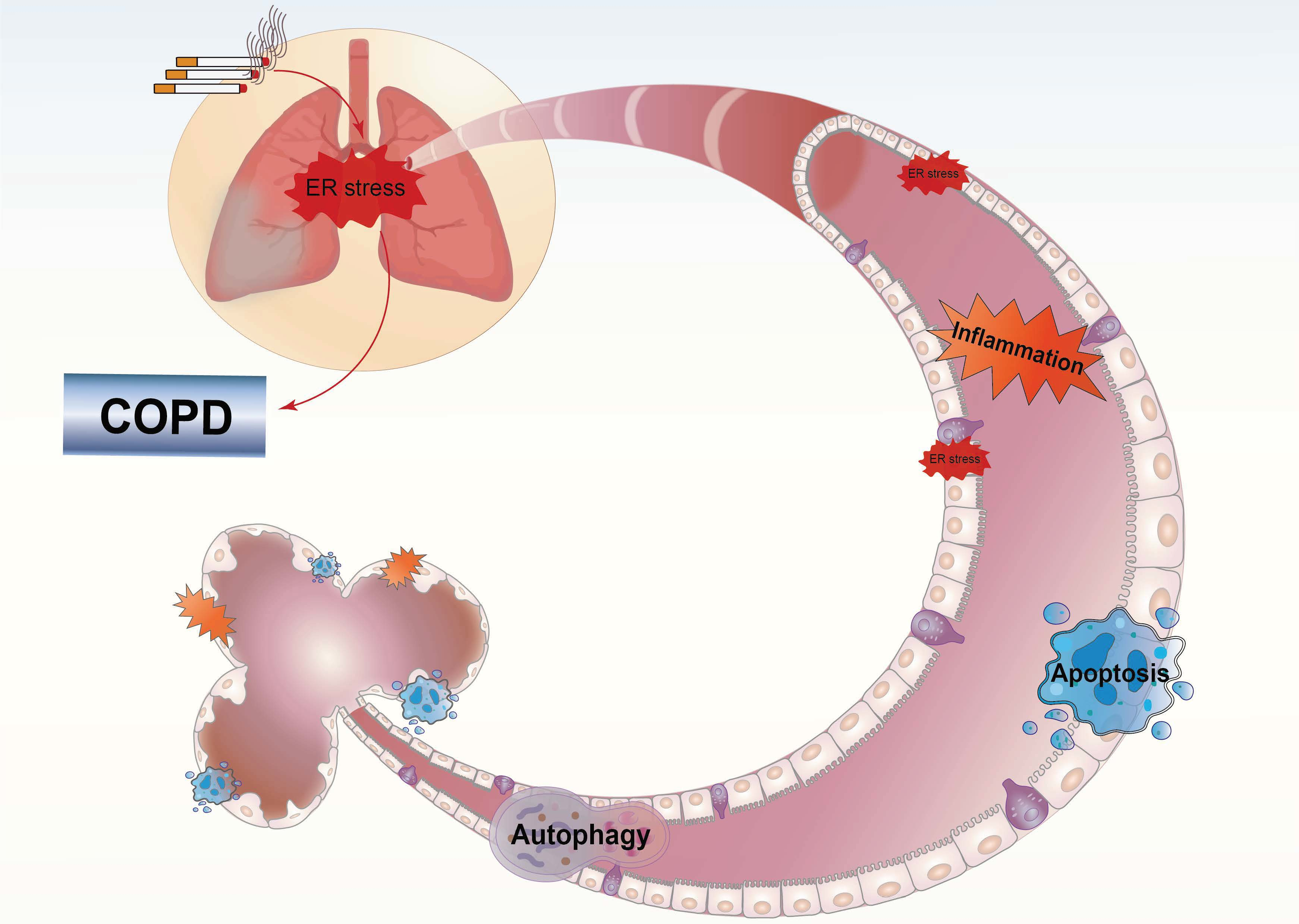
4. COPD and ER Stress

COPD is a progressive lung disease characterized by emphysema and chronic bronchitis. COPD is also associated with systemic inflammation, leading to multiple comorbidities and complications. Two major pathological processes that cause progressive airflow limitation in COPD are small airway remodeling and the destruction of the lung parenchyma [4,6]. The mechanisms of COPD are very complex and have not yet been fully elucidated. Increasing evidence indicates that ER stress is involved in the progression of airway inflammation and epithelial cell apoptosis in COPD, which are the essential mechanisms of early COPD pathogenesis.
Cigarette smoke (CS) is the most common risk factor for lung inflammation in COPD, which can predispose individuals to acute lung injury and pulmonary infections [110]. Some reactive intermediates, such as acrolein, are produced by the reactive oxygen species (ROS) and reactive nitrogen species present in CS. Their cytotoxicity causes oxidative damage and misfolding of proteins in the lung. Misfolded proteins that accumulate in the cells eventually cause ER stress [4].

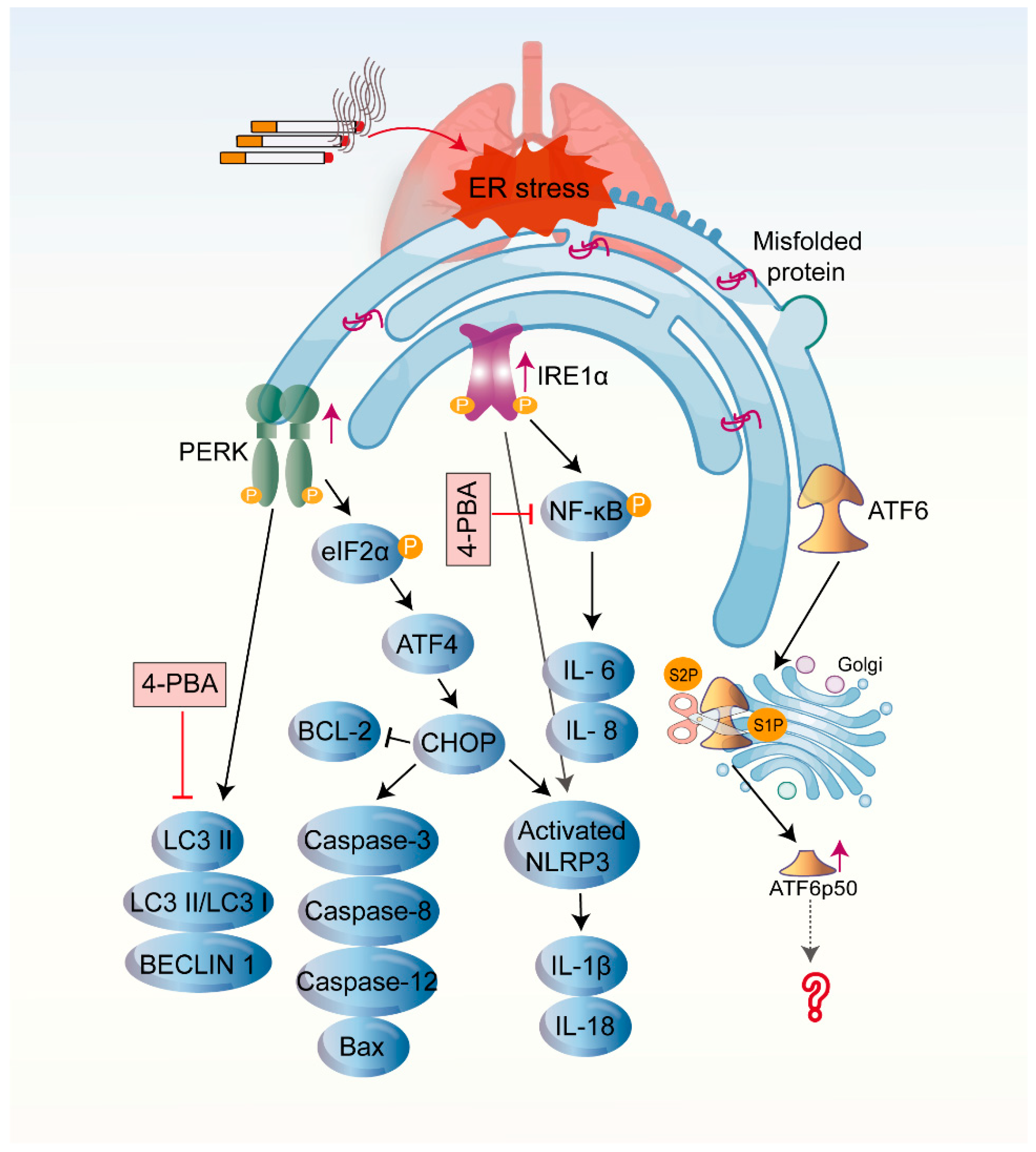
4.1. CS-Induced UPR in COPD

Proteomic analysis showed that long-term smoking induced upregulation of several UPR chaperones and folding enzymes in human lung tissue [111]. Other studies have shown that GRP78 protein levels are increased in the lungs and serum of smokers and patients with COPD, as well as in the bronchoalveolar lavage fluid of smokers, which is connected to decreased lung function and emphysema severity [112,113]. Anti-GRP78 autoreactivity is a risk factor for atherosclerosis and osteoporosis in smokers [114,115]. Compared with those in long-term smokers, the lung levels of GRP78, calreticulin, and protein disulfide isomerase (PDI) are significantly lower in ex-smokers [111]. Weidner and colleagues [13] observed that the ER and endomembrane structures were disorganized in fibroblasts from patients with COPD, and these structural changes did not return to normal after several weeks of in vitro culture. This study suggests that quitting smoking can partially reverse protein unfolding, but structural changes in the lung lining of smokers are permanent, and this cellular fragility in response to stress may explain why some smokers develop COPD. In contrast to these studies, Korfei and colleagues [116] did not observe the expression of UPR-related signals in the lung tissues of patients with COPD, which indicates apparent differences in the expression of UPR-associated proteins in different individuals.
UPR activation was also detected in animal models of CS-induced COPD. Increased protein and mRNA expression levels of CHOP and GRP78 were detected in the lung tissues of COPD rats [117,118]. Long-term exposure to CS also increased the expression levels of the GRP78, ATF4, ATF6, and CHOP proteins and the phosphorylation of PERK and IRE1 in the lung tissues of mice [12]. With prolonged CS exposure, the expression of UPR-related proteins in the lung tissues of rats significantly increased [119]. Interestingly, Kenche and colleagues [120] confirmed that a single cigarette could lead to increased levels of phosphorylated eIF2α and ATF6 (p50) in the lung tissues of mice. Geraghty et al. [121] also demonstrated that acute exposure to CS caused an increase in CHOP levels in mice and guinea pigs. However, after long-term exposure to CS, CHOP expression in lung tissues decreased. In addition, CS exposure increased the expression of ER stress biomarkers in the diaphragms of rats. ER stress-induced apoptosis may be related to the atrophy of the diaphragmatic muscle caused by CS [122]. Unlike that in CS-exposed rats, no increased UPR signaling was found in the diaphragm muscles of stable patients with COPD [10]. This suggests that different individuals and species react differently to CS, and transcriptome analysis also revealed that few genes were universally regulated between mice and humans [123].
Several in vitro studies have demonstrated that exposure to CS extract (CSE) can trigger ER stress in the airway epithelial cells and other associated cells, such as lung cancer cells and alveolar macrophages [12,124,125]. Moreover, CSE exposure causes the oxidation of the ER chaperone PDI, which may contribute to the underlying mechanism of ER stress [126]. CS can also cause oxidative stress and severely damage proteins in the lungs. Exposure to gas-phase CS leads to decreased cystatin, trypsin, and tryptic rennin-like activities in human alveolar epithelial cells. The disruption of intracellular proteostasis under conditions of CS-induced ER stress is due to reduced proteasomal degradation of misfolded proteins, which results in the accumulation of damaged proteins and the reduction of the nascent protein synthesis [127]. Reduced proteasomal activity promotes CSE-induced aggresomal formation and increases apoptosis-inducing factor-mediated cell death [128].

4.2. CS-Induced UPR and Other Cellular Processes in COPD

4.2.1. Inflammation

COPD may be caused by chronic exposure to smoke, dust, and other harmful particles, which induce chronic inflammatory responses in the airways, subsequently leading to irreversible pathological changes, such as small airway remodeling and pulmonary fibrosis. As previously described, all three UPR branches cause inflammatory responses. Significant ER stress was observed in the lung tissues of CS-induced COPD animal models, with elevations in the total inflammatory cell levels and neutrophil percentages in the bronchoalveolar lavage fluid and significantly increased levels of the inflammatory factors IL-6, IL-8, and TNF-α [12,118]. CS stimulation also activates the NLRP3 inflammasome, which promotes the secretion of IL-1β and IL-18 in the lung tissue [129]. Inhibitors or blockers of ER stress signaling can reduce CS-induced airway inflammation and improve emphysema [118,129,130].

4.2.2. Cellular Senescence

Oxidative stress induced by excessive CS exposure can trigger lung cellular senescence through telomere shortening and DNA damage [131]. Acute/premature senescence plays an essential role in the pathogenesis of COPD by inducing changes in the systemic or local immune system and inflammatory factor secretion, interfering with tissue repair mechanisms after injury and weakening pulmonary defenses, among other complex mechanisms that impair lung function [132,133]. Fibroblasts and alveolar epithelial and endothelial cells from patients with COPD display shorter telomere lengths, DNA damage, accelerated cellular senescence, and oxidative stress [134,135]. The study has revealed that activation of UPR promotes the senescence of lung fibroblasts through the deletion of GADD34 [136]. However, studies on the interplay between ER stress and the UPR branch associated with cellular senescence in COPD still need to be further investigated.

4.2.3. Apoptosis

CS exposure promotes the production of oxygen radicals, exacerbates changes in the intracellular redox status, and triggers oxidative stress. In vitro experiments have indicated that CSE stimulation elevates the expression of the apoptotic marker CHOP and induces caspase-dependent apoptosis [137,138]. Tagawa and colleagues [139] observed typical apoptotic manifestations, such as nuclear condensation, membrane blistering, and activation of the apoptosis-specific factors caspase-3 and caspase-4 in human bronchial epithelial cells after 24-h exposure to CSE. Subsequently, Tagawa et al. [140] found that CSE could trigger the PERK/eIF2α/CHOP pathway to induce apoptosis via superoxide anion. The inhibition of this pathway significantly suppressed the CSE-triggered CHOP expression and apoptosis.
The UPR also induces an elevated expression of antioxidants, especially NRF2. Under oxidative conditions, NRF2 translocates to the nucleus to combine with the antioxidant response element and induce the expression of endogenous antioxidant proteins and enzymes [141]. The NRF2 expression is elevated in the peripheral blood mononuclear cells in ex-smoking patients with mild to moderate COPD [142]. The upregulation of NRF2 alleviated CS-induced oxidative stress in rat lungs, attenuated CS-induced emphysema and airway remodeling, and reduced ER stress and apoptosis [143]. These results suggest that the upregulation of NRF2 expression may help prevent oxidative stress-related COPD progression. Interestingly, recent studies have shown that NRF2-deficient mice that were chronically exposed to particulate matter 2.5 had elevated oxidative stress and depressed ER stress levels but did not show obvious emphysema [144]. This may be attributed to the fact that the NRF2 deficiency reduces the expression of cytochrome P450 family 2 subfamily E member 1, thereby diminishing the cellular damage resulting from the metabolism of exogenous compounds [144,145].

4.2.4. Autophagy

Most studies show that the autophagy mechanisms are impaired in COPD. Increased expression of p62 and LC3 in peripheral lung tissues from patients with severe COPD indicates impaired autophagy [146]. Likewise, alveolar macrophages from patients with COPD and smokers display large numbers of autophagosomes but impaired autophagic flux [147]. The same phenomenon was observed in CSE-stimulated human macrophage cell lines [148]. However, it has also been suggested that long-term exposure to CS causes enhanced autophagy in COPD mice, which is associated with COPD progression [149]. Autophagy is a complex regulatory process, and its increase or decrease during COPD progression may be related to different cell types, as well as differences in intensity and duration of stress.
Recent studies have revealed that the interaction between autophagy and ER stress plays an essential role in COPD progression. CSE stimulation induces apoptosis, autophagy, and ER stress-related protein expression in airway epithelial cells. The autophagy inhibitor 3-methyladenine was shown to increase the expression of CHOP, ATF4, and caspase-4, which correlated with CSE-induced apoptosis. Meanwhile, the ER stress inhibitor 4-phenylbutyric acid (4-PBA) inhibited autophagy. Furthermore, CSE-induced autophagy and apoptosis were enhanced by the knockdown of ATF4 or CHOP [150]. Hosaka and colleagues [151] further demonstrated the existence of functional crosstalk between UPR signaling and autophagy during CSE exposure. Impaired chaperone-mediated autophagy contributes to COPD pathogenesis by enhancing UPR-mediated epithelial apoptosis [151].

4.3. The Influence of Three Branches of UPR on the Pathogenesis of COPD

Although studies have confirmed that activation of all three UPR branches can be observed in CS-induced COPD (Figure 3), the investigation of PERK and its downstream pathway signaling is the most extensive. CS-induced activation of the PERK/eIF2α/ATF4/CHOP pathway upregulated apoptosis factor caspase-3, caspase-8, caspase-12, and BCL-2 associated X (Bax) and downregulated the anti-apoptotic factor BCL-2 [137,143]. The action of various drugs is to reduce CS-induced apoptosis by inhibiting PERK, thereby alleviating emphysema. Moreover, CS-induced activation of the PERK pathway could trigger activation of the NLRP3 inflammasome, causing a series of inflammatory cascades that ultimately lead to a pulmonary inflammatory flare [129]. A knockdown of PERK also attenuated CSE-induced autophagy [150]. Efferocytosis is the process by which phagocytes remove apoptotic cells. Impaired efferocytosis is an essential mechanism in inflammatory lung disease. The evidence indicated that CS destroyed efferocytosis via the PERK/eIF2α pathway in macrophages [124].
In addition to modulating the NLRP3 inflammasome, IRE1α can promote CS-induced airway inflammation by activating the NF-κB signaling pathway [129,130]. Furthermore, the activation of IRE1α played a crucial role in the nicotine-induced epithelial-mesenchymal transition and decreased cell migration capacity in human bronchial epithelial cells [152]. Epithelial-mesenchymal transition is a pivotal contributor to airway remodeling in COPD.
The ATF6 pathway is the least investigated in CS-induced COPD. CS-induced ATF6 cleavage was observed in lung lysates from mice [120]. However, some researchers have also found that the ATF6 pathway was inactivated in lung tissue from CS-induced COPD rats [137]. The studies related to ATF6 and its mediated signaling pathways in the pathogenesis of COPD are still insufficient. It is still remarkable that the endogenous cystic fibrosis transmembrane conductance regulator (CFTR) function is diminished under ER stress, which is associated with transcriptional repression of CFTR by ATF6 [153]. CFTR dysfunction can lead to significant impairment of mucociliary clearance and mucus hypersecretion, as well as to airway wall-thickening [154]. Mounting evidence demonstrates that acquired CFTR dysfunction is an essential contributor to the pathophysiology of COPD [154,155,156]. The reversal of CFTR dysfunction is an attractive therapeutic target for ameliorating the airway pathology in COPD [156]. Whether activation of the ATF6 pathway is correlated with CFTR dysfunction in COPD is unclear, and other mechanisms of ATF6 involvement in COPD disease progression need further investigation.

5. The UPR and Potential Therapeutic Interventions in COPD

The evidence of the COPD response to pharmacological manipulations of the UPR is limited to interventions in animal models and cells (Table 1). Wang and colleagues [130] demonstrated that 4-PBA prevented CS-induced emphysema, alveolar apoptosis, and inflammation by inhibiting ER stress and NF-κB signaling. Aggarwal and colleagues [157] indicated that the plasma haem levels and ER stress were elevated in patients with severe COPD and in a ferret model of COPD. Scavenging the haem with salubrinal (eIF2α dephosphorylation inhibitor) suppressed ER stress, reduced elastase levels and activity, and attenuated the development of fibrosis and emphysema pathophysiological phenotypes in COPD. Hydrogen sulfide (H2S), as an endogenous modulator, attenuated smoke-induced apoptosis and epithelial-mesenchymal transition by inhibiting ER stress, thus hindering the progression of lung function decline and emphysema formation in rats [152,158,159]. Melatonin counteracts CS-induced inflammasome activation and apoptosis by inhibiting ER stress and mitochondrial dysfunction [129,160]. Additionally, melatonin diminished CS-induced oxidative stress by upregulating the expression of the antioxidant NRF2 and improving the overall antioxidant status in the lungs of a COPD model [129]. Ursolic acid not only partially inhibits UPR signaling to alleviate ER stress-related apoptosis and oxidative stress but also abrogates CS-induced airway remodeling by modulating the three UPR pathways, which results in a therapeutic effect against COPD [137,143]. Epoxyeicosatrienoic acids (EETs) are produced via the cytochrome P450 epoxy synthase pathway and possess anti-inflammatory and antiapoptotic effects. Exogenous administration of 14,15-EET can prevent CSE-induced apoptosis by blocking UPR signaling [161]. Furthermore, adiponectin, a protein mainly secreted by adipocytes, alleviated the apoptosis of alveolar epithelial cells in COPD rats by suppressing ER stress [162]. Other reports have suggested that herbal ingredients, such as Fengbaisan, ephedrine, and curcumin, could modify COPD in vitro and in vivo by modulating ER stress [12,117,118,138,163].
Additionally, several vital mediators that regulate various processes may be involved in COPD by regulating ER stress. Sirtuin 1 (SIRT1) has become a focus of ER stress research in recent years. SIRT1 is mainly located in the nucleus and can enhance cellular stress resistance and boost cell survival in several ways. The upregulation of SIRT1 could inhibit ER stress and delay COPD progression [117,118,160,164]. AMP-activated protein kinase (AMPK) is an essential sensor of cellular energy status and has protective roles in various diseases. Liu et al. [165] showed that AMPK could activate oxygen-regulated protein 150 via the forkhead box O1 pathway, thereby blocking ER stress and preserving airway epithelial cells from CSE-induced apoptosis. Progranulin (PGRN) is extensively expressed in diverse epithelial cells and can restore epithelial tissue homeostasis in response to tissue injury. In a COPD model, PGRN suppressed apoptosis of airway epithelial cells by regulating the ER stress response and MAPK activation [166]. Furthermore, Chen et al. [167] confirmed that the silencing of orosomucoid 1-like protein 3 (ORMDL3), a transmembrane protein localized in the ER, abrogated smoking-induced human airway smooth muscle cell injury by repressing the UPR pathway. Moreover, miRNAs have been reported to be associated with the progression of many diseases, including COPD. A recent study indicated that miR-150-5p expression is decreased in patients with COPD and in COPD models. The overexpression of miR-150-5p could directly diminish CS-induced inflammation, facilitate cell migration, and achieve protective effects against COPD by targeting IRE1α [168].

6. Conclusions

In this review, we focus on the mechanisms of cellular processes triggered by CS-induced activation of UPR in the development of COPD. Each UPR sensor has a dual role, with some pathways of the initial UPR being adaptive and protective. When this early signaling fails to successfully restore ER homeostasis, the UPR may change into a signal that promotes cell death. UPR-related signals are elevated in patients with COPD and animal models, and a prolonged UPR may induce apoptosis of structural lung cells, ultimately leading to the development of emphysema and pulmonary fibrosis. However, ER stress signaling pathways are complex and tissue-specific, and the effects of ER stress on COPD depend on the different cell types and the degree of stimuli. Currently, pharmacological treatments targeting UPR signaling molecules exhibit great potential in delaying COPD progression. Based on recent research advances, we also found that targeted UPR exhibited remarkable therapeutic effects in COPD. Consequently, therapeutic strategies targeting ER stress may provide new directions for the treatment of COPD.

Author Contributions

Y.Y. and A.Y. contributed equally to the writing and editing of the manuscript and the preparation of the figures and table. G.Y. and H.W. edited the manuscript. All authors have read and agreed to the published version of the manuscript.

Funding

This study was supported by the National Natural Science Foundation of China (Nos. 81870029 and 81700038), the Key Clinical Specialty Construction Program of Beijing (2020–2022), and the Beijing Natural Science Foundation (No. 7204247).

Institutional Review Board Statement

Not applicable.

Informed Consent Statement

Not applicable.

Data Availability Statement

Not applicable.

Conflicts of Interest

The authors declare no conflict of interest.

References

  1. Sarvani, C.; Sireesh, D.; Ramkumar, K.M. Unraveling the role of ER stress inhibitors in the context of metabolic diseases. Pharmacol. Res. 2017, 119, 412–421. [Google Scholar] [CrossRef] [PubMed]
  2. Dufey, E.; Sepulveda, D.; Rojas-Rivera, D.; Hetz, C. Cellular mechanisms of endoplasmic reticulum stress signaling in health and disease. 1. An overview. Am. J. Physiol. Cell Physiol. 2014, 307, C582–C594. [Google Scholar] [CrossRef] [PubMed]
  3. Zhao, H.; Liu, H.; Yang, Y.; Lan, T.; Wang, H.; Wu, D. Hydrogen sulfide plays an important role by regulating endoplasmic reticulum stress in Diabetes-Related diseases. Int. J. Mol. Sci. 2022, 23, 7170. [Google Scholar] [CrossRef] [PubMed]
  4. Kelsen, S.G. The unfolded protein response in chronic obstructive pulmonary disease. Ann. Am. Thorac. Soc. 2016, 13 (Suppl. 2), S138–S145. [Google Scholar] [CrossRef]
  5. Mccaffrey, K.; Braakman, I. Protein quality control at the endoplasmic reticulum. Essays Biochem. 2016, 60, 227–235. [Google Scholar] [CrossRef]
  6. Naiel, S.; Tat, V.; Padwal, M.; Vierhout, M.; Mekhael, O.; Yousof, T.; Ayoub, A.; Abed, S.; Dvorkin-Gheva, A.; Ask, K. Protein misfolding and endoplasmic reticulum stress in chronic lung disease: Will Cell-Specific targeting be the key to the cure? Chest 2020, 157, 1207–1220. [Google Scholar] [CrossRef]
  7. Wang, F.; Ma, J.; Wang, J.; Chen, M.; Xia, H.; Yao, S.; Zhang, D. SIRT1 ameliorated septic associated-lung injury and macrophages apoptosis via inhibiting endoplasmic reticulum stress. Cell. Signal. 2022, 97, 110398. [Google Scholar] [CrossRef]
  8. Bhardwaj, R.; Bhardwaj, A.; Dhawan, D.K.; Tandon, C.; Kaur, T. 4-PBA rescues hyperoxaluria induced nephrolithiasis by modulating urinary glycoproteins: Cross talk between endoplasmic reticulum, calcium homeostasis and mitochondria. Life Sci. 2022, 305, 120786. [Google Scholar] [CrossRef]
  9. Jiang, M.; Li, Z.; Zhu, G. The role of endoplasmic reticulum stress in the pathophysiology of periodontal disease. J. Periodontal Res. 2022, 57, 915–932. [Google Scholar] [CrossRef]
  10. Barreiro, E.; Salazar-Degracia, A.; Sancho-Munoz, A.; Aguilo, R.; Rodriguez-Fuster, A.; Gea, J. Endoplasmic reticulum stress and unfolded protein response in diaphragm muscle dysfunction of patients with stable chronic obstructive pulmonary disease. J. Appl. Physiol. (1985) 2019, 126, 1572–1586. [Google Scholar] [CrossRef]
  11. Wu, G.; Yuan, T.; Zhu, H.; Zhang, H.; Su, J.; Guo, L.; Zhou, Q.; Xiong, F.; Yu, Q.; Yang, P.; et al. Chrysophanol protects human bronchial epithelial cells from cigarette smoke extract (CSE)-induced apoptosis. Int. J. Mol. Epidemiol. Genet. 2020, 11, 39–45. [Google Scholar] [PubMed]
  12. Wang, H.L.; Chen, F.Q.; Wu, L.J. Ephedrine ameliorates chronic obstructive pulmonary disease (COPD) through restraining endoplasmic reticulum (ER) stress in vitro and in vivo. Int. Immunopharmacol. 2022, 103, 107842. [Google Scholar] [CrossRef] [PubMed]
  13. Weidner, J.; Jarenback, L.; Aberg, I.; Westergren-Thorsson, G.; Ankerst, J.; Bjermer, L.; Tufvesson, E. Endoplasmic reticulum, Golgi, and lysosomes are disorganized in lung fibroblasts from chronic obstructive pulmonary disease patients. Physiol. Rep. 2018, 6, e13584. [Google Scholar] [CrossRef] [PubMed]
  14. Schwarz, D.S.; Blower, M.D. The endoplasmic reticulum: Structure, function and response to cellular signaling. Cell. Mol. Life Sci. 2016, 73, 79–94. [Google Scholar] [CrossRef] [Green Version]
  15. Sirish, P.; Diloretto, D.A.; Thai, P.N.; Chiamvimonvat, N. The critical roles of proteostasis and endoplasmic reticulum stress in atrial fibrillation. Front. Physiol. 2021, 12, 793171. [Google Scholar] [CrossRef]
  16. Oakes, S.A.; Papa, F.R. The role of endoplasmic reticulum stress in human pathology. Annu. Rev. Pathol. 2015, 10, 173–194. [Google Scholar] [CrossRef] [Green Version]
  17. van Anken, E.; Braakman, I. Versatility of the endoplasmic reticulum protein folding factory. Crit. Rev. Biochem. Mol. Biol. 2005, 40, 191–228. [Google Scholar] [CrossRef]
  18. Hetz, C.; Papa, F.R. The unfolded protein response and cell fate control. Mol. Cell 2018, 69, 169–181. [Google Scholar] [CrossRef] [Green Version]
  19. Moon, H.W.; Han, H.G.; Jeon, Y.J. Protein quality control in the endoplasmic reticulum and cancer. Int. J. Mol. Sci. 2018, 19, 3020. [Google Scholar] [CrossRef] [Green Version]
  20. Li, Y.; Li, S.; Wu, H. Ubiquitination-Proteasome system (UPS) and autophagy two main protein degradation machineries in response to cell stress. Cells 2022, 11, 851. [Google Scholar] [CrossRef]
  21. Ozgur, R.; Uzilday, B.; Iwata, Y.; Koizumi, N.; Turkan, I. Interplay between the unfolded protein response and reactive oxygen species: A dynamic duo. J. Exp. Bot. 2018, 69, 3333–3345. [Google Scholar] [CrossRef] [PubMed] [Green Version]
  22. Hetz, C.; Zhang, K.; Kaufman, R.J. Mechanisms, regulation and functions of the unfolded protein response. Nat. Rev. Mol. Cell Biol. 2020, 21, 421–438. [Google Scholar] [CrossRef] [PubMed]
  23. Oakes, S.A. Endoplasmic reticulum proteostasis: A key checkpoint in cancer. Am. J. Physiol. Cell Physiol. 2017, 312, C93–C102. [Google Scholar] [CrossRef] [PubMed] [Green Version]
  24. Tirasophon, W.; Welihinda, A.A.; Kaufman, R.J. A stress response pathway from the endoplasmic reticulum to the nucleus requires a novel bifunctional protein kinase/endoribonuclease (Ire1p) in mammalian cells. Genes Dev. 1998, 12, 1812–1824. [Google Scholar] [CrossRef] [Green Version]
  25. Harding, H.P.; Zhang, Y.; Ron, D. Protein translation and folding are coupled by an endoplasmic-reticulum-resident kinase. Nature 1999, 397, 271–274. [Google Scholar] [CrossRef]
  26. Haze, K.; Yoshida, H.; Yanagi, H.; Yura, T.; Mori, K. Mammalian transcription factor ATF6 is synthesized as a transmembrane protein and activated by proteolysis in response to endoplasmic reticulum stress. Mol. Biol. Cell 1999, 10, 3787–3799. [Google Scholar] [CrossRef] [Green Version]
  27. Bhattarai, K.R.; Chaudhary, M.; Kim, H.R.; Chae, H.J. Endoplasmic reticulum (ER) stress response failure in diseases. Trends Cell Biol. 2020, 30, 672–675. [Google Scholar] [CrossRef]
  28. Elfiky, A.A.; Baghdady, A.M.; Ali, S.A.; Ahmed, M.I. GRP78 targeting: Hitting two birds with a stone. Life Sci. 2020, 260, 118317. [Google Scholar] [CrossRef]
  29. Shen, J.; Chen, X.; Hendershot, L.; Prywes, R. ER stress regulation of ATF6 localization by dissociation of BiP/GRP78 binding and unmasking of Golgi localization signals. Dev. Cell. 2002, 3, 99–111. [Google Scholar] [CrossRef] [Green Version]
  30. Sims, S.G.; Cisney, R.N.; Lipscomb, M.M.; Meares, G.P. The role of endoplasmic reticulum stress in astrocytes. Glia 2022, 70, 5–19. [Google Scholar] [CrossRef]
  31. Raymundo, D.P.; Doultsinos, D.; Guillory, X.; Carlesso, A.; Eriksson, L.A.; Chevet, E. Pharmacological targeting of IRE1 in cancer. Trends Cancer 2020, 6, 1018–1030. [Google Scholar] [CrossRef] [PubMed]
  32. Doultsinos, D.; Avril, T.; Lhomond, S.; Dejeans, N.; Guedat, P.; Chevet, E. Control of the unfolded protein response in health and disease. SLAS Discov. 2017, 22, 787–800. [Google Scholar] [CrossRef] [PubMed] [Green Version]
  33. Iwawaki, T.; Akai, R.; Yamanaka, S.; Kohno, K. Function of IRE1 alpha in the placenta is essential for placental development and embryonic viability. Proc. Natl. Acad. Sci. USA 2009, 106, 16657–16662. [Google Scholar] [CrossRef] [Green Version]
  34. Chen, Y.; Brandizzi, F. IRE1: ER stress sensor and cell fate executor. Trends Cell Biol. 2013, 23, 547–555. [Google Scholar] [CrossRef] [PubMed] [Green Version]
  35. Almanza, A.; Carlesso, A.; Chintha, C.; Creedican, S.; Doultsinos, D.; Leuzzi, B.; Luis, A.; Mccarthy, N.; Montibeller, L.; More, S.; et al. Endoplasmic reticulum stress signalling—From basic mechanisms to clinical applications. FEBS J. 2019, 286, 241–278. [Google Scholar] [CrossRef] [Green Version]
  36. Karagoz, G.E.; Acosta-Alvear, D.; Nguyen, H.T.; Lee, C.P.; Chu, F.; Walter, P. An unfolded protein-induced conformational switch activates mammalian IRE1. eLife 2017, 6, e30700. [Google Scholar] [CrossRef]
  37. Calfon, M.; Zeng, H.; Urano, F.; Till, J.H.; Hubbard, S.R.; Harding, H.P.; Clark, S.G.; Ron, D. IRE1 couples endoplasmic reticulum load to secretory capacity by processing the XBP-1 mRNA. Nature 2002, 415, 92–96. [Google Scholar] [CrossRef]
  38. Yoshida, H.; Matsui, T.; Yamamoto, A.; Okada, T.; Mori, K. XBP1 mRNA is induced by ATF6 and spliced by IRE1 in response to ER stress to produce a highly active transcription factor. Cell 2001, 107, 881–891. [Google Scholar] [CrossRef] [Green Version]
  39. Acosta-Alvear, D.; Zhou, Y.; Blais, A.; Tsikitis, M.; Lents, N.H.; Arias, C.; Lennon, C.J.; Kluger, Y.; Dynlacht, B.D. XBP1 controls diverse cell type- and condition-specific transcriptional regulatory networks. Mol. Cell 2007, 27, 53–66. [Google Scholar] [CrossRef]
  40. Liu, J.; Ibi, D.; Taniguchi, K.; Lee, J.; Herrema, H.; Akosman, B.; Mucka, P.; Salazar, H.M.; Uyar, M.F.; Park, S.W.; et al. Inflammation improves glucose homeostasis through IKKbeta-XBP1s interaction. Cell 2016, 167, 1052–1066. [Google Scholar] [CrossRef]
  41. So, J.S.; Hur, K.Y.; Tarrio, M.; Ruda, V.; Frank-Kamenetsky, M.; Fitzgerald, K.; Koteliansky, V.; Lichtman, A.H.; Iwawaki, T.; Glimcher, L.H.; et al. Silencing of lipid metabolism genes through IRE1alpha-mediated mRNA decay lowers plasma lipids in mice. Cell Metab. 2012, 16, 487–499. [Google Scholar] [CrossRef] [PubMed] [Green Version]
  42. Liu, Y.; Adachi, M.; Zhao, S.; Hareyama, M.; Koong, A.C.; Luo, D.; Rando, T.A.; Imai, K.; Shinomura, Y. Preventing oxidative stress: A new role for XBP1. Cell Death Differ. 2009, 16, 847–857. [Google Scholar] [CrossRef] [PubMed] [Green Version]
  43. Akiyama, M.; Liew, C.W.; Lu, S.; Hu, J.; Martinez, R.; Hambro, B.; Kennedy, R.T.; Kulkarni, R.N. X-box binding protein 1 is essential for insulin regulation of pancreatic alpha-cell function. Diabetes 2013, 62, 2439–2449. [Google Scholar] [CrossRef] [PubMed] [Green Version]
  44. Sone, M.; Zeng, X.; Larese, J.; Ryoo, H.D. A modified UPR stress sensing system reveals a novel tissue distribution of IRE1/XBP1 activity during normal Drosophila development. Cell Stress Chaperones 2013, 18, 307–319. [Google Scholar] [CrossRef] [PubMed] [Green Version]
  45. Chen, X.; Iliopoulos, D.; Zhang, Q.; Tang, Q.; Greenblatt, M.B.; Hatziapostolou, M.; Lim, E.; Tam, W.L.; Ni, M.; Chen, Y.; et al. XBP1 promotes triple-negative breast cancer by controlling the HIF1alpha pathway. Nature 2014, 508, 103–107. [Google Scholar] [CrossRef] [Green Version]
  46. Tak, J.; Kim, Y.S.; Kim, T.H.; Park, G.C.; Hwang, S.; Kim, S.G. Galpha12 overexpression in hepatocytes by ER stress exacerbates acute liver injury via ROCK1-mediated miR-15a and ALOX12 dysregulation. Theranostics 2022, 12, 1570–1588. [Google Scholar] [CrossRef]
  47. Bhattarai, K.R.; Riaz, T.A.; Kim, H.R.; Chae, H.J. The aftermath of the interplay between the endoplasmic reticulum stress response and redox signaling. Exp. Mol. Med. 2021, 53, 151–167. [Google Scholar] [CrossRef]
  48. Wiese, W.; Siwecka, N.; Wawrzynkiewicz, A.; Rozpedek-Kaminska, W.; Kucharska, E.; Majsterek, I. IRE1alpha inhibitors as a promising therapeutic strategy in blood malignancies. Cancers 2022, 14, 2526. [Google Scholar] [CrossRef]
  49. Wodrich, A.; Scott, A.W.; Shukla, A.K.; Harris, B.T.; Giniger, E. The unfolded protein responses in health, aging, and neurodegeneration: Recent advances and future considerations. Front. Mol. Neurosci. 2022, 15, 831116. [Google Scholar] [CrossRef]
  50. Cui, X.; Zhang, Y.; Lu, Y.; Xiang, M. ROS and endoplasmic reticulum stress in pulmonary disease. Front. Pharmacol. 2022, 13, 879204. [Google Scholar] [CrossRef]
  51. Hollien, J.; Weissman, J.S. Decay of endoplasmic reticulum-localized mRNAs during the unfolded protein response. Science 2006, 313, 104–107. [Google Scholar] [CrossRef] [PubMed] [Green Version]
  52. Vattem, K.M.; Wek, R.C. Reinitiation involving upstream ORFs regulates ATF4 mRNA translation in mammalian cells. Proc. Natl. Acad. Sci. USA 2004, 101, 11269–11274. [Google Scholar] [CrossRef] [PubMed] [Green Version]
  53. Wortel, I.; van der Meer, L.T.; Kilberg, M.S.; van Leeuwen, F.N. Surviving stress: Modulation of ATF4-Mediated stress responses in normal and malignant cells. Trends Endocrinol. Metab. 2017, 28, 794–806. [Google Scholar] [CrossRef]
  54. Lee, Y.Y.; Cevallos, R.C.; Jan, E. An upstream open reading frame regulates translation of GADD34 during cellular stresses that induce eIF2alpha phosphorylation. J. Biol. Chem. 2009, 284, 6661–6673. [Google Scholar] [CrossRef] [PubMed] [Green Version]
  55. Tao, T.; Wang, J.; Wang, X.; Wang, Y.; Mao, H.; Liu, X. The PERK/Nrf2 pathway mediates endoplasmic reticulum stress-induced injury by upregulating endoplasmic reticulophagy in H9c2 cardiomyoblasts. Life Sci. 2019, 237, 116944. [Google Scholar] [CrossRef] [PubMed]
  56. Stauffer, W.T.; Arrieta, A.; Blackwood, E.A.; Glembotski, C.C. Sledgehammer to scalpel: Broad challenges to the heart and other tissues yield specific cellular responses via transcriptional regulation of the ER-Stress master regulator ATF6alpha. Int. J. Mol. Sci. 2020, 21, 1134. [Google Scholar] [CrossRef] [Green Version]
  57. Yamamoto, K.; Sato, T.; Matsui, T.; Sato, M.; Okada, T.; Yoshida, H.; Harada, A.; Mori, K. Transcriptional induction of mammalian ER quality control proteins is mediated by single or combined action of ATF6alpha and XBP1. Dev. Cell. 2007, 13, 365–376. [Google Scholar] [CrossRef] [Green Version]
  58. Ye, J.; Rawson, R.B.; Komuro, R.; Chen, X.; Dave, U.P.; Prywes, R.; Brown, M.S.; Goldstein, J.L. ER stress induces cleavage of membrane-bound ATF6 by the same proteases that process SREBPs. Mol. Cell 2000, 6, 1355–1364. [Google Scholar] [CrossRef]
  59. Shoulders, M.D.; Ryno, L.M.; Genereux, J.C.; Moresco, J.J.; Tu, P.G.; Wu, C.; Yates, J.R.; Su, A.I.; Kelly, J.W.; Wiseman, R.L. Stress-independent activation of XBP1s and/or ATF6 reveals three functionally diverse ER proteostasis environments. Cell Rep. 2013, 3, 1279–1292. [Google Scholar] [CrossRef] [Green Version]
  60. Li, Y.; Schwabe, R.F.; Devries-Seimon, T.; Yao, P.M.; Gerbod-Giannone, M.C.; Tall, A.R.; Davis, R.J.; Flavell, R.; Brenner, D.A.; Tabas, I. Free cholesterol-loaded macrophages are an abundant source of tumor necrosis factor-alpha and interleukin-6: Model of NF-kappaB- and map kinase-dependent inflammation in advanced atherosclerosis. J. Biol. Chem. 2005, 280, 21763–21772. [Google Scholar] [CrossRef]
  61. Hu, P.; Han, Z.; Couvillon, A.D.; Kaufman, R.J.; Exton, J.H. Autocrine tumor necrosis factor alpha links endoplasmic reticulum stress to the membrane death receptor pathway through IRE1alpha-mediated NF-kappaB activation and down-regulation of TRAF2 expression. Mol. Cell. Biol. 2006, 26, 3071–3084. [Google Scholar] [CrossRef] [PubMed] [Green Version]
  62. Garg, A.D.; Kaczmarek, A.; Krysko, O.; Vandenabeele, P.; Krysko, D.V.; Agostinis, P. ER stress-induced inflammation: Does it aid or impede disease progression? Trends Mol. Med. 2012, 18, 589–598. [Google Scholar] [CrossRef] [PubMed]
  63. Deng, J.; Lu, P.D.; Zhang, Y.; Scheuner, D.; Kaufman, R.J.; Sonenberg, N.; Harding, H.P.; Ron, D. Translational repression mediates activation of nuclear factor kappa B by phosphorylated translation initiation factor 2. Mol. Cell. Biol. 2004, 24, 10161–10168. [Google Scholar] [CrossRef] [PubMed] [Green Version]
  64. Park, S.H.; Choi, H.J.; Yang, H.; Do, K.H.; Kim, J.; Lee, D.W.; Moon, Y. Endoplasmic reticulum stress-activated C/EBP homologous protein enhances nuclear factor-kappaB signals via repression of peroxisome proliferator-activated receptor gamma. J. Biol. Chem. 2010, 285, 35330–35339. [Google Scholar] [CrossRef] [PubMed] [Green Version]
  65. Yue, S.; Zhu, J.; Zhang, M.; Li, C.; Zhou, X.; Zhou, M.; Ke, M.; Busuttil, R.W.; Ying, Q.L.; Kupiec-Weglinski, J.W.; et al. The myeloid heat shock transcription factor 1/beta-catenin axis regulates NLR family, pyrin domain-containing 3 inflammasome activation in mouse liver ischemia/reperfusion injury. Hepatology 2016, 64, 1683–1698. [Google Scholar] [CrossRef] [Green Version]
  66. Lebeaupin, C.; Proics, E.; de Bieville, C.H.; Rousseau, D.; Bonnafous, S.; Patouraux, S.; Adam, G.; Lavallard, V.J.; Rovere, C.; Le Thuc, O.; et al. ER stress induces NLRP3 inflammasome activation and hepatocyte death. Cell Death Dis. 2015, 6, e1879. [Google Scholar] [CrossRef] [Green Version]
  67. Bronner, D.N.; Abuaita, B.H.; Chen, X.; Fitzgerald, K.A.; Nunez, G.; He, Y.; Yin, X.M.; O’Riordan, M.X. Endoplasmic reticulum stress activates the inflammasome via NLRP3- and Caspase-2-Driven mitochondrial damage. Immunity 2015, 43, 451–462. [Google Scholar] [CrossRef] [Green Version]
  68. Xie, P. TRAF molecules in cell signaling and in human diseases. J. Mol. Signal. 2013, 8, 7. [Google Scholar] [CrossRef] [Green Version]
  69. Qu, J.; Zou, T.; Lin, Z. The roles of the Ubiquitin-Proteasome system in the endoplasmic reticulum stress pathway. Int. J. Mol. Sci. 2021, 22, 1526. [Google Scholar] [CrossRef]
  70. Takeda, K.; Nagashima, S.; Shiiba, I.; Uda, A.; Tokuyama, T.; Ito, N.; Fukuda, T.; Matsushita, N.; Ishido, S.; Iwawaki, T.; et al. MITOL prevents ER stress-induced apoptosis by IRE1alpha ubiquitylation at ER-mitochondria contact sites. EMBO J. 2019, 38, e100999. [Google Scholar] [CrossRef]
  71. Zhu, X.; Zhang, J.; Sun, H.; Jiang, C.; Dong, Y.; Shan, Q.; Su, S.; Xie, Y.; Xu, N.; Lou, X.; et al. Ubiquitination of inositol-requiring enzyme 1 (IRE1) by the E3 ligase CHIP mediates the IRE1/TRAF2/JNK pathway. J. Biol. Chem. 2014, 289, 30567–30577. [Google Scholar] [CrossRef] [PubMed] [Green Version]
  72. Kaneko, M.; Ishiguro, M.; Niinuma, Y.; Uesugi, M.; Nomura, Y. Human HRD1 protects against ER stress-induced apoptosis through ER-associated degradation. FEBS Lett. 2002, 532, 147–152. [Google Scholar] [CrossRef] [Green Version]
  73. Xu, Y.; Melo-Cardenas, J.; Zhang, Y.; Gau, I.; Wei, J.; Montauti, E.; Zhang, Y.; Gao, B.; Jin, H.; Sun, Z.; et al. The E3 ligase Hrd1 stabilizes Tregs by antagonizing inflammatory cytokine-induced ER stress response. JCI Insight 2019, 4, e121887. [Google Scholar] [CrossRef] [PubMed]
  74. Toyofuku, T.; Okamoto, Y.; Ishikawa, T.; Sasawatari, S.; Kumanogoh, A. LRRK2 regulates endoplasmic reticulum-mitochondrial tethering through the PERK-mediated ubiquitination pathway. EMBO J. 2020, 39, e105826. [Google Scholar] [CrossRef]
  75. Huang, Y.; Sun, Y.; Cao, Y.; Sun, H.; Li, M.; You, H.; Su, D.; Li, Y.; Liang, X. HRD1 prevents apoptosis in renal tubular epithelial cells by mediating eIF2alpha ubiquitylation and degradation. Cell Death Dis. 2017, 8, 3202. [Google Scholar] [CrossRef]
  76. Yamamoto, K.; Suzuki, N.; Wada, T.; Okada, T.; Yoshida, H.; Kaufman, R.J.; Mori, K. Human HRD1 promoter carries a functional unfolded protein response element to which XBP1 but not ATF6 directly binds. J. Biochem. 2008, 144, 477–486. [Google Scholar] [CrossRef] [Green Version]
  77. Baird, L.; Tsujita, T.; Kobayashi, E.H.; Funayama, R.; Nagashima, T.; Nakayama, K.; Yamamoto, M. A homeostatic shift facilitates endoplasmic reticulum proteostasis through transcriptional integration of proteostatic stress response pathways. Mol. Cell. Biol. 2017, 37, e00439-16. [Google Scholar] [CrossRef] [Green Version]
  78. Wei, Y.; Sinha, S.; Levine, B. Dual role of JNK1-mediated phosphorylation of Bcl-2 in autophagy and apoptosis regulation. Autophagy 2008, 4, 949–951. [Google Scholar] [CrossRef] [Green Version]
  79. Zeng, C.; Zhang, Z.; Luo, W.; Wang, L.; Zhou, H.; Nie, C. JNK initiates Beclin-1 dependent autophagic cell death against Akt activation. Exp. Cell Res. 2022, 414, 113105. [Google Scholar] [CrossRef]
  80. Rubinstein, A.D.; Eisenstein, M.; Ber, Y.; Bialik, S.; Kimchi, A. The autophagy protein Atg12 associates with antiapoptotic Bcl-2 family members to promote mitochondrial apoptosis. Mol. Cell 2011, 44, 698–709. [Google Scholar] [CrossRef]
  81. Chen, L.; Huang, Q.; Bai, Q.; Tong, T.; Zhou, Y.; Li, Z.; Xiao, C.; Chen, L. Chlamydia psittaci Induces Autophagy in Human Bronchial Epithelial Cells via PERK and IRE1alpha, but Not ATF6 Pathway. Infect. Immun. 2022, 90, e7922. [Google Scholar] [CrossRef] [PubMed]
  82. Margariti, A.; Li, H.; Chen, T.; Martin, D.; Vizcay-Barrena, G.; Alam, S.; Karamariti, E.; Xiao, Q.; Zampetaki, A.; Zhang, Z.; et al. XBP1 mRNA splicing triggers an autophagic response in endothelial cells through BECLIN-1 transcriptional activation. J. Biol. Chem. 2013, 288, 859–872. [Google Scholar] [CrossRef] [PubMed] [Green Version]
  83. B’Chir, W.; Maurin, A.C.; Carraro, V.; Averous, J.; Jousse, C.; Muranishi, Y.; Parry, L.; Stepien, G.; Fafournoux, P.; Bruhat, A. The eIF2alpha/ATF4 pathway is essential for stress-induced autophagy gene expression. Nucleic Acids Res. 2013, 41, 7683–7699. [Google Scholar] [CrossRef] [Green Version]
  84. Rouschop, K.M.; van den Beucken, T.; Dubois, L.; Niessen, H.; Bussink, J.; Savelkouls, K.; Keulers, T.; Mujcic, H.; Landuyt, W.; Voncken, J.W.; et al. The unfolded protein response protects human tumor cells during hypoxia through regulation of the autophagy genes MAP1LC3B and ATG5. J. Clin. Investig. 2010, 120, 127–141. [Google Scholar] [CrossRef] [PubMed]
  85. Kim, H.J.; Joe, Y.; Kim, S.K.; Park, S.U.; Park, J.; Chen, Y.; Kim, J.; Ryu, J.; Cho, G.J.; Surh, Y.J.; et al. Carbon monoxide protects against hepatic steatosis in mice by inducing sestrin-2 via the PERK-eIF2alpha-ATF4 pathway. Free Radic. Biol. Med. 2017, 110, 81–91. [Google Scholar] [CrossRef] [PubMed]
  86. Sharma, M.; Bhattacharyya, S.; Sharma, K.B.; Chauhan, S.; Asthana, S.; Abdin, M.Z.; Vrati, S.; Kalia, M. Japanese encephalitis virus activates autophagy through XBP1 and ATF6 ER stress sensors in neuronal cells. J. Gen. Virol. 2017, 98, 1027–1039. [Google Scholar] [CrossRef]
  87. Sun, X.; Li, W.; Deng, Y.; Dong, B.; Sun, Y.; Xue, Y.; Wang, Y. Hepatic conditional knockout of ATF6 exacerbates liver metabolic damage by repressing autophage through MTOR pathway. Biochem. Biophys. Res. Commun. 2018, 505, 45–50. [Google Scholar] [CrossRef]
  88. Wang, J.; Kang, R.; Huang, H.; Xi, X.; Wang, B.; Wang, J.; Zhao, Z. Hepatitis C virus core protein activates autophagy through EIF2AK3 and ATF6 UPR pathway-mediated MAP1LC3B and ATG12 expression. Autophagy 2014, 10, 766–784. [Google Scholar] [CrossRef] [Green Version]
  89. Zhou, Y.; Zhang, S.; Dai, C.; Tang, S.; Yang, X.; Li, D.; Zhao, K.; Xiao, X. Quinocetone triggered ER stress-induced autophagy via ATF6/DAPK1-modulated mAtg9a trafficking. Cell Biol. Toxicol. 2016, 32, 141–152. [Google Scholar] [CrossRef]
  90. L’Hote, V.; Courbeyrette, R.; Pinna, G.; Cintrat, J.C.; Le Pavec, G.; Delaunay-Moisan, A.; Mann, C.; Thuret, J.Y. Ouabain and chloroquine trigger senolysis of BRAF-V600E-induced senescent cells by targeting autophagy. Aging Cell 2021, 20, e13447. [Google Scholar] [CrossRef]
  91. Lee, D.; Hokinson, D.; Park, S.; Elvira, R.; Kusuma, F.; Lee, J.M.; Yun, M.; Lee, S.G.; Han, J. ER stress induces cell cycle arrest at the G2/M phase through eIF2alpha phosphorylation and GADD45alpha. Int. J. Mol. Sci. 2019, 20, 6309. [Google Scholar] [CrossRef] [PubMed] [Green Version]
  92. Brewer, J.W.; Diehl, J.A. PERK mediates cell-cycle exit during the mammalian unfolded protein response. Proc. Natl. Acad. Sci. USA 2000, 97, 12625–12630. [Google Scholar] [CrossRef] [PubMed] [Green Version]
  93. Cormenier, J.; Martin, N.; Desle, J.; Salazar-Cardozo, C.; Pourtier, A.; Abbadie, C.; Pluquet, O. The ATF6alpha arm of the Unfolded Protein Response mediates replicative senescence in human fibroblasts through a COX2/prostaglandin E2 intracrine pathway. Mech. Ageing Dev. 2018, 170, 82–91. [Google Scholar] [CrossRef] [PubMed]
  94. Franco-Juarez, B.; Coronel-Cruz, C.; Hernandez-Ochoa, B.; Gomez-Manzo, S.; Cardenas-Rodriguez, N.; Arreguin-Espinosa, R.; Bandala, C.; Canseco-Avila, L.M.; Ortega-Cuellar, D. TFEB; Beyond its role as an autophagy and lysosomes regulator. Cells. 2022, 11, 3153. [Google Scholar] [CrossRef]
  95. Wang, J.; Qi, Q.; Zhou, W.; Feng, Z.; Huang, B.; Chen, A.; Zhang, D.; Li, W.; Zhang, Q.; Jiang, Z.; et al. Inhibition of glioma growth by flavokawain B is mediated through endoplasmic reticulum stress induced autophagy. Autophagy 2018, 14, 2007–2022. [Google Scholar] [CrossRef] [Green Version]
  96. Rotem-Dai, N.; Muraleedharan, A.; Livneh, E. PKCeta promotes Stress-Induced autophagy and senescence in breast cancer cells, presenting a target for therapy. Pharmaceutics 2022, 14, 1704. [Google Scholar] [CrossRef]
  97. Upton, J.P.; Wang, L.; Han, D.; Wang, E.S.; Huskey, N.E.; Lim, L.; Truitt, M.; Mcmanus, M.T.; Ruggero, D.; Goga, A.; et al. IRE1alpha cleaves select microRNAs during ER stress to derepress translation of proapoptotic Caspase-2. Science 2012, 338, 818–822. [Google Scholar] [CrossRef] [PubMed] [Green Version]
  98. Yin, X.; Zhang, P.; Xia, N.; Wu, S.; Liu, B.; Weng, L.; Shang, M. GPx8 regulates apoptosis and autophagy in esophageal squamous cell carcinoma through the IRE1/JNK pathway. Cell. Signal. 2022, 93, 110307. [Google Scholar] [CrossRef] [PubMed]
  99. Li, Y.Z.; Wu, H.; Liu, D.; Yang, J.; Yang, J.; Ding, J.W.; Zhou, G.; Zhang, J.; Zhang, D. CFLIPL alleviates myocardial Ischemia-Reperfusion injury by inhibiting endoplasmic reticulum stress. Cardiovasc. Drugs Ther. 2021, 1–14. [Google Scholar] [CrossRef]
  100. Nishitoh, H.; Matsuzawa, A.; Tobiume, K.; Saegusa, K.; Takeda, K.; Inoue, K.; Hori, S.; Kakizuka, A.; Ichijo, H. ASK1 is essential for endoplasmic reticulum stress-induced neuronal cell death triggered by expanded polyglutamine repeats. Genes Dev. 2002, 16, 1345–1355. [Google Scholar] [CrossRef]
  101. Shao, C.; Yu, Z.; Luo, T.; Zhou, B.; Song, Q.; Li, Z.; Yu, X.; Jiang, S.; Zhou, Y.; Dong, W.; et al. Chitosan-Coated selenium nanoparticles attenuate PRRSV replication and ROS/JNK-Mediated apoptosis in vitro. Int. J. Nanomed. 2022, 17, 3043–3054. [Google Scholar] [CrossRef] [PubMed]
  102. Lei, K.; Davis, R.J. JNK phosphorylation of Bim-related members of the Bcl2 family induces Bax-dependent apoptosis. Proc. Natl. Acad. Sci. USA 2003, 100, 2432–2437. [Google Scholar] [CrossRef] [PubMed] [Green Version]
  103. Hsieh, M.Y.; Hsieh, M.J.; Lo, Y.S.; Lin, C.C.; Chuang, Y.C.; Chen, M.K.; Chou, M.C. Xanthohumol targets the JNK1/2 signaling pathway in apoptosis of human nasopharyngeal carcinoma cells. Environ. Toxicol. 2022, 37, 1509–1520. [Google Scholar] [CrossRef] [PubMed]
  104. Wang, Y.Y.; Lee, K.T.; Lim, M.C.; Choi, J.H. TRPV1 antagonist DWP05195 induces ER Stress-Dependent apoptosis through the ROS-p38-CHOP pathway in human ovarian cancer cells. Cancers 2020, 12, 1702. [Google Scholar] [CrossRef]
  105. Su, S.; Zhang, D.; Liu, J.; Zhao, H.; Tang, X.; Che, H.; Wang, Q.; Ren, W.; Zhen, D. Folate ameliorates homocysteine-induced osteoblast dysfunction by reducing endoplasmic reticulum stress-activated PERK/ATF-4/CHOP pathway in MC3T3-E1 cells. J. Bone Miner. Metab. 2022, 40, 422–433. [Google Scholar] [CrossRef] [PubMed]
  106. Park, K.M.; Park, J.Y.; Pyo, J.; Lee, S.Y.; Kim, H.S. Induction of DR5-Dependent apoptosis by PGA2 through ATF4-CHOP pathway. Molecules 2022, 27, 3804. [Google Scholar] [CrossRef]
  107. Zhang, F.; Ni, Z.; Zhao, S.; Wang, Y.; Chang, X.; Zhou, Z. Flurochloridone induced cell apoptosis via ER stress and eIF2alpha-ATF4/ATF6-CHOP-Bim/Bax signaling pathways in mouse TM4 sertoli cells. Int. J. Environ. Res. Public Health. 2022, 19, 4564. [Google Scholar] [CrossRef] [PubMed]
  108. Morishima, N.; Nakanishi, K.; Takenouchi, H.; Shibata, T.; Yasuhiko, Y. An endoplasmic reticulum stress-specific caspase cascade in apoptosis. Cytochrome c-independent activation of caspase-9 by caspase-12. J. Biol. Chem. 2002, 277, 34287–34294. [Google Scholar] [CrossRef] [Green Version]
  109. Chang, X.; Tian, M.; Zhang, Q.; Liu, F.; Gao, J.; Li, S.; Liu, H.; Hou, X.; Li, L.; Li, C.; et al. Grape seed proanthocyanidin extract ameliorates cisplatin-induced testicular apoptosis via PI3K/Akt/mTOR and endoplasmic reticulum stress pathways in rats. J. Food Biochem. 2021, 45, e13825. [Google Scholar] [CrossRef]
  110. Lugg, S.T.; Scott, A.; Parekh, D.; Naidu, B.; Thickett, D.R. Cigarette smoke exposure and alveolar macrophages: Mechanisms for lung disease. Thorax 2022, 77, 94–101. [Google Scholar] [CrossRef]
  111. Kelsen, S.G.; Duan, X.; Ji, R.; Perez, O.; Liu, C.; Merali, S. Cigarette smoke induces an unfolded protein response in the human lung: A proteomic approach. Am. J. Respir. Cell Mol. Biol. 2008, 38, 541–550. [Google Scholar] [CrossRef]
  112. Merali, S.; Barrero, C.A.; Bowler, R.P.; Chen, D.E.; Criner, G.; Braverman, A.; Litwin, S.; Yeung, A.; Kelsen, S.G. Analysis of the plasma proteome in COPD: Novel low abundance proteins reflect the severity of lung remodeling. COPD 2014, 11, 177–189. [Google Scholar] [CrossRef] [PubMed] [Green Version]
  113. Aksoy, M.O.; Kim, V.; Cornwell, W.D.; Rogers, T.J.; Kosmider, B.; Bahmed, K.; Barrero, C.; Merali, S.; Shetty, N.; Kelsen, S.G. Secretion of the endoplasmic reticulum stress protein, GRP78, into the BALF is increased in cigarette smokers. Respir. Res. 2017, 18, 78. [Google Scholar] [CrossRef] [Green Version]
  114. Bon, J.; Kahloon, R.; Zhang, Y.; Xue, J.; Fuhrman, C.R.; Tan, J.; Burger, M.; Kass, D.J.; Csizmadia, E.; Otterbein, L.; et al. Autoreactivity to glucose regulated protein 78 links emphysema and osteoporosis in smokers. PLoS ONE 2014, 9, e105066. [Google Scholar] [CrossRef] [PubMed] [Green Version]
  115. Crane, E.D.; Al-Hashimi, A.A.; Chen, J.; Lynn, E.G.; Won, K.D.; Lhotak, S.; Naeim, M.; Platko, K.; Lebeau, P.; Byun, J.H.; et al. Anti-GRP78 autoantibodies induce endothelial cell activation and accelerate the development of atherosclerotic lesions. JCI Insight 2018, 3, e99363. [Google Scholar] [CrossRef]
  116. Korfei, M.; Ruppert, C.; Mahavadi, P.; Henneke, I.; Markart, P.; Koch, M.; Lang, G.; Fink, L.; Bohle, R.M.; Seeger, W.; et al. Epithelial endoplasmic reticulum stress and apoptosis in sporadic idiopathic pulmonary fibrosis. Am. J. Respir. Crit. Care Med. 2008, 178, 838–846. [Google Scholar] [CrossRef] [Green Version]
  117. Tang, F.; Ling, C. Curcumin ameliorates chronic obstructive pulmonary disease by modulating autophagy and endoplasmic reticulum stress through regulation of SIRT1 in a rat model. J. Int. Med. Res. 2019, 47, 4764–4774. [Google Scholar] [CrossRef]
  118. Wang, Y.; Su, N.X.; Pan, S.G.; Ge, X.P.; Dai, X.P. Fengbaisan suppresses endoplasmic reticulum stress by up-regulating SIRT1 expression to protect rats with chronic obstructive pulmonary diseases. Pharm. Biol. 2020, 58, 878–885. [Google Scholar] [CrossRef] [PubMed]
  119. Gan, G.; Hu, R.; Dai, A.; Tan, S.; Ouyang, Q.; Fu, D.; Jiang, D. The role of endoplasmic reticulum stress in emphysema results from cigarette smoke exposure. Cell. Physiol. Biochem. 2011, 28, 725–732. [Google Scholar] [CrossRef]
  120. Kenche, H.; Baty, C.J.; Vedagiri, K.; Shapiro, S.D.; Blumental-Perry, A. Cigarette smoking affects oxidative protein folding in endoplasmic reticulum by modifying protein disulfide isomerase. FASEB J. 2013, 27, 965–977. [Google Scholar] [CrossRef]
  121. Geraghty, P.; Wallace, A.; D’Armiento, J.M. Induction of the unfolded protein response by cigarette smoke is primarily an activating transcription factor 4-C/EBP homologous protein mediated process. Int. J. Chron. Obstruct. Pulmon. Dis. 2011, 6, 309–319. [Google Scholar] [CrossRef] [PubMed] [Green Version]
  122. Zhang, Y.; Gao, J.; Luo, Y. The effect of various durations of cigarette smoke exposure on muscle fibre remodeling in rat diaphragms. Biomed. Pharmacother. 2019, 117, 109053. [Google Scholar] [CrossRef] [PubMed]
  123. Yun, J.H.; Morrow, J.; Owen, C.A.; Qiu, W.; Glass, K.; Lao, T.; Jiang, Z.; Perrella, M.A.; Silverman, E.K.; Zhou, X.; et al. Transcriptomic analysis of lung tissue from cigarette Smoke-Induced emphysema murine models and human chronic obstructive pulmonary disease show shared and distinct pathways. Am. J. Respir. Cell Mol. Biol. 2017, 57, 47–58. [Google Scholar] [CrossRef] [PubMed]
  124. Ito, H.; Yamashita, Y.; Tanaka, T.; Takaki, M.; Le, M.N.; Yoshida, L.M.; Morimoto, K. Cigarette smoke induces endoplasmic reticulum stress and suppresses efferocytosis through the activation of RhoA. Sci. Rep. 2020, 10, 12620. [Google Scholar] [CrossRef]
  125. Yoo, Y.M.; Jung, E.M.; Jeon, B.H.; Tran, D.N.; Jeung, E.B. Cigarette smoke extraxt influences intracellular calcium concentration in A549 cells. J. Physiol. Pharmacol. 2020, 71, 679–687. [Google Scholar] [CrossRef]
  126. Kenche, H.; Ye, Z.W.; Vedagiri, K.; Richards, D.M.; Gao, X.H.; Tew, K.D.; Townsend, D.M.; Blumental-Perry, A. Adverse outcomes associated with cigarette smoke radicals related to damage to protein-disulfide isomerase. J. Biol. Chem. 2016, 291, 4763–4778. [Google Scholar] [CrossRef] [PubMed] [Green Version]
  127. Somborac-Bacura, A.; van der Toorn, M.; Franciosi, L.; Slebos, D.J.; Zanic-Grubisic, T.; Bischoff, R.; van Oosterhout, A.J. Cigarette smoke induces endoplasmic reticulum stress response and proteasomal dysfunction in human alveolar epithelial cells. Exp. Physiol. 2013, 98, 316–325. [Google Scholar] [CrossRef]
  128. Yamada, Y.; Tomaru, U.; Ishizu, A.; Ito, T.; Kiuchi, T.; Ono, A.; Miyajima, S.; Nagai, K.; Higashi, T.; Matsuno, Y.; et al. Decreased proteasomal function accelerates cigarette smoke-induced pulmonary emphysema in mice. Lab. Investig. 2015, 95, 625–634. [Google Scholar] [CrossRef] [Green Version]
  129. Mahalanobish, S.; Dutta, S.; Saha, S.; Sil, P.C. Melatonin induced suppression of ER stress and mitochondrial dysfunction inhibited NLRP3 inflammasome activation in COPD mice. Food Chem. Toxicol. 2020, 144, 111588. [Google Scholar] [CrossRef]
  130. Wang, Y.; Wu, Z.Z.; Wang, W. Inhibition of endoplasmic reticulum stress alleviates cigarette smoke-induced airway inflammation and emphysema. Oncotarget 2017, 8, 77685–77695. [Google Scholar] [CrossRef]
  131. Barnes, P.J. Senescence in COPD and its comorbidities. Annu. Rev. Physiol. 2017, 79, 517–539. [Google Scholar] [CrossRef] [PubMed]
  132. Chilosi, M.; Carloni, A.; Rossi, A.; Poletti, V. Premature lung aging and cellular senescence in the pathogenesis of idiopathic pulmonary fibrosis and COPD/emphysema. Transl. Res. 2013, 162, 156–173. [Google Scholar] [CrossRef]
  133. Koloko, N.M.; Brandsma, C.A.; Gosens, R.; Prakash, Y.S.; Burgess, J.K. The stress of lung aging: Endoplasmic reticulum and senescence Tete-a-Tete. Physiology (Bethesda) 2021, 36, 150–159. [Google Scholar] [CrossRef]
  134. Tsuji, T.; Aoshiba, K.; Nagai, A. Alveolar cell senescence in patients with pulmonary emphysema. Am. J. Respir. Crit. Care Med. 2006, 174, 886–893. [Google Scholar] [CrossRef]
  135. Woldhuis, R.R.; de Vries, M.; Timens, W.; van den Berge, M.; Demaria, M.; Oliver, B.; Heijink, I.H.; Brandsma, C.A. Link between increased cellular senescence and extracellular matrix changes in COPD. Am. J. Physiol. Lung Cell. Mol. Physiol. 2020, 319, L48–L60. [Google Scholar] [CrossRef]
  136. Monkley, S.; Overed-Sayer, C.; Parfrey, H.; Rassl, D.; Crowther, D.; Escudero-Ibarz, L.; Davis, N.; Carruthers, A.; Berks, R.; Coetzee, M.; et al. Sensitization of the UPR by loss of PPP1R15A promotes fibrosis and senescence in IPF. Sci. Rep. 2021, 11, 21584. [Google Scholar] [CrossRef]
  137. Lin, L.; Hou, G.; Han, D.; Kang, J.; Wang, Q. Ursolic acid protected lung of rats from damage induced by cigarette smoke extract. Front. Pharmacol. 2019, 10, 700. [Google Scholar] [CrossRef]
  138. Murata, K.; Fujita, N.; Takahashi, R. Ninjinyoeito ameliorated cigarette smoke extract-induced apoptosis and inflammation through JNK signaling inhibition in human lung fibroblasts. BMC Complement. Med. Ther. 2022, 22, 96. [Google Scholar] [CrossRef] [PubMed]
  139. Tagawa, Y.; Hiramatsu, N.; Kasai, A.; Hayakawa, K.; Okamura, M.; Yao, J.; Kitamura, M. Induction of apoptosis by cigarette smoke via ROS-dependent endoplasmic reticulum stress and CCAAT/enhancer-binding protein-homologous protein (CHOP). Free Radic. Biol. Med. 2008, 45, 50–59. [Google Scholar] [CrossRef] [PubMed]
  140. Tagawa, Y.; Hiramatsu, N.; Kato, H.; Sakoh, T.; Nakajima, S.; Hayakawa, K.; Saito, Y.; Johno, H.; Takahashi, S.; Gu, L.; et al. Induction of CCAAT/enhancer-binding protein-homologous protein by cigarette smoke through the superoxide anion-triggered PERK-eIF2alpha pathway. Toxicology 2011, 287, 105–112. [Google Scholar] [CrossRef]
  141. Lee, J.; Jang, J.; Park, S.M.; Yang, S.R. An update on the role of nrf2 in respiratory disease: Molecular mechanisms and therapeutic approaches. Int. J. Mol. Sci. 2021, 22, 8406. [Google Scholar] [CrossRef] [PubMed]
  142. Fratta, P.A.; Ferrari, M.; Stranieri, C.; Vallerio, P.; Mozzini, C.; Garbin, U.; Zambon, G.; Cominacini, L. Nrf2 expression is increased in peripheral blood mononuclear cells derived from mild-moderate ex-smoker COPD patients with persistent oxidative stress. Int. J. Chron. Obstruct. Pulmon. Dis. 2016, 11, 1733–1743. [Google Scholar] [CrossRef] [PubMed] [Green Version]
  143. Lin, L.; Yin, Y.; Hou, G.; Han, D.; Kang, J.; Wang, Q. Ursolic acid attenuates cigarette smoke-induced emphysema in rats by regulating PERK and Nrf2 pathways. Pulm. Pharmacol. Ther. 2017, 44, 111–121. [Google Scholar] [CrossRef] [PubMed]
  144. Ding, H.; Jiang, M.; Li, D.; Zhao, Y.; Yu, D.; Zhang, R.; Chen, W.; Pi, J.; Chen, R.; Cui, L.; et al. Effects of Real-Ambient PM2.5 Exposure on Lung Damage Modulated by Nrf2−/−. Front. Pharmacol. 2021, 12, 662664. [Google Scholar] [CrossRef] [PubMed]
  145. Tebay, L.E.; Robertson, H.; Durant, S.T.; Vitale, S.R.; Penning, T.M.; Dinkova-Kostova, A.T.; Hayes, J.D. Mechanisms of activation of the transcription factor Nrf2 by redox stressors, nutrient cues, and energy status and the pathways through which it attenuates degenerative disease. Free Radic. Biol. Med. 2015, 88, 108–146. [Google Scholar] [CrossRef] [PubMed] [Green Version]
  146. Vij, N.; Chandramani-Shivalingappa, P.; Van Westphal, C.; Hole, R.; Bodas, M. Cigarette smoke-induced autophagy impairment accelerates lung aging, COPD-emphysema exacerbations and pathogenesis. Am. J. Physiol. Cell Physiol. 2018, 314, C73–C87. [Google Scholar] [CrossRef] [Green Version]
  147. Monick, M.M.; Powers, L.S.; Walters, K.; Lovan, N.; Zhang, M.; Gerke, A.; Hansdottir, S.; Hunninghake, G.W. Identification of an autophagy defect in smokers’ alveolar macrophages. J. Immunol. 2010, 185, 5425–5435. [Google Scholar] [CrossRef] [Green Version]
  148. Kono, Y.; Colley, T.; To, M.; Papaioannou, A.I.; Mercado, N.; Baker, J.R.; To, Y.; Abe, S.; Haruki, K.; Ito, K.; et al. Cigarette smoke-induced impairment of autophagy in macrophages increases galectin-8 and inflammation. Sci. Rep. 2021, 11, 335. [Google Scholar] [CrossRef]
  149. Li, Y.; Yu, G.; Yuan, S.; Tan, C.; Lian, P.; Fu, L.; Hou, Q.; Xu, B.; Wang, H. Cigarette Smoke-Induced pulmonary inflammation and autophagy are attenuated in Ephx2-Deficient mice. Inflammation 2017, 40, 497–510. [Google Scholar] [CrossRef] [Green Version]
  150. He, B.; Chen, Q.; Zhou, D.; Wang, L.; Liu, Z. Role of reciprocal interaction between autophagy and endoplasmic reticulum stress in apoptosis of human bronchial epithelial cells induced by cigarette smoke extract. IUBMB Life 2019, 71, 66–80. [Google Scholar] [CrossRef]
  151. Hosaka, Y.; Araya, J.; Fujita, Y.; Kadota, T.; Tsubouchi, K.; Yoshida, M.; Minagawa, S.; Hara, H.; Kawamoto, H.; Watanabe, N.; et al. Chaperone-Mediated Autophagy Suppresses Apoptosis via Regulation of the Unfolded Protein Response during Chronic Obstructive Pulmonary Disease Pathogenesis. J. Immunol. 2020, 205, 1256–1267. [Google Scholar] [CrossRef]
  152. Lin, F.; Liao, C.; Zhang, J.; Sun, Y.; Lu, W.; Bai, Y.; Liao, Y.; Li, M.; Qi, Y.; Chen, Y. Hydrogen sulfide inhibits bronchial epithelial cell epithelial mesenchymal transition through regulating endoplasm reticulum stress. Front. Mol. Biosci. 2022, 9, 828766. [Google Scholar] [CrossRef] [PubMed]
  153. Bartoszewski, R.; Rab, A.; Twitty, G.; Stevenson, L.; Fortenberry, J.; Piotrowski, A.; Dumanski, J.P.; Bebok, Z. The mechanism of cystic fibrosis transmembrane conductance regulator transcriptional repression during the unfolded protein response. J. Biol. Chem. 2008, 283, 12154–12165. [Google Scholar] [CrossRef] [PubMed] [Green Version]
  154. Dransfield, M.; Rowe, S.; Vogelmeier, C.F.; Wedzicha, J.; Criner, G.J.; Han, M.K.; Martinez, F.J.; Calverley, P. Cystic fibrosis transmembrane conductance regulator: Roles in chronic obstructive pulmonary disease. Am. J. Respir. Crit. Care Med. 2022, 205, 631–640. [Google Scholar] [CrossRef]
  155. De Rose, V.; Molloy, K.; Gohy, S.; Pilette, C.; Greene, C.M. Airway epithelium dysfunction in cystic fibrosis and COPD. Mediat. Inflamm. 2018, 2018, 1309746. [Google Scholar] [CrossRef] [Green Version]
  156. Kaza, N.; Lin, V.Y.; Stanford, D.; Hussain, S.S.; Falk, L.E.; Kim, H.; Borgonovi, M.; Conrath, K.; Mutyam, V.; Byzek, S.A.; et al. Evaluation of a novel CFTR potentiator in COPD ferrets with acquired CFTR dysfunction. Eur. Respir. J. 2022, 60, 2101581. [Google Scholar] [CrossRef] [PubMed]
  157. Aggarwal, S.; Ahmad, I.; Lam, A.; Carlisle, M.A.; Li, C.; Wells, J.M.; Raju, S.V.; Athar, M.; Rowe, S.M.; Dransfield, M.T.; et al. Heme scavenging reduces pulmonary endoplasmic reticulum stress, fibrosis, and emphysema. JCI Insight 2018, 3, e120694. [Google Scholar] [CrossRef] [Green Version]
  158. Lin, F.; Liao, C.; Sun, Y.; Zhang, J.; Lu, W.; Bai, Y.; Liao, Y.; Li, M.; Ni, X.; Hou, Y.; et al. Hydrogen sulfide inhibits cigarette Smoke-Induced endoplasmic reticulum stress and apoptosis in bronchial epithelial cells. Front. Pharmacol. 2017, 8, 675. [Google Scholar] [CrossRef] [Green Version]
  159. Ding, H.B.; Liu, K.X.; Huang, J.F.; Wu, D.W.; Chen, J.Y.; Chen, Q.S. Protective effect of exogenous hydrogen sulfide on pulmonary artery endothelial cells by suppressing endoplasmic reticulum stress in a rat model of chronic obstructive pulmonary disease. Biomed. Pharmacother. 2018, 105, 734–741. [Google Scholar] [CrossRef] [PubMed]
  160. He, B.; Zhang, W.; Qiao, J.; Peng, Z.; Chai, X. Melatonin protects against COPD by attenuating apoptosis and endoplasmic reticulum stress via upregulating SIRT1 expression in rats. Can. J. Physiol. Pharmacol. 2019, 97, 386–391. [Google Scholar] [CrossRef]
  161. Yu, G.; Zeng, X.; Wang, H.; Hou, Q.; Tan, C.; Xu, Q.; Wang, H. 14,15-epoxyeicosatrienoic Acid suppresses cigarette smoke extract-induced apoptosis in lung epithelial cells by inhibiting endoplasmic reticulum stress. Cell. Physiol. Biochem. 2015, 36, 474–486. [Google Scholar] [CrossRef] [PubMed]
  162. Zhao, Y.L.; Li, F.; Liu, Y.W.; Shi, Y.J.; Li, Z.H.; Cao, G.K.; Zhu, W. Adiponectin attenuates endoplasmic reticulum stress and alveolar epithelial apoptosis in COPD rats. Eur. Rev. Med. Pharmacol. Sci. 2017, 21, 4999–5007. [Google Scholar] [PubMed]
  163. Fan, L.; Li, L.; Yu, X.; Liang, Z.; Cai, T.; Chen, Y.; Xu, Y.; Hu, T.; Wu, L.; Lin, L. Jianpiyifei II granules suppress apoptosis of bronchial epithelial cells in chronic obstructive pulmonary disease via inhibition of the reactive oxygen Species-Endoplasmic reticulum Stress-Ca2+ signaling pathway. Front. Pharmacol. 2020, 11, 581. [Google Scholar] [CrossRef] [PubMed]
  164. Zhang, L.; Luo, B.; Ting, Y.; He, S.; Xie, L.; Sun, S. SIRT1 attenuates endoplasmic reticulum stress and apoptosis in rat models of COPD. Growth Factors 2020, 38, 94–104. [Google Scholar] [CrossRef] [PubMed]
  165. Liu, J.Q.; Zhang, L.; Yao, J.; Yao, S.; Yuan, T. AMPK alleviates endoplasmic reticulum stress by inducing the ER-chaperone ORP150 via FOXO1 to protect human bronchial cells from apoptosis. Biochem. Biophys. Res. Commun. 2018, 497, 564–570. [Google Scholar] [CrossRef] [PubMed]
  166. Lee, K.Y.; Park, S.Y.; Park, S.; Hong, G.H.; Moon, K.A.; Kim, Y.S.; Oh, Y.M.; Kwon, H.S.; Kim, T.B.; Moon, H.B.; et al. Progranulin protects lung epithelial cells from cigarette smoking-induced apoptosis. Respirology 2017, 22, 1140–1148. [Google Scholar] [CrossRef] [PubMed]
  167. Chen, R.; Michaeloudes, C.; Liang, Y.; Bhavsar, P.K.; Chung, K.F.; Ip, M.; Mak, J. ORMDL3 regulates cigarette smoke-induced endoplasmic reticulum stress in airway smooth muscle cells. J. Allergy Clin. Immunol. 2022, 149, 1445–1457. [Google Scholar] [CrossRef] [PubMed]
  168. Zhu, M.; Ye, L.; Zhu, G.; Zeng, Y.; Yang, C.; Cai, H.; Mo, Y.; Song, X.; Gao, X.; Peng, W.; et al. ROS-Responsive miR-150-5p downregulation contributes to cigarette Smoke-Induced COPD via targeting IRE1alpha. Oxid. Med. Cell. Longev. 2022, 2022, 5695005. [Google Scholar] [CrossRef]
Figure 1. Endoplasmic reticulum (ER) stress-activated unfolded protein response (UPR) signaling pathways. When misfolded proteins accumulate in the ER, GRP78 dissociates from IRE1α, PERK, and ATF6 and then activates the downstream signaling cascades. Left: Activated PERK phosphorylates eIF2α, which upregulates the translation of ATF4. ATF4 can induce apoptosis by inducing CHOP. GADD34 transcription is upregulated by ATF4 and CHOP and leads to the dephosphorylation of eIF2α. PERK activation also induces the translocation of NRF2 to the nucleus to increase the transcription of antioxidant genes. Middle: IRE1α activation mediates unconventional splicing of the XBP1 mRNA. Spliced XBP1 (XBP1s) is involved in glucose metabolism, lipid biosynthesis, and DNA damage. IRE1α also degrades mRNA through regulated IRE1-dependent decay (RIDD). Right: ATF6 moves to the Golgi, where it is sequentially cleaved by S1P and S2P. The activated ATF6α (p50) fragment mediates the expression of CHOP and several components of ER-associated degradation (ERAD).
Biomolecules 12 01637 g001
Figure 2. Mechanisms of unfolded protein response (UPR) signaling pathways triggered under endoplasmic reticulum (ER) stress. Left: Activated eIF2α can activate NF-κB by inhibiting the translation of IκB. ATF4 transcriptionally upregulates ATG12, ATG5, BECLIN-1, and LC3. CHOP can transcriptionally upregulate TXNIP1 and activate NLRP3 to increase the secretion of IL-1β and IL-18. Middle: IRE1α forms a complex with TRAF2, which activates ASK1 and IKK. Activated ASK1 phosphorylates JNK, leading to the subsequent activation of AP-1. Activated JNK promotes the expression of caspase-3, and BIM and can also phosphorylate BCL-2, leading to the release of BECLIN-1. Phosphorylation of IκB leads to the translocation of NF-κB to the nucleus, triggering inflammation. Cleaved XBP1 promotes the transcription of BECLIN-1 and the conversion of LC3 I to LC3 II and can also activate NLRP3 and increase the release of IL-1β and caspase-1. Right: Activated ATF6 transcriptionally upregulates ATG12 and LC3. Cleaved ATF6 induces the expression of DAPK1, which subsequently enhances ATG9 transcription and BECLIN-1 phosphorylation.
Biomolecules 12 01637 g002
Figure 3. Cigarette smoke (CS)-induced unfolded protein response (UPR). Elevated expression of the three UPR sensors was detected in the lung tissue of smokers and animal models chronically exposed to CS. CS exposure could trigger activation of IRE1α, PERK, and their downstream signaling in airway epithelial and other associated cells, leading to airway inflammation and epithelial cell apoptosis, ultimately causing the development of COPD. The mechanisms of ATF6 involvement in COPD pathogenesis remain to be discussed. Endoplasmic reticulum stress inhibitor 4-PBA prevents the advancement of emphysema and airway inflammation by inhibiting autophagy and NF-κB signaling induced by prolonged CS stimulation.
Biomolecules 12 01637 g003
Table 1. Therapeutic interventions for and key mediators of endoplasmic reticulum (ER) stress and the unfolded protein response.
Experimental Model(s)Reference(s)
ER stress inhibitor
4-PBAIn vivo (murine); in vitro (BEAS-2B)[130]
SalubrinalHuman subjects; in vivo (murine); in vitro (HBECs)[157]
H2SHuman subjects; in vivo (rat); in vitro (16HBE)[152,158,159]
MelatoninIn vivo (murine, rat); in vitro (L-132)[129,160]
Ursolic acidIn vivo (rat)[137,143]
14,15-EETIn vitro (BEAS-2B)[161]
AdiponectinIn vivo (rat)[162]
Herbal ingredients or mixturesIn vivo (murine, rat); in vitro (BEAS-2B, 16HBE, A549, HBECs, HFL1)[12,117,118,138,163]
Key mediator
SIRT1In vivo (rat); in vitro (A549)[117,118,160,164]
AMPKIn vitro (HBEpC)[165]
PGRNIn vivo (murine); in vitro (A549)[166]
ORMDL3In vitro (HASMC)[167]
miR-150-5pHuman subjects; in vivo (murine); in vitro (HBECs)[168]
14,15-EET, 14,15-epoxyeicosatrienoic acid; 16HBE, human bronchial epithelial cell line; 4-PBA, 4-phenylbutyric acid; A549, human non-small cell lung cancer cell line; AMPK, AMP-activated protein kinase; BEAS-2B, bronchial epithelial cell line; HASMC, human aortic smooth muscle cells; HBECs, human bronchial epithelial cells; HBEpC, primary human bronchial epithelial cells; HFL1, human lung fibroblast cell line; L-132, the human lung alveolar epithelium cell line; ORMDL3, orosomucoid 1-like protein 3; PGRN, progranulin; SIRT1, sirtuin 1.
Publisher’s Note: MDPI stays neutral with regard to jurisdictional claims in published maps and institutional affiliations.

