Article

The HMGB Protein KlIxr1, a DNA Binding Regulator of Kluyveromyces lactis Gene Expression Involved in Oxidative Metabolism, Growth, and dNTP Synthesis

by
Agustín Rico-Díaz
,
Aída Barreiro-Alonso
,
Cora Rey-Souto
,
Manuel Becerra
,
Mónica Lamas-Maceiras
,
M. Esperanza Cerdán
and
Ángel Vizoso-Vázquez
*
Grupo EXPRELA, Centro de Investigacións Científicas Avanzadas (CICA), Departamento de Bioloxía, Facultade de Ciencias, INIBIC—Universidade de A Coruña, Campus de A Zapateira, 15071 A Coruña, Spain
*
Author to whom correspondence should be addressed.
Biomolecules 2021, 11(9), 1392; https://doi.org/10.3390/biom11091392
Submission received: 14 August 2021 / Revised: 10 September 2021 / Accepted: 16 September 2021 / Published: 21 September 2021
(This article belongs to the Special Issue HMG Proteins from Molecules to Disease)

Abstract

:
In the traditional fermentative model yeast Saccharomyces cerevisiae, ScIxr1 is an HMGB (High Mobility Group box B) protein that has been considered as an important regulator of gene transcription in response to external changes like oxygen, carbon source, or nutrient availability. Kluyveromyces lactis is also a useful eukaryotic model, more similar to many human cells due to its respiratory metabolism. We cloned and functionally characterized by different methodologies KlIXR1, which encodes a protein with only 34.4% amino acid sequence similarity to ScIxr1. Our data indicate that both proteins share common functions, including their involvement in the response to hypoxia or oxidative stress induced by hydrogen peroxide or metal treatments, as well as in the control of key regulators for maintenance of the dNTP (deoxyribonucleotide triphosphate) pool and ribosome synthesis. KlIxr1 is able to bind specific regulatory DNA sequences in the promoter of its target genes, which are well conserved between S. cerevisiae and K. lactis. Oppositely, we found important differences between ScIrx1 and KlIxr1 affecting cellular responses to cisplatin or cycloheximide in these yeasts, which could be dependent on specific and non-conserved domains present in these two proteins.

1. Introduction

Easy manipulation and availability of high throughput platforms to evaluate the response of yeast cells to directed mutations and drugs strengthen the rationale of using these organisms as disease models, or for testing chemical libraries to find new therapeutic treatments [1,2,3]. Saccharomyces cerevisiae, Candida albicans, or Schizosaccharomyces pombe [4] have been traditionally used with this purpose or as cell factories, but there are many other yeasts that could be also included as far as the knowledge of their biology and regulatory molecular mechanisms controlling principal functions would increase. Kluyveromyces lactis is a good candidate that has been extensively studied in the last twenty years as a non-conventional yeast [5].
The HMGB protein family is characterized for presenting one or more HMG-box domains with DNA/RNA binding abilities. In multicellular eukaryotes, these proteins are implicated not only in many DNA-dependent nuclear processes at the chromatin level [6] (sequence and non-sequence specific), but also in the cytoplasm [7], or extracellularly acting in cell signaling and inflammation [8]. In the nucleus HMGB proteins from yeast and other higher eukaryotes are functionally involved in chromatin interactions, DNA repair, transcriptional regulation, and epigenetic control of gene expression. Cytoplasmic functions of HMGB proteins, like human HMGB1, are related to the balance between apoptosis and autophagy [9]. The binding of HMGB1 to beclin1 promotes autophagy and simultaneously inhibits apoptosis [10], meanwhile the interaction of HMGB1 with p53 diminishes HMGB1-beclin1 interactions in the cytoplasm and promotes apoptosis [11]. Besides, HMGB proteins may function as alarmins and coordinate multicellular responses during immune response and inflammation [12]. Interestingly, HMGB proteins are also associated with diverse diseases like cancer [13], kidney disease [14], rheumatoid arthritis [15], Alzheimer’s disease [16], among many others [17]. HMGB proteins are well characterized in S. cerevisiae, a yeast with a predominant fermentative metabolism [18], but not in other yeasts, like K. lactis, envisaged as an advantageous eukaryote model with respiratory metabolism [19,20]. It has been proposed that during aerobic growth, the high oxidative metabolism observed in K. lactis depends on the pentose phosphate pathway (PPP) and on a very active mitochondrial respiration [20].
In S. cerevisiae, seven HMGB proteins have been characterized, ScAbf2, ScHmo1, ScIxr1, ScNhp6A, ScNhp6B, ScNhp10, and ScRox1, but their homologs in other yeast are less known. ScRox1 (regulation by oxygen 1) and ScIxr1 (intrastrand cross (X)-link recognition 1) proteins take part in the regulation of ScHEM13 (HEMe biosynthesis 13) and other hypoxic genes [21,22,23,24,25,26]. In aerobiosis, ScRox1, in cooperation with ScMot3 (modifier of transcription 3), represses the expression of ScHEM13 [14,27] and also represses ScIXR1 expression [23]. During hypoxia, ScROX1 is downregulated, ScIXR1 expression rises, and ScIxr1 activates ScHEM13 transcription [23]. ScIxr1 also participates in the oxidative response caused by hydrogen peroxide in S. cerevisiae [23]. Besides, the protein binds to the adducts that cisplatin forms with DNA [27] and its depletion causes increased resistance to cisplatin [28]. The transcriptional regulation on ribosomal RNAs and genes encoding ribosomal proteins or involved in ribosome assembly exerted by ScIxr1 is directly related to S. cerevisiae response to cisplatin treatment [29,30]. ScIxr1 is also required for the maintenance of an adequate supply and balance of dNTPs for DNA synthesis [31,32] and repair [33], exerting a regulatory control of ScRNR1 (ribonucleotide reductase 1) expression and participating in the genome integrity checkpoint signaling pathway leaded by the kinases ScMec1 (mitosis entry checkpoint 1) and ScRad53 (radiation sensitive 53).
Considering K. lactis as an alternative model to S. cerevisiae, showing a predominant respiratory metabolism, functional characterization of HMGB proteins in this yeast and comparative analyses in reference to those of S. cerevisiae is a pending task that may report important insights about their role as master regulators in response to changes in environmental conditions, which have huge regulatory influences in unicellular eukaryotes [20,34]. In K. lactis, transcriptional factors are among those proteins showing the lowest similitude to their S. cerevisiae counterparts, and besides, functional homology is also limited [19,20,21]. This rule is applicable to HMGB proteins. KlRox1, the only HMGB protein studied in K. lactis so far [35], does not regulate the hypoxic response [36,37], but participates in the response to cadmium and arsenate by regulating the KlYCF1 promoter [35]. In this work, we cloned and functionally characterized KlIXR1, which encodes a protein with only 34.4% sequence similarity to ScIxr1. Our data indicate that both proteins share common functions, including their involvement in the response to hypoxia or induced oxidative stress, as well as in the control of key regulators for maintenance of the dNTP pool and ribosome synthesis. Nevertheless, we found differences between ScIxr1 and KlIxr1 affecting cellular responses to cisplatin or cycloheximide. These characteristics might be considered when using K. lactis as a eukaryote model for high throughput screenings.

2. Materials and Methods

2.1. Yeast Strains, Media, and Growth Condition

The S. cerevisiae strain W303 and its isogenic derivative W303-ixr1Δ have been previously described [37]. The K. lactis strain MW190-9B (MATa, lac4-8, uraA, Rag+) was kindly provided by M. Wesolowski-Louvel (Université Claude Bernard, France).
Cells were grown at 30 °C in YPD (2% glucose (w/v), 2% bacto-peptone (w/v), 1% yeast extract (w/v)), or complete media, CM, prepared as reported [38] with different carbon sources or specific nutrient omissions as specified. Media for hypoxic growth were supplemented with 0.002% ergosterol in ethanol (w/v) and 0.5% Tween 80 (v/v). Ergosterol biosynthesis in yeast is limited in hypoxic conditions by defects in specific enzymes that depend on oxygen and heme as an essential substrate or cofactor, meanwhile surfactant Tween 80 maintains ergosterol soluble in polar solvent and is available to be taken up by the cell. The AnaeroGen system (Oxoid Ltd., Dublin, Ireland) was used for hypoxic growth, and oxygen levels were monitored by the BR55 indicator strip (Oxoid Ltd., Dublin, Ireland). For analysis of resistance to metals, cisplatin was added to the media in the form of cis-dichlorodiammineplatinum (II) (Sigma Aldrich Chemicals Co., St. Louis, MO, USA), and Cd (II) was added to the media in the form of cadmium sulphate 8/3-hydrate (Sigma Aldrich Chemicals Co., St. Louis, MO, USA). All concentrations are specified in the text and figures. For analysis of the petite phenotype, hemin (Sigma Aldrich Chemicals Co., St. Louis, MO, USA) was added at a final concentration of 50 µg/mL and S-adenosyl homocysteine (Sigma Aldrich Chemicals Co., St. Louis, MO, USA) at a final concentration of 1 mM.

2.2. General Techniques

DNA isolation, propagation, and modification were carried out using standard techniques as previously described [39]. Yeast genomic sequences were obtained from the Saccharomyces Genome Database (SGD) maintained at Stanford University (http://www.yeastgenome.org; last accessed date: 1 April 2021) or from Ensembl (https://fungi.ensembl.org/index.html; last accessed date: 1 April 2021) databases. In silico analyses of promoters were done with RSA (Regulatory Sequence Analysis) tools (http://rsat.sb-roscoff.fr; last accessed date: 11 April 2021) and YEASTRACT (Yeast Search for Transcriptional Regulators and Consensus Tracking) repository (http://www.yeastract.com; last accessed date: 11 April 2021), using the consensus sequences YYYATTGTTCTC and KTTSAAYKGTTYASA, previously described for ScIxr1 binding in S. cerevisiae [23,24]. The primers used in this work are described in Supplementary Table S1.
The clone of KlIXR1 in the shuttle vector YEplac195 [40] was obtained by PCR amplification of genomic DNA from the K. lactis strain YRRL-Y1140 with the primers ECV719 and ECV720 (Table S1). The PCR product, containing the KlIXR1 ORF (Open Reading Frame), a 698 bp upstream region, and a 643 bp downstream region, was digested with the enzymes SphI and SalI (included in the PCR primers) and introduced in the same sites of the MCS (Multi Cloning Site) of the plasmid.
The clone of KlIXR1 ORF in the vector pAG426GAL-ccdB-EGFP (Addgene plasmid #14203) was obtained by PCR amplification of genomic DNA from the K. lactis strain YRRL-Y1140 with the primers AJVV024f and AJVV025r (described in Table S1). A 6xHis sequence was included in AJVV024f primer for N-terminus tagging of protein product.
Both clones, YEplac195-KlIXR1 and pAG426GAL-KlIXR1, were verified by restriction analysis and sequenced.

2.3. Construction and Verification of the KlIXR1 Null Strain

The construction YEplac195-KlIXR1 was used as template for inverse PCR with divergent primers, ECV763 and ECV764, carrying NotI restriction sites at the 5’ ends, and producing an internal deletion between positions +118 and +1075 of the KlIXR1 coding sequence. The kanMX4 cassette obtained from the pFA6-kanMX4 plasmid [41] was then introduced between NotI sites by T4 ligation. This new construction, YEplac195-KlIXR1::kanMX4, was used as template to amplify by PCR using the ECV765 and ECV766 primers. The resulted linear fragment contained the marker for geneticin (G418) selection and two flanking regions of homology to the K. lactis genome, extended to 730 bp in the 5′ region up the KlIxr1 deletion and to 664 bp in the 3′ region. Cells of the K. lactis strain MW190-9B transformed with this fragment were selected in CM plates supplemented with 200 µg/mL geneticin. To verify, the correct replacement in the K. lactis genome was verified by PCR as previously described [42]. Genomic DNAs isolated from the MW190-9B strain (wild type) and the null candidates were amplified by PCR combining one internal primer designed for annealing inside kanMX4 (ECV315K2 and ECV314K3) with one external primer designed for annealing in the genome flanking to KlIXR1 gene, but external to the regions of homology used for the recombination event (ECV808AR and ECV809AR, respectively). The correct size of the expected amplicons was verified (Supplementary Figure S1).

2.4. Northern Blotting

RNA was isolated from S. cerevisiae or K. lactis yeast cells grown in different media to A600 = 0.8, and analyzed as described previously [39] with minor modifications. PCR amplification with specific primers were used to obtain DNA probes for each gene (Table S1). For each condition, three independent Northern blot experiments were performed to ensure reliable results. Data from all the analysis were included in the quantification, but figures only show one representative experiment. Results were normalized for RNA loading against the signal obtained from the control probe, KlSNR17A. The value 1 in arbitrary units (A.U.) was designed to control experiment and used as the reference for re-scaling the other signals.

2.5. Analysis of Expression by RT-qPCR

Total RNA isolated as previously described was converted into cDNA and labeled with the One-step NZYSpeedy RT-qPCR Green kit (NZYTech, Lisboa, Portugal). PCR primers were designed following procedures previously described [43]. The sequence of primers is available in Supplementary Material online resources accompanying this paper (Supplementary Table S1). The StepOnePlus Real-Time PCR System was used for the experiments (Applied Biosystems, Austin, TX, USA) and calculations were made by the 2−ΔΔCt method [44]. Three independent RNA extractions and two technical replicates were assayed for each strain or condition. The mRNA levels of the selected genes were corrected by the geometric mean of the mRNA levels of KlTAF10, previously verified to be constitutive in the assayed conditions and not affected by the deletion. A paired Student’s t-test with two-tails was applied to evaluate the differences between ΔCt values (Ct values normalized with reference gene) of control and treated samples. A p-value lower than 0.05 was considered significant, although results with a significance lower of 0.01 were also indicated.

2.6. Isolation of KlIxr1 Protein

Protein samples used in the EMSA assays were obtained by expressing full-length KlIxr1, fused at its N-terminus to a 6xHis tag, in E. coli BL21-DE3 cells (Sigma Aldrich Chemicals Co., St. Louis, MO, USA). The ORF was amplified from the YEplac195-KlIXR1 construction by PCR, using the primers AVV021 and AVV022 (Table S1). The DNA insert, previously digested with the enzymes VspI and HindIII, was ligated into the expression vector pOPTH ([ampr ori 2μm His6]) digested with the enzymes NdeI and HindIII. The new construction, pOPTH-KlIXR1, was verified by restriction analysis and sequencing. Competent E. coli BL21-DE3 cells were transformed with pOPTH-KlIXR1 and grown in LBA at 37 °C to reach OD600 = 1.0. Then, expression was induced with 0.1 mM IPTG (isopropyl-alfa-D-thiogalactopyranoside, Sigma Aldrich Chemicals Co., St. Louis, MO, USA), and growth was continued during 3 h at 37 °C. After expression, cell pellets were collected and lysed by sonication in high salt lysis buffer (50 mM Tris-HCl buffer pH 8.0, 1 M NaCl, 2 mM dithiothreitol and 2× Complete Mini EDTA-free protease inhibitor cocktail from Roche, Switzerland). After clarification by centrifugation 30 min at 23,000× g, lysates were passed through HisTrap HP Column, 5 mL (GE Healthcare, Chicago, IL, EEUU) equilibrated in wash buffer A (50 mM Tris-HCl buffer pH 8.0, 100 mM NaCl, 2 mM dithiothreitol). Proteins were eluted in an AKTA prime plus (GE Healthcare, EEUU) by linear gradient from 0 to 100% of buffer elution B (50 mM Tris-HCl buffer pH 8.0, 100 mM NaCl, 2 mM dithiothreitol, 300 mM imidazole). After elution, protein was further purified by gel filtration chromatography using a HiLoad™ 16/600 Superdex™ 200 pg column (GE Healthcare, Chicago, IL, EEUU) pre-equilibrated with SEC buffer (20 mM Tris-HCl buffer pH 8.0, 100 mM NaCl, 1 mM dithiothreitol, 1 mM EDTA). The protein was concentrated by ultrafiltration using Amicon Ultra 15 mL Centrifugal Filters, 10 kDa (Merck, Darmstadt, Germany). Final homogeneity of KlIxr1 protein was verified by SDS-PAGE (Supplementary Figure S2), observing a protein with the expected size (51.3 kDa).

2.7. Electrophoretic Mobility Shift Assay

EMSA (Electrophoretic Mobility Shift Assay) assays were carried out to test KlIxr1 binding to the KlHEM13 and KlYCF1 promoters. The desired promoter target was obtained after annealing complementary primers described in Supplementary Table S1, by mixing the complementary oligonucleotides in equimolar amounts, heating to 95 °C for 5 min, and cooling slowly to room temperature in darkness. The 5′-ends of forward primers were fluorescein-labeled (FAM) for fluorescence detection. The binding reactions (20 µL) were performed at 4 °C for 30 min in the buffer previously described [21] with 500 nM DNA 5′ labeled, 1 μg of the assayed protein, and 1 μg of calf thymus DNA (Sigma-Aldrich, USA) as carrier. In competition experiments, an excess (1-, 2-, 5- or 100-fold molar) of the same or mutated versions of unlabeled promoter fragment were used as a specific competitor. A 100-fold molar excess of non-labeled fragmented salmon sperm DNA (Sigma Aldrich Chemicals Co., St. Louis, MO, USA) was used as the nonspecific competitor (data not shown). Samples were electrophoresed as previously described [29] and the gels scanned for fluorescence in a Typhoon FLA 7000 Biomolecular Imager v.1.2 (GE Healthcare, Chicago, IL, USA) to detect the FAM fluorophore, using 473 nm laser excitation and an Y520 filter.

2.8. Fluorescence Anisotropy Experiments

FAM-labeled dsDNA oligonucleotides (see Supplementary Table S1) were extensively dialyzed against the buffer (10 mM Tris-HCl pH 8.0, 100 mM KCl, 2 mM DTT, 1 mM EDTA, 500 μg/mL bovine serum albumin). Fluorescence anisotropy (FA) titrations were performed at 25 °C on a Multi-modal Synergy H1 plate reader (Biotek, Winooski, VT, USA) using 384 Low Volume Black Round Bottom Polystyrene NBS Microplate (Corning, New York, NY, USA) with 15 μL per well. The excitation and detection wavelengths were 485 and 528 nm, respectively, with dichroic mirror (510 nm) and polarizer filter assembled. Tumbling rates or changes in the rotational times of the small labelled-DNAs, when tightly bound to large proteins, were used to calculate fluorescence anisotropy values. In each titration, the fluorescence anisotropy of a solution of 100 nM FAM-tagged duplex DNA was measured, normalized, and represented in a percentage of ligand bound as a function of the added protein concentration. For each competition experiment, the polarization signal was followed over time. As a result, a 30 min incubation period was selected as an adequate time to reach equilibrium (data not shown). Binding data were fitted to a simple one site saturation-binding model by nonlinear least squares regression using GraphPad Prism 6.0 (GraphPad Software Inc., San Diego, CA, USA). Each titration was performed three times, and the final affinity was taken as the mean of these measurements. In competition experiments, IC50 values were determined using GraphPad Prism 6.0.

2.9. Bioinformatics Analysis and 3-D Modelling

Alignment was done by the local alignment tool LALIGN (https://www.ebi.ac.uk/Tools/psa/lalign/; last accessed date: 5 February 2021) using the BLOSUM50 matrix and edited by ESPript 3 (http://espript.ibcp.fr/ESPript/ESPript/index.php; last accessed date: 5 February 2021). 3-D Homology modelling of KlIxr1 and ScIxr1 HMG-box domains was done by Phyre2 server [45] (http://www.sbg.bio.ic.ac.uk/phyre2/html/page.cgi?id=index; last accessed date: 6 May 2021), based on protein templates human HMGB1 (PDB 2E6O), human SOX9 (PDB 4EUW), human SOX17 (PDB 2YUL), and Tox2 protein from Mus musculus (PDB 2CO9) in the Protein Data Bank (http://www.rcsb.org; last accessed date: 6 May 2021). Protein model pictures were made with the PyMol package v1.7 (www.pymol.org, 1st February 2014) and structure similarities were analyzed with the TM-align tool [46] (https://zhanggroup.org/TM-align/; last accessed date: 15 May 2021).

3. Results

3.1. Heterologous Expression of KLLA0E18481g (KlIXR1) in Saccharomyces Cerevisiae Does Not Complement the Increased Resistance to Cisplatin Observed in the S. cerevisiae ixr1Δ Mutant

A pairwise local alignment of the amino acid sequences of the ORFs YKL032c, the ScIxr1 protein of S. cerevisiae, and KLLA0E18481g (KlIxr1) from K. lactis reveals only 34.4% amino acid sequence similarity (considered as substitutions for amino acids of the same chemical group) and 26.7% identity between these two proteins.
In fact, the region actually conserved extends only to the sequence that includes the two HMG-box domains present in both proteins. The long traits of poly-glutamines and asparagines found in ScIxr1 do not exist in KlIxr1 and a region with repeated glutamines and alanines present in KlIxr1 is absent in ScIxr1 (Figure 1a). The conserved amino acid sequences in the HMG domains have been aligned with the program LALIGN showing a 91.3% similarity and 70.3% identity between both HMG-box tandem regions (Figure 1a). Additionally, their structures were modeled and superposed to show that these domains fold apparently in a similar way in the two proteins, with TM-scores of 0.99 and 0.96 for HMG-box A and B, respectively (Figure 1b).
The function of the HMG-box domains in DNA binding have been proven in ScIxr1 [6] and their interaction with DNA has been postulated as a mechanism that, interfering with the machineries of DNA repair, increases cisplatin cytotoxicity [6,27,28]. More recently, we demonstrated that ScIxr1 participates in the yeast response to cisplatin treatment through the control of ribosome biogenesis by direct binding to promoters of specific genes that regulate rRNA, ribosomal proteins, and RiBi (Ribosone Biogenesis) gene expression, as well as partial overlapping of ScIxr1 targets and TOR (Target of Rapamycin) signaling pathway components that regulate cell proliferation [29,30]. Since the HMG domains are the only well conserved regions between ScIxr1 and KlIxr1, we tested if heterologous expression of KLLA0E18481g (KlIXR1) in S. cerevisiae could complement the increased resistance to cisplatin observed in the S. cerevisiae ixr1Δ deletion mutant. The S. cerevisiae strains W303 and its isogenic derivative W303-ixr1Δ [37] were transformed with the construction YEplac195-KlIXR1 and cisplatin sensitivity was analyzed in plate by serial dilutions prepared from cultures at logarithmic growth or stationary phase, as described in Materials and Methods. From results obtained (Figure 2), the absence of complementation for this phenotype between KlIxr1 and ScIxr1 is concluded in both growth phases.

3.2. KlIXR1 Is Not Related to Cisplatin Sensitivity in K. lactis

Since KlIXR1 was unable to complement the function of the ScIXR1 gene increasing cisplatin citotoxicity, we constructed a K. lactis ixr1Δ null strain to test whether the phenotype was intrinsically observable in K. lactis. The results show that deletion of KlIXR1 does not increase cisplatin resistance as reported for the ScIXR1 gene deletion in S. cerevisiae [27], but oppositely, the cytotoxic effect of the drug was increased in the MW190-9b-ixr1Δ null mutant (Figure 3a). Cisplatin has a cytotoxic effect upon K. lactis cells similar to that caused upon S. cerevisiae; however, genes related to this phenotype have not been directly studied in K. lactis yet.

3.3. KlIxr1 Controls the Expression of Genes Related to rRNA Processing and Ribosome Biogenesis in K. lactis

Considering that we had previously found in S. cerevisiae that ScIxr1 regulates the rRNA processing machinery [24], rRNA cellular content was measured by microfluidics-based automated electrophoresis instruments in the K. lactis ixr1Δ null strain and control. Figure 3b shows that KlIxr1 cellular depletion decreases 25S and 18S rRNA levels, similarly to that previously observed for S. cerevisiae [24]. In S. cerevisiae, this regulation was mediated by transcriptional ScSFP1 (Split Finger Protein 1) upregulation and ScCRF1 (Co-Repressor with FHL1) downregulation [29]. ScSfp1 is a transcription factor mainly involved in regulation of RiBi genes, but also of ribosomal proteins, by protein translocation from cytoplasm to nucleus after rapid TORC1 kinase activation under growth promoting conditions [47]. Conversely, ScCrf1 is a transcriptional repressor, activated by ScYak1 (Yet Another Kinase 1) phosphorylation via TOR and PKA pathways during carbon and nitrogen starvation or oxidative stress conditions, to displace the paralogue ScFhl1 (Fork Head-Like 1) and repress ribosomal protein genes [48]. To analyze regulatory parallelisms in TOR signaling pathways in S. cerevisiae and K. lactis, mRNA levels of KlSFP1 (KLLA0B03047g) and KlCRF1 (KLLA0F13222g) genes were measured by RT-qPCR in the absence and presence of cisplatin. The results show that KlIxr1 depletion downregulates the KlSFP1 gene, meanwhile the repressor KlCRF1 gene is upregulated (Figure 3c), indicating that KlIxr1 regulation exerted on ribosome synthesis processes through these two regulators in K. lactis is identical to that observed previously in S. cerevisiae [29].
Additionally, intracellular mRNA levels of another eight genes related to TOR signaling pathway were analyzed by RT-qPCR, including KlABF1 (KLLA0F02970g, previously related to ScIXR1 regulation [29]), as well as KlSCH9 (KLLA0B03586g), KlTOR1 (KLLA0B13948g), KlRAP1 (KLLA0D19294g), KlIFH1 (KLLA0F13222g), KlFHL1 (KLLA0F08206g), and KlDOT6 (KDROE02480). With the exception of ScSch9 and ScTor1 kinases, the other four genes code for transcription factors involved in the regulation of ribosomal protein genes in response to nutritional availability. When nutrients are available, ScRap1 (Repressor/Activator site binding Protein 1), ScIfh1 (Interacts with Fork Head 1), and ScFhl1 bind to ribosomal protein genes and promote active transcriptional rates. Under nutritional stress, ScAbf1 (ARS-Binding Factor 1) is incorporated and ScIfh1 dissociates to minimize ribosomal protein gene expression and to inhibit cell growth [49]. ScSch9, another TORC1 direct phosphorylation target that acts through ScSfp1, is a positive regulator of ribosomal protein and RiBi gene expression [50]. Figure 3c shows that the absence of KlIxr1 protein reduces the transcriptional levels of KlTOR1, but surprisingly KlIFH1 is highly upregulated, suggesting that different mechanisms have a direct or indirect influence in K. lactis regulation of the expression of these genes.
It was previously found that there is a functional link between the response to cisplatin treatment and regulation of the rRNA processing machinery by ScIxr1 in S. cerevisiae [29]. We also analyzed this possibility in K. lactis, but obtained results did not show any significant effect of cisplatin treatment in this regulatory mechanism (Figure 3c).

3.4. KlIXR1 Regulates the Expression of the KlHEM13 Gene

We have previously described that ScIxr1 from S. cerevisiae controls the aerobic repression and hypoxic activation of genes involved in the adaptation to oxygen levels, including ScHEM13 [24]. With the aim to elucidate whether a similar regulatory role is functional for KlIxr1 in K. lactis, we analyzed the transcriptional levels of KlHEM13 (KLLA0F20075g) from the K. lactis strain MW190-9B and its isogenic derivative MW190-9B-ixr1Δ, in normoxic and hypoxic conditions, by Northern blot (Figure 4a) and Real-time qPCR experiments (Figure 4b). The results demonstrated that KlIxr1 acts as a transcriptional activator in both conditions, independently of oxygen levels. Additionally, we tested the possibility that KlIxr1 would exert a cis regulation on KlHEM13 promoter. In silico analysis of the KlHEM13 promoter region, using the AYKGTT core sequence for ScIxr1 sequence-specific DNA binding, rendering two possible binding sites, both situated very closely between –373 and –352 positions from the translational start site of KlHEM13. EMSA assays showed that the KlIxr1 protein is able to interact to both binding sites, observing two retarded bands, which correlate with stoichiometry 1:1 and 2:1 (protein: ligand) (Figure 5a). To calculate equilibrium dissociation constants, both putative binding sites were analyzed by fluorescence anisotropy. The results showed that binding affinity of KlIxr1 for the first site (ATCTTGAATGTATGTTGGTTCAGCCTCTCC) is in the low nanomolar range, meanwhile the binding affinity for the second site (TGGCCCAGCCTCTATTTCTCTCGTACCGGT) is ten times lower (Figure 5b,c). Additionally, mutations of the putative core sequences combined with EMSA or FA assays indicate that KlIxr1 protein binds to both sites in a sequence-specific manner (Figure 5a,c).

3.5. KlIxr1 Is Implicated in the Response to Hydrogen Peroxide and Cadmium-Metals

We had previously demonstrated the implication of ScIxr1 in the cellular response to hypoxia [24] and oxidative stress, two conditions tightly connected in S. cerevisiae [51]. With the aim to determine regulatory parallelisms in K. lactis, the wild-type strain MW190-9b and its derivative ixr1Δ null mutant were grown in the presence of increasing concentrations of hydrogen peroxide. Figure 6a shows that the strain MW190-9b-ixr1Δ was less resistant to oxidative stress caused by 0.5 or 2 mM H2O2, indicating that KlIxr1 is also important for conferring resistance to such stress. Additionally, and since oxidative stress can be also induced by high concentrations of metals [52], we tested the effect of cadmium. A sensitive phenotype was found in the ixr1Δ null mutant to the presence of 10 and 50 µM of cadmium (Figure 6a). Yeast Cadmium Factor 1 (ScYCF1) is one of the main cellular elements to confer cadmium resistance in S. cerevisiae through a mechanism causing vacuolar sequestration of GSH-conjugated cadmium, bis (glutathionate) cadmium (Cd-GS2) [53,54,55]. It was reported that KlRox1 regulates the expression of KlYCF1 gene and mediates the response to cadmium metals [35]. The analysis of the KlYCF1 promoter region (KLLA0F20075g) by RSA tools provided two consecutive putative KlIxr1 binding regions at -568 and -562 positions from the coding start codon. In order to analyze the relationship between cadmium resistance associated to KlIXR1 gene expression and a possible cis regulatory role of KlIxr1 protein on KlYCF1 promoter, EMSA assays were performed. Figure 6b shows that KlIxr1 binds to KlYCF1 promoter sequences containing both putative sites. These binding evidences lower affinity (Kd = 105.26 ± 18.85 nM) in comparison with binding to the KlHEM13 promoter region (with Kd = 15.59 ± 4.27 nM for HEM13A and Kd = 154.88 ± 38.58 nM for HEM13B) (Figure 5 and Figure 6).

3.6. Deletion of KlIXR1 Causes Petite Colonies That Are Not Related to Carbon Source, Cell Cycle Control, or Defects in Heme Production

A phenotype observed in the K. lactis ixr1Δ null strain and not previously reported for the S. cerevisiae ixr1Δ null strains is the smaller size of the colonies (petites). We further investigated this phenotype by diverse approaches. We first verified if the phenotype was dependent on carbon source and we found that it is produced both in fermentable (glucose) and non-fermentable (glycerol) carbon sources (Figure 7a). The observation of petite growth in K. lactis also in glucose could be attributable to a minor fermentative capacity in K. lactis than in S. cerevisiae. Therefore, the growth defect caused by KlIxr1 depletion is not compensated by fermentative metabolism. Additionally, and considering that KlIxr1 activates the transcription of the KlHEM13 gene as above reported, we also tested if this phenotype could be attributed to heme deficiency/shortness. To test this hypothesis, we plate the strain in media supplemented with hemin and results show that externally provided heme-precursor does not overcome this growth decrease (Figure 7a).
Transcriptomic data revealed ScIxr1 is a master regulator of regulators related to cell cycle progression and cell growth [29], since ScIxr1 function modulates mRNA levels of several key transcriptional factors associated to these processes, including ScTEC1 (Transposon Enhancement Control 1), ScSOK2 (Suppressor Of Kinase 2), ScUME6 (Unscheduled Meiotic gene Expression 6) (KDROA03340), or ScDAL81 (Degradation of Allantoin 81) (KDROE03280) among others [29]. ScTec1 participates in the regulation of several signaling pathways, which respond to nutrient availability in a TOR1C-dependent manner, and control cellular developmental programs [56]. On the other hand, ScSok2 interacts with the ScTup1-ScCyc8 co-repressor complex to downregulate the expression of certain genes under plenty nutrient availability [57,58,59]. In K. lactis, deletion of KlIXR1 decreases significantly the mRNA levels of KlSOK2 (KLLA0F04840g), meanwhile KlTEC1 (KLLA0E12497g) was upregulated (Figure 7b). More surprisingly, KlIxr1 depletion causes a resistant phenotype in presence of the protein translation inhibitor cycloheximide (Figure 7c), which blocks meiosis by cell cycle arrest at G1 phase.

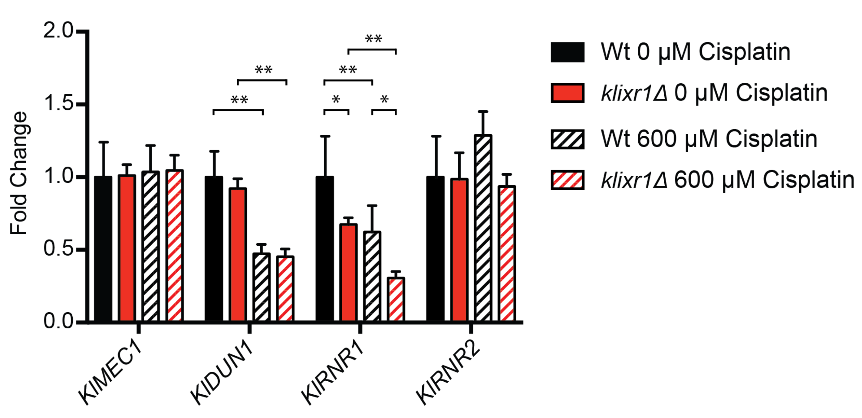
3.7. KlIxr1 and the Regulation of Genes Related to De Novo dNTP Synthesis

In S. cerevisiae, during the normal cell cycle, the adequate supply of dNTPs, which are necessary for DNA synthesis, is regulated through modulation of the activity of ribonucleotide reductase (RNR) or the expression of the RNR genes [60]. The genes ScRNR1 and ScRNR3 encode the large subunit of RNR and ScRNR2 and ScRNR4 encode the small subunit. Transcriptional regulation of ScRNR3, ScRNR2, and ScRNR4 is repressed by the transcriptional factor ScCrt1 (Regulatory Factor X 1) and its phosphorylation, which is ScMec1-ScRad53-ScDun1–dependent after DNA damage or replication stress, promotes activation of their transcription [61]. The kinase activity of ScDun1 (DNA-damage Uninducible 1) also targets for degradation ScSml1 (Suppressor of Mec1 Lethality 1), a protein inhibitor of RNR, and ScDif1 (Damage-regulated Import Facilitator 1), a protein that regulates the nuclear retention of ScRnr2 and ScRnr4, thus promoting RNR activity [62]. ScRox1 and ScMot3 participates in ScRNR3, ScRNR2, and ScRNR4 repression [63]. The transcription of ScRNR1 is independent of ScCrt1, ScRox1, and ScMot3. The deletion of ScIXR1 results in decreased dNTP levels due to a reduced ScRNR1 expression and to ScIxr1 binding to the ScRNR1 promoter. The control of ScRNR1 by ScIxr1 is ScMec1-ScRad53 dependent, but independent of ScDun1 [31].
The biosynthesis of dNTPs has not been studied in K. lactis, but exploring the sequence of the K. lactis genome we have found similarities and differences in the putative homologs of the genes related to the control of the dNTPs pool in comparison with S. cerevisiae. In K. lactis there are two ORFs (KLLA0C07887g and KLLA0F01188g) encoding proteins with significant homology to S. cerevisiae ScRnr1 and ScRnr3, the large subunits of RNR. It is not possible to establish a unique correspondence based merely in their sequence similarity, since KLLA0C07887g is more similar to both ScRnr1 and ScRnr3 than KLLA0F01188g. Regarding the small subunit of RNR in K. lactis, there is only one gene encoding this subunit (KLLA0F15103g). The machinery of signal transduction after DNA-damage or replicative stress (ScRad53, ScMec1 and ScDun1) is well conserved between both yeast species. However, there is low conservation of the transcriptional regulators ScCrt1, ScCrt10 [64] (absent) or other regulatory factors affecting RNR activity like ScSml1 or ScDif1.
RT-qPCR experiments were carried out in order to assess the role of KlIxr1 in the regulation and maintenance of dNTPs pools by analyzing the transcriptional levels of the regulators KlMEC1 (KLLA0C15785g) and KlDUN1 (KLLA0E01585g) and the ribonucleotide reductase genes KlRNR1 and KlRNR2. The results showed that there exists a clear parallelism between transcriptional regulation of this pathway in S. cerevisiae and K. lactis. In this sense, KlRNR1 is downregulated in the K.lactis ixr1Δ strain. However, KlRNR2 and the key regulators KlMEC1 and KlDUN1 (upstream of ScIXR1 in the regulatory pathway of S. cerevisiae) are not affected by KlIXR1 deletion (Figure 8).
Additionally, we have analyzed transcript levels of these genes in the presence of 600 µM cisplatin to induce DNA lesions. The results obtained showed that the expression KlRNR1 and KIRNR2 is increased in the wild-type, meanwhile the reduction of KlRNR1 transcript levels in the Klixr1Δ null strain are even more abrupt (Figure 8), as previously described for S. cerevisiae in the presence of 4-nitroquinoline 1-oxide (4-NQO) or hydroxyurea (HU) [31].

4. Discussion

Nowadays, eukaryotic unicellular models are useful for the easy study of orphan genes without assigned functions and for massive screening of drug targets. Most studies about the hypoxic and oxidative stress responses in eukaryotes, and their connections, were developed initially on S. cerevisiae cells with a predominant fermentative metabolism [34]. S. cerevisiae mutants have been widely used as research models in aging [65] or in human pathologies [66]. Frequently, the mechanisms discovered with this yeast proved to be conserved in multicellular eukaryotes. However, human cells, including those from the nervous system, have usually a respiratory metabolism, differing from S. cerevisiae cells. In this sense, K. lactis arises as an alternative eukaryote model more suitable for studies about the nervous system or their pathologies, since this yeast has a predominantly respiratory metabolism, although sharing essential regulatory machineries with S. cerevisiae [67]. Also important for the selection of K. lactis as an eukaryotic model is the fact that it is evolutionary derived from an ancestral yeast previous to the whole-genome duplication (WGD) that took place over 100 million years ago and resulted later, after genomic losses and chromosome reorganizations, in the existence of two copies of some genes in S. cerevisiae [68]. Each paralog likely sub-functionalized to carry out some, but not all, of its previous functions so that, a more complex functional network arose [69].
HMGB proteins are not extensively studied in K. lactis, although their transcriptional regulatory function might predictably influence the respiratory metabolism of this yeast. In the present work, we show evidence of the cellular functions of the HMGB protein KlIxr1, finding parallelisms with ScIxr1 functions controlling the expression of genes that are modulators of oxidative metabolism and oxidative stress response, dNTP cellular levels, and ribosome biogenesis.
In S. cerevisiae, one of the principal sensors in the yeast response to changes in oxygen levels is heme, since its enzymatic synthesis is oxygen-dependent. The heme biosynthetic pathway is well conserved between S. cerevisiae and K. lactis. Both yeasts have eight genes encoding the enzymes for the biosynthesis of heme, and up to three (KlHEM1, KlHEM12, and KlHEM13) share functional equivalence confirmed experimentally by cross-complementation [70,71,72]. Although K. lactis homologs of key regulators (ScHap1 and ScRox1) of the biosynthesis of heme in S. cerevisiae have been characterized, their sequence and function diverge notably from those described in S. cerevisiae [35,73]. Here, we show that transcriptional activation of KlHEM13 is also dependent on the HMGB protein KlIxr1p, as occurred with ScIxr1 in S. cerevisiae [24]. Moreover, both proteins share similar DNA binding sequences in their target promoters, as shown by EMSA and fluorescence anisotropy experiments.
Several S. cerevisiae genes that are induced during hypoxia are also related to the oxidative stress response caused by hydrogen peroxide or metals, including ScCUP1 (CUPrum 1), ScCUP2 (CUPrum 1), ScHSP12 (Heat Shock Protein 12), ScFMP46 (Found in Mitochondrial Proteome 46), ScGRE1 (stress Responsive Gene 1), ScALK1, ScMGM1 (Mitochondrial Genome Maintenance 1), or ScSOD1 (SuperOxide Dismutase 1), among others [51,65,74]. In this sense, we confirmed that depletion of KlIxr1 protein in K. lactis also produces an increased sensitivity to the presence of hydrogen peroxide and cadmium metal, as occurred in S. cerevisiae [23,75]. Furthermore, we show for the first time that KlIxr1 is implied in the transcriptional regulation of the KlYCF1 gene, involved in cadmium detoxification, and binds to its promoter. Interestingly, other regulators of the respiratory metabolism in response to changes of oxygen levels, KlHap1 and KlRox1, have been previously related to the oxidative stress response in K. lactis [30,37].
It was previously described that the role of ScIxr1 is as a “regulator of regulators” directly or indirectly controlling the expression of a set of 33 yeast transcriptional factors mostly involved in the regulation of cell growth and cell cycle progression. These transcriptional regulators are downregulated in the Scixr1Δ mutant strain [29]. In S. cerevisiae, these regulatory networks sense nutrient availability, external stimuli, or DNA damage through TOR complexes known as Tor Complex 1 (ScTORC1, TOR complex 1) and Tor Complex 2 (ScTORC2, TOR complex 2) [76]. ScTORC1 is sensitive to rapamycin and controls protein synthesis, mRNA synthesis and degradation, ribosome biogenesis, and autophagy. Rapamycin is currently used clinically as an immunosuppressive drug for organ and tissue transplant recipients and a chemotherapy agent against a variety of solid cancers because of its antiproliferative properties [77]. S. cerevisiae cells treated with rapamycin display phenotypes associated with nutrient depletion including G1 cell cycle arrest, cellular volume expansion, protein synthesis inhibition, glycogen accumulation, and autophagy [78]. In K. lactis, although only a TOR complex exists, we have found that KlIxr1 is also implied in the control of ribosomal production and cellular growth by regulation of rRNA levels, as well as transcription of several RiBi genes and other transcriptional factors that modulate ribosomal protein gene expression, including KlSFP1, KlCRF1, KlTOR1, or KlIFH1.
Cellular division depends on the maintenance of dNTP pools for DNA synthesis and genes encoding ribonucleotide reductases (RNR) are tightly regulated [26]. We have also shown that KlIxr1 modulates transcriptional activation of the KlRNR1 gene, a mechanism previously described in S. cerevisiae [31]. RNR expression is tightly controlled by the genome integrity checkpoint, a conserved signaling pathway that is regulated in S. cerevisiae by the ScMec1 (homologous to human ATR) and ScRad53 (homologous to human Chk1) kinases. Under DNA damage or DNA replication stress, this pathway promotes the activation of ScRNR2, ScRNR3, and ScRNR4 through the ScMec1-ScRad53-ScDun1 kinase cascade, which targets and inactivates the transcriptional inhibitor ScCrt1 [62]. However, upregulation of ScRNR1 in response to DNA damage requires ScMec1 and ScRad53, but not ScDun1. If this molecular mechanism is also maintained, K. lactis will need to be elucidated in future experiments.
It is interesting to note that despite all similitudes found between KlIxr1 and ScIxr1, showing that KlIxr1 is also a master regulator of oxidative metabolism, oxidative stress response, cell growth, and cell division in K. latis, several functions are not conserved between ScIxr1 and KlIXr1. We have found that KlIxr1p is not able to supplement the absence of ScIxr1 in S. cerevisiae and unable to revert the phenotype of increased resistance to cisplatin treatment associated to the null mutation [37]. Furthermore, KlIXR1 gene knockout generates the opposite phenotype in K. lactis, conferring a sharp sensitivity to cisplatin treatment. Some transcriptional regulators that were previously related to cisplatin resistance or ribosomal biosynthesis in S. cerevisiae [73,74,75] are also differently regulated by ScIxr1 and KlIxr1 in both species, respectively. We have observed these differences comparing mRNA changes of KlABF1, KlUME6, or KlDAL81 after KlIxr1 depletion with those previously reported in S. cerevisiae after ScIXr1 depletion [29]. Related to this, we have found that KlIxr1 depletion causes a resistant phenotype to cyclohexymide not previously described for the Scixr1∆ null mutant, and that could be associated to alterations of certain ribosomal protein targets of the drug [76], among other causes. Future structural studies are necessary to dilucidate if non-conserved domains in the Ixr1 proteins from S. cerevisiae and K. lactis are responsible of differences observed in their functions in reference to responses to cisplatin or cyclohexymide treatments.

5. Conclusions

Taken together with the data reported here, Ixr1 from K. lactis shows similarities with its S. cerevisiae counterpart to how these two proteins participate in the transcriptional regulation of genes related to cellular growth, dNTP levels, and ribosome biosynthesis, as well as of genes implicated in the cellular response changes in oxygen availability, which consequently alters cellular levels of heme and ROS. This supports the idea that these regulation mechanisms are essentially well conserved for Ixr1 in S. cerevisiae and K. lactis yeasts, which differ in lifestyle with respect to carbon sources. However, the finding of new roles for KlIxr1 protein in the response to platinum compounds and the occurrence of a petite phenotype after KlIXR1 gene deletion reveal that it is necessary to further understanding of the mechanisms of KlIxr1 function and also a broader knowledge of the regulatory pathways associated to all these processes in K. lactis.

Supplementary Materials

The following are available online at https://www.mdpi.com/article/10.3390/biom11091392/s1, Table S1: Oligonucleotides used in this study; Figure S1: Verification of MW190-9b klixr1Δ null strain by PCR and agarose gel electrophoresis; Figure S2: SDS-PAGE gel of KlIxr1 purification.

Author Contributions

All the authors contributed to conceptualization and design of experiments. A.R.-D. and A.B.-A. created all the constructs, null mutants, and conducted cell culture; A.R.-D. and M.L.-M. ran the experiments for transcriptional analysis by Northern blot and qPCR experiments; C.R.-S. performed protein purification, EMSA and FA assays; M.E.C. and Á.V.-V. performed data analysis and interpretation of the experimental results; A.R.-D., M.B., M.E.C. and Á.V.-V. wrote the original draft and figures. All authors have read and agreed to the published version of the manuscript.

Funding

This work has been funded by the contract ED431C 2020–08 from Xunta de Galicia (Consolidación Grupos Referencia Competitiva). A.R.-D. was funded by a research fellowship from Diputacion da Coruña-2017 (Spain).

Institutional Review Board Statement

Not applicable.

Informed Consent Statement

Not applicable.

Data Availability Statement

Not applicable.

Conflicts of Interest

The authors declare no conflict of interest.

References

  1. Dhakal, S.; Macreadie, I. Protein Homeostasis Networks and the Use of Yeast to Guide Interventions in Alzheimer’s Disease. Int. J. Mol. Sci. 2020, 21, 8014. [Google Scholar] [CrossRef]
  2. Chernova, T.A.; Chernoff, Y.O.; Wilkinson, K.D. Yeast Models for Amyloids and Prions: Environmental Modulation and Drug Discovery. Molecules 2019, 24, 3388. [Google Scholar] [CrossRef] [PubMed] [Green Version]
  3. Rzepnikowska, W.; Kaminska, J.; Kabzińska, D.; Binięda, K.; Kochański, A. A Yeast-Based Model for Hereditary Motor and Sensory Neuropathies: A Simple System for Complex, Heterogeneous Diseases. Int. J. Mol. Sci. 2020, 21, 4277. [Google Scholar] [CrossRef] [PubMed]
  4. Wendland, J. Special Issue: Non-Conventional Yeasts: Genomics and Biotechnology. Microorganisms 2020, 8, 21. [Google Scholar] [CrossRef] [PubMed] [Green Version]
  5. Rebello, S.; Abraham, A.; Madhavan, A.; Sindhu, R.; Binod, P.; Karthika Bahuleyan, A.; Aneesh, E.M.; Pandey, A. Non-conventional yeast cell factories for sustainable bioprocesses. FEMS Microbiol. Lett. 2018, 365. [Google Scholar] [CrossRef] [PubMed]
  6. Vizoso-Vázquez, A.; Lamas-Maceiras, M.; Fernández-Leiro, R.; Rico-Díaz, A.; Becerra, M.; Cerdán, M.E. Dual function of Ixr1 in transcriptional regulation and recognition of cisplatin-DNA adducts is caused by differential binding through its two HMG-boxes. Biochim. Biophys. Acta-Gene Regul. Mech. 2017, 1860, 256–269. [Google Scholar] [CrossRef]
  7. Ye, Y.; Zeng, Z.; Jin, T.; Zhang, H.; Xiong, X.; Gu, L. The Role of High Mobility Group Box 1 in Ischemic Stroke. Front. Cell. Neurosci. 2019, 13, 127. [Google Scholar] [CrossRef] [Green Version]
  8. Štros, M.; Kučírek, M.; Sani, S.A.; Polanská, E. HMGB1-mediated DNA bending: Distinct roles in increasing p53 binding to DNA and the transactivation of p53-responsive gene promoters. Biochim. Biophys. Acta-Gene Regul. Mech. 2018, 1861, 200–210. [Google Scholar] [CrossRef] [PubMed]
  9. Tang, D.; Kang, R.; Livesey, K.M.; Cheh, C.-W.; Farkas, A.; Loughran, P.; Hoppe, G.; Bianchi, M.E.; Tracey, K.J.; Zeh III, H.J.; et al. Endogenous HMGB1 regulates autophagy. J. Cell Biol. 2010, 190, 881–892. [Google Scholar] [CrossRef]
  10. Tang, D.; Kang, R.; Livesey, K.M.; Kroemer, G.; Billiar, T.R.; Van Houten, B.; Zeh III, H.J.; Lotze, M.T. High-Mobility Group Box 1 Is Essential for Mitochondrial Quality Control. Cell Metab. 2011, 13, 701–711. [Google Scholar] [CrossRef] [Green Version]
  11. Livesey, K.M.; Kang, R.; Zeh Herbert, J., III; Lotze, M.T.; Tang, D. Direct molecular interactions between HMGB1 and TP53 in colorectal cancer. Autophagy 2012, 8, 846–848. [Google Scholar] [CrossRef] [PubMed] [Green Version]
  12. Andersson, U.; Yang, H.; Harris, H. High-mobility group box 1 protein (HMGB1) operates as an alarmin outside as well as inside cells. Semin. Immunol. 2018, 38, 40–48. [Google Scholar] [CrossRef] [PubMed]
  13. Niu, L.; Yang, W.; Duan, L.; Wang, X.; Li, Y.; Xu, C.; Liu, C.; Zhang, Y.; Zhou, W.; Liu, J.; et al. Biological functions and theranostic potential of HMGB family members in human cancers. Ther. Adv. Med. Oncol. 2020, 12, 1758835920970850. [Google Scholar] [CrossRef] [PubMed]
  14. Zhao, Z.; Hu, Z.; Zeng, R.; Yao, Y. HMGB1 in kidney diseases. Life Sci. 2020, 259, 118203. [Google Scholar] [CrossRef]
  15. Kaur, I.; Behl, T.; Bungau, S.; Kumar, A.; Mehta, V.; Setia, D.; Uddin, M.S.; Zengin, G.; Aleya, L.; Arora, S. Exploring the therapeutic promise of targeting HMGB1 in rheumatoid arthritis. Life Sci. 2020, 258, 118164. [Google Scholar] [CrossRef]
  16. Paudel, Y.N.; Angelopoulou, E.; Piperi, C.; Othman, I.; Aamir, K.; Shaikh, M.F. Impact of HMGB1, RAGE, and TLR4 in Alzheimer’s Disease (AD): From Risk Factors to Therapeutic Targeting. Cells 2020, 9, 383. [Google Scholar] [CrossRef] [PubMed] [Green Version]
  17. Vijayakumar, E.C.; Bhatt, L.K.; Prabhavalkar, K.S. High Mobility Group Box-1 (HMGB1): A Potential Target in Therapeutics. Curr. Drug Targets 2019, 20, 1474–1485. [Google Scholar] [CrossRef] [PubMed]
  18. Flores, C.-L.; Rodríguez, C.; Petit, T.; Gancedo, C. Carbohydrate and energy-yielding metabolism in non-conventional yeasts1. FEMS Microbiol. Rev. 2000, 24, 507–529. [Google Scholar] [CrossRef] [PubMed] [Green Version]
  19. González-Siso, M.I.; García-Leiro, A.; Tarrío, N.; Cerdán, M.E. Sugar metabolism, redox balance and oxidative stress response in the respiratory yeast Kluyveromyces lactis. Microb. Cell Fact. 2009, 8, 46. [Google Scholar] [CrossRef] [Green Version]
  20. González–Siso, M.I.; Freire–Picos, M.A.; Ramil, E.; González-Domínguez, M.; Rodríguez Torres, A.; Cerdán, M.E. Respirofermentative metabolism in Kluyveromyces lactis: Insights and perspectives. Enzyme Microb. Technol. 2000, 26, 699–705. [Google Scholar] [CrossRef]
  21. Lambert, J.R.; Bilanchone, V.W.; Cumsky, M.G. The ORD1 gene encodes a transcription factor involved in oxygen regulation and is identical to IXR1, a gene that confers cisplatin sensitivity to Saccharomyces cerevisiae. Proc. Natl. Acad. Sci. USA 1994, 91, 7345–7349. [Google Scholar] [CrossRef] [PubMed] [Green Version]
  22. Bourdineaud, J.-P.; De Sampaïo, G.; Lauquin, G.J.-M. A Rox1-independent hypoxic pathway in yeast. Antagonistic action of the repressor Ord1 and activator Yap1 for hypoxic expression of the SRP1/TIR1 gene. Mol. Microbiol. 2000, 38, 879–890. [Google Scholar] [CrossRef] [Green Version]
  23. Castro-Prego, R.; Lamas-Maceiras, M.; Soengas, P.; Carneiro, I.; González-Siso, I.; Cerdán, M.E. Regulatory factors controlling transcription of Saccharomyces cerevisiae IXR1 by oxygen levels: A model of transcriptional adaptation from aerobiosis to hypoxia implicating ROX1 and IXR1 cross-regulation. Biochem. J. 2009, 425, 235–243. [Google Scholar] [CrossRef] [Green Version]
  24. Castro-Prego, R.; Lamas-Maceiras, M.; Soengas, P.; Fernández-Leiro, R.; Carneiro, I.; Becerra, M.; González-Siso, M.I.; Cerdán, M.E. Ixr1p regulates oxygen-dependent HEM13 transcription. FEMS Yeast Res. 2010, 10, 309–321. [Google Scholar] [CrossRef] [PubMed] [Green Version]
  25. Vizoso-Vázquez, Á.; Lamas-Maceiras, M.; Becerra, M.; González-Siso, M.I.; Rodríguez-Belmonte, E.; Cerdán, M.E. Ixr1p and the control of the Saccharomyces cerevisiae hypoxic response. Appl. Microbiol. Biotechnol. 2012, 94, 173–184. [Google Scholar] [CrossRef] [PubMed]
  26. Klinkenberg, L.G.; Mennella, T.A.; Luetkenhaus, K.; Zitomer, R.S. Combinatorial Repression of the Hypoxic Genes of Saccharomyces cerevisiae by DNA Binding Proteins Rox1 and Mot3. Eukaryot. Cell 2005, 4, 649–660. [Google Scholar] [CrossRef] [PubMed] [Green Version]
  27. Brown, S.J.; Kellett, P.J.; Lippard, S.J. Ixr1, a yeast protein that binds to platinated DNA and confers sensitivity to cisplatin. Science 1993, 261, 603–605. [Google Scholar] [CrossRef] [PubMed]
  28. McA’Nulty, M.M.; Lippard, S.J. The HMG-domain protein Ixr1 blocks excision repair of cisplatin-DNA adducts in yeast. Mutat. Res. Repair 1996, 362, 75–86. [Google Scholar] [CrossRef]
  29. Vizoso-Vázquez, Á.; Lamas-Maceiras, M.; González-Siso, M.I.; Cerdán, M.E. Ixr1 Regulates Ribosomal Gene Transcription and Yeast Response to Cisplatin. Sci. Rep. 2018, 8, 3090. [Google Scholar] [CrossRef] [Green Version]
  30. Vizoso-Vázquez, A.; Barreiro-Alonso, A.; González-Siso, M.I.; Rodríguez-Belmonte, E.; Lamas-Maceiras, M.; Cerdán, M.E. HMGB proteins involved in TOR signaling as general regulators of cell growth by controlling ribosome biogenesis. Curr. Genet. 2018, 64, 1205–1213. [Google Scholar] [CrossRef]
  31. Tsaponina, O.; Barsoum, E.; Åström, S.U.; Chabes, A. Ixr1 Is Required for the Expression of the Ribonucleotide Reductase Rnr1 and Maintenance of dNTP Pools. PLoS Genet. 2011, 7, e1002061. [Google Scholar] [CrossRef] [Green Version]
  32. Ros-Carrero, C.; Ramos-Alonso, L.; Romero, A.M.; Bañó, M.C.; Martínez-Pastor, M.T.; Puig, S. The yeast Aft1 transcription factor activates ribonucleotide reductase catalytic subunit RNR1 in response to iron deficiency. Biochim. Biophys. Acta-Gene Regul. Mech. 2020, 1863, 194522. [Google Scholar] [CrossRef]
  33. Tsaponina, O.; Chabes, A. Pre-activation of the genome integrity checkpoint increases DNA damage tolerance. Nucleic Acids Res. 2013, 41, 10371–10378. [Google Scholar] [CrossRef]
  34. Breunig, K.D.; Bolotin-Fukuhara, M.; Bianchi, M.M.; Bourgarel, D.; Falcone, C.; Ferrero, I.; Frontali, L.; Goffrini, P.; Krijger, J.J.; Mazzoni, C.; et al. Regulation of primary carbon metabolism in Kluyveromyces lactis. Enzyme Microb. Technol. 2000, 26, 771–780. [Google Scholar] [CrossRef]
  35. Rodríguez Torres, A.M.; Lamas Maceiras, M.; Rodríguez Belmonte, E.; Núñez Naveira, L.; Blanco Calvo, M.; Cerdán, M.E. KlRox1p contributes to yeast resistance to metals and is necessary for KlYCF1 expression in the presence of cadmium. Gene 2012, 497, 27–37. [Google Scholar] [CrossRef] [PubMed]
  36. Fang, Z.-A.; Wang, G.-H.; Chen, A.-L.; Li, Y.-F.; Liu, J.-P.; Li, Y.-Y.; Bolotin-Fukuhara, M.; Bao, W.-G. Gene Responses to Oxygen Availability in Kluyveromyces lactis: An Insight on the Evolution of the Oxygen-Responding System in Yeast. PLoS ONE 2009, 4, e7561. [Google Scholar] [CrossRef] [PubMed] [Green Version]
  37. Rodríguez Lombardero, S.; Vizoso Vázquez, Á.; Rodríguez Belmonte, E.; González Siso, M.I.; Cerdán, M.E. SKY1 and IXR1 interactions, their effects on cisplatin and spermine resistance in Saccharomyces cerevisiae. Can. J. Microbiol. 2012, 58, 184–188. [Google Scholar] [CrossRef] [PubMed]
  38. Zitomer, R.S.; Hall, B.D. Yeast cytochrome c messenger RNA. In vitro translation and specific immunoprecipitation of the CYC1 gene product. J. Biol. Chem. 1976, 251, 6320–6326. [Google Scholar] [CrossRef]
  39. Ito, H.; Fukuda, Y.; Murata, K.; Kimura, A. Transformation of intact yeast cells treated with alkali cations. J. Bacteriol. 1983, 153, 163–168. [Google Scholar] [CrossRef] [PubMed] [Green Version]
  40. Gietz, R.D.; Akio, S. New yeast-Escherichia coli shuttle vectors constructed with in vitro mutagenized yeast genes lacking six-base pair restriction sites. Gene 1988, 74, 527–534. [Google Scholar] [CrossRef]
  41. Wach, A.; Brachat, A.; Pöhlmann, R.; Philippsen, P. New heterologous modules for classical or PCR-based gene disruptions in Saccharomyces cerevisiae. Yeast 1994, 10, 1793–1808. [Google Scholar] [CrossRef]
  42. Tizón, B.; Rodríguez-Torres, A.M.; Cerdán, M.E. Disruption of six novel Saccharomyces cerevisiae genes reveals that YGL129c is necessary for growth in non-fermentable carbon sources, YGL128c for growth at low or high temperatures and YGL125w is implicated in the biosynthesis of methionine. Yeast 1999, 15, 145–154. [Google Scholar] [CrossRef]
  43. Rodríguez-Lombardero, S.; Vizoso-Vázquez, Á.; Lombardía, L.J.; Becerra, M.; González-Siso, M.I.; Cerdán, M.E. Sky1 regulates the expression of sulfur metabolism genes in response to cisplatin. Microbiology 2014, 160, 1357–1368. [Google Scholar] [CrossRef]
  44. Livak, K.J.; Schmittgen, T.D. Analysis of Relative Gene Expression Data Using Real-Time Quantitative PCR and the 2−ΔΔCT Method. Methods 2001, 25, 402–408. [Google Scholar] [CrossRef] [PubMed]
  45. Kelley, L.A.; Mezulis, S.; Yates, C.M.; Wass, M.N.; Sternberg, M.J.E. The Phyre2 web portal for protein modeling, prediction and analysis. Nat. Protoc. 2015, 10, 845–858. [Google Scholar] [CrossRef] [Green Version]
  46. Zhang, Y.; Skolnick, J. TM-align: A protein structure alignment algorithm based on the TM-score. Nucleic Acids Res. 2005, 33, 2302–2309. [Google Scholar] [CrossRef]
  47. Albert, B.; Tomassetti, S.; Gloor, Y.; Dilg, D.; Mattarocci, S.; Kubik, S.; Hafner, L.; Shore, D. Sfp1 regulates transcriptional networks driving cell growth and division through multiple promoter-binding modes. Genes Dev. 2019, 33, 288–293. [Google Scholar] [CrossRef] [PubMed] [Green Version]
  48. Martin, D.E.; Soulard, A.; Hall, M.N. TOR Regulates Ribosomal Protein Gene Expression via PKA and the Forkhead Transcription Factor FHL1. Cell 2004, 119, 969–979. [Google Scholar] [CrossRef] [PubMed] [Green Version]
  49. Fermi, B.; Bosio, M.C.; Dieci, G. Promoter architecture and transcriptional regulation of Abf1-dependent ribosomal protein genes in Saccharomyces cerevisiae. Nucleic Acids Res. 2016, 44, 6113–6126. [Google Scholar] [CrossRef] [Green Version]
  50. Urban, J.; Soulard, A.; Huber, A.; Lippman, S.; Mukhopadhyay, D.; Deloche, O.; Wanke, V.; Anrather, D.; Ammerer, G.; Riezman, H.; et al. Sch9 Is a Major Target of TORC1 in Saccharomyces cerevisiae. Mol. Cell 2007, 26, 663–674. [Google Scholar] [CrossRef] [PubMed]
  51. Dirmeier, R.; O’Brien, K.M.; Engle, M.; Dodd, A.; Spears, E.; Poyton, R.O. Exposure of yeast cells to anoxia induces transient oxidative stress. Implications for the induction of hypoxic genes. J. Biol. Chem. 2002, 277, 34773–34784. [Google Scholar] [CrossRef] [PubMed] [Green Version]
  52. Hosiner, D.; Gerber, S.; Lichtenberg-Fraté, H.; Glaser, W.; Schüller, C.; Klipp, E. Impact of Acute Metal Stress in Saccharomyces cerevisiae. PLoS ONE 2014, 9, e83330. [Google Scholar] [CrossRef]
  53. Li, Z.-S.; Lu, Y.-P.; Zhen, R.-G.; Szczypka, M.; Thiele, D.J.; Rea, P.A. A New Pathway for Vacuolar Cadmium Sequestration in Saccharomyces cerevisiae: YCF1-Catalyzed Transport of bis(glutathionato)cadmium. Proc. Natl. Acad. Sci. USA 1997, 94, 42–47. [Google Scholar] [CrossRef] [PubMed] [Green Version]
  54. Tommasini, R.; Evers, R.; Vogt, E.; Mornet, C.; Zaman, G.J.; Schinkel, A.H.; Borst, P.; Martinoia, E. The human multidrug resistance-associated protein functionally complements the yeast cadmium resistance factor 1. Proc. Natl. Acad. Sci. USA 1996, 93, 6743–6748. [Google Scholar] [CrossRef] [Green Version]
  55. Wei, W.; Smith, N.; Wu, X.; Kim, H.; Seravalli, J.; Khalimonchuk, O.; Lee, J. YCF1-Mediated Cadmium Resistance in Yeast Is Dependent on Copper Metabolism and Antioxidant Enzymes. Antioxid. Redox Signal. 2014, 21, 1475–1489. [Google Scholar] [CrossRef] [PubMed] [Green Version]
  56. Brückner, S.; Kern, S.; Birke, R.; Saugar, I.; Ulrich, H.D.; Mösch, H.-U. The TEA Transcription Factor Tec1 Links TOR and MAPK Pathways to Coordinate Yeast Development. Genetics 2011, 189, 479–494. [Google Scholar] [CrossRef] [Green Version]
  57. Galit, S.; Yona, K. A Positive Regulator of Mitosis, Sok2, Functions as a Negative Regulator of Meiosis in Saccharomyces cerevisiae. Mol. Cell. Biol. 2001, 21, 1603–1612. [Google Scholar] [CrossRef] [Green Version]
  58. Hanlon, S.E.; Rizzo, J.M.; Tatomer, D.C.; Lieb, J.D.; Buck, M.J. The Stress Response Factors Yap6, Cin5, Phd1, and Skn7 Direct Targeting of the Conserved Co-Repressor Tup1-Ssn6 in S. cerevisiae. PLoS ONE 2011, 6, e19060. [Google Scholar] [CrossRef] [Green Version]
  59. Tam, J.; van Werven, F.J. Regulated repression governs the cell fate promoter controlling yeast meiosis. Nat. Commun. 2020, 11, 2271. [Google Scholar] [CrossRef] [PubMed]
  60. Desany, B.A.; Alcasabas, A.A.; Bachant, J.B.; Elledge, S.J. Recovery from DNA replicational stress is the essential function of the S-phase checkpoint pathway. Genes Dev. 1998, 12, 2956–2970. [Google Scholar] [CrossRef] [Green Version]
  61. Zaim, J.; Speina, E.; Kierzek, A.M. Identification of New Genes Regulated by the Crt1 Transcription Factor, an Effector of the DNA Damage Checkpoint Pathway in Saccharomyces cerevisiae. J. Biol. Chem. 2005, 280, 28–37. [Google Scholar] [CrossRef] [Green Version]
  62. Huang, M.; Zhou, Z.; Elledge, S.J. The DNA replication and damage checkpoint pathways induce transcription by inhibition of the Crt1 repressor. Cell 1998, 94, 595–605. [Google Scholar] [CrossRef] [Green Version]
  63. Klinkenberg, L.G.; Webb, T.; Zitomer, R.S. Synergy among differentially regulated repressors of the ribonucleotide diphosphate reductase genes of Saccharomyces cerevisiae. Eukaryot. Cell 2006, 5, 1007–1017. [Google Scholar] [CrossRef] [Green Version]
  64. Fu, Y.; Xiao, W. Identification and characterization of CRT10 as a novel regulator of Saccharomyces cerevisiae ribonucleotide reductase genes. Nucleic Acids Res. 2006, 34, 1876–1883. [Google Scholar] [CrossRef] [Green Version]
  65. Lepperdinger, G.; Berger, P.; Breitenbach, M.; Frohlich, K.U.; Grillari, J.; Grubeck-Loebenstein, B.; Madeo, F.; Minois, N.; Zwerschke, W.; Jansen-Durr, P. The use of genetically engineered model systems for research on human aging. Front. Biosci. 2008, 13, 7022–7031. [Google Scholar] [CrossRef] [PubMed] [Green Version]
  66. Petranovic, D.; Nielsen, J. Can yeast systems biology contribute to the understanding of human disease? Trends Biotechnol. 2008, 26, 584–590. [Google Scholar] [CrossRef] [PubMed]
  67. González Siso, M.I.; Cerdán, M.E. Kluyveromyces lactis: A Suitable Yeast Model to Study Cellular Defense Mechanisms against Hypoxia-Induced Oxidative Stress. Oxid. Med. Cell. Longev. 2012, 2012, 634674. [Google Scholar] [CrossRef] [PubMed] [Green Version]
  68. Wolfe, K.H. Origin of the Yeast Whole-Genome Duplication. PLoS Biol. 2015, 13, 1–7. [Google Scholar] [CrossRef] [PubMed] [Green Version]
  69. Shertz, C.A.; Bastidas, R.J.; Li, W.; Heitman, J.; Cardenas, M.E. Conservation, duplication, and loss of the Tor signaling pathway in the fungal kingdom. BMC Genom. 2010, 11, 510. [Google Scholar] [CrossRef] [PubMed] [Green Version]
  70. Núñez, L.; Rodríguez-Torres, A.; Cerdán, M.E. Regulatory elements in the KlHEM1 promoter. Biochim. Biophys. Acta-Gene Regul. Mech. 2008, 1779, 128–133. [Google Scholar] [CrossRef] [PubMed]
  71. González-Domínguez, M.; Méndez-Carro, C.; Esperanza Cerdán, M. Isolation and Characterization of the KlHEM1 Gene in Kluyveromyces lactis. Yeast 1997, 13, 961–971. [Google Scholar] [CrossRef]
  72. Blanco, M.; Becerra, M.; González-Siso, M.I.; Cerdán, M.E. Functional characterization of KlHEM13, a hypoxic gene of Kluyveromyces lactis. Can. J. Microbiol. 2005, 51, 241–249. [Google Scholar] [CrossRef] [PubMed]
  73. Lamas-Maceiras, M.; Núñez, L.; Rodríguez-Belmonte, E.; González-Siso, M.I.; Cerdán, M.E. Functional characterization of KlHAP1: A model to foresee different mechanisms of transcriptional regulation by Hap1p in yeasts. Gene 2007, 405, 96–107. [Google Scholar] [CrossRef] [PubMed]
  74. Becerra, M.; Lombardía-Ferreira, L.J.; Hauser, N.C.; Hoheisel, J.D.; Tizon, B.; Cerdán, M.E. The yeast transcriptome in aerobic and hypoxic conditions: Effects of hap1, rox1, rox3 and srb10 deletions. Mol. Microbiol. 2002, 43, 545–555. [Google Scholar] [CrossRef] [Green Version]
  75. Leiro, A.; Rodríguez-Lombardero, S.; Vizoso-Vázquez, A.; González Siso, M.I.; Cerdán, M.E. The Yeast Genes ROX1, IXR1, SKY1 and Their Effect upon Enzymatic Activities Related to Oxidative Stress. Intech 2012, 16, 297–322. [Google Scholar] [CrossRef]
  76. Loewith, R.; Jacinto, E.; Wullschleger, S.; Lorberg, A.; Crespo, J.L.; Bonenfant, D.; Oppliger, W.; Jenoe, P.; Hall, M.N. Two TOR Complexes, Only One of which Is Rapamycin Sensitive, Have Distinct Roles in Cell Growth Control. Mol. Cell 2002, 10, 457–468. [Google Scholar] [CrossRef]
  77. Tsang, C.K.; Qi, H.; Liu, L.F.; Zheng, X.F.S. Targeting mammalian target of rapamycin (mTOR) for health and diseases. Drug Discov. Today 2007, 12, 112–124. [Google Scholar] [CrossRef] [PubMed]
  78. Barbet, N.C.; Schneider, U.; Helliwell, S.B.; Stansfield, I.; Tuite, M.F.; Hall, M.N. TOR controls translation initiation and early G1 progression in yeast. Mol. Biol. Cell 1996, 7, 25–42. [Google Scholar] [CrossRef] [Green Version]
Figure 1. (a) In silico comparison of Ixr1 amino acid sequence from S. cerevisiae and K. lactis.(upper) Scheme of protein domain distribution in Ixr1 protein from S. cerevisiae and K. lactis. (lower) Pairwise alignment of the Ixr1 double HMG-box domains arranged in tandem from S. cerevisiae (Uniprot code P33417) and K. lactis (Uniprot code Q6CMQ4). Conserved amino acids are highlighted in bold and conserved regions indicated by grey boxes. Small and hydrophobic amino acids (less tyrosine) are indicated in red, acidic amino acids in blue, basic amino acids (less histidine) in magenta and hydroxyl + sulfhydryl + amine (STYHCNGQ) in green. (b) Model superposition of HMG-box A (S. cerevisiae in dark green and K. lactis in dark blue) and HMG-box B (S. cerevisiae in light green and K. lactis in light blue). Enlarged frames show the conserved aromatic residues that form the cluster stabilizing the three-helix HMG folding.
Biomolecules 11 01392 g001
Figure 2. Effect of cisplatin treatment on cell growth (in logarithmic and stationary phase) of strain W303 and derivatives. ixr1Δ null strain was transformed with the plasmid pAG426GAL-ccdB, either empty (Ø), or containing the IXR1 ORFs from S. cerevisiae or K. lactis under the control of the GAL1 promoter. Serial dilutions of the cells (1, 10−1, 10−2, 10−3, 10−4, 10−5; from left to right) were made from an initial culture of OD600 = 1.0 and were grown at 30 °C for three days on YPGal plates with different concentrations of cisplatin as specified.
Biomolecules 11 01392 g002
Figure 3. Study of the cellular response of MW190-9b strain (black) and its derivative ixr1Δ (red) to cisplatin treatment. (a) Resistance to cisplatin in the wild type and klixr1Δ null strains. Cells from the K. lactis strain MW190-9B and its isogenic derivative MW190-9B-ixr1Δ were treated with several concentrations of cisplatin (0, 50, 100, 200, 400, and 600 µM). After 24 h of growth at 30 °C, optical density measurements at 600 nm were taken, and survival rates were calculated by normalization from untreated cultures. (b) rRNA cellular content determination by microfluidic-based automated electrophoresis. (c) mRNA levels of KlSFP1, KlCRF1, KlSCH9, KlTOR1, KlRAP1, KlIFH1, KlFHL1, and KlDOT6 were analyzed by RT-qPCR, before (solid) and after (streaked) the treatment with 600 µM of the chemotherapeutic agent cisplatin. Housekeeping gene KlTAF10 was used for gene normalization. * p < 0.05; ** p < 0.01.
Biomolecules 11 01392 g003
Figure 4. Effect of KlIXR1 disruption on KlHEM13 expression under aerobic (solid) or hypoxic conditions (grid) in the MW190-9b strain from K. lactis were analyzed by (a) Northern blot and (b) RT-qPCR experiments. Blots for (a) were obtained in triplicate experiments and a representative picture is shown in upper left panel. KlSNR17A and KlTAF10 signal levels were used as references for normalization in (a,b), respectively. * p < 0.05; ** p < 0.01.
Biomolecules 11 01392 g004
Figure 5. Analysis of KlIxr1 protein binding to the KlHEM13 promoter region. (a) EMSA assays showing sequence specificity to both binding sites found using the consensus sequences YYYATTGTTCTC and KTTSAAYKGTTYASA previously described [23,24]. Competition assays were conducted with increasing amounts of non-labeled ligands indicated. WT: ligand not mutated; M1: ligand mutated in first site; M2: ligand mutated in second site; NB: no retarded band (no protein added). (b) Klotz plots representing quantitative analysis of KlIxr1 binding to both KlHEM13 promoter deduced sites. DNA binding measured by fluorescence anisotropy changes of the 5′ fluorescein-labelled DNA (100 nM ligand) upon protein titration (see Section 2). The resulting semi-log binding isotherms were fitted to a 1:1 binding model with non-linear least squares regression. Data points are the average of 3 independent experiments, error bars representing standard deviations. HEM13A: first binding site deduced from KTTSAAYKGTTYASA [23] consensus sequence; HEM13B: second binding site deduced from YYYATTGTTCTC [24] consensus sequence; * indicates FAM labeled ligand. (c) Klotz plots representing competition analysis of KlIxr1 binding to both KlHEM13 promoter-assayed sites. DNA binding measured by fluorescence anisotropy changes of the 5′ fluorescein-labelled DNA (100 nM ligand) bound to KlIxr1 protein (200 nM) and upon non-labeled DNA competitor titration (see Section 2). The resulting semi-log binding isotherms were fitted to a logIC50 competitive model with non-linear least squares regression. * indicates FAM labeled ligand.
Biomolecules 11 01392 g005
Figure 6. Analysis of the cellular response of MW190-9b strain and its derivative ixr1Δ to hydrogen peroxide and cadmium treatments. (a) Resistance to H2O2 and cadmium in the wild type and klixr1Δ null strains. Serial dilutions of the cells (1, 10−1, 10−2, 10−3, 10−4, 10−5; from left to right) were made from an initial culture of OD600 = 1.0 and were grown at 30 °C for three days on CM plates supplemented with the indicated concentrations of compound. (b) EMSA assays for the KlYCF1 promoter region contains two putative contiguous binding sites for KlIXR1 deduced by using the AYKGTT core consensus sequence [23] in silico searches. Competition assays were conducted with increasing amounts of non-labeled ligands indicated. WT: ligand not mutated; M1: ligand mutated in first site; M2: ligand mutated in second site. (c) Klotz plots representing quantitative analysis of KlIxr1 binding to KlYCF1 promoter-deduced site. DNA binding measured by fluorescence anisotropy changes of the 5′ fluorescein labelled DNA (100 nM ligand) upon protein titration (Section 2). The resulting semi-log binding isotherms were fitted to a 1:1 binding model with non-linear least squares regression. Data points are the average of 3 independent experiments. Competition analysis of KlIxr1 binding to both KlYCF1 promoter deduced sites were measured by fluorescence anisotropy changes of the 5′ fluorescein labelled DNA (100 nM ligand non mutated) bound to KlIxr1 protein (200 nM) and upon non-labeled DNA competitor titration (KlIxr1 binding sites individually mutated). The resulting semi-log binding isotherms were fitted to a logIC50 competitive model with non-linear least squares regression. * indicates FAM labeled ligand.
Biomolecules 11 01392 g006
Figure 7. Analysis of petite phenotype in MW190-9b ixr1Δ null mutant. (a) (left) Colony size of MW190-9b strain and its derivative ixr1Δ in 2% w/v glucose, 2% w/v glycerol, or 2% w/v glucose supplemented with 50 µg/mL hemin. (right) Growth tracking of MW190-9b strain (black) and its derivative ixr1Δ (red) 2% w/v galactose or 2% w/v glycerol during 48h. Growth rates were calculated using respective 2% w/v glucose cultures as normalization elements. (b) RT-qPCR results of KlABF1, KlTEC1, KlSOK2, KlUME6, and KlDAL81 genes in wild-type (black) and ixr1Δ null strain (red), before (solid) and after (streaked) the treatment with 600 µM of the chemotherapeutic agent cisplatin. Housekeeping gene KlTAF10 was used for gene normalization. * p < 0.05; ** p <0.01. (c) Cellular response to cycloheximide treatment. Cell cultures of the K. lactis strain MW190-9B (black) and its isogenic derivative MW190-9B-ixr1Δ (red) were treated with several concentrations of cycloheximide (0, 25, 50, 100, 200, and 300 µg/mL). After 24 h of growth at 30 °C, optical density measurements at 600 nm were taken, and survival rates were calculated by normalization from untreated cultures.
Biomolecules 11 01392 g007
Figure 8. Regulatory role of KlIxr1 in the KlMec1-KlRad53-KlDun1–dependent ribonucleotide reductase pathway of K. lactis. mRNA levels of KlMEC1, KlDUN1, KlRNR1, and KlRNR2 were analyzed by RT-qPCR in the MW190-9b strain (black) and its derivative ixr1Δ (red), before (solid) and after (streaked) the treatment with 600 µM of the chemotherapeutic agent cisplatin. Housekeeping gene KlTAF10 was used for gene normalization. * p < 0.05; ** p < 0.01.
Biomolecules 11 01392 g008
Publisher’s Note: MDPI stays neutral with regard to jurisdictional claims in published maps and institutional affiliations.

