
Postprandial Bioactivity of a Spread Cheese Enriched with Mountain Tea and Orange Peel Extract in Plasma Oxidative Stress Status, Serum Lipids and Glucose Levels: An Interventional Study in Healthy Adults

 ,
,  ,
,  ,
,  ,
,  and
and 
Abstract
:1. Introduction
2. Materials and Methods
2.1. Subjects
2.2. Characterization of the Enriched Spread Cheese Preparation
2.3. Meals
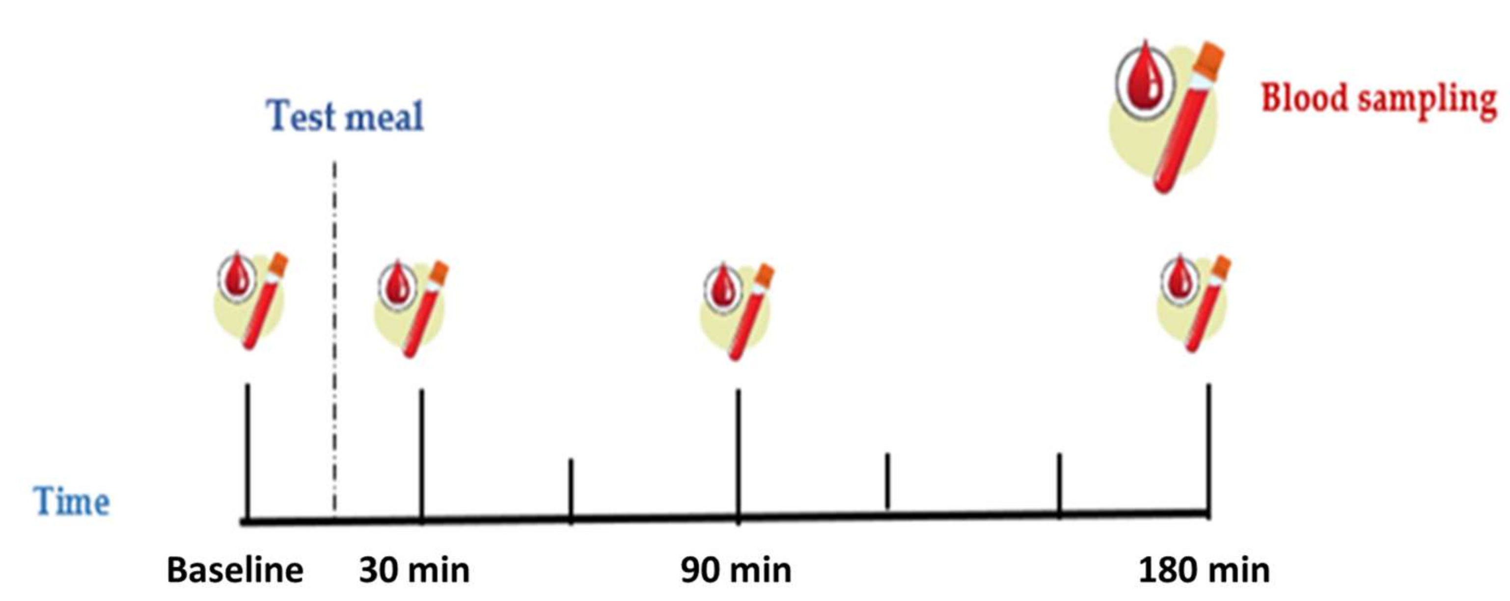
2.4. Study Design
2.5. Chemical Analysis of Blood Samples
2.5.1. Total Antioxidant Capacity and Resistance of Plasma to Copper-Induced Oxidation
2.5.2. Biochemical Parameters
2.6. Statistical Analysis
3. Results
3.1. In Vitro Antioxidant Capacity and Total Phenolic Content of Spread Cheeses and Infusion Extract
3.2. Baseline Characteristics
3.3. Effects of Control and Functional Meal Consumption on Blood Biomarkers
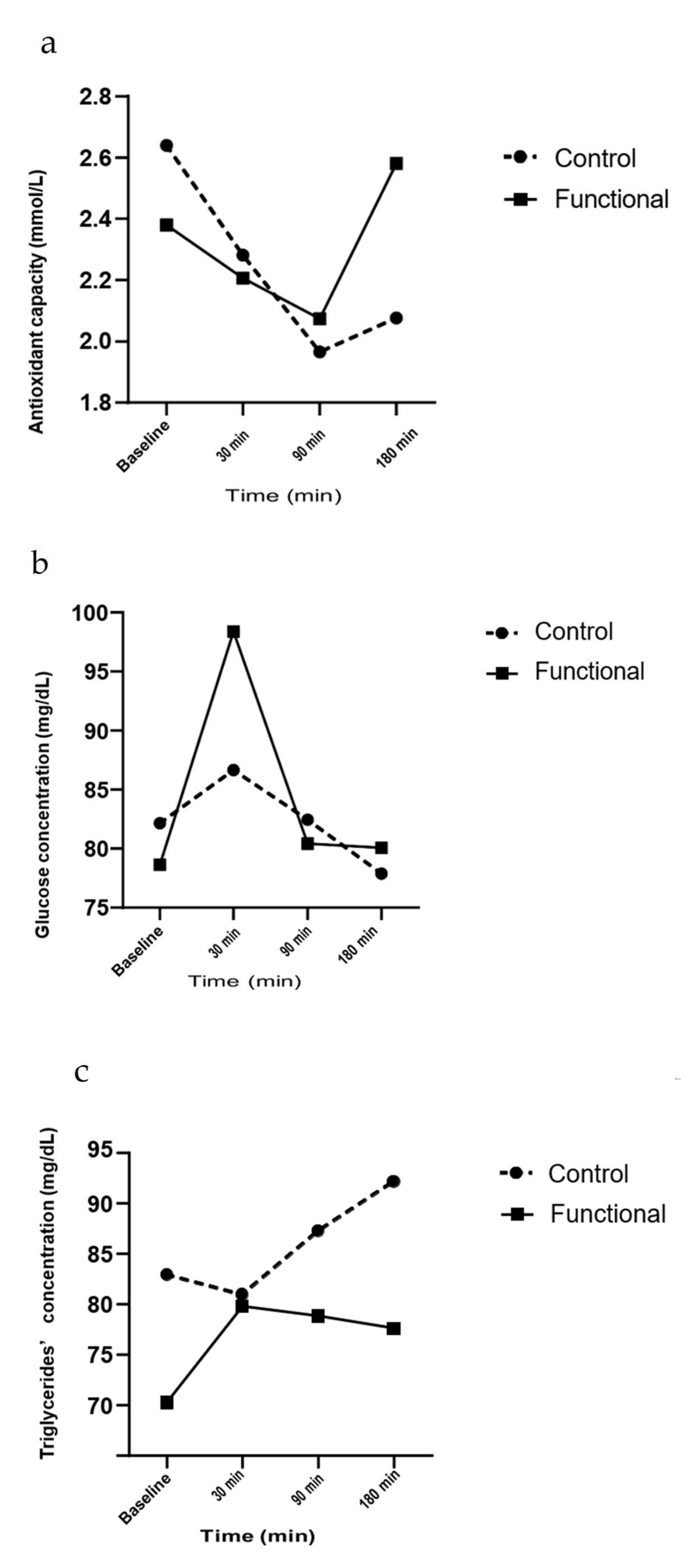
3.4. Effect of Functional Meal Consumption on Plasma Total Antioxidant Capacity
3.5. Effect of Functional Meal Consumption on Plasma Resistance to Oxidation
3.6. Effect of Functional Meal Consumption on Serum Glucose, Lipids and Uric Acid Levels
4. Discussion
5. Conclusions
Author Contributions
Funding
Institutional Review Board Statement
Informed Consent Statement
Data Availability Statement
Acknowledgments
Conflicts of Interest
References
- Tuccar, T.; Akbulut, G. Role of Meal Consumption on Postprandial Oxidative Stress and Inflammation. Gazi. Univ. J. Sci. 2020, 2, 30–37. [Google Scholar]
- Meessen, Ε.; Warmbrunn, M.V.; Nieuwdorp, Μ.; Soeters, Μ.Ρ. Human postprandial nutrient metabolism and low-grade inflammation: A narrative review. Nutrients 2019, 11, 3000. [Google Scholar] [CrossRef] [Green Version]
- Dimina, L.; Mariotti, F. The postprandial appearance of features of cardiometabolic risk: Acute induction and prevention by nutrients and other dietary substances. Nutrients 2019, 11, 1963. [Google Scholar] [CrossRef] [Green Version]
- Leary, Μ.; Tanaka, H. Role of fluid milk in attenuating postprandial hyperglycemia and hypertriglyceridemia. Nutrients 2020, 12, 3806. [Google Scholar] [CrossRef]
- Bozzetto, L.; Della Pepa, G.; Vetrani, C.; Rivellese, A.A. Dietary Impact on Postprandial Lipemia. Front. Endocrinol. (Lausanne) 2020, 11, 337. [Google Scholar] [CrossRef] [PubMed]
- Desmaschelier, C.; Borel, P.; Lairon, D.; Maraninchi, Μ.; Valero, R. Effect of Nutrient and Micronutrient Intake on Chylomicron Production and Postprandial Lipemia. Nutrients 2019, 11, 1299. [Google Scholar] [CrossRef] [PubMed] [Green Version]
- Diamanti-Kandarakis, E.; Papalou, O.; Kandaraki, Ε.A.; Kassi, G. Nutrition as a mediator of oxidative stress in metabolic and reproductive disorders in women. Eur. J. Endocrinol. 2017, 176, R79–R99. [Google Scholar] [CrossRef] [PubMed] [Green Version]
- Dias, C.B.; Moughan, P.J.; Wood, L.G.; Singh, H.; Garg, M.L. Postprandial lipemia: Factoring in lipemic response for ranking foods for their healthiness. Lipids Health Dis. 2017, 16, 178. [Google Scholar] [CrossRef] [Green Version]
- Sirtori, C.R.; Galli, C.; Anderson, J.W.; Sirtori, E.; Arnoldi, A. Functional foods for dyslipidaemia and cardiovascular risk prevention. Nutr. Res. Rev. 2009, 22, 244–261. [Google Scholar] [CrossRef] [Green Version]
- Koutelidakis, A.; Dimou, C. The effects of functional food and bioactive compounds on biomarkers of cardiovascular diseases. In Functional Foods Text Book, 1st ed.; Martirosyan, D., Ed.; Functional Food Center: Dallas, TX, USA, 2017; pp. 89–117. [Google Scholar]
- Martirosyan, D.; Miller, Ε. Bioactive Compounds: The Key to Functional Foods. Bioact. Compd. Health Dis. 2018, 1, 36. [Google Scholar] [CrossRef]
- Bagchi, D. Nutraceutical and Functional Food Regulations in the United States and around the World, 3rd ed.; Academic Press: London, UK, 2019. [Google Scholar]
- Birch, C.S.; Bonwick, G.A. Ensuring the future of functional foods. Int. J. Food Sci. Technol. 2019, 54, 1467–1485. [Google Scholar] [CrossRef]
- Cheng, Y.C.; Sheen, J.M.; Hu, W.L.; Hung, H.M. Polyphenols and Oxidative Stress in Atherosclerosis-Related Ischemic Heart Disease and Stroke. Oxid. Med. Cell Longev. 2017, 2017, 8526438. [Google Scholar] [CrossRef] [Green Version]
- Palma, Χ.; Thomas-Valdés, S.; Cruz, G. Acute Consumption of Blueberries and Short-Term Blueberry Supplementation Improve Glucose Management and Insulin Levels in Sedentary Subjects. Nutrients 2021, 13, 1458. [Google Scholar] [CrossRef] [PubMed]
- Sadowska-Bartosz, I.; Galiniak, S.; Bartosz, G. Kinetics of glycoxidation of bovine serum albumin by glucose, fructose and ribose and its prevention by food components. Molecules 2014, 19, 18828–18849. [Google Scholar] [CrossRef] [Green Version]
- Żyżelewicz, D.; Kulbat-Warycha, Κ.; Oracz, J.; Żyżelewicz, Κ. Polyphenols and other bioactive compounds of sideritis plants and their potential biological activity. Molecules 2020, 25, 3763. [Google Scholar] [CrossRef] [PubMed]
- Kassi, Ε.; Dimas, C.; Dalamaga, Μ.; Panagiotou, A.; Papoutsi, Ζ.; Spilioti, Ε.; Moutsatsou, P. Sideritis euboea extract lowers total cholesterol but not LDL cholesterol in humans: A randomized controlled trial. Clin. Lipidol. 2013, 8, 627–634. [Google Scholar] [CrossRef]
- Chen, X.; Tait, A.; Kitts, D.D. Flavonoid composition of orange peel and its association with antioxidant and anti-inflammatory activities. Food Chem. 2017, 218, 15–21. [Google Scholar] [CrossRef]
- Dimou, C.; Karantonis, H.C.; Skalkos, D.; Koutelidakis, A.Ε. Valorization of Fruits by-products to Unconventional Sources of Additives, Oil, Biomolecules and Innovative Functional Foods. Curr. Pharm. Biotechnol. 2019, 20, 776–786. [Google Scholar] [CrossRef] [PubMed]
- Drouin-Chartier, J.P.; Côté, J.A.; Labonté, Μ.-Ε.; Brassard, D.; Tessier-Grenier, Μ.; Desroches, S.; Couture, P.; Lamarche, Β. Comprehensive review of the impact of dairy foods and dairy fat on cardiometabolic risk. Adv. Nutr. 2016, 7, 1041–1051. [Google Scholar] [CrossRef]
- Lamothe, S.; Langlois, A.; Bazinet, L.; Couillard, C.; Britten, Μ. Antioxidant activity and nutrient release from polyphenol-enriched cheese in a simulated gastrointestinal environment. Food Funct. 2016, 7, 1634–1644. [Google Scholar] [CrossRef]
- Koutelidakis, A.Ε.; Argyri, Κ.; Sevastou, Ζ.; Lamprinaki, D.; Panagopoulou, Ε.; Paximada, Ε.; Sali, A.; Papalazarou, V.; Mallouchos, A.; Evageliou, V.; et al. Bioactivity of Epigallocatechin Gallate Nanoemulsions Evaluated in Mice Model. J. Med. Food 2017, 20, 923–931. [Google Scholar] [CrossRef] [PubMed]
- Pico, J.; Pismag, R.Y.; Laudouze, Μ.; Martinez, Μ.Μ. Systematic evaluation of the Folin-Ciocalteu and Fast Blue BB reactions during the analysis of total phenolics in legumes, nuts and plant seeds. Food Funct. 2020, 11, 9868–9880. [Google Scholar] [CrossRef] [PubMed]
- Karantonis, H.K.; Antonopoulou, S.; Perrea, D.N.; Sokolis, D.P.; Theocharis, S.E.; Kavantzas, Ν.; Iliopoulos, D.G.; Demopoulos, C.A. In vivo antiatherogenic properties of olive oil and its constituent lipid classes in hyperlipidemic rabbits. Nutr. Metab. Cardiovasc. Dis. 2006, 16, 174–185. [Google Scholar] [CrossRef] [PubMed]
- Papagianni, O.; Loukas, Τ.; Magkoutis, A.; Biagki, Τ.; Dimou, C.; Karantonis, C.; Koutelidakis., A. Postprandial Bioactivity of Spread Cheese, Enhanced with Mountain Tea and Orange Peel Extract, in Healthy Volunteers. A Pilot Study. Proceedings 2021, 70, 19. [Google Scholar] [CrossRef]
- Anuyahong, Τ.; Chusak, C.; Thilavech, Τ.; Adisakwattana, S. Postprandial effect of yogurt enriched with anthocyanins from riceberry rice on glycemic response and antioxidant capacity in healthy adults. Nutrients 2020, 12, 2930. [Google Scholar] [CrossRef]
- Koutelidakis, A.Ε.; Rallidis, L.; Koniari, K.; Panagiotakos, D.; Komaitis, M.; Zampelas, A.; Anastasiou-Nana, M.; Kapsokefalou, M. Effect of green tea on postprandial antioxidant capacity, serum lipids, C Reactive Protein and glucose levels in patients with coronary artery disease. Eur. J. Nutr. 2013, 53, 479–486. [Google Scholar] [CrossRef]
- Azman, N.F.; Azlan, A.; Khoo, H.E.; Razman, Μ.R. Antioxidant properties of fresh and frozen peels of citrus species. Curr. Res. Nutr. Food Sci. 2019, 7, 331–339. [Google Scholar] [CrossRef] [Green Version]
- Montero-Calderon, A.; Cortes, A.; Zulueta, A.; Frigola, A.; Esteve, Μ. Green solvents and Ultrasound-Assisted Extraction of bioactive orange (Citrus sinensis) peel compounds. Sci. Rep. 2019, 9, 16120. [Google Scholar] [CrossRef]
- Cardoso, A.; Di Pietro, P.F.; Kunradi Vieira, F.; Bremer Boaventura, B.C.; de Liza, S.; Campelo Borges, G.; Fett, R.; Andrade, D.; da Silva, E.L. Acute consumption of juçara juice (Euterpe edulis) and antioxidant activity in healthy individuals. J. Funct. Foods 2015, 17, 152–162. [Google Scholar] [CrossRef]
- Tasaki, E.; Sakurai, H.; Nitao, Μ.; Matsuura, Κ.; Iuchi, Y. Uric acid, an important antioxidant contributing to survival in termites. PLoS ONE 2017, 12, e0179426. [Google Scholar] [CrossRef] [Green Version]
- Nakai, M.; Fukui, Y.; Asami, S.; Toyoda-Ono, Y.; Iwashita, Τ.; Shibata, H.; Mitsunaga, Τ.; Hashimoto, F.; Kiso, Y. Inhibitory effects of oolong tea polyphenols on pancreatic lipase in vitro. J. Agric. Food Chem. 2005, 53, 4593–4598. [Google Scholar] [CrossRef]
- Buchholz, T.; Melzig, Μ. Polyphenolic Compounds as Pancreatic Lipase Inhibitors. Planta. Med. 2015, 81, 771–783. [Google Scholar] [CrossRef] [PubMed] [Green Version]
- Suzuki, R.; Kawasaki, Τ.; Honda, Y.; Fujita, S.; Shirataki, Y. Effects of oolong tea on postprandial triglyceride levels systematic review and meta-Analysis. Jpn. Pharmacol. Ther. 2018, 46, 1339–1346. [Google Scholar]
- Unno, Τ.; Tago, M.; Suzuki, Υ.; Nozawa, A.; Sagesaka, Y.; Kakuda, T.; Egawa, Κ.; Kondo, K. Effect of tea catechins on postprandial plasma lipid responses in human subjects. Br. J. Nutr. 2005, 93, 543–547. [Google Scholar] [CrossRef] [PubMed] [Green Version]
- Mathew, A.S.; Capel-Williams, G.M.; Berry, S.E.; Hall, W.L. Acute Effects of Pomegranate Extract on Postprandial Lipaemia, Vascular Function and Blood Pressure. Plant. Foods Hum. Nutr. 2012, 67, 351–357. [Google Scholar] [CrossRef] [PubMed]
- Coe, S.; Ryan, L. Impact of polyphenol-rich sources on acute postprandial glycaemia: A systematic review. J. Nutr. Sci. 2016, 5, e24. [Google Scholar] [CrossRef] [PubMed]
- Uchida, N.; Yoshimoto, Ν.; Nakamura, A.; Takagi, A.; Honda, Κ.; Moto, Μ.; Katsuraya, Κ.; Hashizume, Ν. Difference of inhibitory effect ofα-glucosidase by tealeaves species and extraction condition and effect of black tea on postprandial blood glucose level elevation in ICR mice. J. Anal. Bio-Sci. 2013, 36, 193–202. [Google Scholar]
- Bryans, J.A.; Ellis, P.R.; Judd, P.A. The Effect of Consuming Instant Black Tea on Postprandial Plasma Glucose and Insulin Concentrations in Healthy Humans. J. Am. Coll. Nutr. 2007, 26, 471–477. [Google Scholar] [CrossRef] [PubMed]
- Marrocco, Ι.; Altieri, F.; Peluso, Ι. Measurement and Clinical Significance of Biomarkers of Oxidative Stress in Humans. Oxid. Med. Cell Longev. 2017, 2017, 6501046. [Google Scholar] [CrossRef]
- Zhang, H.; Tsao, R. Dietary polyphenols, oxidative stress and antioxidant and anti-inflammatory effects. Curr. Opin. Food Sci. 2016, 8, 33–42. [Google Scholar] [CrossRef]
- Isik, B.; Ceylan, A.; Isik, R. Oxidative stress in smokers and non-smokers. Inhal. Toxicol. 2007, 19, 767–769. [Google Scholar] [CrossRef] [PubMed]
- Argyri, E.; Piromalis, S.; Koutelidakis, A.; Kafetzopoulos, D.; Petsas, A.S.; Skalkos, D.; Nasopoulou, C.; Dimou, C.; Karantonis, H.C. Applied Sciences Olive Paste-Enriched Cookies Exert Increased Antioxidant Activities. Appl. Sci. 2021, 11, 5515. [Google Scholar] [CrossRef]


| Nutrients of the Control Meal (130 g) | |
| Energy (kcal) | 445.89 |
| Carbohydrates (g) | 41.75 |
| Fat, total (g) | 27.45 |
| Protein (g/kg) | 8.41 |
| Saturated fat (g) | 16.06 |
| Unsaturated fat (g) | 9.65 |
| Cholesterol (mg) | 70.6 |
| Dietary fiber, total (g) | 1.92 |
| Sugar, total (g) | 4.5 |
| Samples | Total Antioxidant Capacity (μmol FeSO4/mL) | Total Phenolic Content (μg GA/mL) |
|---|---|---|
| Mountain tea extract | 11.54 ± 3.47 | 58.7 ± 1.7 |
| Orange peel extract | 6.7 ± 2.5 | 45.82 ± 9.1 |
| Mountain tea–orange peel extract (massive extraction) | 34.67 ± 6.5 | 49.9 ± 2.9 |
| Control spread cheese | 0.41 ± 0.14 | 3.65 ± 1.8 |
| Spread cheese, enhanced with mountain tea–orange peel extract (massive extraction) | 1.22 ± 0.22 | 6.2 ± 4.3 |
| Participants’ Characteristics | |
|---|---|
| Participants (number) | 14 |
| Men (number) | 6 |
| Women (number) | 8 |
| Dietary supplementation (number of participants) | 1 |
| Physical activity medium or high (number of participants) | 11 |
| Age (years) | 22.8 ± 1.9 |
| Weight (kg) | 65.6 ± 9.6 |
| Height (cm) | 169 ± 13.4 |
| BMI | 23 ± 0.6 |
| Serum Cholesterol (mg/dL) | Baseline | Δ 30 min | Δ 1.5 h | Δ 3 h |
|---|---|---|---|---|
| (30 min–Baseline) | (1.5 h–Baseline) | (3 h–Baseline) | ||
| Control | 127.8 ± 32.6 | −15.05 ± 16.6 | −7.37 ± 12.03 | −4.1 ± 11.6 |
| Functional | 156 ± 0 | 6.3 ± 6.83 | 4.35 ± 16.8 | 2.6 ± 16 |
| Serum Glucose (mg/dL) | ||||
| Control | 84.2 ± 19.3 | 4.5 ± 24 | 7.25 ± 19.2 | 2.6 ± 8 |
| Functional | 78.2 ± 26.5 | 19.75 ± 13.8 | 4.64 ± 16 | 4.28 ± 8.2 |
| Serum Triglycerides (mg/dL) | ||||
| Control | 82.9 ± 32.9 | −1.97 ± 22.7 | 11.6 ± 19.1 | 16.5 ± 24.3 |
| Functional | 70.3 ± 11.6 | 9.5 ± 7.45 | 13.7 ± 8.2 | 12.5 ± 6.05 |
| HDL cholesterol (mg/dL) | ||||
| Control | 56.6 ± 22 | −4.15 ± 5.5 | −1.8 ± 3.7 | −1.18 ± 3.9 |
| Functional | 49.7 ± 31.2 | 2.87 ± 2.6 | 2.14 ± 4.1 | 2.85 ± 3.02 |
| LDL cholesterol (mg/dL) | ||||
| Control | 91.4 ± 38.9 | −8.99 ± 8.6 | −9.01 ± 9.5 | −5.9 ± 10 |
| Functional | 82.5 ± 15,3 | 1.15 ± 4.7 | −3.9 ± 13.2 | −1.4 ± 11.9 |
| Uric Acid (mg/dL) | ||||
| Control | 5.5 ± 2 | 6.48 ± 17.8 | 1.08 ± 3.84 | 1 ± 2.3 |
| Functional | 7.2 ± 23.9 | 1.76 ± 2.4 | 0.74 ± 6.5 | 1.82 ± 8.5 |
| Lag Time of Plasma Oxidation (min) | ||||
| Control | 2.6 ± 0.95 | −0.35 ± 1.08 | −0.8 ± 1.02 | −1.36 ± 1.18 |
| Functional | 2.4 ± 0.85 | −0.17 ± 0.88 | −0.44 ± 1.3 | 0.69 ± 1.25 |
| Plasma Antioxidant Capacity (mmoL FeSO4/L) | ||||
| Control | 94.5 ± 20.1 a | −1.4 ± 4.5 a | −3.4 ± 4.3 a | −4.5 ± 6.7 a |
| Functional | 77.3 ± 20.5 a | 16.8 ± 9.5 b | 11.7 ± 12.6 b | 20.8 ± 23.7 b |
| Biomarker | p Value | Timepoint |
|---|---|---|
| Plasma Total Antioxidant Capacity (mmol/L) | ||
| Control | 0.015 * | 3 h |
| Functional | 0.05 * | |
| Serum Glucose (mg/dL) | ||
| Control | 0.124 | 30 min |
| Functional | 0.024 * | |
| Serum Triglycerides (mg/dL) | ||
| Control | 0.082 | 1.5 h–3 h |
| Functional | 0.001 * |
| a. Differences over Time | ||||||
| Biomarker | Treatment | Time | Treatment * Time | Paired-Samples t-Test | ||
| p value a | p value a | p value a | Timepoint | p value b | ||
| Total antioxidant capacity (mmol/L) | 0.471 | 0.382 | 0.007 * | 3 h | 0.005 * | |
| Glucose (mg/dL) | 0.131 | 0.064 | 0.145 | p > 0.05 | ||
| Triglycerides (mg/dL) | 0.212 | 0.062 | 0.067 | |||
| Cholesterol (mg/dL) | 0.313 | 0.936 | 0.37 | |||
| HDL cholesterol | 0.766 | 0.172 | 0.247 | |||
| LDL cholesterol (mg/dL) | 0.335 | 0.36 | 0.782 | |||
| Uric acid (mg/dL) | 0.537 | 0.183 | 0.617 | |||
| b. Differences from Baseline | ||||||
| Biomarkers | Treatment | Time Period | Treatment * Time Period | Mean Difference | Paired-Samples t-Test | |
| p value c | p value c | p value c | Time period | p value d | ||
| Total Antioxidant Capacity (mmol/L) | 0.19 | 0.275 | 0.006 * | Δ 3 h–Baseline | 0.024 * | |
| Glucose (mg/dL) | 0.212 | 0.155 | 0.034 * | −9.5 | Δ 30 min-Baseline | 0.05 * |
| Triglycerides (mg/dL) | 0.002 * | 0.552 | 0.854 | Δ 30 min-Baseline | 0.006 * | |
| Δ 1.5 h–Baseline | 0.05 * | |||||
| Biomarkers | Δ 3 h–Baseline | 0.04 * | ||||
| Cholesterol (mg/dL) | 0.232 | 0.728 | 0.788 | p > 0.05 | ||
| HDL cholesterol | 0.13 | 0.066 | 0.385 | |||
| LDL cholesterol (mg/dL) | 0.473 | 0.493 | 0.796 | |||
| Uric Acid (mg/dL) | 0.145 | 0.188 | 0.588 |
Publisher’s Note: MDPI stays neutral with regard to jurisdictional claims in published maps and institutional affiliations. |
© 2021 by the authors. Licensee MDPI, Basel, Switzerland. This article is an open access article distributed under the terms and conditions of the Creative Commons Attribution (CC BY) license (https://creativecommons.org/licenses/by/4.0/).
Share and Cite
Papagianni, O.; Argyri, K.; Loukas, T.; Magkoutis, A.; Biagki, T.; Skalkos, D.; Kafetzopoulos, D.; Dimou, C.; Karantonis, H.C.; Koutelidakis, A.E. Postprandial Bioactivity of a Spread Cheese Enriched with Mountain Tea and Orange Peel Extract in Plasma Oxidative Stress Status, Serum Lipids and Glucose Levels: An Interventional Study in Healthy Adults. Biomolecules 2021, 11, 1241. https://doi.org/10.3390/biom11081241
Papagianni O, Argyri K, Loukas T, Magkoutis A, Biagki T, Skalkos D, Kafetzopoulos D, Dimou C, Karantonis HC, Koutelidakis AE. Postprandial Bioactivity of a Spread Cheese Enriched with Mountain Tea and Orange Peel Extract in Plasma Oxidative Stress Status, Serum Lipids and Glucose Levels: An Interventional Study in Healthy Adults. Biomolecules. 2021; 11(8):1241. https://doi.org/10.3390/biom11081241
Chicago/Turabian StylePapagianni, Olga, Konstantina Argyri, Thomas Loukas, Athanasios Magkoutis, Theodora Biagki, Dimitrios Skalkos, Dimitrios Kafetzopoulos, Charalampia Dimou, Haralampos C. Karantonis, and Antonios E. Koutelidakis. 2021. "Postprandial Bioactivity of a Spread Cheese Enriched with Mountain Tea and Orange Peel Extract in Plasma Oxidative Stress Status, Serum Lipids and Glucose Levels: An Interventional Study in Healthy Adults" Biomolecules 11, no. 8: 1241. https://doi.org/10.3390/biom11081241
APA StylePapagianni, O., Argyri, K., Loukas, T., Magkoutis, A., Biagki, T., Skalkos, D., Kafetzopoulos, D., Dimou, C., Karantonis, H. C., & Koutelidakis, A. E. (2021). Postprandial Bioactivity of a Spread Cheese Enriched with Mountain Tea and Orange Peel Extract in Plasma Oxidative Stress Status, Serum Lipids and Glucose Levels: An Interventional Study in Healthy Adults. Biomolecules, 11(8), 1241. https://doi.org/10.3390/biom11081241