Case Report

Troponin T Mutation as a Cause of Left Ventricular Systolic Dysfunction in a Young Patient with Previous Surgical Correction of Aortic Coarctation

by
Martina Caiazza
1,†,
Michele Lioncino
1,†,
Emanuele Monda
1,
Francesco Di Fraia
1,
Federica Verrillo
1,
Roberta Pacileo
1,
Federica Amodio
1,
Marta Rubino
1,
Annapaola Cirillo
1,
Adelaide Fusco
1,
Emanuele Romeo
2,
Alessandra Scatteia
3,
Santo Dellegrottaglie
3,
Paolo Calabrò
4,
Berardo Sarubbi
2,
Anwar Baban
5,
Giulia Frisso
6,
Maria Giovanna Russo
1 and
Giuseppe Limongelli
1,7,*
1
Inherited and Rare Cardiovascular Diseases, Department of Translational Medical Sciences, University of Campania “Luigi Vanvitelli”, Monaldi Hospital, 80131 Naples, Italy
2
Adult Congenital Heart Disease Unit, Department of Cardiology, Monaldi Hospital, 80131 Naples, Italy
3
Division of Cardiology “Villa dei Fiori” Hospital, Acerra, 80011 Naples, Italy
4
Division of Clinical Cardiology, A.O.R.N. “Sant’Anna e San Sebastiano”, Department of Translational Medical Sciences, University of Campania “Luigi Vanvitelli”, 80131 Napoli, Italy
5
Department of Pediatric Cardiology and Cardiac Surgery, Bambino Gesù Children’s Hospital and Research Institute, 00165 Rome, Italy
6
Department of Molecular Medicine and Medical Biotechnology, Federico II University of Naples, CEINGE Scarl-Advanced Biotechnologies, 80131 Naples, Italy
7
Institute of Cardiovascular Sciences, University College of London and St. Bartholomew’s Hospital, London WC1E 6DD, UK
*
Author to whom correspondence should be addressed.
These authors have equally contributed.
Biomolecules 2021, 11(5), 696; https://doi.org/10.3390/biom11050696
Submission received: 17 April 2021 / Revised: 1 May 2021 / Accepted: 1 May 2021 / Published: 6 May 2021
(This article belongs to the Special Issue 10th Anniversary of Biomolecules—Recent Advances in Cardiomyopathy)

Abstract

:
Coarctation of the aorta is a leading cause of morbidity and mortality among adults with congenital heart disease (ACHD). Lifelong surveillance is mandatory to screen for possible long-term cardiovascular events. Left ventricular systolic dysfunction has been reported in association with recoarctation, and association with dilated cardiomyopathy (DCMP) is very rare. Herein, we report the case of a 19-year-old boy with coarctation of the aorta who complained of mild exertional dyspnea. Cardiac magnetic resonance revealed a moderately dilated, hypokinetic left ventricle (LV), with mildly reduced EF (45%), and residual isthmic coarctation was excluded. Genetic tests revealed a heterozygous missense variant in TNNT2 (NM_001001430.2): c.518G>A (p. Arg173Gln). This case highlights the role of careful history taking: a family history of cardiomyopathy should not be overlooked even when the clinical setting seems to suggest a predisposition to hemodynamic factors for LVSD.

1. Introduction

With an estimated prevalence of 1 in 2500 live births, coarctation of the aorta (COA) is a major cause of morbidity and mortality among adults with congenital heart disease (ACHD) [1]. Lifelong surveillance after surgical correction is mandatory, as increased risk of cardiovascular events is well documented, particularly after the third decade of life, including systemic hypertension, aortic valve abnormalities, and risk of aortic aneurysm [2]. Although it has been widely accepted that increased left ventricle (LV) afterload plays a major role in cardiovascular events, to date, there is a lack of data about the association of COA and left ventricular systolic dysfunction (LVSD) [3,4]. Few cases of LVSD have been reported in literature and mostly, recoarctation has been considered causative, as hemodynamic relief is associated to complete regression of LVSD [5].
Herein we report the case of a 19-year-old boy who was referred to our clinic because of unexplained LVSD and positive family history of dilated cardiomyopathy (DCM).

2. Case Report

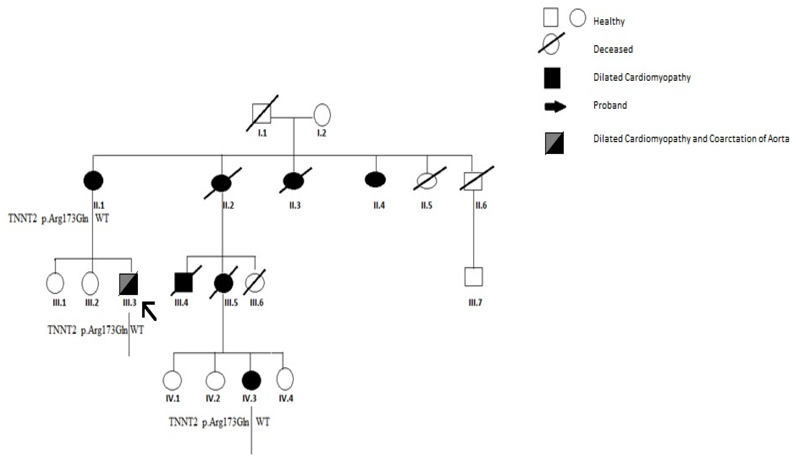
The proband is the third son of non-consanguineous parents. At the age of 19, he was referred to our clinic for a comprehensive evaluation after he experienced mild exertional dyspnea in the last few weeks. The family history was positive to sudden death (his cousin died, aged 3 months) and to DCMP and heart transplant (HT): a maternal aunt and her son who then died at 42 and 24 years old, respectively. Figure 1 summarizes the family pedigree. At birth, he was diagnosed with COA and therefore surgically corrected by end-to-end anastomosis and patch aortoplasty. Since then, he had been routinely evaluated at the Pediatric Cardiology Unit in our Hospital clinic with serial clinical and echocardiographic evaluations, which showed normal myocardial function until our evaluation. He had normal growth parameters (weight 85 Kg and height 165 cm) and reached regularly developmental milestones suggesting an isolated form of COA.
During routine evaluation, the patient complained of progressive exertional dyspnea, NYHA II-III. There was no medical history for recent infectious nor inflammatory events. On physical examination: lower limb pulses were bilaterally palpable, there was no significant upper and lower limb pressure differences and there were no signs of peripheral congestion (neither crepitation nor hepatomegaly). Echocardiographic examination showed a mildly reduced left ventricular ejection fraction (LVEF) (∼42%) with nondilated LV. Residual isthmic gradient was not significant (<20 mmHg) and absence of holodiastolic run off was ascertained. Echocardiography excluded other findings that may suggest hemodynamic consequences. The aortic valve was tricuspid and no mitral valve abnormalities were found. Ambulatory blood pressure monitoring excluded hypertensive status suggesting recoarctation. Complete blood count, thyroid hormones, serum electrolytes, lipid profile, hepatic function, serum creatinine and NT pro BNP laboratory values were within the reference ranges. Medical therapy was introduced and well tolerated with beta-blocker (Bisoprolol) and ACE inhibitor (Ramipril) [6]. The patient was referred to the Inherited and Rare Diseases Unit on the suspicion of an underlying cardiomyopathy. For tissue characterization, a cardiac magnetic resonance (CMR) was performed showing a moderately dilated, hypokinetic left ventricle (LV), with mildly reduced EF (45%). T2-weighted images and double inversion recovery sequences excluded myocardial edema. Contrast enhanced fast spin echo T1-weighted sequences did not show any abnormality. No evidence of early and late gadolinium enhancement (LGE) was detected and residual isthmic coarctation was excluded (Figure 2).

3. Results

Based on the clinical presentation and family history, genetic counselling was performed and molecular testing was suggested. According to the dispositions of the local ethics committee, informed consent was appropriately obtained for genetic investigations through next generation sequencing (NGS) with a panel of 325 genes, known to be associated with cardiomyopathies (Supplementary Table S1).
A blood sample in EDTA was collected from the subject. Genomic DNA was extracted using the Maxwell 16 instrument (Promega, Madison, WI, USA), and DNA quality was assessed using a Nanodrop machine. Molecular testing was carried out by analyzing a panel of target genes through NGS-based procedure. The Alyssa software (Agilent, Santa Clara, CA, USA) was used to perform sequence data analysis. This tool allows the alignment of the sequences to the reference genome in order to obtain a list of genomic variants that can be prioritized using a bioinformatic pipeline in order to highlight likely pathogenic or pathogenic variants. Genome data processing was performed using a home-made bioinformatic pipeline. The MAF threshold was set to 5% using the Illumina Variant Interpreter Software. Genetic testing identified a heterozygous variant in TNNT2 (NM_001001430.2):c.518G>A (p.Arg173Gln). According to the American College of Medical Genetics (ACMG), the variant was classified as pathogenic/likely pathogenic (class 5-4) which confirmed the clinical suspicion of cardiomyopathy. The NGS variant was also validated by Sanger sequencing. The variant has been previously reported in literature and is associated with the phenotype of cardiomyopathies [7]. Among the 325 genes included in the panel, no other clinically relevant variant of unknown significance, according to ACMG criteria, was identified. A genetic test performed through Next Generation Sequencingdid not find any CNV. However, SNP/CGHarrays were not performed, as the patient withdraw his consent for further investigations. Thus, the relatives were invited to join the cascade program, including both phenotyping (cardiac screening) and genotyping (analysis of the pathogenic variant identified in the proband). Cascade family screening revealed a TNNT2 variant in the mother (II.1) and his cousin (IV.3). Both of them were phenotype positive (affected by DCM) at cardiac screening at our Hospital clinic. The two sisters of the proband were unavailable for screening. Figure 1 reports both phenotyping and genotyping of the reported family.

4. Discussion

To date, the association of dilated cardiomyopathy and repaired COA is very rare. LVSD has been commonly associated with recoarctation in infancy, nonetheless heart failure symptoms seem to regress after hemodynamic relief of aortic gradients [5,6,7,8,9]. Among adult patients, DCM has been commonly reported in association with recoarctation [10,11,12]; similarly, hypokinetic phenotype regressed after stent implantation or surgical repair. In one case, due to severe heart failure, orthotopic heart transplantation [13] was performed. The potential pathophysiologic mechanism of progression to DCM in severe COA in infancy seems to occur after closure of the arterial duct, because formation of an adequate collateral circulation is not allowed promptly [14]. Mechanical wall stress imposed by hemodynamic overload in COA may trigger left ventricular hypertrophy (LVH) and concentric remodeling. Different pathways seem to be involved in adaptive LVH, including increased transcription of sarcomeric proteins, release and production of cytokines and growth factors and production of neurohormonal mediators that contribute to the increase in myocardial mass [15]. Nonetheless, the burden of chronic pressure overload may trigger the compensatory hypertrophy to evolve into maladaptive hypertrophy with cardiac dilatation and loss of systolic function [16,17]. Isthmic recoarctation should be included in the differential diagnosis of children and infants with DCM, regardless of previous surgical or percutaneous correction, and aortic imaging using CMR or CTA is recommended by current Guidelines [18]; nonetheless this was ruled out in our case. Differential diagnosis included myocarditis, acute coronary syndrome or dilated cardiomyopathy (DCM). Absence of hypertensive status and residual isthmic gradient with positive family history of early onset DCM and SCD, suggested a genetic form of cardiomyopathy. Myocarditis was highly excluded on the basis of negative personal history and negative signs on CMR for foci of increased signal compatible with myocardial edema. Genotyping was positive for a heterozygous missense variant in TNNT2 (NM_001001430.2): c.518G>A (p.Arg173Gln) which affects a highly conserved domain that encodes for the sarcomeric protein troponin T, and is currently classified as pathogenic/likely pathogenic for hypertrophic cardiomyopathy(HCM), restrictive cardiomyopathy and DCM in several genetic databases [19,20,21,22]. Of note, this variant hasnot been associated to left-sided obstructive lesions according to existing databases. The panel of analyzed genes included genes with causative association with left-sided obstructive lesions, including aortic coarctation. In particular, the proband was tested for NKX2.5, NOTCH1, GATA4, SMAD6 and TBX1 and was found to be negative for pathogenic/likely pathogenic variants.
Cardiomyocytes generated from induced pluripotent stem cells (iPSc) harboring mutations in Troponin T gene may exhibit altered calcium handling and impaired myofilament contraction [23]. Interestingly, iPSc derived cardiomyocytes harboring TNNT2 mutations seemed more susceptible to β-adrenergic stimulation and morphological analysis showed that their myocardial sarcomeric disarray was improved by treatment with β1 selective-β blocker metoprolol [24].
This case illustrates several issues related to diagnosis and follow up of DCM in the setting of repaired congenital heart defects as COA. Careful history taking, including a family history of cardiomyopathy, should not be overlooked even when the clinical findings seem to suggest a predisposition to hemodynamic factors for LVSD (such as recoarctation). The diagnosis of DCM could not be achieved without detailed family history, including the construction of a three-generation pedigree. Since it is a time-consuming approach, it would not be routinely performed at the time of routine cardiac screening. Referral to an inherited cardiovascular disease center is highly recommended in children and ACHD positive family history for inherited cardiac diseases (cardiomyopathies and genetically determined arrhythmias) since these conditions might be overlooked. Additional genetic testing is essential in the genotyping of family members who may benefit from early diagnosis and regular follow up [25].

5. Conclusions

We described a rare case of a young adult with a surgically repaired coarctation of the aorta who complained of mild exertional dyspnea, carrying a heterozygous missense variant in TNNT2 (NM_001001430.2): c.518G>A (p.Arg173Gln). This case highlights the role of careful history taking: a family history of cardiomyopathy should not be overlooked even when the clinical setting seems to suggest a predisposition to hemodynamic factors for LVSD.

Supplementary Materials

The following are available online at https://www.mdpi.com/article/10.3390/biom11050696/s1, Supplementary Table S1: List of the 325 genes included in the NGS panel.

Author Contributions

M.C., M.L. and G.L. contributed to the conception and design of the work. G.F. contributed to genetic analysis. A.F., A.C., R.P., M.R., E.M., S.D., E.R., A.S., B.S., F.V., F.D.F., F.A., P.C. and M.G.R. contributed to clinical or genetic studies. M.C., M.L., E.M., A.B. and G.L. drafted the manuscript. All the authors critically revised the manuscript. All gave final approval and agreed to be accountable for all aspects of work ensuring integrity and accuracy. All authors have read and agreed to the published version of the manuscript.

Funding

This research received no external funding.

Institutional Review Board Statement

Ethical review and approval were waived for this study, due to the local law and the type of research. All tests were performed for clinical purposes using routine techniques, thus ethical approval was not sought.

Informed Consent Statement

Informed consent was obtained from all subjects involved in the study to publish this paper.

Data Availability Statement

The data that support the findings of this study are available from the corresponding author upon reasonable request.

Acknowledgments

We would like to thank our nurses Michela Piscopo, Daniela Lafera and Ciro De Prisco for their assistance with the proband and family members.

Conflicts of Interest

The authors declare no potential conflict of interest.

References

  1. Brown, M.L.; Burkhart, H.M.; Connolly, H.M.; Dearani, J.A.; Cetta, F.; Li, Z.; Oliver, W.C.; Warnes, C.A.; Schaff, H.V. Coarctation of the aorta: Lifelong surveillance is mandatory following surgical repair. J. Am. Coll. Cardiol. 2013, 62, 1020–1025. [Google Scholar] [CrossRef] [PubMed] [Green Version]
  2. Lee, M.G.Y.; Babu-Narayan, S.V.; Kempny, A.; Uebing, A.; Montanaro, C.; Shore, D.F.; D’Udekem, Y.; Gatzoulis, M.A. Long-term mortality and cardiovascular burden for adult survivors of coarctation of the aorta. Heart 2019, 105, 1190–1196. [Google Scholar] [CrossRef] [PubMed]
  3. Fawzy, M.E.; Awad, M.; Hassan, W.; Al Kadhi, Y.; Shoukri, M.; Fadley, F. Long-term outcome (up to 15 years) of balloon angioplasty of discrete native coarctation of the aorta in adolescents and adults. J. Am. Coll. Cardiol. 2004, 43, 1062–1067. [Google Scholar] [CrossRef] [PubMed] [Green Version]
  4. Moonsamy, P.; Bhatt, A.B.; Mohan, N.; Masaki, F.; Serguei, M.; David, A.; D’Alessandro, G.T.; Thoralf, M.S.; Duke, E.C.; Arminder, J. Long term follow-up of patients with coarctation of the aorta. J. Am. Coll. Cardiol. 2020, 75, 609. [Google Scholar] [CrossRef]
  5. Hwang, M.S.; Chu, J.J.; Chang, Y.S.; Su, W.J. Dilated cardiomyopathy: An unusual presentation of aortic coarctation in an infant. Cardiology 2006, 106, 56–58. [Google Scholar] [CrossRef] [PubMed]
  6. Ponikowski, P.; Voors, A.A.; Anker, S.D.; Héctor, B.; John, G.F.; Coats, A.J.S.; Falk, V.; González-Juanatey, J.R.; Veli-Pekka, H.; Jankowska, E.A.; et al. 2016 ESC Guidelines for the diagnosis and treatment of acute and chronic heart failure. Eur. Heart J. 2016, 37, 2129–2200. [Google Scholar] [CrossRef] [PubMed]
  7. ClinVar Database (ClinVar-NCBI-NIH). Available online: https://www.ncbi.nlm.nih.gov/clinvar/variation/43649/ (accessed on 16 April 2021).
  8. Salahuddin, N.; Wilson, A.D.; Rao, P.S. An unusual presentation of coarctation of the aorta in infancy: Role of balloon angioplasty in the critically ill infant. Am. Heart J. 1991, 122, 1772–1775. [Google Scholar] [CrossRef]
  9. Alehan, D.; Kafali, G.; Demircin, M. Middle aortic syndrome as a cause of dilated cardiomyopathy. Anadolu Kardiyol. Derg. 2004, 4, 178–180. [Google Scholar]
  10. Aǧaç, M.T.; Acar, Z.; Akdemir, R.; Korkmaz, L.; Kiris, A.; Akyuz, A.R.; Erkan, H. Dilated cardiomyopathy secondary to coarctation of the aorta was completely resolved after stent implantation. Cardiovasc. J. Afr. 2012, 23, 6. [Google Scholar] [CrossRef]
  11. Alcibar, J.; Peña, N.; Oñate, A.; Gochi, R.; Barrenechea, J.I. Stent implantation in an adult with coarctation of the aorta in the presence of advanced secondary heart failure. Texas Heart Inst. J. 1999, 26, 143–147. [Google Scholar]
  12. Pauly, D.F.; Morss, S.E.; Tanio, J.W.; Irani, K.; Cameron, D.E.; Schulman, S.P.; Hare, J.M. Reduced left ventricular dimension and normalized atrial natriuretic hormone level after repair of aortic coarctation in an adult. Clin. Cardiol. 1999, 22, 33–235. [Google Scholar] [CrossRef] [PubMed]
  13. Raffel, O.C.; Abraham, A.; Ruygrok, P.N.; Finucane, A.K.; McGeorge, A.D.; French, R.L. Cardiac transplantation and aortic coarctation repair in severe heart failure. Asian Cardiovasc. Thorac. Ann. 2006, 14, 522–524. [Google Scholar] [CrossRef] [PubMed]
  14. Kenny, D.; Polson, J.; Martin, R.P.; Paton, J.F.R.; Wolf, A.R. Hypertension and coarctation of the aorta: An inevitable consequence of developmental pathophysiology. Hypertens Res. 2011, 34, 543–547. [Google Scholar] [CrossRef] [Green Version]
  15. Frey, N.; Olson, E.N. Cardiac Hypertrophy: The Good, the Bad, and the Ugly. Annu. Rev. Physiol. 2003, 65, 45–79. [Google Scholar] [CrossRef] [PubMed]
  16. De Acetis, M.; Notte, A.; Accornero, F.; Selvetella, G.; Brancaccio, M.; Vecchione, C.; Sbroggiò, M.; Collino, F.; Pacchioni, B.; Lanfranchi, G.; et al. Cardiac overexpression of melusin protects from dilated cardiomyopathy due to long-standing pressure overload. Circ. Res. 2005, 96, 1087–1094. [Google Scholar] [CrossRef] [Green Version]
  17. Brancaccio, M.; Fratta, L.; Notte, A.; Hirsch, E.; Poulet, R.; Guazzone, S.; De Acetis, M.; Vecchione, C.; Marino, G.; Altruda, F. Melusin a muscle-specific integrin β1-interacting protein, is required to prevent cardiac failure in response to chronic pressure overload. Nat. Med. 2003, 9, 68–75. [Google Scholar] [CrossRef] [PubMed]
  18. Stout, K.K.; Daniels, C.J.; Aboulhosn, J.A.; Bozkurt, B.; Broberg, C.S.; Colman, J.M.; Crumb, S.R.; Dearani, J.A.; Fuller, S.; Gurvitz, M.; et al. 2018 AHA/ACC guideline for the management of adults with congenital heart disease: Executive summary: A report of the American College of Cardiology/American Heart Association Task Force on clinical practice guidelines. Circulation 2019, 139, E637–E697. [Google Scholar] [CrossRef]
  19. Jáchymová, M.; Muravská, A.; Paleček, T.; Kuchynka, P.; Řeháková, H.; Magage, S.; Král, A.; Zima, T.; Horký, K.; Linhart, A. Genetic variation screening of TNNT2 gene in a cohort of patients with hypertrophic and dilated cardiomyopathy. Physiol. Res. 2012, 61, 169–175. [Google Scholar] [CrossRef]
  20. Millat, G.; Bouvagnet, P.; Chevalier, P.; Sebbag, L.; Dulac, A.; Dauphin, C.; Jouk, P.S.; Delrue, M.A.; Thambo, J.B.; Le Metayer, P. Clinical and mutational spectrum in a cohort of 105 unrelated patients with dilated cardiomyopathy. Eur. J. Med. Genet. 2011, 54, e570–e575. [Google Scholar] [CrossRef]
  21. Hershberger, R.E.; Pinto, J.R.; Parks, S.B.; Kushner, J.D.; Duanxiang, L.; Ludwigsen, S.; Cowan, J.; Morales, A.S.; Parvatiyar, M.; Potter, J.D. Clinical and functional Characterization of TNNT2 mutations identified in patients with dilated cardiomyopathy. Circ. Cardiovasc. Genet. 2009, 2, 306–313. [Google Scholar] [CrossRef] [Green Version]
  22. Kamisago, M.; Sharma, S.D.; DePalma, S.R.; Solomon, S.; Sharma, P.; McDonough, B.; Smoot, L.; Mullen, M.P.; Woolf, P.K.; Wigle, E.D.; et al. Mutations in Sarcomere Protein Genes as a Cause of Dilated Cardiomyopathy. N. Engl. J. Med. 2000, 343, 1688–1696. [Google Scholar] [CrossRef] [PubMed]
  23. Sousa, A.; Canedo, P.; Campelo, M.; Moura, B.; Leite, S.; Baixia, M.; Belo, A.; Rocha-Gonçalves, F.; Machado, J.C.; Silva-Cardoso, J. Genetic Variants Are Not Rare in ICD Candidates with Dilated Cardiomyopathy: Time for Next-Generation Sequencing? Cardiol. Res. Pract. 2019, 2019. [Google Scholar] [CrossRef] [PubMed] [Green Version]
  24. Morimoto, S. Molecular pathogenic mechanisms of cardiomyopathies caused by mutations in cardiac troponin T. In Advances in Experimental Medicine and Biology; Springer: New York, NY, USA, 2007; Volume 592, pp. 227–239. [Google Scholar]
  25. Crispell, K.A.; Wray, A.; Ni, H.; Nauman, D.J.; Hershberger, R.E. Clinical profiles of four large pedigrees with familial dilated cardiomyopathy: Preliminary recommendations for clinical practice. J. Am. Coll. Cardiol. 1999, 34, 837–847. [Google Scholar] [CrossRef] [Green Version]
Figure 1. Family pedigree of the proband. Open symbols: phenotype negative. Full symbols: phenotype positive (affected by dilated cardiomyopathy). III,6 died suddenly at 3 months. II,2 III,4 underwent cardiac transplantation. II,2; II,3; III,4; III,5 died aged 42, 18, 24, 38 years old, respectively.
Biomolecules 11 00696 g001
Figure 2. 1,5 Tesla cardiac magnetic resonance (CMR) showing a moderately dilated, hypokinetic left ventricle at 4 chamber (a) and midventricular short-axis view (b); (c) shows post contrast short axis view excluding late gadolinium enhancement (LGE). (d) Three-dimensional reconstruction of aortic arch where no signs of recoarctation were detected.
Biomolecules 11 00696 g002
Publisher’s Note: MDPI stays neutral with regard to jurisdictional claims in published maps and institutional affiliations.