Next Article in Journal
Biogenesis of Mitochondrial Metabolite Carriers
Next Article in Special Issue
The Interaction of Bovine β-Lactoglobulin with Caffeic Acid: From Binding Mechanisms to Functional Complexes
Previous Article in Journal
Myxovirus Resistance Protein 1 (MX1), a Novel HO-1 Interactor, Tilts the Balance of Endoplasmic Reticulum Stress towards Pro-Death Events in Prostate Cancer
Previous Article in Special Issue
Co-Delivery of Imiquimod and Curcumin by Nanoemugel for Improved Topical Delivery and Reduced Psoriasis-Like Skin Lesions
 
 
Retraction published on 14 March 2024, see Biomolecules 2024, 14(3), 351.
Article

RETRACTED: Optimized Ellagic Acid–Ca Pectinate Floating Beads for Gastroprotection against Indomethacin-Induced Gastric Injury in Rats

by
Nabil A. Alhakamy
1,2,3,
Shaimaa M. Badr-Eldin
1,4,*,
Osama A. A. Ahmed
1,2,
Abdulrahman A. Halwani
1,
Hibah M. Aldawsari
1,
Mohamed A. El-Moselhy
5,6,
Aliaa Anter
6,
Sara S. Sharkawi
6,
Muhammad H. Sultan
7,
Osama A. A. Madkhali
7,
Muhammed A. Bakhrebah
8,
Mohammad N. Alomary
8,
Wesam H. Abdulaal
9 and
Usama A. Fahmy
1
1
Department of Pharmaceutics, Faculty of Pharmacy, King Abdulaziz University, Jeddah 21589, Saudi Arabia
2
Advanced Drug Delivery Research Group, Faculty of Pharmacy, King Abdulaziz University, Jeddah 21589, Saudi Arabia
3
Center of Excellence for Drug Research and Pharmaceutical Industries, King Abdulaziz University, Jeddah 21589, Saudi Arabia
4
Department of Pharmaceutics and Industrial Pharmacy, Faculty of Pharmacy, Cairo University, Cairo 12613, Egypt
5
Department of Clinical Pharmacy and Pharmacology, Ibn Sina National College for Medical Studies, Jeddah 22413, Saudi Arabia
6
Department of Pharmacology and Toxicology, Faculty of Pharmacy, Minia University, Minia 61519, Egypt
7
Department of Pharmaceutics, College of Pharmacy, Jazan University, Jazan 45142, Saudi Arabia
8
Life Science and Environment Research Institute, King Abdulaziz City for Science and Technology (KACST), Riyadh 12354, Saudi Arabia
9
Department of Biochemistry, Cancer Metabolism and Epigenetic Unit, Faculty of Science, King Abdulaziz University, Jeddah 21589, Saudi Arabia
*
Author to whom correspondence should be addressed.
Biomolecules 2020, 10(7), 1006; https://doi.org/10.3390/biom10071006
Submission received: 17 May 2020 / Revised: 23 June 2020 / Accepted: 1 July 2020 / Published: 6 July 2020 / Retracted: 14 March 2024
(This article belongs to the Special Issue Bioactive Formulations in Agri-Food-Pharma: Source and Applications)

Abstract

:
A peptic ulcer is an alimentary tract injury that leads to a mucosal defect reaching the submucosa. This work aimed to optimize and maximize ellagic acid (EA) loading in Ca pectinate floating beads to maximize the release for 24 h. Three factors were selected: Ca pectinate concentration (X1, 1–3 w/v %), EA concentration (X2, 1–3 w/v %) and the dropping time (X3, 10–30 min). The factorial design proposed eight formulations. The optimized EA–Ca pectinate formulation was evaluated for the gastric ulcer index and the oxidative stress parameter determination of gastric mucosa. The results indicated that the optimum EA–Ca pectinate formula significantly improved the gastric ulcer index in comparison with raw EA. The protective effect of the optimized EA–Ca pectinate formula was further indicated by the histopathological features of the stomach. The results of the study indicate that an EA formulation in the form of Ca pectinate beads would be effective for protection against gastric ulcers because of Nonsteroidal anti-inflammatory drugs (NSAID) administration.

1. Introduction

A peptic ulcer is a common health problem that occurs in young and old patients all over the world. The incidence of peptic ulcer disease (PUD) increases as patients get older, with an estimated prevalence of about 10% in the general population [1,2]. Peptic ulcers are described as defects or injuries in the gastroduodenal mucosa caused by peptic acid [3]. This ulceration is associated with stomach pain and often leads to gastrointestinal bleeding. PUD usually occurs in the lower part of the esophagus, the lower portion of stomach and the upper duodenum. Two factors are considered as the main cause of PUD, which are Helicobacter pylori (H. pylori) infection and the long-term use of non-steroidal anti-inflammatory drugs (NSAIDs) [4,5].
NSAIDs are the most common prescribed medications for patients suffering from pain and inflammation. They are commonly prescribed for patients over 65 years old with an approximate prevalence of 7.3% [6,7,8]. NSAIDs work as anti-inflammatory agents through the inhibition of cyclooxygenase (COX) enzymes. However, NSAIDs inhibit both COX-1 and COX-2 enzymes in a non-selective way [9,10]. COX-1 plays an important role in the protection of the gastric mucosa by producing prostaglandins and thromboxane A2, which control the mucosal barrier in the gastrointestinal tract (GIT). On the other hand, COX-2 is responsible for prostaglandin-mediated pain and inflammation. Indomethacin is one of the most commonly prescribed non-selective NSAIDs, which is mainly used for its analgesic and anti-inflammatory effects [11]. However, ulcerations and gastric mucosal damage are the common side effects associated with using indomethacin [12].
Several studies have shown the potential of Ellagic acid (EA) as an anti-ulcer agent, which is due to its ability to modulate the pathway activation of nuclear factor-κB (NF-κB) [13,14]. A previous report showed that EA has the ability to control the production of COX-2 mRNA through the inhibition of reactive oxygen species (ROS) production, which in turn inhibits NF-κB activation. It was also shown that EA has a higher binding affinity to COX-2 active sites through the formation of four hydrogen bonds compared to other NSAIDs, such as diclofenac and meloxicam. The anti-inflammatory effect of EA in acute inflammation has been explained by authors as occurring through the blocking of the COX-2 receptor [15,16]. In addition, various pharmacological activities of EA have been reviewed and explained in different studies. Sodium pectinate has been shown to be more effective and cost-effective in the treatment of symptoms presented with reflux without extreme esophagitis when compared with cisapride [17]. Studies suggested the use of sodium pectinate to achieve continuous drug release, gastric mucosa area targeting and improved drug bioavailability as a result of sodium pectinate’s ability to form a bio-adhesive calcium ion gel [18]. Additionally, pectinate preparation in the form of beads requires the use of water as a solvent without the need for high temperatures and harmful organic solvents. Accordingly, the pectinate beads produced show the characteristics of buoyancy, bio-adhesion and non-immunogenic behavior. These characteristics of pectinate beads are useful for stomach-targeted drug delivery systems that are considered a promising vehicle for the delivery of EA in this investigation [19,20,21,22,23].
This work investigates the design and formulation of floating EA–pectinate beads that were expected to retain and localize the effect of EA in the stomach (gastric epithelium) for an extended period. This approach of targeted delivery could improve the efficacy of EA to protect against gastric ulcers induced by NSAIDs.

2. Materials and Methods

2.1. Materials

Ellagic acid, calcium chloride and methanol were acquired from Sigma-Aldrich (St. Louis, MO, USA). Sodium pectinate low methoxyl pectin (degree of esterification), with a degree of methylation of approximately 38% was a gift from DEEF Pharmaceutical Industries & Co. (Riyadh, Saudi Arabia).

2.2. Experimental Design of EA–Ca Pectinate Beads

A 23 factorial design was applied for the preparation of EA–Ca pectinate beads. The impact of the independent variables, namely, EA percentage (%, X1), Ca pectinate percentage (%, X2) and dropping time (X3, min) are shown in Table 1. Each factor was studied at two levels and a total of eight formulations were prepared, as shown in Table 2. The dependent variables (responses), including drug entrapment efficiency (EE%, Y1) and cumulative EA percent released (EA%, Y2), were subjected to statistical analysis using Design-Expert® Software Version 12 (Stat-Ease Inc, Minneapolis, MN, USA). An ANOVA test was employed to assess the main effects of the variables on the studied responses at a 95% level of significance.

2.3. EA–Ca Pectinate Bead Preparation

Sodium pectinate and EA were utilized in this study in concentrations of 1–3% w/v each, according to the experimental design. Sodium pectinate and EA aqueous solution (50 mL) were dropped within 10–30 min from a syringe (22-gauge needle) into 100 mL of 0.5 M CaCl2 aqueous solution and stirred at 400 rpm. After the addition of the aqueous solution, stirring was continued for one hour. After that, Ca pectinate beads formed and were removed from the CaCl2 solution by filtration. Beads were then washed twice with distilled water and freeze-dried (Christ Beta 1–8 LD Freeze Dryer; Martin Christ Gefriertrocknungsanlagen GmbH, Osterode am Harz, Germany) [24]. Plain beads (beads without EA) were prepared with the same procedure mentioned for the EA-loaded Ca pectinate beads.

2.4. Determination of EA–Ca Pectinate Buoyancy Time

EA–Ca pectinate beads (50 mg) were added to 200 mL simulated gastric fluid (without enzyme) pH 1.2 solution and stirred at 50 rpm. The temperature of the simulated gastric fluid was 37 ± 0.5 °C. EA–Ca pectinate buoyancy time was measured by visual observation. Buoyancy was considered when all beads floated in the test solution immediately or within a 30 s lag time. The average of three replicates was estimated.

2.5. Determination of EA Entrapped Percent

For determination of EA, 10 mg of the prepared EA–Ca pectinate beads were crushed and dispersed in methanol (100 mL). The dispersion was then centrifuged at 3500 rpm for 45 min and filtered using a 0.22-μm filter before analysis with the previously reported High performance liquid chromatography HPLC method [22].

2.6. In Vitro Release Evaluation of EA–Ca Pectinate Beads

Optimized EA–Ca pectinate bead in vitro release was determined using dissolution tester apparatus II (paddle method). EA–Ca pectinate beads (50 mg) were added to 900 mL simulated gastric fluid (without enzyme) pH 1.2 solution (contains 0.25% w/v sodium lauryl sulphate) and rotated at 100 rpm. The temperature of the simulated gastric fluid was 37 ± 0.5 °C. At specific time intervals, a 5 mL sample was withdrawn and analyzed for EA cumulative % released using HPLC. Fresh media (5 mL) replaced the withdrawn samples.

2.7. In Vitro Release Evaluation of EA–Ca Pectinate Beads

All the studied responses were integrated using the desirability function to select the optimal levels of the independent variables. The desired goals were maximizing both the drug entrapment and cumulative percent EA released, shown in Table 1.

2.8. In Vivo Evaluation of Optimized EA–Ca Pectinate Beads

2.8.1. Animals

Adult male Wistar rats weighing 180–200 g obtained from the animal care unit (Faculty of Pharmacy, ElNahda University, Egypt) were used after 2 weeks of proper acclimatization to the animal house conditions (12:12 h light/dark cycles and 25 ± 2 °C temperature) and had free access to standard rodent chow and water ad libitum. Experiments were conducted in accordance with the international ethical guidelines for animal care of the United States Naval Medical Research Center, Unit no. 3, Abbaseya, Cairo, Egypt, accredited by the Association for Assessment and Accreditation of Laboratory Animal Care International (AAALAC International). The adopted guidelines were in accordance with “Principles of Laboratory Animals Care” (NIH publication no. 85–23, revised 1985). The study protocol was approved by members of the Research Ethics Committee and by the Pharmacology and Toxicology Department, Faculty of Pharmacy, Minia University, Egypt (No. 53/2019).

2.8.2. Animal Groups

Four groups (eight animals each) were randomly divided. The first group, group (1) or control group, had no induction of ulcers (non-treated). The second group, group (2) or indomethacin group), rats received 50 mg/kg of indomethacin. The third group, group (3) or pure EA group, rats orally received raw EA 30 min before the injection of indomethacin. The fourth group, group (4) or EA formula group, rats orally received optimized EA–Ca pectinate beads 30 min before the injection of indomethacin. The fifth group, group (5) or plain formula group, rats orally received optimized Ca pectinate beads (without EA) 30 min before the injection of indomethacin.

2.8.3. Induction of Gastric Ulcers

All rats were fasted for one day, with free access to water, before the induction of gastric ulcers. Indomethacin (50 mg/kg) was injected through the intraperitoneal route for the induction of gastric ulceration. EA or an equivalent dose of the optimized EA formula was orally administered at a dose of 7 mg/kg. Four hours later, rats were sacrificed by decapitation. Blood samples were immediately collected in clean tubes, left for 30 min and sera were separated by centrifugation at 3000 rpm for 15 min at 4 °C and stored at −80 °C until use. Then, the abdomen was opened, and the stomachs were removed and opened along the greater curvature. The stomachs were washed with ice-cold saline and scored for macroscopic gross mucosal lesions. Gastric mucosae were collected and stored at −80 °C until used for the estimation of lipid peroxidation. Another set of stomachs from each group was immersed in 10% formalin for histopathological examination using hematoxylin and eosin (H&E) staining.

2.8.4. Assessment of Gastric Mucosal Lesions

Mucosal lesions were quantified in the investigated groups (eight rats/group) as previously indicated by Szabo and Hollander [24]. Images of pinned stomach areas of mucosal damage were expressed as a percentage of the total surface area of the stomach utilizing ImageJ software. The ulcer index (U.I.) is the mean ulcer score for the investigated group. The ulcer inhibition percent against indomethacin-induced ulcers was determined using Equation (1).
U l c e r   i n h i b i t i o n   % = U .   I .   i n   i n d o m e t h a c i n U .   I   i n   t r e a t e d   r a t s U .   I .   i n   i n d o m e t h a c i n × 100

2.8.5. Determination of Gastric Mucosal Lipid Peroxidation

Gastric mucosal tissues were homogenized in ice-cold phosphate-buffered saline at 10% (0.1 g/mL) using a polytron homogenizer, then centrifuged for 20 min at 4 °C. The supernatant was then aspirated and used for the determination of malondialdehyde (MDA), according to the method indicated by Mihara and Uchiyama [25].

2.8.6. Determination of Serum Total Antioxidant Capacity

Serum total antioxidant capacity was determined using a commercially available kit (Bio-diagnostic, Giza, Egypt), according to the method of Koracevic and other colleagues [25].

2.8.7. Determination of Gastric Secretion Parameters

The same rat groups as mentioned in Section 2.8.2 were used to evaluate the effect of EA-Formula (EA-F) on the stomach pH. Gastric acid output (volume) was determined in the supernatant (2 mL) by titration with 0.0025 N NaOH using Toepfer’s reagent as an indicator. The pH of gastric juice was determined using a pH meter.

2.8.8. Statistical Analysis

In vivo evaluation statistical analysis was carried out using IBM SPSS software version 25 (SPSS Inc., Chicago, IL, USA). The mean comparison was made using analysis of variance (ANOVA), followed by Tukey’s test as a post hoc study. The data are presented as the standard error of the mean (SEM). The differences were found to be significant at p < 0.05.

3. Results

3.1. Determination of EA–Ca Pectinate Buoyancy Time

All the prepared beads presented excellent (100%) floating capacity in simulated gastric fluid (without enzyme) pH 1.2 solution. All beads floated immediately after contact with the release medium and remained floating for more than 24 h (Figure 1A,B).

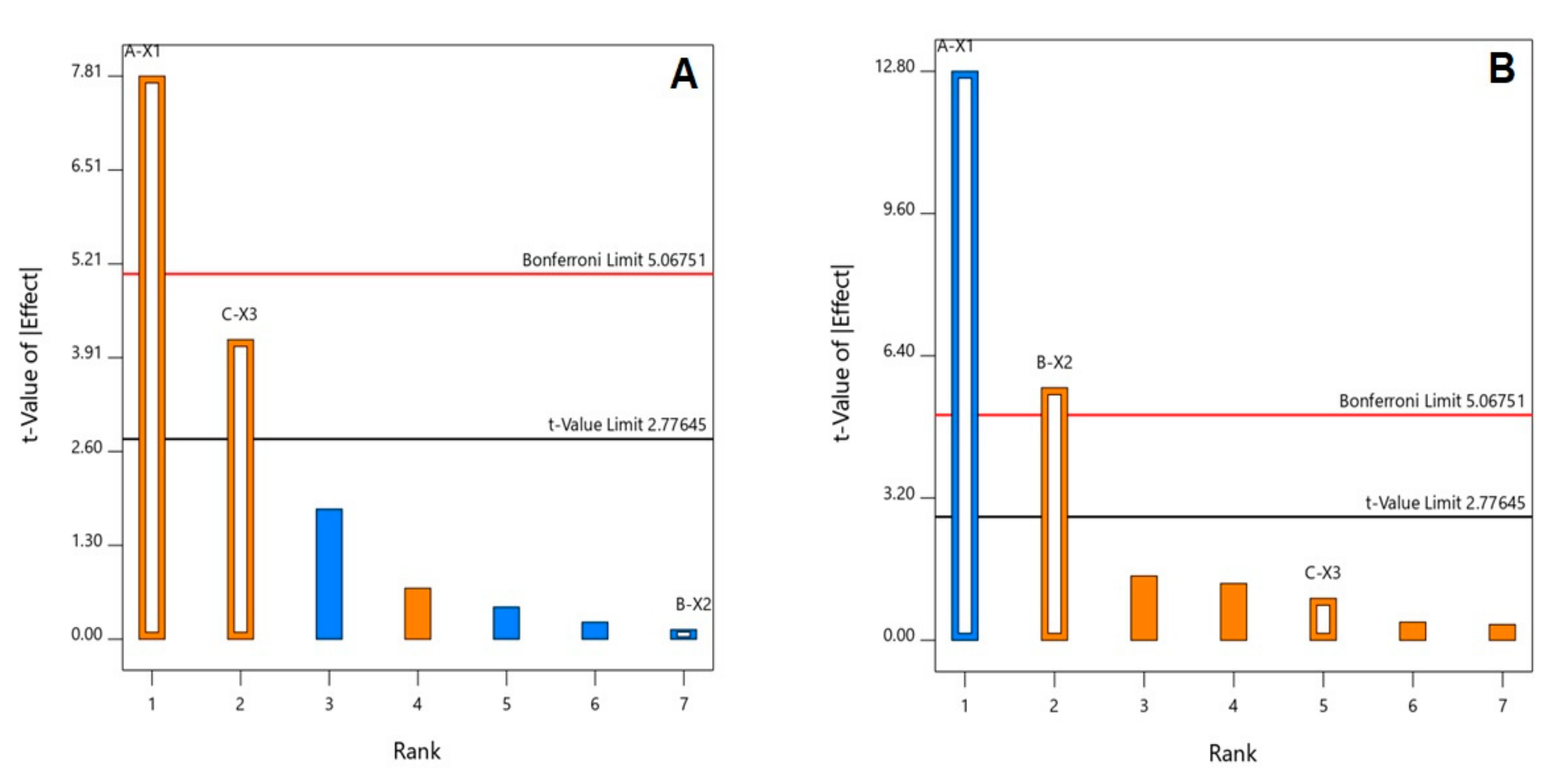
3.2. Statistical Analysis of the Factorial Design

In this study, the factors and their corresponding levels were selected according to the preliminary trials. ANOVA was applied for each response to assess the main effects of the studied variables. For both responses, the predicted R2 values were in reasonable agreement with the adjusted R2 values. Adequate precision was greater than 4 (Table 3), proving the suitability of the model to explore the design space [24].

3.2.1. Effect of Variables on Entrapment Efficiency (Y1)

The percent EA entrapment of the formulations ranged from 74.7 ± 1.45 to 88.1 ± 2.31%, as shown in Table 2. According to the factorial design, the selected model (classical sum of squares type III) was significant (model F-value = 26.10; p = 0.0044). There is only a 0.44% chance that this F-value could occur due to noise. The equation representing the sequential model generated is shown in Equation (2).
Y1 = 80.79 + 3.74 X1 − 0.0625 X2 + 1.00 X3
Analysis of variance (ANOVA) showed that both EA% (p-value = 0.0014) and dropping time (p-value = 0.0142) showed a positive significant impact on the entrapment efficiency of the beads, as presented in the Pareto chart, Figure 2A. However, the effect of the EA% was more predominant, as is evident by its lower p-value. The individual effects of the investigated variables on the drug entrapment are graphically illustrated in Figure 3. As is evident, the drug entrapment increases with higher EA%. In addition, the drug entrapment significantly increases with increasing dropping time.

3.2.2. Effect of Variables on Cumulative EA% Released (Y2)

Figure 4 illustrates the in vitro release profiles of EA–Ca pectinate beads. All formulations showed a gradual release over a period of 24 h. Statistical analysis revealed that the selected model (classical sum of squares type III) was significant (model F-value = 65.64; p = 0.0007). There is only a 0.07% chance that this F-value could occur due to noise. The sequential model representative Equation (3) was generated.
Y2 = 71.81 − 11.41 X1 + 5.06 X2 + 0.8375 X3
Analysis of variance (ANOVA) showed that EA% (p-value = 0.0002) and Ca pectinate % (p-value = 0.0048) had significant effects on the cumulative EA% of the beads, as presented in the Pareto chart, Figure 2B.
As illustrated in Figure 5, which represents the individual effects of the variables, EA release was inversely related to EA%. In addition, the EA release significantly decreased with increasing EA%. EA release was inversely related to EA%.

3.2.3. Selection of the Optimized EA–Ca Pectinate Beads

A desirability function was employed for choosing the optimized EA–Ca pectinate beads. The criteria set for selection were attaining maximum EA entrapment and release, as presented in Table 1. It was found that beads prepared with an EA% of 1.97%, 3% Ca pectinate and a dropping time of 30 min fulfilled the required criteria with a desirability value of 0.641. Thus, this formulation was selected for further studies. The proposed formulation was prepared and evaluated for entrapment and cumulative EA% released, giving results of 82.6% and 77.9%, respectively. The results were in good agreement with the predicted values, with a residual percentage of less than 5%.

3.3. In Vivo Evaluation of Optimized EA Formulation

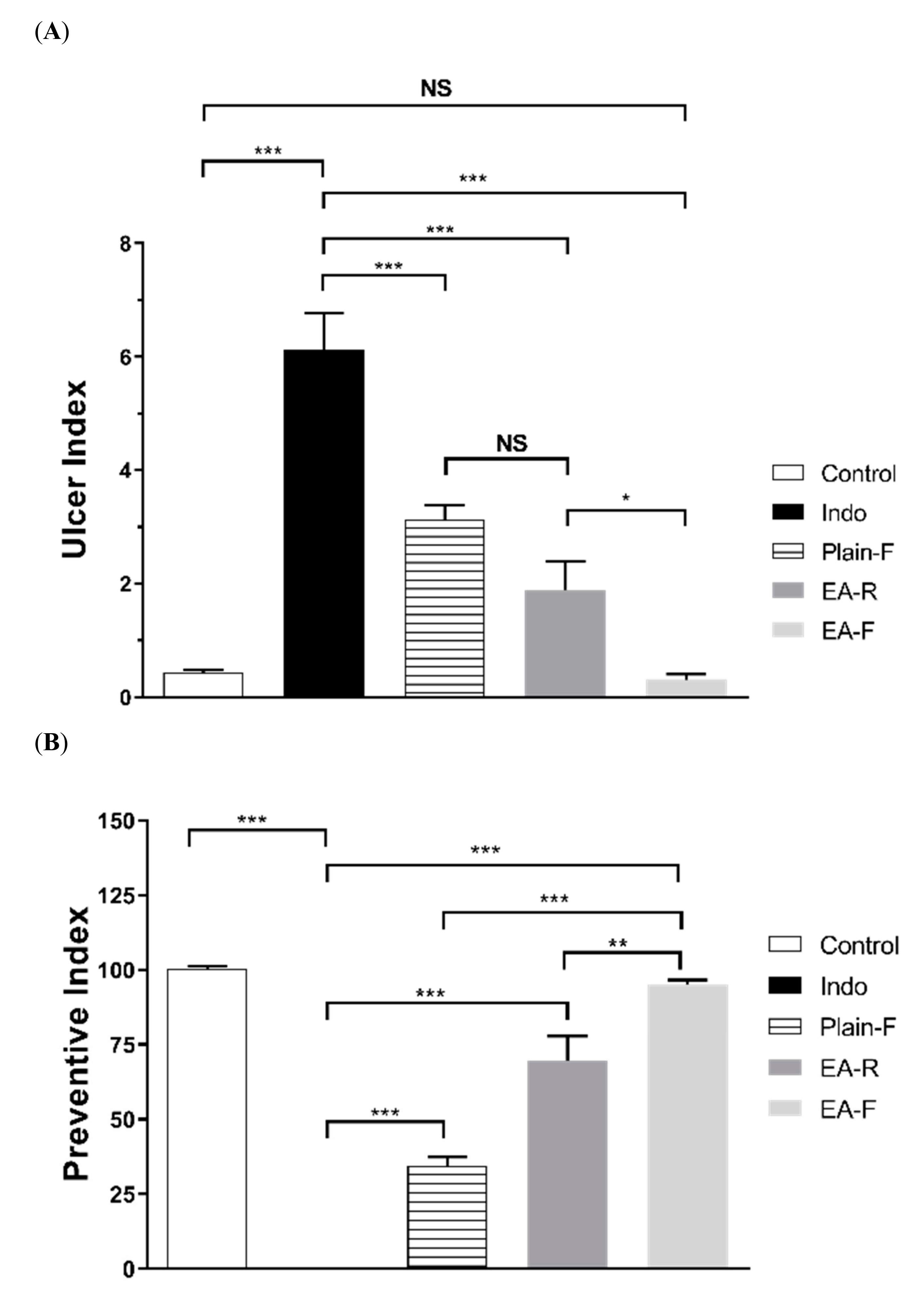
3.3.1. Effect of Pure EA and Optimized EA–Ca Pectinate Beads on Indomethacin-Induced Gastric Lesions

Figure 6A shows the effect of pure EA and the optimized EA formula on indomethacin-induced gastric lesions. Both pure EA and the optimized EA formula resulted in significantly (p < 0.001) fewer mucosal lesions (1.89 ± 0.51, 0.31 ± 0.10, respectively) compared to indomethacin in the assessment of the ulcer index. Additionally, Figure 6B shows a significant difference (p < 0.01) between the effect of pure EA and the optimized EA formula in ulcer inhibition percent (preventive index).

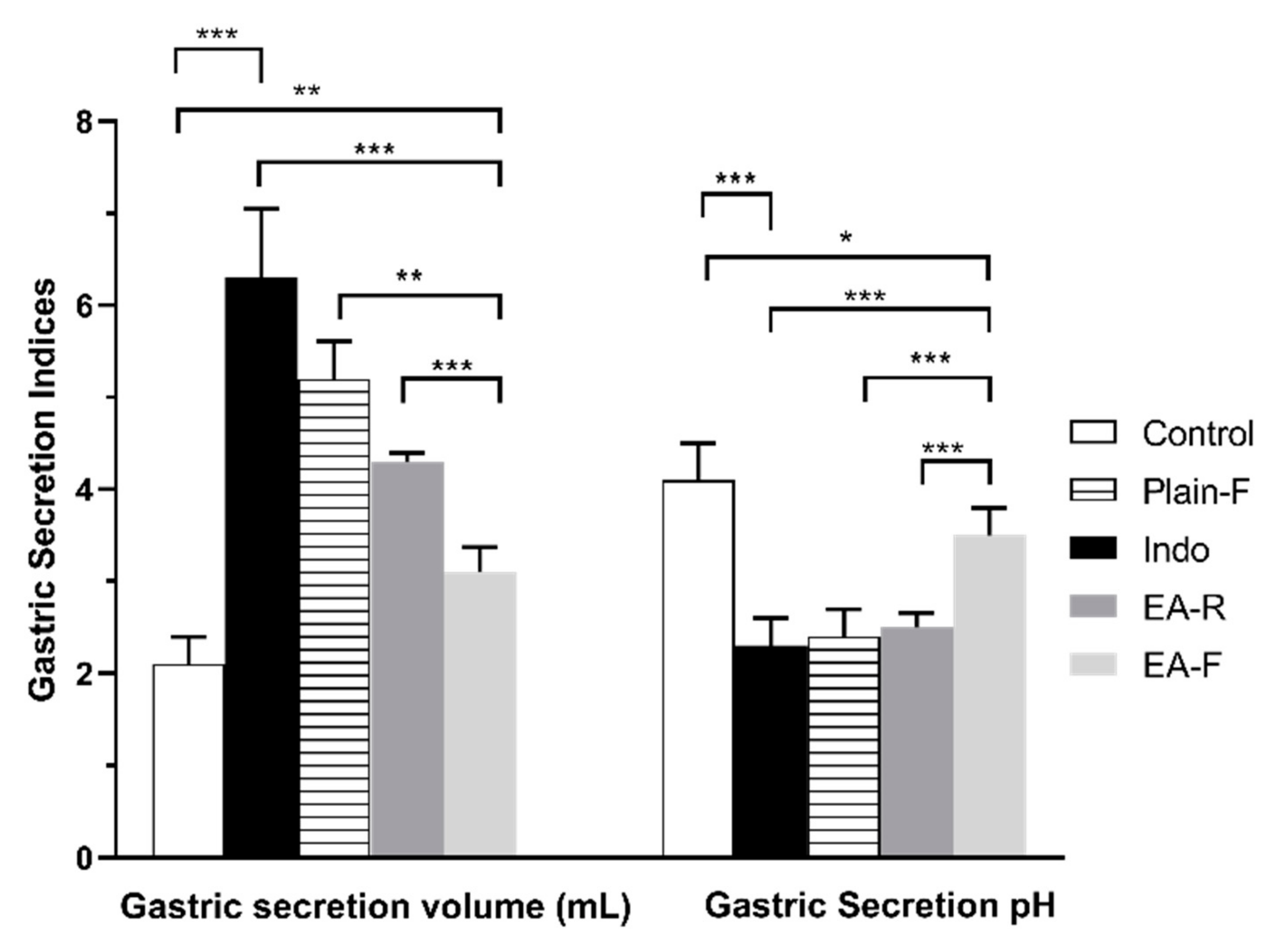
3.3.2. Effect of Pure EA and Optimized EA Formulation on Gastric pH

Gastric secretion volume and pH are represented in Figure 7, and the results revealed a significant difference (p < 0.001) between the indomethacin group and control group for both gastric secretion volume and pH. Additionally, the data revealed that there is a significant difference between the EA-F and indomethacin groups (p < 0.001) for both volume and pH. The plain formula and pure EA showed no significant difference in gastric secretion pH in comparison with the indomethacin group. On the other hand, the results revealed that both the plain formula and pure EA showed a significant difference (p < 0.01) for gastric secretion volume.

3.3.3. Effect of Pure EA and Optimized EA Formulation on Gastric Mucosal Oxidative Stress

The lipid peroxidation end-product, MDA, was used as a marker of oxidation. Figure 8 represents the effect of pure EA and the optimized EA formula on the mucosal MDA level. Indomethacin resulted in a significant (p < 0.001) elevation in the mucosal MDA level compared to the control. This elevation of mucosal MDA was significantly (p < 0.01) attenuated with the optimized EA formula but not with pure EA.

3.3.4. Effect of Pure EA and Optimized EA Formulation on Serum Total Antioxidant Capacity

The lipid peroxidation end-product, MDA, was used as a marker of oxidation. Figure 9 represents the effect of pure EA and the optimized EA formula on the mucosal MDA level. Indomethacin resulted in a significant (p < 0.001) elevation in the mucosal MDA level compared to the control. This elevation of mucosal MDA was significantly (p < 0.01) attenuated with the optimized EA formula but not with pure EA.

3.3.5. Effect of Pure EA and Optimized EA Formulation on the Histopathological Features of the Stomach

Figure 10 shows the results of the histopathological examination of H&E-stained stomach sections, which show the normal structure of gastric mucosa, revealing mucous glands of the fundic region composed of compact parietal and chief cells with underlying histologically normal lamina propria. There is no evidence of inflammation or ulceration in control rats (Figure 10A). Sections from indomethacin-treated groups show features of acute gastritis grade I in the form of compact mucous glands composed of parietal and chief cells with areas of superficial ulceration, congested mucosa and underlying lamina propria with focal lymphocytic inflammatory infiltrates and few eosinophils (Figure 10B). Sections from pure ellagic acid-treated rats show branching mucous glands formed of parietal and chief cells with underlying histologically normal lamina propria. There is no surface mucosal ulceration and few inflammatory cellular infiltrates in the form of lymphocytes (Figure 10C). The stomach of ellagic acid formula-treated rats (D) show compact branching mucous glands of the fundic region formed of parietal and chief cells without superficial ulceration or intraepithelial lymphocytic infiltrates in the lamina propria.

4. Discussion

Floating drug delivery is considered one of the most successful mechanisms for controlling dosage and gastric retention [26,27]. Among the approaches, a gel-forming or swellable cellulose type of matrix-forming polymer is utilized [24,28]. In this study, floating EA–pectinate beads were designed to retain and localize the effect of EA in the stomach. All beads floated immediately after contact with the release medium and remained buoyant for more than 24 h. An aqueous solution of sodium pectinate (with a low degree of esterification) formed spherical gel beads instantaneously by ionotropic gelation with divalent cations of Ca2+ and Ba2+ [29]. Intermolecular cross-links were formed between the divalent ions and the negatively charged carboxyl groups of the polymer molecules. The gel beads were easily prepared without any sophisticated equipment or techniques [30]. The lyophilization process imparts the floating behavior to the prepared beads. This is attributed to the highly porous internal structure of lyophilized beads as a result of the water sublimation during lyophilization that formed cavities in the core matrix of the formed Ca pectinate beads [24]. A previous report by Das and Ng revealed that Ca pectinate beads reserve the stability of entrapped drugs at various temperatures for more than 6 months [31]. The study showed 99.5% at 4 °C and at room temperature. Additionally, no significant change in drug entrapment was seen at the accelerated storing condition of 40 °C.
Determining the formulation and process parameters that might affect the drug delivery system properties is essential. Factorial design is advantageous regarding this aspect, as it can analyze the effect of various factors simultaneously [32,33,34]. The drug entrapment and sustained release characteristics of EA–Ca pectinate beads have been shown to be influenced by the formula composition and process parameters. After several preliminary tests, the viscosity and concentration of Ca pectinate were observed to be efficient factors in EA loading; the concentration of EA, the inner diameter of the needle, the drip height of the needle and the proportion of drugs and adjunctions were all important.
The drug entrapment significantly increases with increasing dropping time and at higher EA%. Increased dropping time allows EA to be entrapped efficiently between the viscous solution, in addition to the higher wettability of EA with a CaCl2 solution that allows cross linkage between pectin and Ca++ ions. In addition, the EA release was significantly decreased with increasing EA%, which could be attributed to a decrease in the EA to pectin ratio. Previous work confirmed the emulsification role of the pectin. Pectin can perform as a surfactant and reduce surface tension to facilitate the formation of a stable suspension. Pectin also forms a coating around the dispersed EA core particles. Thus, a charged surface is imparted to the coated particles, which then repel each other and maintain a stable, dispersed state [35,36,37]. The main sources of pectins are apple pomace, the peels of citrus fruits and sugar beet chips. Neutral sugars and methoxy substituents in sugar beet pectin also contain considerable quantities of acetyl groups. The presence of the acetyl groups enhances the hydrophobic nature of the pectin molecule, giving it a surface-active character.
The gastroprotective properties of EA have been proven against various ulcer inducers [38,39,40,41]. The inhibitory action of EA is related to its antioxidant characteristics, the inhibitory action on the gastric H+, K+-ATPase and anti-H. pylori activity [39,41,42]. NSAIDs are main cause of peptic ulcers [43]. The in vivo study in this work showed that optimized EA–Ca pectinate beads significantly reduced the ulcer index, preventive index, and MDA levels in the indomethacin ulcer model and when compared with raw EA (Figure 6, Figure 8 and Figure 9). This could be attributed to the prolongation of EA gastric residence time in the optimized EA–Ca pectinate beads when compared with raw EA. Additionally, the optimized EA–Ca pectinate beads significantly reduced serum total antioxidant capacity in the indomethacin ulcer model, as shown in Figure 9. The pectin was also previously reported to show mild antiulcer activity. These findings indicate that pectin could serve as an anti-ulcerative agent. Some researchers have documented that the anti-ulcerative activity of pectin can be triggered by pectic polysaccharides. One mechanism proposed to explain this activity is to bind the pectic polysaccharides to the surface mucosa, which is thought to produce a protective coating against the acidic secretions of the stomach [44,45].
EA has been previously reported to reduce the 5-lipooxygenase (LOX) enzyme that is responsible for LTB4, a potent inflammatory and vasoconstriction mediator, that plays a significant role in the pathogenesis of NSAID-induced gastric injury [38,46]. Indomethacin inhibits the biosynthesis of cytoprotective prostaglandins that cause the overproduction of leukotrienes and other products of the LOX pathway [47]. Accordingly, the inhibitory effect of EA on the LOX enzyme and other leukotrienes leads to the reduction in free radicals and gastric injury. These results revealed that the optimized EA–Ca pectinate beads are a promising formula for peptic ulcers associated with the use of NSAIDs.

5. Conclusions

In this research, the goal was to augment the protective effects of EA for indomethacin-induced ulcers in rats. An optimized formula was developed to achieve optimum drug release with the highest EA loading. Optimized EA–Ca pectinate reported significant improvements in the gastric ulcer and preventive indices in the indomethacin ulcer model and relative to pure EA. Histopathological observations demonstrated the beneficial effect of optimized EA–Ca pectinate beads when compared with EA. These findings suggest that the use of optimized EA–Ca pectinate in the form of floating beads would be promising and more effective in the prevention of peptic ulcers associated with the use of NSAIDs.

Author Contributions

Conceptualization, U.A.F. and O.A.A.A.; methodology, U.A.F. and O.A.A.A.; software, S.M.B.-E.; validation, N.A.A., H.M.A. and A.A.H.; formal analysis, S.M.B.-E.; investigation, M.A.E.-M., A.A., and S.S.S.; resources, M.H.S., O.A.A.M., M.A.B., W.H.A., and M.N.A.; data curation, U.A.F. and S.M.B.-E.; writing—U.A.F. and O.A.A.A., and M.A.E.-M.; writing—review and editing, S.M.B.-E. and U.A.F.; visualization, H.M.A., A.A.H., and W.H.A..; supervision, O.A.A.A.; project administration, N.A.A.; funding acquisition, N.A.A. All authors have read and agreed to the published version of the manuscript.

Funding

This project was funded by the Deanship of Scientific Research (DSR) at King Abdulaziz University, Jeddah, under grant no. (RG-2-166-41). The authors, therefore, acknowledge DSR with thanks for technical and financial support.

Conflicts of Interest

The authors declare no conflict of interest.

References

  1. Stack, W.A.; Hawkey, G.M.; Atherton, J.C.; Logan, R.F.A. Interactions between Helicobacter pylori and other risk factors for peptic ulcer bleeding. Aliment. Pharmacol. Ther. 2002, 16, 497–506. [Google Scholar] [CrossRef]
  2. Talley, N.J.; Locke, G.R.; Moayyedi, P.; West, J.; Ford, A.C.; Saito, Y.A. GI Epidemiology: Diseases and Clinical Methodology, 2nd ed.; Wiley Blackwell: Hoboken, NJ, USA, 2014; ISBN 9781118727072. [Google Scholar]
  3. Dunlap, J.; Patterson, S. PEPTIC ULCER DISEASE. Gastroenterol. Nurs. 2019, 42, 451–454. [Google Scholar] [CrossRef]
  4. Dumic, I.; Nordin, T.; Jecmenica, M.; Lalosevic, M.S.; Milosavljevic, T.; Alempijević, T. Gastrointestinal Tract Disorders in Older Age. Can. J. Gastroenterol. Hepatol. 2019, 2019, 6757524. [Google Scholar] [CrossRef] [PubMed]
  5. Goriacko, P.; Veltri, K.T. Adverse Drug Effects Involving the Gastrointestinal System (Pharmacist Perspective); Springer Science and Business Media LLC: Berlin/Heidelberg, Germany, 2019; pp. 1–44. [Google Scholar]
  6. Rodríguez, L.A.G.; Lanas, A.; Soriano-Gabarró, M.; Soriano, L.C. Low-dose aspirin and risk of upper/lower gastrointestinal bleeding by bleed severity: A cohort study with nested case-control analysis using primary care electronic health records from the United Kingdom. Ann. Med. 2019, 51, 182–192. [Google Scholar] [CrossRef] [PubMed]
  7. Fu, Y.; Wu, H.-Q.; Cui, H.-L.; Li, Y.-Y.; Li, C.-Z. Gastroprotective and anti-ulcer effects of oxymatrine against several gastric ulcer models in rats: Possible roles of antioxidant, antiinflammatory, and prosurvival mechanisms. Phyther. Res. 2018, 32, 2047–2058. [Google Scholar] [CrossRef]
  8. Cheong, A.M.; Tan, Z.W.; Patrick, N.O.; Tan, C.; Lim, Y.M.; Nyam, K.-L. Improvement of gastroprotective and anti-ulcer effect of kenaf seed oil-in-water nanoemulsions in rats. Food Sci. Biotechnol. 2018, 27, 1175–1184. [Google Scholar] [CrossRef] [PubMed]
  9. Vaxman, I.; Dispenzieri, A.; Muchtar, E.; Gertz, M. New developments in diagnosis, risk assessment and management in systemic amyloidosis. Blood Rev. 2020, 40, 100636. [Google Scholar] [CrossRef] [PubMed]
  10. Goenka, M.K.; Majumder, S.; Sethy, P.K.; Chakraborty, M. Helicobacter pylori negative, non-steroidal anti-inflammatory drug-negative peptic ulcers in India. Indian J. Gastroenterol. 2011, 30, 33–37. [Google Scholar] [CrossRef]
  11. Ng, E.K.W.; Chung, S.C.S.; Sung, J.J.Y.; Lam, Y.H.; Lee, D.W.H.; Lau, J.Y.W.; Ling, T.K.W.; Lau, W.Y.; Li, A.K.C. High prevalence of Helicobacter pylori infection in duodenal ulcer perforations not caused by non-steroidal anti-inflammatory drugs. Br. J. Surg. 1996, 83, 1779–1781. [Google Scholar] [CrossRef]
  12. Voutilainen, M.; Mäntynen, T.; Fär, M. Impact of Non-steroidal Anti-inflammatory Drug and Aspirin Use on the Prevalence of Dyspepsia and Uncomplicated Peptic Ulcer Disease. Scand. J. Gastroenterol. 2001, 36, 817–821. [Google Scholar] [CrossRef]
  13. González-Sarrías, A.; Larrosa, M.; Tomás-Barberán, F.A.; Dolara, P.; Espín, J.C. NF-kB-dependent anti-inflammatory activity of urolithins, gut microbiota ellagic acid-derived metabolites, in human colonic fibroblasts. Br. J. Nutr. 2010, 104, 503–512. [Google Scholar] [CrossRef]
  14. Kaya, I.; Karapehlivan, M. The Investigation of Biochemical and Histological Effects of Ellagic Acid in Experimental Chronic Fluorosis Induced Mice. Int. J. Health Sci. Res. 2017, 7, 396–406. [Google Scholar]
  15. Masamune, A.; Satoh, M.; Kikuta, K.; Suzuki, N.; Satoh, K.; Shimosegawa, T. Ellagic acid blocks activation of pancreatic stellate cells. Biochem. Pharmacol. 2005, 70, 869–878. [Google Scholar] [CrossRef]
  16. Ahad, A.; Ganai, A.A.; Mujeeb, M.; Siddiqui, W.A. Ellagic acid, an NF-κB inhibitor, ameliorates renal function in experimental diabetic nephropathy. Chem. Biol. Interact. 2014, 219, 64–75. [Google Scholar] [CrossRef] [PubMed]
  17. Poynard, T.; Vernisse, B.; Agostini, H. Randomized, multicentre comparison of sodium alginate and cisapride in the symptomatic treatment of uncomplicated gastro-oesophageal reflux. Aliment. Pharmacol. Ther. 1998, 12, 159–165. [Google Scholar] [CrossRef] [PubMed]
  18. Jain, A.; Jain, R.; Jain, S.; Khatik, R.; Kohli, D.V. Minicapsules encapsulating nanoparticles for targeting, apoptosis induction and treatment of colon cancer. Artif. Cells Nanomed. Biotechnol. 2019, 47, 1085–1093. [Google Scholar] [CrossRef]
  19. Liu, Q.S.; Deng, R.; Li, S.; Li, X.; Li, K.; Kebaituli, G.; Li, X.; Liu, R. Ellagic acid protects against neuron damage in ischemic stroke through regulating the ratio of Bcl-2/Bax expression. Appl. Physiol. Nutr. Metab. 2017, 42, 855–860. [Google Scholar] [CrossRef]
  20. Malik, A.; Afaq, S.; Assiri, A.; Shahid, M.; Akhtar, K. Influence of ellagic acid on prostate cancer cell proliferation: A caspase–dependent pathway. Asian Pac. J. Trop. Med. 2011, 4, 550–555. [Google Scholar] [CrossRef]
  21. Rosillo, M.A.; Sánchez-Hidalgo, M.; Cárdeno, A.; De La Lastra, A. Protective effect of ellagic acid, a natural polyphenolic compound, in a murine model of Crohn’s disease. Biochem. Pharmacol. 2011, 82, 737–745. [Google Scholar] [CrossRef]
  22. Ratnam, D.V.; Bhardwaj, V.; Kumar, M.N.V.R. Simultaneous analysis of ellagic acid and coenzyme Q10 by derivative spectroscopy and HPLC. Talanta 2006, 70, 387–391. [Google Scholar] [CrossRef]
  23. Kuo, M.-Y.; Ou, H.-C.; Lee, W.-J.; Kuo, W.-W.; Hwang, L.-L.; Song, T.-Y.; Huang, C.-Y.; Chiu, T.-H.; Tsai, K.-L.; Tsai, C.-S.; et al. Ellagic Acid Inhibits Oxidized Low-Density Lipoprotein (OxLDL)-Induced Metalloproteinase (MMP) Expression by Modulating the Protein Kinase C-α/Extracellular Signal-Regulated Kinase/Peroxisome Proliferator-Activated Receptor γ/Nuclear Factor-κB (PKC-α/ERK/PPAR-γ/NF-κB) Signaling Pathway in Endothelial Cells. J. Agric. Food Chem. 2011, 59, 5100–5108. [Google Scholar] [CrossRef]
  24. Ahmed, O.A.A.; Abourehab, M.A.; Khaled, K.A.; Sarhan, H.A. Evaluation of combined famotidine with quercetin for the treatment of peptic ulcer: In vivo animal study. Drug Des. Dev. Ther. 2015, 9, 2159–2169. [Google Scholar] [CrossRef] [PubMed]
  25. Uchiyama, M.; Mihara, M. Determination of malonaldehyde precursor in tissues by thiobarbituric acid test. Anal. Biochem. 1978, 86, 271–278. [Google Scholar] [CrossRef]
  26. Ponchel, G.; Irache, J.M. Specific and non-specific bioadhesive particulate systems for oral delivery to the gastrointestinal tract. Adv. Drug Deliv. Rev. 1998, 34, 191–219. [Google Scholar] [CrossRef]
  27. Mandal, U.K.; Chatterjee, B.; Senjoti, F.G. Gastro-retentive drug delivery systems and their in vivo success: A recent update. Asian J. Pharm. Sci. 2016, 11, 575–584. [Google Scholar] [CrossRef]
  28. Arora, S.; Ali, J.; Ahuja, A.; Khar, R.K.; Baboota, S. Floating drug delivery systems: A review. AAPS PharmSciTech 2005, 6, E372–E390. [Google Scholar] [CrossRef] [PubMed]
  29. Ciriminna, R.; Fidalgo, A.; DeLisi, R.; Tamburino, A.; Carnaroglio, D.; Cravotto, G.; Ilharco, L.M.; Pagliaro, M. Controlling the Degree of Esterification of Citrus Pectin for Demanding Applications by Selection of the Source. ACS Omega 2017, 2, 7991–7995. [Google Scholar] [CrossRef]
  30. Sriamornsak, P.; Thirawong, N.; Puttipipatkhachorn, S. Emulsion gel beads of calcium pectinate capable of floating on the gastric fluid: Effect of some additives, hardening agent or coating on release behavior of metronidazole. Eur. J. Pharm. Sci. 2005, 24, 363–373. [Google Scholar] [CrossRef]
  31. Das, S.; Ng, K.-Y. Resveratrol-loaded calcium-pectinate beads: Effects of formulation parameters on drug release and bead characteristics. J. Pharm. Sci. 2010, 99, 840–860. [Google Scholar] [CrossRef]
  32. Ahmed, O.; Fahmy, U.; Bakhaidar, R.; El-Moselhy, M.A.; Okbazghi, S.Z.; Ahmed, A.-S.F.; Hammad, A.S.A.; Alhakamy, N. Omega-3 Self-Nanoemulsion Role in Gastroprotection against Indomethacin-Induced Gastric Injury in Rats. Pharmaceutics 2020, 12, 140. [Google Scholar] [CrossRef]
  33. Hosny, K.M.; Bahmdan, R.H.; Alhakamy, N.A.; AlFaleh, M.A.; Ahmed, O.A.; Elkomy, M.H. Physically Optimized Nano-Lipid Carriers Augment Raloxifene and Vitamin D Oral Bioavailability in Healthy Humans for Management of Osteoporosis. J. Pharm. Sci. 2020. [Google Scholar] [CrossRef] [PubMed]
  34. Roy, P.; Shahiwala, A. Statistical optimization of ranitidine HCl floating pulsatile delivery system for chronotherapy of nocturnal acid breakthrough. Eur. J. Pharm. Sci. 2009, 37, 363–369. [Google Scholar] [CrossRef] [PubMed]
  35. Priyadarshi, R.; Sauraj; Kumar, B.; Deeba, F.; Kulshreshtha, A.; Negi, Y.S. Chitosan films incorporated with Apricot (Prunus armeniaca) kernel essential oil as active food packaging material. Food Hydrocoll. 2018, 85, 158–166. [Google Scholar] [CrossRef]
  36. Fathi, N.; Almasi, H.; PirouziFard, M.K. Effect of ultraviolet radiation on morphological and physicochemical properties of sesame protein isolate based edible films. Food Hydrocoll. 2018, 85, 136–143. [Google Scholar] [CrossRef]
  37. Verkempinck, S.; Kyomugasho, C.; Salvia-Trujillo, L.; Denis, S.; Bourgeois, M.; Van Loey, A.; Hendrickx, M.; Grauwet, T. Emulsion stabilizing properties of citrus pectin and its interactions with conventional emulsifiers in oil-in-water emulsions. Food Hydrocoll. 2018, 85, 144–157. [Google Scholar] [CrossRef]
  38. Beserra, A.M.S.E.S.; Calegari, P.I.; Souza, M.D.C.; Dos Santos, R.A.N.; Lima, J.C.D.S.; Silva, R.M.; Balogun, S.O.; Martins, D.T.D.O. Gastroprotective and Ulcer-Healing Mechanisms of Ellagic Acid in Experimental Rats. J. Agric. Food Chem. 2011, 59, 6957–6965. [Google Scholar] [CrossRef] [PubMed]
  39. Murakami, S.; Isobe, Y.; Kijima, H.; Nagai, H.; Muramatu, M.; Otomo, S. Inhibition of Gastric H+, K (+)-ATPase and Acid Secretion by Ellagic Acid. Planta Med. 1991, 57, 305–308. [Google Scholar] [CrossRef]
  40. Iino, T.; Nakahara, K.; Miki, W.; Kiso, Y.; Ogawa, Y.; Kato, S.; Takeuchi, K. Less Damaging Effect of Whisky in Rat Stomachs in Comparison with Pure Ethanol. Digestion 2001, 64, 214–221. [Google Scholar] [CrossRef]
  41. Iino, T.; Tashima, K.; Umeda, M.; Ogawa, Y.; Takeeda, M.; Takata, K.; Takeuchi, K. Effect of ellagic acid on gastric damage induced in ischemic rat stomachs following ammonia or reperfusion. Life Sci. 2002, 70, 1139–1150. [Google Scholar] [CrossRef]
  42. Chung, J.-G.; Wu, L.-T.; Chang, S.H.; Lo, H.H.; Hsieh, S.E.; Li, Y.C.; Hung, C.F. Inhibitory Actions of Berberine on Growth and Arylamine N-Acetyltransferase Activity in Strains of Helicobacter Pylori from Peptic Ulcer Patients. Int. J. Toxicol. 1999, 18, 35–40. [Google Scholar] [CrossRef]
  43. Brzozowski, T.; Konturek, P.C.; Sliwowski, Z.; Kwiecień, S.; Drozdowicz, D.; Pawlik, M.; Mach, K.; Konturek, S.J.; Pawlik, W.W. Interaction of nonsteroidal anti-inflammatory drugs (NSAID) with Helicobacter pylori in the stomach of humans and experimental animals. J. Physiol. Pharmacol. 2006, 57, 67–79. [Google Scholar]
  44. Woodward, M.C.; Huff, N.K.; Garza, F.; Keowen, M.L.; Kearney, M.T.; Andrews, F.M. Effect of pectin, lecithin, and antacid feed supplements (Egusin®) on gastric ulcer scores, gastric fluid pH and blood gas values in horses. BMC Veter. Res. 2014, 10, S4. [Google Scholar] [CrossRef] [PubMed]
  45. Murray, M.J.; Grady, T.C. The effect of a pectin-lecithin complex on prevention of gastric mucosal lesions induced by feed deprivation in ponies. Equine Veter. J. 2002, 34, 195–198. [Google Scholar] [CrossRef] [PubMed]
  46. Kirchner, T.; Aparicio, B.; Argentieri, D.; Lau, C.; Ritchie, D. Effects of tepoxalin, a dual inhibitor of cyclooxygenase/5-lipoxygenase, on events associated with NSAID-induced gastrointestinal inflammation. Prostaglandins, Leukot. Essent. Fat. Acids 1997, 56, 417–423. [Google Scholar] [CrossRef]
  47. Rainsford, K.D. Gastric ulcerogenicity of non-steroidal anti-inflammatory drugs in mice with mucosa sensitized by cholinomimetic treatment. J. Pharm. Pharmacol. 1987, 39, 669–670. [Google Scholar] [CrossRef]
Figure 1. Buoyancy (floating ability) of the prepared EA–Ca pectinate beads. (A) Top view and (B) side view.
Biomolecules 10 01006 g001
Figure 2. Standardized Pareto chart for the (A) entrapment efficiency % and (B) cumulative percent EA released of EA–Ca pectinate floating beads.
Biomolecules 10 01006 g002
Figure 3. Main effects of (A) ellagic acid % (X1), (B) Ca pectinate % (X2) and (C) dropping time (X3) on ellagic acid entrapment efficiency of EA–Ca pectinate floating beads.
Biomolecules 10 01006 g003
Figure 4. Release of EA from different EA–Ca pectinate floating beads prepared according to the 23 factorial design.
Biomolecules 10 01006 g004
Figure 5. Main effects of (A) ellagic acid %; X1, (B) Ca pectinate %; X2, and (C) dropping time; X3 on ellagic acid cumulative percentage released from EA–Ca pectinate floating beads.
Biomolecules 10 01006 g005
Figure 6. The effect of indomethacin, plain formula, raw EA and optimized EA–Ca pectinate formula on ulcer index (A) and ulcer inhibition percent (preventive index) (B). Data are presented as mean ± SEM. Significantly different at * p < 0.05, ** p < 0.01, *** p < 0.001. Indo: indomethacin, Plain-F: Plain formula, EA-R: pure ellagic acid, EA-F: ellagic acid formula.
Biomolecules 10 01006 g006
Figure 7. The effect of indomethacin, raw EA, plain formula and optimized EA–Ca pectinate formula on gastric secretion volume (left section) and gastric secretion pH (right section). Data are presented as mean ± SEM. Significantly different at * p < 0.05, ** p < 0.01, *** p < 0.001. Indo: indomethacin, Plain-F: Plain formula, EA-R: pure ellagic acid, EA-F: ellagic acid formula.
Biomolecules 10 01006 g007
Figure 8. Bar graph showing the effect of indomethacin, pure EA and optimized EA formula on MDA level as a marker of lipid peroxidation. Data are presented as mean ± SEM. Significantly different at * p < 0.05, ** p < 0.01, *** p < 0.001. Indo: indomethacin, EA-R: pure ellagic acid, EA-F: ellagic acid formula.
Biomolecules 10 01006 g008
Figure 9. Bar graph showing the effect of indomethacin, pure EA and optimized EA formula on serum total antioxidant capacity. Data are presented as mean ± SEM. *** Significantly different at p < 0.001. Indo: indomethacin, EA-R: pure ellagic acid, EA-F: ellagic acid formula.
Biomolecules 10 01006 g009
Figure 10. Representative photomicrographs of hematoxylin and eosin (H&E)-stained stomachs of: (A) control: showing normal mucosal thickness with intact mucosa and more gastric glands; (B) Indomethacin-treated (ulcer model): showing acute gastritis grade I in the form of an area of superficial ulceration, congested mucosa with focal lymphocytic inflammatory infiltrates; (C) pure ellagic acid + indomethacin showing mild damage with no surface mucosal ulceration and few inflammatory cellular infiltrates in the form of lymphocytes; (D) ellagic acid formula + indomethacin: showing mucous glands of the fundic region without superficial ulceration or intraepithelial lymphocytic infiltrates in the lamina propria. (Magnification = 200×). H&E stain.
Biomolecules 10 01006 g010
Table 1. Independent variables and responses used in the 23 full factorial experimental design for the formulation and optimization of EA–Ca pectinate beads.
Independent VariablesLevels
(−1)(+1)
X1: EA%1.003.00
X2: Ca pectinate %1.003.00
X3: dropping time (min)10.0030.00
ResponsesDesirability Constraints
Y1: % EE (%)Maximize
Y2: cumulative % EA releasedMaximize
EA: ellagic acid, Ca pectinate: calcium pectinate.
Table 2. Experimental runs and the observed of responses of EA–Ca pectinate beads prepared according to the 23 factorial design.
Experimental RunIndependent VariablesEE% ± SDCum. % EA Released ± SD
EA%Ca Pectinate %Dropping Time (Min)
F-13.001.0030.0088.1 ± 2.3153.8 ± 1.12
F-23.001.0010.0082.8 ± 1.8954.6 ± 1.34
F-33.003.0030.0085.6 ± 3.2169.4 ± 2.76
F-41.001.0010.0074.7 ± 1.4579.8 ± 1.99
F-51.003.0030.0079.6 ± 2.3888.6 ± 3.22
F-63.003.0010.0081.6 ± 3.1163.8 ± 2.39
F-71.001.0030.0077.8 ± 1.9878.8 ± 1.76
F-81.003.0010.0076.1 ± 2.2685.7 ± 2.67
EA: ellagic acid, Ca pectinate: calcium pectinate, EE: entrapment efficiency, SD: standard deviation.
Table 3. Statistical analysis output of response data of the 23 factorial design used for the formulation of EA–Ca pectinate beads.
ResponsesR2Adjusted R2Predicted R2Adequate PrecisionSignificant Factors
Y1: EE%0.95140.91490.805612.09X1, X3
Y2: Cum. EA% released0.98010.96520.920419.42X1, X2
EA: ellagic acid, Ca pectinate: calcium pectinate, EE: entrapment efficiency.

