Abstract
Euterpe oleracea Mart. (EO), popularly known as açaí, belongs to the Arecaceae family and grows abundantly in Brazil. The fruit of this palm tree is widely used because of its anti-inflammatory and antioxidant properties. In this review, a search for literature and patent technological prospecting has been performed on the use of EO to treat and prevent diseases as well as to prepare pharmaceutical formulations. EO leaves, fruits, and oil stand out for their large number of pharmacological activities such as anti-inflammatory, antioxidant, antimicrobial, antinociceptive, anticancer, anti-atherogenic, and healing activities, protection against metabolic syndromes such as diabetes, hypertension, and hyperlipidemia, and protection of organs such as lung, kidney, liver, heart, and nervous system. While the phytochemical composition is intrinsically linked to identified biological activities, discoveries of the past decade concerning the use of this species have shown pharmacological alternatives mainly in the treatment and prevention of breast cancer and metabolic syndromes. Although studies and inventions on the use of EO though are believed to have been important in light of the pharmacological activities found, few clinical and toxicity tests have been performed. Nevertheless, with the increase of interest in EO, this species is believed to be only at the beginning of the breakthroughs in the development of promising products for the pharmaceutical industry.
1. Introduction
The use of plants for therapeutic and healing purposes has been a habit all over the world since primordial times [1,2]. With the rise of allopathic medicines, in the 1930s and 1940s there was a decrease in incentives for the use of plant extracts, reducing the initiative for studies to demonstrate their efficacy and safety [3].
However, most pharmaceutical products are known to consist of metabolites isolated from plants [4], which drive “alternative medicine”. Bruning et al. [3] also reported that the interest in the search for new pharmacological alternatives among plants dates back to the 1970s, 1980s, and especially the 1990s, thus giving rise to phytotherapy [5]. This activity is maintained to the present day, constituting the traditional medicine of many cultures [6,7,8].
The growth in sales of herbal supplements and pharmaceutical formulations occurs mainly in Europe and Asia-Pacific, while Brazil remains the country with the greatest plant diversity [9,10]. Brazilian rainforest does in fact hold great biodiversity, containing approximately 23% of the existing plant species worldwide [11]. This biome is relevant to the pharmaceutical industry, since physicochemical and pharmacological studies enable the discovery of important biological activities, suggesting the development of new products from native species.
Plants of the genus Euterpe have deeply been studied. Santamarina et al. reported the anti-inflammatory and hepatoprotective effects of the pulp of Euterpe edulis Mart. [12,13], while Cižauskait et al. [14] evaluated the use of açaí as a dye and an enhancer of product sensory properties. The species Euterpe oleracea Mart. (EO), popularly known as “açaí”, is widely consumed because of its antidiarrheal, anti-inflammatory and antioxidant properties [15,16,17,18], but numerous studies have also shown antinociceptive [19], anticonvulsant [20], antioxidant [21], antiangiogenic [22], antimicrobial [23,24,25], antileishmania [26], anti-aging [27], and neuroprotective [28] activities related to extracts and fruit oil. In this context, the present study aims to develop a literature review and technological prospecting for patents on the use of EO as a pharmacological alternative for the treatment and prevention of diseases.
2. Euterpe oleracea Mart. Plant Phytochemical Composition
Interest in EO is currently increasing not only because of its wide use in the food and cosmetic industries, but also because of its potential in the pharmaceutical sector [17]. Different parts of EO such as fruits, leaves, roots, and fruit oil have in fact been studied for pharmacological application, suggesting different biological activities according to their chemical composition. A variety of phenolic acids, among which are vanillic, syringic, p-hydroxybenzoic, protocatechuic, and ferulic acids, as well as (+)-catechin and procyanidin oligomers, have been identified in high concentrations in EO pulp and oil extracts [29] (Figure 1 and Table 1).
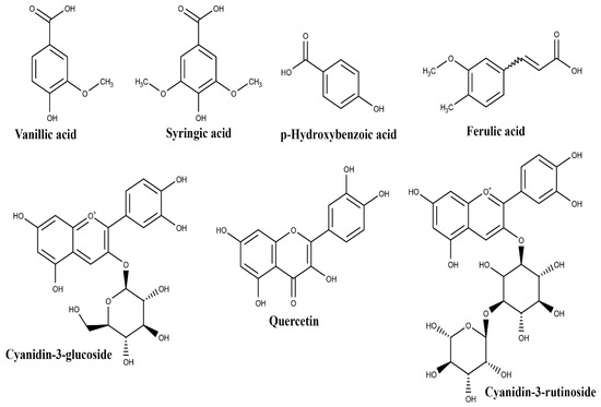
Figure 1.
Structures of the main compounds identified in Euterpe oleracea Mart. fruit.

Table 1.
Chemical profile of different parts of Euterpe oleracea Mart. with pharmacological application.
Pacheco-Palencia et al. [29] states that extracts enriched with these compounds are responsible for antioxidant activity. Moreover, Agawa et al. [30] and Poulose et al. [31] reported the presence of the flavonoid quercetin and anthocyanins, such as cyanidin-3-glucoside and cyanidin-3-rutinoside (Figure 1 and Table 1) in EO extracts, confirming their antioxidant activity. Poulose et al. [31] also reported an anti-inflammatory effect, corroborating the results of Kang et al. [32], Cedrim et al. [33], and Mathias et al. [34].
Several studies performed on EO fruit extracts and oil have shown other biological activities, among which are the antinociceptive [19], antiangiogenic [22], antimicrobial [23,24,25], antileishmanial [26], anticonvulsant [35], and antiproliferative [36] ones, which suggested their application in the treatment and prevention of metabolic syndromes such as cardiovascular disease, diabetes and dyslipidemias [37,38,39,40]. They are listed in Table 2 and discussed in Section 3.1, among other diverse biological activities.
Table 2.
In vitro and in vivo studies reporting the biological activities of different parts of Euterpe oleracea Mart.
EO oil, which is extracted from the pulp or seed of the fruit, consists mainly of fatty acids, whose major constituent is oleic acid (47.58%), followed by palmitic (24.06%) and linoleic (13.58%) acids (Figure 2), while palmitoleic, vaccenic, lauric and stearic acids were detected in minor concentrations [25]. Melhorança Filho and Pereira [23] and Magalhães et al. [25] ascribed significant antibacterial activity, Favacho et al. [19] anti-inflammatory and antinociceptive effects, and Souza et al. [40] antilipemic action to the oil. A study by Pacheco-Palencia et al. [29] also reported the presence of polyphenolic compounds and anthocyanins retained in the oil, among which are procyanidin dimers, procyanidin trimers, vanillic acid, and syringic acid (Table 1), which contribute to its significant antioxidant activity.
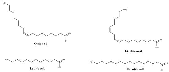
Figure 2.
Structures of the main fatty acids in Euterpe oleracea Mart. oil.
A phytochemical study on EO root and leaf extracts pointed out high concentrations of hydroxycinnamic acids such as caffeoylquinic acids, caffeoylshikimic acids, and flavone derivatives such as C-glycosyl derivatives of apigenin and luteolin. Table 1 lists the main compounds identified in the respective extracts. Therein, 5-Caffeoylquinic acid, 4-caffeoylshikimic acid and 5-caffeoylshikimic acid stand out in root extracts, while 5-caffeoylquinic acid and 6,8-di-C-hexosyl apigenin sulfate, which has a sulfate in position 7 or on the sugar moieties, stand out in the leaf extract. Figure 3 shows their molecular structures [41]. These authors highlighted the interest of the pharmaceutical, cosmetic, and food industries, especially for leaves, due to their antioxidant potential.
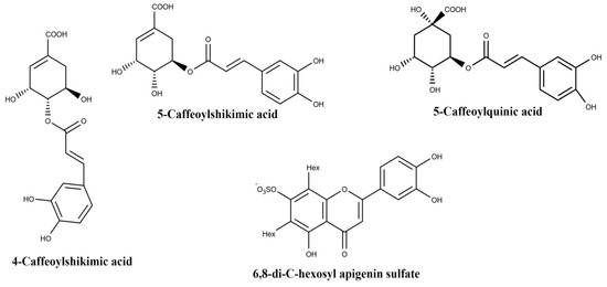
Figure 3.
Structures of the main components of Euterpe oleracea Mart. root and leaf; Hex: hexose.
3. Biological EO Activities: Application in the Prevention and Treatment of Diseases
EO has been long used in folk medicine. Plotkin and Balick [15] reported its empirical use against diarrhea. Only in 2002 did a study on EO leaf and steam extracts report a reduction in abdominal contortions and peripheral analgesic action [42], while in 2004 and 2005 the antioxidant capacity of EO fruit was attributed to flavonic compounds present in its extract [43,44]. Over the years, several studies have reported pharmacological applications related to biological activities of this species. The biological activities found in the literature are summarized in Table 2 together with the respective pharmacological applications.
3.1. Pharmacological Applications of Euterpe oleracea Mart. Fruit
3.1.1. Pro-Apoptotic Effect
A polyphenolic fraction obtained from juice was tested against human colorectal adenocarcinoma (HT-29), colon adenocarcinoma (SW-80) and colon fibroblast cells (CCD-18Co). The extract inhibited the growth of SW-480 cells more than that of HT-29 cells but had no toxicity to the non-malignant CCD-18Co cells. The mechanisms involved in colon cancer cell growth suppression include protection against reactive oxygen species (ROS) production and downregulations of nuclear factor-kappa B (NF-kB), NF-kB-target vascular cell adhesion molecule-1 (VCAM-1), intercellular adhesion molecule-1 (ICAM-1), Sp prooncogenic transcription factors, Sp-target vascular endothelial growth factor (VEGF), Bcl-2, and survivin. It also activated the mitochondrial pro-apoptotic pathway that resulted in cytochrome c release, caspase-3 cleavage, and poly (ADP-ribose) polymerase-1 (PARP-1) degradation [45].
3.1.2. Anticancer Effect
In vitro and in vivo studies using different parts of EO have pointed out anticancer effects against different cell lines (Table 2). Choi et al. [46] observed that the administration of pellets containing 5% of lyophilized EO berries reduced significantly the incidence of both adenoma (from 76.9% to 23.1%) and cancer (from 76.9% to 15.4%) as well as the expressions of myeloperoxidase (MPO) and proinflammatory cytokines (tumor necrosis factor α [TNF-α], interleukin [IL]-1β, and IL-6) in colorectal cancer. It also inhibited the expression of proliferating cell nuclear antigen (PCNA) and B2 cell lymphoma (Bcl-2), through increased anti-Bcl-2-associated death (Bad) as well as the expression and activation of cleaved caspase-3 of the mitochondrial pro-apoptotic pathway. On the other hand, Martinez et al. [49] attributed the high antioxidant activity, detected for the EO hydroalcoholic extract by the 2,2-diphenyl-1-picrylhydrazyl (DPPH), the 2,2’-azinobis-3-ethylbenzothiazoline-6-sulfonic acid/Trolox Equivalent Antioxidant Capacity (ABTS/TEAC), the ferric reducing ability of plasma (FRAP) and the oxygen radical absorbance capacity (ORAC) assays, to its large content of total phenolic compounds (37.08 ± 8.56 g of gallic acid equivalent/100g). In a cell viability test, the same authors observed a decrease in A549 lung cancer cell viability, with cell cycle regulation due to cell increase in the G0/G1 diploid phases (2 n), but a reduction in the S (>2 n but <4 n) and G2/M (4 n) phases, in addition to a high increase in the apoptotic cells when compared to the untreated ones.
3.1.3. Anticlastogenic Effect
The ability of EO extract to inhibit osteoclastogenesis and osteoclast activity was investigated by Brito et al. [51]. The 3-[4,5-dimethylthiazole-2-yl]-2,5-diphenyltetrazolium bromide (MTT), half maximal inhibitory concentration (IC50) and total protein assays showed that the EO extract concentration did not influence cell viability of monocyte macrophage cell line (RAW 264.7). However, its administration resulted in the inhibition of osteoclast differentiation and activity, possibly due to the modulation of cytokines produced by osteoclast precursor cells [51].
3.1.4. Anticonvulsant Effect
Arrifano et al. [35] reported that EO juice can improve GABAergic neurotransmission, thus helping to treat seizures and epilepsies. Treatment of primary cultures of cortical neurons and astrocytes with EO (0–25%) resulted in an increase in binding of the agonist ([3H] flunitrazepam) and a decrease of the antagonist ([3H] TBOB) to their receptor binding sites on cortical neurons. Low concentrations still significantly inhibited GABA uptake, suggesting an accumulation of endogenous GABA in the synaptic cleft. Souza-Monteiro et al. [20] demonstrated the ability of EO juice to increase latencies to the first myoclonic jerk and first generalized tonic-clonic seizure, while reducing the total duration of tonic-clonic seizures caused by pentylenetetrazol administration, hence displaying an anticonvulsant response. It was also able to prevent lipid peroxidation in the cerebral cortex, highlighting an additional neuroprotective effect on humans against lipid peroxidation associated with seizures.
3.1.5. Antidepressive and Anti-Aging Effects
EO juice was also tested for possible antidepressive and anti-aging effects [52]. Only 4 doses of juice were enough to prevent despair- and anhedonia-like behaviors, and changes observed in electromyographic measurements were comparable to those of imipramine. Tests by two-step quantitative reverse transcription polymerase chain reaction (qRT-PCR) highlighted an increase in the expression of telomerase reverse transcriptase (TERT mRNA) and an aging-associated enzyme whose level decreased in people with depression, revealing for the first time an anti-aging and neuroprotective action related to old age.
3.1.6. Antidiabetic Effect
De Bem et al. [38] observed a positive antidiabetic effect when administering EO seed extracts. Dietary complications were induced in rats by a high-fat diet and streptozotocin; however, the reduction in glycemic indexes, insulin resistance, leptin and IL-6 levels, lipid profile and vascular dysfunction were evident in the treated group. When physical exercise was added to the treatment, it not only potentiated the reductions of glycemic indexes and TNF-α levels, but also increased the expressions of phosphorylated protein kinase B (pAKT), adiponectin in adipose tissue, and insulin receptor (IR) and phosphorylated adenosine monophosphate-activated protein kinase (pAMPK) in the skeletal muscle of type 2 diabetic rats.
3.1.7. Antihypertensive Effect
The vasodilating effect on the mesenteric vascular layer of a rat pretreated with noradrenaline was evaluated. Rocha et al. [37] observed that the exposure to EO seed extract induced endothelium-dependent vasodilation, likely due to activation of the NO-cGMP pathway, which may involve the release of endothelium-derived hyperpolarizing factor (EDHF). According to Cordeiro et al. [53], EO seed extracts were able to attenuate spontaneously hypertensive episodes in rats due to high levels of protein carbonylation combined with low levels of nitrite, both attenuated in mesenteric arteries and heart homogenates. The up-regulation of nitric oxide synthase from endothelial tissue (eNOS) and superoxide dismutase 1 (SOD1) expression and increased SOD activity were also observed. EO also prevented endothelial dysfunctions in the aorta with increases in media thickness and media/lumen ratio and a decrease in the percentage of elastic fibers. These authors ascribed the antihypertensive activity to the antioxidant effects related to the high amounts of procyanidins and catechins in the extract along with the production of endothelial NO.
3.1.8. Anti-Inflammatory Effect
As can be seen in Table 2, the anti-inflammatory effect of EO is one of its pharmacological activities most reported in the literature. Most in vitro studies report positive results of the ORAC assay and decreased ROS production even when the cell line used was exposed to lipopolysaccharides. Machado et al. [57] observed a reduction in the activation of murine RAW 264.7 macrophage line cells induced by the treatment with an EO hydroalcoholic extract, due to a decreased activation of nod-like receptor pyrin containing 3 (NLRP3). In another study, Poulose et al. [31], using BV-2 murine microglial cells, observed that the p38 mitogen-activated protein kinase (p38-MAPK) and the transcription factor NF-kB had their activities attenuated by EO, which regulated the production of Ca2+-independent nitric oxide synthase (iNOS) as well as the activation of cyclooxygenase 2 (COX -2). Zhou et al. [54] also reported that EO extract reduced the inflammation mediated by interleukin 8 (IL-8), tumor necrosis factor-α (TNF-α) and transforming growth factor-β (TGF-β) and reduced the alcohol-induced expression of nuclear factor NF -κB and CD68 in hepatocytes of Wistar rats. This study also reports a reduction in oxidative stress and liver damage.
3.1.9. Antilipemic Effect
Martino et al. [39] claim that EO polyphenols reduce the accumulation of lipids in adipocyte cells by a reduction of the genetic expression of adipogenic transcription factors C/ebpα, C/ebpβ, Kruppel-like factor and sterol regulatory element-binding protein 1c, accompanied by a reduction in adipogenic genes, adipocyte fatty acid-binding protein 2, lipoprotein lipase, fatty acid transport proteins, fatty acid synthase, leptin, total plasminogen activator inhibitor, and by an increase in adiponectin level. These authors also reported an anti-inflammatory and antioxidant activity. In vivo studies demonstrated a hypocholesterolemic action of EO in rats that underwent diets rich in fat and cholesterol [60,61,62,63,64,65,66]. Feio et al. [65] observed that the consumption of EO juice improved the lipidic profile of adult male New Zealand white rabbits after 12 weeks of treatment, besides mitigating atherosclerosis. These effects were justified by an improvement of the balance of synthesis and absorption of sterols. In a clinical study by Pala et al. [66], a group of women who incorporated in their diet an amount of 200 g of lyophilized EO pulp for 4 weeks, showed reduced levels of ROS and oxidized-low density lipoproteins (ox-LDL) along with an increased activity of antioxidative paraoxonase 1. As far as the apolipoproteins involved in cholesterol metabolism are concerned, an improvement in the metabolism of Apo-I was observed, which implies an improvement in plasma high density lipoprotein (HDL) levels.
3.1.10. Antimicrobial Effect
A study by Sprenger et al. [67] observed that hydroalcoholic extracts from EO seeds and fruit exerted an antimicrobial activity against strains of Clostridium perfringens, Streptococcus aureus, and Pseudomonas aeruginosa. Dias-Souza et al. [24], who tested a methanolic extract from EO pulp according to the serial dilution method, obtained positive results against S. aureus and its biofilm formation and observed a synergism when the extract was used in combination with antimicrobial drugs. For tests with EO extract, Borges et al. [68] used two strains of Aspergillus fumigatus, AFAR and AF4091, commonly found adhered to abiotic surfaces of medical-hospital material. Both the adhesion and the amount of fungal biomass, especially those of the AFAR strain, were remarkably reduced after treatment with EO.
3.1.11. Antinociceptive Effect
A clinical study by Jensen et al. [69] demonstrated that the consumption of juice from EO fruits significantly improved amplitude and pain score data as well as the activities of daily living in patients. The antioxidant activity assessed by the novel cell-based antioxidant protection in erythrocytes (CAP-e) bioassay was improved over 12 weeks of treatment. Whereas tests with the C-reactive protein (CRP) inflammatory marker showed no significant difference, the lipid peroxidation was decreased. These results suggest an improvement in the physical well-being of patients with movement restrictions associated with pain caused by their daily load. Marinho et al. [42], who reported the first evidence of an antinociceptive effect of extracts from EO flowers and stalks, observed in rats a reduction of up to 50% in the total number of abdominal contortions, showing a peripheral effect of either extract in a dose of 30 mg/kg. Even though stalk extracts caused an increase in the rate of analgesia in the tail-removal model at doses of 10 and 30 mg/kg, no extract was able to change the analgesia index in the hot plate test, suggesting that there was no central action. Sudo et al. [70] used different methodologies to investigate the antinociceptive activity of EO seed extract (ASE) in an acute and chronic way on male Swiss mice and male Winstar rats, as well as the mechanisms underlying these effects. The hot plate test was used to evaluate antinociceptive agents that act centrally, but not peripherally. The treatment with ASE (30, 100, or 300 mg/kg/day) showed a dose dependent antinociceptive activity, as well as other methodologies such as the formalin-induced hind paw-licking test, the carrageenan-induced pain test and the acetic-acid writhing test. The study was not able to completely demonstrate the exact mechanism of action of the extract, suggesting the involvement of various pathophysiological systems.
3.1.12. Antioxidant Effect
Hydroalcoholic extracts from EO seeds were used in antioxidant tests. The in vitro study on immortalized human umbilical vein cells (HUVEC) carried out by Soares et al. [72] proved that EO was able to prevent the deleterious effects caused by the H2O2-induced oxidative stress, besides positively modulating the signaling cascade of NRF2. An in vivo study showed that the same extract had a beneficial effect on the general framework of the cachectic syndrome caused in rats [73]. Carvalho et al. [74] demonstrated the antioxidant activity by the DPPH and ORAC assays of both a lyophilized EO pulp and an extract-containing gel in different concentrations (8%, 12%, 16%, and 20%).
3.1.13. Antiplasmodial Effect
Ferreira et al. [76] tested three fractions obtained from the EO pulp, namely total phenolics, total anthocyanins and non-anthocyanidin phenolics. The first two fractions were inefficient in reducing parasitic DNA, while the third one showed moderate antiplasmodial activity in strains of Plasmodium falciparum. A murine model of infection was also used to investigate whether EO polyphenols could mitigate parasitemia in vivo, and Plasmodium chabaudi-infected mice were treated orally with the total phenolics fraction (10, 15, and 20 mg/kg) for 12 days. During parasitemia peak occurred after six and seven days, the 20 mg/kg dose reduced parasite growth by 89.4% and 77.3%, respectively, compared to the untreated control group, while the 15 mg/kg one did so by 81% and 62.2%, respectively.
3.1.14. Antiproliferative Effect
Pozo-Insfran et al. [77] exposed HL-60 leukemic cells to different fractions rich in anthocyanins and other polyphenolic compounds known for their antioxidant activities. The biological response demonstrated a decrease in dose-dependent cell viability. Fractions containing non-hydrolyzed anthocyanins effectively suppressed HL-60 cell proliferation by inducing apoptosis via caspase-3 activation. Pacheco-Palencia et al. [29], in a comparative study on the proliferative action of polyphenolic fractions obtained from EO pulp and oil, noticed that both fractions caused a decrease in the viability of concentration-dependent human colon adenocarcinoma (HT-29) cells. The polyphenolic extract obtained from the oil proved to be twice as effective in all tested concentrations. When the same research-group tested monomeric and polymeric anthocyanins obtained from the fruit pulp on the same cell line, the former proved more effective [36].
3.1.15. Antiprotozoal Effect
EO juice reduced the number of Leishmania promastigotes, caused morphological changes and increased ROS production [26]. The phenotypes of induced cell death were likely associated with apoptosis in promastigotes of L. mazonensis and L. infantum (= L. chagasi). The treatment against amastigotes incubated in isolated macrophages of peritonitis showed a reduction in the levels of cytokines of IL-17 family, which are involved in the pathogenic process, as well as a decrease in the number of intracellular amastigotes of the macrophages infected by both species. In addition, no cytotoxic effects were observed in infected macrophages, demonstrating leishmanicidal activity and safety to the host cell. Flavonoids present in the EO juice may have been responsible for the observed property [26].
3.1.16. Cardioprotective Effect
Treatment performed with EO seed extract improved cardiac dysfunction and exercise intolerance in rats with induced myocardial infarction (MI) [79]. After treatment, the low systolic blood pressure (86.88 ± 4.62 mmHg) and high diastolic pressure (17.62 ± 1.21 mmHg) in animals with MI underwent inversion to values (130.00 ± 8.16 mmHg and 3.69 ± 2.69 mmHg, respectively) close to those of the control group. The distance covered by the treated group was 5.46-fold that of rats with MI, preventing cardiac hypertrophy, fibrosis, and dysfunction. A brief study showed a significant reduction in systolic blood pressure in healthy volunteers who had ingested EO gelatin capsules, but hemodynamic and electrocardiographic effects did not show any significant variation [80].
3.1.17. Healing Effect
A study on cell proliferation showed increased migration of Hs68 human fibroblastic cells after administration of aqueous EO extract, which may play an important role in wound healing. EO increased the level of fibronectin mRNA expression and decreased that of matrix metalloproteinase MMP-1 mRNA expression. In vivo tests with macroscopic and histopathological observations showed a wound healing effect, indicating that EO is a potential healing agent [81]. The same research-group, treating an oral healing model with EO, observed effects on the healing of wounds in the oral mucosa consistent with previous histopathological observations [82].
3.1.18. Cytotoxic effect
Cardiotoxicity studies showed that EO supplementation was able to prevent changes caused by doxorubicin, commonly used in chemotherapy [94]. The added EO diet improved fractional shortening of the left ventricle and increased the levels of enzymes associated with cardiac metabolism, such as β-hydroxyacyl-CoA dehydrogenase, phosphofructokinase, citrate synthase, complex II enzyme activities, and ATP synthase. On the other hand, the concentration of myocardial lipid hydroperoxide and the activity of the matrix metalloproteinase MMP-2 decreased when doxorubicin was used alone [34].
3.1.19. Hepatoprotective Effect
Açaí seed extract reduced body weight gain, food intake, and glycemic, cholesterol, and triglyceride levels in the liver. Expressions of phosphorylated adenosine monophosphate activated protein kinase (pAMPK), phosphorylated acetyl-CoA carboxylase (pACC), acetyl-CoA carboxylase (ACC) and cholesterol excretion transporters [ATP-binding cassette, subfamily G transporter 5 (ABCG5) and ATP-binding cassette, subfamily G transporter 8 (ABCG8)] were increased, whereas those of lipogenic proteins [sterol-regulatory-element binding protein-1c (SREBP-1c) and 3-hydroxy-3-methylglutaryl CoA reductase (HMG-CoA reductase)] were reduced. These responses contributed to the reduction of obesity and hepatic steatosis. Antioxidant activity was also assessed, and there was an improvement in the activities of SOD, catalase and glutathione peroxidase [60]. De Freitas Carvalho et al. [84], who investigated non-alcoholic fatty liver disease, obtained answers in agreement with the study by De Oliveira et al. [60], when male mice of the C57BL/6 strain were treated with EO pulp extracts. The extract attenuated liver damage, inflammatory process and oxidative stress and modulated glutathione reductase, SOD and catalase, hence conferring a hepatoprotective effect ascribed to EO phenolic compounds. Both studies were cited earlier in the antilipemic effect subsection.
3.1.20. Immune System Inhibition
Exposure of IgE-sensitized mouse primary cultured mast cells to EO pulp resulted in suppression of IgE-mediated degranulation and transcription of the cytokine genes from a cultured mast cell line originated from rat basophilic leukemia (RBL-2H3), selectively inhibited FcεRI signaling pathways and suppressed the FcεRI-mediated complementary signaling pathway. In general, EO inhibited IgE-mediated mast cell activation [85].
3.1.21. Neuroprotective Effect
Exposure of rat pheochromocytoma cells (Ordway PC12) to human amyloid-protein 1–42 (Aβ1–42) or 25–35 (Aβ25–35) or to tert-butyl hydroperoxide decreased cell viability, whereas a pretreatment with EO extract only improved cell viability after exposure to Aβ1–42. EO extract and isolated metabolites such as (-)-epigallocatechin-3-gallate, cyaniding glucoside, cyaniding rutinoside caused loss of thioflavin T (ThT) fluorescence, with cyaniding glucoside also disrupting Aβ1–42 fibril and aggregate morphology [86]. A study by Machado et al. [87] suggested the use of EO as an alternative therapy for the treatment of bipolar disorder associated with mitochondrial dysfunction and cell oxidative stress. Exposure to EO extract increased enzyme activity of the mitochondrial complex I, amounts of proteins and, mainly, protein overexpression in mitochondrial complex I Q module subunits NDUFS7 and NDUFS8, and reduced levels of ROS and cellular lipid peroxidation.
3.1.22. Lung Protective Effect
Polysaccharide fractions from EO fruit stimulated γδ T cell activity in peripheral blood mononuclear cells (PBMC) from humans, mice and cattle. High molecular weight polysaccharides isolated from crude EO were the most active in vitro activating myeloid T cells and γδ, while in vivo they induced the recruitment of myeloid cells and the production of IL-12, thus favoring a downstream Th-1 response able to alleviate asthma symptoms [88]. In a second study, the same polysaccharide fraction, when administered via nasal route in mice, a) protected animals against Francisella tularensis with survival rates of up to 80%, b) increased intracellular expression of interferon–gamma (IFN-γ) by natural killer (NK) cells in the lungs of infected animals, c) drastically reduced the number of Burkholderia pseudomallei cells in the lung, d) blocked the bacterial spread to the spleen and liver, and e) increased the IFN-γ response by NK and γδ T cells [89], thereby suggesting an agonistic action on the innate immune system.
3.1.23. Renoprotective Effect
Exposure to EO seed extracts attenuated kidney damage in rats that had suffered diabetes induction, prevented its dysfunction, lowered serum levels of urea, creatinine and albumin, and reduced renal fibrosis due to the decreased expression of collagen IV and transforming growth factor beta 1 (TGF-β1), an early marker of fibrosis. Despite the contrast to the increase in pro-inflammatory biomarkers (IL-6, TNF-α and MCP-1) and in apoptosis (caspase-3), markers of oxidative damage [thiobarbituric acid reactive substances (TBARS), carbonyl and 8-isoprostane levels] were reduced [91]. On the other hand, there was an evident increase in the number of glomeruli and activity of antioxidant enzymes (SOD, catalase and GPx) as well as a reduction in inflammation and oxidative stress. A similar study carried out by Da Costa et al. [92] demonstrated that the treatment with EO seed extracts reduced kidney damage in rats with renovascular hypertension, likely due to antihypertensive and antioxidant effects.
3.1.24. Hematopoietic Effect
A study by Shibuya et al. [93] showed a significant increase in the number of erythrocytes, hemoglobin and hematocrit in the C57BL/6NCrSlc mouse strain treated with a dose of EO fruit extract for 4 days. This erythropoietic effect was associated with renal hypoxia caused by EO. Tests showed an increase in the hematopoietic hormone and in the transient gene expression of the hematopoietic factor (EPO), expressed mainly in the kidney. There was also an increase in the expression of VEGFA, mediated by the transcription factor HIF induced by hypoxia, after exposure to EO. These authors suggested investigating other mechanisms of action for this EO function.
3.2. Pharmacological Applications of Euterpe oleracea Mart. Oil
3.2.1. Antineoplasic Effect
Monge-Fuentes et al. [95] demonstrated an effective anticancer activity of a nanoemulsion formulated with EO oil against melanoma cell lines, leading to death by apoptosis/necrosis of 85% of line B16F10 cells in vitro and an 82% reduction of tumor volume in tumor-bearing C57BL/6 mice. These results suggest photodynamic therapy for the treatment of melanomas (Table 3).
Table 3.
In vitro and in vivo studies reporting the effect of Euterpe oleracea Mart. oil.
3.2.2. Anti-Inflammatory Effect
Favacho et al. [19] reported inhibitions in the formation of subcutaneous granulomatous tissue and ear edema, decreases in vascular permeability response and prostaglandin synthesis, and a reduction in up to 80.14% of migrating neutrophils in peritoneum inflammation, when compared to the control group.
3.2.3. Antilipemic Effect
Sousa et al. [40], after inducing dyslipidemia in rats and treating them with EO, observed a reduction in the levels of triglycerides (TG), total cholesterol (TC), and direct low-density lipoprotein cholesterol (LDL-c), but an increase in that of HDL.
3.2.4. Antimicrobial Effect
After Pseudomonas aeruginosa and Staphylococcus aureus growth in a medium composed of agar Muller–Hinton, disks containing 10 µg of EO seed oil were added. The oil-impregnated discs showed no inhibition halo after 24 and 48 h of contact with the medium containing P. aeruginosa, while that containing S. aureus induced an inhibition halo, even if significantly smaller than those induced by vancomycin and penicillin [23]. The minimum inhibitory concentration of the oil obtained from the pulp was determined against Enterococcus faecalis, S. aureus, P. aeruginosa and Escherichia coli strains by diffusion in broth. Oil inclusion complexes in (β) or (HP-β)-cyclodextrins showed a reduction in their minimum inhibitory concentration, increasing the oil’s antibacterial effect, which was related to the improvement of oil solubility and stability induced by complexation [25].
3.2.5. Antinociceptive Effect
Favacho et al. [19] carried out an abdominal contortion test in rats that showed inhibition of up to 55.58% at a dose of 1500 mg/kg of oil. This effect was attributed to an action of peripheral origin and linked to the previously mentioned inhibition of prostaglandin biosynthesis.
3.2.6. Cytotoxic Effect
An oral toxicity test using EO oil showed that doses of 30, 100 and 300 mg/kg caused changes in follicular morphology of thyroid cells, mainly marked by the loss of follicular cell lumen size, hypertrophy and unorganized growth. Meanwhile, hepatocytes underwent vacuolization and change from eosinophilic to basophilic characteristics. This suggests that morphological change caused a significant cytotoxic physiological change in both tissues [96]
3.2.7. Genotoxic Effect
A genotoxicity study was carried out with 1% EO oil in Tween 80 at doses of 30, 100, and 300 mg/kg. The genotoxic property of EO oil was evaluated by comet assay, and samples of leukocytes obtained from peripheral blood, liver, bone marrow, and testicular cells did not show any indication of genotoxic effect [97].
4. Inventions Related to EO as an Alternative Medicine
Due to the above broad pharmacological arsenal, it is understandable that patentable inventions dealing with EO are developed for therapeutic purposes. In 2008, two patents were granted by Soares de Moura, researcher from the Federal University of Rio de Janeiro (Brazil) (Table 4). Patents were pertinent to the development of gelatin capsules and tablets obtained from lyophilized hydroalcoholic extracts of EO fruits and seeds, even though they point to different biological activities of the formulations.
Table 4.
Patents related to pharmaceutical formulations containing Euterpe oleracea Mart. for the treatment and prevention of general diseases.
The first patent filed, PI0418614-1 A2 [98], discloses obtaining said extracts and pharmaceutical forms for their application in pain treatment. The analgesic effect was determined from the antinociceptive response by the abdominal writhing test in mice. Abdominal writhing was induced intraperitoneally by acetic acid, and then the lyophilized extracts obtained by hydroalcoholic extraction from fruits were administered at doses of 1 and 3 mg/kg, while those from seeds at doses of 0.3, 1.0, 3.0, 10.0, and 30.0 mg/kg. All extracts were able to significantly reduce the number of abdominal contortions, and the antinociceptive response was improved as the concentration was increased. The patent suggests the use of these extracts for pain treatment as a general attempt, since the method is sensitive to antinociception in the central and peripheral nervous system, muscle relaxants, and sedatives [99,100].
The second patent, PI 0604281-3 A2 [101], reports the application of these same lyophilized extracts to treat and prevent vasospastic, ischemic diseases and hypertension. A vasodilation test was performed on mesenteric vessels of Wistar rats, cannulated and perfused with Krebs nourishing solution, after euthanasia laparotomy. After perfusion had been established, vasoconstriction with norepinephrine was induced, and then increasing doses of lyophilized extracts were administered, thereby obtaining a vasodilatory dose-response curve as a function of the decreased norepinephrine pressor effect. Approximately 2.58 and 1.21 µg of fruit and seed extracts, respectively, ensured 50% of the vasodilator effect observed. The experimental response suggests the application of these extracts to reduce vascular spasms, which are responsible for ischemic heart disease and ischemic peripheral vascular disease [102].
Examination of the selected patents revealed that several studies were still developed with hydroalcoholic extracts from EO fruits. In 2011 a third patent, WO 2011/1036448 A1, was issued by Soares de Moura [103], where he claimed the preparation of ointments or creams containing antioxidants from plants, especially those of the genus Euterpe, capable of accelerating the healing process (Table 4). After injuries to the epithelial tissue, inflammatory processes were triggered in response to the immune system and tissue healing. Such a mechanism stimulated the formation of ROS and reduced that of nitric oxide. When pro-oxidant responses exceed the potential of cellular antioxidant mechanisms, they may compromise cell viability and generation of secondary reactive species, rendering the healing response deficient [104].
The invention was based on scientific data obtained from international literature, which showed the presence of polyphenolic compounds in EO fruit and important antioxidant activity by preclinical assays [21,37,75,105]. In addition to the antioxidant activity, experiments also demonstrated the ability of these extracts to stimulate the expression of eNOS, which is responsible for the synthesis of nitric oxide [37] involved in wound repair.
Another invention has been developed with a similar purpose of the patent above. The invention, US 61/814791 [106], requested by Neocutis SA (Pully, Switzerland) and published in 2015, describes the addition of various plant extracts, quoting EO berries, to a preparation also containing vitamin C and E with therapeutic and preventive potential against skin damage (Table 4). Application of the EO extract was likely suggested by the high content of polyphenols in berries, which are responsible for a reduction of the oxidative stress caused by inflammatory, infectious, injury and tissue aging processes [17,21]. However, the addition of EO berry extracts to the above pharmaceutical formulations was only mentioned in the summary and claims with no additional information.
Nascimento, in 2018, patented (BR 102015017543-4 A2) the development of gelatin capsules and syrups for the treatment of breast cancer [107] (Table 4). The formulations were developed from hydroalcoholic extracts of EO and Euterpe precatoria seeds submitted to the freeze-drying process. The lyophilizates were applied to the MCF-7 human breast cancer cell line to investigate inhibition of cell proliferation and morphological changes. For this purpose, cells were seeded and incubated in 96-well plates under biochemical oxygen demand conditions and subjected to transmission electron microscopy. The results showed that the EO lyophilizates were cytotoxic at concentrations of 10 µg/mL and significantly reduced MCF-7 cell viability at concentrations of 20 and 40 µg/mL. Moreover, cells treated with 40 µg/mL of the freeze-dried extracts suffered severe morphological changes such as cytoplasmic retraction, vacuolization and apparent lysis with loss of cytoplasmic content. Accordingly, the invention patents the use of lyophilizates for chemotherapeutic purposes as described in claim 7.
Due to the EO fruit chemical composition, its application becomes desirable because of the high concentrations of metabolites responsible for significant antioxidant activity [30,31,36,39]. The patent BR 102017007451-0 A2 [108], published in 2018 (Table 4), discloses the formulation of tablets, capsules, granulates and controlled-release pharmaceutical preparations from ethanolic EO extracts. Plant extracts were subjected to quality control analysis and quantification of chemical constituents by thermal analysis, microscopic examination, spectroscopy in the infrared region and high-performance liquid chromatography (HPLC) coupled to mass detectors, while formulations were subjected to physicochemical tests as recommended by the V Brazilian Pharmacopoeia. Preclinical tests of acute toxicity [109] and determination of antioxidant activity by DPPH and ABTS•+ assays were performed to justify application.
The patent BR 102018005450 [110] discloses the development of, possibly lyophilized, powders obtained from açaí seed extracts and fractions thereof (Table 4). The invention has been developed to treat and prevent diseases or disorders individually or in conjunction with metabolic syndromes such as abdominal obesity, disorders of lipoprotein lipid metabolism, high levels of triglycerides, high uric acid levels, increased blood pressure, insulin resistance, glucose intolerance, increased fasting glucose and liver steatosis. Quantification of the extract and its fractions by HPLC confirmed the presence of procyanidin B2, epicatechin, rutin, catechin, vicenin 2, vitexin, chlorogenic acid, while in vitro artery relaxation assays evidenced effects ascribable to the presence of such compounds, especially procyanidin B2. These are the only trials reported in the patent, although the claim suggests broad pharmacological action.
The patent BR 10201807679332018 [111], unlike the others, shows the production of cycloamylases by encapsulating EO oil in cyclodextrins. The authors suggested that this system is a pharmacological alternative to increase the oil’s solubility and stability, thereby enabling the compound’s anti-inflammatory, antimicrobial and antioxidant effect. In this patent, no in vitro or in vivo analysis was shown, but the authors stressed the importance of using inclusion complexes with EO as a contribution to optimize the therapeutic arsenal. This invention arose from the wide knowledge and growing importance attributed to EO and its oil.
Recently, in 2019, the patent BR 102017013494-6 A2 [112] was published with the purpose of developing an antimicrobial phytotherapic from EO leaf extracts (Table 4). Even though the invention does not show the development of in vitro or preclinical trials, it proposes the development of experiments to support the suggested biological activity.
EO extracts and oils are commonly consumed by the population because of their biological properties, especially in the treatment of inflammation. The number of studies conducted with fruits, leaves, seeds, or oil of EO shows the importance and interest of researchers to this plant, especially due to its chemical composition and biological activities discovered in scientific experiments. However, it is understood as contradictory the number of patents located, whose purpose suggests their pharmacological application.
When searching for publications on EO biological properties, only one scientific article was found in 1984 by Plotkin and Balick [15], in which the authors described the use of EO oil as antidiarrheal in folk medicine. Until 2002, no other article related to its pharmacological application was found in literature. In the past ten years there has been an increasing interest in the discovery of possible biological activities of EO. Six publications were recorded in 2011 and eight in 2015 and 2016. However, the largest record was in 2018, with 11 publications (Figure 4).
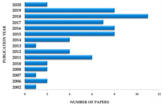
Figure 4.
Number of papers reporting the pharmacological activities of Euterpe oleracea Mart. for the treatment and prevention of general diseases.
After a detailed search of patents and their submission to the inclusion and exclusion criteria of this study, only nine patents suggested pharmacological EO application. A second point analyzed was the geographical distribution of patents. Moreover, 88.9% are Brazilian and 11.1% belong to the United States. The high percentage of Brazilian patents may be justified by the origin of EO, which is well distributed in the states of Pará, Maranhão, Amazônia, and Amapá in the north of Brazil [26,113]. The registration of the selected patents began in 2008 and the largest number of publications was in 2018 (Figure 5).
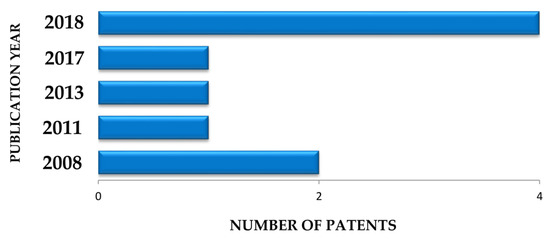
Figure 5.
Number of patents reporting pharmacological activities of Euterpe oleracea Mart.
The inventions suggest the application of various EO fruit, seed and leaf extracts to treat bacterial and fungal infections, treat and prevent skin damage, accelerate healing, antioxidant activity, antinociceptive action to pain, chemotherapeutic and chemo preventive action against breast cancer, treat and prevent vasospastic and ischemic diseases, high blood pressure, and metabolic syndromes in general.
In 50% of the patents analyzed, preclinical trials of the formulations were done, while the other half only pointed to the development and pharmacological application of products based on previous studies reported in the international literature or suggested evidential experimental development. For these, the inventors recognize EO fruit composition and link it to possible biological activities. An example is the invention of an EO fruit extract ointment [103], in which the identification of polyphenolic compounds, well described in the literature, directly influenced the development of this patent. Therefore, if there is no defined mechanism of action and clinical evidence, subjectivity is generated in the content of such patents. In vitro and in vivo assays are primordial and necessary for accreditation of the invention.
In the same context, the patent filed by Neocutis S.A. [106] that pointed out a formulation containing vitamins C and E as well as an antioxidant compound, included EO berries among the possible natural extract of choice for the composition. EO was likely selected because of its contents of phenolic compounds, anthocyanins and phytosteroids, which give the fruit antioxidant and anti-inflammatory activities [31,36]. Nevertheless, the invention does not show any pharmacological effect or synergistic activity among the components of the formulation, raising questions about its efficiency.
Inventions corroborated by preclinical studies generate high expectations among patients, as is the case of studies carried out against breast cancer cells, which have shown chemotherapeutic activity of EO seed extracts [107]. The invention of tablets, capsules, and granules obtained from fruit extracts was the only one that evidenced physicochemical, quality control, efficacy, and safety tests throughout the development of the formulation [108], becoming extremely relevant for product accreditation. Importantly, no patent mentions clinical trials.
Considering that physicochemical, pre-clinical and clinical tests must be carried out in all stages of the development of a new medicine, it is evident that more accurate studies are needed on pharmaceutical formulations.
5. Survey Methodology and Criteria
This review provided a survey of the papers and patents related to the therapeutic and preventive activity of EO and the advances in its pharmacological application. For this purpose, a search was performed to identify articles in the scientific literature, while specialized databases were consulted for patents such as the National Institute of Industrial Property (INPI), Latin American Patent Bank (Latipat), European Patent Office (ESPACENET), World Intellectual Property Organization (WIPO) and United States Patent and Trademark Office (USPTO). For such a search we used the terms “Euterpe oleracea” or “açaí” appearing in the title or abstract. To prepare Table 2, we considered articles that suggested therapeutic and preventive activities for pharmacological purposes, while excluding those that only addressed biological activities without pharmacological indication, or simply suggested EO as a source of nutraceuticals or cosmetics, and review articles. Likewise, only patents reporting EO pharmacological and preventive activities were taken into consideration. The International Patent Classification (IPC), section A, related to human needs, and subclass A61K, preparations for medical, dental or hygienic purposes, were adopted. For both searches, there was no restriction in the years of publication.
The search revealed a total of 459 patents. Patents related to cosmetic, food and hygiene applications, even those conforming to the previously mentioned classification, have been excluded. Only some patents presented preclinical trials showing EO biological activity, others only suggested its pharmacological application, but all were considered in the present study. Also, as an inclusion criterium, only patents written in English and Spanish were selected. Thus, after applying the inclusion and exclusion criteria and withdrawal of multiple patents, eight patents were selected and analyzed according to the objective of the study (Figure 6).
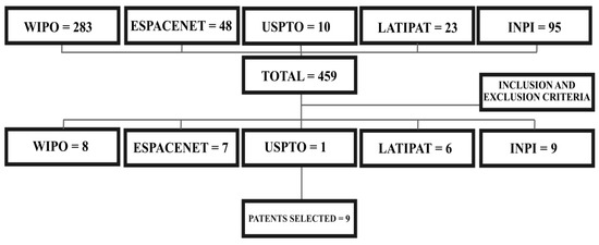
Figure 6.
Selection of patents concerning pharmaceutical formulations of Euterpe oleracea Mart. for the treatment and prevention of general diseases.
6. Conclusions and Future Perspectives
Euterpe oleracea (EO) is a plant known all over the world especially for its biological activities, whose chemical composition varies according to the part considered. Extracts from the fruit, roots, and leaves are mainly composed of phenolic metabolites, while EO oil is basically composed of fatty acids. Therefore, they are considered abundant sources of bioactive extracts capable of maintaining health.
Several in vitro and in vivo tests have demonstrated a direct relationship between EO chemical composition and numerous pharmacological activities such as anti-inflammatory, antioxidant, antimicrobial, antinociceptive, anticancer, anti-atherogenic and healing activities, which may be usefully exploited to treat metabolic syndromes and protect the lungs, kidneys, liver, heart, and nervous system. As for the analyzed inventions, they showed the use of EO in the treatment of infections by bacteria and fungi, the treatment and prevention of skin damage, breast cancer, and metabolic syndrome and the acceleration of healing, besides its antinociceptive and antioxidant properties. The scientific literature and patents concerning EO suggest a recent and, at the same time, growing interest in this plant, with a set of actions pointing to the development of promising new drugs for clinical use. However, very few studies and no patents have prioritized clinical trials, suggesting that more efforts should be made to clarify the effectiveness and safety of promising crude extracts, as well as isolated components of this plant for the subsequent consolidation of a pharmaceutical product.
Author Contributions
T.S.S.d.A.M. conducted the study and wrote the manuscript; P.C.d.O.M. assisted in the writing of the manuscript; A.C. interpreted, read and revised the work; Á.A.N.d.L. coordinated, read and reviewed the work. All authors read and approved the final manuscript.
Funding
This study was financed in part by the Coordenação de Aperfeiçoamento de Pessoal de Nível Superior-Brasil (CAPES)–Finance Code 001.
Acknowledgments
The authors thank CNPq and CAPES for their support and Federal University of Rio Grande do Norte.
Conflicts of Interest
The authors declare no conflict of interest. CNPq and CAPES have no role in the design of the study, in the collection, analyses or interpretation of data, in the writing of the manuscript, or in the decision to publish the results.
References
- Yuan, H.; Ma, Q.; Ye, L.; Piao, G. The traditional medicine and modern medicine from natural products. Molecules 2016, 21, 559. [Google Scholar] [CrossRef] [PubMed]
- Jamshidi-Kia, F.; Lorigooini, Z.; Amini-Khoei, H. Medicinal plants: Past history and future perspective. J. Herbmed Pharmacol. 2018, 7, 1–7. [Google Scholar] [CrossRef]
- Bruning, M.C.R.; Mosegui, G.B.G.; Vianna, C.M.D.M. A utilização da fitoterapia e de plantas medicinais em unidades básicas de saúde nos municípios de Cascavel e Foz do Iguaçu–Paraná: A visão dos profissionais de saúde. Ciênc. Saúde Coletiva 2011, 17, 2675–2686. [Google Scholar] [CrossRef] [PubMed]
- Oteng Mintah, S.; Asafo-Agyei, T.; Archer, M.A.; Atta-Adjei Junior, P.; Boamah, D.; Kumadoh, D.; Appiah, A.; Ocloo, A.; Boakye, Y.U.; Agyare, C. Medicinal Plants for Treatment of Prevalent Diseases. In Pharmacognosy Medicinal Plants; Perveen, S., Al-Taweel, A., Eds.; Intech Open: Londres, Reino Unido, 2019. [Google Scholar] [CrossRef]
- Lima-Saraiva, S.R.G.; De César, H.C.; Saraiva, C.; Oliveira-Júnior, R.G.; De Silva, J.C.; Damasceno, D.C.M.; Almeida, J.R.G.S.; Amorim, E.L.C. A implantação do programa de plantas medicinais e fitoterápicos no sistema público de saúde no Brasil: Uma revisão de literatura. Rev. Interdiscip. Pesqui. Inov. 2015, 1, 1–11. Available online: https://seer.ufs.br/index.php/revipi/article/view/3095/3406 (accessed on 2 February 2020).
- Silva, N.C.S.; Vítor, A.M.; Bessa, H.H.D.S.; Barros, R.M.S. A utilização de plantas medicinais e fitoterápicos em prol da saúde. UNICA Cad. Acad. 2017, 3, 1–5. [Google Scholar]
- Alves, R.R.N.; Rosa, I.M.L. Biodiversity, traditional medicine and public health: Where do they meet? J. Ethnobiol. Ethnomed. 2007, 3, 1–9. [Google Scholar] [CrossRef]
- Li, X.; Chen, Y.; Lai, Y.; Yang, Q.; Hu, H.; Wang, Y. Sustainable utilization of traditional Chinese medicine resources: Systematic evaluation on different production modes. Evidence-Based Complement. Altern. Med. 2015, 2015, e218901. [Google Scholar] [CrossRef]
- Rinaldi, A.; Shetty, P. Traditional Medicine for Modern Times: Facts and Figures. 2015. Available online: https://www.scidev.net/global/medicine/feature/traditional-medicine-modern-times-facts-figures.html (accessed on 12 February 2020).
- Chugh, N.A.; Bali, S.; Koul, A. Integration of botanicals in contemporary medicine: Road blocks, checkpoints and go-ahead signals. Integr. Med. Res. 2018, 7, 109–125. [Google Scholar] [CrossRef]
- Klein, T.; Longhini, R.; Bruschi, M.L.; Mello, J.C.P. Fitoterápicos: Um mercado promissor. Rev. Cienc. Farm. Basica Apl. 2009, 30, 241–248. [Google Scholar]
- Santamarina, A.B.; Jamar, G.; Mennitti, L.V.; de Rosso, V.V.; Cesar, H.C.; Oyama, L.M.; Pisani, L.P. The use of juçara (Euterpe edulis Mart.) supplementation for suppression of nf-κb pathway in the hypothalamus after high-fat diet in wistar rats. Molecules 2018, 21, e1814. [Google Scholar] [CrossRef]
- Santamarina, A.B.; Jamar, G.; Mennitti, L.V.; Ribeiro, D.A.; Cardoso, C.M.; de Rosso, V.V.; Oyama, L.M.; Pisani, L.P. Polyphenols-rich fruit (Euterpe edulis Mart.) prevents peripheral inflammatory pathway activation by the short-term high-fat diet. Molecules 2019, 24, e1655. [Google Scholar] [CrossRef] [PubMed]
- Čižauskaitė, U.; Jakubaitytė, G.; Žitkevičius, V.; Kasparavičienė, G. Natural ingredients-based gummy bear composition designed according to texture analysis and sensory evaluation in vivo. Molecules 2019, 24, e1442. [Google Scholar] [CrossRef] [PubMed]
- Plotkin, M.J.; Balick, M.J. Medicinal uses of South American palms. J. Ethnopharmacol. 1984, 10, 157–179. [Google Scholar] [CrossRef]
- Oliveira, A.G.; Costa, M.C.D.; Rocha, S.M.B.D.M. Benefícios funcionais do açaí na prevenção de doenças cardiovasculares. J. Amazon Health Sci. 2015, 1, 1–10. Available online: https://periodicos.ufac.br/revista/index.php/ahs/article/view/39 (accessed on 22 January 2020).
- Yamaguchi, K.K.D.L.; Pereira, L.F.R.; Lamarão, C.V.; Lima, E.S.; Da Veiga-Junior, V.F. Amazon açai: Chemistry and biological activities: A review. Food Chem. 2015, 179, 137–151. [Google Scholar] [CrossRef]
- Garzón, G.A.; Narváez-Cuenca, C.E.; Vincken, J.P.; Gruppen, H. Polyphenolic composition and antioxidant activity of açai (Euterpe oleracea Mart.) from Colombia. Food Chem. 2017, 217, 364–372. [Google Scholar] [CrossRef]
- Favacho, H.A.S.; Oliveira, B.R.; Santos, K.C.; Medeiros, B.J.L.; Sousa, P.J.C.; Perazzo, F.F.; Carvalho, J.C.T. Anti-inflammatory and antinociceptive activities of Euterpe oleracea oil. Braz. J. Pharmacogn. 2011, 21, 105–114. [Google Scholar] [CrossRef]
- Souza-Monteiro, J.R.; Hamoy, M.; Santana-Coelho, D.; Arrifano, G.P.F.; Paraense, R.S.O.; Costa-Malaquias, A.; Mendonça, J.R.; da Silva, R.F.; Monteiro, W.S.; Rogez, H.; et al. Anticonvulsant properties of Euterpe oleracea in mice. Neurochem. Int. 2015, 90, 20–27. [Google Scholar] [CrossRef]
- Pacheco-Palencia, L.A.; Mertens-Talcott, S.; Talcott, S.T. Chemical composition, antioxidant properties, and thermal stability of a phytochemical enriched oil from açai (Euterpe oleracea Mart.). J. Agric. Food Chem. 2008, 56, 4631–4636. [Google Scholar] [CrossRef]
- Alessandra-Perini, J.; Perini, J.A.; Rodrigues-Baptista, K.C.; Moura, R.S.D.; Junior, A.P.; Santos, T.A.D.; Souza, P.J.C.; Nasciutti, L.E.; Machado, D.E. Euterpe oleracea extract inhibits tumorigenesis effect of the chemical carcinogen DMBA in breast experimental cancer. BMC Complement. Altern. Med. 2018, 18, 1–11. [Google Scholar] [CrossRef]
- Melhorança Filho, A.L.; Pereira, M.R.R. Atividade antimicrobiana de óleos extraídos de açaí e de pupunha sobre o desenvolvimento de Pseudomonas aeruginosa e Staphylococcus aureus. Biosci. J. 2012, 28, 598–603. [Google Scholar]
- Dias-Souza, M.V.; Santos, R.M.D.; Cerávolo, I.P.; Cosenza, G.; Ferreira Marçal, P.H.; Figueiredo, F.J.B. Euterpe oleracea pulp extract: Chemical analyses, antibiofilm activity against Staphylococcus aureus, cytotoxicity and interference on the activity of antimicrobial drugs. Microb. Pathog. 2018, 114, 29–35. [Google Scholar] [CrossRef] [PubMed]
- Magalhães, T.S.S.D.A.; Macedo, P.C.D.O.; Pacheco, S.Y.K.; Silva, S.S.; da Barbosa, E.G.; Pereira, R.R.; Costa, R.M.R.; Junior, J.O.C.S.; Ferreira, M.A.D.S.; Almeida, J.C.D.; et al. Development and evaluation of antimicrobial and modulatory activity of inclusion complex of Euterpe oleracea Mart oil and β-cyclodextrin or hp-β–cyclodextrin. Int. J. Mol. Sci. 2020, 21, e942. [Google Scholar] [CrossRef]
- Silva, B.J.M.D.; Souza-Monteiro, J.R.; Rogez, H.; Crespo-López, M.E.; Nascimento, J.L.M.D.; Silva, E.O. Selective effects of Euterpe oleracea (açai) on Leishmania (Leishmania) amazonensis and Leishmania infantum. Biomed. Pharm. 2018, 97, 1613–1621. [Google Scholar] [CrossRef]
- Petruk, G.; Illiano, A.; Del Giudice, R.; Raiola, A.; Amoresano, A.; Rigano, M.M.; Piccoli, R.; Monti, D.M. Malvidin and cyanidin derivatives from açai fruit (Euterpe oleracea Mart.) counteract UV-A-induced oxidative stress in immortalized fibroblasts. J. Photochem. Photobiol. B Biol. 2017, 172, 42–51. [Google Scholar] [CrossRef]
- Crespo-López, M.E.; Soares, E.S.; Macchi, B.M.; Santos-Sacramento, L.; Takeda, P.Y.; Lopes-Araújo, A.; Paraense, R.S.O.; Souza-Monteiro, J.R.; Augusto-Oliveira, M.; Luz, D.A.; et al. Towards therapeutic alternatives for mercury neurotoxicity in the amazon: Unraveling the pre-clinical effects of the superfruit açaí (Euterpe oleracea, Mart.) as juice for human consumption. Nutrients 2019, 26, e2585. [Google Scholar] [CrossRef]
- Pacheco-Palencia, L.A.; Talcott, S.T.; Safe, S.; Mertens-Talcott, S. Absorption and biological activity of phytochemical-rich extracts from açai (Euterpe oleracea Mart.) pulp and oil in vitro. J. Agric. Food Chem. 2008, 56, 3593–3600. [Google Scholar] [CrossRef]
- Agawa, S.; Sakakibara, H.; Iwata, R.; Shimoi, K.; Hergesheimer, A.; Kumazawa, S. Anthocyanins in mesocarp/epicarp and endocarp of fresh açai (Euterpe oleracea Mart.) and their antioxidant activities and bioavailability. Food Sci. Technol. Res. 2011, 17, 327–334. [Google Scholar] [CrossRef]
- Poulose, S.M.; Fisher, D.R.; Larson, J.; Bielinski, D.F.; Rimando, A.M.; Carey, A.N.; Schauss, A.G.; Shukitt-Hale, B. Anthocyanin-rich açai (Euterpe oleracea Mart.) fruit pulp fractions attenuate inflammatory stress signaling in mouse brain BV-2 microglial cells. J. Agric. Food Chem. 2012, 60, 1084–1093. [Google Scholar] [CrossRef]
- Kang, J.; Thakali, K.M.; Xie, C.; Kondo, M.; Tong, Y.; Ou, B.; Jensen, G.; Medina, M.B.; Schauss, A.G.; Wu, X. Bioactivities of açai (Euterpe precatoria Mart.) fruit pulp, superior antioxidant and anti-inflammatory properties to Euterpe oleracea Mart. Food Chem. 2012, 133, 671–677. [Google Scholar] [CrossRef]
- Cedrim, P.C.A.S.; Barros, E.M.A.; Nascimento, T.G.D. Antioxidant properties of acai (Euterpe oleracea) in the metabolic syndrome. Braz. J. Food Technol. 2018, 21, e2017092. [Google Scholar] [CrossRef]
- Mathias, L.M.B.S.; Alegre, P.H.C.; Santos, I.O.F.D.; Bachiega, T.; Figueiredo, A.M.; Chiuso-Minicucci, F.; Fernandes, A.A.; Bazan, S.G.Z.; Minicucci, M.F.; Azevedo, P.S.; et al. Euterpe oleracea Mart. (açai) supplementation attenuates acute doxorubicin-induced cardiotoxicity in rats. Cell. Physiol. Biochem. 2019, 53, 388–399. [Google Scholar] [CrossRef] [PubMed]
- Arrifano, G.P.F.; Lichtenstein, M.P.; Souza-Monteiro, J.R.; Farina, M.; Rogez, H.; Carvalho, J.C.T.; Suñol, C.; Crespo-López, M.E. Clarified açai (Euterpe oleracea) juice as an anticonvulsant agent: In vitro mechanistic study of GABAergic targets. Oxidative Med. Cell. Longev. 2018, 2018, e2678089. [Google Scholar] [CrossRef] [PubMed]
- Pacheco-Palencia, L.A.; Mertens-Talcott, S.U.; Talcott, S.T. In vitro absorption and antiproliferative activities of monomeric and polymeric anthocyanin fractions from açai fruit Euterpe oleracea Mart. Food Chem. 2010, 119, 1071–1078. [Google Scholar] [CrossRef]
- Rocha, A.P.M.; Carvalho, L.C.R.M.; Sousa, M.A.V.; Madeira, S.V.F.; Sousa, P.J.C.; Tano, T.; Moura, R.S.D. Endothelium-dependent vasodilator effect of Euterpe oleracea Mart. (açai) extracts in mesenteric vascular bed of the rat. Vasc. Pharmacol. 2007, 46, 97–104. [Google Scholar] [CrossRef] [PubMed]
- De Bem, G.F.; Costa, C.A.; Santos, I.B.; Cristino Cordeiro, V.D.S.; Marins de Carvalho, L.C.R.; Vieira de Souza, M.A.; de Andrade Soares, R.; da Cunha Sousa, P.J.; Ognibene, D.T.; Resende, A.C.; et al. Antidiabetic effect of Euterpe oleracea mart. (açai) extract and exercise training on high-fat diet and streptozotocin-induced diabetic rats: A positive interaction. PLoS ONE 2018, 13, e0199207. [Google Scholar] [CrossRef] [PubMed]
- Martino, H.S.D.; Dias, M.M.D.S.; Noratto, G.; Talcott, S.; Mertens-Talcott, S.U. Anti-lipidaemic and anti-inflammatory effect of açai (Euterpe oleracea Martius) polyphenols on 3T3-L1 adipocytes. J. Funct. Foods 2016, 23, 432–443. [Google Scholar] [CrossRef]
- Souza, B.S.F.; Carvalho, H.O.; Ferreira, I.M.; da Cunha, E.L.; Barros, A.S.; Taglialegna, T.; Carvalho, J.C.T. Effect of the treatment with Euterpe oleracea Mart. oil in rats with Triton-induced dyslipidemia. Biomed. Pharm. 2017, 90, 542–547. [Google Scholar] [CrossRef]
- Brunschwig, C.; Leba, L.J.; Saout, M.; Martial, K.; Bereau, D.; Robinson, J.C. Chemical composition and antioxidant activity of Euterpe oleracea roots and leaflets. Int. J. Mol. Sci. 2016, 18, e61. [Google Scholar] [CrossRef]
- Marinho, B.G.; Herdy, S.A.; Sá, A.C.; Santos, G.B.; Matheus, M.E.; Menezes, F.S.; Fernandes, P.D. Atividade antinociceptiva de extratos de açaí (Euterpe oleracea Mart.). Rev. Bras. Farmacogn. 2002, 12, 52–53. [Google Scholar] [CrossRef]
- Arruda, D.C.; Felippi, R.; Mantovani, I.S.B.; Santos, G.B.; Gabriel, F.T.; de Sa, A.C.; Fernandez, S.B.D.O.; Ribeiro-do-Valle, R.M.; Menezes, F.S.; Ckless, K. Antioxidant activity and levels of phenolics in Euterpe oleracea Mart extract (açai). Rev. Bras. Plantas Med. 2004, 6, 5–10. [Google Scholar]
- Lichtenthäler, R.; Rodrigues, R.B.; Maia, J.G.S.; Papagiannopoulos, M.; Fabricius, H.; Marx, F. Total oxidant scavenging capacities of Euterpe oleracea Mart. (açai) fruit. Int. J. Food Sci. Nutr. 2005, 56, 53–64. [Google Scholar] [CrossRef] [PubMed]
- Dias, M.M.D.S.; Noratto, G.; Martino, H.S.D.; Arbizu, S.; Peluzio, M.D.C.G.; Talcott, S.; Ramos, A.M.; Mertens-Talcott, S.U. Pro-apoptotic activities of polyphenolics from açai (Euterpe oleracea Martius) in human SW-480 colon cancer cells. Nutr. Cancer 2014, 66, 1394–1405. [Google Scholar] [CrossRef] [PubMed]
- Choi, Y.J.; Choi, Y.J.; Kim, N.; Nam, R.H.; Lee, S.; Lee, H.S.; Lee, H.-N.; Surh, Y.-J.; Lee, D.H. Açai berries inhibit colon tumorigenesis in azoxymethane/dextran sulfate sodium-treated mice. Gut Liver 2017, 11, 243–252. [Google Scholar] [CrossRef] [PubMed]
- Fragoso, M.F.; Romualdo, G.R.; Vanderveer, L.A.; Franco-Barraza, J.; Cukierman, E.; Clapper, M.L.; Carvalho, R.F.; Barbisan, L.F. Lyophilized açaí pulp (Euterpe oleracea Mart) attenuates colitis-associated colon carcinogenesis while its main anthocyanin has the potential to affect the motility of colon cancer cells. Food Chem. Toxicol. 2018, 80, 237–245. [Google Scholar] [CrossRef] [PubMed]
- Romualdo, G.R.; Fragoso, M.F.; Borguini, R.G.; Santiago, M.C.P.A.; Fernandes, A.A.H.; Barbisan, L.F. Protective effects of spray-dried açaí (Euterpe oleracea Mart) fruit pulp against initiation step of colon carcinogenesis. Food Res. Int. 2015, 77, 432–440. [Google Scholar] [CrossRef]
- Martinez, R.; Guimarães, D.; Berniz, C.; Abreu, J.; Rocha, A.; Moura, R.; Resende, A.C.; Teodoro, A. Açai (Euterpe oleracea Mart.) seed extract induces cell cycle arrest and apoptosis in human lung carcinoma cells. Foods 2018, 7, e178. [Google Scholar] [CrossRef]
- Silva, D.F.; Vidal, F.C.B.; Santos, D.M.C.P.C.; Morgado-Díaz, J.A.; Nascimento, M.D.D.S.B.; Moura, R.S.D. Cytotoxic effects of Euterpe oleracea Mart. in malignant cell lines. BMC Complement. Altern. Med. 2014, 14, e175. [Google Scholar] [CrossRef]
- Brito, C.; Stavroullakis, A.T.; Ferreira, A.C.; Li, K.; Oliveira, T.; Nogueira-Filho, G.; Prakki, A. Extract of acai-berry inhibits osteoclast differentiation and activity. Arch. Oral Biol. 2016, 68, 29–34. [Google Scholar] [CrossRef]
- Souza-Monteiro, J.R.; Arrifano, G.P.F.; Queiroz, A.I.D.G.; Mello, B.S.F.; Custódio, C.S.; Macêdo, D.S.; Hamoy, M.; Paraense, R.S.O.; Bittencourt, L.O.; Lima, R.R.; et al. Antidepressant and antiaging effects of açaí (Euterpe oleracea Mart.) in mice. Oxidative Med. Cell. Longev. 2019, 2019, e3614960. [Google Scholar] [CrossRef]
- Cordeiro, V.S.C.; Carvalho, L.C.R.M.; de Bem, G.F.; Costa, C.A.; Souza, M.A.V.; Sousa, P.J.C.; Rocha, V.N.; Carvalho, J.J.; Moura, R.S.; Resende, A.C. Euterpe oleracea Mart extract prevents vascular remodeling and endothelial dysfunction in spontaneously hypertensive rats. Int. J. Appl. Res. Nat. Prod. 2015, 8, 6–16. [Google Scholar]
- Zhou, J.; Zhang, J.; Wang, C.; Qu, S.; Zhu, Y.; Yang, Z.; Wang, L. Açaí (Euterpe oleracea Mart.) attenuates alcohol-induced liver injury in rats by alleviating oxidative stress and inflammatory response. Exp. Ther. Med. 2017, 15, 166–172. [Google Scholar] [CrossRef] [PubMed]
- Kang, J.; Xie, C.; Li, Z.; Nagarajan, S.; Schauss, A.G.; Wu, T.; Wu, X. Flavonoids from açai (Euterpe oleracea Mart.) pulp and their antioxidant and anti-inflammatory activities. Food Chem. 2011, 128, 152–157. [Google Scholar] [CrossRef] [PubMed]
- De Moura, R.S.; Ferreira, T.S.; Lopes, A.A.; Pires, K.M.P.; Nesi, R.T.; Resende, A.C.; Souza, P.J.C.; Da Silva, A.J.R.; Borges, R.M.; Porto, L.C.; et al. Effects of Euterpe oleracea Mart. (açaí) extract in acute lung inflammation induced by cigarette smoke in the mouse. Phytomedicine 2012, 19, 262–269. [Google Scholar] [CrossRef] [PubMed]
- Machado, A.K.; Cadoná, F.C.; Assmann, C.E.; Andreazza, A.C.; Duarte, M.M.M.F.; dos Santos Branco, C.; Zhou, X.; Souza, D.V.D.; Ribeiro, E.E.; da Cruz, I.B.M. Açaí (Euterpe oleracea Mart.) has anti-inflammatory potential through NLRP3-inflammasome modulation. J. Funct. Foods 2019, 56, 364–371. [Google Scholar] [CrossRef]
- Dias, M.M.D.S.; Martino, H.S.D.; Noratto, G.; Roque-Andrade, A.; Stringheta, P.C.; Talcott, S.; Ramos, A.M.; Mertens-Talcott, S.U. Anti-inflammatory activity of polyphenolics from açai (Euterpe oleracea Martius) in intestinal myofibroblasts CCD-18Co cells. Food Funct. 2015, 6, 3249–3256. [Google Scholar] [CrossRef]
- Machado, D.E.; Rodrigues-Baptista, K.C.; Alessandra-Perini, J.; Soares de Moura, R.; Santos, T.A.D.; Pereira, K.G.; da Silva, Y.M.; Souza, P.J.C.; Nasciutti, L.E.; Perini, J.A. Euterpe oleracea extract (açaí) is a promising novel pharmacological therapeutic treatment for experimental endometriosis. PLoS ONE 2016, 11, e0166059. [Google Scholar] [CrossRef]
- De Oliveira, P.R.B.; da Costa, C.A.; de Bem, G.F.; Cordeiro, V.S.C.; Santos, I.B.; de Carvalho, L.C.R.M.; Conceição, E.P.S.D.; Lisboa, P.C.; Ognibene, D.T.; Sousa, P.J.C.; et al. Euterpe oleracea Mart.-derived polyphenols protect mice from diet-induced obesity and fatty liver by regulating hepatic lipogenesis and cholesterol excretion. PLoS ONE 2015, 10, e0143721. [Google Scholar] [CrossRef]
- Trindade, P.; Soares, E.; Monteiro, E.; Moura-Nunes, N.; Costa, D.; Daleprane, J. Antiadipogenic effects of açai polyphenols on high fat diet-fed mice and 3T3-L1 adipocytes: A potential mechanism of action (OR34-04-19). Curr. Dev. Nutr. 2019, 3, 500–502. [Google Scholar] [CrossRef]
- Oliveira de Souza, M.; Silva, M.; Silva, M.E.; de Paula Oliveira, R.; Pedrosa, M.L. Diet supplementation with açai (Euterpe oleracea Mart.) pulp improves biomarkers of oxidative stress and the serum lipid profile in rats. Nutrition 2010, 26, 804–810. [Google Scholar] [CrossRef]
- Da Silva, R.C.; Batista, A.; Costa, D.C.F.D.; Moura-Nunes, N.; Koury, J.C.; da Costa, C.A.; Resende, Â.C.; Daleprane, J.B. Açai (Euterpe oleracea Mart.) seed flour prevents obesity-induced hepatic steatosis regulating lipid metabolism by increasing cholesterol excretion in high-fat diet-fed mice. Food Res. Int. 2018, 111, 408–415. [Google Scholar] [CrossRef] [PubMed]
- De Souza, M.O.; Souza e Silva, L.; de Brito Magalhães, C.L.; De Figueiredo, B.B.; Costa, D.C.; Silva, M.E.; Pedrosa, M.L. The hypocholesterolemic activity of açaí (Euterpe oleracea Mart.) is mediated by the enhanced expression of the ATP-binding cassette, subfamily G transporters 5 and 8 and low-density lipoprotein receptor genes in the rat. Nutr. Res. 2012, 32, 976–984. [Google Scholar] [CrossRef] [PubMed]
- Feio, C.A.; Izar, M.C.; Ihara, S.S.; Kasmas, S.H.; Martins, C.M.; Feio, M.N.; Maués, L.A.; Borges, N.C.; Moreno, R.A.; Póvoa, R.M.; et al. Euterpe oleracea (açai) modifies sterol metabolism and attenuates experimentally-induced atherosclerosis. J. Atheroscler. Thromb. 2012, 19, 237–245. [Google Scholar] [CrossRef]
- Pala, D.; Barbosa, P.O.; Silva, C.T.; De Souza, M.O.; Freitas, F.R.; Volp, A.C.P.; Maranhão, R.C.; Freitas, R.N.D. Açai (Euterpe oleracea Mart.) dietary intake affects plasma lipids, apolipoproteins, cholesteryl ester transfer to high-density lipoprotein and redox metabolism: A prospective study in women. Clin. Nutr. 2018, 37, 618–623. [Google Scholar] [CrossRef] [PubMed]
- Sprenger, L.K.; Giese, E.G.; Dos Santos, J.N.; Molento, M.B. Efeito antibacteriano in vitro de Euterpe oleracea Mart. e extratos hidroalcoólicos de Theobroma grandiflorum. Arq. Ciênc. Vet. 2016, 21, 21–32. [Google Scholar] [CrossRef]
- Borges, K.R.; Rodrigues, I.V.; Pereira, L.A.; Silva, G.X.; Filho, W.E.; Silva, M.A.C.N.; Alves, R.N.S.; Bezerra, C.R.F.; Rosa, I.G.; Brito, L.M.O.; et al. Euterpe oleracea Mart. inibe os fatores de virulência de Aspergillus fumigatus. Microbiol. Futuro 2019, 14, 717–728. [Google Scholar] [CrossRef]
- Jensen, G.S.; Ager, D.M.; Redman, K.A.; Mitzner, M.A.; Benson, K.F.; Schauss, A.G. Pain reduction and improvement in range of motion after daily consumption of an açai (Euterpe oleracea Mart.) pulp-fortified polyphenolic-rich fruit and berry juice blend. J. Food Med. 2011, 14, 702–711. [Google Scholar] [CrossRef]
- Sudo, R.T.; Neto, M.L.; Monteiro, C.E.S.; Amaral, R.V.; Resende, Â.C.; Souza, P.J.C.; Zapata-Sudo, G.; Moura, R.S. Antinociceptive effects of hydroalcoholic extract from Euterpe oleracea Mart. (açaí) in a rodent model of acute and neuropathic pain. BMC Complement. Altern. Med. 2015, 15, e208. [Google Scholar] [CrossRef]
- Chin, Y.-W.; Chai, H.-B.; Keller, W.J.; Kinghorn, A.D. Lignans and other constituents of the fruits of Euterpe oleracea (açai) with antioxidant and cytoprotective activities. J. Agric. Food Chem. 2008, 56, 7759–7764. [Google Scholar] [CrossRef]
- Soares, E.R.; Monteiro, E.B.; de Bem, G.F.; Inada, K.O.P.; Torres, A.G.; Perrone, D.; Soulage, C.O.; Monteiro, M.C.; Resende, A.C.; Moura-Nunes, N.; et al. Up-regulation of Nrf2-antioxidant signaling by açaí (Euterpe oleracea Mart.) extract prevents oxidative stress in human endothelial cells. J. Funct. Foods 2017, 37, 107–115. [Google Scholar] [CrossRef]
- Nascimento, V.H.N.; do Lima, C.D.S.; Paixão, J.T.C.; Freitas, J.J.D.S.; Kietzer, K.S. Antioxidant effects of açaí seed (Euterpe oleracea) in anorexia-cachexia syndrome induced by Walker-256 tumor. Acta Cir. Bras. 2016, 31, 597–601. [Google Scholar] [CrossRef] [PubMed]
- Carvalho, L.M.J.; Viana, D.S.; Leite, D.D.C.; Peixoto, J.C.; Moura, M.R. Total phenolics and antioxidant activity of a functional gel based on açaí (Euterpe oleracea Martius) pulp. J. Adv. Agric. 2015, 3, 252–259. [Google Scholar] [CrossRef]
- Schauss, A.G.; Wu, X.; Prior, R.L.; Ou, B.; Huang, D.; Owens, J.; Shanbrom, E. Antioxidant capacity and other bioactivities of the freeze-dried Amazonian palm berry, Euterpe oleracea Mart. (Açai). J. Agric. Food Chem. 2006, 54, 8604–8610. [Google Scholar] [CrossRef] [PubMed]
- Ferreira, L.T.; Venancio, V.P.; Kawano, T.; Abrão, L.C.C.; Tavella, T.A.; Almeida, L.D.; Pires, G.S.; Bilsland, E.; Sunnerhagen, P.; Azevedo, L.; et al. Chemical genomic profiling unveils the in vitro and in vivo antiplasmodial mechanism of açaí (Euterpe oleracea Mart.) polyphenols. Am. Chem. Soc. Omega 2019, 4, 15628–15635. [Google Scholar] [CrossRef]
- Pozo-Insfran, D.D.; Percival, S.S.; Talcott, S.T. Açai (Euterpe oleracea Mart.) polyphenolics in their glycoside and aglycone forms induce apoptosis of HL-60 leukemia cells. J. Agric. Food Chem. 2006, 54, 1222–1229. [Google Scholar] [CrossRef] [PubMed]
- Hogan, S.; Chung, H.; Zhang, L.; Li, J.; Lee, Y.; Dai, Y.; Zhou, K. Antiproliferative and antioxidant properties of anthocyanin-rich extract from açai. Food Chem. 2010, 118, 208–214. [Google Scholar] [CrossRef]
- Zapata-Sudo, G.; da Silva, J.S.; Pereira, S.L.; Souza, P.J.; de Moura, R.S.; Sudo, R.T. Oral treatment with Euterpe oleracea Mart. (açaí) extract improves cardiac dysfunction and exercise intolerance in rats subjected to myocardial infarction. BMC Complement. Altern. Med. 2014, 14, e227. [Google Scholar] [CrossRef]
- Gale, A.M.; Kaur, R.; Baker, W.L. Hemodynamic and electrocardiographic effects of açaí berry in healthy volunteers: A randomized controlled trial. Int. J. Cardiol. 2014, 174, 421–423. [Google Scholar] [CrossRef]
- Kang, M.H.; Choi, S.; Kim, B. Skin wound healing effects and action mechanism of acai berry water extracts. Toxicol. Res. 2017, 33, 149–156. [Google Scholar] [CrossRef]
- Kang, M.H.; Kim, B.-H. Oral wound healing effects of acai berry water extracts in rat oral mucosa. Toxicol. Res. 2018, 34, 97–102. [Google Scholar] [CrossRef]
- Freitas, D.D.S.; Morgado-Díaz, J.A.; Gehren, A.S.; Vidal, F.C.B.; Fernandes, R.M.T.; Romão, W.; Tose, L.V.; Frazão, F.N.S.; Costa, M.C.P.; Silva, D.F.; et al. Cytotoxic analysis and chemical characterization of fractions of the hydroalcoholic extract of the Euterpe oleracea Mart. seed in the MCF-7 cell line. J. Pharm. Pharmacol. 2017, 69, 714–721. [Google Scholar] [CrossRef] [PubMed]
- De Freitas Carvalho, M.M.; Lage, N.N.; de Souza Paulino, A.H.; Pereira, R.R.; de Almeida, L.T.; da Silva, T.F.; Magalhães, C.L.B.; Lima, W.G.; de Silva, M.E.; Pedrosa, M.L.; et al. Effects of açai on oxidative stress, ER stress, and inflammation-related parameters in mice with high fat diet-fed induced NAFLD. Sci. Rep. 2019, 9, e8107. [Google Scholar] [CrossRef] [PubMed]
- Horiguchi, T.; Ishiguro, N.; Chihara, K.; Ogi, K.; Nakashima, K.; Sada, K.; Hori-Tamura, N. Inhibitory effect of açaí (Euterpe oleracea Mart.) pulp on IgE-mediated mast cell activation. J. Agric. Food Chem. 2011, 59, 5595–5601. [Google Scholar] [CrossRef] [PubMed]
- Wong, D.Y.S.; Musgrave, I.F.; Harvey, B.S.; Smid, S.D. Açaí (Euterpe oleracea Mart.) berry extract exerts neuroprotective effects against β-amyloid exposure in vitro. Neurosci. Lett. 2013, 556, 221–226. [Google Scholar] [CrossRef]
- Machado, A.K.; Andreazza, A.C.; da Silva, T.M.; Boligon, A.A.; do Nascimento, V.; Scola, G.; Duong, A.; Cadoná, F.C.; Ribeiro, E.E.; da Cruz, I.B.M. Neuroprotective effects of açaí (Euterpe oleracea Mart.) against rotenone in vitro exposure. Oxidative Med. Cell. Longev. 2016, 2016, e8940850. [Google Scholar] [CrossRef]
- Holderness, J.; Schepetkin, I.A.; Freedman, B.; Kirpotina, L.N.; Quinn, M.T.; Hedges, J.F.; Jutila, M.A. Polysaccharides isolated from açai fruit induce innate immune responses. PLoS ONE 2011, 6, e17301. [Google Scholar] [CrossRef]
- Skyberg, J.A.; Rollins, M.F.; Holderness, J.S.; Marlenee, N.L.; Schepetkin, I.A.; Goodyear, A.; Dow, S.W.; Jutila, M.A.; Pascual, D.W. Nasal acai polysaccharides potentiate innate immunity to protect against pulmonary Francisella tularensis and Burkholderia pseudomallei infections. PLoS Pathog. 2012, 8, e1002587. [Google Scholar] [CrossRef]
- El Morsy, E.M.; Ahmed, M.A.E.; Ahmed, A.A.E. Attenuation of renal ischemia/reperfusion injury by açaí extract preconditioning in a rat model. Life Sci. 2015, 123, 35–42. [Google Scholar] [CrossRef]
- Cordeiro, V.S.C.; Bem, G.F.; Costa, C.A.; Santos, I.B.; Carvalho, L.C.R.M.; Ognibene, D.T.; Rocha, A.M.; Carvalho, J.J.; Moura, R.S.; Resende, A.C. Euterpe oleracea Mart. seed extract protects against renal injury in diabetic and spontaneously hypertensive rats: Role of inflammation and oxidative stress. Eur. J. Nutr. 2016, 57, 817–832. [Google Scholar] [CrossRef]
- Da Costa, C.A.; Ognibene, D.T.; Cordeiro, V.S.C.; de Bem, G.F.; Santos, I.B.; Soares, R.A.; Cunha, L.L.M.; Carvalho, L.C.R.M.; Moura, R.S.; de Resende, A.C. Effect of Euterpe oleracea Mart. seeds extract on chronic ischemic renal injury in renovascular hypertensive rats. J. Med. Food 2017, 20, 1002–1010. [Google Scholar] [CrossRef]
- Shibuya, S.; Toda, T.; Ozawa, Y.; Yata, M.J.V.; Shimizu, T. Acai extract transiently upregulates erythropoietin by inducing a renal hypoxic condition in mice. Nutrients 2020, 12, e533. [Google Scholar] [CrossRef] [PubMed]
- Maia, T.N.; de Araujo, G.B.R.; Teixeira, J.A.C.; Alves Junior, E.d.D.; Dias, K.P. Cardiotoxicity of Doxorubicin Treatment and Physical Activity: A Systematic Review. Int. J. Cardiovasc. Sci. 2015, 30, 70–80. [Google Scholar] [CrossRef]
- Monge-Fuentes, V.; Muehlmann, L.A.; Longo, J.P.F.; Silva, J.R.; Fascineli, M.L.; de Souza, P.; Faria, F.; Degterev, I.A.; Rodriguez, A.; Carneiro, F.P.; et al. Photodynamic therapy mediated by acai oil (Euterpe oleracea Martius) in nanoemulsion: A potential treatment for melanoma. J. Photochem. Photobiol. B Biol. 2017, 166, 301–310. [Google Scholar] [CrossRef] [PubMed]
- Marques, E.S.; Froder, J.G.; Oliveira, P.R.; de Perazzo, F.F.; Rosa, P.C.P.; Gaivão, I.O.M.; Mathias, M.I.C.; Maistro, E.L. Cytotoxic effects of Euterpe oleracea fruit oil (açaí) in rat liver and thyroid tissues. Rev. Bras. Farmacogn. 2018, 29, 54–61. [Google Scholar] [CrossRef]
- Marques, E.S.; Froder, J.G.; Carvalho, J.C.T.; Rosa, P.C.P.; Perazzo, F.F.; Maistro, E.L. Evaluation of the genotoxicity of Euterpe oleracea Mart. (Arecaceae) fruit oil (açaí), in mammalian cells in vivo. Food Chem. Toxicol. 2016, 93, 13–19. [Google Scholar] [CrossRef]
- Soares de Moura, R.; inventor; Federal University of Rio de Janeiro, assignee. Process for Obtaining Euterpe oleracea (açaí) Fruit and Stone Clumps; Process of Obtaining Hydroalcoholic Extracts from Decollets; Process of Obtaining Lyophilisates and/or Spray Dryer; Pharmaceutical Compositions Containing the Lyophilisates and/or Spray Dryers of Said Extracts and Therapeutic Use of the Compositions in the Treatment of Pain in General. Brazilian Patent Application No. PI 0418614-1 A2, 25 November 2008. [Google Scholar]
- Santos, V. Antinociceptive and antioxidant activities of phytol in vivo and in vitro models manganese-induced oxidative stress in rat primary astrocyte cultures. J. Toxicol. Environ. Health Part A Curr. Issues 2014, 77, 390–404. [Google Scholar] [CrossRef]
- Martinez, R.M.; Zarpelon, A.C.; Domiciano, T.P.; Georgetti, S.R.; Baracat, M.M.; Moreira, I.C.; Andrei, C.C.; Andrei, C.C.; Verri, W.A., Jr.; Casagrande, R. Antinociceptive effect of Tephrosia sinapou extract in the acetic acid, phenyl-p-benzoquinone, formalin, and complete Freund’s adjuvant models of overt pain-like behavior in mice. Scientifica 2016, 2016, e8656397. [Google Scholar] [CrossRef]
- Soares de Moura, R.S.; inventor; Federal University of Rio de Janeiro, assignee. Process for Obtaining Fruit and Seed Decollets of Euterpe oleracea (açaí). Process for Obtaining Hydroalcoholic Extracts from the Decollets; Process of Obtaining Lyophilisate and/or Spray Dryer of the Hydroalcoholic Extract; Pharmaceutical Compositions Containing the Lyophilisates and/or Spray Dryer of Said Extracts and Therapeutic Use of the Compositions as a Vasodilator in the Treatment of Ischemic, Vaso-Spastic and Arterial Hypertension Syndromes. Brazilian Patent Application No. PI 0604281-3 A2, 4 March 2008. [Google Scholar]
- Ford, T.J.; Rocchiccioli, P.; Good, R.; McEntegart, M.; Eteiba, H.; Watkins, S.; Shaukat, A.; Lindsay, M.; Robertson, K.; Hood, S.; et al. Systemic microvascular dysfunction in microvascular and vasospastic angina. Eur. Heart J. 2018, 39, 4086–4097. [Google Scholar] [CrossRef]
- Soares de Moura, R.; inventor; Federal University of Rio de Janeiro, assignee. Method for Preparing Ointments Containing Antioxidants from Polyphenol-Rich Plants for Use in the Treatment of Wounds of Various Origins Which Involve an Increase in Oxidation-Promoting Agents and/or Increased Formation of Oxygen-Derived Reactive Species and/or Reduce Formation of Nitric Oxide. International Patent Application No. WO 2011/1036448 A1, 1 September 2011. [Google Scholar]
- Kurutas, E.B. The importance of antioxidants which play the role in cellular response against oxidative/nitrosative stress: Current state. Nutr. J. 2016, 15, e71. [Google Scholar] [CrossRef]
- Rodrigues, R.B.; Lichtenthäler, R.; Zimmermann, B.F.; Papagiannopoulos, M.; Fabricius, H.; Marx, F.; Maia, J.G.; Almeida, O. Total oxidant scavenging capacity of Euterpe oleracea Mart. (açaí) seeds and identification of their polyphenolic compounds. J. Agric. Food Chem. 2006, 54, 4162–4167. [Google Scholar] [CrossRef]
- Dreher, F.; Neocutis, S.A. Composiciones Antioxidantes y sus Métodos de Uso, Composición Farmaceutica, KIT. U.S. Patent Application No. US 61/814791, 22 April 2013. [Google Scholar]
- Nascimento, M.D.S.B.; inventor; Federal University of Maranhão, assignee. Composition of Alcoholic Extracts Obtained from Seeds of Euterpe oleracea Mart. (acai) and Euterpe edulis Mart. (Juçara) and its Pharmaceutical Forms Containing the Lyophilisates of Said Extracts and Therapeutic Use of the Formulations as Cancer Treatment. Brazilian Patent Application No. BR 102015017543-4 A2, 6 February 2018. [Google Scholar]
- Da Silva, H.R.; inventor; Federal University of Amapá, assignee. Technological Process for Obtaining Tablets Containing Ethanolic Extract (Standardized) from the Fruits of Euterpe oleracea Mart. (Acai), Its Application as an Antioxidant. Brazilian Patent Application No. BR 102017007451-0 A2, 30 October 2018. [Google Scholar]
- OECD 2001. Guideline 423: Acute Oral Toxicity-Acute Toxic Class Method Paris: Head of Publications Service. Available online: http://www.oecd.org/publications (accessed on 18 December 2008).
- Moreira Castilho, A.; Pianowski, L.F.; Soares de Moura, R.; inventor; Power Seed Comércio e Representações LTDA.; assignee. Açai Seed Extract; Use of Acai Seed Extracts; Process of Obtaining Food for the Treatment of Diseases or Disorders. Brazilian Patent Application No. BR 102018005450, 30 October 2018. [Google Scholar]
- Almeida, T.S.S.; de Lima, A.A.N.; de Ostroski, E.A. Sistemas Binários do Óleo Essencial de Açaí e Cicloamiloses. Brazilian Patent Application No. BR 10201807679332018, 20 December 2018. [Google Scholar]
- Figueirêdo, P.M.S.; inventor; Federal University of Maranhão, assignee. Antimicrobial Herbal Medicine Obtained from the Extract of the Leaves of Euterpe oleracea (açaí). Brazilian Patent Application No. BR 102017013494-6 A2, 15 January 2019. [Google Scholar]
- Cavalcante, P. Frutas Comestíveis da Amazônia; CEJUP: Belém, Brazil, 1991; p. 271. [Google Scholar]


© 2020 by the authors. Licensee MDPI, Basel, Switzerland. This article is an open access article distributed under the terms and conditions of the Creative Commons Attribution (CC BY) license (http://creativecommons.org/licenses/by/4.0/).