Article

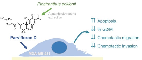
Anti-Migratory and Pro-Apoptotic Properties of Parvifloron D on Triple-Negative Breast Cancer Cells

1
CBIOS, Universidade Lusófona Research Center for Biosciences & Health Technologies, Campo Grande 376, 1749-024 Lisboa, Portugal
2
Research Institute for Medicines (iMed.ULisboa), Faculty of Pharmacy, Universidade de Lisboa, Av. Professor Gama Pinto, 1649-003 Lisboa, Portugal
*
Author to whom correspondence should be addressed.
Biomolecules 2020, 10(1), 158; https://doi.org/10.3390/biom10010158
Submission received: 19 December 2019 / Revised: 9 January 2020 / Accepted: 10 January 2020 / Published: 19 January 2020
(This article belongs to the Special Issue Selected Papers from Bio.Natural Meeting 2019)

Abstract

:
Medicinal plants are important sources of new bioactive compounds with potential anticancer activity. Parvifloron D (ParvD) is an abietane diterpenoid, isolated in high amounts from Plectranthus ecklonii Benth. Previous reports have suggested potential therapeutic properties for ParvD. ParvD has shown pro-apoptotic and cytotoxic effects in leukemia and melanoma cell lines. However, to the best of our knowledge, there are no studies in triple-negative breast cancer (TNBC) models. TNBC is a breast cancer subtype characterized by an aggressive behavior with poor clinical outcomes and weak overall therapeutic responses to the current treatment options. This work aimed at evaluating the anticancer effect of ParvD in MDA-MB-231 cells, a model of human TNBC. To obtain sufficient amounts of purified ParvD the efficiency of several extraction methods was compared. ParvD (0.1–10 µM) decreased cell viability in a concentration-dependent manner. Treatment with ParvD (5 µM) significantly increased the percentage of apoptotic nuclei and exposure to 3 µM ParvD increased the sub-G1 population. Since altered cell adherence, migration, and invasion are determinant processes for the formation of metastases, the effect of ParvD on these cellular processes was tested. Although treatment with ParvD (1 µM) had no effect on cell-substrate attachment, ParvD (1 µM) significantly reduced cell chemotaxis and invasion. This is the first report describing the proapoptotic effect of ParvD in TNBC cells. Moreover, for the first time we have shown that ParvD reduces cell motility, unraveling potential anti-metastatic properties.

Graphical Abstract

1. Introduction

Phytotherapy is as old as the human civilization itself and has been used over the centuries for a variety of health conditions [1,2]. Nowadays, multiple efforts are in motion to assess the potential pharmacological activity of plant-derived compounds as antimicrobial, antifungal, anti-inflammatory, antioxidant, and anticancer agents [2,3]. Many existing classical anticancer drugs have a plant source. This is the case for taxol (derived from Taxus brevifolia), camptothecin (derived from Camptotheca acuminate), among others [1,4]. Plants constitute thus a prolific source to search for new drugs for cancer prevention or treatment [1]. The development of extraction and synthesis techniques aims to allow the mass production of anticancer bioactive plant derivatives [4].
Plants from the Plectranthus genus, which belongs to the family Lamiaceae have been studied for their multiple pharmacological properties [5,6]. Plectranthus species are rich in naturally occurring compounds like the abietane diterpenes, which exhibit a broad spectrum of biological activities, including anticancer activity [5,7,8,9,10]. Parvifloron D (ParvD; Figure 1) is a major diterpenoid of P. ecklonii and its anticancer properties were described against human leukemia, melanoma, colon, pancreatic, and non-metastatic breast cancer cell lines, as well in human keratinocytes [7,11,12,13].
Breast cancer is the most prevalent type of cancer in women from developed countries, accounting for approximately 20% of female cancer deaths. Breast cancer has a complex and heterogeneous pattern regarding histology, mutations, metastatic potential, cellular origin, progression, treatment response, and clinical outcome [14]. Triple-negative breast cancer (TNBC) is characterized by the lacking expression of the three markers that define breast cancer subtypes and treatment, namely, the estrogen receptor, progesterone receptor, and human epidermal growth factor receptor 2 (HER-2). TNBC represents approximately 20% of all breast cancers [15,16,17]. TNBC has generally a more aggressive behavior and is associated with a poor clinical outcome [16,17,18]. The treatment guidelines for TNBC are currently limited to systemic chemotherapy for early and later stages, with unsatisfactory results, shown by weak overall therapeutic response rates and low median progression-free survival [16,17,19]. The aim of this study was to isolate ParvD from P. ecklonii Benth by a selected extraction method and to assess the impact of this isolated compound on TNBC cell motility, cell cycle and cell death using an in vitro model of TNBC. Here, we describe for the first time the proapoptotic and anti-migratory effects of ParvD in a TNBC cell model.

2. Materials and Methods

2.1. Chemicals

Dulbecco’s modified Eagle’s medium (DMEM), fetal bovine serum (FBS), penicillin-streptomycin solution, trypsin, and crystal violet (CV) were purchased from Sigma-Aldrich (St Louis, MO, USA). Dimethylsulphoxide (DMSO), ethanol, and acetic acid were purchased from Merck (Darmstadt, Germany). Matrigel was purchased from BD Biosciences (San Jose, CA, USA). DAPI, PI, and RNAase were purchased from Thermo Fisher Scientific (Waltham, MA, USA).
P. ecklonii Benth air-dried and powdered plant material was given by the Faculty of Pharmacy of the University of Lisbon and it was collected from seeds provided by the herbarium of the National Botanical Garden of Kirstenbosch, South Africa. Voucher specimens (S/No. LISC) have been deposited in the herbarium of the Tropical Research Institute in Lisbon [11]. Acetone and other organic solvents were from analytic grade and provided by (VWR international S.A.S., Briare, France); Silica for the isolation was obtained from Merck (grade 60, 230–400 mesh, Merck KGaA, Darmstadt, Germany).

P. ecklonii Extracts Preparation

In this study, different extracts of P. ecklonii were prepared using multiple extraction techniques (decoction, infusion, microwave, supercritical fluid, and ultrasound-assisted extractions) [9]. Acetone, water, and supercritical CO2 were used as extraction solvents for the different extraction procedures. The extraction quantification of ParvD was assessed by HPLC-DAD. Aqueous and acetonic extracts were prepared using 10% (w of plant, g; v of extraction solvent, mL).
Aqueous plant extracts were prepared by:
1. Infusion, using freshly boiled distilled water.
2. Decoction, boiling distilled water with plant material for 10 min.
3. Microwave extraction, using distilled water under a conventional microwave for 2 min at continuous irradiation of 2.45 GHz.
4. Ultrasound extraction using distilled water in an ultrasonic bath (Sonorex Super RK 510 H; Bandelin, Berlin, Germany) at room temperature for 30 min.
All aqueous extracts were further filtered using a Whatman paper no 5 (Whatman, Inc., Clifton, NJ, USA) and lyophilized (Freezone 2.5 L, Freeze-dryer Labconco, Kansas City, MO, USA) prior to use in HPLC analysis [9].
Acetonic extracts were obtained using an ultrasonic bath (Sonorex Super RK 510 H; Bandelin, Berlin, Germany) operated for 1 h, at 35 Hz with maximum input power of 320 W in ultrasound extraction or maceration 1 h under magnetic stirring.
The obtained organic extracts were filtered, and the solvent was removed by rotary evaporation [9].
The supercritical fluid extraction (SFE) was done as previously described [20].

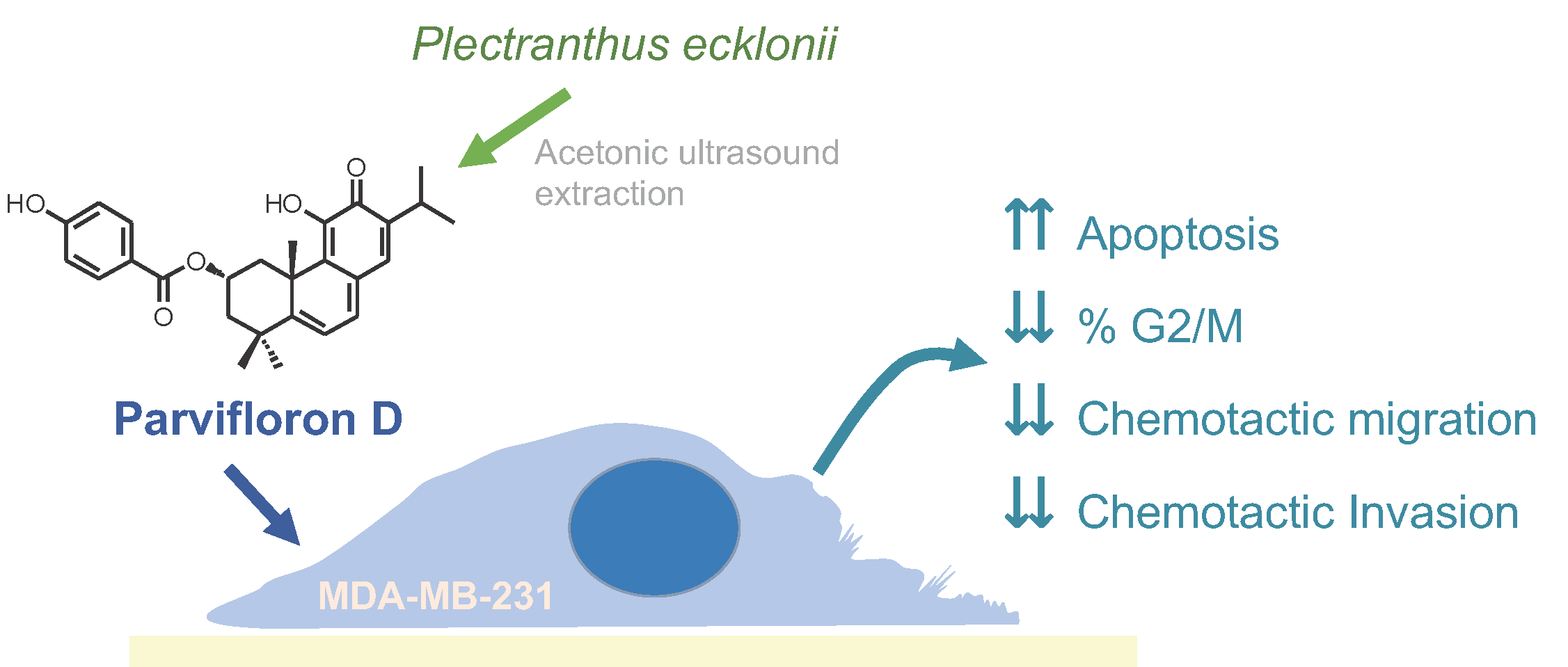
2.2. Isolation and Quantification of ParvD by HPLC-DAD

An authentic sample of ParvD was isolated from an acetonic ultrasound-assisted extract of P. ecklonii Benth., as described in a previous work [11]. The authentic sample of ParvD was in agreement with the spectroscopic means (purity assessed by 1H-NMR spectrum—Figure S1. see Supporting Information) and was further used in the HPLC-DAD quantification and biological assays. The HPLC analysis was performed in a high-performance liquid chromatography with diode array detection (HPLC-DAD) with an Agilent Technologies 1200 Infinity Series LC system (Agilent Technologies, Santa Clara, CA, USA) coupled to a diode array detector (DAD), using a ChemStation Software and a reverse phase LiChrospher® 100 RP-18 5 μm (4.0 × 250 mm) column (Merck). The ParvD of all the extracts was quantified by injecting 20 µL of each sample at 1 mg/mL, using a gradient composed of Solution A (methanol), Solution B (acetonitrile), and Solution D (0.3% trichloroacetic acid in water) as follows: 0 min, 15% A, 5% B, and 80% D; 20 min, 80% A, 10% B, and 10% D; 25 min, 80% A, 10% B, and 10% D. The flow rate was set at 1 mL/min. ParvD was run under the same conditions in methanol, and the detection was carried out between 200 and 600 nm with a diode array detector (DAD). All analyses were performed in triplicate.

2.3. Cell Culture

The human breast cancer cell line MDA-MB-231 was obtained from ATCC (HTB-26). Cells were cultured in DMEM supplemented with 10% fetal bovine serum, 100 U/mL penicillin, and 0.1 mg/mL streptomycin. The cultures were maintained at 37 °C, under a humidified atmosphere containing 5% CO2 in the air [21].

2.4. Cell Viability

Cell viability was evaluated by the crystal violet (CV) staining assay. Approximately 5 × 10³ cells in 200 μL of culture medium per well were plated in 96-well plates and incubated for 24 h. Cells were then exposed to ParvD (0.1–25 µM) for 48 h. The CV assay was carried out according to previously described protocols [22,23,24]. Two or three independent experiments were performed, each comprising four replicate cultures. IC50 was calculated using GraphPad software 7.0.

2.5. Nuclear Morphology

ParvD-induced apoptosis in MDA-MB-231 cells was examined according to nuclear morphological changes using DAPI staining [25]. Cells were treated with different concentrations of ParvD (0; 0.1; 3 and 5 µM) for 8, 24, and 48 h. Later cells were fixed and stained with DAPI and analyzed by fluorescence microscopy. Cell image acquisition was performed using a wide field BX51 fluorescent Olympus microscope with a 40× objective. The number of apoptotic cells was determined according to changes in nuclear morphology, including shrinkage, condensation, margination, and chromatin fragmentation.

2.6. Cell DNA Content Analysis

MDA-MB-231 cells were cultured in 6-well plates for 24 h. Cells were then treated with different ParvD concentrations (0; 0.2 and 3 µM) with a 48-h incubation period. Subsequently, cells were harvested and with 5 mM EDTA in PBS and fixed with 80% ethanol. Following treatment with RNase A (20 µg/mL) and staining with PI (10 µg/mL) the cellular DNA content was analyzed by flow cytometry using a FACSCalibur flow cytometer (BD) [26,27]. Data acquisition and analysis were performed using CellQuest software (BD) and FlowJo (Tree Star, San Carlos, CA, USA), respectively.

2.7. Chemotaxis and Chemoinvasion

The chemotactic migration of MDA-MB-231 cells was assessed in 24-well plates which contained transwell inserts with a transparent PET membrane with 8 µm sized pores (BD Falcon, Bedford, MA, USA), according to Florido et al. [21]. Cells resuspended in the FBS-free medium were seeded on the top of the insert, and complete medium was placed in the lower chamber of the culture well. A ParvD concentration of 1 µM was added in both chambers and cells were incubated for 24 h. The evaluation of the chemotactic migration was performed following a previously described protocol [28]. The results were expressed as percentages of non-treated control cultures. The chemoinvasion assay was executed as described above for the chemotaxis assay, but coating the transwell inserts membrane with Matrigel diluted 30 times in serum-free medium. Three independent experiments were performed.

2.8. Cell Detachment Assay

To evaluate cell adhesion, MDA-MB-231 cells were cultured in 24-well plates for 24 h, to achieve a confluence of approximately 30%. Cells were then incubated with several ParvD concentrations (0, 1 and 2 µM) for a period of 24 h. The EDTA-induced cell detachment assay was performed following previously described protocols [28,29].

3. Results

3.1. P. ecklonii Extracts Preparation and HPLC-DAD Quantification

A multiple extraction study of P. ecklonii, which included aqueous, supercritical CO2 and acetonic extracts, was performed to select the method to isolate ParvD in highest amount. The extraction techniques involved were decoction, infusion, microwave, supercritical fluid, and ultrasound-assisted extractions. The quantification of ParvD on the different extracts was performed by HPLC-DAD, which indicated that the acetonic extracts have a higher ParvD concentration, when compared with the aqueous extracts; this was expected considering the chemical structure of ParvD. Acetonic extracts obtained by maceration or ultrasounds achieved ParvD concentrations of 136.8 and 166.1 µg/mg, respectively (Table 1).
The remaining extractions using water and supercritical CO2, independently of the extraction methods used (supercritical fluid extraction, decoction, infusion, microwave, and ultrasound extractions) only reached values of around 2.4 and 1.0 µg/mg, which are shown in Table 1. Considering the extraction efficiency, the acetonic ultrasound method was selected to isolate ParvD and spectroscopically confirmed ParvD was subsequently used in biological studies.

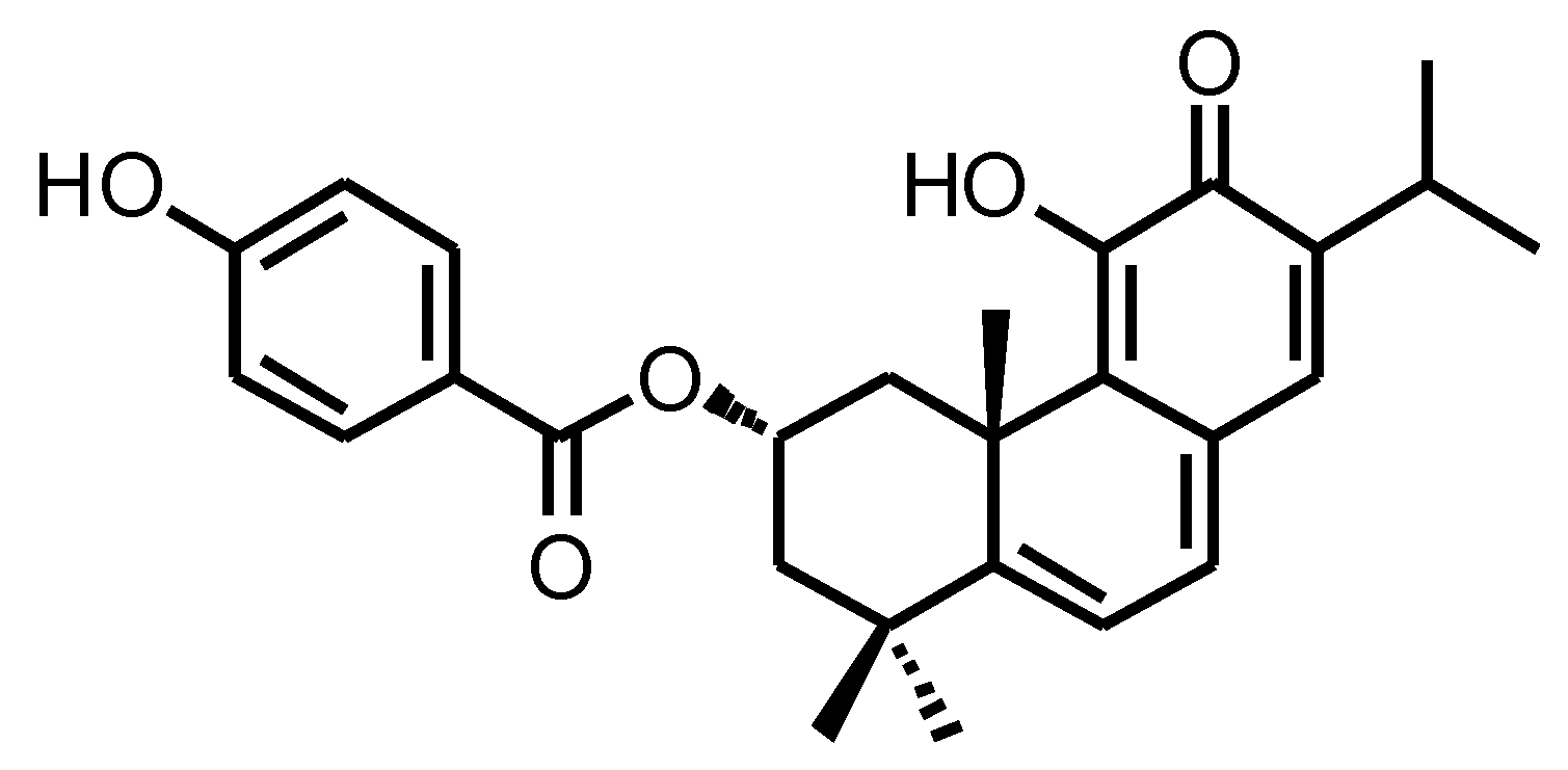
3.2. ParvD Reduces the Viability of MDA-MB-231 Cells

The cytotoxicity of ParvD was evaluated in MDA-MB-231 cells, a well-established in vitro model of TNBC. ParvD decreased cell viability in a concentration-dependent manner, following a 48 h incubation period. ParvD 0.1 and 1 µM demonstrated only a slight decrease in cell viability, with 89% of live cells by the end of the 48 h incubation period. Concentrations of ParvD above 5 µM showed a remarkable decrease in the cell viability, as it can be observed in Figure 2. The IC50 value was 2.48 µM.

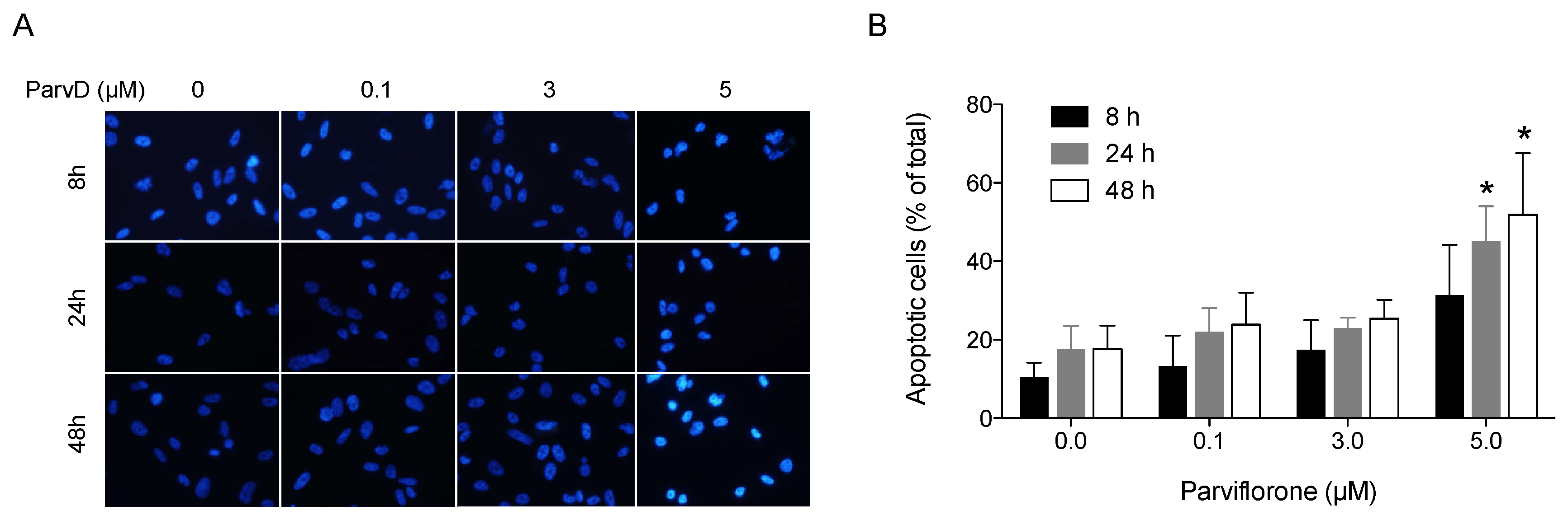
3.3. ParvD Induces Apoptosis in MDA-MB-231 Cells

Aiming at characterizing the reduction in cell viability provoked by ParvD, the induction of apoptosis was assessed by analyzing the cell nuclear morphology using DAPI staining. The results are depicted in Figure 3. The percentage of apoptotic cells increased with the concentration of ParvD and with the period of exposure. ParvD 5 µM showed a significant pro-apoptotic effect in MDA-MB-231 cells after periods of 24 and 48 h of incubation, increasing the % of apoptotic cells by 2.5- and 2.9-fold, respectively.

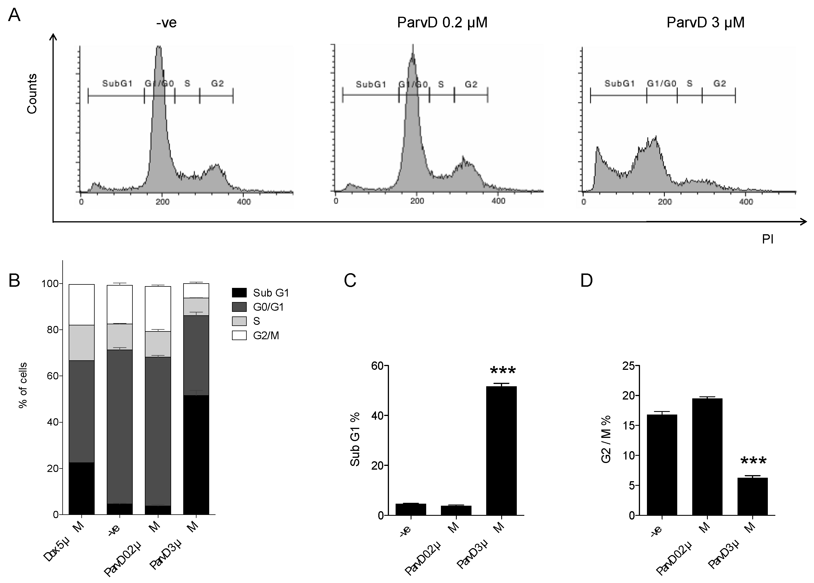
3.4. ParvD Increases the Sub-G1 Population

The impact of ParvD on the cell cycle progression was evaluated by assessing the cellular DNA content by flow cytometry. As shown in Figure 4, while a low concentration of ParvD (0.2 µM) did not impact the cell cycle distribution, the exposure of cells for 48 h to a higher concentration (3 µM) led to a marked increase in the Sub-G1 phase. Indeed, a 13-fold increase in Sub-G1 cell population versus controls was observed. In parallel, the cell G2/M population suffered a three-fold decrease when compared with control cells. Cells treated with ParvD 3 µM show a two-fold higher percentage of Sub-G1 phase and three-fold lower percentage of G2/M phase when compared with cells treated with doxorubicin (Dox) 5 µM, a well-established anticancer drug used in breast cancer treatment.

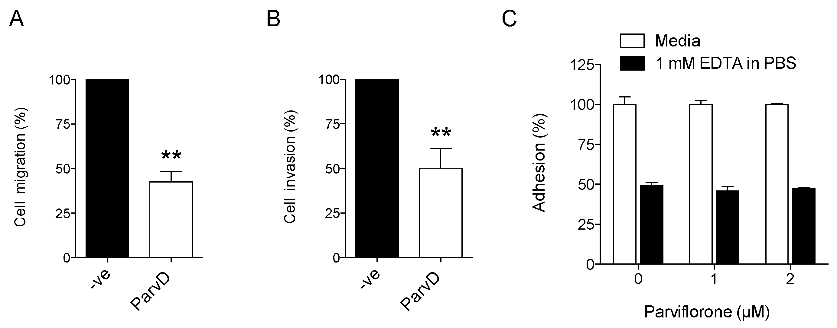
3.5. ParvD Reduces Breast Cancer Cell Migration and Invasion

Given the importance of cell migration and cell invasion for cancer progression, especially for the formation of metastases, the effects of ParvD at this level was also assessed. The results are shown in Figure 5. ParvD (1 µM, 24 h), decreased the chemotactic migration of MDA-MB-231 cells by 57% when compared with the non-treated control cells (Figure 5A). Regarding cell invasion, a decrease of 50% was observed in cells treated with ParvD (Figure 5B).
As ParvD treatment influenced cell migration, the impact of this compound on cell adhesion/detachment was also determined. This was assessed using an EDTA-induced cell detachment assay, using experimental conditions that lead to ~50% cell detachment in cells not treated with ParvD. As shown in Figure 5C, ParvD (0–2 µM) did not affect cell detachment.

4. Discussion

Plants constitute a rich source of bioactive natural compounds with potential therapeutic effects for a wide number of diseases, including cancer. The selection of the procedure to extract bioactive compounds from their plants of origin is a critical step to obtain enough amounts of pure compounds that allow biological evaluation. In this work, the extraction method using acetone and an ultrasound-assisted technique is the one that results in the higher quantity of isolated ParvD. The acetone extraction results are in agreement with previous studies, where other bioactive abietane diterpenoids from Plectranthus spp. were isolated [9]. The highest efficiency of this extraction method is likely because of a more effective rupture of the plant cell wall, enabling the leaching of the ParvD into the acetone solvent [30].
Although ParvD is scarcely studied, previous reports have demonstrated a cytotoxic effect of ParvD in different cancer cell lines. Burmistrova et al. showed that ParvD had a remarkable cytotoxic effect in different human cancer cell lines. The leukemia cell lines HL-60, U-937, MOLT-3, and K-562 were especially susceptible to this compound, with IC50 values ranging from 0.35 to 1.2 µM after a 72-h period of incubation [7]. In another study, Silva et al. reported the cytotoxic effect of ParvD in melanoma cell lines B16V5 and A375 with IC50 values of 6.86 µM and 6.24 µM, following a 24 h period [12]. In comparison with the only reported study so far using the non-metastatic breast cancer cell line MCF7 [13] (IC50 value of 35.10 µM), our results demonstrate that ParvD is highly cytotoxic for MDA-MB-231 cells, with an IC50 value of 2.48 µM. This is an encouraging finding, since MDA-MB-231 cells exhibit a high level of resistance to an array of chemotherapeutic drugs [31]. Other diterpenoids previously isolated from distinct plant sources were also studied in MDA-MB-231 cells. The reported IC50 values were higher than those obtained in the present work [32,33,34].
Given the pronounced cytotoxic effect observed, we explored whether ParvD promoted apoptosis. Our data shows that ParvD increased the % of nuclei with morphological alterations characteristic of apoptotic cells. In line with this observation, an increase in the sub-G1 population was also found. Interestingly, the increase in the Sub-G1 population was more notorious for ParvD 3 µM than for Dox 5 µM, a chemotherapy drug frequently used in TBNC treatment modalities [35,36]. The pro-apoptotic effect described here for MDA-MB-231 cells was previously reported in other cancer cell lines. Burmistrova et al. reported an apoptotic effect of ParvD in the human myeloid leukemia cell lines HL-60 and U-937, demonstrated by the condensation and fragmentation of chromatin as well as by the increase in sub-G1 cell populations [7]. Similarly, Silva et al. reported an increase in Sub-G1 phase in the melanoma cell lines B16V5 and A375 [12]. Gelomulide K, another abietane diterpene, also induced apoptotic cell death in MDA-MB-231 cells, but a much higher concentration was needed to achieve a significant effect in comparison with ParvD (50 µM vs. 5 µM), after a 24 h period of incubation [33]. Although the mechanism by which ParvD induces apoptosis is not fully understood, a previous study [7] suggests that it might involve loss of mitochondrial membrane potential, extracellular signal-regulated kinases (ERK) 1/2 signaling, and reactive oxygen species accumulation.
The mortality associated with breast cancer is mostly due to the metastatic behavior of breast cancer cells [37]. Therefore, we studied the effects of ParvD on cell adhesion, cell migration, and cell invasion, which are determinant features for the formation of metastases. This is the first study addressing the impact of ParvD in these cellular processes. While ParvD had no effect in the adhesion/detachment of MDA-MB-231 cells, it led to a clear reduction in chemotactic cell migration and in cell invasion. Chemotaxis is a single-cell and directed type of cell migration, essential for tumor dissemination during progression and metastasis, including in the case of breast cancer [24,38]. The observed impact of ParvD in cell migration and cell invasion suggests that this compound might be useful in managing cancer dissemination and growth [38].
In summary, ParvD was isolated using a preliminary comparative extraction study, which allowed selecting an efficient ultrasound-assisted extraction. ParvD has shown anti-migration and anti-invasion properties and, at higher concentrations, cytotoxic and pro-apoptotic effects in an in vitro model of TNBC. Despite these encouraging results, ParvD is not selective toward cancer cells [7,12], as also happens with anticancer drugs currently in clinical use for TNBC [39,40]. A previous study using human normal-like fibroblasts (Detroit 551) determined an IC50 value of 9 µM for ParvD [12]. Therefore, future studies should be taken to develop adequate delivery systems that allow the targeting of this compound specifically to breast cancer cells. Such approaches were proposed by Silva et al. (2016) and Santos-Rebelo et al. (2018) using hybrid nanoparticles to target ParvD to melanoma and pancreatic cancer cells [12,13].

5. Conclusions

This is the first report describing the pro-apoptotic properties of ParvD on TNBC cells. Additionally, for the first time the anti-migratory effects of this compound were demonstrated, unraveling potential anti-metastatic properties of this natural compound. Although ParvD has shown encouraging effects in the breast cancer cell model used, further efforts must be undertaken to enhance the selectivity of this compound toward cancer cells, limiting undesirable side effects in non-cancer cells. Additionally, the dissection of the molecular mechanisms underlying the observed effects, as well as in vivo experiments will contribute to the understanding of the therapeutic relevance of this compound.

Supplementary Materials

The following are available online at https://www.mdpi.com/2218-273X/10/1/158/s1. Figure S1: 1H-NMR spectrum of Parvifloron D (400 MHz, CDCl3). The datasets used and/or analyzed during the current study are available from the corresponding author on reasonable request.

Author Contributions

A.S.F., P.R., and N.S. contributed to the conception and design. N.S., P.R., C.R., and A.S.F. contributed to the development of methodology. A.S.F., P.R., N.S., and N.A. contributed to the acquisition of data. A.S.F., N.S., N.A., and J.G.C. contributed to the analysis and interpretation of data. All authors contributed to the writing, review, and/or revision of the manuscript. A.S.F. and N.S. supervised the study. All authors have read and agreed to the published version of the manuscript.

Funding

This work was financially supported by Fundação para a Ciência e a Tecnologia (FCT, Portugal), through projects UID/DTP/04567/2016 (research grant attributed to N.A.) and UID/DTP/04567/2019 (general funding to CBIOS).

Conflicts of Interest

The authors declare that there is no conflict of interest regarding the publication of this paper.

References

  1. Nahata, A. Anticancer Agents: A Review of Relevant Information on Important Herbal Drugs. Int. J. Clin. Pharmacol. Toxicol. 2017, 6, 250–255. [Google Scholar] [CrossRef]
  2. Vallejo, M.J.; Salazar, L.; Grijalva, M. Oxidative Stress Modulation and ROS-Mediated Toxicity in Cancer: A Review on. Oxid. Med. Cell. Longev. 2017, 2017, 4586068. [Google Scholar] [CrossRef] [PubMed] [Green Version]
  3. Greenwell, M.; Rahman, P.K. Medicinal Plants: Their Use in Anticancer Treatment. Int. J. Pharm. Sci. Res. 2015, 6, 4103–4112. [Google Scholar] [CrossRef]
  4. Wallace, D. Natural Products as a Source of Anti-Cancer Lead Compounds: Ginger and Breast Cancer. J. Pharm. Clin. Res. 2016, 1, 1–6. [Google Scholar] [CrossRef]
  5. Lukhoba, C.W.; Simmonds, M.S.; Paton, A.J. Plectranthus: A review of ethnobotanical uses. J. Ethnopharmacol. 2006, 103, 1–24. [Google Scholar] [CrossRef] [PubMed]
  6. Figueiredo, N. Phytochemical Analysis of Plectranthus sp. Extracts and Application in Inhibition of Dental Bacteria, Streptococcus sobrinus and Streptococcus mutans. Eur. J. Med. Plants 2014, 4, 794–809. [Google Scholar] [CrossRef]
  7. Burmistrova, O.; Perdomo, J.; Simões, M.F.; Rijo, P.; Quintana, J.; Estévez, F. The abietane diterpenoid parvifloron D from Plectranthus ecklonii is a potent apoptotic inducer in human leukemia cells. Phytomedicine 2015, 22, 1009–1016. [Google Scholar] [CrossRef] [PubMed]
  8. Garcia, C.; Teodósio, C.; Oliveira, C.; Díaz-Lanza, A.; Reis, C.; Duarte, N.; Rijo, P. Naturally Occurring Plectranthus-derived Diterpenes with Antitumoral Activities. Curr. Pharm. Des. 2018, 24, 4207–4236. [Google Scholar] [CrossRef] [PubMed]
  9. Matias, D.; Nicolai, M.; Fernandes, A.S.; Saraiva, N.; Almeida, J.; Saraiva, L.; Faustino, C.; Díaz-Lanza, A.M.; Reis, C.P.; Rijo, P. Comparison Study of Different Extracts of Plectranthus madagascariensis, P. neochilus and the Rare, P. porcatus (Lamiaceae): Chemical Characterization, Antioxidant, Antimicrobial and Cytotoxic Activities. Biomolecules 2019, 9, 179. [Google Scholar] [CrossRef] [Green Version]
  10. Matias, D.; Nicolai, M.; Saraiva, L.; Pinheiro, R.; Faustino, C.; Diaz Lanza, A.; Pinto Reis, C.; Stankovic, T.; Dinic, J.; Pesic, M.; et al. Cytotoxic Activity of Royleanone Diterpenes from Plectranthus madagascariensis Benth. ACS Omega 2019, 4, 8094–8103. [Google Scholar] [CrossRef] [Green Version]
  11. Simões, M.F.; Rijo, P.; Duarte, A.; Matias, D.; Rodríguez, B. An easy and stereoselective rearrangement of an abietane diterpenoid into a bioactive microstegiol derivative. Phytochem. Lett. 2010, 3, 234–237. [Google Scholar] [CrossRef]
  12. Silva, C.O.; Molpeceres, J.; Batanero, B.; Fernandes, A.S.; Saraiva, N.; Costa, J.G.; Rijo, P.; Figueiredo, I.V.; Faísca, P.; Reis, C.P. Functionalized diterpene parvifloron D-loaded hybrid nanoparticles for targeted delivery in melanoma therapy. Ther. Deliv. 2016, 7, 521–544. [Google Scholar] [CrossRef] [PubMed]
  13. Santos-Rebelo, A.; Garcia, C.; Eleutério, C.; Bastos, A.; Coelho, S.C.; Coelho, M.A.N.; Molpeceres, J.; Viana, A.S.; Ascensão, L.; Pinto, J.F.; et al. Development of Parvifloron D-loaded Smart Nanoparticles to Target Pancreatic Cancer. Pharmaceutics 2018, 10, 216. [Google Scholar] [CrossRef] [PubMed] [Green Version]
  14. Cetin, I.; Topcul, M. Triple negative breast cancer. Asian Pac. J. Cancer Prev. 2014, 15, 2427–2431. [Google Scholar] [CrossRef] [Green Version]
  15. Foulkes, W.D.; Smith, I.E.; Reis-Filho, J.S. Triple-negative breast cancer. N. Engl. J. Med. 2010, 363, 1938–1948. [Google Scholar] [CrossRef] [Green Version]
  16. Avery, T.P. Triple-Negative Breast Cancer. In Changing Paradigms in the Management of Breast Cancer; Springer International Publishing: Manhattan, NY, USA, 2017; pp. 155–166. [Google Scholar]
  17. Costa, R.L.B.; Han, H.S.; Gradishar, W.J. Targeting the PI3K/AKT/mTOR pathway in triple-negative breast cancer: A review. Breast Cancer Res. Treat. 2018, 169, 397–406. [Google Scholar] [CrossRef]
  18. Lehmann, B.D.; Bauer, J.A.; Chen, X.; Sanders, M.E.; Chakravarthy, A.B.; Shyr, Y.; Pietenpol, J.A. Identification of human triple-negative breast cancer subtypes and preclinical models for selection of targeted therapies. J. Clin. Investig. 2011, 121, 2750–2767. [Google Scholar] [CrossRef] [Green Version]
  19. Hudis, C.A.; Gianni, L. Triple-Negative Breast Cancer: An Unmet Medical Need. Oncologist 2011, 16, 1–11. [Google Scholar] [CrossRef] [Green Version]
  20. Bernardes, C.E.S.; Garcia, C.; Pereira, F.; Mota, J.; Pereira, P.; Cebola, M.J.; Reis, C.P.; Correia, I.; Piedade, M.F.M.; Minas da Piedade, M.E.; et al. Extraction Optimization and Structural and Thermal Characterization of the Antimicrobial Abietane 7α-Acetoxy-6β-hydroxyroyleanone. Mol. Pharm. 2018, 15, 1412–1419. [Google Scholar] [CrossRef]
  21. Flórido, A.; Saraiva, N.; Cerqueira, S.; Almeida, N.; Parsons, M.; Batinic-Haberle, I.; Miranda, J.P.; Costa, J.G.; Carrara, G.; Castro, M.; et al. The manganese(III) porphyrin MnTnHex-2-PyP(5+) modulates intracellular ROS and breast cancer cell migration: Impact on doxorubicin-treated cells. Redox Biol. 2019, 20, 367–378. [Google Scholar] [CrossRef]
  22. Fernandes, A.S.; Gaspar, J.; Cabral, M.F.; Rueff, J.; Castro, M.; Batinic-Haberle, I.; Costa, J.; Oliveira, N.G. Protective role of ortho-substituted Mn(III) N-alkylpyridylporphyrins against the oxidative injury induced by tert-butylhydroperoxide. Free Radic. Res. 2010, 44, 430–440. [Google Scholar] [CrossRef] [PubMed]
  23. Caparica, R.; Júlio, A.; Baby, A.R.; Araújo, M.E.M.; Fernandes, A.S.; Costa, J.G.; Santos de Almeida, T. Choline-Amino Acid Ionic Liquids as Green Functional Excipients to Enhance Drug Solubility. Pharmaceutics 2018, 10, 288. [Google Scholar] [CrossRef] [PubMed] [Green Version]
  24. Costa, J.G.; Saraiva, N.; Batinic-Haberle, I.; Castro, M.; Oliveira, N.G.; Fernandes, A.S. The SOD Mimic MnTnHex-2-PyP(5+) reduces the viability and migration of 786-O human renal cancer cells. Antioxidants 2019, 8, 490. [Google Scholar] [CrossRef] [PubMed] [Green Version]
  25. Chazotte, B. Labeling nuclear DNA using DAPI. Cold Spring Harb. Protoc. 2011, 2011, pdb.prot5556. [Google Scholar] [CrossRef] [Green Version]
  26. Costa, J.G.; Saraiva, N.; Guerreiro, P.S.; Louro, H.; Silva, M.J.; Miranda, J.P.; Castro, M.; Batinic-Haberle, I.; Fernandes, A.S.; Oliveira, N.G. Ochratoxin A-induced cytotoxicity, genotoxicity and reactive oxygen species in kidney cells: An integrative approach of complementary endpoints. Food Chem. Toxicol. 2016, 87, 65–76. [Google Scholar] [CrossRef]
  27. Guerreiro, P.S.; Corvacho, E.; Costa, J.G.; Saraiva, N.; Fernandes, A.S.; Castro, M.; Miranda, J.P.; Oliveira, N.G. The APE1 redox inhibitor E3330 reduces collective cell migration of human breast cancer cells and decreases chemoinvasion and colony formation when combined with docetaxel. Chem. Biol. Drug Des. 2017, 90, 561–571. [Google Scholar] [CrossRef]
  28. Fernandes, A.S.; Flórido, A.; Saraiva, N.; Cerqueira, S.; Ramalhete, S.; Cipriano, M.; Cabral, M.F.; Miranda, J.P.; Castro, M.; Costa, J.; et al. Role of the Copper(II) Complex Cu[15]pyN5 in Intracellular ROS and Breast Cancer Cell Motility and Invasion. Chem. Biol. Drug Des. 2015, 86, 578–588. [Google Scholar] [CrossRef]
  29. Saraiva, N.; Prole, D.L.; Carrara, G.; Johnson, B.F.; Taylor, C.W.; Parsons, M.; Smith, G.L. hGAAP promotes cell adhesion and migration via the stimulation of store-operated Ca2+ entry and calpain 2. J. Cell. Biol. 2013, 202, 699–713. [Google Scholar] [CrossRef]
  30. Rijo, P.; Falé, P.L.; Serralheiro, M.L.; Simões, M.F.; Gomes, A.; Reis, C. Optimization of medicinal plant extraction methods and their encapsulation through extrusion technology. Measurement 2014, 58, 249–255. [Google Scholar] [CrossRef]
  31. Chen, J.; Lu, L.; Feng, Y.; Wang, H.; Dai, L.; Li, Y.; Zhang, P. PKD2 mediates multi-drug resistance in breast cancer cells through modulation of P-glycoprotein expression. Cancer Lett. 2011, 300, 48–56. [Google Scholar] [CrossRef]
  32. Lee, C.L.; Chang, F.R.; Hsieh, P.W.; Chiang, M.Y.; Wu, C.C.; Huang, Z.Y.; Lan, Y.H.; Chen, M.; Lee, K.H.; Yen, H.F.; et al. Cytotoxic ent-abietane diterpenes from Gelonium aequoreum. Phytochemistry 2008, 69, 276–287. [Google Scholar] [CrossRef] [PubMed]
  33. Yang, J.C.; Lu, M.C.; Lee, C.L.; Chen, G.Y.; Lin, Y.Y.; Chang, F.R.; Wu, Y.C. Selective targeting of breast cancer cells through ROS-mediated mechanisms potentiates the lethality of paclitaxel by a novel diterpene, gelomulide K. Free Radic. Biol. Med. 2011, 51, 641–657. [Google Scholar] [CrossRef] [PubMed]
  34. Song, J.T.; Han, Y.; Wang, X.L.; Shen, T.; Lou, H.X.; Wang, X.N. Diterpenoids from the twigs and leaves of Croton caudatus var. tomentosus. Fitoterapia 2015, 107, 54–59. [Google Scholar] [CrossRef] [PubMed]
  35. Illiano, M.; Sapio, L.; Salzillo, A.; Capasso, L.; Caiafa, I.; Chiosi, E.; Spina, A.; Naviglio, S. Forskolin improves sensitivity to doxorubicin of triple negative breast cancer cells via Protein Kinase A-mediated ERK1/2 inhibition. Biochem. Pharmacol. 2018, 152, 104–113. [Google Scholar] [CrossRef]
  36. Inao, T.; Iida, Y.; Moritani, T.; Okimoto, T.; Tanino, R.; Kotani, H.; Harada, M. Bcl-2 inhibition sensitizes triple-negative human breast cancer cells to doxorubicin. Oncotarget 2018, 9, 25545–25556. [Google Scholar] [CrossRef] [Green Version]
  37. Al-Mahmood, S.; Sapiezynski, J.; Garbuzenko, O.B.; Minko, T. Metastatic and triple-negative breast cancer: Challenges and treatment options. Drug Deliv. Transl. Res. 2018, 8, 1483–1507. [Google Scholar] [CrossRef] [Green Version]
  38. Roussos, E.T.; Condeelis, J.S.; Patsialou, A. Chemotaxis in cancer. Nat. Rev. Cancer 2011, 11, 573–587. [Google Scholar] [CrossRef]
  39. Razak, N.A.; Abu, N.; Ho, W.Y.; Zamberi, N.R.; Rizi, N.; Tan, S.W.; Alitheen, N.B.; Banu, N.; Long, K.; Yeap, S.K. Cytotoxicity of eupatorin in MCF-7 and MDA-MB-231 human breast cancer cells via cell cycle arrest, anti-angiogenesis and induction of apoptosis. Sci. Rep. 2019, 9, 1514. [Google Scholar] [CrossRef]
  40. Takeshita, T.; Wu, W.; Koike, A.; Fukuda, M.; Ohta, T. Perturbation of DNA repair pathways by proteasome inhibitors corresponds to enhanced chemosensitivity of cells to DNA damage-inducing agents. Cancer Chemother. Pharmacol. 2009, 64, 1039–1046. [Google Scholar] [CrossRef] [Green Version]
Figure 1. Chemical structure of Parvifloron D.
Biomolecules 10 00158 g001
Figure 2. Exposure of cells to ParvD for 48 h reduces MDA-MB-231 cell viability measured by a crystal violet assay. Results are expressed as means ± SD (n = 2–3). *** p < 0.001 versus control (one-way ANOVA with Tukey’s multiple comparison test).
Biomolecules 10 00158 g002
Figure 3. ParvD-mediated apoptosis in MDA-MB-231 cells was examined by nucleus morphological changes using DAPI. (A) Cells were treated with the indicated concentrations of ParvD for 8, 24, and 48 h, fixed stained with DAPI, analyzed under fluorescence microscopy, and the number of apoptotic cells was determined according to changes in nuclear morphology, including shrinkage, condensation, margination, and fragmentation of chromatin. Representative fields of cells are shown. (B) Summary results (means ± SD from at least 200 cells for each condition) show the percentage of apoptotic cells. * p < 0.05 (Student’s t-test, relative to untreated cells for the same incubation period).
Biomolecules 10 00158 g003
Figure 4. ParvD induces an increase in Sub-G1 and a reduction of G2/M populations. MDA-MB-231 cells were treated with the indicated concentrations of ParvD for 48 h. Dox 5 µM was used as a positive control. Cells were fixed, permeabilized, and stained with PI. (A) Cellular DNA content was analyzed by flow cytometry, representative histograms are shown. (BD) Cell Sub-G1, G0/G1, S, and G2/M population summary results (Means ± SEM). *** p < 0.001 (Student’s t-test, relative to untreated cells).
Biomolecules 10 00158 g004
Figure 5. Impact of ParvD on cell migration, invasion, and detachment. Cell migration (A) and cell invasion (B) were evaluated using a transwell system. Results are expressed as mean values ± SD (n = 3) ** p < 0.01 (Student’s t-test, relative to untreated cells). Cell adhesion was assessed using an EDTA-induced cell detachment assay (C). Results are expressed as mean values ± SD (n = 2).
Biomolecules 10 00158 g005
Table 1. Amount of parvifloron D in 1 mg of P. ecklonii.
Extraction MethodParvifloron D (µg/mg)
Acetone Maceration136.8 1
Acetone Ultrasound166.1 1
Supercritical fluid extraction2.2 1
Decoction2.4 1
Infusion1.0 1
Microwave1.2 1
Ultrasound1.2 1
1 Amount of ParvD quantified by HPLC-DAD.

