Article

Linear and Nonlinear Optical Absorption of CdSe/CdS Core/Shell Quantum Dots in the Presence of Donor Impurity

by
Grigor A. Mantashian
1,
Nare A. Zaqaryan
1,
Paytsar A. Mantashyan
1,2,
Hayk A. Sarkisyan
1,
Sotirios Baskoutas
3 and
David B. Hayrapetyan
1,*
1
Department of General Physics and Quantum Nanostructures, Russian-Armenian University, Yerevan 0051, Armenia
2
Institute for Physical Research, The National Academy of Sciences of the Republic of Armenia, Ashtarak 0203, Armenia
3
Department of Materials Science, University of Patras, GR-26504 Patras, Greece
*
Author to whom correspondence should be addressed.
Atoms 2021, 9(4), 75; https://doi.org/10.3390/atoms9040075
Submission received: 20 July 2021 / Revised: 17 September 2021 / Accepted: 19 September 2021 / Published: 5 October 2021

Abstract

:
Linear and nonlinear optical properties in colloidal CdSe/CdS core/shell quantum dots with different sizes have been theoretically investigated in the framework of effective mass approximation. The electron states in colloidal CdSe/CdS core/shell quantum dots have been calculated using the finite element method. The intraband linear and nonlinear absorption spectra have been calculated for colloidal CdSe/CdS core/shell quantum dots with different sizes. In addition, the dependences of the linear and nonlinear refractive index change on the incident light energy have been calculated. In the last section of the paper the second- and third-order harmonic generation spectra have been presented.

1. Introduction

Despite the fact that modern semiconductor nanotechnologies provide an opportunity to grow quantum dots (QDs) of different shapes and sizes, the methods of manufacturing QDs are expensive, require specialized equipment, and cannot provide sufficient volumes for industrial production [1,2,3]. The way out of this predicament was the introduction of the colloidal QD manufacturing process, which is carried out on the basis of solutions at low temperatures, and provides a unique opportunity for mass chemical synthesis and a narrow size distribution [4,5,6]. Colloidal QDs open up the possibility of manufacturing a variety of devices, namely, photodiodes, photovoltaic cells, photoconductors, electroluminescent devices, modern batteries, etc. [7,8,9,10,11].
Among colloidal QDs, a special place is occupied by QDs with a core/shell structure. To obtain a core/shell structure, epitaxial layers of inorganic material (shell) are grown on the QD’s core material [12,13,14,15]. Nanoparticles, spheres, wires, rods, tubes, ribbons, fibers, plates, cubes, sheets, flowers [16,17,18,19,20,21] and many other considerable results have been obtained from the growth of core/shell systems. Core/shell quantum dots have a number of advantages: the shell structure protects the core from environmental influences, enhances physical capabilities, or brings forth new ones, limits volumetric expansion, thereby preserving structural integrity, and prevents the formation of large particles in the core. It should also be noted that most of the properties of the core/shell QDs depend both on the core materials and the shell material.
Exciton-related Raman scattering, interband absorption and photoluminescence in colloidal CdSe/CdS core/shell QD ensembles was investigated in [22]. It is very important to note that nonlinear optical effects can potentially be used in the creation of various next-generation optoelectronic devices [23]. Therefore, the study of the nonlinear optical properties of semiconductor QDs continues to be a relevant problem.
For example, one of the first articles which was devoted to the study of nonlinear optical properties of QDs was [23], in which the authors considered nonlinear optical transitions in semiconductor QDs. In particular, the authors of the article have shown that, depending on the magnitude of the broadening, the nonlinear absorption and the refractive index change can be quite large. The effects of exciton optical nonlinearity in semiconductor QDs were studied in [24]. The authors of [25] theoretically investigated the second harmonic generation coefficient in parabolic QDs in the presence of external electric and magnetic fields. The generation of the third harmonic in a cylindrical GaAs/AlGaAs QD with a parabolic-confining potential in the presence of an electric field was studied in [26]. The authors have shown that the inherent substantial optical nonlinearity is due to large values of the matrix elements of dipole transitions, as well as resonance conditions. Polaron effects and optical rectification, as well as second harmonic generation (SHG) in cylindrical QDs in the presence of an external magnetic field, were theoretically considered in [27]. The second-order nonlinear optical susceptibility in a cylindrical QD containing a donor impurity in the presence of an external uniform magnetic field was considered in [28,29]. Moreover, the experimental realization of the coherent SHG from single core/shell CdTe/CdS nanocrystals with a diameter of 10 to 15 nm is reported in [30]. The nonlinear optical properties of doped semiconductor QDs in the presence of quantum noise were studied in [31].
The aim of this work was to study the linear and nonlinear optical properties of a spherical colloidal CdSe/CdS quantum dot when an impurity is located inside the QD at the geometric center. For this problem, we will assume that the electron is localized in the core; therefore, in this work, all calculations were performed using CdSe material data [32]: effective electron mass— m e * = 0.156 m 0 , the dielectric constant— ε = 10.16 , effective Rydberg energy— E R = 18.846   meV , Bohr electron radius— a B = 2.98 × 10 9   m , n r = 2.329 —refractive index. Calculations for nonlinear optical properties were performed for optical intensities I = 10 5   W / m 2 and I = 10 6   W / m 2 . In addition, the mismatch between dielectric constants for CdSe and CdS materials are negligible, and the contribution is not taken into account.

2. Theory

In this section, we study the energy spectrum and wave function of CdSe/CdS core/shell QDs in the presence of a donor impurity at the center of the QD. However, first, let us consider the structure of the CdSe/CdS QD itself and the model confining potential comparable to it.
Figure 1 shows the energy diagram (a) and schematic view (b) of CdSe/CdS core/shell QDs for the conduction band. From Figure 1a, it becomes clear that in the process of growth, due to diffusion, a blurring of sharp boundaries occurs; as a result, the band structure can be represented using a smoother confinement potential. Therefore, in further calculations, we used the modified Pöschl–Teller potential (MPTP) as a confinement potential model. In [33,34,35,36], to model the conduction band structure of core/shell QD, the Pöschl–Teller potential was used.
In the following calculations, we considered only the ground states of the magnetic and orbital quantum numbers; therefore, the angular parts of the Schrödinger equation were not considered within the framework of this problem. Thus, the contribution of the angular part to the wave function was a constant: Y m ( θ , φ ) = 1 / 4 π . Hence, the radial part of the Hamiltonian was considered, which in the effective mass approximation for one-electron system in the presence of a donor impurity center in spherical coordinates takes the following form:
H ^ = 2 2 m * Δ e 2 ε r + U c o n f ( r ) ,  
where the first term is the kinetic energy of the electron, the second term corresponds to the Coulomb interaction between the electron and the impurity, U c o n f ( r ) is the confinement potential of the electron, and r is the distance of the electron from the impurity. The analytical model potential will be:
U c o n f ( r ) = U U cos h 2 ( r / β ) ,
where U and β are the depth and half-width of the MPTP, respectively. It is obvious that the β half-width is connected to the dot radius, namely, with the radius of the core, and manipulation of the half-width means variation in the QD sizes. The correlation between the β half-width and the R 1 inner radius of the core/shell QD can be given in the following form: β = γ R 1 , where the γ parameter can be taken from the experiment. Hence, the main geometrical parameter for the characterization of the core/shell QD is the half-width.
Thus, the Hamiltonian of the system in spherical coordinates will have the following form:
H ^ = 2 2 m * ( d 2 d r 2 + 2 r d d r ) e 2 ε r + U U cos h 2 ( r / β ) .  
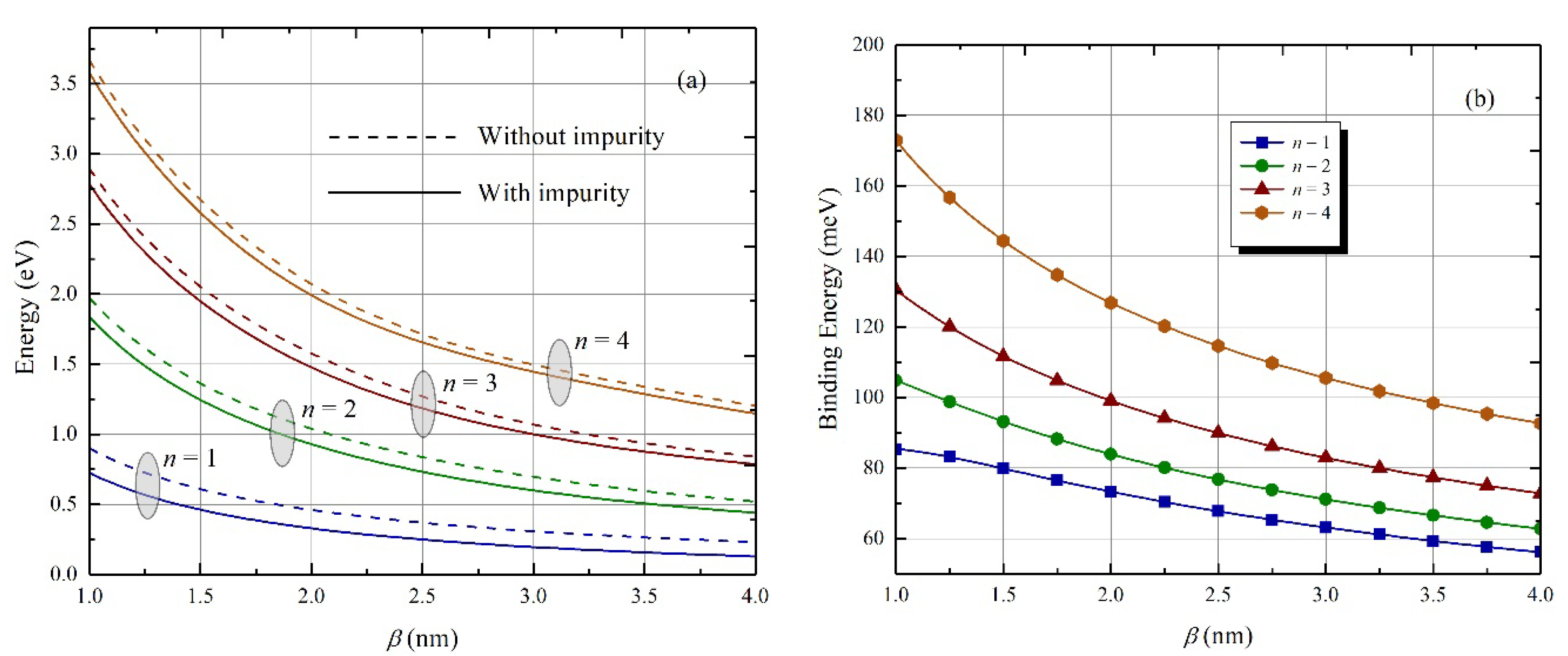
Calculations for the Schrödinger equation and the values of the energy spectrum were performed numerically. The finite element method was used for the numerical calculations. Results for the energy dependence on the half-width β for the first four levels, with and without an impurity center, are presented in Figure 2a. It can be seen from the figure that with a decrease in the QD size, the manifestation of the size quantization effect increases, as a result of which the energy levels rise upward, and the interlevel distance also increases. Accordingly, with an increase in the size of QDs, the energy levels decrease and the interlevel distances decrease. It is obvious that the energy levels taking into account the impurity center are located lower than the energy levels without an impurity center.
Figure 2b shows the dependence of the binding energies of the same levels on the QD half-width β . The binding energy of an electron is defined as the difference between the energies without and in the presence of a donor impurity center. Note that the energy with the impurity tended to the fixed negative value as the size of the QD increased. This value corresponded to the impurity energy in the bulk material [37]. The energy of the electron without impurity remains positive, corresponding to the electron in the bulk material. We have plotted the range of the half-width from 3 nm to 12 nm, corresponding to the experimentally obtained core/shell CdSe/CdS QD [38]. As the size of the QD decreases, the positive contribution of the QD walls to the binding energy increases, and in parallel, as the electron becomes closer to impurity, the negative contribution to the binding energy increases. However, the first effect prevails over the second; therefore, the binding energy remains positive.
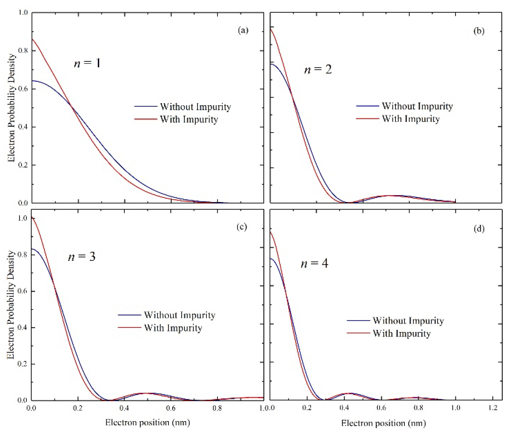
The distribution of the probability density of finding an electron in QDs corresponding to the first four levels of an electron is shown in Figure 3. In all cases, the probability density of the electron tends to the center of QD when the influence of the impurity center has been taken into account.

3. Linear and Nonlinear Optical Properties

Once the energy levels and their corresponding wave functions are obtained, the linear and third-order nonlinear optical absorption coefficients can be calculated. The linear and nonlinear absorption coefficients can be derived from the density matrix approach and perturbation expansion method [39,40,41,42,43]. In the two-level system approach, the linear and nonlinear third-order optical absorption coefficient of QD will be written in the following form:
α ( 1 ) ( ω ) = σ e 2 μ ε | M f i | 2 ω Γ f i ( E f i ω ) 2 + ( Γ f i ) 2 ,
α ( 3 ) ( ω ) = 2 σ e 4 μ ε ( I ε 0 n r c ) | M f i | 4 ω Γ f i ( ( E f i ω ) 2 + ( Γ f i ) 2 ) 2 × ( 1 | M f f M i i | 2 4 | M f i | 2 × 3 E f i 2 4 ω E f i + ( ω ) 2 ( Γ f i ) 2 E f i 2 + ( Γ ) 2 )
where e is the electron charge; σ is the charge concentration divided by system volume σ = n V = 3 n 4 π a 2 c ; μ is the system magnetic permeability; ε is the dielectric constant; n r is the relative refractive index; ω is the energy of the incident photon; E f i = E f E i is the difference between the energies of the initial and final states; Γ f i = 1 / τ f i is an off-diagonal matrix element known as the relaxation rate of the final and initial states, with τ f i as the relaxation time; M f i = ψ i | z | ψ f is transition matrix element; and I is the intensity of the incident electromagnetic radiation. Additionally, the total absorption coefficient α ( ω , I ) will be equal to the sum of the linear and nonlinear optical absorption coefficients:
α ( ω , I ) = α ( 1 ) ( ω ) + α ( 3 ) ( ω , I ) ,
The linear and nonlinear refractive index changes are given by the following formulas:
Δ n ( 1 ) ( ω ) n r = σ e 2 2 n r 2 ε 0 M f i 2 ( E f i ω ) ( E f i ω ) 2 + Γ f i 2 ,  
Δ n ( 3 ) ( ω ) n r = μ c I σ e 4 2 n r 3 ε 0 M f i 4 ( E f i ω ) [ ( E f i ω ) 2 + Γ f i 2 ] 2 × { 1 ( M f f M i i ) 4 M f i 2 ( E f i 2 Γ f i 2 ) ( E f i ( E f i ω ) Γ f i 2 Γ f i 2 2 E f i ω E f i ω ) } .
The total refractive index change is:
Δ n ( ω ) n r = Δ n ( 1 ) ( ω ) n r + Δ n ( 3 ) ( ω ) n r .  
Using the density matrix formalism and an iterative procedure, analytical expressions can be obtained for the second and third harmonic generations (THG), which are associated with optical transition [44,45]. The corresponding expressions can be written in the following form:
χ 2 ω ( 2 ) ( ω ) = σ e 3 ε 0 2 M 01 M 12 M 20 ( ω ω 10 i Γ 0 ) ( 2 ω ω 20 i Γ 0 / 2 )
χ 3 ω ( 3 ) ( ω ) = σ e 4 ε 0 3 M 01 M 12 M 23 M 30 ( ω ω 10 i Γ 0 ) ( 2 ω ω 20 i Γ 0 / 2 ) ( 3 ω ω 30 i Γ 0 / 3 )
where ε 0 is the dielectric constant in vacuum; Γ 0 is the phenomenological parameter, responsible for attenuation due to electron–phonon interaction, collisions among electrons, and ω i j = ( E i E j ) / is the transition frequency; and M 01 M 12 M 23 M 30 is the matrix element of transitions.

4. Results and Discussion

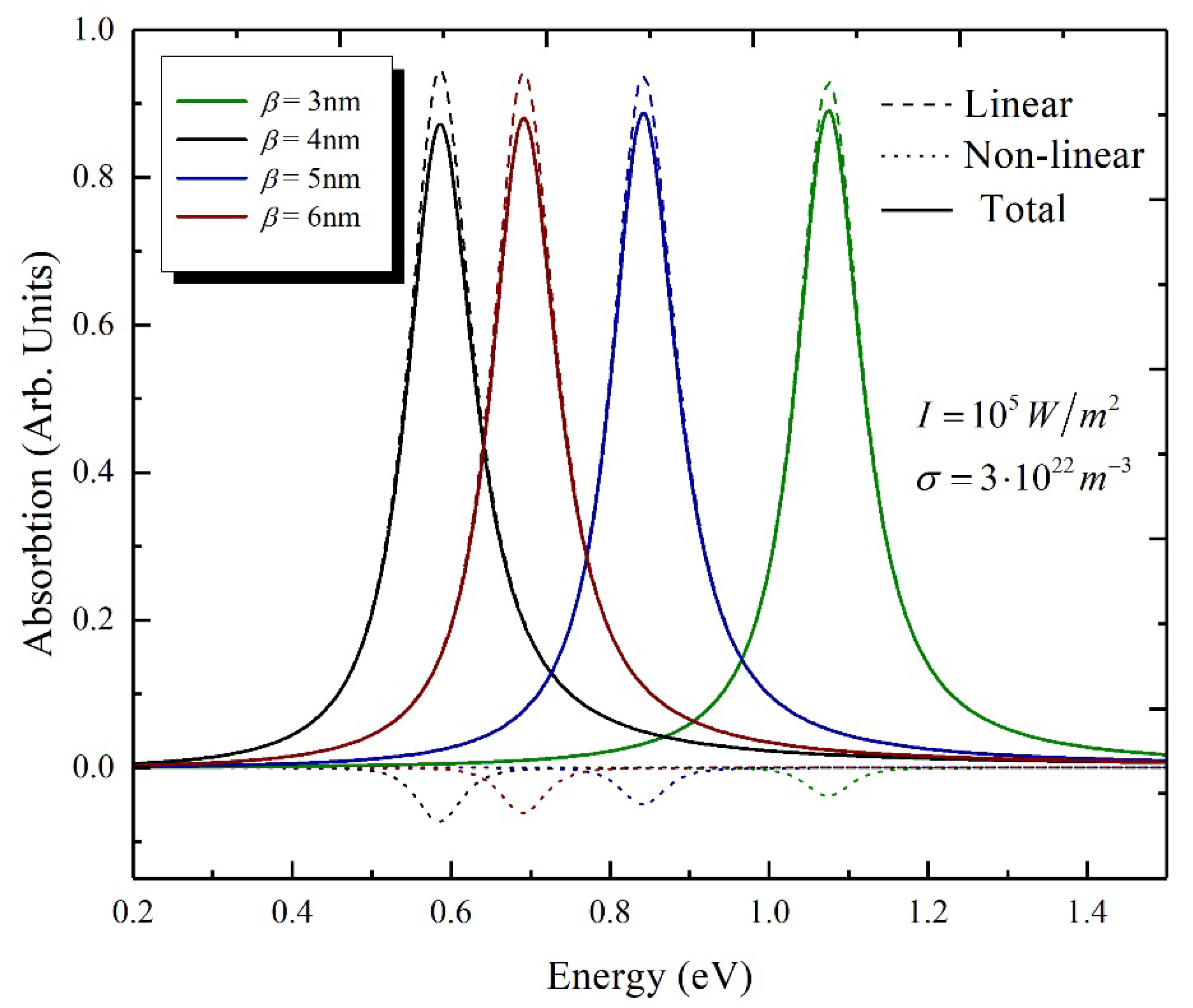
Now, we can proceed to the discussion of the obtained results. Calculated results for the dependence of the linear, non-linear, and total absorption coefficient on the incident light energy for the four different half-widths have been presented in Figure 4. The absorption curves were plotted for the following parameters: I = 10 5   W / m 2 and σ = 3 10 22   m 3 . It can be seen that at smaller QDs, the peak position is shifted to the area of high energy; thus, a blue shift takes place. It is obvious from the figure that the non-linear correction is greater for the large-radius QDs, so the intensity of the total absorption coefficient will be higher for the smaller QDs.
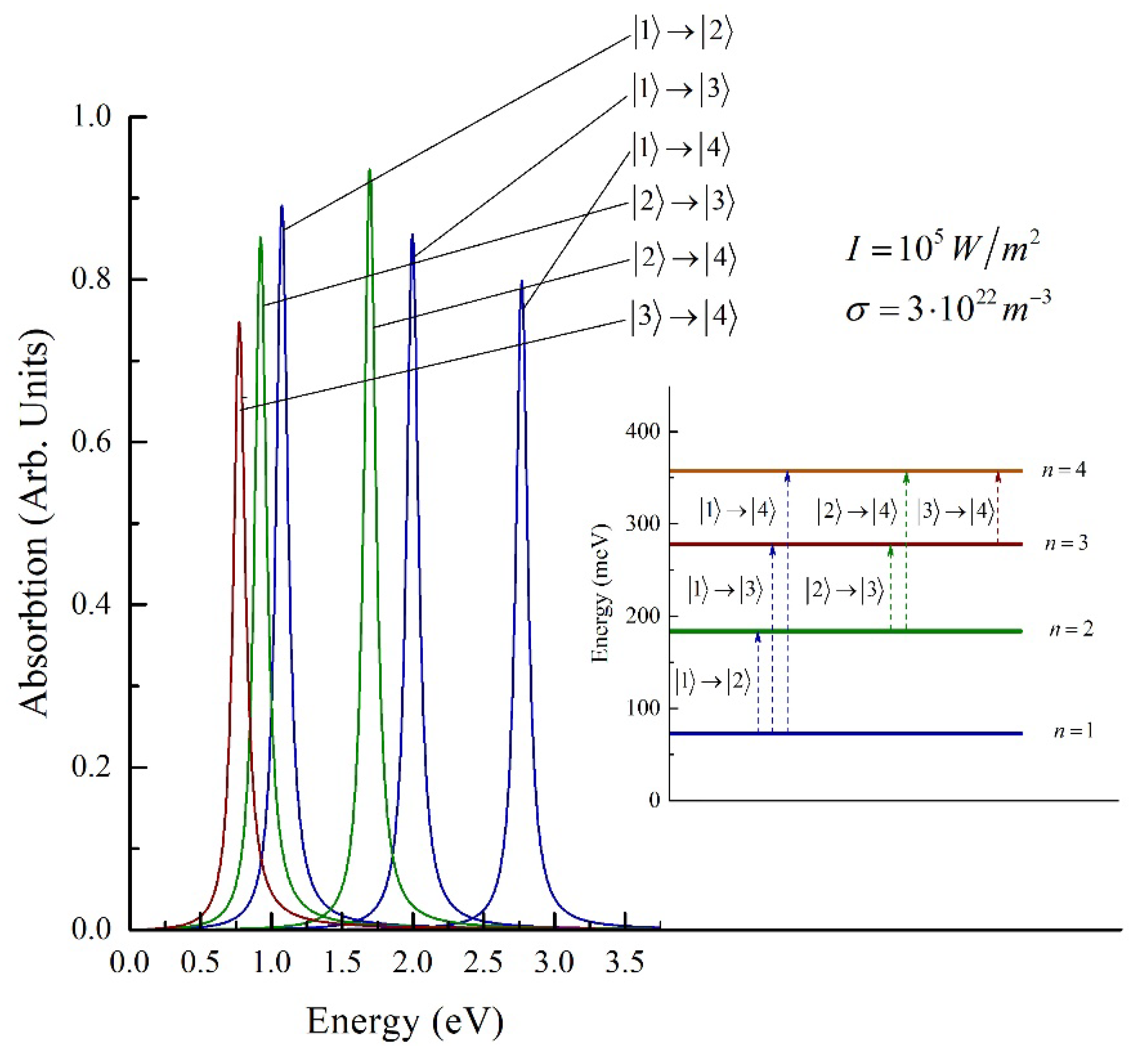
The presented absorption coefficients are calculated for the intraband quantum transition | 1 | 2 , from the ground state to the first excited state. The angular parts were considered in the ground state, with the notation | 1 , 0 , 0 ; therefore, we will use the short version, | 1 . The dependence for absorption curves for the all-possible transitions between the first four levels are presented in Figure 5. The quantum transitions for the four levels are divided in three sets: transitions started from the | 1 , | 2 and | 3 levels. For the first set, the intensities decreased with the transition to the higher levels, whereas for the second set, the intensities increased. The same was true for the third set of quantum transitions, although it is not presented in the figure.
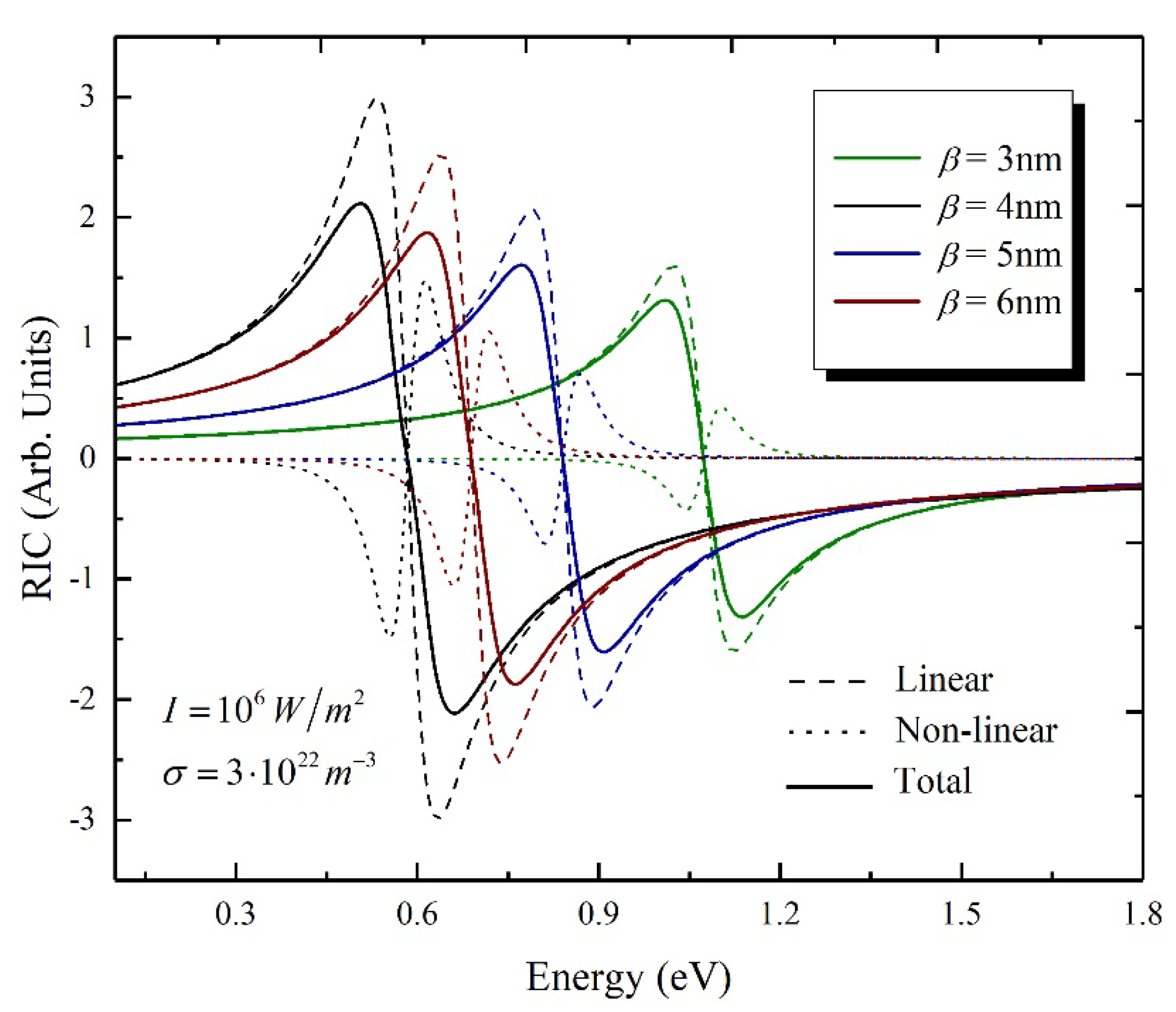
Next, we considered the results obtained for the refractive index change. Figure 6 shows the linear, non-linear and total third-order values of the refractive index. The dependence is given on the energy of the incident light for four different values of the half-width β . This figure clearly shows that the total change in the refractive index strictly depends on the QD radius. As the QD radius increases, the overall change in the refractive index increases and shifts towards lower energies. This change occurs because the energy difference decreases E f i = E f E i with the increase in the half-width (QD radius increases) for a fixed potential height.
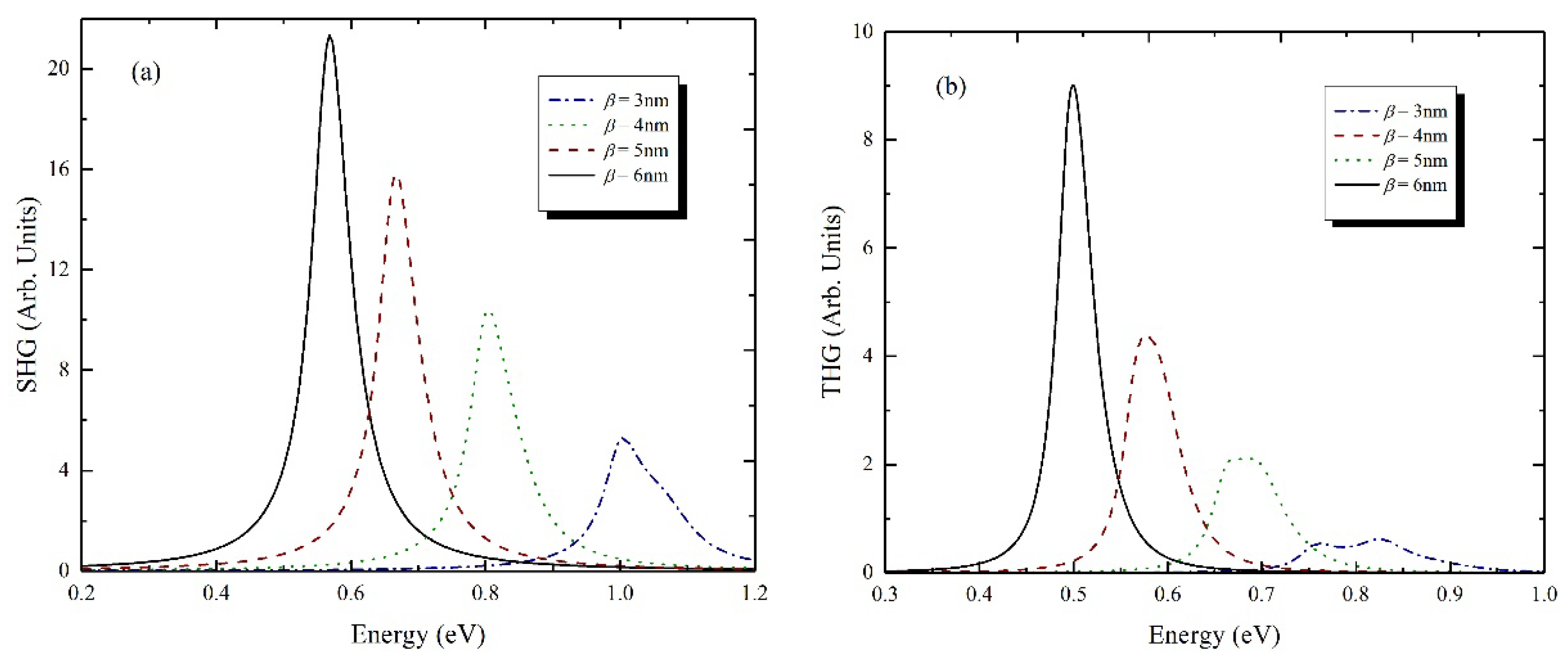
Figure 7a shows the dependence of SHG on the photon energy of the incident light for QDs for four different values of the half-width β . It can be seen from the figure that SHG strongly depends on the radius of the quantum dot. As the radius of QD increases, SHG (and all resonance peaks) increases and also has a red shift. The main reason for these resonance shifts is a decrease in the energy difference of two different electronic states between which an optical transition occurs. We know that with increases in the QD radius, the size quantization decreases; thus, the difference between energies decreases. Actually, the peak of the SHG consists of two peaks, although these two peaks are close to each other and appear in the spectra as merged. Note also that the peaks of the spectrum corresponding to one fixed value of QD are more cohesive at larger than smaller values of QD dimensions; for instance, for the value β = 3   nm , the peaks are more distinct.
Figure 7b shows a graph of the dependence of THG on the energy of incident photons for QDs with different radii. It can be seen from the figure that the THG strongly depends on the size of the QD. Moreover, it becomes clear from the figure that the value of THG will increase with an increase in QD size. In addition, all resonance peaks shifted towards lower energies with increasing radius. This is due to the fact that the energy difference, ∆E, decreased with an increase in QD size. The physical reason for such behavior is the quantum confinement effect. As the QD size increased, the effect of quantum confinement decreases. It can be seen from the figure that the line width of the resonance peaks increases with increasing size. As is the case for SHG, for the THG as well, the peaks are merged, with the difference being that in this case, three peaks have been merged.

5. Conclusions

In this article, the linear and nonlinear intraband optical properties of colloidal spherical CdSe/CdS core/shell QDs in the presence of donor impurity in the center are theoretically investigated. The modified Pöschl–Teller potential was chosen as the model confinement potential for the spherical core/shell QD.
The energy and binding energy of an electron were calculated as functions of the half-width of spherical core/shell QD and the dependence was shown to be monotonic. The spectra of the linear, nonlinear and total absorption coefficients of the incident light energy were obtained for various values of the half-width. It is shown that at smaller QDs, light absorption occurred with higher energy than at large QDs, and the non-linear correction was bigger for the large radius QDs; therefore, the intensity of the total absorption coefficient will be more for the smaller QDs.
The change in the refractive index as a function of the incident light energy was also considered. Thus, the calculation of the change in the refractive index using only the linear term may be incorrect for systems operating especially with high optical intensity, due to the strong dependence of the nonlinear component on the incident optical intensity.
As the final step, the generation of the second and third harmonics has been calculated for the system under consideration. As the radius of QD increased, SHG and THG increased and also exhibited a red shift. The peaks of SHG and THG occurred in a merged form, which were split for the smaller values of half-width β .

Author Contributions

G.A.M., N.A.Z. and P.A.M.—numerical calculations, G.A.M., D.B.H., H.A.S. and S.B.—conceptualization and development of the theory, G.A.M., P.A.M. and D.B.H.—original draft preparation, H.A.S. and S.B.—review and editing. All authors have read and agreed to the published version of the manuscript.

Funding

This work was financially supported by the Horizon-2020 research and innovation program of the European Union (grant no. 952335, NanoQIQO project).

Data Availability Statement

Not applicable.

Conflicts of Interest

The authors declare no conflict of interest. The funders had no role in the design of the study; in the collection, analyses, or interpretation of data; in the writing of the manuscript, or in the decision to publish the results.

References

  1. Bimberg, D.; Grundmann, M.; Ledentsov, N.N.; Ruvimov, S.S.; Werner, P.; Richter, U.; Heydenreich, J.; Ustinov, V.M.; Kop’ev, P.S.; Alferov, Z.I. Self-organization processes in MBE-grown quantum dot structures. Thin Solid Films 1995, 267, 32–36. [Google Scholar] [CrossRef]
  2. Bosacchi, A.; Frigeri, P.; Franchi, S.; Allegri, P.; Avanzini, V. InAsGaAs self-assembled quantum dots grown by ALMBE and MBE. J. Cryst. Growth 1997, 175, 771–776. [Google Scholar] [CrossRef]
  3. Nötzel, R.; Ploog, K.H. MBE of quantum wires and quantum dots. J. Cryst. Growth 2001, 227, 8–12. [Google Scholar] [CrossRef]
  4. Mahler, B.; Spinicelli, P.; Buil, S.; Quelin, X.; Hermier, J.P.; Dubertret, B. Towards non-blinking colloidal quantum dots. Nat. Mater. 2008, 7, 659–664. [Google Scholar] [CrossRef]
  5. Baskoutas, S.; Terzis, A.F. Size-dependent band gap of colloidal quantum dots. J. Appl. Phys. 2006, 99, 013708. [Google Scholar] [CrossRef]
  6. Litvin, A.P.; Martynenko, I.V.; Purcell-Milton, F.; Baranov, A.V.; Fedorov, A.V.; Gun’Ko, Y.K. Colloidal quantum dots for optoelectronics. J. Mater. Chem. A 2017, 5, 13252–13275. [Google Scholar] [CrossRef]
  7. Kagan, C.R.; Lifshitz, E.; Sargent, E.H.; Talapin, D.V. Building devices from colloidal quantum dots. Science 2016, 353. [Google Scholar] [CrossRef]
  8. Lu, W.; Guo, X.; Luo, Y.; Li, Q.; Zhu, R.; Pang, H. Core-shell materials for advanced batteries. Chem. Eng. J. 2019, 355, 208–237. [Google Scholar] [CrossRef]
  9. Clifford, J.P.; Konstantatos, G.; Johnston, K.W.; Hoogland, S.; Levina, L.; Sargent, E.H. Fast, sensitive and spectrally tuneable colloidal-quantum-dot photodetectors. Nat. Nanotechnol. 2009, 4, 40–44. [Google Scholar] [CrossRef]
  10. Konstantatos, G.; Howard, I.; Fischer, A.; Hoogland, S.; Clifford, J.; Klem, E.; Levina, L.; Sargent, E.H. Ultrasensitive solution-cast quantum dot photodetectors. Nature 2006, 442, 180–183. [Google Scholar] [CrossRef] [PubMed]
  11. Chen, O.; Zhao, J.; Chauhan, V.P.; Cui, J.; Wong, C.; Harris, D.K.; Wei, H.; Han, H.S.; Fukumura, D.; Jain, R.K.; et al. Compact high-quality CdSe–CdS core–shell nanocrystals with narrow emission linewidths and suppressed blinking. Nat. Mater. 2013, 12, 445–451. [Google Scholar] [CrossRef] [Green Version]
  12. Vasudevan, D.; Gaddam, R.R.; Trinchi, A.; Cole, I. Core–shell quantum dots: Properties and applications. J. Alloys Compd. 2015, 636, 395–404. [Google Scholar] [CrossRef]
  13. Selopal, G.S.; Zhao, H.; Wang, Z.M.; Rosei, F. Core/shell quantum dots solar cells. Adv. Funct. Mater. 2020, 30, 1908762. [Google Scholar] [CrossRef]
  14. Yuan, X.; Zheng, J.; Zeng, R.; Jing, P.; Ji, W.; Zhao, J.; Yang, W.; Li, H. Thermal stability of Mn2+ ion luminescence in Mn-doped core–shell quantum dots. Nanoscale 2014, 6, 300–307. [Google Scholar] [CrossRef]
  15. Bhattacharyya, B.; Pandey, A. CuFeS2 quantum dots and highly luminescent CuFeS2 based core/shell structures: Synthesis, tunability, and photophysics. J. Am. Chem. Soc. 2016, 138, 10207–10213. [Google Scholar] [CrossRef]
  16. Zeiri, N.; Naifar, A.; Nasrallah, S.A.B.; Said, M. Third nonlinear optical susceptibility of CdS/ZnS core-shell spherical quantum dots for optoelectronic devices. Optik 2019, 176, 162–167. [Google Scholar] [CrossRef]
  17. Loitsch, B.; Winnerl, J.; Grimaldi, G.; Wierzbowski, J.; Rudolph, D.; Morkoötter, S.; Doöblinger, M.; Abstreiter, G.; Koblmüller, G.; Finley, J.J. Crystal phase quantum dots in the ultrathin core of GaAs–AlGaAs core–shell nanowires. Nano Lett. 2015, 15, 7544–7551. [Google Scholar] [CrossRef] [PubMed]
  18. Zhu, X.; Yang, X.; Lv, C.; Guo, S.; Li, J.; Zheng, Z.; Zhu, H.; Yang, D. New approach to create TiO2 (B)/carbon core/shell nanotubes: Ideal structure for enhanced lithium ion storage. ACS Appl. Mater. Interfaces 2016, 8, 18815–18821. [Google Scholar] [CrossRef]
  19. Jiang, H.; Chen, Y.; Li, L.; Liu, H.; Ren, C.; Liu, X.; Tian, G. Hierarchical ZnO nanorod/ZnFe2O4 nanosheet core/shell nanoarray decorated with PbS quantum dots for efficient photoelectrochemical water splitting. J. Alloys Compd. 2020, 828, 154449. [Google Scholar] [CrossRef]
  20. Kayaci, F.; Vempati, S.; Ozgit-Akgun, C.; Donmez, I.; Biyikli, N.; Uyar, T. Transformation of polymer-ZnO core–shell nanofibers into ZnO hollow nanofibers: Intrinsic defect reorganization in ZnO and its influence on the photocatalysis. Appl. Catal. B Environ. 2015, 176, 646–653. [Google Scholar] [CrossRef]
  21. Wang, M.; Peng, Z.; Qian, J.; Li, H.; Zhao, Z.; Fu, X. Highly efficient solar-driven photocatalytic degradation on environmental pollutants over a novel C fibers@ MoSe2 nanoplates core-shell composite. J. Hazard. Mater. 2018, 347, 403–411. [Google Scholar] [CrossRef] [PubMed]
  22. Mantashian, G.A.; Mantashyan, P.A.; Sarkisyan, H.A.; Kazaryan, E.M.; Bester, G.; Baskoutas, S.; Hayrapetyan, D.B. Exciton-Related Raman Scattering, Interband Absorption and Photoluminescence in Colloidal CdSe/CdS Core/Shell Quantum Dots Ensemble. Nanomaterials 2021, 11, 1274. [Google Scholar] [CrossRef] [PubMed]
  23. Schmitt-Rink, S.D.A.B.M.; Miller, D.A.B.; Chemla, D.S. Theory of the linear and nonlinear optical properties of semiconductor microcrystallites. Phys. Rev. B 1987, 35, 8113. [Google Scholar] [CrossRef] [PubMed]
  24. Takagahara, T. Excitonic optical nonlinearity and exciton dynamics in semiconductor quantum dots. Phys. Rev. B 1987, 36, 9293. [Google Scholar] [CrossRef]
  25. Li, B.; Guo, K.X.; Zhang, C.J.; Zheng, Y.B. The second-harmonic generation in parabolic quantum dots in the presence of electric and magnetic fields. Phys. Lett. A 2007, 367, 493–497. [Google Scholar] [CrossRef]
  26. Shao, S.; Guo, K.X.; Zhang, Z.H.; Li, N.; Peng, C. Studies on the third-harmonic generations in cylindrical quantum dots with an applied electric field. Superlattices Microstruct. 2010, 48, 541–549. [Google Scholar] [CrossRef]
  27. Liu, G.; Guo, K.; Wu, Q.; Wu, J.H. Polaron effects on the optical rectification and the second harmonic generation in cylindrical quantum dots with magnetic field. Superlattices Microstruct. 2013, 53, 173–183. [Google Scholar] [CrossRef]
  28. Portacio, A.A.; Rodríguez, B.A.; Villamil, P. Influence of the position of a donor impurity on the second-order nonlinear optical susceptibility in a cylindrical quantum dot. Superlattices Microstruct. 2018, 113, 550–557. [Google Scholar] [CrossRef]
  29. Xie, W. Nonlinear optical properties of a hydrogenic donor quantum dot. Phys. Lett. A 2008, 372, 5498–5500. [Google Scholar] [CrossRef]
  30. Zielinski, M.; Oron, D.; Chauvat, D.; Zyss, J. Second-Harmonic Generation from a Single Core/Shell Quantum Dot. Small 2009, 5, 2835–2840. [Google Scholar] [CrossRef] [PubMed]
  31. Bera, A.; Ghosh, A.; Ghosh, M. Nonlinear optical properties of doped quantum dots: Interplay between noise and carrier density. Opt. Mater. 2017, 69, 352–357. [Google Scholar] [CrossRef]
  32. Poerschke, O.R. Madelung, Semiconductors: Other than Group IV Elements and 2I-V Compounds; Springer: Berlin/Heidelberg, Germany, 1992. [Google Scholar]
  33. Parang, Z.; Keshavarz, A.; Zamani, N. Optimization of optical absorption coefficient in double modified Pöschl–Teller quantum wells. J. Comput. Electron. 2014, 13, 1020–1025. [Google Scholar] [CrossRef]
  34. Hayrapetyan, D.B.; Kazaryan, E.M.; Tevosyan, H.K. Optical properties of spherical quantum dot with modified Pöschl–Teller potential. Superlattices Microstruct. 2013, 64, 204–212. [Google Scholar] [CrossRef]
  35. Sargsian, T.A.; Mkrtchyan, M.A.; Sarkisyan, H.A.; Hayrapetyan, D.B. Effects of external electric and magnetic fields on the linear and nonlinear optical properties of InAs cylindrical quantum dot with modified Pöschl-Teller and Morse confinement potentials. Phys. E Low-Dimens. Syst. Nanostructures 2021, 126, 114440. [Google Scholar] [CrossRef]
  36. Ünlü, S.; Karabulut, İ.; Şafak, H. Linear and nonlinear intersubband optical absorption coefficients and refractive index changes in a quantum box with finite confining potential. Phys. E Low-Dimens. Syst. Nanostructures 2006, 33, 319–324. [Google Scholar] [CrossRef]
  37. Chuu, D.S.; Hsiao, C.M.; Mei, W.N. Hydrogenic impurity states in quantum dots and quantum wires. Phys. Rev. B 1992, 46, 3898. [Google Scholar] [CrossRef] [Green Version]
  38. Badlyan, N.M.; Biermann, A.; Aubert, T.; Hens, Z.; Maultzsch, J. Thermal expansion of colloidal CdSe/CdS core/shell quantum dots. Phys. Rev. B 2019, 99, 195425. [Google Scholar] [CrossRef]
  39. İbrahim, K.; Baskoutas, S. Linear and nonlinear optical absorption coefficients and refractive index changes in spherical quantum dots: Effects of impurities, electric field, size, and optical intensity. J. Appl. Phys. 2008, 103, 073512. [Google Scholar] [CrossRef]
  40. Xie, W. Impurity effects on optical property of a spherical quantum dot in the presence of an electric field. Phys. B Condens. Matter 2010, 405.16, 3436–3440. [Google Scholar] [CrossRef]
  41. Baira, M.; Salem, B.; Madhar, N.A.; Ilahi, B. Linear and Nonlinear Intersubband Optical Properties of Direct Band Gap GeSn Quantum Dots. Nanomaterials 2019, 9, 124. [Google Scholar] [CrossRef] [Green Version]
  42. Liu, C.H.; Xu, B.R. Theoretical study of the optical absorption and refraction index change in a cylindrical quantum dot. Phys. Lett. A 2008, 372, 888–892. [Google Scholar] [CrossRef]
  43. Pandey, A.; Guyot-Sionnest, P. Intraband spectroscopy and band offsets of colloidal II-VI core/shell structures. J. Chem. Phys. 2007, 127, 104710. [Google Scholar] [CrossRef] [PubMed]
  44. Duque, C.M.; Mora-Ramos, M.E.; Duque, C.A. Properties of the second and third harmonics generation in a quantum disc with inverse square potential. A modeling for nonlinear optical responses of a quantum ring. J. Lumin. 2013, 138, 53–60. [Google Scholar] [CrossRef]
  45. Bahramiyan, H.; Khordad, R. Optical properties of a GaAs pyramid quantum dot: Second-and third-harmonic generation. Int. J. Mod. Phys. B 2014, 28, 1450053. [Google Scholar] [CrossRef]
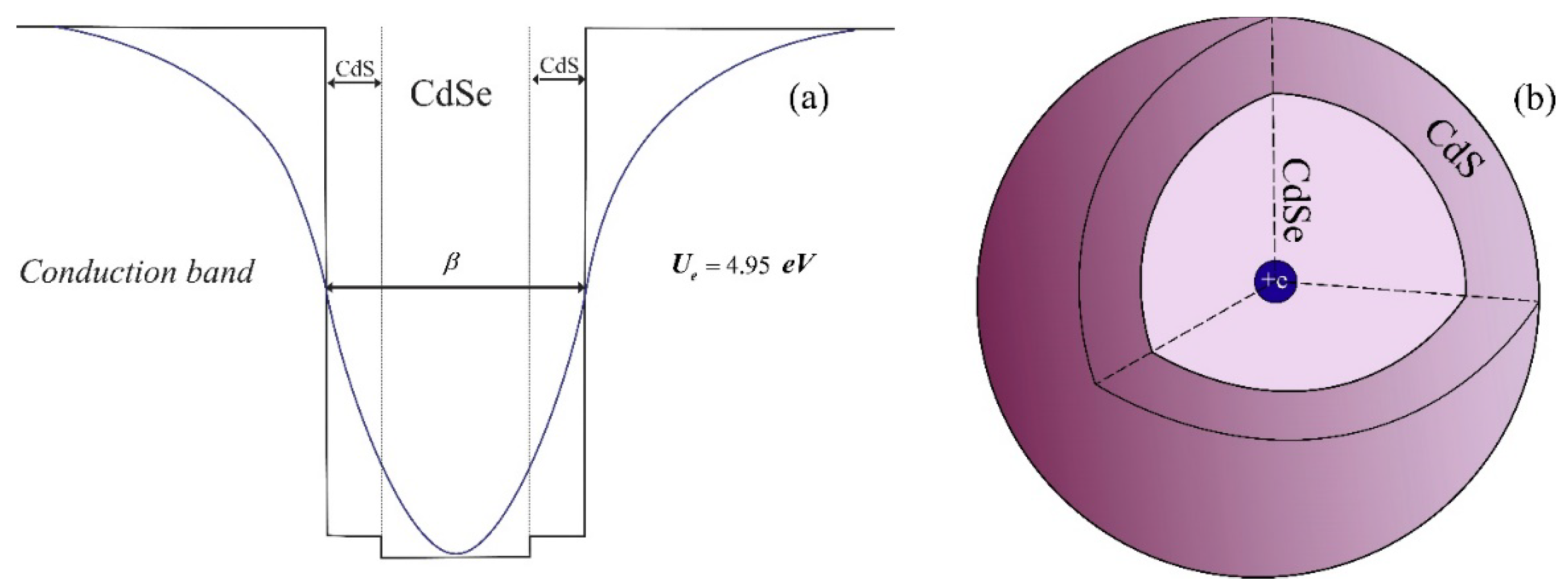
Figure 1. Structure of the conduction band of CdSe/CdS core/shell QDs (a), and a 3D drawing of a CdSe/CdS core/shell QD (b).
Atoms 09 00075 g001
Figure 2. Dependence of the first four energy levels on the half-width β with and without the impurity center (a), and binding energy (b) on the half-width β for the same levels.
Atoms 09 00075 g002
Figure 3. Distribution of the probability density of finding an electron in a QD for the first four levels of an electron with and without impurity center. (a) n = 1 , (b) n = 2 , (c) n = 3 , (d) n = 4 .
Atoms 09 00075 g003
Figure 4. Dependence of the linear, non-linear and total absorption coefficient on the energy of the incident light for four different values of β .
Atoms 09 00075 g004
Figure 5. Dependence of the total absorption coefficient on the energy of the incident light for different transitions, (inset) schematic view of the energy diagram with quantum transitions.
Atoms 09 00075 g005
Figure 6. Dependence of linear, nonlinear and total values of the refractive index change on the energy of the incident light for four different values of β .
Atoms 09 00075 g006
Figure 7. Dependence of the second harmonic generation (a) and third harmonic generation (b) on the energy of the incident light for four different values of β .
Atoms 09 00075 g007
Publisher’s Note: MDPI stays neutral with regard to jurisdictional claims in published maps and institutional affiliations.