Gallstone Magnesium Distributions from Optical Emission Spectroscopy


 ,
, 

Abstract
:1. Introduction
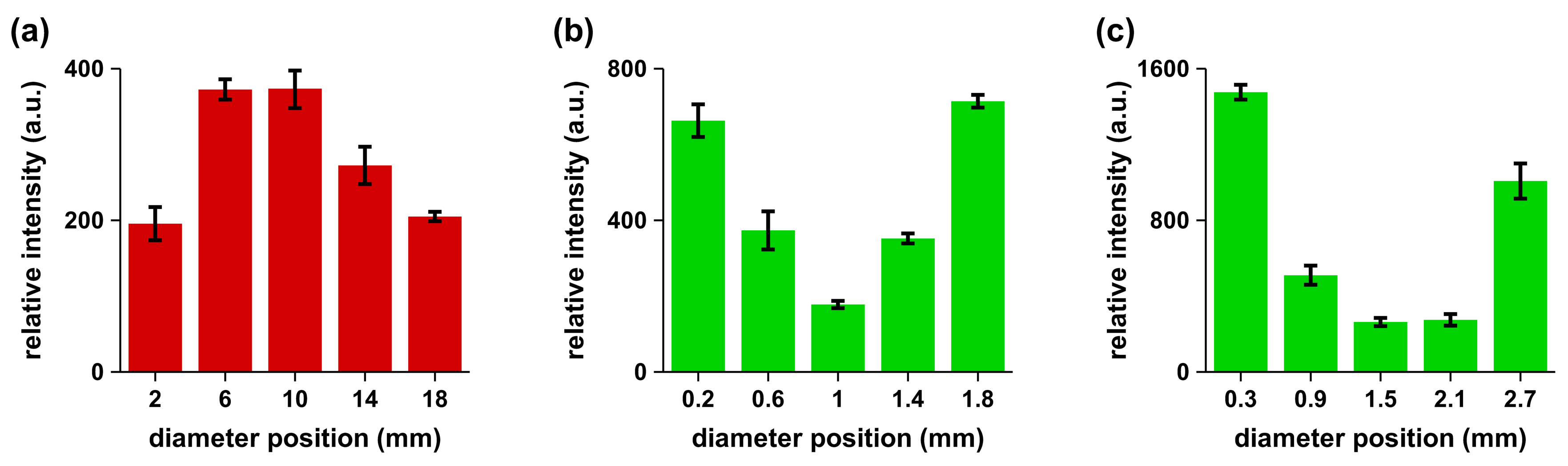
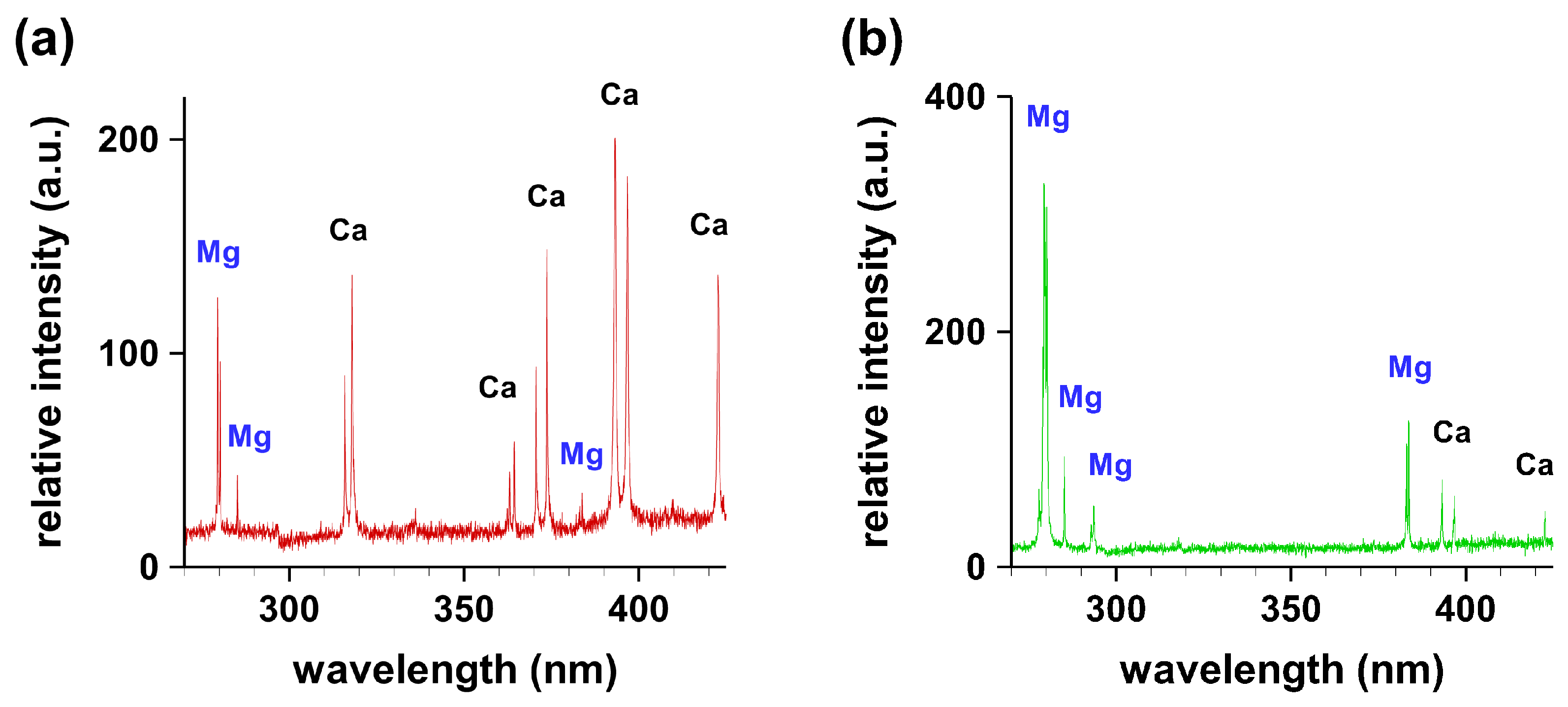
2. Results
3. Conclusions
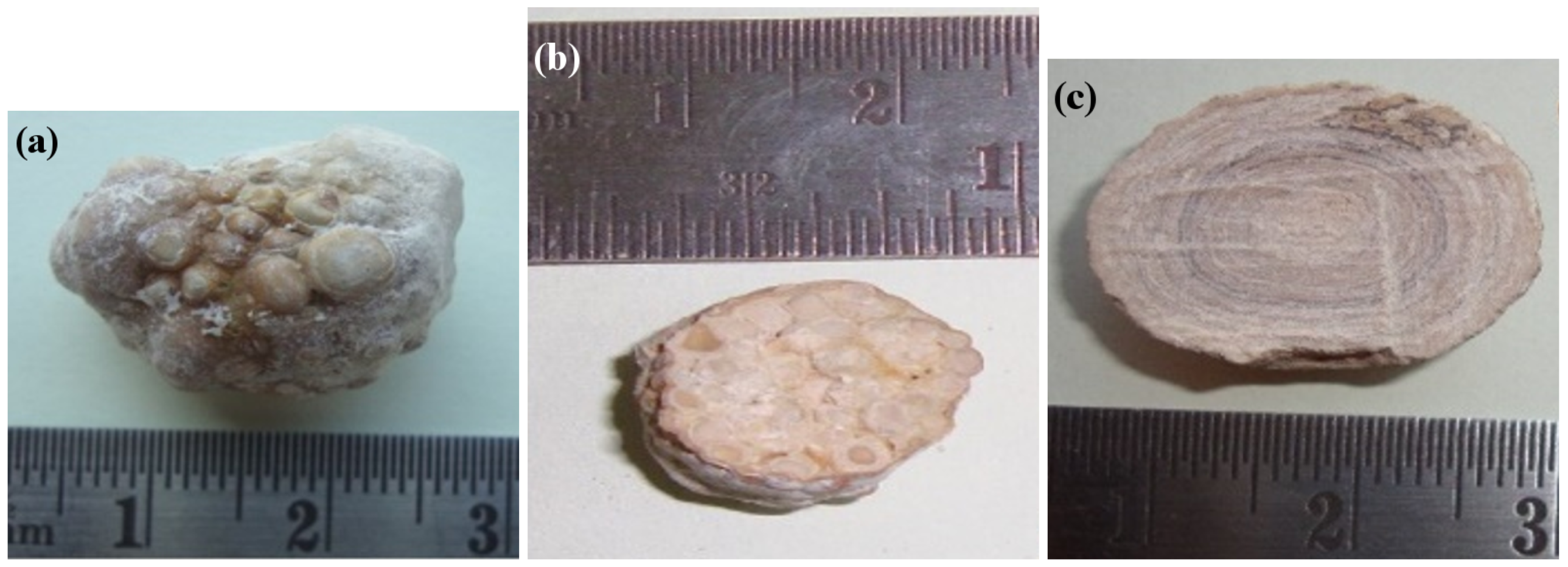
4. Materials and Methods
Author Contributions
Funding
Acknowledgments
Conflicts of Interest
References
- Mohan, H.; Punia, R.P.S.; Dhawan, S.B.; Ahal, S.; Shekhon, M.S. Morphological spectrum of gallstone disease in 1100 cholecystectomies in North India. Indian J. Surg. 2005, 67, 140–142. [Google Scholar]
- Saris, N.E.L.; Mervaala, E.; Karppanen, H.; Khawaja, J.A.; Lewenstam, A. Magnesium: An update on physiological, clinical and analytical aspects. Clin. Chim. Acta 2000, 294, 1–26. [Google Scholar] [CrossRef]
- Chakraborti, S.; Chakraborti, T.; Mandal, M.; Mandal, A.; Das, S.; Ghosh, S. Protective role of magnesium in cardiovascular diseases: A review. Mol. Cell Biochem. 2002, 238, 163–179. [Google Scholar] [CrossRef] [PubMed]
- He, K.; Liu, K.; Daviglus, M.L.; Morris, S.J.; Loria, C.M.; Horn, L.V.; Jacobs, D.R., Jr.; Savage, P.J. Magnesium intake and incidence of metabolic syndrome among young adults. Circulation 2006, 113, 1675–1682. [Google Scholar] [CrossRef] [PubMed]
- Touyz, R.M. Magnesium in clinical medicine. Front Biosci. 2004, 9, 1278–1293. [Google Scholar] [CrossRef] [PubMed]
- Fawcett, W.J.; Haxby, E.J.; Male, D.A. Magnesium: Physiology and pharmacology. Br. J. Anaesth. 1999, 83, 302–320. [Google Scholar] [CrossRef] [PubMed]
- National Institute of Standards and Technology (NIST) Electronic Database. Available online: http://physics.nist.gov/PhysRefData/ASD/lines_form.html (accessed on 5 March 2018).
- Singh, V.K.; Singh, V.; Rai, A.K.; Thakur, S.N.; Rai, P.K.; Singh, J.P. Quantitative analysis of gallstones using laser-induced breakdown spectroscopy. Appl. Opt. 2008, 47, G38–G47. [Google Scholar] [CrossRef] [PubMed]
- Singh, V.K.; Rai, V.; Rai, A.K. Variational study of the constituents of cholesterol stones by laser-induced breakdown spectroscopy. Lasers Med. Sci. 2009, 24, 27–33. [Google Scholar] [CrossRef] [PubMed]
- Tsai, C.J.; Leitzmann, M.F.; Willett, W.C.; Giovannucci, E.L. Long-term effect of Magnesium Consumption on the risk of symptomatic gallstone disease among men. Am. J. Gastroenterol. 2008, 103, 375. [Google Scholar] [CrossRef] [PubMed]
- Pathak, A.K.; Singh, V.K.; Rai, N.K.; Rai, A.K.; Rai, P.K.; Rai, P.K.; Rai, S.; Baruah, G.D. Study of different concentric rings inside gallstones with LIBS. Lasers Med. Sci. 2011, 26, 531–537. [Google Scholar] [CrossRef] [PubMed]
- Pathak, A.K.; Kumar, R.; Singh, V.K.; Agrawal, R.; Rai, S.; Rai, A.K. Assessment of LIBS for Spectrochemical Analysis: A Review. Appl. Spectrosc. Rev. 2012, 47, 14–40. [Google Scholar] [CrossRef]

© 2018 by the authors. Licensee MDPI, Basel, Switzerland. This article is an open access article distributed under the terms and conditions of the Creative Commons Attribution (CC BY) license (http://creativecommons.org/licenses/by/4.0/).
Share and Cite
Pathak, A.K.; Rai, N.K.; Kumar, R.; Rai, P.K.; Rai, A.K.; Parigger, C.G. Gallstone Magnesium Distributions from Optical Emission Spectroscopy. Atoms 2018, 6, 42. https://doi.org/10.3390/atoms6030042
Pathak AK, Rai NK, Kumar R, Rai PK, Rai AK, Parigger CG. Gallstone Magnesium Distributions from Optical Emission Spectroscopy. Atoms. 2018; 6(3):42. https://doi.org/10.3390/atoms6030042
Chicago/Turabian StylePathak, Ashok K., Nilesh K. Rai, Rohit Kumar, Pradeep K. Rai, Awadhesh K. Rai, and Christian G. Parigger. 2018. "Gallstone Magnesium Distributions from Optical Emission Spectroscopy" Atoms 6, no. 3: 42. https://doi.org/10.3390/atoms6030042
APA StylePathak, A. K., Rai, N. K., Kumar, R., Rai, P. K., Rai, A. K., & Parigger, C. G. (2018). Gallstone Magnesium Distributions from Optical Emission Spectroscopy. Atoms, 6(3), 42. https://doi.org/10.3390/atoms6030042