Review

Methylglyoxal, a Knot to Be Untied in Brain Glucose Hypometabolism

by
Vitor Gayger-Dias
1,†,
Vanessa-Fernanda Da Silva
1,†,
Thomas Michel Sobottka
1,
Marina Concli Leite
1,
Adriana Fernanda K. Vizuete
1,2 and
Carlos-Alberto Gonçalves
1,3,*
1
Programa de Pós-Graduação em Bioquímica, Instituto de Ciências Básicas da Saúde, Universidade Federal do Rio Grande do Sul, Porto Alegre 90035-003, Brazil
2
Laboratório de Fisiologia Translacional, Programa de Pós-Graduação em Ciências da Saúde, Universidade Federal de Ciências da Saúde de Porto Alegre, Porto Alegre 90050-170, Brazil
3
Programa de Pós-Graduação em Neurociências, Instituto de Ciências Básicas da Saúde, Universidade Federal do Rio Grande do Sul, Porto Alegre 90035-003, Brazil
*
Author to whom correspondence should be addressed.
These authors contributed equally to this work.
Metabolites 2025, 15(11), 690; https://doi.org/10.3390/metabo15110690 (registering DOI)
Submission received: 1 October 2025 / Revised: 17 October 2025 / Accepted: 22 October 2025 / Published: 24 October 2025
(This article belongs to the Special Issue Metabolic Profiling in Neurometabolisms)

Abstract

Background: Advanced glycation end products (AGEs) and receptors for AGEs (RAGE) have been extensively implicated in metabolic and neurodegenerative disorders due to their capacity to alter protein structure and function through non-enzymatic glycation. More recently, methylglyoxal (MG), a highly reactive glycolytic byproduct, has gained attention as a critical mediator of AGE formation and an independent contributor to cellular distress, particularly in the context of diabetes mellitus and Alzheimer’s disease. Objectives: This review synthesizes evidence from experimental and clinical studies addressing MG generation and metabolism in brain tissue, emphasizing the glyoxalase system as the primary detoxification mechanism, the functional contribution of astrocytes, and the downstream consequences of MG accumulation. In addition, we examined the interplay between MG, RAGE signaling, unfolded protein response, and regulatory mechanisms involving the hexosamine biosynthesis pathway and O-GlcNAcylation of key proteins in glucose metabolism and insulin signaling. Results and Conclusions: Brain glucose hypometabolism is a consequence of insulin resistance and results in a metabolic rearrangement that expands the glycolytic pathway and generates more MG, which, in turn, can affect insulin signaling, further compromising the molecular basis of insulin resistance and creating a vicious cycle. Astrocytes are key cells in the generation and detoxification of MG in the brain, making them a therapeutic target.

Graphical Abstract

1. Introduction

Methylglyoxal (MG) has gained prominence as a pivotal factor in the formation of advanced glycation end products (AGEs) and, consequently, the basis of ageing and prevalent neurodegenerative diseases, such as diabetic encephalopathy (DE) and Alzheimer’s disease (AD) [1,2]. We have highlighted the role of astrocytes in MG detoxification in vitro [3] and in vivo models of AD [4]. MG is increasingly being viewed as an undesirable side product of glucose metabolism.
Since MG is derived from glucose, exposure of cells to high levels of glucose commonly elevates MG levels, suggesting simply that this is due to high glucose and not the underlying insulin resistance. Herein, we review the insulin resistance as a mechanism behind MG elevation, as well as the MG detoxification pathway, its physiological and pathological effects, the pathophysiological conditions associated with its accumulation, and the prospects for its use as a marker in pathological conditions, always emphasizing the role of astrocytes in the generation and clearance of this compound.

2. Sources of Brain MG

MG is 2-oxopropanal, a small cell-permeable compound that demonstrates high (glycating) activity with the amino groups of proteins and DNA [5]. This molecule is principally derived from the phosphotrioses of the glycolytic pathway (Figure 1); however, given that the ratio of the two phosphotrioses, dihydroxyacetone phosphate (DHAP)/glyceraldehyde 3-phosphate (GA3P) is 9/1, DHAP is the only metabolite frequently cited as a source of MG. The reaction is spontaneous and involves DHAP dephosphorylation and isomerization of glyceraldehyde to MG, but some bacteria possess an enzyme for this reaction, contradicting the idea that it is purely an accidental byproduct of metabolism. In addition to the phosphotrioses, there are two other minor sources of MG in mammals. The first is acetone, one of the ketone bodies, which can generate MG when oxidized by cytochrome P450 oxidase 2E1 (CYP2E1). This oxidase, which metabolizes ethanol to acetaldehyde, is therefore capable of generating MG under conditions of ketosis, such as fasting and diabetes mellitus (DM). The second source derives from threonine catabolism. Threonine is an essential amino acid that is metabolized to succinyl-CoA (mostly) or pyruvate. This second pathway can lead to the formation of aminoacetone, which, through the action of an amino oxidase, results in MG formation. It is estimated that physiologically, 0.1–0.4% of metabolized glucose results in MG [6]. However, less than 1% of the MG generated results in glycation, despite its high reactivity. More than 99% is detoxified by the glyoxalase system, which varies between tissues [2]. Thus, while we find approximately 4 μM of MG in the liver and muscle tissues, we find approximately 1 μM in brain tissue [7]. Levels of 10 μM are found in the extracellular medium.
Of note, the degradation of DHAP to MG is an uncatalyzed reaction, but that does not mean that it is constant. At least two metabolic variables affect this reaction: pH and the presence of reactive oxygen species (ROS). Since DHAP phosphorolysis begins with deprotonation (see [8] for a review), an alkaline pH would favor MG formation. Furthermore, if there is consumption of glutathione (GSH) (due to the presence of ROS) the GSH/GSSG ratio drops and this favors the glutathionylation of proteins, including GA3P dehydrogenase (GAPD), the enzyme in the reaction subsequent to the isomerization of phosphotrioses in the glycolytic pathway. Glutathionylation protects cysteine groups from ROS attack, but in the case of GAPD, there is also an inhibition of activity, retaining DHAP and favoring MG formation. Thus, under conditions of oxidative stress, there is greater MG formation. Commonly MG is seen as a glycotoxin, generated under conditions of oxidative stress; however, the concept of hormosis is worth mentioning, where a toxic substance can be protective at low concentrations (see [9] for a definition). Like ROS, physiological concentrations of MG stimulate the expression of metabolic pathway enzymes that protect cells from MG toxicity at higher concentrations, as will be discussed below.

3. Brain Detoxification

In the neuropil region, comparison of the cytosolic glycolytic pathways in neurons and astrocytes reveals stronger astroglial activity (see Barros et al., 2024 for a review) [10]. During neuronal activity, astrocytes provide lactate to sustain this activity. This more vigorous glycolytic activity results in greater MG production, which also explains the greater MG detoxification activity in astrocytes, primarily through the glyoxalase system [11].
The system is composed of two enzymes, glyoxalase 1 and 2 (GLO 1 and 2, respectively) (Figure 1). Although the enzymes are ubiquitous, studies in primary cortical cultures indicate that GLO-1 is almost 10 times more active in astrocytes than in neurons, while GLO-2 is twice as active [12]. In the first, uncatalyzed reaction, MG conjugates with GSH, forming hemithioacetal, which is then converted into S-lactoylglutathione by the catalytic action of GLO-1. This compound is subsequently cleaved by the action of GLO-2 to D-lactate, regenerating GSH. The reaction catalyzed by GLO-1 is the rate-limiting step in the detoxification process. Considering the greater activity of the glyoxalase system in astrocytes, as well as their ability to generate and recycle GSH, the importance of these cells in MG clearance in brain disorders is unquestionable [2,11]. It is worth noting that GLO-2, from S-lactoylglutathione, can catalyze the protein glutathionylation previously described [13].
The D-lactate generated can be transported to mitochondria through a D-lactate/H+ symport and a malate antiport. The incoming D-lactate is oxidized to pyruvate by D-lactate dehydrogenase, a mitochondrial flavoenzyme. Thus, the electrons from this reaction go directly to the respiratory chain. The outgoing malate also generates pyruvate, through the action of the malic enzyme, and the electrons generate NADPH, which can potentially be used in the antioxidant response [13].

4. Extracellular MG Signaling via RAGE

The discovery of AGEs and signaling via the receptor for AGEs (RAGE) has expanded our understanding of many chronic diseases, particularly DM [14] and neurodegenerative diseases such as Alzheimer’s dementia [15]. Despite the denomination of RAGE, it is not a specific immunoglobulin receptor for AGEs, and also binds, at different sites, lipopolysaccharide (LPS) from Gram-negative bacteria, S100 calcium-binding protein B (S100B), and amyloid beta peptide from neurons [16,17]. This receptor, when activated, mobilizes pathways such as nuclear factor kappa B (NFkB) and mitogen-activated protein kinase (MAPK), which are sensitive to oxidants, and NADPH oxidase (NOX), which generate superoxide and intercommunicate, triggering an inflammatory response. If this inflammatory response occurs chronically, due to a high production of AGEs and/or lack of their clearance, cell death processes are induced [4,18,19].
Evidence increasingly indicates that MG is the main generator of AGEs due to its permanent endogenous production, its permeability and its high reactivity with amine groups in proteins and DNA [15]. Thus, the extracellular effects of MG, primarily mediated by RAGE, have been widely investigated both in vivo and in vitro, either directly using MG exposure or indirectly through exposure to high glucose levels. Notably, MG has stronger glycating activity than glucose; however, while glucose is present in the extracellular medium at mM concentrations, MG is found only at μM concentrations. This indicates that the reactivity of MG (preferentially for arginine) is thousands of times greater than that of glucose, which preferentially targets lysine [20].
Several studies, in vitro and in vivo, have characterized the neuroinflammatory changes that are induced by increased extracellular MG [21,22] as well as changes in neurotransmission [23], blood–brain barrier permeability [20,24], generation of amyloid beta peptides [25] and beta amyloidogenesis [26], which may underlie behavioral changes and cognitive impairments [27,28]. However, it is important to note that the effects of MG may also be RAGE-independent [29,30] and that GABAA receptors have more recently been shown to be direct targets of modulation by MG [31,32].

5. Direct Intracellular Consequences of Increased MG in the UPR

In addition to the formation of extracellular AGEs, intracellular MG directly affects protein machinery, including the machinery involved in protein homeostasis, causing and/or aggravating the unfolded protein response (UPR) (see [33,34] for reviews on UPR in the CNS). Cellular protein homeostasis depends greatly on the endoplasmic reticulum (ER), which involves, in addition to protein synthesis, protein folding (mediated by chaperones), co-translational modifications and even degradation in cases of misfolding [35]. This operational flow is modulated by various nuclear and cytosolic factors, through proteins in the membrane ER proteins called sensors. These sensors trigger the UPR, which is essentially an adaptive response; however, if prolonged, it can lead to cell death. Furthermore, persistent UPR appears to be the basis of neurodegenerative diseases.
Three classes of sensor proteins have been characterized: PERK (PKR-like ER kinase), IRE1α (inositol-requiring transmembrane kinase/endoribonuclease 1α), and ATF6 (activating transcription factor 6). MG, which escapes detoxification mechanisms, reacts predominantly with arginine (forming hydroimidazolones), but can react to a lesser extent with lysine. This glycation alters the charge and hydrophobicity of the protein chain, potentially leading to misfolding [34]. Protein misfolding in the ER triggers the UPR, which ultimately results in reduced protein synthesis, but also in the clearance of misfolded proteins and the increased expression of “restorative proteins”, such as chaperone proteins or proteins protective against oxidative stress, reticular calcium leak, or glucose starvation.
Studies on the direct effects of MG on human lens epithelial cells have characterized the activation of UPR by oxidative stress and increased reticular calcium release. With time, these effects cause a reduction in sensor proteins, providing insights into cataract formation in diabetic patients [36]. A more recent study with human aortic endothelial cells reinforces the activation of the UPR by MG, as well as the key role of GLO-1 in cellular defense [37].
GLO-1 expression is regulated by nuclear factor erythroid 2-related factor 2 (Nrf2) and NFkB. Under basal conditions, Nrf2 is ubiquitinated and undergoes proteasomal degradation dependent on KEAP1, a ROS-sensitive protein. Under oxidative stress, Nrf2 is released from KEAP1 and migrates to the nucleus, increasing the expression of antioxidant proteins such as glutamate–cysteine ligase (GCL), the rate-limiting enzyme for GSH synthesis. Interestingly, MG also acts as a crosslinker, forming mercaptomethylimidazole between thiol (cysteine) and amine (arginine) groups (MICA) [20]. When KEAP1 has MG-induced MICA bridges, it also releases Nrf2, increasing the expression of antioxidant enzymes [38]. It is not yet known which other UPR proteins may undergo MICA-induced modifications by MG.
In addition to KEAP1 regulation, Nrf2 is modulated by phosphorylation (see [39] for a review). For example, phosphorylation by PKC at Ser40 or by AMPK at Ser550 increases Nrf2 stability and activity, respectively. In contrast, phosphorylation by glycogen synthase kinase 3β (GSK-3β) at Ser335 facilitates its ubiquitination and degradation. PERK also positively modulates Nfr2 expression in the UPR. However, under conditions of prolonged UPR stimulation, calpain 1/2 could lead to the cleavage of p35, a positive modulator of cyclin-dependent kinase 5 (CDK5), which also positively modulates Nrf2. In other words, calpain would lead to a reduction in Nrf2 activity and stability. This mechanism involving calpain activation, which has been hypothesized to occur particularly in astrocytes, would explain the reduction in Nrf2 under conditions of chronic UPR activation, as found in neurodegenerative diseases [40,41].

6. But What Is the Knot in the MG Metabolism?

Cells exposed to high glucose levels, whose glucose entry is independent of insulin, are commonly considered permeable to glucose and, therefore, generate MG through the glycolytic pathway. However, in DM and AD, there is no glucose hypermetabolism with uncontrolled glucose entry; instead, glucose hypometabolism occurs due to insulin resistance, as shown in vivo by reduced glucose transport (and, indirectly, reduced metabolization) [42]. As such, elevated MG is now viewed both as a consequence and also a cause of insulin resistance.
But how can we explain the increase in MG in glucose hypometabolism? To understand the increase in MG, we need to examine other pathways of glucose metabolism, particularly in astrocytes (see [43] for a review). Glucose is not only an immediate fuel for brain tissue, but it is also storable (as glycogen) and serves as a substrate for neurotransmitter synthesis (glutamate, GABA), NADPH for GSH recycling and lipid synthesis, and as a carbon source for membrane lipids (cholesterol and phospholipids). We need to understand that these pathways are reduced due to insulin resistance, and that this redistribution favors the glycolytic/lactogenic pathway, increasing MG formation.
Note that astrocytes exhibit broad morphological and functional heterogeneity, which is even greater than that of neurons [44]. Furthermore, it is important to remember that the various studies show snapshots of the reality of AD and DE, whose manifestations are a continuum, reflecting temporal (i.e., a given parameter may be elevated at one time and reduced at another) and regional (i.e., a given marker may be elevated in one brain region and reduced in another) metabolic heterogeneity.
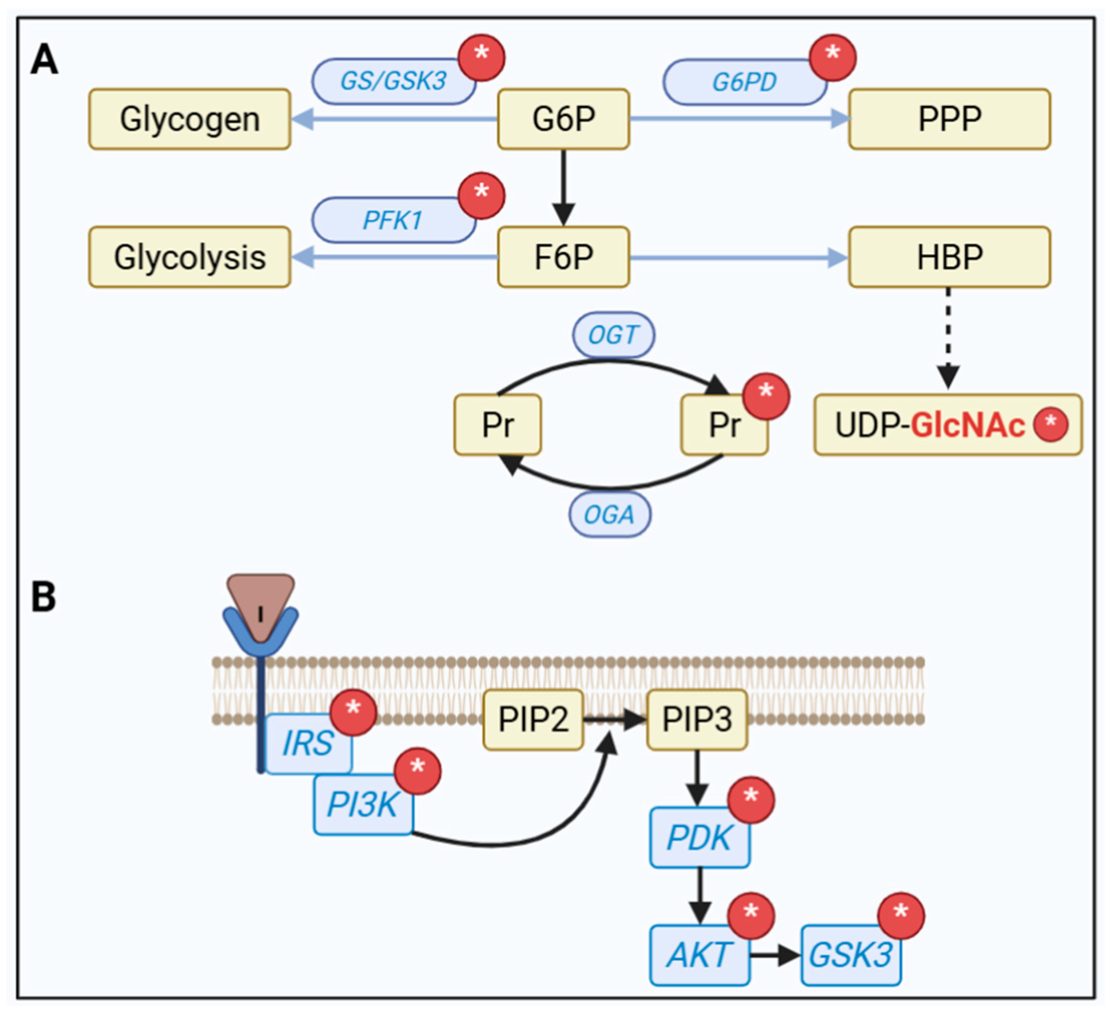
At the beginning of both AD and/or ED, oxidative distress may demand a greater flow of the pentose phosphate pathway (PPP) to produce and recycle GSH (see Figure 2). However, in more advanced stages, this system may be compromised, as indicated by reduced activity of glucose-6-phosphate dehydrogenase (G6PD) [45]. On the other hand, glycogen synthesis may be reduced from the onset of AD, possibly due to an alteration in the AKT/GSK-3 communication of insulin signaling, and this per se contributes to cognitive alterations, with impairment of learning and memory [46].
In addition to the glycolytic pathway and the two routes mentioned above, the hexosamine biosynthesis pathway (HBP), although a quantitatively small destination, has a regulatory effect on the other pathways. The glucose pathway that produces UDP-GlcNAc, used for post-translational modification of O-GlcNAcylation (or simply NAGylation) on Ser or Thr residues [43], is also reduced in DM and AD, possibly reflecting the lower availability of glucose [47]. However, we know relatively little about the diverse significance of specific protein modifications by NAGylation and the crosstalk with phosphorylation, particularly in brain tissue. This is an important aspect for consideration, especially given our observation, in a model of dementia with streptozotocin, that the reduction in NAGylation apparently precedes insulin resistance, as indicated by the phosphorylation of IRS-1 at Ser309 [48]. Indeed, several enzymes in the insulin signaling pathway and key enzymes for glucose metabolization pathways, such as glycogen synthase, glucose-6P dehydrogenase and phosphofrutokinase-1, are NAGylated (see Figure 2).
As a consequence, changes in intracellular glucose flux result in increased glycolytic activity with increased lactate and MG production. Accordingly, AD patients demonstrate increased lactate in their cerebrospinal fluid, as well as a reduction in pyruvate levels [49]. Notably, pyruvate is also used to produce cholesterol for exportation to neurons. Although we know that increased neuronal cholesterol worsens disease progression, it seems logical that this is due to the neuron’s inability to perform clearance (via cytochrome P450 46A1), and not to the increase in astroglial cholesterol synthesis [50]. In summary, the greater glycolytic flux (in this hypometabolic condition) may also result in greater MG production, as confirmed by the increased activity of the glyoxalase system [51].

7. Pharmacological and Nonpharmacological Strategies for Controlling MG

Given the central role of the central glyoxalase system in MG detoxification, with GLO-1 as the rate-limiting step, pharmacological neuroprotective strategies focus on GLO-1 activators (by increasing the activity and/or expression of the enzyme) and scavengers that complement the antioxidant activity of the system. Drugs such as metformin [52,53,54], aminoguanidine [55,56,57], simvastatin [58], and glucagon-like peptide-1 agonists [59,60,61] have been identified as GLO-1 activators. Scavengers include resveratrol [62,63,64], sulforaphane [65,66], curcumin [67], glycine [68,69], pyridoxamine [70] and flavonoids such as hesperetin [71,72] and quercetin [73,74]. It is worth noting that most studies with these compounds did not directly evaluate the expression, content, or activity of GLO-1 in neural cells. However, quercetin positively modulated GLO-1 in both cerebellar neuronal culture [73] and in the cerebral cortex of STZ-induced diabetic rats [74].
Nonpharmacological approaches, such as physical exercise and diet, modulate energy metabolism and antioxidant defense. Physical exercise has been identified as a modulator of Nrf2, increasing GSH and GLO-1 levels and contributing to MG detoxification [75]. A study with mice subjected to treadmill exercise observed a direct increase in the GLO-1 and GLO-2 enzymes in the cerebral cortex [76] and GLO-1 in the hippocampus [77]. In humans, after running exercise, a decrease in MG levels and an increase in D-lactate levels in the blood were observed [78]. With regard to diet, caloric restriction and ketogenic diets are also reported to increase the expression of Nrf2 [79,80].

8. Final Comments and Perspectives

There is no doubt that insulin resistance, as observed in metabolic syndrome, accompanies and/or underlies DE and AD [81]. Note that insulin resistance is also observed in major depressive disorder [82], another condition considered to be a risk factor for AD.
Glucose hypometabolism is a consequence of insulin resistance and results in a metabolic rearrangement that expands the glycolytic pathway and generates more MG. MG, in turn, can affect insulin signaling, further compromising the molecular basis of insulin resistance and creating a vicious cycle. Astrocytes are key cells in the generation and detoxification of MG in the brain, making them a therapeutic target.
Although several biomarkers have been proposed for monitoring the course of DM, fasting plasma glucose and glycated hemoglobin (HbA1c) are still the biomarkers of choice for DM. However, considering the limitations of using HbA1c in cases of low Hb (anemia) or accelerated red blood cell turnover (due to chronic kidney disease), MG, due to its close relationship with insulin resistance, must be considered as a potential diagnostic and prognostic marker, not limited to DM [83]. However, methodological approaches and measurement variability are still important difficulties [84].
MG, due to its high capacity to generate AGEs (sometimes referred to as gerontoxins) [85] and the activation of the UPR, is a key element in the basis of neurodegenerative diseases and aging. Taken together with the data available on its production and detoxification, we propose that while under physiological conditions, MG may stimulate antioxidant defenses via Nrf2, chronic exposure may lead to continuous activation of the NFkB pathway, both intracellularly via the UPR, and extracellularly via RAGE.
Finally, more recently, the direct effects of MG on GABA receptors [86] and the dopamine pathway [27] have been proposed and investigated, which could justify the alterations observed in neuropsychiatric disorders, and may be independent of a degenerative basis.

Author Contributions

Conceptualization, C.-A.G., V.G.-D. and V.-F.D.S.; writing—original draft preparation, C.-A.G., V.G.-D. and V.-F.D.S.; writing—review and editing, C.-A.G., V.G.-D., V.-F.D.S., T.M.S., M.C.L. and A.F.K.V. All authors have read and agreed to the published version of the manuscript.

Funding

This research was funded by Conselho Nacional de Desenvolvimento Científico Tecnológico (CNPq), Coordenação de Aperfeiçoamento de Pessoal de Nível Superior (CAPES), Fundação de Amparo à Pesquisa do Estado do Rio Grande do Sul (FAPERGS), and the Instituto Nacional de Ciência e Tecnologia for Excitotoxicity and Neuroprotection (INCTEN, 465671/2014-4).

Institutional Review Board Statement

Not applicable.

Informed Consent Statement

Not applicable.

Data Availability Statement

The raw data supporting the conclusions of this article will be made available by the authors on request.

Conflicts of Interest

The authors declare no conflicts of interest.

Abbreviations

The following abbreviations are used in this manuscript:
ADAlzheimer’s disease
AGEsadvanced glycation end products
ATF6activating transcription factor 6
CDK5cyclin-dependent kinase 5
CYP2E1cytochrome P450 oxidase 2E1
DEdiabetic encephalopathy
DHAPdihydroxyacetone phosphate
DMdiabetes mellitus
G6PDglucose-6-phosphate dehydrogenase
GA3Pglyceraldehyde 3-phosphate
GAPDglyceraldehyde 3-phosphate dehydrogenase
GCLglutamate-cysteine ligase
GLO1glyoxalase 1
GLO2glyoxalase 2
GSHglutathione
GSK-3βglycogen synthase kinase 3β
HBPhexosamine biosynthesis pathway
IRE1αinositol-requiring transmembrane kinase/endoribonuclease 1α
LPSlipopolysaccharide
MAPKmitogen-activated protein kinase
MGmethyliglyoxal
NFκBnuclear factor kappa B
NOXNADPH oxidase
Nrf2nuclear factor erythroid 2-related factor 2
PERKPKR-like ER kinase
PPPpentose phosphate pathway
RAGEadvanced glycation end products receptor
ROSreactive oxygen species
S100BS100 calcium binding protein B
UPRunfolded protein response

References

  1. Li, W.Y.; Lee, C.Y.; Lee, K.M.; Zhang, G.; Lyu, A.; Yue, K.K.M. Advanced Glycation End-Product Precursor Methylglyoxal May Lead to Development of Alzheimer’s Disease. Diabetes Metab. Syndr. Obes. 2022, 15, 3153–3166. [Google Scholar] [CrossRef] [PubMed]
  2. Allaman, I.; Bélanger, M.; Magistretti, P.J. Methylglyoxal, the dark side of glycolysis. Front. Neurosci. 2015, 9, 23. [Google Scholar] [CrossRef]
  3. Hansen, F.; Galland, F.; Lirio, F.; De Souza, D.F.; Da Ré, C.; Pacheco, R.F.; Vizuete, A.F.; Quincozes-Santos, A.; Leite, M.C.; Gonçalves, C.-A. Methylglyoxal Induces Changes in the Glyoxalase System and Impairs Glutamate Uptake Activity in Primary Astrocytes. Oxidative Med. Cell Longev. 2017, 2017, 9574201. [Google Scholar] [CrossRef] [PubMed]
  4. Moreira, A.P.; Vizuete, A.F.K.; Zin, L.E.F.; de Marques, C.O.; Pacheco, R.F.; Leal, M.B.; Gonçalves, C.-A. The Methylglyoxal/RAGE/NOX-2 Pathway is Persistently Activated in the Hippocampus of Rats with STZ-Induced Sporadic Alzheimer’s Disease. Neurotox. Res. 2022, 40, 395–409. [Google Scholar] [CrossRef] [PubMed]
  5. Donnellan, L.; Fenech, M.; Dhillon, V.S.; Young, C.; Hoffmann, P.; Deo, P. Role of methylglyoxal protein modifications in DNA damage and chromosomal instability: Emerging molecular mechanisms. Mutat. Res. Rev. Mutat. Res. 2025, 796, 108558. [Google Scholar] [CrossRef]
  6. Kalapos, M.P. The tandem of free radicals and methylglyoxal. Chem. Biol. Interact. 2008, 171, 251–271. [Google Scholar] [CrossRef]
  7. Rabbani, N.; Xue, M.; Thornalley, P.J. Dicarbonyls and glyoxalase in disease mechanisms and clinical therapeutics. Glycoconj. J. 2016, 33, 513–525. [Google Scholar] [CrossRef] [PubMed]
  8. Vašková, J.; Kováčová, G.; Pudelský, J.; Palenčár, D.; Mičková, H. Methylglyoxal Formation-Metabolic Routes and Consequences. Antioxidants 2025, 14, 212. [Google Scholar] [CrossRef]
  9. Mattson, M.P. Hormesis defined. Ageing Res. Rev. 2008, 7, 1–7. [Google Scholar] [CrossRef]
  10. Barros, L.F.; Schirmeier, S.; Weber, B. The Astrocyte: Metabolic Hub of the Brain. Cold Spring Harb. Perspect. Biol. 2024, 16, a041355. [Google Scholar] [CrossRef]
  11. Dringen, R.; Arend, C. Glutathione Metabolism of the Brain-The Role of Astrocytes. J. Neurochem. 2025, 169, e70073. [Google Scholar] [CrossRef]
  12. Bélanger, M.; Yang, J.; Petit, J.-M.; Laroche, T.; Magistretti, P.J.; Allaman, I. Role of the glyoxalase system in astrocyte-mediated neuroprotection. J. Neurosci. 2011, 31, 18338–18352. [Google Scholar] [CrossRef] [PubMed]
  13. de Bari, L.; Atlante, A.; Armeni, T.; Kalapos, M.P. Synthesis and metabolism of methylglyoxal, S-D-lactoylglutathione and D-lactate in cancer and Alzheimer’s disease. Exploring the crossroad of eternal youth and premature aging. Ageing Res. Rev. 2019, 53, 100915. [Google Scholar] [CrossRef] [PubMed]
  14. Wątroba, M.; Grabowska, A.D.; Szukiewicz, D. Effects of Diabetes Mellitus-Related Dysglycemia on the Functions of Blood-Brain Barrier and the Risk of Dementia. Int. J. Mol. Sci. 2023, 24, 10069. [Google Scholar] [CrossRef]
  15. Ahmed, N.; Ahmed, U.; Thornalley, P.J.; Hager, K.; Fleischer, G.; Münch, G. Protein glycation, oxidation and nitration adduct residues and free adducts of cerebrospinal fluid in Alzheimer’s disease and link to cognitive impairment. J. Neurochem. 2005, 92, 255–263. [Google Scholar] [CrossRef]
  16. Yan, S.D.; Chen, X.; Fu, J.; Chen, M.; Zhu, H.; Roher, A.; Slattery, T.; Zhao, L.; Nagashima, M.; Morser, J.; et al. RAGE and amyloid-beta peptide neurotoxicity in Alzheimer’s disease. Nature 1996, 382, 685–691. [Google Scholar] [CrossRef]
  17. Leerach, N.; Harashima, A.; Munesue, S.; Kimura, K.; Oshima, Y.; Goto, H.; Yamamoto, H.; Higashida, H.; Yamamoto, Y. Glycation reaction and the role of the receptor for advanced glycation end-products in immunity and social behavior. Glycoconj. J. 2021, 38, 303–310. [Google Scholar] [CrossRef]
  18. Pugazhenthi, S.; Qin, L.; Reddy, P.H. Common neurodegenerative pathways in obesity, diabetes, and Alzheimer’s disease. Biochim. Biophys. Acta Mol. Basis Dis. 2017, 1863, 1037–1045. [Google Scholar] [CrossRef]
  19. Tang, H.; Ma, T.; Wang, Y.; Zhang, C.; Chu, Y.; Guo, Y.; Xi, J.; Jiao, D.; Li, B.; Xie, C.; et al. Paeoniflorin modulates AGEs/RAGE/P38MAPK/ERK/mTOR autophagy pathway to improve cognitive dysfunction in MRL/lpr mice. Int. J. Biol. Macromol. 2025, 307, 141765. [Google Scholar] [CrossRef] [PubMed]
  20. Berends, E.; van Oostenbrugge, R.J.; Foulquier, S.; Schalkwijk, C.G. Methylglyoxal, a highly reactive dicarbonyl compound, as a threat for blood brain barrier integrity. Fluids Barriers CNS 2023, 20, 75. [Google Scholar] [CrossRef]
  21. Vizuete, A.F.K.; Gonçalves, C.-A. Is Methylglyoxal a Potential Biomarker for the Warburg Effect Induced by the Lipopolysaccharide Neuroinflammation Model? Neurochem. Res. 2024, 49, 1823–1837. [Google Scholar] [CrossRef]
  22. Subedi, L.; Lee, J.H.; Gaire, B.P.; Kim, S.Y. Sulforaphane Inhibits MGO-AGE-Mediated Neuroinflammation by Suppressing NF-κB, MAPK, and AGE-RAGE Signaling Pathways in Microglial Cells. Antioxidants 2020, 9, 792. [Google Scholar] [CrossRef]
  23. Vizuete, A.F.K.; Fróes, F.; Seady, M.; Hansen, F.; Ligabue-Braun, R.; Gonçalves, C.-A.; Souza, D.O. A Mechanism of Action of Metformin in the Brain: Prevention of Methylglyoxal-Induced Glutamatergic Impairment in Acute Hippocampal Slices. Mol. Neurobiol. 2024, 61, 3223–3239. [Google Scholar] [CrossRef]
  24. Lissner, L.J.; Wartchow, K.M.; Rodrigues, L.; Bobermin, L.D.; Borba, E.; Dias, V.G.; Hansen, F.; Quincozes-Santos, A.; Gonçalves, C.-A. Acute Methylglyoxal-Induced Damage in Blood-Brain Barrier and Hippocampal Tissue. Neurotox. Res. 2022, 40, 1337–1347. [Google Scholar] [CrossRef] [PubMed]
  25. Ko, S.-Y.; Lin, Y.-P.; Lin, Y.-S.; Chang, S.-S. Advanced glycation end products enhance amyloid precursor protein expression by inducing reactive oxygen species. Free Radic. Biol. Med. 2010, 49, 474–480. [Google Scholar] [CrossRef]
  26. More, S.S.; Vince, R. Potential of a γ-glutamyl-transpeptidase-stable glutathione analogue against amyloid-β toxicity. ACS Chem. Neurosci. 2012, 3, 204–210. [Google Scholar] [CrossRef]
  27. de Almeida, G.R.L.; Szczepanik, J.C.; Selhorst, I.; Cunha, M.P.; Dafre, A.L. The expanding impact of methylglyoxal on behavior-related disorders. Prog. Neuropsychopharmacol. Biol. Psychiatry 2023, 120, 110635. [Google Scholar] [CrossRef] [PubMed]
  28. Samanta, S.; Akhter, F.; Xue, R.; Sosunov, A.; Wu, L.; Chen, D.; Arancio, O.; Yan, S.F.; Yan, S.S. Synaptic mitochondria glycation contributes to mitochondrial stress and cognitive dysfunction. Brain 2025, 148, 262–275. [Google Scholar] [CrossRef]
  29. Hansen, F.; Battú, C.E.; Dutra, M.F.; Galland, F.; Lirio, F.; Broetto, N.; Nardin, P.; Gonçalves, C.-A. Methylglyoxal and carboxyethyllysine reduce glutamate uptake and S100B secretion in the hippocampus independently of RAGE activation. Amino Acids 2016, 48, 375–385. [Google Scholar] [CrossRef]
  30. Prantner, D.; Nallar, S.; Richard, K.; Spiegel, D.; Collins, K.D.; Vogel, S.N. Classically activated mouse macrophages produce methylglyoxal that induces a TLR4- and RAGE-independent proinflammatory response. J. Leukoc. Biol. 2021, 109, 605–619. [Google Scholar] [CrossRef] [PubMed]
  31. Distler, M.G.; Plant, L.D.; Sokoloff, G.; Hawk, A.J.; Aneas, I.; Wuenschell, G.E.; Termini, J.; Meredith, S.C.; Nobrega, M.A.; Palmer, A.A. Glyoxalase 1 increases anxiety by reducing GABAA receptor agonist methylglyoxal. J. Clin. Investig. 2012, 122, 2306–2315. [Google Scholar] [CrossRef] [PubMed]
  32. Vizuete, A.F.K.; Hansen, F.; Da Ré, C.; Leal, M.B.; Galland, F.; Leite, M.C.; Gonçalves, C.-A. GABAA Modulation of S100B Secretion in Acute Hippocampal Slices and Astrocyte Cultures. Neurochem. Res. 2019, 44, 301–311. [Google Scholar] [CrossRef] [PubMed]
  33. Sims, S.G.; Cisney, R.N.; Lipscomb, M.M.; Meares, G.P. The role of endoplasmic reticulum stress in astrocytes. Glia 2022, 70, 5–19. [Google Scholar] [CrossRef]
  34. Rabbani, N.; Xue, M.; Thornalley, P.J. Dicarbonyl stress, protein glycation and the unfolded protein response. Glycoconj. J. 2021, 38, 331–340. [Google Scholar] [CrossRef] [PubMed]
  35. Hemagirri, M.; Chen, Y.; Gopinath, S.C.B.; Sahreen, S.; Adnan, M.; Sasidharan, S. Crosstalk between protein misfolding and endoplasmic reticulum stress during ageing and their role in age-related disorders. Biochimie 2024, 221, 159–181. [Google Scholar] [CrossRef]
  36. Palsamy, P.; Bidasee, K.R.; Ayaki, M.; Augusteyn, R.C.; Chan, J.Y.; Shinohara, T. Methylglyoxal induces endoplasmic reticulum stress and DNA demethylation in the Keap1 promoter of human lens epithelial cells and age-related cataracts. Free Radic. Biol. Med. 2014, 72, 134–148. [Google Scholar] [CrossRef]
  37. Xue, M.; Irshad, Z.; Rabbani, N.; Thornalley, P.J. Increased cellular protein modification by methylglyoxal activates endoplasmic reticulum-based sensors of the unfolded protein response. Redox Biol. 2024, 69, 103025. [Google Scholar] [CrossRef]
  38. Bollong, M.J.; Lee, G.; Coukos, J.S.; Yun, H.; Zambaldo, C.; Chang, J.W.; Chin, E.N.; Ahmad, I.; Chatterjee, A.K.; Lairson, L.L.; et al. A metabolite-derived protein modification integrates glycolysis with KEAP1–NRF2 signalling. Nature 2018, 562, 600–604. [Google Scholar] [CrossRef]
  39. Liu, T.; Lv, Y.-F.; Zhao, J.-L.; You, Q.-D.; Jiang, Z.-Y. Regulation of Nrf2 by phosphorylation: Consequences for biological function and therapeutic implications. Free Radic. Biol. Med. 2021, 168, 129–141. [Google Scholar] [CrossRef]
  40. Schultz, B.; Taday, J.; Menezes, L.; Cigerce, A.; Leite, M.C.; Gonçalves, C.-A. Calpain-Mediated Alterations in Astrocytes Before and During Amyloid Chaos in Alzheimer’s Disease. J. Alzheimers Dis. 2021, 84, 1415–1430. [Google Scholar] [CrossRef]
  41. Jimenez-Blasco, D.; Santofimia-Castaño, P.; Gonzalez, A.; Almeida, A.; Bolaños, J.P. Astrocyte NMDA receptors’ activity sustains neuronal survival through a Cdk5-Nrf2 pathway. Cell Death Differ. 2015, 22, 1877–1889. [Google Scholar] [CrossRef]
  42. Soltani, S.; Dolatshahi, M.; Soltani, S.; Khazaei, K.; Rahmani, M.; Raji, C.A. Relationships Between Brain Glucose Metabolism Patterns and Impaired Glycemic Status: A Systematic Review of FDG-PET Studies With a Focus on Alzheimer’s Disease. Hum. Brain Mapp. 2025, 46, e70180. [Google Scholar] [CrossRef]
  43. Gonçalves, C.-A.; Rodrigues, L.; Bobermin, L.D.; Zanotto, C.; Vizuete, A.; Quincozes-Santos, A.; Souza, D.O.; Leite, M.C. Glycolysis-Derived Compounds From Astrocytes That Modulate Synaptic Communication. Front. Neurosci. 2018, 12, 1035. [Google Scholar] [CrossRef]
  44. Escartin, C.; Guillemaud, O.; Carrillo-de Sauvage, M.-A. Questions and (some) answers on reactive astrocytes. Glia 2019, 67, 2221–2247. [Google Scholar] [CrossRef] [PubMed]
  45. Sharallah, O.A.; Poddar, N.K.; Alwadan, O.A. Delineation of the role of G6PD in Alzheimer’s disease and potential enhancement through microfluidic and nanoparticle approaches. Ageing Res. Rev. 2024, 99, 102394. [Google Scholar] [CrossRef]
  46. Bak, L.K.; Walls, A.B.; Schousboe, A.; Waagepetersen, H.S. Astrocytic glycogen metabolism in the healthy and diseased brain. J. Biol. Chem. 2018, 293, 7108–7116. [Google Scholar] [CrossRef] [PubMed]
  47. Yuan, Y.; Zhao, G.; Zhao, Y. Dysregulation of energy metabolism in Alzheimer’s disease. J. Neurol. 2024, 272, 2. [Google Scholar] [CrossRef]
  48. Dos Santos, J.P.A.; Vizuete, A.; Hansen, F.; Biasibetti, R.; Gonçalves, C.-A. Early and Persistent O-GlcNAc Protein Modification in the Streptozotocin Model of Alzheimer’s Disease. J. Alzheimers Dis. 2018, 61, 237–249. [Google Scholar] [CrossRef] [PubMed]
  49. Manyevitch, R.; Protas, M.; Scarpiello, S.; Deliso, M.; Bass, B.; Nanajian, A.; Chang, M.; Thompson, S.M.; Khoury, N.; Gonnella, R.; et al. Evaluation of Metabolic and Synaptic Dysfunction Hypotheses of Alzheimer’s Disease (AD): A Meta-Analysis of CSF Markers. Curr. Alzheimer Res. 2018, 15, 164–181. [Google Scholar] [CrossRef]
  50. Ahmed, H.; Wang, Y.; Griffiths, W.J.; Levey, A.; Pikuleva, I.; Liang, S.H.; Haider, A. Brain cholesterol and Alzheimer’s disease: Challenges and opportunities in probe and drug development. Brain 2024, 147, 1622–1635. [Google Scholar] [CrossRef]
  51. Haddad, M.; Perrotte, M.; Ben Khedher, M.R.; Madec, E.; Lepage, A.; Fülöp, T.; Ramassamy, C. Levels of Receptor for Advanced Glycation End Products and Glyoxalase-1 in the Total Circulating Extracellular Vesicles from Mild Cognitive Impairment and Different Stages of Alzheimer’s Disease Patients. J. Alzheimers Dis. 2021, 84, 227–237. [Google Scholar] [CrossRef]
  52. Peters, A.S.; Wortmann, M.; Fleming, T.H.; Nawroth, P.P.; Bruckner, T.; Böckler, D.; Hakimi, M. Effect of metformin treatment in patients with type 2 diabetes with respect to glyoxalase 1 activity in atherosclerotic lesions. Vasa 2019, 48, 186–192. [Google Scholar] [CrossRef] [PubMed]
  53. Oliveira, A.L.; de Oliveira, M.G.; Medeiros, M.L.; Mónica, F.Z.; Antunes, E. Metformin abrogates the voiding dysfunction induced by prolonged methylglyoxal intake. Eur. J. Pharmacol. 2021, 910, 174502. [Google Scholar] [CrossRef]
  54. Kender, Z.; Fleming, T.; Kopf, S.; Torzsa, P.; Grolmusz, V.; Herzig, S.; Schleicher, E.; Rácz, K.; Reismann, P.; Nawroth, P.P. Effect of metformin on methylglyoxal metabolism in patients with type 2 diabetes. Exp. Clin. Endocrinol. Diabetes 2014, 122, 316–319. [Google Scholar] [CrossRef]
  55. Nikray, N.; Abharian, N.; Jafari Ashtiani, S.; Kobarfard, F.; Faizi, M. Comparative Evaluation of Aminoguanidine, Semicarbazide and Thiosemicarbazide Treatment for Methylglyoxal-Induced Neurological Toxicity in Experimental Models. Iran. J. Pharm. Res. 2024, 23, e153322. [Google Scholar] [CrossRef] [PubMed]
  56. Lo, T.W.; Selwood, T.; Thornalley, P.J. The reaction of methylglyoxal with aminoguanidine under physiological conditions and prevention of methylglyoxal binding to plasma proteins. Biochem. Pharmacol. 1994, 48, 1865–1870. [Google Scholar] [CrossRef] [PubMed]
  57. Webster, J.; Urban, C.; Berbaum, K.; Loske, C.; Alpar, A.; Gärtner, U.; De Arriba, S.G.; Arendt, T.; Münch, G. The carbonyl scavengers aminoguanidine and tenilsetam protect against the neurotoxic effects of methylglyoxal. Neurotox. Res. 2005, 7, 95–101. [Google Scholar] [CrossRef]
  58. Bobermin, L.D.; Sesterheim, P.; da Costa, D.S.; Rezena, E.; Schmitz, I.; da Silva, A.; de Moraes, A.D.M.; Souza, D.O.; Wyse, A.T.; Leipnitz, G.; et al. Simvastatin Differentially Modulates Glial Functions in Cultured Cortical and Hypothalamic Astrocytes Derived from Interferon α/β Receptor Knockout mice. Neurochem. Res. 2024, 49, 732–743. [Google Scholar] [CrossRef]
  59. Zanotto, C.; Hansen, F.; Galland, F.; Batassini, C.; Federhen, B.C.; da Silva, V.F.; Leite, M.C.; Nardin, P.; Gonçalves, C.-A. Glutamatergic Alterations in STZ-Induced Diabetic Rats Are Reversed by Exendin-4. Mol. Neurobiol. 2019, 56, 3538–3551. [Google Scholar] [CrossRef]
  60. Rodrigues, T.; Borges, P.; Mar, L.; Marques, D.; Albano, M.; Eickhoff, H.; Carrêlo, C.; Almeida, B.; Pires, S.; Abrantes, M.; et al. GLP-1 improves adipose tissue glyoxalase activity and capillarization improving insulin sensitivity in type 2 diabetes. Pharmacol. Res. 2020, 161, 105198. [Google Scholar] [CrossRef]
  61. Tseng, Y.-T.; Tsai, Y.-H.; Fülöp, F.; Chang, F.-R.; Lo, Y.-C. 2-Iodo-4’-Methoxychalcone Attenuates Methylglyoxal-Induced Neurotoxicity by Activation of GLP-1 Receptor and Enhancement of Neurotrophic Signal, Antioxidant Defense and Glyoxalase Pathway. Molecules 2019, 24, 2249. [Google Scholar] [CrossRef] [PubMed]
  62. Sovrani, V.; Bobermin, L.D.; Sesterheim, P.; Rezena, E.; Cioccari, M.S.; Netto, C.A.; Gonçalves, C.-A.; Leipnitz, G.; Quincozes-Santos, A. Glioprotective effects of resveratrol in hypothalamic astrocyte cultures obtained from interferon receptor knockout (IFNα/βR−/−) mice. In Vitr. Cell Dev. Biol. Anim. 2023, 59, 366–380. [Google Scholar] [CrossRef]
  63. Quincozes-Santos, A.; Bobermin, L.D.; Tramontina, A.C.; Wartchow, K.M.; Da Silva, V.-F.; Gayger-Dias, V.; Thomaz, N.K.; de Moraes, A.D.M.; Schauren, D.; Nardin, P.; et al. Glioprotective Effects of Resveratrol Against Glutamate-Induced Cellular Dysfunction: The Role of Heme Oxygenase 1 Pathway. Neurotox. Res. 2025, 43, 7. [Google Scholar] [CrossRef]
  64. Santini, S.J.; Cordone, V.; Mijit, M.; Bignotti, V.; Aimola, P.; Dolo, V.; Falone, S.; Amicarelli, F. SIRT1-Dependent Upregulation of Antiglycative Defense in HUVECs Is Essential for Resveratrol Protection against High Glucose Stress. Antioxidants 2019, 8, 346. [Google Scholar] [CrossRef]
  65. Alfarano, M.; Pastore, D.; Fogliano, V.; Schalkwijk, C.G.; Oliviero, T. The Effect of Sulforaphane on Glyoxalase I Expression and Activity in Peripheral Blood Mononuclear Cells. Nutrients 2018, 10, 1773. [Google Scholar] [CrossRef]
  66. Bobermin, L.D.; Weber, F.B.; Dos Santos, T.M.; Belló-Klein, A.; Wyse, A.T.S.; Gonçalves, C.-A.; Quincozes-Santos, A. Sulforaphane Induces Glioprotection After LPS Challenge. Cell Mol. Neurobiol. 2022, 42, 829–846. [Google Scholar] [CrossRef] [PubMed]
  67. Lima, T.F.O.; Costa, M.C.; Figueiredo, I.D.; Inácio, M.D.; Rodrigues, M.R.; Assis, R.P.; Baviera, A.M.; Brunetti, I.L. Curcumin, Alone or in Combination with Aminoguanidine, Increases Antioxidant Defenses and Glycation Product Detoxification in Streptozotocin-Diabetic Rats: A Therapeutic Strategy to Mitigate Glycoxidative Stress. Oxidative Med. Cell Longev. 2020, 2020, 1036360. [Google Scholar] [CrossRef] [PubMed]
  68. Wang, Z.; Zhang, J.; Chen, L.; Li, J.; Zhang, H.; Guo, X. Glycine Suppresses AGE/RAGE Signaling Pathway and Subsequent Oxidative Stress by Restoring Glo1 Function in the Aorta of Diabetic Rats and in HUVECs. Oxidative Med. Cell Longev. 2019, 2019, 4628962. [Google Scholar] [CrossRef] [PubMed]
  69. Wang, Z.; Zhao, D.; Chen, L.; Li, J.; Yuan, G.; Yang, G.; Zhang, H.; Guo, X.; Zhang, J. Glycine increases glyoxalase-1 function by promoting nuclear factor erythroid 2-related factor 2 translocation into the nucleus of kidney cells of streptozotocin-induced diabetic rats. J. Diabetes Investig. 2019, 10, 1189–1198. [Google Scholar] [CrossRef] [PubMed]
  70. Jiang, M.; Yakupu, A.; Guan, H.; Dong, J.; Liu, Y.; Song, F.; Tang, J.; Tian, M.; Niu, Y.; Lu, S. Pyridoxamine ameliorates methylglyoxal-induced macrophage dysfunction to facilitate tissue repair in diabetic wounds. Int. Wound J. 2022, 19, 52–63. [Google Scholar] [CrossRef]
  71. Chen, Y.-J.; Kong, L.; Tang, Z.-Z.; Zhang, Y.-M.; Liu, Y.; Wang, T.-Y.; Liu, Y.-W. Hesperetin ameliorates diabetic nephropathy in rats by activating Nrf2/ARE/glyoxalase 1 pathway. Biomed. Pharmacother. 2019, 111, 1166–1175. [Google Scholar] [CrossRef]
  72. Zhu, X.; Zhang, Y.-M.; Zhang, M.-Y.; Chen, Y.-J.; Liu, Y.-W. Hesperetin ameliorates diabetes-associated anxiety and depression-like behaviors in rats via activating Nrf2/ARE pathway. Metab. Brain Dis. 2021, 36, 1969–1983. [Google Scholar] [CrossRef]
  73. Frandsen, J.; Narayanasamy, P. Flavonoid Enhances the Glyoxalase Pathway in Cerebellar Neurons to Retain Cellular Functions. Sci. Rep. 2017, 7, 5126. [Google Scholar] [CrossRef]
  74. Zhu, X.; Cheng, Y.-Q.; Lu, Q.; Du, L.; Yin, X.-X.; Liu, Y.-W. Enhancement of glyoxalase 1, a polyfunctional defense enzyme, by quercetin in the brain in streptozotocin-induced diabetic rats. Naunyn Schmiedebergs Arch. Pharmacol. 2018, 391, 1237–1245. [Google Scholar] [CrossRef] [PubMed]
  75. Dieter, B.P.; Vella, C.A. A proposed mechanism for exercise attenuated methylglyoxal accumulation: Activation of the ARE-Nrf pathway and increased glutathione biosynthesis. Med. Hypotheses 2013, 81, 813–815. [Google Scholar] [CrossRef]
  76. Falone, S.; D’Alessandro, A.; Mirabilio, A.; Petruccelli, G.; Cacchio, M.; Di Ilio, C.; Di Loreto, S.; Amicarelli, F. Long term running biphasically improves methylglyoxal-related metabolism, redox homeostasis and neurotrophic support within adult mouse brain cortex. PLoS ONE 2012, 7, e31401. [Google Scholar] [CrossRef]
  77. Falone, S.; D’Alessandro, A.; Mirabilio, A.; Cacchio, M.; Di Ilio, C.; Di Loreto, S.; Amicarelli, F. Late-onset running biphasically improves redox balance, energy- and methylglyoxal-related status, as well as SIRT1 expression in mouse hippocampus. PLoS ONE 2012, 7, e48334. [Google Scholar] [CrossRef]
  78. Kondoh, Y.; Kawase, M.; Ohmori, S. D-lactate concentrations in blood, urine and sweat before and after exercise. Eur. J. Appl. Physiol. Occup. Physiol. 1992, 65, 88–93. [Google Scholar] [CrossRef] [PubMed]
  79. Ildarabadi, A.; Mir Mohammad Ali, S.N.; Rahmani, F.; Mosavari, N.; Pourbakhtyaran, E.; Rezaei, N. Inflammation and oxidative stress in epileptic children: From molecular mechanisms to clinical application of ketogenic diet. Rev. Neurosci. 2024, 35, 473–488. [Google Scholar] [CrossRef]
  80. Sharma, A.; Singh, A.K. Molecular mechanism of caloric restriction mimetics-mediated neuroprotection of age-related neurodegenerative diseases: An emerging therapeutic approach. Biogerontology 2023, 24, 679–708. [Google Scholar] [CrossRef] [PubMed]
  81. Ezkurdia, A.; Ramírez, M.J.; Solas, M. Metabolic Syndrome as a Risk Factor for Alzheimer’s Disease: A Focus on Insulin Resistance. Int. J. Mol. Sci. 2023, 24, 4354. [Google Scholar] [CrossRef]
  82. Meng, F.; Wang, J.; Wang, L.; Zou, W. Glucose metabolism impairment in major depressive disorder. Brain Res. Bull. 2025, 221, 111191. [Google Scholar] [CrossRef]
  83. Beisswenger, P.J. Methylglyoxal in diabetes: Link to treatment, glycaemic control and biomarkers of complications. Biochem. Soc. Trans. 2014, 42, 450–456. [Google Scholar] [CrossRef] [PubMed]
  84. Ramachandra Bhat, L.; Vedantham, S.; Krishnan, U.M.; Rayappan, J.B.B. Methylglyoxal—An emerging biomarker for diabetes mellitus diagnosis and its detection methods. Biosens. Bioelectron. 2019, 133, 107–124. [Google Scholar] [CrossRef] [PubMed]
  85. Krautwald, M.; Münch, G. Advanced glycation end products as biomarkers and gerontotoxins—A basis to explore methylglyoxal-lowering agents for Alzheimer’s disease? Exp. Gerontol. 2010, 45, 744–751. [Google Scholar] [CrossRef] [PubMed]
  86. McMurray, K.M.J.; Ramaker, M.J.; Barkley-Levenson, A.M.; Sidhu, P.S.; Elkin, P.K.; Reddy, M.K.; Guthrie, M.L.; Cook, J.M.; Rawal, V.H.; A Arnold, L.; et al. Identification of a novel, fast-acting GABAergic antidepressant. Mol. Psychiatry 2018, 23, 384–391. [Google Scholar] [CrossRef]
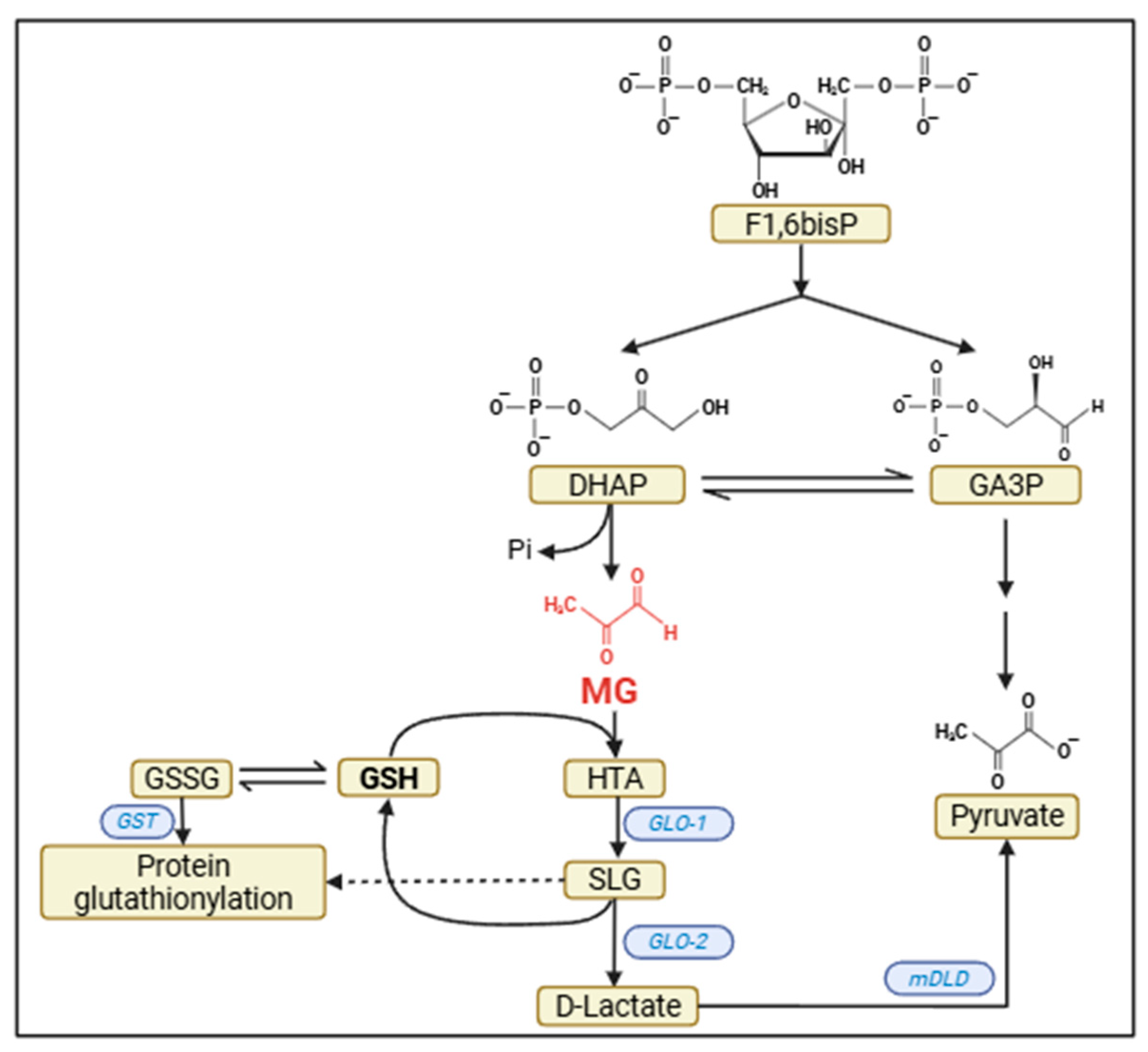
Figure 1. Origin and detoxification of MG. In the glycolytic pathway, fructose-1,6-bisphosphate (F1,6bisP) is cleaved to dihydroxyacetone phosphate (DHAP) and glyceraldehyde-3P (GA3P). Part of DHAP is spontaneously dephosphorylated and isomerized to methylglyoxal (MG). MG reacts with glutathione (GSH) to form hemithioacetal (HTA). HTA, in turn, in a reaction catalyzed by glyoxalase-1 (GLO-1), produces S-lactoyl-glutathione (SLG). SLG, in a reaction catalyzed by glyoxalase-2 (GLO-2), produces D-lactate and regenerates GSH. D-lactate is converted to pyruvate, delivering its electrons directly to the respiratory chain, in a reaction catalyzed by mitochondrial D-lactate dehydrogenase (mDLD). Note that oxidized glutathione (GSSG) can, spontaneously and through the action of GST (glutathione-S-transferase), glutathionylate -SH groups of proteins. SLG can also do this (as indicated by the dotted line), in a process catalyzed by GLO-2.
Metabolites 15 00690 g001
Figure 2. Regulation of glucose metabolism pathways and the insulin pathway by NAGylation. (A) Glucose-6P (G6P) fates: glycogen synthesis and pentose phosphate pathway (PPP); Fructose-6P (F6P) fates: glycolysis to pyruvate/lactate and hexosamine biosynthesis pathway (HBP), where UDP-GlcNAc is synthesized. The cycle between the N-acetylglucosamine transferase (OGT) enzyme that adds NAG (represented by an asterisk in a red circle) and the O-GlcNAcase (OGA) enzyme that removes NAG from proteins is shown below. Key regulatory enzymes in these pathways regulated by NAGylation are indicated: glycogen synthase (GS), glycogen synthase kinase 3 (GSK3), glucose -6-phospate dehydrogenase (G6PD), and phosphofructokinase 1 (PFK1). (B) Key proteins of the insulin signaling pathway that are regulated by NAGylation (represented by an asterisk in a red circle): insulin receptor substrate (IRS), phosphatidyl inositol 3 kinase (PI3K) and PIP3 dependent kinase (PDK).
Metabolites 15 00690 g002
Disclaimer/Publisher’s Note: The statements, opinions and data contained in all publications are solely those of the individual author(s) and contributor(s) and not of MDPI and/or the editor(s). MDPI and/or the editor(s) disclaim responsibility for any injury to people or property resulting from any ideas, methods, instructions or products referred to in the content.