Article

Measurement of Oxidative Stress Index (OSI) in Penile Corpora Cavernosa and Peripheral Blood of Peyronie’s Disease Patients: A Report of 49 Cases

by
Gianni Paulis
1,*,
Andrea Paulis
2,
Giovanni De Giorgio
3 and
Salvatore Quattrocchi
4
1
Peyronie’s Care Center, Department of Urology and Andrology, Castelfidardo Clinical Analysis Center, 00185 Rome, Italy
2
Neurosystem for Applied Psychology and Neuroscience, Janet Clinical Centre, 00195 Rome, Italy
3
Section of Ultrasound Diagnostics, Department of Urology and Andrology, Castelfidardo Clinical Analysis Center, 00185 Rome, Italy
4
Clinical Analysis Laboratory, Castelfidardo Clinical Analysis Center, 00185 Rome, Italy
*
Author to whom correspondence should be addressed.
Metabolites 2024, 14(1), 55; https://doi.org/10.3390/metabo14010055
Submission received: 11 December 2023 / Revised: 10 January 2024 / Accepted: 13 January 2024 / Published: 15 January 2024
(This article belongs to the Special Issue Exploring Oxidative Stress Biomarkers in Human Disease)

Abstract

:
Peyronie’s disease (PD) is a chronic inflammatory disease affecting the penile albuginea. Oxidative stress (OS) is important for the development of the disease; therefore, it seemed interesting to us to directly measure OS at both the site of the disease and in peripheral blood. For a precise OS study, it is necessary to evaluate not only the single results of the total oxidant status (TOS) and total antioxidant status (TAS) but also their ratio: OS index (OSI) (arbitrary unit) = TOS/TAS × 100. This study included 49 PD patients examined and diagnosed in our Peyronie’s care center and a control group of 50 cases. We collected blood samples from both the penis and a vein in the upper extremity; we used d-ROMs and PAT-test (FRAS kit) for OS measurement. Pearson’s study found a statistical correlation between penile OSI values and PD plaque volumes: p-value = 0.002. No correlation was found between systemic OSI values and PD plaque volumes: p-value = 0.27. Penile OSI values were significantly reduced after the elimination of the PD plaque (p < 0.00001). The mean value of the penile OSI indices in the PD patients after plaque elimination corresponded to 0.090 ± 0.016 (p = 0.004). The comparison between the penile OSI values of the PD patients (with plaque elimination) and the control group revealed no statistically significant differences (p = 0.130). The absence of a correlation between Peyronie’s plaque volume and systemic OSI values indicates that it is preferable to carry out the OS study by taking a sample directly from the site of the disease. By carrying out a penile OSI study, it would be possible to obtain a precise plaque-volume-dependent oxidative marker. Even if the study did not demonstrate any correlation between OSI indices and anxious–depressive state, we detected a high prevalence of anxiety (81.6%) and depression (59.1%) in PD patients.

1. Introduction

Peyronie’s disease (PD) is a disease that consists of chronic inflammation located in the tunica albuginea of the penile corpora cavernosa. There is a genetic predisposition for this disease with dominant autonomic inheritance [1,2]. In the literature, the prevalence of PD varies from 3.2 to 13% [3,4,5,6]. This disease progressively evolves into a fibrous area (plaque) that focally reduces the elasticity of the penis, causing the following symptoms: deformation (curving and/or shortening, depression, incision, hourglass appearance), pain, erectile dysfunction (ED), and anxious–depressive state [7,8,9,10]. Although the pathogenesis has not yet been completely clarified, the most accredited etiopathogenetic theory is that of local trauma [11,12,13,14]. Following penile trauma, a blood collection would form that, instead of being reabsorbed, would attract inflammatory cells and large quantities of fibrogenic factors, cytokines, and free radicals (oxidative stress) with the secondary local hyperproduction of collagen (plaque) [15,16,17,18,19,20,21,22]. PD presents in a unifocal manner (single plaque) in the majority of cases (78–84%) but can also occur in multiple areas of the penile corpora cavernosa [9]. Over the last two decades, oxidative stress (OS) has proven to be very important and crucial for plaque formation and the evolution of the disease itself [15,16,17,18,19,20,21,22,23]. The disease presents and evolves in two phases. The first (active phase of inflammation) lasts approximately 12–18 months and coincides with the formation and growth of the plaque. It is in this phase that conservative treatment is indicated: oral substances (vitamin E, colchicine, potaba, tamoxifen, pentoxifylline/PTX and other antioxidants, nonsteroidal anti-inflammatory drugs/NSAIDs, phosphodiesterase 5/PDE-5 inhibitors); penile infiltrations with antifibrogenic substances, such as verapamil, corticosteroids, interferon-α2b, pentoxifylline/PTX, hyaluronic acid, and clostridium histolyticum collagenase/CCH; and physical therapies, such as extracorporeal shock wave therapy/ESWT, iontophoresis, penile traction devices, and vacuum devices. The second phase of PD consists of the stabilization of PD with the consequent stabilization of the deformation and the disappearance of pain. This is the phase of the disease where surgical intervention is indicated (corporoplasty, plaque incision, and/or implantation of penile prosthesis) [7,19,24,25,26,27,28,29,30,31,32,33,34,35,36,37,38,39,40,41,42,43,44,45,46]. Peyronie’s fibrous plaque can calcify in approximately 20–36% of cases [47,48].
Since OS plays an important role in PD, it is our opinion that, to obtain a more precise evaluation of OS in the disease under consideration, it is more appropriate to evaluate the oxidative state in both the penile corpora cavernosa and peripheral blood. There are already studies in the literature that have evaluated OSI in secretions (tears, saliva) taken from disease sites (ocular rosacea, periodontitis) [49,50].
This article is the first study in which OS is evaluated by calculating the OS index in blood samples taken directly from the corpora cavernosa of the penis of patients suffering from PD. The study of OS and OSI is also useful, above all, to evaluate the effectiveness of treatments for different pathologies, and in our case, it was very useful to evaluate the effectiveness of antioxidant therapy in patients suffering from PD. Until some time ago, our evaluation was based solely on ultrasound investigation, which allowed for us to examine the progressive reduction in PD plaque until its elimination.
With this new approach, which involves the study of OS in the penile corpora cavernosa, it would be possible to precisely detect the “chemical” evidence of the regression of the disease in the site where it was previously present. Until now, we could only use the ultrasound image as documentation of the disappearance of PD plaque.

2. Materials and Methods

2.1. Study Population

This study included 49 patients found to be affected by PD; these patients received the diagnosis and were examined in our Peyronie’s Care Center between June 2017 and January 2023. This study included a control group composed of 50 cases of healthy subjects without PD or other acute or chronic organic pathologies. This study was conducted in compliance with the principles contained in the Declaration of Helsinki (Fortaleza, 2013): all study subjects were contacted and provided their informed consent to the study. The sensitive data were, however, anonymized in compliance with privacy according to LEGISLATIVE DECREE 10 August 2018, n. 101 adapted to the GDPR.
The authors conducted this study on OS by collecting and analyzing the existing data from all patients, including those in the control group, from their medical records archived at our andrology clinic.
We specifically focused on the analysis of data related to the study of OS in PD patients before starting treatment with antioxidants and also after treatment. However, these treatments are not the subject of this article.

2.2. Purpose of This Study

OS can be measured by evaluating blood levels derived from the reactive oxygen metabolites (ROMs) (total oxidant status/TOS) and blood levels of total antioxidant status (TAS). To determine these values, the tests we used were the d-ROMs test and the plasma antioxidant test (PAT); concentrations of hydrogen peroxide (d-ROMs) and vitamin C (PAT) are used as reference standards for oxidative metabolites and antioxidant capacity (AC), respectively [51,52].
For a precise evaluation of oxidative stress (OS), it is necessary to evaluate both the single results of d-ROMs and PAT and their ratio: oxidative stress index (arbitrary unit) = d-ROMs/PAT × 100. The OS index (OSI), initially proposed in 2003 (Erel et al.), increases proportionally to any imbalance in the oxidative state [53,54,55,56].
There are numerous kits on the market to evaluate OS in blood samples; more specifically, we used a machine (FRAS 5) with related available kits [57]. With this device, it is possible to determine the redox balance in the sample using the d-ROMs Fast (measurement of free radicals) and PAT (measurement of antioxidant power) tests and, therefore, to calculate the OSI. In most cases, the study of OS in various diseases is conducted on blood samples taken from a peripheral vein; however, in this way, only the “general” oxidative state of the patient is analyzed [53,54,55,56,58,59,60,61,62].
The main aim of this study was to measure free radicals (d-ROM) and antioxidant capacity (PAT) in the peripheral blood and penile corpora cavernosa of patients with PD in order to measure OS directly at the site of the disease. With this study, we want to demonstrate whether OSI is reduced after treatment with antioxidants. This study includes both PD patients still undergoing treatment (24 cases) and PD patients who have already achieved ultrasound evidence of the disappearance of the PD plaque (25 cases).
To specify the actual state of the OS, we calculated the OS index using the relative formula.
Other aims of this study were as follows:
-
Evaluation of the OS index in healthy subjects without PD or other acute or chronic organic pathologies;
-
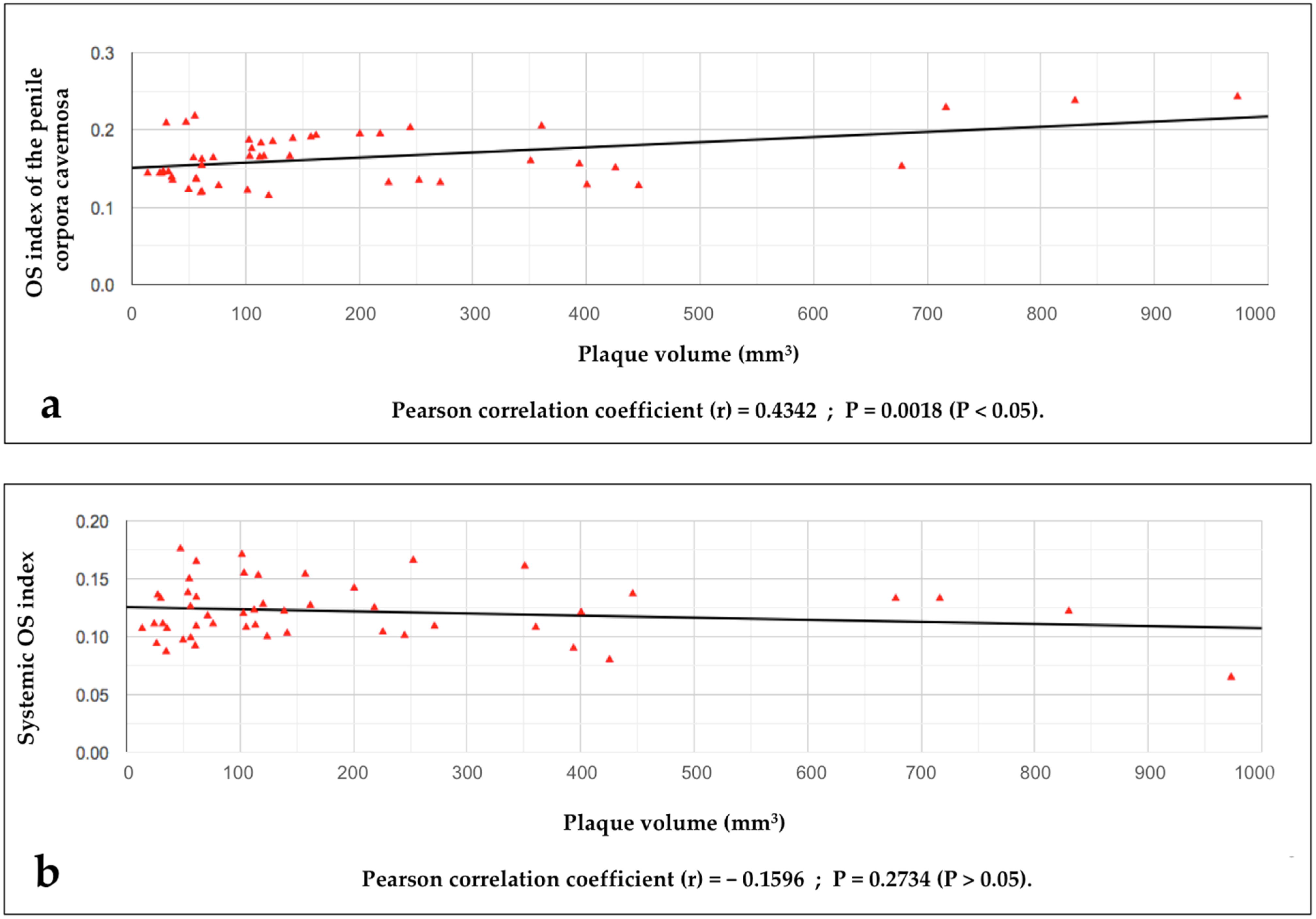
The search for a possible relationship between the value of the OS index detected in the penile corpora cavernosa (penile OSI value) and the volume of the disease area (plaque);
-
The search for a possible relationship between the values of the systemic OSI (blood sampling from a peripheral vein) and the volumes of the disease area (plaque);
-
The search for a possible relationship between the values of systemic OSI and penile OSI;
-
The identification of penile OSI values indicative of regression of the disease area (PD plaque);
-
The search for a possible relationship between values of systemic OSI and ongoing chronic pathology;
-
The search for a possible relationship between penile OSI values and ongoing chronic pathology;
-
The search for a possible relationship between penile OSI values and an anxious–depressive state (and its prevalence);
-
The search for a possible relationship between systemic OSI values and an anxious–depressive state (and its prevalence).

2.2.1. Inclusion and Exclusion Criteria

Inclusion criteria were patients affected by PD who were between 21 and 70 years of age and had available data that reported the results of a thorough clinical history examination (including all diseases); and the diagnosis of PD at the conclusion of the following assessments: performing penile palpation, photographic documentation of the penile deformation (according to Kelâmi) with a goniometric measurement of the angulation, penile eco-color Doppler ultrasound with plaque measurements (in the three dimensions, in mm) and volume calculation (mm3) using the ellipsoid formula (volume = 0.524 × length × width × thickness), and completion of the International Index of Erectile Function (IIEF) questionnaire for erectile function measurement, the Patient Health Questionnaire-9 (PHQ-9, concerning depression), the Generalized Anxiety Disorder Questionnaire-7 (GAD-7, concerning anxiety), and the visual analogue scale (VAS) questionnaire for measuring pain [63,64,65,66,67,68,69].
All patients (including the subjects of the control group) must have performed the OS study (d-ROMs and PAT) with subsequent calculation of the OSI with blood samples directly from the penile corpora cavernosa with a 25 G needle and from the peripheral blood (standard sampling from a vein of the upper extremity). Before the OS study, the patient must have fasted since the previous evening.
Each patient with PD must have performed the OS study before and after their antioxidant treatment (with a time interval of at least 6 months); the subsequent check-up after treatment must have been carried out more than once (at least one check-up) and always at least 6 months apart during the course of treatment.
During subsequent checks, each patient must have temporarily interrupted the antioxidant treatment for at least 10 days prior to the blood tests.
Exclusion criteria were PD patients with the following pathological situations (ongoing or recent within the last three months): infection (viral, bacterial, or other), acute inflammatory disease (gout attack, tooth abscess, burn, hemorrhoidal crisis, thrombophlebitis, prostatitis, acute dermatitis, allergic episode, etc.), thyroid hyperfunction, obesity, hypertensive crisis, ischemic and/or infarct episode, diabetes mellitus, liver dysfunction, renal failure, neoplastic disease, surgery, periodontal dental treatment, and, in any case, all conditions that cause a disturbance of the physiological redox balance (OS).
A total of 49 patients who had undergone an OS study and met the inclusion criteria were extracted from the total group of PD patients treated at our care center and included in this study.
Of these 49 patients (aged between 27 and 68 years), 25 of them had already been successfully treated with the disease being resolved, whilst the other 24 patients had not yet completed the entire treatment cycle.
All PD patients underwent an OS examination before and after treatment, at least 6 months after therapy. The treatment to which the PD patients were subjected was as follows: oral L-carnitine 1000 mg + bilberry 180 mg + propolis 700 mg + ginkgo biloba 240 mg + silymarin 400 mg + coenzyme Q-10 100 mg + vitamin C 50 mg + vitamin E 48 mg + superoxide dismutase 11,000 IU/g 10 mg/daily and topical diclofenac gel 4%/2 times daily + peri-plaque penile injections (only in the case of plaques with volume ≥ 100 mm3): pentoxifylline 100 mg (30 G needle) every month for 12 months and, then, 1 penile injection every 2 months for 12 months (18 total injections).
Given the difficulty in finding “normal” cases to include in the control group, we included 50 cases of subjects not affected by PD who had undergone OS studies (both of the peripheral blood and the penile corpora cavernosa) before carrying out a penile dynamic color Doppler ultrasound for suspected ED in the same session. In all 50 cases, the results of their respective dynamic ultrasound studies had shown normal results with the final diagnosis of exclusively psychogenic ED (note that their hormone tests were also normal).
We excluded from the present study all cases where a vascular or hormonal problem was found to be the cause of ED; furthermore, all patients who fell within the previously listed exclusion criteria were excluded.

2.2.2. Data Collection

We collected all the demographic data of the PD patients and the characteristics of the ultrasound examination (location, size, and volume of the plaque), the type of deformation and the degree of penile curvature (if present), the degree of ED possibly present, the degree of any associated anxious–depressive state, and the degree of pain possibly present.
We also recorded all the demographic and clinical data relating to the 50 cases of the control group.

2.2.3. Sample Collection

Blood samples for the measurement of OS were collected from both the penile corpora cavernosa and a vein of the upper extremity. The sampling from the penile corpora cavernosa (measurement of local OS) was carried out with a syringe and 25 G needle, avoiding puncturing the PD plaque. The amount of blood collected from the penile corpora cavernosa was 0.5 mL (500 µL). The sampling from the peripheral vein of the upper extremity, necessary for the evaluation of systemic OS, was carried out with a standard method; the quantity of blood aspirated was approximately 1–2 mL in order to ensure at least 0.5 mL (500 µL) for use for the exam. As soon as they were collected, the blood samples were immediately poured into a heparinized (and not EDTA) tube.

2.2.4. Plasma Collection

The samples were then centrifuged (1600 rpm for 90 s) to separate the plasma from the rest of the sample. After reacting the plasma samples (10 µL for each of the two tests) with the respective reagents for the d-ROMs test and PAT, the respective cuvettes were placed in the photometric analytical device (FRAS 5) [57].

2.2.5. d-ROMs and PAT Measurements

The d-ROMs Fast test (for determining the concentration of peroxides) was used to measure reactive oxygen species. The plasma antioxidant test (PAT, measurement of iron-reducing capacity) was used to measure the antioxidant capacity. Both tests are part of the FRAS kit (Parma, IT) [57].
The measurement of the d-ROMs and the antioxidant capacity of plasma was carried out both in the peripheral blood sample (measurement of systemic OS) and in the blood sample taken from the penile corpora cavernosa (measurement of local OS).
The unit of measurement used in the d-ROMs test was Carratelli units (Carr. U.) [51,52]. The unit of measurement used in the PAT was Cornelli units (Cor. U.) [51,52]. For d-ROMs (d-ROMs test), the reference range of normal values is between 250 and 300 Carr. U. For the measurement of the antioxidant capacity of plasma (PAT), values between 2200 and 2800 Cor. U. are considered normal, while values < 1800 Cor. U. are considered deficient [51,52].
We calculated the OS index (OSI) using the following formula: OSI (arbitrary unit) = d-ROMs/PAT × 100 [51,52,53,54,55,56].

2.2.6. Statistical Analysis

We used MedCalc statistical software (version 16.4.3, 2016) for the unpaired t-test and chi-square test. For the statistical study of the standard deviation, mean, median, and interquartile range calculation (IQR), we used CalculatorSoup® software (version of 7 March 2023). SPSS Statistics software version 22.0 (2013) was used to calculate the Pearson correlation coefficient (size, strength, and direction of the relationship between two variables) and to perform the Mann–Whitney U-test (Wilcoxon rank sum) and Shapiro–Wilk test to test the normality of values. To explore the diagnostic ability of penile OSI and create a ROC curve, we used Eng J. ROC analysis: web-based calculator for ROC curves. Baltimore: Johns Hopkins University (version of 17 February 2022), link “http://www.rad.jhmi.edu/jeng/javarad/roc/JROCFITi.html (accessed on 31 October 2023)”. To calculate the optimal cut-off, related to the ROC curve (Youden index), for measuring the sensitivity and specificity of the test, we used the Youden index calculator (MDApp, (version of 29 June 2020, Manchester, UK). For the statistical study of logistic regression, we used AgriMetSoft’s software (2023 version) and Excel (MS Office, Redmond, WA, USA, 2011 version). In the statistical analyses, a threshold of 5% for alpha error (significant p-value < 0.05) was considered to demonstrate statistical significance.

3. Results

The demographic and clinical characteristics of the 49 PD patients and control group subjects are shown in Table 1 and Table 2.
No statistically significant differences were detected between the demographic and clinical characteristics of the group of PD patients and the control group subjects (see Table 2).
The values of the d-ROMs, PAT, and the relative OS index (both systemic and penile) relating to the PD patients and control group subjects are listed in Tables S1 and S2 in the Supplementary Materials.
The Pearson correlation coefficient (PCC) statistical study demonstrated a statistical correlation between the penile OSI values and the volumes of the disease area (PD plaque): PCC = r 0.4342, r2 0.1885, p-value = 0.001827 (p < 0.05) (see Figure 1).
The Pearson correlation coefficient statistical study did not demonstrate a statistical correlation between the systemic OSI values and the volumes of the disease area (plaque): PCC = r − 0.1596, r2 0.02546, p = 0.2734 (p > 0.05) (see Figure 1).
The Pearson correlation coefficient statistical study did not demonstrate a statistical correlation between the systemic OSI values and penile OSI values: Pearson correlation coefficient, p-value = 0.198 (p > 0.05); furthermore, there was a statistically significant difference between the systemic OSI values and penile OSI values: unpaired t-test, p-value < 0.0001 (p < 0.05).
The comparison between the penile OSI values of the PD patients (before treatment) and the penile OSI values of the control group subjects revealed statistically significant differences (p-value < 0.0001), (p < 0.05).
We ascertained that the penile OSI values were significantly reduced after treatment and specifically after elimination of PD plaque (Mann–Whitney U-test, Z-score = 6.05369, p-value < 0.00001).
Figure 2 highlights the decrease in the penile OSI values after the treatment and the disappearance of Peyronie’s plaque.
Our study allowed for us to identify in the PD patients (after plaque elimination) a mean value of penile OSI indices corresponding to 0.090 ± 0.016 (p-value = 0.004, p < 0.05). In the control group subjects, a mean value of penile OSI indices was obtained corresponding to 0.096 ± 0.016 (p-value = 0.006, p < 0.05).
The comparison between the penile OSI values of the PD patients with plaque elimination and the penile OSI values of the control group subjects revealed no statistically significant differences (p-value = 0.130, p < 0.05).
In the ROC curve analysis, the area under the curve that represents the actual measure of accuracy was found to be 0.959, significance level p-value < 0.0001, sensitivity 71.4%, and specificity 90%; furthermore, after calculating the Youden index, it was found that the penile OSI value equal to 0.61 represented the optimal cutoff value (see Figure 3).
No correlation has been established between OSI values (both systemic and penile) and associated pathological conditions including cigarette smoking (see Table 3).
Table 4 illustrates our results regarding the prevalence of depression and anxiety in 49 PD patients.

4. Discussion

This is the first study where the OS index (OSI) was calculated by analyzing OS directly in the penile corpora cavernosa of PD patients by taking a blood sample.
To measure the antioxidant capacity, until some time ago, the biological antioxidant potential test (BAP test) was generally used, which is based on the ability of blood plasma to reduce the ferric ion to a ferrous ion. However, this method has an evaluation bias, as the colorimetric determination of the ferric ion is influenced by the phosphate ions. In practice, part of the ferric ions complexed with the thiocyanate ion binds to the phosphate ions normally present in the plasma sample, causing discoloration of the solution, which does not depend on the actual concentration of antioxidants but on the concentration of the phosphate ions. The result of this chemical reaction translates into a significant overestimation of the antioxidant capacity. With the more modern PAT, which is similarly based on the colorimetric determination of the ferric ion, a zirconium salt with adequate concentrations was introduced into the test, which is a transition metal that has a greater affinity with phosphates [52]. This made it possible to eliminate the interference caused by plasma phosphates.
The results of our study reinforce the observation that OS plays an important role in the pathogenesis of PD [15,16,17,18,19,20,21,22,23,70]. Penile OSI could be considered a biochemical parameter to diagnose the extent of PD given that OSI values were found to correlate closely with plaque volume.
Having detected a significant correlation between the volumes of Peyronie’s plaque and the penile OSI values, we could have access not only to the ultrasound study (which allows for us to detect the volume of Peyronie’s plaque) but also an important marker that could indicate the real presence of the disease, confirming the therapeutic result not only visually but also chemically.
Furthermore, in the absence of a certain ultrasound diagnosis, which is possible in some cases, the study of OS in the penile corpora cavernosa could prove to be a test for a more certain and definitive diagnosis.
The absence of a correlation between the volumes of Peyronie’s plaque and the systemic OSI indices indicates that, to be truly meaningful, the study of OS must be performed by taking a sample directly from the site of the disease, as has already been carried out in other studies for other pathologies [49,50,71].
Given that the comparison between the penile OSI values of the PD patients with plaque elimination and the penile OSI values of the control group subjects did not reveal statistically significant differences, it indicates that the study of penile OSI can represent an effective test for the study of oxidative markers in this disease.
Even if the aim of this study was not the following, our results testified to the elimination of the plaque after antioxidant treatment, as other articles in the literature have already described [72,73,74,75].
Even if this study did not demonstrate any correlation between penile OSI values and anxious–depressive state, we detected a high prevalence of anxiety (81.6%) and depression (59.1%) in the PD patients. These data confirm that the anxious–depressive state represents one of the symptoms of PD (along with penile pain, penile curvature/deformation, and ED) [10,76,77,78]. The finding of significant percentages of anxious–depressive disorders has also been noted by other authors who have used the Peyronie’s Disease Questionnaire (PDQ) domains [79]. Although our results are interesting, the limitation of our study was that we did not have a very large sample of patients.
Further studies are needed to investigate OS in the penile corpora cavernosa of subjects with PD. To optimize this clinical research, experimental studies on animal models could also be implemented. For example, experimental studies on rats would be necessary to induce penile fibrosis by injecting fibrogenic agents (TGF-β, fibrin, or others) into the penile tunica albuginea, as has already been conducted in other studies [80,81,82]. After the formation of fibrotic areas in the penis, oxidative stress could be measured in both the corpora cavernosa and peripheral blood of rats. Subsequently, antioxidants or antifibrotics (such as anthocyanin, platelet-rich plasma, or others) could be administered into the penile corpora cavernosa of rats to reduce fibrosis [81,82]. Once this effect was achieved, OSI could be measured as it was performed before the treatment.

5. Conclusions

The absence of a correlation between the PD plaque volumes and systemic OSI indices and, on the contrary, the existence of a significant correlation between the PD plaque volumes and the penile OSI values, indicate that the study of OS must be carried out by taking a sample directly from the site of the disease; other authors have demonstrated this in other fields of specialist medicine.
The penile OSI study associated with the ultrasound study, which allows for us to examine the plaque volume, would also provide a precious oxidative marker that could indicate the real chemical presence of the disease.
Although our data are very interesting, further studies are needed to investigate OS (with the calculation of the OS index) in the penile corpora cavernosa of PD patients.

Supplementary Materials

The following supporting information can be downloaded at: https://www.mdpi.com/article/10.3390/metabo14010055/s1, Table S1: Values of d-ROMs, PAT and relative OSI index (“systemic” and “in penile corpora cavernosa”) of the 49 patients with Peyronie’s disease, Table S2: Values of d-ROMs, PAT and relative OSI index (“systemic” and “in penile corpora cavernosa”) of the 50 normal cases (control group).

Author Contributions

G.P., G.D.G. and S.Q. contributed to the realization of the article (excluding the parts regarding anxious and depressive state) as follows: supervision; conceptualization; methodology; software; validation; formal analysis; investigation; data curation; writing—original draft preparation; writing—review and editing; visualization; supervision; and project administration. A.P. edited the parts of the article concerning the anxious and depressive state, taking care in particular of validation; formal analysis; investigation; data curation; writing—review and editing; and supervision. All authors have read and agreed to the published version of the manuscript.

Funding

This research received no external funding.

Institutional Review Board Statement

This study was conducted in accordance with the Declaration of Helsinki and received approval from the Castelfidardo ethical commission (protocol code #00231, date of approval 20 February 2023) for studies involving humans.

Informed Consent Statement

Informed consent was obtained from all subjects involved in this study. The sensitive data were, however, anonymized in compliance with privacy according to Legislative Decree, 10 August 2018, n. 101, adapted to the GDPR (Official Gazette of the Italian Republic, General Series n. 205, dated 4 September 2018).

Data Availability Statement

The original contributions presented in the study are included in the article/Supplementary Materials, further inquiries can be directed to the corresponding author.

Conflicts of Interest

The authors declare no conflicts of interest.

References

  1. Herati, A.S.; Pastuszak, A.W. The Genetic Basis of Peyronie’s Disease: A Review. Sex Med. Rev. 2016, 4, 85–94. [Google Scholar] [CrossRef]
  2. Bias, W.B.; Nyberg, L.M., Jr.; Hochberg, M.C.; Walsh, P.C.; Opitz, J.M. Peyronie’s disease: A newly recognized autosomal-dominant trait. Am. J. Med. Genet. 1982, 12, 227–235. [Google Scholar] [CrossRef] [PubMed]
  3. Schwarzer, U.; Sommer, F.; Klotz, T.; Braun, M.; Reifenrath, B.; Engelmann, U. The prevalence of Peyronie’s disease: Results of a large survey. BJU Int. 2001, 88, 727–730. [Google Scholar] [CrossRef]
  4. Stuntz, M.; Perlaky, A.; des Vignes, F.; Kyriakides, T.; Glass, D. The prevalence of Peyronie’s disease in the United States: A population-based study. PLoS ONE 2016, 11, e0150157. [Google Scholar] [CrossRef] [PubMed]
  5. La Pera, G.; Pescatori, E.S.; Calabrese, M.; Boffini, A.; Colombo, F.; Andriani, E.; Natali, A.; Vaggi, L.; Catuogno, C.; Giustini, M.; et al. Peyronie’s disease: Prevalence and association with cigarette smoking. A multicenter population-based study in men aged 50–69 years. Eur. Urol. 2001, 40, 525–530. [Google Scholar] [CrossRef] [PubMed]
  6. DiBenedetti, D.B.; Nguyen, D.; Zografos, L.; Ziemiecki, R.; Zhou, X. A Population-based study of peyronie’s disease: Prevalence and treatment patterns in the United States. Adv. Urol. 2011, 2011, 282503. [Google Scholar] [CrossRef]
  7. Hellstrom, W.J.; Bivalacqua, T.J. Peyronie’s disease: Etiology, medical, and surgical therapy. J. Androl. 2000, 21, 347–354. [Google Scholar] [CrossRef]
  8. Weidner, W.; Schroeder-Printzen, I.; Weiske, W.-H.; Vosshenrich, R. Sexual dysfunction in Peyronie’s disease: An analysis of 222 patients without previous local plaque therapy. J. Urol. 1997, 157, 325–328. [Google Scholar] [CrossRef]
  9. Pryor, J.P.; Ralph, D.J. Clinical presentations of Peyronie’s disease. Int. J. Impot. Res. 2002, 14, 414–417. [Google Scholar] [CrossRef]
  10. Nelson, C.J.; Diblasio, C.; Kendirci, M.; Hellstrom, W.; Guhring, P.; Mulhall, J.P. The chronology of depression and distress in men with Peyronie’s disease. J. Sex. Med. 2008, 5, 1985–1990. [Google Scholar] [CrossRef]
  11. Devine, C.J., Jr.; Somers, K.D.; Ladaga, L.E. Peyronie’s disease: Pathophysiology. Prog. Clin. Biol. Res. 1991, 370, 355–358. [Google Scholar]
  12. Devine, C.J., Jr.; Somers, K.D.; Jordan, G.H.; Schlossberg, S.M. Proposal: Trauma as a cause of Peyronie’s lesion. J. Urol. 1997, 157, 285–290. [Google Scholar] [CrossRef] [PubMed]
  13. Jarow, J.P.; Lowe, F.C. Penile trauma: An etiologic factor in Peyronie’s disease and erectile dysfunction. J. Urol. 1997, 158, 1388–1390. [Google Scholar] [CrossRef] [PubMed]
  14. Somers, K.D.; Dawson, D.M. Fibrin deposition in Peyronie’s disease plaque. J. Urol. 1997, 157, 311–315. [Google Scholar] [CrossRef] [PubMed]
  15. Sikka, S.C.; Hellstrom, W.J. Role of oxidative stress and antioxidants in Peyronie’s disease. Int. J. Impot. Res. 2002, 14, 353–360. [Google Scholar] [CrossRef]
  16. El-Sakka, A.I.; Salabas, E.; Dinçer, M.; Kadioglu, A. The pathophysiology of Peyronie’s disease. Arab. J. Urol. 2013, 11, 272–277. [Google Scholar] [CrossRef]
  17. Paulis, G.; Brancato, T. Inflammatory mechanisms and oxidative stress in Peyronie’s disease: Therapeutic “rationale” and related emerging treatment strategies. Inflamm. Allergy Drug Targets 2012, 11, 48–57. [Google Scholar] [CrossRef]
  18. Paulis, G.; Romano, G.; Paulis, L.; Barletta, D. Recent Pathophysiological Aspects of Peyronie’s Disease: Role of Free Radicals, Rationale, and Therapeutic Implications for Antioxidant Treatment-Literature Review. Adv. Urol. 2017, 2017, 4653512. [Google Scholar] [CrossRef]
  19. Paulis, G.; De Giorgio, G.; Paulis, L. Role of Oxidative Stress in Peyronie’s Disease: Biochemical Evidence and Experiences of Treatment with Antioxidants. Int. J. Mol. Sci. 2022, 23, 15969. [Google Scholar] [CrossRef]
  20. Davila, H.H.; Magee, T.R.; Vernet, D.; Rajfer, J.; Gonzalez-Cadavid, N.F. Gene transfer of inducible nitric oxide synthase complementary DNA regresses the fibrotic plaque in an animal model of Peyronie’s disease. Biol. Reprod. 2004, 71, 1568–1577. [Google Scholar] [CrossRef]
  21. Bivalacqua, T.J.; Champion, H.C.; Hellstrom, W.J. Implications of nitric oxide synthase isoforms in the pathophysiology of Peyronie’s disease. Int. J. Impot. Res. 2002, 14, 345–352. [Google Scholar] [CrossRef]
  22. Gonzalez-Cadavid, N.F.; Magee, T.R.; Ferrini, M.; Qian, A.; Vernet, D.; Rajfer, J. Gene expression in Peyronie’s disease. Int. J. Impot. Res. 2002, 14, 361–374. [Google Scholar] [CrossRef] [PubMed]
  23. Moreland, R.B.; Nehra, A. Pathophysiology of Peyronie’s disease. Int. J. Impot. Res. 2002, 14, 406–410. [Google Scholar] [CrossRef] [PubMed]
  24. Mulhall, J.P.; Schiff, J.; Guhring, P. An analysis of the natural history of Peyronie’s disease. J. Urol. 2006, 175, 2115–2118. [Google Scholar] [CrossRef]
  25. Garaffa, G.; Trost, L.W.; Serefoglu, E.C.; Ralph, D.; Hellstrom, W.J.G. Understanding the course of Peyronie’s disease. Int. J. Clin. Pract. 2013, 67, 781–788. [Google Scholar] [CrossRef]
  26. Paulis, G.; Cavallini, G. Clinical evaluation of natural history of Peyronie’s disease: Our experience, old myths and new certainties. Inflamm. Allergy Drug Targets 2013, 12, 341–348. [Google Scholar] [CrossRef] [PubMed]
  27. Jalkut, M.; Gonzalez-Cadavid, N.; Rajfer, J. Peyronie’s disease: A review. Rev. Urol. 2003, 5, 142–148. [Google Scholar]
  28. Levine, L.A.; Larsen, S. Diagnosis and management of Peyronie disease. In Campbell-Walsh Urology, 11th ed.; Wein, A.J., Kavoussi, L.R., Partin, A.W., Peters, C.A., Eds.; Elsevier Saunders: Philadelphia, PA, USA, 2015; pp. 722–748. [Google Scholar]
  29. Brimley, S.C.; Yafi, F.A.; Greenberg, J.; Hellstrom, W.J.; Nguyen, H.M.T.; Hatzichristodoulou, G. Review of management options for active-phase Peyronie’s disease. Sex. Med. Rev. 2019, 7, 329–337. [Google Scholar] [CrossRef]
  30. Capoccia, E.; Levine, L.A. Contemporary review of Peyronie’s disease treatment. Curr. Urol. Rep. 2018, 19, 51. [Google Scholar] [CrossRef]
  31. Gur, S.; Limin, M.; Hellstrom, W.J. Current status and new developments in Peyronie’s disease: Medical, minimally invasive and surgical treatment options. Expert Opin. Pharmacother. 2011, 12, 931–944. [Google Scholar] [CrossRef]
  32. Tsambarlis, P.; Levine, L.A. Nonsurgical management of Peyronie’s disease. Nat. Rev. Urol. 2019, 16, 172–186. [Google Scholar] [CrossRef] [PubMed]
  33. Tan, R.B.W.; Sangkum, P.; Mitchell, G.C.; Hellstrom, W.J.G. Update on medical management of Peyronie’s disease. Curr. Urol. Rep. 2014, 15, 415. [Google Scholar] [CrossRef] [PubMed]
  34. Paulis, G.; Brancato, T.; D’ascenzo, R.; De Giorgio, G.; Nupieri, P.; Orsolini, G.; Alvaro, R. Efficacy of vitamin E in the conservative treatment of Peyronie’s disease: Legend or reality? A controlled study of 70 cases. Andrology 2013, 1, 120–128. [Google Scholar] [CrossRef] [PubMed]
  35. Paulis, G.; Cavallini, G.; De Giorgio, G.; Quattrocchi, S.; Brancato, T.; Alvaro, R. Long-term multimodal therapy (verapamil associated with propolis, blueberry, vitamin E and local diclofenac) on patients with Peyronie’s disease (chronic inflammation of the tunica albuginea). Results of a controlled study. Inflamm. Allergy Drug Targets 2013, 12, 403–409. [Google Scholar] [CrossRef]
  36. Paulis, G.; Barletta, D.; Turchi, P.; Vitarelli, A.; Dachille, G.; Fabiani, A.; Gennaro, R. Efficacy and safety evaluation of pentoxifylline associated with other antioxidants in medical treatment of Peyronie’s disease: A case-control study. Res. Rep. Urol. 2016, 8, 1–10. [Google Scholar]
  37. Gennaro, R.; Barletta, D.; Paulis, G. Intralesional hyaluronic acid: An innovative treatment for Peyronie’s disease. Int. Urol. Nephrol. 2015, 47, 1595–1602. [Google Scholar] [CrossRef]
  38. Zucchi, A.; Costantini, E.; Cai, T.; Cavallini, G.; Liguori, G.; Favilla, V.; De Grande, G.; D’Achille, G.; Silvani, M.; Franco, G.; et al. Intralesional Injection of Hyaluronic Acid in Patients Affected with Peyronie’s Disease: Preliminary Results from a Prospective, Multicenter, Pilot Study. Sex. Med. 2016, 4, e85–e90. [Google Scholar] [CrossRef]
  39. Kendirci, M.; Hellstrom, W.J. Critical analysis of surgery for Peyronie’s disease. Curr. Opin. Urol. 2004, 6, 381–388. [Google Scholar] [CrossRef]
  40. Levine, L.A.; Burnett, A.L. Standard operating procedures for Peyronie’s disease. J. Sex. Med. 2013, 10, 230–244. [Google Scholar] [CrossRef]
  41. Kadioglu, A.; Akman, T.; Sanli, O.; Gurkan, L.; Cakan, M.; Celtik, M. Surgical treatment of Peyronie’s disease: A critical analysis. Eur. Urol. 2006, 50, 235–248. [Google Scholar] [CrossRef]
  42. Rice, P.G.; Somani, B.K.; Rees, R.W. Twenty Years of Plaque Incision and Grafting for Peyronie’s Disease: A Review of Literature. Sex. Med. 2019, 7, 115–128. [Google Scholar] [CrossRef] [PubMed]
  43. Ralph, D.; Gonzalez-Cadavid, N.; Mirone, V.; Perovic, S.; Sohn, M.; Usta, M.; Levine, L. The management of Peyronie’s disease: Evidence-based 2010 guidelines. J. Sex. Med. 2010, 7, 2359–2374. [Google Scholar] [CrossRef] [PubMed]
  44. Hatzimouratidis, K.; Eardley, I.; Giuliano, F.; Hatzichristou, D.; Moncada, I.; Salonia, A.; Vardi, Y.; Wespes, E. EAU guidelines on penile curvature. Eur. Urol. 2012, 62, 543–552. [Google Scholar] [CrossRef]
  45. Nehra, A.; Alterowitz, R.; Culkin, D.J.; Faraday, M.M.; Hakim, L.S.; Heidelbaugh, J.J.; Khera, M.; Kirkby, E.; McVary, K.T.; Miner, M.M.; et al. Peyronie’s disease: AUA guideline. J. Urol. 2015, 194, 745–753. [Google Scholar] [CrossRef] [PubMed]
  46. Bella, A.J.; Lee, J.C.; Grober, E.D.; Carrier, S.; Benard, F.; Brock, G.B. Canadian Urological Association guideline for Peyronie’s disease and congenital penile curvature. Can. Urol. Assoc. J. 2018, 12, E197–E209. [Google Scholar] [CrossRef] [PubMed]
  47. Levine, L.; Rybak, J.; Corder, C.; Farrel, M.R. Peyronie’s disease plaque calcification--prevalence, time to identification, and development of a new grading classification. J. Sex. Med. 2013, 10, 3121–3128. [Google Scholar] [CrossRef] [PubMed]
  48. Paulis, G.; Paulis, A. Calcification in Peyronie’s disease: Its role and clinical influence on the various symptoms and signs of the disease, including psychological impact. Our study of 551 patients. Arch. Ital. Urol. Androl. 2023, 95, 11549. [Google Scholar] [CrossRef] [PubMed]
  49. Yesilirmak, N.; Bukan, N.; Kurt, B.; Yuzbasioglu, S.; Zhao, M.; Rodrigues-Braz, D.; Aktas, A.; Behar-Cohen, F.; Bourges, J.-L. Evaluation of Ocular and Systemic Oxidative Stress Markers in Ocular Rosacea Patients. Investig. Opthalmol. Vis. Sci. 2023, 64, 22. [Google Scholar] [CrossRef]
  50. Dinç, G.; Fentoğlu, Ö.; Doğru, A.; İlhan, I.; Kırzıoğlu, F.Y.; Orhan, H. The evaluation of salivary oxidative stress in patients with familial Mediterranean fever and chronic periodontitis. J. Periodontol. 2018, 89, 1112–1120. [Google Scholar] [CrossRef]
  51. Valtuille, R.A.; Rossi, G.; Gimenez, E. Protective Effect of Autologous Arteriovenous Fistulae Against Oxidative Stress in Hemodialyzed Patients. Cureus 2021, 13, e15398. [Google Scholar] [CrossRef]
  52. Serena, B.; Primiterra, M.; Catalani, S.; Finco, A.; Canestrari, F.; Cornelli, U. Performance evaluation of the innovative PAT test, comparison with the common BAP test and influence of interferences on the evaluation of the plasma antioxidant capacity. Clin. Lab. 2013, 59, 1091–1097. [Google Scholar] [PubMed]
  53. Harma, M.; Harma, M.; Erel, O. Increased oxidative stress in patients with hydatidiform mole. Swiss. Med. Wkly. 2003, 133, 563–566. [Google Scholar] [CrossRef]
  54. Sánchez-Rodríguez, M.A.; Mendoza-Núñez, V.M. Oxidative Stress Indexes for Diagnosis of Health or Disease in Humans. Oxidative Med. Cell. Longev. 2019, 2019, 4128152. [Google Scholar] [CrossRef] [PubMed]
  55. Demirbag, R.; Gur, M.; Yilmaz, R.; Kunt, A.S.; Erel, O.; Andac, M.H. Influence of oxidative stress on the development of collateral circulation in total coronary occlusions. Int. J. Cardiol. 2007, 116, 14–19. [Google Scholar] [CrossRef] [PubMed]
  56. Yilmaz, S.; Ozgu-Erdinc, A.S.; Demirtas, C.; Ozturk, G.; Erkaya, S.; Uygur, D. The oxidative stress index increases among patients with hyperemesis gravidarum but not in normal pregnancies. Redox Rep. 2015, 20, 97–102. [Google Scholar] [CrossRef] [PubMed]
  57. The FRAS System. 2023. Available online: https://hedsrl.it/en/fras-5/ (accessed on 24 December 2023).
  58. Coaccioli, S.; Panaccione, A.; Biondi, R.; Sabatini, C.; Landucci, P.; Del Giorno, R.; Fantera, M.; Mondo, A.M.; Di Cato, L.; Paladini, A.; et al. Evaluation of oxidative stress in rheumatoid and psoriatic arthritis and psoriasis. Clin. Ter. 2009, 160, 467–472. [Google Scholar] [PubMed]
  59. Mukhopadhyay, K.; De, S.; Kundu, S.; Ghosh, P.; Chatterjee, S.; Chatterjee, M. Evaluation of levels of oxidative stress as a potential biomarker in patients with rheumatoid arthritis. J. Fam. Med. Prim. Care 2021, 10, 1981–1986. [Google Scholar] [CrossRef] [PubMed]
  60. Karaagac, L.; Koruk, S.T.; Koruk, I.; Aksoy, N. Decreasing oxidative stress in response to treatment in patients with brucellosis: Could it be used to monitor treatment? Int. J. Infect. Dis. 2011, 15, e346–e349. [Google Scholar] [CrossRef]
  61. Motor, S.; Ozturk, S.; Ozcan, O.; Gurpinar, A.B.; Can, Y.; Yuksel, R.; Yenin, J.Z.; Seraslan, G.; Ozturk, O.H. Evaluation of total antioxidant status, total oxidant status and oxidative stress index in patients with alopecia areata. Int. J. Clin. Exp. Med. 2014, 7, 1089–1093. [Google Scholar]
  62. Yalcin, S.; Ulas, T.; Eren, M.A.; Aydogan, H.; Camuzcuoglu, A.; Kucuk, A.; Yuce, H.H.; Demir, M.E.; Vural, M.; Aksoy, N. Relationship between oxidative stress parameters and cystatin C levels in patients with severe preeclampsia. Medicina 2013, 49, 19. [Google Scholar] [CrossRef]
  63. Kelâmi, A. Autophotography in evaluation of functional penile disorders. Urology 1983, 21, 628–629. [Google Scholar] [CrossRef] [PubMed]
  64. Eri, L.M.; Thomassen, H.; Brennhovd, B.; Håheim, L.L. Accuracy and repeatability of prostate volume measurements by transrectal ultrasound. Prostate Cancer Prostatic Dis. 2002, 5, 273–278. [Google Scholar] [CrossRef] [PubMed]
  65. Lee, J.S.; Chung, B.H. Transrectal ultrasound versus magnetic resonance imaging in the estimation of prostate volume as compared with radical prostatectomy specimens. Urol. Int. 2007, 78, 323–327. [Google Scholar] [CrossRef] [PubMed]
  66. Rosen, R.C.; Riley, A.; Wagner, G.; Osterloh, I.H.; Kirkpatrick, J.; Mishra, A. The international index of erectile function (IIEF): A multidimensional scale for assessment of erectile dysfunction. Urology 1997, 49, 822–830. [Google Scholar] [CrossRef]
  67. Kroenke, K.; Spitzer, R.L.; Williams, J.B. The PHQ-9: Validity of a brief depression severity measure. J. Gen. Intern. Med. 2001, 16, 606–613. [Google Scholar] [CrossRef] [PubMed]
  68. Spitzer, R.L.; Kroenke, K.; Williams, J.B.; Löwe, B. A brief measure for assessing generalized anxiety disorder: The GAD-7. Arch. Intern. Med. 2006, 166, 1092–1097. [Google Scholar] [CrossRef]
  69. Kahl, C.; Cleland, J.A. Visual analogue scale, numeric pain rating scale and the McGill pain Questionnaire: An overview of psychometric properties. Phys. Ther. Rev. 2005, 10, 123–128. [Google Scholar] [CrossRef]
  70. Mitsui, Y.; Yamabe, F.; Hori, S.; Uetani, M.; Kobayashi, H.; Nagao, K.; Nakajima, K. Molecular Mechanisms and Risk Factors Related to the Pathogenesis of Peyronie’s Disease. Int. J. Mol. Sci. 2023, 24, 10133. [Google Scholar] [CrossRef]
  71. Özdemir, E.Ç.; Erciyas, K.; Ünsal, B.; Sezer, U.; Taysi, S.; Araz, M. The Effects of Chronic Periodontitis and Obesity on Total Antioxidant/Oxidant Status and Oxidative Stress Index. Acta Endocrinol. 2022, 18, 294–300. [Google Scholar] [CrossRef]
  72. Paulis, G.; De Giorgio, G. Complete Plaque Regression in Patients with Peyronie’s Disease after Multimodal Treatment with Antioxidants: A Report of 2 Cases. Am. J. Case Rep. 2022, 23, e936146. [Google Scholar] [CrossRef]
  73. Paulis, G.; De Giorgio, G. Full Regression of Peyronie’s Disease Plaque Following Combined Antioxidant Treatment: A Three-Case Report. Antioxidants 2022, 11, 1661. [Google Scholar] [CrossRef]
  74. Paulis, G.; De Giorgio, G. Patients with Peyronie’s disease achieve complete plaque regression after multimodal treatment with antioxidants: A case series. J. Med. Case Rep. 2022, 16, 359. [Google Scholar] [CrossRef]
  75. Paulis, G.; De Giorgio, G. Disappearance of Plaque Following Treatment with Antioxidants in Peyronie’s Disease Patients—A Report of 3 Cases. Clin. Pract. 2022, 12, 1020–1033. [Google Scholar] [CrossRef]
  76. Terrier, J.E.; Nelson, C.J. Psychological aspects of Peyronie’s disease. Transl. Androl. Urol. 2016, 5, 290–295. [Google Scholar] [CrossRef] [PubMed]
  77. Smith, J.F.; Walsh, T.J.; Conti, S.L.; Turek, P.; Lue, T. Risk factors for emotional and relationship problems in Peyronie’s disease. J. Sex. Med. 2008, 5, 2179–2184. [Google Scholar] [CrossRef] [PubMed]
  78. Nelson, C.J.; Mulhall, J.P. Psychological impact of Peyronie’s disease: A review. J. Sex. Med. 2013, 10, 653–660. [Google Scholar] [CrossRef] [PubMed]
  79. Cilio, S.; Fallara, G.; Capogrosso, P.; Candela, L.; Belladelli, F.; Pozzi, E.; Corsini, C.; Raffo, M.; Schifano, N.; Boeri, L.; et al. The symptomatic burden of Peyronie’s disease at presentation according to patient age: A critical analysis of the Peyronie’s disease questionnaire (PDQ) domains. Andrology 2023, 11, 501–507. [Google Scholar] [CrossRef]
  80. Culha, M.G.; Erkan, E.; Cay, T.; Yücetaş, U. The Effect of Platelet-Rich Plasma on Peyronie’s Disease in Rat Model. Urol. Int. 2019, 102, 218–223. [Google Scholar] [CrossRef]
  81. Bivalacqua, T.J.; Diner, E.K.; Novak, T.E.; Vohra, Y.; Sikka, S.C.; Champion, H.C.; Kadowitz, P.J.; Hellstrom, W.J. A rat model of Peyronie’s disease associated with a decrease in erectile activity and an increase in inducible nitric oxide synthase protein expression. J. Urol. 2000, 163, 1992–1998. [Google Scholar] [CrossRef]
  82. Sohn, D.W.; Bae, W.J.; Kim, H.S.; Kim, S.W.; Kim, S.W. The anti-inflammatory and antifibrosis effects of anthocyanin extracted from black soybean on a Peyronie disease rat model. Urology 2014, 84, 1112–1116. [Google Scholar] [CrossRef]
Figure 1. Graph highlighting the relationship between plaque volumes and penile OS index values (a) and between plaque volumes and systemic OS index values (b).
Metabolites 14 00055 g001
Figure 2. OSI values of penile corpora cavernosa before and after treatment and elimination of PD plaque.
Metabolites 14 00055 g002
Figure 3. Analysis of the ROC curve of penile OSI and identification of the cutoff (Youden index).
Metabolites 14 00055 g003
Table 1. Demographic characteristics and plaque volume of the 49 patients with Peyronie’s disease.
All n. 49
PD Patients
n. 25 PD Patients
with Plaque
Elimination
n. 24 PD Patients
with Partial Plaque
Elimination
Statistical Analysis
25 PD p.
versus
24 PD p.
p-Value (t-Test)
Mean age (years)
(SD)
49.65
(±11.01)
46.68
(±11.29)
50.66
(±10.85)
0.214 *
Plaque volume (mm3)
(SD)
194.33
(±218.55)
152.64
(±192.30)
237.75
(±239.30)
0.175 *
n. 25 PD patients
with plaque elimination
n. 24 PD patients
with partial plaque
elimination
Patient no.Plaque volume
(mm3)
Plaque volume
(mm3)
1.34.9 113.4
2.26.4 76.2
3.13.7 350.8
4.35.6 101.5
5.24.3 138.8
6.123.8 112.3
7.105.3 200.5
8.27.4 71.4
9.716.4 102.9
10.120.2 973.0
11.393.7 445.9
12.56.3 425.4
13.60.4 830.0
14.54.0 30.0
15.61.4 400.4
16.61.4 271.3
17.56.4 225.7
18.157.4 116.0
19.103.5 61.3
20.49.7 252.5
21.244.8 141.4
22.677.5 161.9
23.218.3 47.4
24.360.4 55.2
25.31.9 -
NOTE: PD = Peyronie’s disease; SD = standard deviation; * = no statistically significant difference found.
Table 2. Demographic and clinical characteristics of PD patients and control group subjects.
Group of PD Patients (n. 49)
Mean Age
49.65 Years (SD ± 11.01)
Control Group (n. 50)
Mean Age
49.94 Years (SD ± 11.42)
Statistical Analysis
Group-PD
versus
Control Group
p-Value (t-Test)
0.912
Demographic
characteristics
N. patients (out 49)
(%)
N. patients (out 50)
(%)
p-value
(chi-square test)
Race
Caucasian49 (100)50 (100)1.000
Age Range---
From 20 to 30 years2 (4.08)3 (6.0)0.713
From 31 to 40 years10 (20.4)9 (18.0)0.762
From 41 to 50 years13 (26.53)8 (16.0)0.202
From 51 to 60 years14 (28.57)20 (40.0)0.233
From 61 to 70 years10 (20.4)10 (20.0)0.959
Type of school education--
elementary school2 (4.08)1 (2.0)0.547
secondary school38 (77.55)39 (78.0)0.957
university degree9 (18.36)10 (20.0)0.837
Clinical condition
associated in PD patients
N. patients
(out 49)
N. patients
(out 50)
Statistical analysis
Group-PD
versus
Control group
p-value (t-test)
Anxiety40 Mean GAD-7 score = 16.442Mean GAD-7 score = 16.20.806
Depression29 Mean PHQ-9 score = 15.232Mean PHQ-9 score = 14.40.383
Penile curvature45Average penile curvature
angle (degrees) = 35.1°
0Average penile curvature
angle (degrees) = 0°
<0.0001
Penile pain26 Mean VAS score = 4.60Mean VAS score = 0<0.0001
Erectile
dysfunction
19 Mean IIEF score = 21.950Mean IIEF score = 22.40.469
Cigarette smoking16 Mean No. of cigarettes
per day = 9.3
17Mean No. of cigarettes
per day = 8.76
0.808
NOTE: PD = Peyronie’s disease; SD = standard deviation; p-value significant when <0.05; GAD-7 = Generalized Anxiety Disorder-7 questionnaire; PHQ-9, = Patient Health Questionnaire-9; IIEF = International Index of Erectile Function; VAS = visual analogue scale.
Table 3. Associated clinical conditions in 49 patients with Peyronie’s disease and the study of their possible statistical correlation with the “OS index values of the penile corpora cavernosa” or “systemic OS index values”.
Clinical Condition Associated in PD
Patients
N.
Patients
(out 49)
Type of Statistical
Analysis Used
and (p-Value)
Correlation
with
Penile OS
Index Values
(YES or NO)
Type of Statistical
Analysis Used
and (p-Value)
Correlation
with
Systemic OS
Index Values
(YES or NO)
Anxiety40t-test
(p = 0.153)
Pearson correlation
coefficient
(p = 0.094)
NOt-test
(p = 0.281)
Pearson correlation
coefficient
(p = 0.524)
NO
Depression29t-test
(p = 0.781)
Pearson correlation
coefficient
(p = 0.901)
NOt-test
(p = 0.184)
Pearson correlation
coefficient
(p = 0.171)
NO
Penile pain26t-test
(p = 0.221)
Pearson correlation
coefficient
(p = 0.081)
NOt-test
(p = 0.676)
Pearson correlation
coefficient
(p = 0.834)
NO
Penile
curvature
45t-test
(p = 0.920)
Pearson correlation
coefficient
(p = 0.704)
NOt-test
(p = 0.796)
Pearson correlation
coefficient
(p = 0.290)
NO
Erectile dysfunction19t-test
(p = 0.753)
Pearson correlation
coefficient
(p = 0.774)
NOt-test
(p = 0.611)
Pearson correlation
coefficient
(p = 0.655)
NO
Cigarette smoking16t-test
(p = 0.488)
Pearson correlation
coefficient
(p = 0.380)
NOt-test
(p = 0.564)
Pearson correlation
coefficient
(p = 0.381)
NO
Table 4. Prevalence of depression and anxiety in 49 PD patients.
PHQ-9 Score Range No. Total Cases (%)
No depression00
Minimal or mild depression1–920 (40.8)
Moderate–Severe depression10–2729 (59.1)
- Moderate depression10–1416 (32.6)
- Moderately severe depression15–1911 (22.4)
- Severe depression20–272 (4.08)
TOTAL 49
GAD-7 score rangeNo. total cases (%)
No anxiety00
Minimal or mild anxiety1–96 (12.2)
Moderate–Severe anxiety10–2140 (81.6)
- Moderate anxiety10–1420 (40.8)
- Severe anxiety15–2120 (40.8)
TOTAL 49
NOTE: PD = Peyronie’s disease; PHQ-9 = Patient Health Questionnaire-9; GAD-7 = Generalized Anxiety Disorder Questionnaire-7.
Disclaimer/Publisher’s Note: The statements, opinions and data contained in all publications are solely those of the individual author(s) and contributor(s) and not of MDPI and/or the editor(s). MDPI and/or the editor(s) disclaim responsibility for any injury to people or property resulting from any ideas, methods, instructions or products referred to in the content.

