Article

Muscle Pathology in Dystrophic Rats and Zebrafish Is Unresponsive to Taurine Treatment, Compared to the mdx Mouse Model for Duchenne Muscular Dystrophy

1
School of Molecular Sciences, The University of Western Australia, Perth 6009, Australia
2
TaRGeT Lab, Translational Research for Gene Therapy, INSERM, UMR 1089, Nantes Université, CHU Nantes, 440200 Nantes, France
3
Therassay Platform, CAPACITES, Nantes Université, 44007 Nantes, France
4
School of Biological Sciences, Monash University, Melbourne 3800, Australia
5
School of Human Sciences, the University of Western Australia, Perth 6009, Australia
*
Author to whom correspondence should be addressed.
Metabolites 2023, 13(2), 232; https://doi.org/10.3390/metabo13020232
Submission received: 30 November 2022 / Revised: 31 January 2023 / Accepted: 1 February 2023 / Published: 4 February 2023

Abstract

:
Inflammation and oxidative stress are strongly implicated in the pathology of Duchenne muscular dystrophy (DMD), and the sulphur-containing amino acid taurine ameliorates both and decreases dystropathology in the mdx mouse model for DMD. We therefore further tested taurine as a therapy using dystrophic DMDmdx rats and dmd zebrafish models for DMD that have a more severe dystropathology. However, taurine treatment had little effect on the indices of dystropathology in both these models. While we and others have previously observed a deficiency in taurine in mdx mice, in the current study we show that the rat and zebrafish models had increased taurine content compared with wild-type, and taurine treatment did not increase muscle taurine levels. We therefore hypothesised that endogenous levels of taurine are a key determinate in potential taurine treatment efficacy. Because of this, we felt it important to measure taurine levels in DMD patient plasma samples and showed that in non-ambulant patients (but not in younger patients) there was a deficiency of taurine. These data suggest that taurine homeostasis varies greatly between species and may be influenced by age and disease progression. The potential for taurine to be an effective therapy may depend on such variables.

1. Introduction

Duchenne Muscular Dystrophy (DMD) is a lethal, X-chromosome-linked muscle disease affecting about 1 in 3500–6000 boys worldwide (Reviewed in [1,2,3]). DMD is characterised by severe skeletal muscle loss caused by mutations in the dystrophin gene, resulting in defects or the absence of functional dystrophin protein in muscle. Dystrophin deficiency increases susceptibility to sarcolemma damage after muscle contraction, leading to myofibre necrosis (myonecrosis) associated with inflammation and oxidative stress, and subsequent myogenesis and regeneration plus fibrosis [4,5,6,7,8]. Repeated cycles of widespread myonecrosis result in the loss of muscle function in DMD patients, with premature death often due to respiratory or cardiac failure occurring usually by the third or fourth decade of life (reviewed in [1,9,10]).
There is currently no cure or effective treatment for DMD; research into therapies for DMD has focused on strategies to replace the missing dystrophin protein and on drugs to protect the dystrophic muscles from necrosis and reduce the severe dystropatholgy [10]. These drugs include anti-inflammatory agents, antioxidants, and drugs that target calcium homeostasis and fibrosis [10]. Most DMD preclinical drug research has utilised the classic mdx mouse, which has a naturally occurring mutation in the dystrophin gene [11,12]. In this widely used mdx mouse model, dystropathology is relatively mild and varies across the lifespan with an acute onset and peak of myonecrosis between about 21 and 28 days; after this growth period myonecrosis is reduced by about 8–12 weeks of age and stabilises to a progressive low level [13,14,15,16,17].
Taurine is an amino acid with many functions in tissue including anti-inflammatory and antioxidant effects [18]; our laboratory and several others have tested taurine in the mdx mouse with much success [19,20,21,22,23,24,25,26,27,28]. These combined studies showed that taurine treatment has a wide range of benefits on mdx dystropathology, including decreased myonecrosis and improved muscle strength [19,20,21,22,23,24,25,26,27,28] and that mdx muscle and plasma is deficient in taurine, relative to normal control wild-type (WT) levels, particularly during this early growth period of active myonecrosis [19,20,27,29,30,31,32]. We have similarly shown a deficiency of plasma taurine in the golden retriever muscular dystrophy (GRMD) dog model for DMD, that has a much more severe phenotype that closely resembles the pronounced dystropathology of DMD patients [33]. The GRMD dog model also exhibits high levels of oxidative stress and inflammation and would be an ideal candidate for further preclinical trials of taurine to progress this research to clinical trials [33,34]. However, preclinical trials in GMRD dogs are very expensive, variability between animals is high, and colonies are limited. Therefore, in order to advance our preclinical studies testing the potential of taurine as a therapy for DMD, we examined two other models of DMD that are considered to have a more severe phenotype than the mdx mouse. The dmdpc2 zebrafish contains a nonsense mutation in exon 32 of the dmd gene, rendering it non-functional: these fish display a severe dystrophic phenotype [35] and are a powerful research tool for large scale and rapid screening of therapeutic drugs for DMD [35,36,37]. Another model investigated was the DMDmdx rat, which was developed using transcription activator-like effector nucleases to target exon 23 of the dmd gene [38,39]. Normal rats are about 10 times larger than mice with a similar lifespan and the DMDmdx rats appear to have a more severe and progressive dystropathology (compared with mdx mice) that more closely resembles the DMD patients, with sustained muscle necrosis, more pronounced fibrosis and adipose tissue, loss of strength, earlier changes in heart and nerves, and a reduced lifespan [38,40,41,42].
We have proposed that cells of the immune system, particularly activated neutrophils, exacerbate dystropathology, specifically because hypochlorous acid (HOCl) a potent reactive oxygen species is generated from myeloperoxidase (MPO) produced by neutrophils [33], and that the antioxidant effects of taurine in mdx mice are a consequence of taurine ameliorating the production, or effects, of HOCl [8,20]. Therefore, one aim of this study in DMDmdx rats was to establish whether taurine treatment ameliorated oxidative stress and reduced the severity of dystropathology in dystrophic rat muscle. Taurine was administered to young male DMDmdx rats aged 4 weeks for 8 weeks. Unexpectedly, taurine levels were higher in untreated DMDmdx rats in both plasma and muscle, compared with WT normal rats, and taurine treatment of DMDmdx rats had no impact on the taurine content in muscles.
To clarify our observations of conflicting taurine effects in the dystrophic mouse and rat models, we also tested the impact of taurine treatment in dystrophic dmdpc2/pc2 zebrafish. Similarly, dmdpc2/pc2 zebrafish exhibited significantly higher taurine levels compared with WT controls. In addition, taurine treatment from 2 to 6 days post-fertilisation (dpf) increased taurine content in WT control fish but did not further increase taurine levels in dmdpc2/pc2 zebrafish.
Since the ability of taurine to protect dystrophic muscle from damage is potentially dependent on intrinsic taurine levels, we felt it pertinent to establish the levels of taurine in DMD patients. Archived human plasma samples showed that plasma taurine levels were significantly lower in non-ambulant DMD boys (compared with healthy controls), in accordance with the data for dystrophic mdx mice, but in marked contrast with DMDmdx rats and dystrophic zebrafish.

2. Materials and Methods

All reagents were obtained from Sigma-Aldrich Australia (Maquarie Park, NSW, Australia) unless specified.

2.1. Animals—Rats

Rat experiments were carried out in Nantes, France on dystrophic DMDmdx rats [31,38] and normal control littermate wildtype (WT) (Sprague–Dawley) male rats. All animal experiments were conducted in strict accordance with the guidelines of the French National Research Council for the Care and Use of Laboratory Animals (Permit Number: APAFIS#10792–2017061316021120). Rats were maintained at the Institut de Recherche en Santé 2—Nantes University on a 12-h light/dark cycle, under standard conditions, with free access to food and drinking water. From 4 weeks of age, rats were randomly assigned to the following groups—untreated wildtype (WT) control rats, untreated DMDmdx rats, taurine-treated DMDmdx rats (2 g/kg/day) and taurine-treated DMDmdx rats (5 g/kg/day). These doses were chosen based on doses used in previous studies in our laboratory and others [20,22,23]. Taurine was administered via the drinking water for 8 weeks; every two days taurine concentration was adjusted in water in order to correspond to each individual’s approximate daily water intake. During the experiment, body weights and taurine consumption were measured.
At 12 weeks of age at the end of the experiment (after 8 weeks of treatment), forelimb grip strength and locomotor and behavioural activity of all rats were measured. To measure grip strength, rats were placed with their forepaws on a T-bar, and gently pulled backwards until they released their grip. A grip meter (Bio-GT3, Bioseb, Vitrolles, France) attached to a force transducer measured the generated peak force. For each session, 5 trials were performed sequentially, and the results are expressed as the mean of 3 median values in grams (g) and normalised by the body weight (g/g). Locomotion and behavior analysis was performed in an open-field arena (dimensions of 100 cm × 100 cm × 40 cm) for 5 min (OF-3C, Bioseb, Vitrolles, France). This open-field system was composed of a 3D sensor-based technology accurately capable of rearing detection by direct height measurements. This 3D camera was connected to software analyzing the spontaneous locomotion of the rat placed in a novel environment. Several parameters were analyzed: distance travelled (cm), activity time (s), and number of rearings.
Thirty minutes to 6 h after a subcutaneous injection of buprenorphine for analgesia (Buprecare, 0.03 mg/mL, Axience, Pantin, France), rats were anesthetised with etomidate (Hypnomidate 2 mg/mL, Maphar, Casablanca, Morocco) and ketamine (Imalgene, 100 mg/mL, Merial, Ingelheim am Rhein, Germany). Rats were sacrificed by blood sampling from the renal vein. Blood was taken for hematology analysis (to quantify blood neutrophils and other blood parameters), and the remainder of the blood was centrifuged to prepare plasma samples that were stored frozen at −80 °C. Both biceps femoris muscles were taken, and either frozen in pre-cooled isopentane for histological analysis, or snap frozen in liquid nitrogen for biochemical analysis. These frozen samples of plasma and muscle were flown to Perth, Australia for analyses. The tibialis anterior, extensor digitorum longus, and soleus muscles were also removed, and weight recorded.

2.2. Animals—Zebrafish

Zebrafish experiments were carried out in Melbourne, Australia. Fish maintenance and handling were performed as per the standard operating procedures approved by the Monash Animal Research Precinct Ethics Committee 3, Monash University and maintained according to standard protocols [43]. The dmdPC2/+ zebrafish parental strains [35] were in-crossed and their progeny were treated with either 1 mM Taurine (as per [44]) or water (vehicle-treated control) dissolved in embryo media (E3) or left in E3 without treatment, from 2 to 6 days post fertilisation (dpf). For locomotion assays, fish were plated in a randomised manner in 24-well plates at 5 dpf and their movement was recorded at 6 dpf [45]. Following this, fish were genotyped using a KASP genotyping protocol (LGC Genomics-UK), and the distance travelled was determined using Ethovision XT, (Noldus, Wageningen, The Netherlands). At 6 dpf heads were removed and used for KASP genotyping, the tails were then snap frozen and flown to Perth, Australia. For the analysis, tails were pooled by genotype.

2.3. Human Plasma Samples

Archival human plasma samples from DMD patients and normal boys were obtained from the Newcastle University Biobank (UK). Samples from two groups of DMD patients were used, 10 ambulant young boys (aged 2–8) and 7 non-ambulant (aged 16–20). Control samples were taken from normal healthy boys of various ages and used as age matched controls (aged 2–8 and 18–20, respectively). Frozen samples were flown to Perth, Australia for analysis.

2.4. HPLC Quantification of Taurine in Rat and Human Plasma, Rat Muscles, and Zebrafish Tails

Taurine was measured in the plasma and muscle using reverse phase high performance liquid chromatography (HPLC) as previously described [20]. In brief, plasma samples were precipitated by the addition of 20 times by weight of 5% trichloroacetic acid (TCA). Frozen muscle was crushed using a mortar and pestle under liquid nitrogen and homogenised in 100 times 5% TCA. Zebrafish tails were collected from four independent replicates (12 tails for each experimental condition). The sample for each replicate was split into two, to allow technical replication, and the mean of the technical replicates was used in the subsequent analysis. Samples were homogenised in 200 µL 5% TCA. After centrifugation, supernatants were removed and stored at −80 °C before analysis. Analytes were separated using HPLC with fluorescent detection, with pre-column derivatisation with o-phthalaldehyde (OPA) and 2-mercaptoethanol (2ME). OPA reacts rapidly with amino acids and sulfhydryl groups to yield intensely fluorescent derivatives, and 2ME, a reducing agent, prevents the OPA reagent from oxidising. An internal standard, O-phospho-DL-serine, dissolved in 5% TCA was added. Sodium borate was used to adjust the pH to 9. Samples were placed in an autosampler, which was maintained at 4 °C. Samples were mixed on a sample loop with a derivatising solution containing 20 mM OPA and 60 mM 2ME in 100 mM sodium borate, pH 10, for 30 s before injection onto the column. Separation was achieved with a C18 column (4 µm, 4.6 × 100 mm, Agilent, Santa Clara, CA, USA) using an Agilent 1260 Infinity HPLC system. Mobile phase A consisted of 50 mM potassium phosphate buffer, methanol, and tetrahydrofuran (94:3:3). Mobile phase B consisted of 90% methanol, with a gradient increase in B from 0 to 100%. Fluorescence was set at 360 nm and 455 nm for excitation and emission, respectively. The protein content of the muscle and zebrafish samples were quantified by solubilising the pellet in 0.5M sodium hydroxide, before incubation at 80 °C for 15 min. Once fully dissolved, protein concentrations of supernatants were quantified using a Bradford protein assay (Bio-Rad Australia, South Granville, NSW, Australia).

2.5. Plasma Creatine Kinase (CK)

Plasma CK reflects the leak of CK from muscles into the blood and is a classic systemic measure of damage and necrosis of dystrophic muscles [46]. CK levels were measured using a CK-NAC kit (Randox Laboratories, Crumlin, United Kingdom) and analysed kinetically using a BioTek Powerwave XS Spectrophotometer (Currumbin, QLD, Australia) using the KC4 (V34) program.

2.6. Quantification of Myeloperoxidase (MPO) in the Muscle as a Measure of Neutrophil Activity

Myeloperoxidase (MPO) is an enzyme secreted by neutrophils and MPO activity is a useful biomarker of neutrophils in tissues [47,48]. The enzyme MPO catalyses the production of HOCl from hydrogen peroxide and chloride [49] and HOCl acid reacts with 2-[6-(4-aminophenoxy)-3-oxo-3H-xanthen-9-yl]benzoic acid (APF) to form the highly fluorescent compound fluorescein, that is measured in this method, as previously described [33]. Briefly, frozen biceps femoris muscles from rats were crushed under liquid nitrogen and homogenised in 0.5% hexadecyltrimethylammonium bromide in phosphate buffered saline (PBS). Samples were centrifuged and supernatants diluted in PBS. Human MPO was used as the standard for the assay (Cayman Chemical, Ann Arbor, MI, USA). Aliquots of each experimental sample or MPO standard were pipetted into a 384 well plate, before the addition of APF working solution (20 µM APF and 20 µM hydrogen peroxide in PBS) was added. The plate was incubated at room temperature (protected from light) for 30 min, with the fluorescence being measured every minute using excitation at 485 nm and emission at 515–530 nm. The rate of change of fluorescence for each sample was compared to that of the standards and results were expressed per mg of protein, quantified using the DC protein assay (Bio-Rad Australia, South Granville, NSW, Australia).

2.7. Quantification of Muscle Oxidative Stress (Protein Thiol Oxidation)

Protein thiols are susceptible to oxidation by oxidants, leading to the reversible formation of disulphide bonds, and thus the percentage of thiols undergoing oxidation is a sensitive biomarker of oxidative stress [50]. Reduced and oxidised protein thiols were measured in rat biceps femoris muscles using the 2-tag technique as described previously [20]. In brief, frozen muscle was crushed under liquid nitrogen, before protein was extracted with 20% TCA/acetone. Protein was solubilised in an SDS buffer and protein thiols were labelled with the fluorescent dye BODIPY FL-N-(2-aminoethyl) maleimide (FLM, Invitrogen, Waltham, MA, USA). Following the removal of the unbound dye using cysteine, protein was re-solubilised in SDS buffer and oxidised thiols were reduced with tris(2-carboxyethyl)phosphine (TCEP) before the subsequent unlabelled reduced thiols were labelled with a second fluorescent dye Texas Red C2-maleimide (Texas red, Invitrogen, Waltham, MA, USA). The sample was washed in 100% TCA, followed by acetone, and resuspended in SDS buffer. Samples were read using a fluorescent plate reader (Fluostar Optima, BMG Labtech, Ortenberg, Germany) with wavelengths set at excitation 485 nm, emission 520 nm for FLM, and excitation 595 nm, emission 610 nm for Texas red. A standard curve for each dye was generated using ovalbumin and the results were expressed per mg of protein, quantified using the DC protein assay (Bio-Rad Australia, South Granville, NSW, Australia).

2.8. Statistics

For rat and human experiments, data were analysed using GraphPad Prism software (Boston, MA, USA). One-way ANOVAs and post-hoc (Tukey) testing to correct for multiple comparisons were used. For zebrafish assays, data were analyzed using a linear model in IBM SPSS Statistics (version 28) with replicate, genotype, and treatment as factors for the taurine analysis and replicate, genotype, treatment, and tracking system as factors for the swimming analysis. In both cases planned tests were conducted to explore genotype–treatment interactions. All results are presented as mean ± SEM with a significance level set at p < 0.05.

3. Results

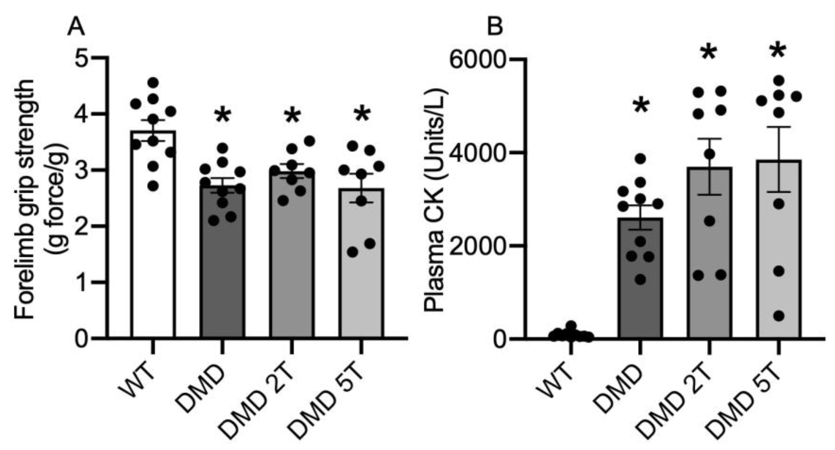
3.1. Dystropathology (Grip Strength, Plasma CK, and Other Parameters) in WT Compared with Untreated and Taurine-Treated DMDmdx Rats

At 12 weeks of age, the DMDmdx rat muscle produced 30% less forelimb grip force than WT rats, and neither dose of taurine had any effect on grip strength (Figure 1A).
DMDmdx rats had 11-fold more plasma CK than WT, with no effect of either dose of taurine (Figure 1B). Other measures of phenotype and dystropathology for WT and untreated and treated DMDmdx rats are summarised in Table 1. Differences for untreated DMDmdx rats (compared with WT) included significantly higher (1.1-fold) muscle weight for tibialis anterior muscle, and for plasma, (1.7-fold) higher levels of platelets, a higher (~3-fold) percentage of neutrophils, fewer (30%) lymphocytes, and a small increase in haematocrit. Neither dose of taurine had any effect on any parameter, except that both doses decreased the haematocrit back to WT levels.

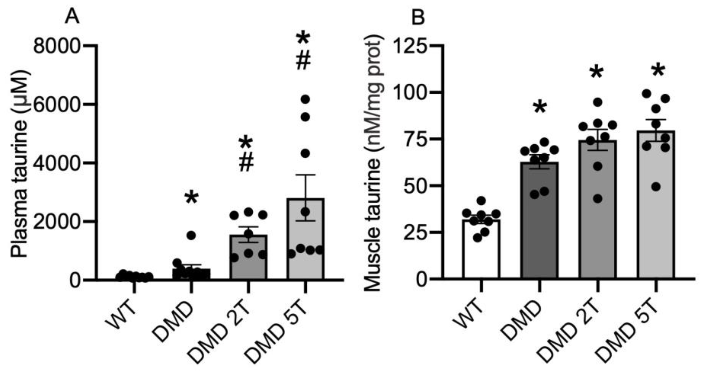
3.2. Plasma and Muscle Taurine Content in WT Compared with Untreated and Taurine-Treated DMDmdx Rats

Untreated DMDmdx rats had 3.5-fold more plasma taurine than WT control rats (Figure 2A). The 2 g/kg/day and 5 g/kg/day doses of taurine increased DMDmdx plasma taurine 4 and 7-fold, respectively (Figure 2A).
Skeletal muscles (biceps femoris) of untreated DMDmdx rats had 2-fold more taurine, compared with WT (Figure 2B) with no effect of either dose of taurine (Figure 2B).

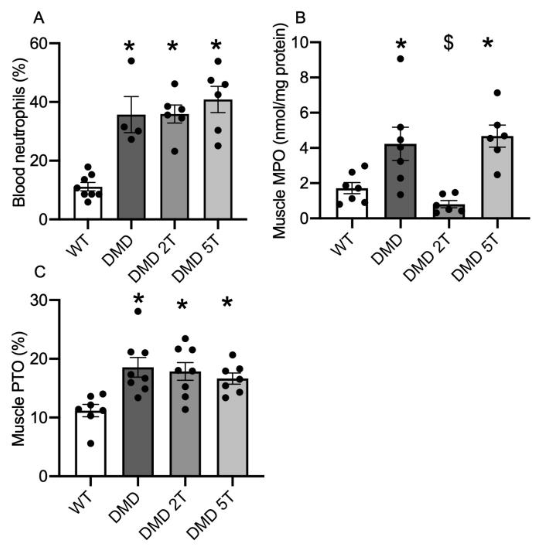
3.3. Inflammation and Oxidative Stress in the Plasma and Muscles of Taurine-Treated DMDmdx Rats

DMDmdx rats had 3.3-fold higher plasma neutrophils (%) than WT (Table 1, Figure 3A) and neither dose of taurine had any effect on plasma neutrophil levels (Figure 3A). Muscles (biceps femoris) of untreated DMDmdx rats also had 2-fold more MPO than WT (Figure 3B) and the 2 g/kg/day dose of taurine decreased DMDmdx muscle MPO by 80% (Figure 3B): in contrast the higher 5 g/kg/day taurine dose had no effect (Figure 3B). Protein thiol oxidation was 1.7-fold higher in muscles of DMDmdx rats compared with WT (Figure 3C) and neither dose of taurine had any effect on muscle protein thiol oxidation (Figure 3C).

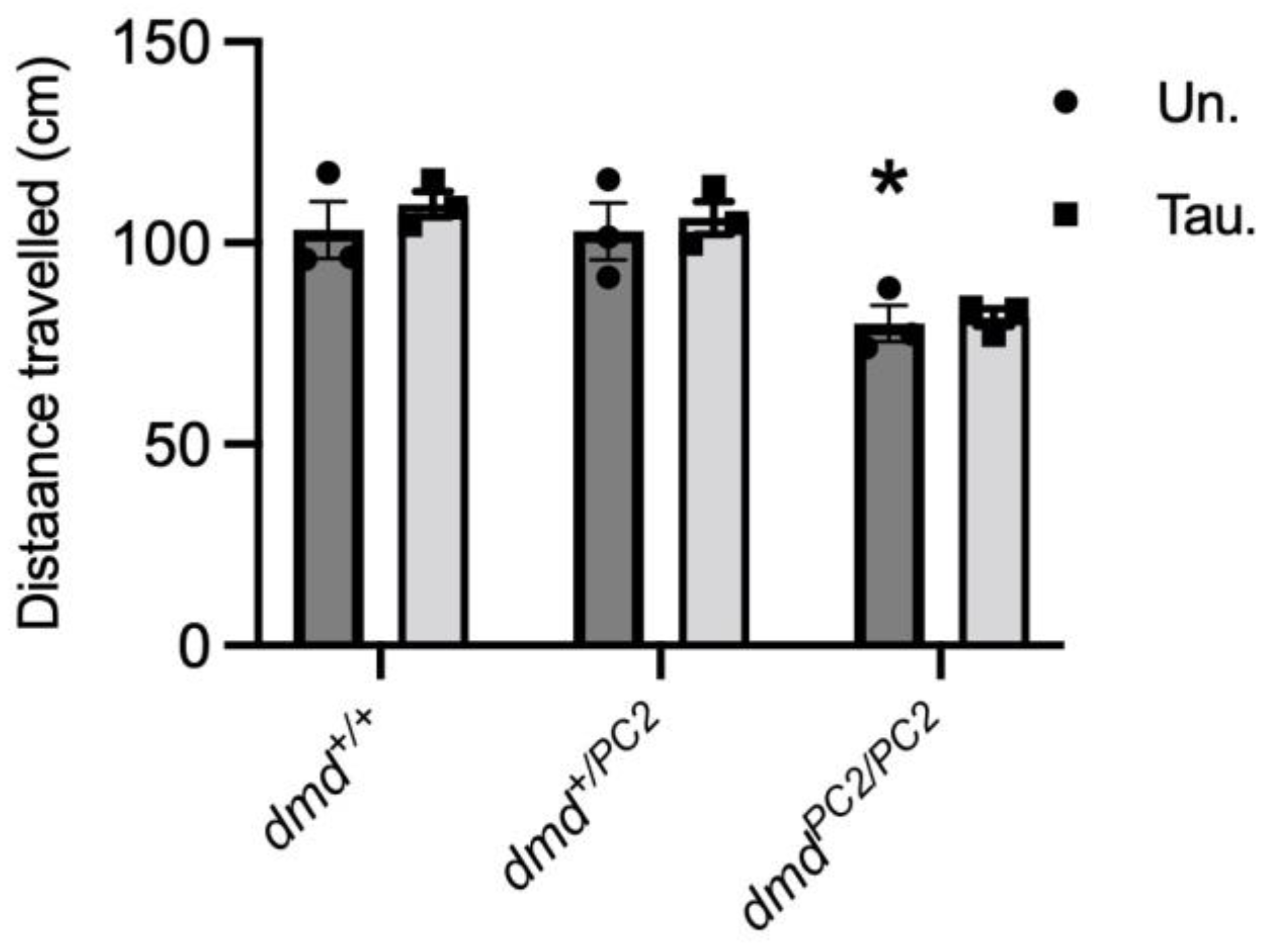
3.4. Muscle Function in Untreated and Taurine-Treated WT (dmd+/+), Heterozygous (dmd+/pc2) and Dystrophic Homozygous (dmdpc2/pc2) Zebrafish

We used locomotion assays on zebrafish at 6 dpf to determine if taurine treatment improved muscle function. Dystrophin-deficient dmdpc2/pc2 zebrafish travelled a distance of 25% and 24% less than WT dmd+/+ and dmd+/pc2 fish, respectively (p < 0.01) (Figure 4). Taurine treatment had no significant effect on the swimming performance of any genotype (Figure 4).

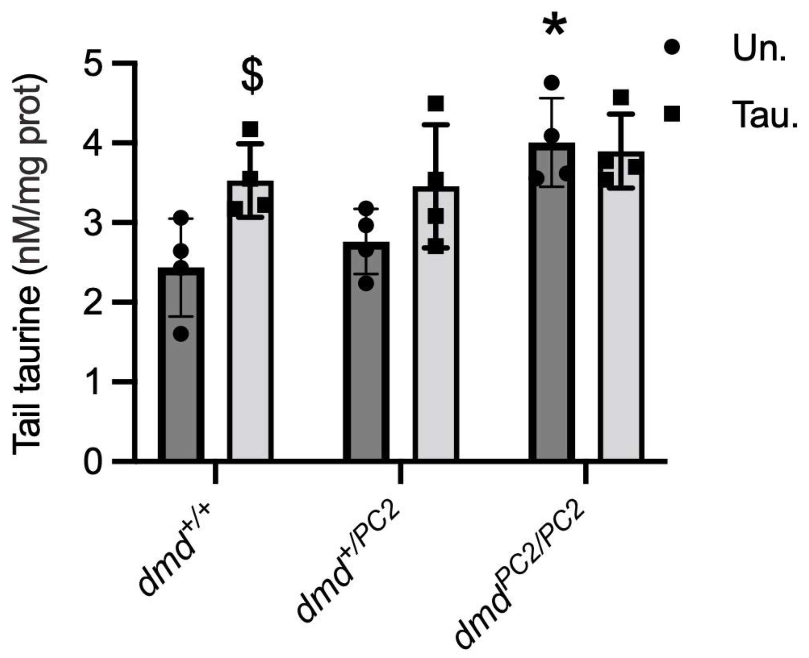
3.5. Taurine Content in Untreated and Taurine-Treated WT (dmd+/+), Heterozygous (dmd+/pc2) and Dystrophic Homozygous (dmdpc2/pc2) Zebrafish

Zebrafish tails were used to determine taurine levels since they are comprised mostly of skeletal muscle. Dystrophin-deficient dmdpc2/pc2 zebrafish tails had 1.7- and 1.5-fold higher taurine than WT dmd+/+ and heterozygote dmd+/pc2, respectively (Figure 5). While taurine treatment had no effect on taurine levels of dystrophin-deficient fish tails, taurine treatment significantly increased the taurine content in dmd+/+ (p < 0.001) and dmd+/pc2 (p = 0.014) tails (Figure 5).

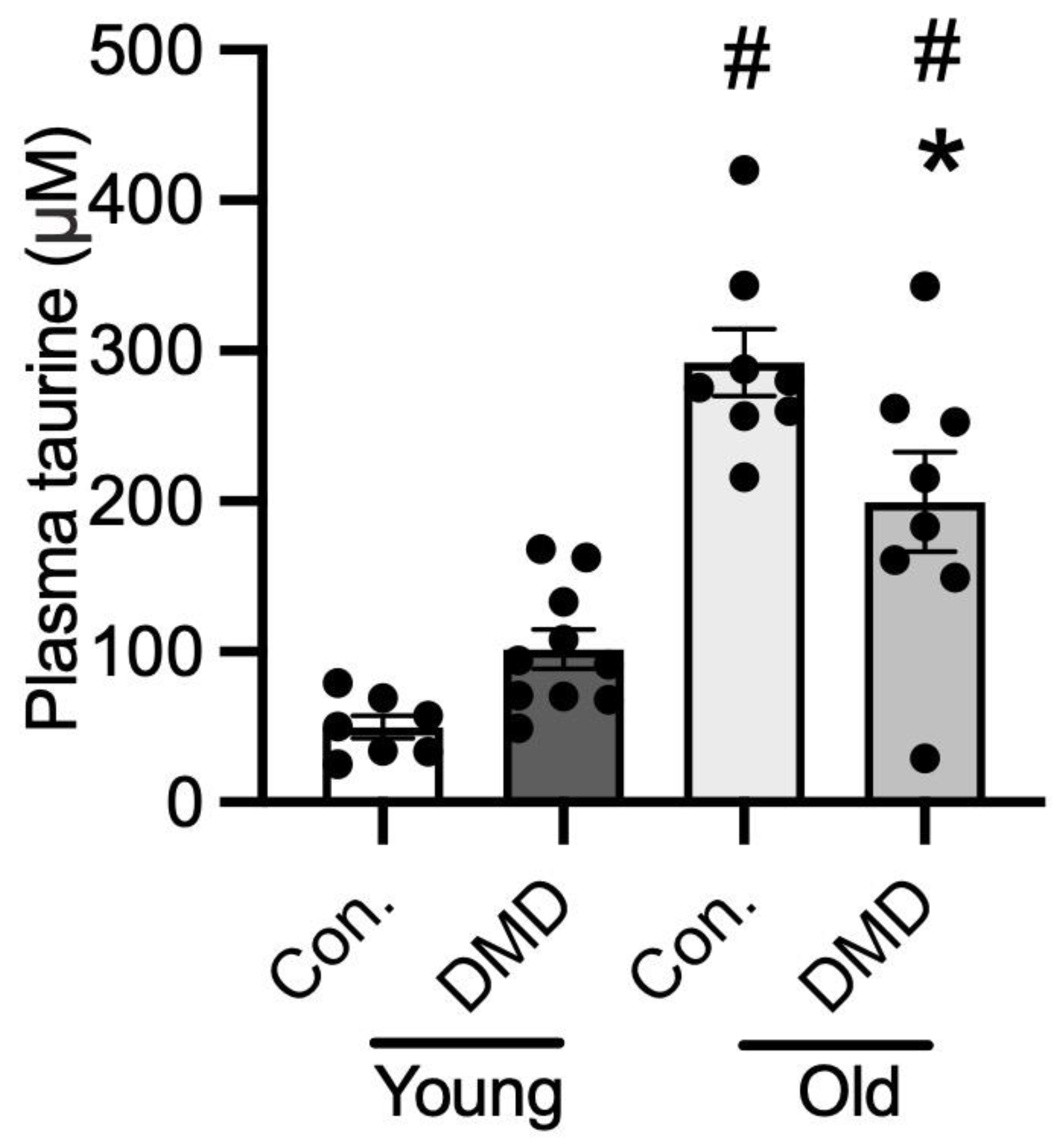
3.6. Taurine Content in Human Plasma of Normal Controls and Patients with DMD

There was no difference in plasma taurine content between young healthy controls (2–8 years) and ambulant DMD boys aged 2–8 (Figure 6). However, the older non-ambulant DMD patients (aged 16–20 years) had 30% lower plasma taurine than age-matched controls (aged 18–20 years). Non-ambulant DMD patients had 3-fold more plasma taurine than ambulant DMD boys (Figure 6). Also of note, plasma taurine in older controls in their late teens was 6-fold higher than for the younger controls aged under 10 years (Figure 6).

4. Discussion

We hypothesised that taurine treatment would reduce the severity of dystropathology in the DMDmdx rat and dmd zebrafish, two models for DMD that are considered more severe compared to the widely used mdx mouse. Taurine is considered important for the function of skeletal muscle, where it modulates ion channel function, membrane stability, and calcium homeostasis, as well as having anti-inflammatory and antioxidant properties [51,52,53,54,55,56]. However, we showed that taurine administration to DMDmdx rats had very little effect on the indices of dystropathology. Likewise, taurine administration to young dystrophic dmd zebrafish had no effect on muscle function (distance travelled). The surprising results for these two animal models contrast with our previous studies in young mdx mice (aged up to 6 weeks) where taurine had many benefits, with improved grip strength and decreased muscle necrosis, inflammation, oxidative stress, and plasma CK content [19,20,21]. Various benefits of taurine administration in mdx mice have also been widely reported by other groups for mice aged up to 12 months [22,23,24,25,26,27,28,57].
Interestingly, the endogenous plasma and muscle taurine levels in young adult DMDmdx rats aged 12 weeks were unexpectedly higher relative to WT rats and, while both doses of taurine increased levels of plasma taurine in DMDmdx rats, neither dose had an effect on muscle taurine levels. Likewise, taurine levels in dystrophic dmdpc2/pc2 zebrafish tails (mainly composed of skeletal muscle) were higher than WT dmd+/+ fish, and taurine treatment did not increase the taurine content of dystrophic dmdpc2/pc2 tails (but did in WT dmd+/+ fish). Taurine content of mdx muscle has been measured across the lifespan of mdx mice (summarised in Table 2). In most studies, muscle taurine levels in mdx muscle were either lower than, or equivalent, to WT (C57), and taurine was effective at reducing dystropathology. In one study (at 10 weeks), muscle taurine levels were observed to be higher than WT, and at this age, taurine was not effective at reducing dystropathology [28].
In these studies where taurine treatment was effective in reducing the severity of dystropathology, it was also observed that taurine treatment increased the taurine content of mdx muscle (summarised in Table 2). These previous results, combined with the new data in this current study, suggest that taurine is only efficacious in models where treatment improves the muscle content of taurine. This increase after exogenous taurine treatment was not observed in dystrophic models where intrinsic endogenous muscle taurine levels were high (i.e., DMDmdx rats, dmdpc2/pc2 zebrafish, and 10 week mdx mice [28]). Interestingly, in older mice (from 12 weeks) taurine in mdx plasma was higher than WT (summarised in Table 2). However, taurine treatment was still effective in older mdx mice, suggesting that the muscle content of taurine, rather than plasma content, is the important factor in determining efficacy. While the exact cause of perturbations in taurine levels in these models is not understood, we have previously explored taurine metabolism and transport in mdx mice [30]. We showed that taurine homeostasis, including synthesis in the liver, excretion in the kidney, and transport into the muscle was affected by dystrophy (and age). Future studies into taurine homeostasis in DMDmdx rats, dmdpc2/pc2 zebrafish, and even DMD patients would greatly help our understanding of the great variability in endogenous taurine levels in all of these species.
Since these combined results suggest that the ability of taurine to protect dystrophic muscle from damage is potentially determined by endogenous taurine levels, we thought it pertinent to measure taurine levels in DMD boys. As DMD muscle biopsies were not available, we measured taurine in archival plasma samples from DMD patients. While there was no significant difference in plasma taurine levels between young ambulant DMD (2–8 years) and normal boys of the same age, the older non-ambulant DMD boys with severe loss of muscle mass (16–20 years) had lower plasma taurine compared with normal boys. The observation that the normal teenage boys had markedly more taurine than younger normal boys, accords with previous reports that attributed this difference to the increased requirements of taurine during periods of peak growth [58]. Interestingly, in normal mice we observed that taurine levels were 5 and 10 times higher in the liver and plasma, respectively, in juvenile 18-day WT mice (during a period of peak growth just prior to weaning) compared with older WT mice [30]. These data support the need for high levels of taurine during normal growth. In mdx mice, this is also the age where we observed the biggest deficiencies in liver and plasma taurine in juvenile animals; this is also the time of peak myonecrosis [30] and the high metabolic demands of growth [59]. Benefits of taurine administration were pronounced in young mdx mice; however, this is in marked contrast with the situation in young zebrafish, presumably due to species differences. To further understand taurine metabolism in dystrophy, and to help assess the potential of taurine as a beneficial therapy for DMD, it would be informative to measure taurine levels in DMD muscles at different ages: unfortunately, this is difficult in DMD patients as it requires invasive muscle biopsies.
Finally, since the DMDmdx rat is a relatively new model, we thought it pertinent to discuss the phenotypical observations of the model from this current study (Table 1). Previous research has suggested that the model has a severe progressive phenotype compared with mdx mice [38] and our new data for DMDmdx rats show that at 12 weeks of age (young adult) dystropathology was evident as reduced grip strength and increased inflammation, oxidative stress, and plasma CK content compared with normal WT rats (although many other parameters measured were not affected). Studies of dystrophic sciatic nerves in DMDmdx rats (aged 8 months) suggest that there is more pronounced ongoing myonecrosis, compared with mdx mice [41]. A level of sustained myonecrosis was also reported for another dystrophic rat model with no significant difference in the extent of muscle degeneration between week 4 and week 13; these authors also concluded that the pathological progression is more pronounced in dystrophic rats compared with classic mdx mice [60]. It would be useful to have a more detailed description of the extent of myonecrosis, regeneration, and fibrosis in a range of skeletal muscles (e.g., various limb muscles, diaphragm, tongue) across the lifespan for dystrophic DMDmdx rats to compare with the wealth of information published for mdx mice.
In conclusion, taurine administration was not an effective treatment in young dystrophic DMDmdx rats analysed at 12 weeks of age, nor in young dmdpc2/pc2 zebrafish at 6 dpf. We hypothesise that this is due to the intrinsic high levels of taurine we observed in both models (compared to WT), and the resultant inability to therapeutically increase muscle taurine levels. We also showed that plasma taurine levels varied between young and older DMD patients. Limitations of this study include the specific ages of animals used, since pathology of dystrophic animals varies greatly across lifespan. Additionally, to understand the regulation in taurine levels in these different models, future studies could investigate taurine metabolism and transport, as both have been shown to be dysregulated in the mdx mouse [30]. Therefore, more research is required to better understand the potential of taurine to be used as a therapy for DMD boys.

Author Contributions

Conceptualisation, J.R.T., C.H., C.L.G., A.T., R.J.B.-R., T.E.S., M.D.G. and P.G.A.; methodology, J.R.T., C.H., C.L.G., A.L., D.C., A.T., R.J.B.-R. and T.E.S.; data curation, J.R.T.; writing—original draft preparation, J.R.T.; writing—review and editing, C.H., C.L.G., A.T., R.J.B.-R., T.E.S., M.D.G. and P.G.A.; project administration, P.G.A.; funding acquisition, P.G.A. All authors have read and agreed to the published version of the manuscript.

Funding

This research was supported by funding from Duchenne UK, Save Our Sons Duchenne Foundation (Australia), and a donation from Muscular Dystrophy Australia.

Institutional Review Board Statement

The study was conducted in accordance with the Declaration of Helsinki and approved by the Institutional Review Board (or Ethics Committee) of the University of Western Australia (2019/RA/4/20/6447). The animal study protocols were approved by the Ethics Committees of Nantes Université (APAFIS#10792–2017061316021120) and the University of Melbourne (22161).

Informed Consent Statement

Informed consent was obtained from all subjects involved in the study.

Data Availability Statement

Not applicable.

Acknowledgments

We thank Volker Straub and the Newcastle Biobank at Newcastle University for kindly providing the archival DMD samples.

Conflicts of Interest

The authors declare no conflict of interest.

References

  1. Bushby, K.; Finkel, R.; Birnkrant, D.J.; Case, L.E.; Clemens, P.R.; Cripe, L.; Kaul, A.; Kinnett, K.; McDonald, C.; Pandya, S.; et al. Diagnosis and management of Duchenne muscular dystrophy, part 1: Diagnosis, and pharmacological and psychosocial management. Lancet Neurol. 2010, 9, 77–93. [Google Scholar] [CrossRef]
  2. Emery, A.E. The muscular dystrophies. Lancet 2002, 359, 687–695. [Google Scholar] [CrossRef] [PubMed]
  3. Duan, D.; Goemans, N.; Takeda, S.I.; Mercuri, E.; Aartsma-Rus, A. Duchenne muscular dystrophy. Nat. Rev. Dis. Prim. 2021, 7, 13. [Google Scholar] [CrossRef] [PubMed]
  4. Grounds, M.D. Two-tiered hypotheses for Duchenne muscular dystrophy. Cell. Mol. Life Sci. 2008, 65, 1621–1625. [Google Scholar] [CrossRef] [PubMed]
  5. Kharraz, Y.; Guerra, J.; Pessina, P.; Serrano, A.L.; Munoz-Canoves, P. Understanding the process of fibrosis in Duchenne muscular dystrophy. Biomed. Res. Int. 2014, 2014, 965631. [Google Scholar] [CrossRef] [PubMed]
  6. Lapidos, K.A.; Kakkar, R.; McNally, E.M. The dystrophin glycoprotein complex: Signaling strength and integrity for the sarcolemma. Circ. Res. 2004, 94, 1023–1031. [Google Scholar] [CrossRef] [PubMed]
  7. Petrof, B.J.; Shrager, J.B.; Stedman, H.H.; Kelly, A.M.; Sweeney, H.L. Dystrophin protects the sarcolemma from stresses developed during muscle contraction. Proc. Natl. Acad. Sci. USA 1993, 90, 3710–3714. [Google Scholar] [CrossRef]
  8. Grounds, M.D.; Terrill, J.R.; Al-Mshhdani, B.A.; Duong, M.N.; Radley-Crabb, H.G.; Arthur, P.G. Biomarkers for Duchenne muscular dystrophy: Myonecrosis, inflammation and oxidative stress. Dis. Model. Mech. 2020, 13, dmm043638. [Google Scholar] [CrossRef]
  9. Biggar, W.D. Duchenne muscular dystrophy. Pediatr. Rev. 2006, 27, 83–88. [Google Scholar] [CrossRef]
  10. Verhaart, I.E.; Aartsma-Rus, A. Therapeutic developments for Duchenne muscular dystrophy. Nat. Rev. Neurol. 2019, 15, 373–386. [Google Scholar] [CrossRef]
  11. Bulfield, G.; Siller, W.; Wight, P.; Moore, K. X chromosome-linked muscular dystrophy (mdx) in the mouse. Proc. Natl. Acad. Sci. USA 1984, 81, 1189–1192. [Google Scholar] [CrossRef] [PubMed]
  12. Partridge, T.A. The mdx mouse model as a surrogate for Duchenne muscular dystrophy. FEBS J. 2013, 280, 4177–4186. [Google Scholar] [CrossRef]
  13. McGeachie, J.K.; Grounds, M.D.; Partridge, T.A.; Morgan, J.E. Age-related changes in replication of myogenic cells in mdx mice: Quantitative autoradiographic studies. J. Neurol. Sci. 1993, 119, 169–179. [Google Scholar] [CrossRef]
  14. Radley, H.G.; Grounds, M.D. Cromolyn administration (to block mast cell degranulation) reduces necrosis of dystrophic muscle in mdx mice. Neurobiol. Dis. 2006, 23, 387–397. [Google Scholar] [CrossRef] [PubMed]
  15. Radley, H.G.; Davies, M.J.; Grounds, M.D. Reduced muscle necrosis and long-term benefits in dystrophic mdx mice after cV1q (blockade of TNF) treatment. Neuromuscul. Disord. 2008, 18, 227–238. [Google Scholar] [CrossRef]
  16. Grounds, M.D.; Torrisi, J. Anti-TNF alpha (Remicade (R)) therapy protects dystrophic skeletal muscle from necrosis. FASEB J. 2004, 18, 676–682. [Google Scholar] [CrossRef]
  17. Lefaucheur, J.P.; Pastoret, C.; Sebille, A. Phenotype of dystrophinopathy in old mdx mice. Anat. Rec. 1995, 242, 70–76. [Google Scholar] [CrossRef] [PubMed]
  18. De Luca, A.; Pierno, S.; Camerino, D.C. Taurine: The appeal of a safe amino acid for skeletal muscle disorders. J. Transl. Med. 2015, 13, 243. [Google Scholar] [CrossRef]
  19. Terrill, J.R.; Webb, S.M.; Arthur, P.G.; Hackett, M.J. Investigation of the effect of taurine supplementation on muscle taurine content in the mdx mouse model of Duchenne muscular dystrophy using chemically specific synchrotron imaging. Analyst 2020, 145, 7242–7251. [Google Scholar] [CrossRef]
  20. Terrill, J.R.; Pinniger, G.J.; Graves, J.A.; Grounds, M.D.; Arthur, P.G. Increasing taurine intake and taurine synthesis improves skeletal muscle function in the mdx mouse model for Duchenne muscular dystrophy. J. Physiol. 2016, 594, 3095–3110. [Google Scholar] [CrossRef] [Green Version]
  21. Terrill, J.R.; Grounds, M.D.; Arthur, P.G. Increased taurine in pre-weaned juvenile mdx mice greatly reduces the acute onset of myofibre necrosis and dystropathology and prevents inflammation. PLoS Curr. 2016, 8, ecurrents. [Google Scholar] [CrossRef] [PubMed]
  22. Cozzoli, A.; Rolland, J.F.; Capogrosso, R.F.; Sblendorio, V.T.; Longo, V.; Simonetti, S.; Nico, B.; De Luca, A. Evaluation of potential synergistic action of a combined treatment with alpha-methyl-prednisolone and taurine on the mdx mouse model of Duchenne muscular dystrophy. Neuropathol. Appl. Neurobiol. 2011, 37, 243–256. [Google Scholar] [CrossRef] [PubMed]
  23. De Luca, A.; Pierno, S.; Liantonio, A.; Cetrone, M.; Camerino, C.; Fraysse, B.; Mirabella, M.; Servidei, S.; Ruegg, U.T.; Conte Camerino, D. Enhanced dystrophic progression in mdx mice by exercise and beneficial effects of taurine and insulin-like growth factor-1. J. Pharmacol. Exp. Ther. 2003, 304, 453–463. [Google Scholar] [CrossRef]
  24. De Luca, A.; Pierno, S.; Liantonio, A.; Cetrone, M.; Camerino, C.; Simonetti, S.; Papadia, F.; Camerino, D.C. Alteration of excitation-contraction coupling mechanism in extensor digitorum longus muscle fibres of dystrophic mdx mouse and potential efficacy of taurine. Br. J. Pharmacol. 2001, 132, 1047–1054. [Google Scholar] [CrossRef] [PubMed]
  25. Capogrosso, R.F.; Cozzoli, A.; Mantuano, P.; Camerino, G.M.; Massari, A.M.; Sblendorio, V.T.; De Bellis, M.; Tamma, R.; Giustino, A.; Nico, B.; et al. Assessment of resveratrol, apocynin and taurine on mechanical-metabolic uncoupling and oxidative stress in a mouse model of duchenne muscular dystrophy: A comparison with the gold standard, alpha-methyl prednisolone. Pharmacol. Res. 2016, 106, 101–113. [Google Scholar] [CrossRef] [PubMed]
  26. Mele, A.; Mantuano, P.; De Bellis, M.; Rana, F.; Sanarica, F.; Conte, E.; Morgese, M.G.; Bove, M.; Rolland, J.-F.; Capogrosso, R.F. A long-term treatment with taurine prevents cardiac dysfunction in mdx mice. Transl. Res. 2019, 204, 82–99. [Google Scholar] [CrossRef] [PubMed]
  27. Horvath, D.M.; Murphy, R.M.; Mollica, J.P.; Hayes, A.; Goodman, C.A. The effect of taurine and β-alanine supplementation on taurine transporter protein and fatigue resistance in skeletal muscle from mdx mice. Amino Acids 2016, 48, 2635–2645. [Google Scholar] [CrossRef] [PubMed]
  28. Barker, R.G.; Horvath, D.; van der Poel, C.; Murphy, R.M. Benefits of prenatal taurine supplementation in preventing the onset of acute damage in the Mdx mouse. PLoS Curr. 2017, 9, ecurrents.md.9a3e357a0154d01050b591601cbd4fdb. [Google Scholar]
  29. Terrill, J.R.; Boyatzis, A.; Grounds, M.D.; Arthur, P.G. Treatment with the cysteine precursor l-2-oxothiazolidine-4-carboxylate (OTC) implicates taurine deficiency in severity of dystropathology in mdx mice. Int. J. Biochem. Cell Biol. 2013, 45, 2097–2108. [Google Scholar] [CrossRef]
  30. Terrill, J.R.; Grounds, M.D.; Arthur, P.G. Taurine deficiency, synthesis and transport in the mdx mouse model for Duchenne Muscular Dystrophy. Int. J. Biochem. Cell Biol. 2015, 66, 141–148. [Google Scholar] [CrossRef]
  31. Griffin, J.; Williams, H.; Sang, E.; Clarke, K.; Rae, C.; Nicholson, J. Metabolic Profiling of Genetic Disorders: A Multitissue1 H Nuclear Magnetic Resonance Spectroscopic and Pattern Recognition Study into Dystrophic Tissue. Anal. Biochem. 2001, 293, 16–21. [Google Scholar] [CrossRef] [PubMed]
  32. McIntosh, L.; Granberg, K.E.; Brière, K.M.; Anderson, J.E. Nuclear magnetic resonance spectroscopy study of muscle growth, mdx dystrophy and glucocorticoid treatments: Correlation with repair. NMR Biomed. 1998, 11, 1–10. [Google Scholar] [CrossRef]
  33. Terrill, J.R.; Duong, M.N.; Turner, R.; Le Guiner, C.; Boyatzis, A.; Kettle, A.J.; Grounds, M.D.; Arthur, P.G. Levels of inflammation and oxidative stress, and a role for taurine in dystropathology of the Golden Retriever Muscular Dystrophy dog model for Duchenne Muscular Dystrophy. Redox Biol. 2016, 9, 276–286. [Google Scholar] [CrossRef] [PubMed]
  34. Terrill, J.R.; Al-Mshhdani, B.A.; Duong, M.N.; Wingate, C.D.; Abbas, Z.; Baustista, A.P.; Bettis, A.K.; Balog-Alvarez, C.J.; Kornegay, J.N.; Nghiem, P.P.; et al. Oxidative damage to urinary proteins from the GRMD dog and mdx mouse as biomarkers of dystropathology in Duchenne muscular dystrophy. PLoS ONE 2020, 15, e0240317. [Google Scholar] [CrossRef]
  35. Berger, J.; Berger, S.; Jacoby, A.S.; Wilton, S.D.; Currie, P.D. Evaluation of exon-skipping strategies for Duchenne muscular dystrophy utilizing dystrophin-deficient zebrafish. J. Cell. Mol. Med. 2011, 15, 2643–2651. [Google Scholar] [CrossRef]
  36. Gibbs, E.M.; Horstick, E.J.; Dowling, J.J. Swimming into prominence: The zebrafish as a valuable tool for studying human myopathies and muscular dystrophies. FEBS J. 2013, 280, 4187–4197. [Google Scholar] [CrossRef]
  37. Kawahara, G.; Karpf, J.A.; Myers, J.A.; Alexander, M.S.; Guyon, J.R.; Kunkel, L.M. Drug screening in a zebrafish model of Duchenne muscular dystrophy. Proc. Natl. Acad. Sci. USA 2011, 108, 5331–5336. [Google Scholar] [CrossRef]
  38. Larcher, T.; Lafoux, A.; Tesson, L.; Remy, S.; Thepenier, V.; François, V.; Le Guiner, C.; Goubin, H.; Dutilleul, M.; Guigand, L. Characterization of dystrophin deficient rats: A new model for Duchenne muscular dystrophy. PLoS ONE 2014, 9, e110371. [Google Scholar] [CrossRef]
  39. Szabó, P.L.; Ebner, J.; Koenig, X.; Hamza, O.; Watzinger, S.; Trojanek, S.; Abraham, D.; Todt, H.; Kubista, H.; Schicker, K. Cardiovascular phenotype of the Dmdmdx rat–a suitable animal model for Duchenne muscular dystrophy. Dis. Model. Mech. 2021, 14, dmm047704. [Google Scholar] [CrossRef]
  40. Caudal, D.; François, V.; Lafoux, A.; Ledevin, M.; Anegon, I.; Le Guiner, C.; Larcher, T.; Huchet, C. Characterization of brain dystrophins absence and impact in dystrophin-deficient Dmdmdx rat model. PLoS ONE 2020, 15, e0230083. [Google Scholar] [CrossRef]
  41. Krishnan, V.S.; Thanigaiarasu, L.P.; White, R.; Crew, R.; Larcher, T.; Le Guiner, C.; Grounds, M.D. Dystrophic Dmdmdx rats show early neuronal changes (increased S100β and Tau5) at 8 months, supporting severe dystropathology in this rodent model of Duchenne muscular dystrophy. Mol. Cell. Neurosci. 2020, 108, 103549. [Google Scholar] [CrossRef] [PubMed]
  42. Bourdon, A.; François, V.; Zhang, L.; Lafoux, A.; Fraysse, B.; Toumaniantz, G.; Larcher, T.; Girard, T.; Ledevin, M.; Lebreton, C. Evaluation of the dystrophin carboxy-terminal domain for micro-dystrophin gene therapy in cardiac and skeletal muscles in the DMDmdx rat model. Gene Ther. 2022, 29, 520–535. [Google Scholar] [CrossRef]
  43. Westerfield, M. The Zebrafish Book: A Guide for the Laboratory Use of Zebrafish. 2000. Available online: http://zfin.org/zf_info/zfbook/zfbk.html (accessed on 11 May 2021).
  44. Messineo, A.M.; Gineste, C.; Sztal, T.E.; McNamara, E.L.; Vilmen, C.; Ogier, A.C.; Hahne, D.; Bendahan, D.; Laing, N.G.; Bryson-Richardson, R.J. L-tyrosine supplementation does not ameliorate skeletal muscle dysfunction in zebrafish and mouse models of dominant skeletal muscle α-actin nemaline myopathy. Sci. Rep. 2018, 8, 11490. [Google Scholar] [CrossRef] [PubMed]
  45. Sztal, T.E.; Ruparelia, A.A.; Williams, C.; Bryson-Richardson, R.J. Using touch-evoked response and locomotion assays to assess muscle performance and function in zebrafish. JoVE (J. Vis. Exp.) 2016, e54431. [Google Scholar] [CrossRef]
  46. Burch, P.M.; Pogoryelova, O.; Goldstein, R.; Bennett, D.; Guglieri, M.; Straub, V.; Bushby, K.; Lochmüller, H.; Morris, C. Muscle-derived proteins as serum biomarkers for monitoring disease progression in three forms of muscular dystrophy. J. Neuromuscul. Dis. 2015, 2, 241–255. [Google Scholar] [CrossRef] [PubMed]
  47. Setsukinai, K.; Urano, Y.; Kakinuma, K.; Majima, H.J.; Nagano, T. Development of novel fluorescence probes that can reliably detect reactive oxygen species and distinguish specific species. J. Biol. Chem. 2003, 278, 3170–3175. [Google Scholar] [CrossRef] [PubMed]
  48. Winterbourn, C.C.; Vissers, M.C.M.; Kettle, A.J. Myeloperoxidase. Curr. Opin. Hematol. 2000, 7, 53–58. [Google Scholar] [CrossRef] [PubMed]
  49. Winterbourn, C.C.; Kettle, A.J. Biomarkers of myeloperoxidase-derived hypochlorous acid. Free Radic. Biol. Med. 2000, 29, 403–409. [Google Scholar] [CrossRef]
  50. Armstrong, A.E.; Zerbes, R.; Fournier, P.A.; Arthur, P.G. A fluorescent dual labeling technique for the quantitative measurement of reduced and oxidized protein thiols in tissue samples. Free Radic. Biol. Med. 2011, 50, 510–517. [Google Scholar] [CrossRef]
  51. Bakker, A.J.; Berg, H.M. Effect of taurine on sarcoplasmic reticulum function and force in skinned fast-twitch skeletal muscle fibres of the rat. J. Physiol. 2004, 538, 185–194. [Google Scholar] [CrossRef]
  52. Hamilton, E.J.; Berg, H.M.; Easton, C.J.; Bakker, A.J. The effect of taurine depletion on the contractile properties and fatigue in fast-twitch skeletal muscle of the mouse. Amino Acids 2006, 31, 273–278. [Google Scholar] [CrossRef] [PubMed]
  53. Huxtable, R.J. Physiological actions of taurine. Physiol. Rev. 1992, 72, 101–163. [Google Scholar] [CrossRef] [PubMed]
  54. Warskulat, U.; Flogel, U.; Jacoby, C.; Hartwig, H.G.; Thewissen, M.; Merx, M.W.; Molojavyi, A.; Heller-Stilb, B.; Schrader, J.; Haussinger, D. Taurine transporter knockout depletes muscle taurine levels and results in severe skeletal muscle impairment but leaves cardiac function uncompromised. FASEB J. 2004, 18, 577–579. [Google Scholar] [CrossRef] [PubMed]
  55. Warskulat, U.; Heller-Stilb, B.; Oermann, E.; Zilles, K.; Haas, H.; Lang, F.; Haussinger, D. Phenotype of the taurine transporter knockout mouse. Methods Enzymol. 2007, 428, 439–458. [Google Scholar] [CrossRef]
  56. Camerino, D.C.; Tricarico, D.; Pierno, S.; Desaphy, J.F.; Liantonio, A.; Pusch, M.; Burdi, R.; Camerino, C.; Fraysse, B.; De Luca, A. Taurine and skeletal muscle disorders. Neurochem. Res. 2004, 29, 135–142. [Google Scholar] [CrossRef]
  57. Terrill, J.R.; Pinniger, G.J.; Nair, K.V.; Grounds, M.D.; Arthur, P.G. Beneficial effects of high dose taurine treatment in juvenile dystrophic mdx mice are offset by growth restriction. PLoS ONE 2017, 12, e0187317. [Google Scholar] [CrossRef]
  58. Gregory, D.M.; Sovetts, D.; Clow, C.L.; Scriver, C.R. Plasma free amino acid values in normal children and adolescents. Metabolism 1986, 35, 967–969. [Google Scholar] [CrossRef]
  59. Radley-Crabb, H.G.; Marini, J.C.; Sosa, H.A.; Castillo, L.I.; Grounds, M.D.; Fiorotto, M.L. Dystropathology increases energy expenditure and protein turnover in the mdx mouse model of duchenne muscular dystrophy. PLoS ONE 2014, 9, e89277. [Google Scholar] [CrossRef]
  60. Nakamura, K.; Fujii, W.; Tsuboi, M.; Tanihata, J.; Teramoto, N.; Takeuchi, S.; Naito, K.; Yamanouchi, K.; Nishihara, M. Generation of muscular dystrophy model rats with a CRISPR/Cas system. Sci. Rep. 2014, 4, 5635. [Google Scholar] [CrossRef] [Green Version]
Figure 1. Markers of dystropathology, grip strength (A), and plasma CK (B), in WT and untreated and taurine-treated DMDmdx rats (at 2 doses) aged 12 weeks. * = significantly (p < 0.05) different to WT. Bars represent mean ± SEM and n = 8–10 rats per group.
Metabolites 13 00232 g001
Figure 2. Levels of taurine in plasma (A) and muscle (B) in plasma, from WT and untreated and taurine-treated DMDmdx rats (at 2 doses), aged 12 weeks. * = significantly (p < 0.05) different to WT. # = significant (p < 0.05) different to untreated DMDmdx rats. Bars represent mean ± SEM and n = 8–10 per group.
Metabolites 13 00232 g002
Figure 3. Markers of inflammation and oxidative stress in WT and untreated and taurine-treated DMDmdx rats (at 2 doses) aged 12 weeks. Levels of plasma neutrophils (A) and muscle MPO (B) and oxidative stress measured as protein thiol oxidation (PTO) (C). * = significantly (p < 0.05) different to WT. $ = significantly (p < 0.05) different to untreated DMDmdx rats. Bars represent mean ± SEM and n = 8–10 per group.
Metabolites 13 00232 g003
Figure 4. Muscle function (distance travelled) of 6 dpf WT (dmd+/+), heterozygous (dmd+/−) and homozygous (pc2/pc) dystrophic (dmd−/−) zebrafish treated with either 1 mM taurine or water (control) from 2 to 6 dpf. * = significantly (p < 0.05) different to WT. Bars represent mean ± SEM. For dmd+/+: n = 30, 27, 33 for untreated and n = 27, 22, 26 for taurine-treated; for dmd+/pc2: n = 48, 48, 55 for untreated and n = 56, 62, 55 for taurine-treated, and for dmdpc2/pc2: n = 41, 23, 28 for untreated and n = 32, 25, 37 for taurine-treated.
Metabolites 13 00232 g004
Figure 5. Levels of taurine in tails of WT (dmd+/+), heterozygous (dmd+/−) and homozygous (pc2/pc) dystrophic (dmd−/−) zebrafish treated with either 1 mM taurine (tau) or water (un) from 2 to 6 dpf. * = significantly (p < 0.05) different to WT. $ = significantly (p < 0.05) different to the untreated of same strain. Bars represent mean ± SEM and n = 4 (pooled samples of 12) per group.
Metabolites 13 00232 g005
Figure 6. Human plasma levels of taurine in control males and patients with DMD. Compared for healthy normal aged-matched (A–M) controls and patients with DMD: ambulant (2–6 years) and non-ambulant (16–20 years). * = significantly (p < 0.05) different to healthy age-matched control. # = significantly (p < 0.05) different to young healthy age-matched control and ambulant DMD. Bars represent mean ± SEM and n = 8–10 per group.
Metabolites 13 00232 g006
Table 1. Measures of phenotype and dystropathology in normal WT rats, compared with untreated and taurine-treated DMDmdx rats at 12 weeks of age (data collected in France). * = DMDmdx rats significantly (p < 0.05) different to WT control. $ = taurine treated DMDmdx rats significantly (p < 0.05) different to untreated DMDmdx rats. Values represent mean ± SEM and n = 8–10 per group. TA = tibialis anterior muscle, EDL = extensor digitorum longus muscle and Sol = soleus muscle.
ParameterWildtypeDMDmdxDMDmdx
Taurine Treated-
2 g/kg/day
DMDmdx
Taurine Treated-
5 g/kg/day
Body weight (g)497 ± 42433 ± 35438 ± 27433 ± 33
TA weight (mg)831 ± 12952 ± 38 *987 ± 291019 ± 71
EDL weight (mg)221 ± 5237 ± 9241 ± 9218 ± 13
Sol weight (mg)207 ± 4213 ± 7214 ± 8192 ± 12
Distance travelled (cm)2305 ± 2091937 ± 1971885 ± 1622012 ± 113
Activity time (s)196 ± 9181 ± 13179 ± 10191 ± 6
Number rearings18 ± 211 ± 27 ± 2 *12 ± 2
Platelets (Giga/L)963 ± 351645 ± 52 *1527 ± 37 *1543 ± 50 *
Neutrophils (%)11 ± 136 ± 6 *36 ± 3 *41 ± 5 *
Lymphocytes (%)87 ± 260 ± 7 *58 ± 3 *54 ± 5 *
Hematocrit (%)45 ± 148 ± 1 *44 ± 0.4 $45 ± 1$
Leucocytes (Giga/L)8 ± 110 ± 110 ± 111 ± 1
Monocytes (%)1.2 ± 13.7 ± 14.7 ± 0.53.5 ± 1
Table 2. Taurine content of normal and DMD animal models and DMD patients at various ages. Models bolded are data from the current study. ↑ = increased taurine content compared to wildtype/normal control, ↓ = decreased, and — = no change in taurine content compared to wildtype/normal control, ✓ = improvement in dystropathology after taurine treatment, ✘ = no improvement in dystropathology after taurine treatment, N = not measured.
Model AgeTaurine in
Muscle
Taurine in PlasmaTaurine EfficacyReference
mdx mouse 18 days [30]
<3 weeksNN[32]
23 daysN[21]
4 weeks ↓/—[30]/[28]
6 weeks [19,20,30,57]
10 weeks[28]
12 weeks N[29]
6 monthsN[27]
6–8 monthsN[24]
12 months Cardiac muscle only[26]
DMDmdx rats12 weeks
dmdzebrafish6 dpf↑ (tails)N
GRMD dogs8 months N[33]
DMD patients2–6 years NN
16–20 years NN
Disclaimer/Publisher’s Note: The statements, opinions and data contained in all publications are solely those of the individual author(s) and contributor(s) and not of MDPI and/or the editor(s). MDPI and/or the editor(s) disclaim responsibility for any injury to people or property resulting from any ideas, methods, instructions or products referred to in the content.

