Article

Upregulation of Nrf2/HO-1 Signaling and Attenuation of Oxidative Stress, Inflammation, and Cell Death Mediate the Protective Effect of Apigenin against Cyclophosphamide Hepatotoxicity

by
Wesam Al-Amarat
1,
Mohammad H. Abukhalil
2,3,
Reem S. Alruhaimi
4,
Haifa A. Alqhtani
4,
Nouf Aldawood
4,
Manal A. Alfwuaires
5,
Osama Y. Althunibat
2,
Saleem H. Aladaileh
2,6,
Abdulmohsen I. Algefare
5,
Abdulkareem A. Alanezi
7,
Ali M. AbouEl-ezz
8,
Ahmad F. Ahmeda
9,10 and
Ayman M. Mahmoud
11,12,*
1
Department of Medical Support, Al-karak University College, Al-Balqa’ Applied University, As-Salt 206, Jordan
2
Department of Medical Analysis, Princess Aisha Bint Al-Hussein College of Nursing and Health Sciences, Al-Hussein Bin Talal University, Ma’an 71111, Jordan
3
Department of Biology, College of Science, Al-Hussein Bin Talal University, Ma’an 71111, Jordan
4
Department of Biology, College of Science, Princess Nourah bint Abdulrahman University, Riyadh 11671, Saudi Arabia
5
Department of Biological Sciences, Faculty of Science, King Faisal University, Al-Ahsa 31982, Saudi Arabia
6
Department of Pharmacy Practice, College of Pharmacy, University of Hafr Al-Batin, Hafr Al-Batin 31991, Saudi Arabia
7
Department of Pharmaceutics, College of Pharmacy, University of Hafr Al-Batin, Hafr Al-Batin 31991, Saudi Arabia
8
Department of Molecular Biology, Faculty of Science, New Mansoura University, Mansoura 35511, Egypt
9
Department of Basic Medical Sciences, College of Medicine, Ajman University, Ajman 346, United Arab Emirates
10
Center of Medical and Bio-allied Health Sciences Research, Ajman University, Ajman 346, United Arab Emirates
11
Physiology Division, Zoology Department, Faculty of Science, Beni-Suef University, Beni-Suef 62514, Egypt
12
Department of Life Sciences, Faculty of Science and Engineering, Manchester Metropolitan University, Manchester M1 5GD, UK
*
Author to whom correspondence should be addressed.
Metabolites 2022, 12(7), 648; https://doi.org/10.3390/metabo12070648
Submission received: 21 June 2022 / Revised: 8 July 2022 / Accepted: 12 July 2022 / Published: 14 July 2022
(This article belongs to the Special Issue Dietary Antioxidants and Metabolic Diseases)

Abstract

:
Liver injury is among the adverse effects of the chemotherapeutic agent cyclophosphamide (CP). This study investigated the protective role of the flavone apigenin (API) against CP-induced liver damage, pointing to the involvement of Nrf2/HO-1 signaling. Rats were treated with API (20 and 40 mg/kg) for 15 days and received CP (150 mg/kg) on day 16. CP caused liver damage manifested by an elevation of transaminases, alkaline phosphatase (ALP), and lactate dehydrogenase (LDH), and histological alterations, including granular vacuolation, mononuclear cell infiltration, and hydropic changes. Hepatic reactive oxygen species (ROS), malondialdehyde (MDA), and nitric oxide (NO) were increased and glutathione (GSH) and antioxidant enzymes were decreased in CP-administered rats. CP upregulated the inflammatory markers NF-κB p65, TNF-α, IL-6, and iNOS, along with the pro-apoptotic Bax and caspase-3. Pre-treatment with API ameliorated circulating transaminases, ALP, and LDH, and prevented histopathological changes in CP-intoxicated rats. API suppressed ROS, MDA, NO, NF-κB p65, iNOS, inflammatory cytokines, oxidative DNA damage, Bax, and caspase-3 in CP-intoxicated rats. In addition, API enhanced hepatic antioxidants and Bcl-2 and boosted the Nrf2 and HO-1 mRNA abundance and protein. In conclusion, API is effective in preventing CP hepatotoxicity by attenuating oxidative stress, the inflammatory response, and apoptosis. The hepatoprotective efficacy of API was associated with the upregulation of Nrf2/HO-1 signaling.

1. Introduction

Cyclophosphamide (CP) is an alkylating chemotherapeutic agent broadly employed in the therapy of numerous human malignancies [1,2,3]. CP has been approved by the FDA for use in the treatment of stage III and IV lymphomas, including lymphocytic Hodgkin and non-Hodgkin lymphomas [4,5]. CP is used as an immunosuppressant to prevent post-transplant rejection and graft-vs.-host complications; however, its therapeutic application is associated with serious adverse effects, such as hemorrhagic cystitis, myelosuppression, kidney injury, amenorrhea, lung injury, cardiotoxicity, and hepatotoxicity [1,3,6,7,8,9,10,11,12,13]. The metabolism of CP within the liver through the action of cytochrome P-450 produces acrolein and phosphoramide mustard. Acrolein has no antitumor efficacy while phosphoramide mustard is responsible for the majority of the antineoplastic activity of CP [2]. Phosphoramide mustard induces apoptosis via irreversible inhibition of protein synthesis through DNA cross-linking [2]. Acrolein is a highly reactive metabolite with a short half-life and can damage cellular macromolecules [14]. Acrolein can induce toxicity via various direct and indirect mechanisms, including DNA and protein adduction, excessive reactive oxygen species (ROS) generation, and induction of mitochondrial and endoplasmic reticulum (ER) stresses [14]. Excess ROS can damage cellular macromolecules, resulting in lipid peroxidation (LPO) and oxidative DNA and protein damage [15,16]. In addition, ROS can elicit proinflammatory responses via the activation of nuclear factor-kappaB (NF-κB) and the release of proinflammatory mediators [17,18]. Excess ROS and proinflammatory cytokines work in concert to induce cell death via apoptosis [19,20]. Therefore, attenuation of surplus ROS and cytokine production can protect hepatocytes against CP toxicity.
Apigenin (API; 4,5,7-trihydroxyflavone, Figure 1) is a member of the flavone class that is naturally abundant in various fruits and vegetables. Since ancient times, API has been wieldy applied in traditional medicine because of its antioxidant, hepatoprotective, antiviral, and anti-inflammatory activities [21]. Many recent reports have also emphasized that API exhibits a protective role against chemically induced hepatic toxicity via boosting the antioxidant and ROS scavenging properties [22,23,24,25]. Although numerous pharmacological activities of API have been explored, its potential to attenuate CP-induced hepatic damage has not been investigated. Therefore, this study evaluated the potential of API to protect against oxidative stress, inflammation, and liver injury induced by CP in rats, pointing to the possible involvement of nuclear factor (erythroid-derived 2)-like 2/heme oxygenase1 (Nrf2/HO-1) signaling. Nrf2 is a cytoprotective transcription factor that controls antioxidant defense enzymes and prevents oxidative damage [26,27,28]. Cytosolic Nrf2 is sequestered by Keap1 under physiological conditions and dissociates and translocates into the nucleus upon exposure to ROS or electrophiles, where it binds the antioxidant response element (ARE) and promotes the transcription of HO-1 and other defensive enzymes [26]. The role of Nrf2 activation in preventing CP-induced liver injury has been previously demonstrated [17,29,30,31,32]. Thus, we hypothesized that upregulation f Nrf2/HO-1 is involved in the protective effect of API against liver injury induced by CP.

2. Results

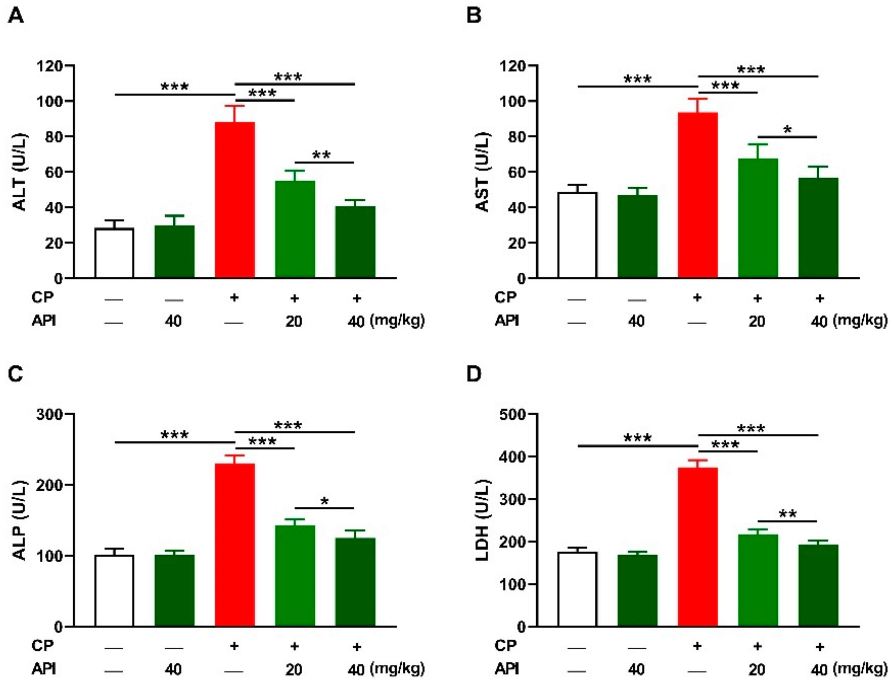
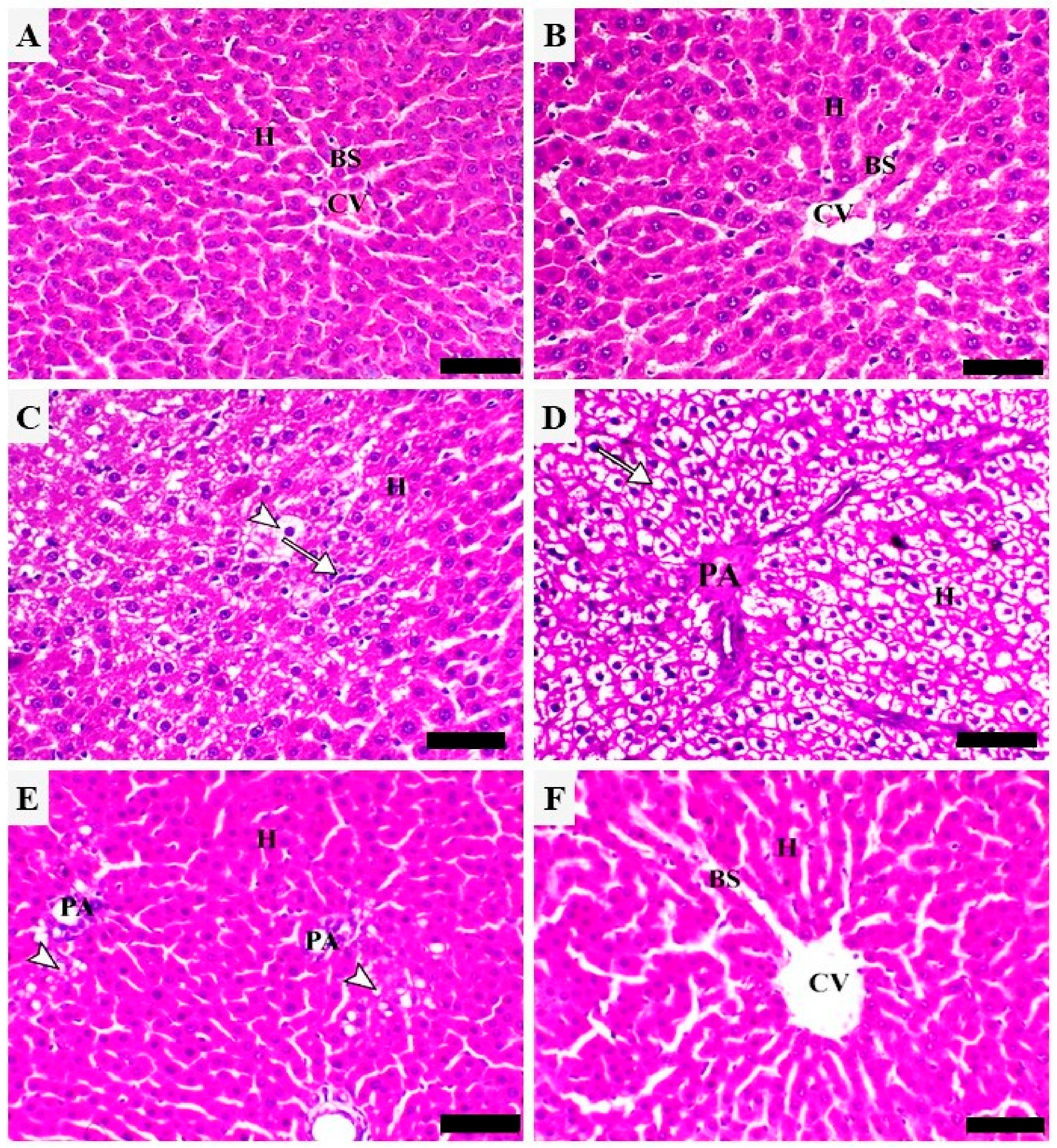
2.1. API Prevents CP-Induced Hepatocyte Injury in Rats

The protective effect of API against hepatocyte damage induced by CP was evaluated via measurement of circulating markers (Figure 2) and microscopic examination of stained sections (Figure 3). CP increased alanine aminotransferase (ALT), aspartate aminotransferase (AST), alkaline phosphatase (ALP), and lactate dehydrogenase (LDH) in the serum of rats (p < 0.001) as depicted in Figure 2A–D. API ameliorated transaminases, ALP, and LDH in CP-intoxicated rats in a dose-dependent manner. Rats that received 40 mg/kg API exhibited no changes in liver function markers.
Both control (Figure 3A) and API-treated rats (Figure 3B) showed normal histology of the hepatic tissue, where hepatocytes appeared normal and radially arranged in cords around central veins. CP-intoxicated rats exhibited multiple histopathological changes, including degenerative changes, marked granular hepatic vacuolation, mononuclear cell infiltration, and advanced hydropic changes (Figure 3C,D). Treatment with the low dose of API improved the tissue architecture, which showed focally limited periportal hepatic vacuolation (Figure 3E). The 40 mg/kg API dose markedly decreased hepatic vacuolation and other degenerative changes and the sections revealed mild congestion of the blood sinusoids (Figure 3F).

2.2. API Attenuates CP-Induced Oxidative Stress and Oxidative DNA Damage in Rats

Excessive ROS levels play a key role in CP hepatotoxicity, and the results revealed a significant elevation in the liver of CP-intoxicated rats (Figure 4A) as compared to the control (p < 0.001). Hepatic malondialdehyde (MDA; Figure 4B) and nitric oxide (NO; Figure 4C) were markedly elevated in rats that received CP (p < 0.001). Treatment with either dose of API decreased hepatic ROS, MDA, and NO significantly (p < 0.001) in CP-intoxicated rats. Reduced glutathione (GSH), superoxide dismutase (SOD), and catalase (CAT) were significantly declined in CP-intoxicated rats (p < 0.001), an effect that was reversed in the API-treated groups (Figure 4D–F). The effect of API on hepatic oxidants and antioxidants in CP-intoxicated rats was dose-dependent, whereas it had no effect in normal rats. 8-Oxo-2′-deoxyguanosine (8-oxo-dG), a marker of oxidative DNA damage, was significantly increased in the liver of CP-intoxicated rats, an effect that was dose-dependently prevented by API (Figure 5).

2.3. API Activates Nrf2/HO-1 Signaling in CP-Intoxicated Rats

CP downregulated the hepatic Nrf2 (Figure 6A), NAD(P)H quinone dehydrogenase (NQO)-1 (Figure 6B), and HO-1 (Figure 6C) mRNA abundance when compared with the control group (p < 0.001). Nrf2 protein (Figure 6D–E) was downregulated and HO-1 activity (Figure 6F) was suppressed in the liver of rats that received CP (p < 0.001). API dose-dependently upregulated hepatic Nrf2, NQO-1, and HO-1 in CP-intoxicated rats (p < 0.001) while having a non-significant effect on Nrf2/HO-1 signaling in normal rats.

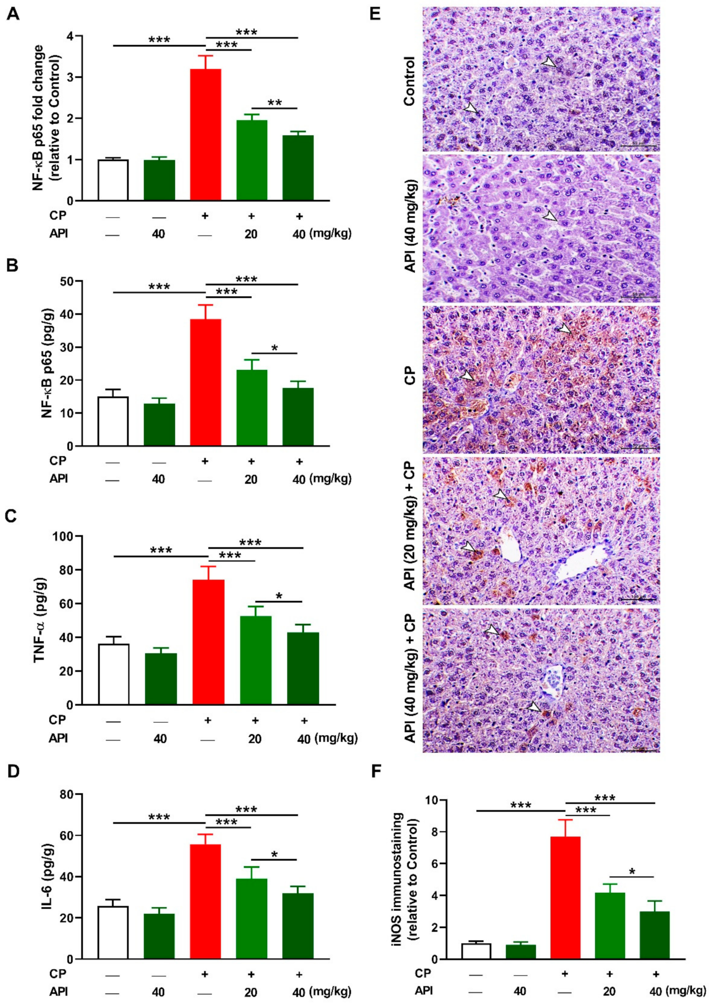
2.4. API Mitigates CP-Induced Hepatic Inflammation in Rats

NF-κB, tumor necrosis factor (TNF)-α, interleukin (IL)-6, and inducible NO synthase (iNOS) were measured to evaluate the anti-inflammatory activity of API. CP upregulated hepatic NF-κB p65 mRNA (Figure 7A) and protein (Figure 7B) in CP-intoxicated rats (p < 0.001). Similarly, hepatic TNF-α (Figure 7C), IL-6 (Figure 7D), and iNOS (Figure 7E,F) showed a significant increase in CP-administered rats (p < 0.001). Treatment with 20 or 40 mg/kg API downregulated NF-κB p65 and the proinflammatory mediators in CP-administered rats, with the higher dose exerting a more potent effect. Normal rats treated with 40 mg/kg API exhibited no changes in the assayed inflammatory markers.

2.5. API Prevents CP-Induced Hepatic Apoptosis in Rats

CP provoked apoptosis in the liver of rats as shown by the significant increase in Bax (Figure 8A,D,E) and caspase-3 (Figure 8B,D,F), and decreased Bcl-2 (Figure 8C,D,G), both RNA and protein levels, as compared to the control group (p < 0.001). While it had no effect in normal rats, API dose-dependently decreased hepatic Bax and caspase-3 and boosted Bcl-2 in CP-intoxicated rats.

3. Discussion

Hepatotoxicity is one of the main health complications related to the use of anticancer agents [33]. CP is an effective chemotherapeutic agent against various cancers; however, hepatotoxicity and other toxic complications limit its applications. CP causes liver injury at both low [12] and high doses [13], with oxidative stress playing a major role [14,17,31,32,34]. Herein, we investigated the protective efficacy of the antioxidant and anti-inflammatory flavonoid API [22,23,24,25] against oxidative stress, the inflammatory response, and liver injury in CP-intoxicated rats.
CP injection resulted in liver injury manifested by the elevated serum transaminases, ALP, and LDH, along with multiple histopathological changes, including diffuse vacuolation consistent with advanced hydropic changes associated with mononuclear cell infiltration. Elevated transaminases and other enzymes in serum are a sensitive marker of liver injury since their release indicates hepatocyte injury [35]. The findings of this investigation were consistent with previous studies from Mahmoud’s lab showing a substantial increase in circulating liver function markers and liver tissue injury in rats that received a single injection of CP [17,29,31,32,34,36]. Pre-treatment with API exerted a dose-dependent hepatoprotective effect marked by the ameliorated liver function markers in CP-intoxicated rats. API mitigated CP-induced histopathological alterations and improved the histological architecture of the liver, demonstrating a potent hepatoprotective efficacy. The ability of API to protect against different hepatotoxicants has been previously reported in rodents challenged with NDEA, furan, acetaminophen, and methotrexate (MTX) [22,23,24,25]. In these studies, treatment with API was effective in ameliorating serum transaminases and preventing hepatocyte injury [22]. Our findings add support to the hepatoprotective effect of API by showing its ability to prevent CP-induced liver injury.
Owing to the role of oxidative stress in CP hepatotoxicity [14,17,31,32,34], we assumed that the protective efficacy of API was mediated through attenuation of oxidative injury and improvement of cellular antioxidants. CP provoked excessive generation of ROS that is extensively involved in its deleterious effect on the liver [31]. In addition, acrolein produced during its metabolism increases ROS release and can damage membranes via its direct covalent binding with proteins and lipids [14]. Excessive ROS can damage lipids by provoking LPO, proteins, DNA, and other cellular macromolecules, and deplete GSH and other antioxidants, thereby leading to cell death [37]. Accordingly, increased hepatic MDA, NO, and 8-oxo-dG, and decreased antioxidants in conjunction with surplus ROS were observed in CP-administered rats in this study. LPO destroys the cells by disrupting the membrane fluidity and permeability and inactivating the bound proteins [37]. NO, produced by the upregulated iNOS, can produce the versatile oxidant peroxynitrite (ONOO⁻) by reacting with superoxide radicals. ONOO⁻ can increase ROS production, impair mitochondrial function, and modify DNA bases, leading to DNA breaks and cell injury [38]. This study supports our previous studies showing elevated hepatic ROS, LPO, NO, and oxidative DNA damage along with declined antioxidant defenses in rats that received CP [17,29,31,32,36]. API significantly suppressed hepatic ROS, LPO, NO, and oxidative DNA damage and boosted GSH, SOD, and CAT in CP-intoxicated rats, highlighting its antioxidant efficacy. In this context, API ameliorated LPO and improved GSH and antioxidant enzymes in the liver of rodents intoxicated with NDEA [22], furan [23], acetaminophen [24], and MTX [25]. API possesses a radical scavenging property mediated by the hydrogen atom transfer and single electron transfer pathways [39]. The OH group in its B ring enables API to stabilize free radicals by donating its H and electron, producing a stable flavonoid radical [39]. The 4-oxo group in the C ring and the m-5,7-dihydroxy arrangements in the A ring participate in the radical scavenging property of API [39]. Electron delocalization from the B ring to the 4 keto group and the C2-C3 double bond helps API to scavenge free radicals [40]. The antioxidant activity of API could also be connected to its interaction and modulation with cell signaling molecules. We assumed that activation of Nrf2 plays a role in the antioxidant and hepatoprotective efficacy of API. In this study, CP suppressed hepatic Nrf2/HO-1 signaling as evidenced by the decreased mRNA abundance of Nrf2, NQO-1, and HO-1, Nrf2 protein, and HO-1 activity, demonstrating the surplus production of ROS. Although activated by ROS under physiological conditions, Nrf2 downregulation has been reported under conditions of excessive and prolonged ROS, including CP hepatotoxicity [30,31,41,42,43,44]. API upregulated hepatic Nrf2/HO-1 signaling in CP-intoxicated rats as shown by the upregulated Nrf2, NQO-1, and HO-1, which explained the enhanced antioxidant enzymes. Previous investigators have reported the role of Nrf2 in mediating the therapeutic actions of API. This flavone activated Nrf2-mediated expression of antioxidant enzymes and induced anti-inflammatory effects in HepG2 cells [45], and protected PC12 cells [46] and melanocytes against the deleterious effects of ROS by activating Nrf2 signaling [47]. In addition, API activated Nrf2 and protected the liver against oxidative stress in a rodent model of NAFLD [48].
In addition to oxidative stress, API attenuated the hepatic inflammatory response and apoptosis in CP-intoxicated rats. NF-κB, iNOS, TNF-α, and IL-6 were increased in the liver of CP-intoxicated rats. Sustained and surplus ROS activates NF-κB, which plays a key role in inflammation by increasing the transcription of iNOS, TNF-α, IL-6, and other mediators [49]. In liver diseases, the proinflammatory mediators recruit inflammatory cells and provoke the production of more ROS, leading to cell death [50]. Upregulation of NF-κB and increased proinflammatory mediators and inflammatory cell infiltration were associated with surplus ROS levels in the liver of CP-intoxicated rats [17,29,31,51]. Free radicals and proinflammatory mediators work in concert to elicit cell death via apoptosis by activating Bax, which promotes the loss of mitochondrial membrane potential [52]. Subsequently, cytochrome c is released to the cytoplasm, where it activates the executioner caspase-3 [52], which provokes DNA fragmentation, degradation of cytoskeletal proteins, and further release of cytochrome c, leading to cell death [53]. Here, CP increased both gene and protein expression of Bax and caspase-3 and decreased Bcl-2. Similar findings were observed in the liver of rats that received CP [17,29,31]. API suppressed hepatic NF-κB, iNOS, cytokines, Bax, and caspase-3 and upregulated Bcl-2 in CP-intoxicated rats, demonstrating its anti-inflammatory and anti-apoptosis efficacies. Accordingly, API decreased proinflammatory cytokines in the serum of rodent models of furan- [23] and MTX-induced hepatotoxicity [25], and decreased Bax, and increased Bcl-2 in a model of ischemia/reperfusion-induced liver injury [54]. Similar to its antioxidant property, the anti-inflammatory effect of API could be linked to the activation of Nrf2/HO-1 signaling, which prevents NF-ĸB activation and the release of proinflammatory mediators [27,28].
In conclusion, this study introduced new information that the flavone API can protect the liver against CP toxicity by activating Nrf2 signaling and attenuating oxidative injury and the inflammatory response. API alleviated liver function markers, prevented hepatocyte injury, and suppressed ROS, LPO, NF-κB, proinflammatory mediators, and apoptosis markers. These beneficial effects were associated with activation of Nrf2/HO-1 signaling and enhancement of antioxidant defenses. Therefore, API could be a potent protective agent against CP hepatotoxicity in clinical settings, pending further investigations and clinical trials to explore its exact therapeutic mechanism(s).

4. Materials and Methods

4.1. Experimental Design and Treatments

Thirty male Wistar rats (200–220 g) were housed under standard conditions on a 12 h light/dark cycle and provided with free access to food and water. The rats were acclimatized for 7 days and then allocated into 5 groups (n = 6). Group I served as a control and received the vehicle 0.5% carboxymethyl cellulose (CMC, Sigma, St. Louis, MO, USA), and group II received 40 mg/kg API (Sigma, St. Louis, MO, USA) suspended in 0.5% CMC [55] via oral gavage for 15 days. Groups III, IV, and V received a single intraperitoneal injection of 150 mg/kg CP (Endoxan®, Baxter Oncology, Halle, Germany) diluted in sterile saline on day 16 [29,31]. Group III received 0.5% CMC and groups IV and V received 20 and 40 mg/kg API in 0.5% CMC, respectively, via oral gavage for 15 days [55].
On the 19th day, blood was collected via cardiac puncture under ketamine/xylazine anesthesia and serum was prepared by centrifuging the samples at 3000 for 15 min. The rats were sacrificed and dissected to excise the livers of specimens, which were fixed in 10% neutral buffered formalin (NBF) for histology and immunohistochemistry. Specimens from the liver were stored in RNAlater at −80 °C for RNA isolation while other specimens were homogenized (10% w/v) in cold Tris-HCl buffer (pH = 7.4), centrifuged, and the clear supernatant was kept at −80 °C for biochemical analyses.

4.2. Determination of Liver Function Enzymes, Cytokines, and 8-Oxo-dG

The activities of transaminases (ALT and AST), ALP, and LDH were assayed in the serum of rats using reagent kits supplied by Spinreact (Girona, Spain). TNF-α and IL-6 levels were measured in liver homogenates using R&D Systems (Minneapolis, MN, USA) ELISA kits and NF-κB p65 was determined using an ELISA kit (Elabscience, Houston, TX, USA). 8-Oxo-dG was assayed by an ELISA kit supplied by Cusabio (Wuhan, China). All assays were conducted following the manufacturers’ instructions.

4.3. Determination of ROS, LPO, NO, and Antioxidants

Hepatic ROS levels were assayed by mixing 100 μL sample with 5 μL H2DCF-DA (Sigma, St. Louis, MO, USA) in 1 mL PBS and incubation of the mixture at 37 °C protected from the light. After 30 min, the fluorescence was determined at 490 nm [56]. MDA, a marker of LPO, was measured as described by Ohkawa et al. [57], and NO was assayed using Griess reagent [58]. GSH, SOD, and CAT were assayed following the methods described by Ellman [59], Marklund and Marklund [60], and Cohen et al. [61], respectively. Total HO-1 activity was assayed as reported by Abraham et al. [62].

4.4. Histopathology and Immunohistochemistry (IHC) Examination

Liver samples fixed in 10% NBF for 24 h were processed for paraffin embedding and 5-µm sections were cut using a microtome. The sections were stained with hematoxylin and eosin (H&E) as previously described [63]. To evaluate changes in the expression levels of iNOS, Bax, Bcl-2, caspase-3, and Nrf2, sections from the liver were dewaxed and 0.05 M citrate buffer (pH 6.8) was used for antigen retrieval. The slides were treated with 0.3% hydrogen peroxide (H2O2), washed in phosphate-buffered saline (PBS), and probed with anti-iNOS (ThermoFisher Scientific, Waltham, MA, USA; 1:20 dilution), anti-Bax (Abcam, Cambridge, MA, USA; 1:100 dilution), anti-Bcl-2 (Abcam, Cambridge, UK; 1:100 dilution), anti-caspase-3 (ThermoFisher Scientific, Waltham, MA, USA; 1:100 dilution), and anti-Nrf2 (ThermoFisher Scientific, Waltham, MA, USA; 1:100 dilution) overnight at 4 °C. The slides were washed in PBS, incubated with secondary antibodies, and color was developed by incubation with DAB in H2O2 [64]. Mayer’s hematoxylin was used for counterstaining, and the slides were visualized under a light microscope. The intensity of the developed color was determined in 6 fields/slide using ImageJ (NIH, Bethesda, MD, USA).

4.5. qRT-PCR

The effect of CP and API on hepatic Nrf2, HO-1, NQO-1, Bax, Bcl-2, and caspase-3 was evaluated using qRT-PCR as previously described [65]. TRIzol (ThermoFisher Scientific, Waltham, MA, USA) was used to isolate RNA, which was treated with RNase-free Dnase (Qiagen, Germany), and its quantity and purity were determined using a nanodrop. RNA samples with A260/A280 of 1.8 or more were used for reverse transcription using a cDNA synthesis kit (ThermoFisher Scientific, Waltham, MA, USA). Amplification of the cDNA was carried out by SYBR Green (ThermoFisher Scientific, Waltham, MA, USA) and the primers in Table 1. The obtained data were analyzed using the 2−ΔΔCt method [66] and normalized to β-actin.

4.6. Statistical Analysis

The results are expressed as mean ± standard error of the mean (SEM) and the statistical comparisons were carried out by one-way ANOVA and Tukey’s test n GraphPad Prism 8 software (GraphPad, San Diego, CA, USA). A p value < 0.05 was considered significant.

Author Contributions

Conceptualization, A.M.M., W.A.-A. and M.H.A.; methodology, W.A.-A., R.S.A., H.A.A., N.A., M.A.A., O.Y.A., A.F.A., M.H.A. and A.M.M.; validation, A.M.M. and M.H.A.; formal analysis, A.M.M.; investigation, W.A.-A., R.S.A., H.A.A., N.A., S.H.A., O.Y.A., A.F.A., M.H.A. and A.M.M.; resources, M.A.A., A.I.A., A.A.A., A.M.A.-e., R.S.A., H.A.A. and N.A.; data curation, A.M.M., W.A.-A. and M.H.A.; writing—original draft preparation, A.M.M. and W.A.-A.; writing—review and editing, A.M.M.; visualization, A.M.M. and M.H.A.; supervision, A.M.M. and M.H.A. All authors have read and agreed to the published version of the manuscript.

Funding

This research received no external funding.

Institutional Review Board Statement

The experiment was conducted according to the guidelines of the National Institutes of Health (NIH publication No. 85–23, revised 2011) and was approved by the research ethics Committee at Al-Hussein Bin Talal University.

Informed Consent Statement

Not applicable.

Data Availability Statement

The data presented in this study are available in article.

Conflicts of Interest

The authors declare no conflict of interest.

References

  1. Emadi, A.; Jones, R.J.; Brodsky, R.A. Cyclophosphamide and cancer: Golden anniversary. Nat. Rev. Clin. Oncol. 2009, 6, 638–647. [Google Scholar] [CrossRef] [PubMed]
  2. Colvin, O.M. An overview of cyclophosphamide development and clinical applications. Curr. Pharm. Des. 1999, 5, 555–560. [Google Scholar] [PubMed]
  3. Moignet, A.; Hasanali, Z.; Zambello, R.; Pavan, L.; Bareau, B.; Tournilhac, O.; Roussel, M.; Fest, T.; Awwad, A.; Baab, K. Cyclophosphamide as a first-line therapy in lgl leukemia. Leukemia 2014, 28, 1134–1136. [Google Scholar] [CrossRef] [PubMed]
  4. Collins, C.; Mortimer, J.; Livingston, R.B. High-dose cyclophosphamide in the treatment of refractory lymphomas and solid tumor malignancies. Cancer 1989, 63, 228–232. [Google Scholar] [CrossRef]
  5. Michot, J.M.; Annereau, M.; Danu, A.; Legoupil, C.; Bertin, L.; Chahine, C.; Achab, N.; Antosikova, A.; Cerutti, A.; Rossignol, J.; et al. High-dose cyclophosphamide for hard-to-treat patients with relapsed or refractory b-cell non-hodgkin’s lymphoma, a phase ii result. Eur. J. Haematol. 2020, 104, 281–290. [Google Scholar] [CrossRef]
  6. Abd El-Ghafar, O.A.M.; Hassanein, E.H.M.; Sayed, A.M.; Rashwan, E.K.; Shalkami, A.S.; Mahmoud, A.M. Acetovanillone prevents cyclophosphamide-induced acute lung injury by modulating pi3k/akt/mtor and nrf2 signaling in rats. Phytother. Res. PTR 2021, 35, 4499–4510. [Google Scholar] [CrossRef]
  7. Hassanein, E.H.M.; Abd El-Ghafar, O.A.M.; Ahmed, M.A.; Sayed, A.M.; Gad-Elrab, W.M.; Ajarem, J.S.; Allam, A.A.; Mahmoud, A.M. Edaravone and acetovanillone upregulate nrf2 and pi3k/akt/mtor signaling and prevent cyclophosphamide cardiotoxicity in rats. Drug Des. Dev. Ther. 2020, 14, 5275–5288. [Google Scholar] [CrossRef]
  8. Martin, F.; Lauwerys, B.; Lefèbvre, C.; Devogelaer, J.P.; Houssiau, F.A. Side-effects of intravenous cyclophosphamide pulse therapy. Lupus 1997, 6, 254–257. [Google Scholar] [CrossRef]
  9. Stillwell, T.J.; Benson, R.C., Jr. Cyclophosphamide-induced hemorrhagic cystitis. A review of 100 patients. Cancer 1988, 61, 451–457. [Google Scholar] [CrossRef]
  10. Segura, A.; Yuste, A.; Cercos, A.; López-Tendero, P.; Gironés, R.; Pérez-Fidalgo, J.A.; Herranz, C. Pulmonary fibrosis induced by cyclophosphamide. Ann. Pharmacother. 2001, 35, 894–897. [Google Scholar] [CrossRef]
  11. Dhesi, S.; Chu, M.P.; Blevins, G.; Paterson, I.; Larratt, L.; Oudit, G.Y.; Kim, D.H. Cyclophosphamide-induced cardiomyopathy: A case report, review, and recommendations for management. J. Investig. Med. High Impact Case Rep. 2013, 1, 2324709613480346. [Google Scholar] [CrossRef] [PubMed]
  12. Akay, H.; Akay, T.; Secilmis, S.; Kocak, Z.; Donderici, O. Hepatotoxicity after low-dose cyclophosphamide therapy. South. Med. J. 2006, 99, 1399–1401. [Google Scholar] [CrossRef]
  13. De Jonge, M.; Huitema, A.; Beijnen, J.; Rodenhuis, S. High exposures to bioactivated cyclophosphamide are related to the occurrence of veno-occlusive disease of the liver following high-dose chemotherapy. Br. J. Cancer 2006, 94, 1226–1230. [Google Scholar] [CrossRef] [Green Version]
  14. Moghe, A.; Ghare, S.; Lamoreau, B.; Mohammad, M.; Barve, S.; McClain, C.; Joshi-Barve, S. Molecular mechanisms of acrolein toxicity: Relevance to human disease. Toxicol. Sci. 2015, 143, 242–255. [Google Scholar] [CrossRef] [PubMed]
  15. Hamzeh, M.; Hosseinimehr, S.J.; Khalatbary, A.R.; Mohammadi, H.R.; Dashti, A.; Amiri, F.T. Atorvastatin mitigates cyclophosphamide-induced hepatotoxicity via suppression of oxidative stress and apoptosis in rat model. Res. Pharm. Sci. 2018, 13, 440. [Google Scholar] [PubMed]
  16. Zhu, H.; Long, M.-H.; Wu, J.; Wang, M.-M.; Li, X.-Y.; Shen, H.; Xu, J.-D.; Zhou, L.; Fang, Z.-J.; Luo, Y. Ginseng alleviates cyclophosphamide-induced hepatotoxicity via reversing disordered homeostasis of glutathione and bile acid. Sci. Rep. 2015, 5, 17536. [Google Scholar] [CrossRef]
  17. Mahmoud, A.M.; Germoush, M.O.; Alotaibi, M.F.; Hussein, O.E. Possible involvement of nrf2 and pparγ up-regulation in the protective effect of umbelliferone against cyclophosphamide-induced hepatotoxicity. Biomed. Pharmacother. 2017, 86, 297–306. [Google Scholar] [CrossRef]
  18. Mansour, D.F.; Saleh, D.O.; Mostafa, R.E. Genistein ameliorates cyclophosphamide-induced hepatotoxicity by modulation of oxidative stress and inflammatory mediators. Open Access Maced. J. Med. Sci. 2017, 5, 836. [Google Scholar] [CrossRef] [Green Version]
  19. Elgebaly, H.A.; Mosa, N.M.; Allach, M.; El-massry, K.F.; El-Ghorab, A.H.; Al Hroob, A.M.; Mahmoud, A.M. Olive oil and leaf extract prevent fluoxetine-induced hepatotoxicity by attenuating oxidative stress, inflammation and apoptosis. Biomed. Pharmacother. 2018, 98, 446–453. [Google Scholar] [CrossRef]
  20. Mohamed, H.M.; Mahmoud, A.M. Chronic exposure to the opioid tramadol induces oxidative damage, inflammation and apoptosis, and alters cerebral monoamine neurotransmitters in rats. Biomed. Pharmacother. 2019, 110, 239–247. [Google Scholar] [CrossRef]
  21. Salehi, B.; Venditti, A.; Sharifi-Rad, M.; Kręgiel, D.; Sharifi-Rad, J.; Durazzo, A.; Lucarini, M.; Santini, A.; Souto, E.B.; Novellino, E. The therapeutic potential of apigenin. Int. J. Mol. Sci. 2019, 20, 1305. [Google Scholar] [CrossRef] [PubMed] [Green Version]
  22. Ali, F.; Naz, F.; Jyoti, S.; Siddique, Y.H. Protective effect of apigenin against n-nitrosodiethylamine (ndea)-induced hepatotoxicity in albino rats. Mutat. Res. Genet. Toxicol. Environ. Mutagenesis 2014, 767, 13–20. [Google Scholar] [CrossRef] [PubMed]
  23. Wang, E.; Chen, F.; Hu, X.; Yuan, Y. Protective effects of apigenin against furan-induced toxicity in mice. Food Funct. 2014, 5, 1804–1812. [Google Scholar] [CrossRef] [PubMed]
  24. Yang, J.; Wang, X.-Y.; Xue, J.; Gu, Z.-L.; Xie, M.-L. Protective effect of apigenin on mouse acute liver injury induced by acetaminophen is associated with increment of hepatic glutathione reductase activity. Food Funct. 2013, 4, 939–943. [Google Scholar] [CrossRef] [PubMed]
  25. Goudarzi, M.; Kalantar, M.; Sadeghi, E.; Karamallah, M.H.; Kalantar, H. Protective effects of apigenin on altered lipid peroxidation, inflammation, and antioxidant factors in methotrexate-induced hepatotoxicity. Naunyn Schmiedeberg’s Arch. Pharmacol. 2021, 394, 523–531. [Google Scholar] [CrossRef]
  26. Satta, S.; Mahmoud, A.M.; Wilkinson, F.L.; Yvonne Alexander, M.; White, S.J. The role of nrf2 in cardiovascular function and disease. Oxidative Med. Cell. Longev. 2017, 2017, 9237263. [Google Scholar] [CrossRef] [PubMed]
  27. Wardyn, J.D.; Ponsford, A.H.; Sanderson, C.M. Dissecting molecular cross-talk between nrf2 and nf-κb response pathways. Biochem. Soc. Trans. 2015, 43, 621–626. [Google Scholar] [CrossRef] [PubMed] [Green Version]
  28. Pan, H.; Wang, H.; Wang, X.; Zhu, L.; Mao, L. The absence of nrf2 enhances nf-kappab-dependent inflammation following scratch injury in mouse primary cultured astrocytes. Mediat. Inflamm. 2012, 2012, 217580. [Google Scholar] [CrossRef]
  29. Mahmoud, A.M.; Al Dera, H.S. 18β-glycyrrhetinic acid exerts protective effects against cyclophosphamide-induced hepatotoxicity: Potential role of pparγ and nrf2 upregulation. Genes Nutr. 2015, 10, 41. [Google Scholar] [CrossRef]
  30. Mahmoud, A.M.; Hozayen, W.G.; Ramadan, S.M. Berberine ameliorates methotrexate-induced liver injury by activating nrf2/ho-1 pathway and pparγ, and suppressing oxidative stress and apoptosis in rats. Biomed. Pharmacother. 2017, 94, 280–291. [Google Scholar] [CrossRef]
  31. Aladaileh, S.H.; Abukhalil, M.H.; Saghir, S.A.; Hanieh, H.; Alfwuaires, M.A.; Almaiman, A.A.; Bin-Jumah, M.; Mahmoud, A.M. Galangin activates nrf2 signaling and attenuates oxidative damage, inflammation, and apoptosis in a rat model of cyclophosphamide-induced hepatotoxicity. Biomolecules 2019, 9, 346. [Google Scholar] [CrossRef] [PubMed] [Green Version]
  32. Kamel, E.M.; Mahmoud, A.M.; Ahmed, S.A.; Lamsabhi, A.M. A phytochemical and computational study on flavonoids isolated from trifolium resupinatum l. And their novel hepatoprotective activity. Food Funct. 2016, 7, 2094–2106. [Google Scholar] [CrossRef] [PubMed]
  33. Vincenzi, B.; Armento, G.; Spalato Ceruso, M.; Catania, G.; Leakos, M.; Santini, D.; Minotti, G.; Tonini, G. Drug-induced hepatotoxicity in cancer patients-implication for treatment. Expert Opin. Drug Saf. 2016, 15, 1219–1238. [Google Scholar] [CrossRef] [PubMed]
  34. Mahmoud, A.M. Hesperidin protects against cyclophosphamide-induced hepatotoxicity by upregulation of pparγ and abrogation of oxidative stress and inflammation. Can. J. Physiol. Pharmacol. 2014, 92, 717–724. [Google Scholar] [CrossRef]
  35. McGill, M.R. The past and present of serum aminotransferases and the future of liver injury biomarkers. EXCLI J. 2016, 15, 817–828. [Google Scholar]
  36. Germoush, M.O.; Mahmoud, A.M. Berberine mitigates cyclophosphamide-induced hepatotoxicity by modulating antioxidant status and inflammatory cytokines. J. Cancer Res. Clin. Oncol. 2014, 140, 1103–1109. [Google Scholar] [CrossRef]
  37. Smathers, R.L.; Galligan, J.J.; Stewart, B.J.; Petersen, D.R. Overview of lipid peroxidation products and hepatic protein modification in alcoholic liver disease. Chem. Biol. Interact. 2011, 192, 107–112. [Google Scholar] [CrossRef] [Green Version]
  38. Pacher, P.; Beckman, J.S.; Liaudet, L. Nitric oxide and peroxynitrite in health and disease. Physiol. Rev. 2007, 87, 315–424. [Google Scholar] [CrossRef] [Green Version]
  39. Messaadia, L.; Bekkar, Y.; Benamira, M.; Lahmar, H. Predicting the antioxidant activity of some flavonoids of arbutus plant: A theoretical approach. Chem. Phys. Impact 2020, 1, 100007. [Google Scholar] [CrossRef]
  40. Zheng, Y.-Z.; Chen, D.-F.; Deng, G.; Guo, R. The substituent effect on the radical scavenging activity of apigenin. Molecules 2018, 23, 1989. [Google Scholar] [CrossRef] [Green Version]
  41. Khadrawy, S.M.; Mohamed, H.M.; Mahmoud, A.M. Mesenchymal stem cells ameliorate oxidative stress, inflammation, and hepatic fibrosis via nrf2/ho-1 signaling pathway in rats. Environ. Sci. Pollut. Res. 2021, 28, 2019–2030. [Google Scholar] [CrossRef] [PubMed]
  42. Mahmoud, A.M.; Desouky, E.M.; Hozayen, W.G.; Bin-Jumah, M.; El-Nahass, E.S.; Soliman, H.A.; Farghali, A.A. Mesoporous silica nanoparticles trigger liver and kidney injury and fibrosis via altering tlr4/nf-κb, jak2/stat3 and nrf2/ho-1 signaling in rats. Biomolecules 2019, 9, 528. [Google Scholar] [CrossRef] [Green Version]
  43. Al-Amarat, W.; Abukhalil, M.H.; Althunibat, O.Y.; Alfwuaires, M.A.; Alnamshan, M.M.; Alqosaibi, A.I.; Ahmeda, A.F.; Kamel, E.M.; Arab, H.H.; Mahmoud, A.M. Galangin attenuates liver injury, oxidative stress and inflammation, and upregulates nrf2/ho-1 signaling in streptozotocin-induced diabetic rats. Processes 2021, 9, 1562. [Google Scholar] [CrossRef]
  44. Mahmoud, A.M.; Zaki, A.R.; Hassan, M.E.; Mostafa-Hedeab, G. Commiphora molmol resin attenuates diethylnitrosamine/phenobarbital-induced hepatocarcinogenesis by modulating oxidative stress, inflammation, angiogenesis and nrf2/are/ho-1 signaling. Chem. Biol. Interact. 2017, 270, 41–50. [Google Scholar] [CrossRef] [PubMed]
  45. Paredes-Gonzalez, X.; Fuentes, F.; Jeffery, S.; Saw, C.L.; Shu, L.; Su, Z.Y.; Kong, A.N. Induction of nrf2-mediated gene expression by dietary phytochemical flavones apigenin and luteolin. Biopharm. Drug Dispos. 2015, 36, 440–451. [Google Scholar] [CrossRef] [PubMed]
  46. Wu, P.-S.; Yen, J.-H.; Kou, M.-C.; Wu, M.-J. Luteolin and apigenin attenuate 4-hydroxy-2-nonenal-mediated cell death through modulation of upr, nrf2-are and mapk pathways in pc12 cells. PLoS ONE 2015, 10, e0130599. [Google Scholar] [CrossRef] [Green Version]
  47. Zhang, B.; Wang, J.; Zhao, G.; Lin, M.; Lang, Y.; Zhang, D.; Feng, D.; Tu, C. Apigenin protects human melanocytes against oxidative damage by activation of the nrf2 pathway. Cell Stress Chaperones 2020, 25, 277–285. [Google Scholar] [CrossRef]
  48. Feng, X.; Yu, W.; Li, X.; Zhou, F.; Zhang, W.; Shen, Q.; Li, J.; Zhang, C.; Shen, P. Apigenin, a modulator of pparγ, attenuates hfd-induced nafld by regulating hepatocyte lipid metabolism and oxidative stress via nrf2 activation. Biochem. Pharmacol. 2017, 136, 136–149. [Google Scholar] [CrossRef]
  49. Luedde, T.; Schwabe, R.F. Nf-κb in the liver--linking injury, fibrosis and hepatocellular carcinoma. Nat. Rev. Gastroenterol. Hepatol. 2011, 8, 108–118. [Google Scholar] [CrossRef] [Green Version]
  50. Kolios, G.; Valatas, V.; Kouroumalis, E. Role of kupffer cells in the pathogenesis of liver disease. World J. Gastroenterol. 2006, 12, 7413–7420. [Google Scholar] [CrossRef]
  51. Alqahtani, S.; Mahmoud, A.M. Gamma-glutamylcysteine ethyl ester protects against cyclophosphamide-induced liver injury and hematologic alterations via upregulation of pparγ and attenuation of oxidative stress, inflammation, and apoptosis. Oxid. Med. Cell Longev. 2016, 2016, 4016209. [Google Scholar] [CrossRef] [PubMed] [Green Version]
  52. Shi, Y.; Chen, J.; Weng, C.; Chen, R.; Zheng, Y.; Chen, Q.; Tang, H. Identification of the protein-protein contact site and interaction mode of human vdac1 with bcl-2 family proteins. Biochem. Biophys. Res. Commun. 2003, 305, 989–996. [Google Scholar] [CrossRef]
  53. Redza-Dutordoir, M.; Averill-Bates, D.A. Activation of apoptosis signalling pathways by reactive oxygen species. Biochim. Et Biophys. Acta BBA Mol. Cell Res. 2016, 1863, 2977–2992. [Google Scholar] [CrossRef] [PubMed]
  54. Tsaroucha, A.K.; Tsiaousidou, A.; Ouzounidis, N.; Tsalkidou, E.; Lambropoulou, M.; Giakoustidis, D.; Chatzaki, E.; Simopoulos, C. Intraperitoneal administration of apigenin in liver ischemia/reperfusion injury protective effects. Saudi J. Gastroenterol. 2016, 22, 415–422. [Google Scholar] [PubMed]
  55. Du, H.; Hao, J.; Liu, F.; Lu, J.; Yang, X. Apigenin attenuates acute myocardial infarction of rats via the inhibitions of matrix metalloprotease-9 and inflammatory reactions. Int. J. Clin. Exp. Med. 2015, 8, 8854. [Google Scholar]
  56. Hozayen, W.G.; Mahmoud, A.M.; Desouky, E.M.; El-Nahass, E.S.; Soliman, H.A.; Farghali, A.A. Cardiac and pulmonary toxicity of mesoporous silica nanoparticles is associated with excessive ros production and redox imbalance in wistar rats. Biomed. Pharmacother. Biomed. Pharmacother. 2019, 109, 2527–2538. [Google Scholar] [CrossRef]
  57. Ohkawa, H.; Ohishi, N.; Yagi, K. Assay for lipid peroxides in animal tissues by thiobarbituric acid reaction. Anal. Biochem. 1979, 95, 351–358. [Google Scholar] [CrossRef]
  58. Grisham, M.B.; Johnson, G.G.; Lancaster, J.R., Jr. Quantitation of nitrate and nitrite in extracellular fluids. Methods Enzymol. 1996, 268, 237–246. [Google Scholar]
  59. Ellman, G.L. Tissue sulfhydryl groups. Arch. Biochem. Biophys. 1959, 82, 70–77. [Google Scholar] [CrossRef]
  60. Marklund, S.; Marklund, G. Involvement of the superoxide anion radical in the autoxidation of pyrogallol and a convenient assay for superoxide dismutase. Eur. J. Biochem. 1974, 47, 469–474. [Google Scholar] [CrossRef]
  61. Cohen, G.; Dembiec, D.; Marcus, J. Measurement of catalase activity in tissue extracts. Anal. Biochem. 1970, 34, 30–38. [Google Scholar] [CrossRef]
  62. Abraham, N.G.; Lutton, J.D.; Levere, R.D. Heme metabolism and erythropoiesis in abnormal iron states: Role of delta-aminolevulinic acid synthase and heme oxygenase. Exp. Hematol. 1985, 13, 838–843. [Google Scholar]
  63. Bancroft, J.D.; Gamble, M. Theory and Practice of Histological Techniques; Elsevier Health Sciences: Amsterdam, The Netherlands, 2008. [Google Scholar]
  64. Jennes, L. Chapter 7-immunohistochemical detection of neuropeptides/transmitters in mammalian brain. In Methods in Cell Biology; Conn, P.M., Ed.; Academic Press: Cambridge, MA, USA, 2013; Volume 113, pp. 123–147. [Google Scholar]
  65. Mahmoud, A.M.; Hussein, O.E.; Hozayen, W.G.; Abd El-Twab, S.M. Methotrexate hepatotoxicity is associated with oxidative stress, and down-regulation of ppargamma and nrf2: Protective effect of 18beta-glycyrrhetinic acid. Chem. Biol. Interact. 2017, 270, 59–72. [Google Scholar] [CrossRef] [PubMed]
  66. Livak, K.J.; Schmittgen, T.D. Analysis of relative gene expression data using real-time quantitative pcr and the 2(-delta delta c(t)) method. Methods 2011, 25, 402–408. [Google Scholar] [CrossRef] [PubMed]
Figure 1. Chemical structure of apigenin.
Metabolites 12 00648 g001
Figure 2. API ameliorated serum (A) ALT, (B) AST, (C) ALP, and (D) LDH in CP-intoxicated rats. Data are mean ± SEM, (n = 6). * p < 0.05, ** p < 0.01, and *** p < 0.001.
Metabolites 12 00648 g002
Figure 3. API prevented CP-induced tissue injury. Photomicrographs of sections in the liver of (A) control and (B) API-administered rats showing a normal histological architecture with normal hepatocytes (H) radially arranged in cords around central veins (CVs) and normal sinusoids (BS); (C,D) CP-intoxicated rats showing marked granular hepatic vacuolation (arrowheads), mononuclear cells infiltration (arrow) [C], and advanced hydropic changes (arrow) [D], PA indicates portal area and H indicates hepatocytes; (E,F) CP-intoxicated rats treated with 20 mg/kg API (E) showing focally limited periportal hepatic vacuolation (arrowheads), and 40 mg/kg API (F) showing a marked decrease in hepatic vacuolation with mild congestion of the blood sinusoids (H&E, Scale bar = 50 µm).
Metabolites 12 00648 g003
Figure 4. API attenuated CP-induced hepatic oxidative stress. API decreased (A) ROS, (B) MDA, and (C) NO, and increased (D) GSH, (E) SOD, and (F) CAT in the liver of CP-administered rats. Data are mean ± SEM, (n = 6). * p < 0.05, ** p < 0.01, and *** p < 0.001.
Metabolites 12 00648 g004
Figure 5. API ameliorated oxidative DNA damage in CP-intoxicated rats. Pre-treatment with API decreased hepatic 8-oxo-dG in a dose-dependent manner. Data are mean ± SEM, (n = 6). * p < 0.05 and *** p < 0.001.
Metabolites 12 00648 g005
Figure 6. API activated hepatic Nrf2/HO-1 signaling in CP-intoxicated rats. API increased the (A) Nrf2, (B) NQO-1, and (C) HO-1 mRNA abundance, and (F) HO-1 activity in CP-administered rats. (D) Immunostaining of hepatic Nrf2 (arrowheads) showed a decrease in the expression in CP-administered rats and upregulation in the API-treated groups. (E) Image analysis of Nrf2 immunostaining. Data are mean ± SEM, (n = 6). * p < 0.05, ** p < 0.01, and *** p < 0.001.
Metabolites 12 00648 g006
Figure 7. API mitigated inflammation in CP-intoxicated rats. API decreased hepatic (A) NF-κB p65 mRNA, (B) NF-κB p65 protein, (C) TNF-α, and (D) IL-6 in CP-administered rats. (E,F) Photomicrographs showing increased immunostaining of iNOS (E) and image analysis (F) showing that API decreased hepatic iNOS in CP-administered rats. Data are mean ± SEM, (n = 6). * p < 0.05, ** p < 0.01, and *** p < 0.001.
Metabolites 12 00648 g007
Figure 8. API prevented CP-induced hepatocyte apoptosis in rats. API decreased (A) Bax and (B) caspase-3 and increased (C) Bcl-2 mRNA in CP-administered rats. (D) Immunostaining of Bax, caspase-3, and Bcl-2, and (EG) image analysis showing decreased (E) Bax and (F) caspase-3 and increased (G) Bcl-2 in CP-intoxicated rats treated with API. Data are mean ± SEM, (n = 6). * p < 0.05, ** p < 0.01, and *** p < 0.001.
Metabolites 12 00648 g008
Table 1. Primers used for qRT-PCR.
GeneForward Primer (5′-3′)Reverse Primer (5′-3′)
Nrf2TTGTAGATGACCATGAGTCGCTGTCCTGCTGTATGCTGCTT
NQO-1GGCCATCATTTGGGCAAGTCTCCTTGTGGAACAAAGGCGA
HO-1GTAAATGCAGTGTTGGCCCCATGTGCCAGGCATCTCCTTC
NF-κB p65CCTCATCTTTCCCTCAGAGCCGGTCCCGTGTAGCCATTGAT
Caspase-3GGAGCTTGGAACGCGAAGAAACACAAGCCCATTTCAGGGT
BaxAGGACGCATCCACCAAGAAGCAGTTGAAGTTGCCGTCTGC
Bcl-2ACTCTTCAGGGATGGGGTGATGACATCTCCCTGTTGACGC
β-actinAGGAGTACGATGAGTCCGGCCGCAGCTCAGTAACAGTCCG
Publisher’s Note: MDPI stays neutral with regard to jurisdictional claims in published maps and institutional affiliations.

