Article

Pumpkin Seeds Harbor Hidden Agonists: Adenosine-Mediated A1 Receptor Activation and Antioxidant Activity

by
Adina-Elena Grasu
1,
Roman Senn
2,
Christiane Halbsguth
2,
Alexander Schenk
2,
Veronika Butterweck
2,*,
Giulia Zecchin
2,
Ionel I. Mangalagiu
3,4,
Cătălina-Ionica Ciobanu
4 and
Anca Miron
1,*
1
Department of Pharmacognosy-Phytotherapy, Faculty of Pharmacy, “Grigore T. Popa” University of Medicine and Pharmacy, 700115 Iasi, Romania
2
Max Zeller & Söhne AG, Seeblickstrasse 4, 8590 Romanshorn, Switzerland
3
Faculty of Chemistry, Alexandru Ioan Cuza University of Iasi, 11 Carol I Boulevard, 700506 Iasi, Romania
4
Institute of Interdisciplinary Research—CERNESIM Centre, Alexandru Ioan Cuza University of Iasi, 11 Carol I Boulevard, 700506 Iasi, Romania
*
Authors to whom correspondence should be addressed.
Sci. Pharm. 2025, 93(4), 48; https://doi.org/10.3390/scipharm93040048
Submission received: 15 August 2025 / Revised: 18 September 2025 / Accepted: 26 September 2025 / Published: 30 September 2025

Abstract

Hydroethanolic Cucurbita pepo seed extracts are traditionally used for alleviating lower urinary tract symptoms (LUTS), yet their mechanisms remain unclear. Adenosine, a purine nucleoside involved in neuromodulation and smooth muscle relaxation, was recently identified in C. pepo seeds. Since A1 adenosine receptors (A1AR) suppress parasympathetic bladder overactivity by inhibiting acetylcholine (ACh) release, we investigated to which extent purines from pumpkin seed extracts contribute to A1AR activation. Complementary antioxidant capacity was assessed using the 2,2-diphenyl-1-picrylhydrazyl (DPPH) assay. Three hydrophilic seed extracts containing different adenosine levels (0.60–1.18 mg/g dw) were evaluated for agonist activity using a cAMP inhibition assay. The most active extract showed an EC50 of 40.22 µg/mL. Selective removal of adenosine shifted the dose–response curve rightward, while further elimination of an adenosine derivative increased the EC50 to 212.10 µg/mL, confirming adenosine as the principal active compound. Guanosine and inosine did not exhibit A1AR agonist or allosteric effects. All samples exhibited measurable but weak antioxidant activity (IC50 = 1.02–4.19 mg/mL), consistent with their low total phenolic content. These findings underscore the importance of accounting for naturally occurring agonists in plant extracts to avoid overestimating receptor-mediated effects in vitro which are not translatable in vivo.

1. Introduction

Benign prostatic hyperplasia (BPH) and overactive bladder (OAB) are prevalent urogenital disorders in aging populations [1,2,3], often manifesting concomitantly as lower urinary tract symptoms (LUTS) [4]. BPH typically presents with voiding symptoms such as weak stream, straining, and incomplete bladder emptying, due to prostate-induced bladder outlet obstruction [4,5]. In contrast, OAB is characterized by storage symptoms including urinary urgency, frequency, and nocturia [6]. Conventional treatments of these urogenital disorders are often limited by side effects. 5α-Reductase inhibitors such as finasteride and dutasteride can cause sexual dysfunction, gynecomastia, and depression [7,8]. Antimuscarinic agents frequently induce dry mouth, constipation, and cognitive impairment, especially in older adults [9,10] while β3-adrenergic agonists such as mirabegron may lead to hypertension, headache, and urinary tract infections [11]. These adverse effects negatively affect the quality of life [12] and can impact treatment adherence [13,14], highlighting the need for safer alternatives [15].
In this context, the seeds of C. pepo, which have a long history of use for urinary disorders, have emerged as a promising nutraceutical intervention [16,17,18]. Clinical studies have shown that various preparations of C. pepo seeds, including comminuted seeds, hydroethanolic extracts (dry and soft forms), and seed oil can ameliorate LUTS, supporting their long-standing use [19,20,21,22]. In recent years, oil-free and aqueous extracts from C. pepo seeds have attracted increasing attention for their potential in managing lower urinary tract disorders. Randomized controlled trials and pilot studies reported improvements in urinary frequency, urgency, and incontinence in women with OAB [23,24], as well as reductions in LUTS in men with BPH [25]. Given the high prevalence of OAB and BPH in aging populations and the limitations of conventional pharmacotherapy, these findings highlight pumpkin seed extracts as a clinically relevant and well-tolerated alternative.
The efficacy of C. pepo seeds is mainly attributed to their complex phytochemical composition, including Δ7-sterols (e.g., Δ7,22-stigmasterol and α-spinasterol), tocopherols (γ-tocopherol), unsaturated fatty acids such as linoleic and oleic acids, and various polyphenolic compounds [16,26,27,28]. In addition to their well-known lipophilic profile, C. pepo seeds also contain water-soluble constituents such as proteins, peptides, free amino acids, and saccharides [29,30,31]. In addition, the occurrence of purine nucleosides [32] such as adenosine in C. pepo seed extracts has recently been reported [25,33]. These hydrophilic compounds are increasingly recognized for their potential to influence neuromodulatory and receptor-mediated pathways [34].
The effect of C. pepo seeds on bladder function was demonstrated in preclinical studies using OAB models where administration of a water-soluble pumpkin seed extract significantly increased bladder capacity and reduced urination frequency in male rats [35]. These effects were likely mediated through the arginine-nitric oxide (NO) pathway which facilitates detrusor relaxation independent of adrenergic and cholinergic signaling [36]. In the urinary bladder, detrusor smooth muscle tone is regulated by a dynamic balance between parasympathetically induced contraction and adrenergic mediated relaxation [37]. During voiding, acetylcholine (ACh) released from parasympathetic nerves binds to muscarinic receptors on the detrusor myocytes, triggering contraction. In the storage phase, β3-adrenoceptor activation raises intracellular cyclic adenosine monophosphate (cAMP), promoting smooth muscle relaxation. Moreover, β3 stimulation induces adenosine triphosphate (ATP) release and its rapid conversion to adenosine, which, via presynaptic A1 adenosine receptors (A1AR), further inhibits ACh release and suppresses involuntary contractions [38,39]. This functional interaction between purinergic and adrenergic pathways suggests that direct activation of A1AR may replicate or enhance the bladder-relaxant effects mediated by β3-adrenergic agonists [39]. Under physiological conditions, A1ARs act as a prejunctional inhibitory control on cholinergic signaling in the human bladder. However, in case of poor adenosine availability, A1AR engagement is reduced, thereby facilitating detrusor overactivity [40]. Under inflammatory conditions, luminal stimulation of epithelial A1AR modulates afferent signaling and lowers the threshold pressure for voiding [41]. Central A1AR activation has also been shown to prolong the voiding intervals in conscious rats [42]. Additionally, reduced adenosine availability resulting from impaired ATP hydrolysis in bladder outlet obstruction contributes to exaggerated cholinergic activity by diminishing A1AR-mediated inhibitory tone [40]. These findings support the relevance of A1AR as a pharmacological target and underscore the importance of clarifying whether receptor activation by C. pepo seed extracts results from direct agonist activity or indirect effects of the extract matrix.
Beyond their urodynamic impact, pumpkin seed extracts have exhibited antidiabetic, antitumor, and antioxidant properties [43]. In vitro experiments revealed that the extracts inhibited lipid peroxidation and exhibited radical scavenging activities [44], supporting their potential role in mitigating oxidative damage associated with urological disorders. Oxidative stress is a key contributor to the pathophysiology of both BPH and OAB, particularly in aging populations. Elevated levels of reactive oxygen species (ROS) and diminished antioxidant defenses promote inflammation, smooth muscle dysfunction, and fibrosis within the prostate and bladder tissues [45,46]. In BPH, ROS promote cellular proliferation and inhibit apoptosis [45,47], while in OAB, ischemia–reperfusion cycles trigger oxidative damage, afferent nerve sensitization, and detrusor overactivity [48].
Given the pharmacological context and the multifactorial pathophysiology of LUTS, the present study aimed to clarify to which extent adenosine, previously detected in C. pepo seeds, and related purines present in hydrophilic extracts, contribute to the observed A1AR activation in C. pepo seed hydrophilic extracts. While clinical and preclinical studies support the benefits of C. pepo in LUTS, the role of hydrophilic compounds has not been systematically addressed. To fill this gap, we employed a stepwise depletion strategy to selectively remove adenosine and its derivative and assess to what extent A1AR activity can be attributed to adenosine versus other extract constituents in vitro. To the best of our knowledge, this is the first study to directly demonstrate A1AR activation by hydrophilic C. pepo seed extracts. By combining depletion experiments with testing of pure compounds and isolated fractions in both agonist and positive allosteric modulation (PAM) mode, we were able to distinguish the contribution of individual nucleosides from matrix effects. The isolated adenosine derivative was further subjected to NMR-based structural analysis. In parallel, we assessed total phenolic content (TPC) and the antioxidant potential of the extracts using the 2,2-diphenyl-1-picrylhydrazyl (DPPH) assay, providing a complementary mechanism of potential relevance for urinary health. Together, this integrated approach provides both mechanistic insight and a methodological framework for other extracts beyond pumpkin seed extracts.

2. Materials and Methods

2.1. Materials

Hydroethanolic extracts of C. pepo seeds were prepared according to a previously described method [33]. Briefly, milled seeds were extracted with 60% (v/v) ethanol at 50° C for 3 h, followed by ethanol removal, liquid–liquid separation of the oil phase and freeze-drying, yielding dry extracts with a drug to extract ratio (DER) of 40–60:1 (w/w). The samples were coded as follows: SK (CP convar. citrullina var. styriaca), SKR (CP var. styriaca cultivar Gleisdorfer Rustikal), LN (CP—Lady Nail), SW (CP—Snow White), RZ (CP var. giromontia—Radu), SS (CP—Shine Skin), GA (CP—Greek Cultivar), GV (CP—Gray Volga), BBZ (CP var. cylindrica—Black Beauty), HV (CP—Hungarian Cultivar). The adenosine-depleted extracts were generated from their respective native hydroethanolic extracts as described in Section 2.2.2.
Adenosine (cat. no. A9251), guanosine (cat. no. G6752), inosine (cat. no. I4125), DPPH radical (cat.no. 300267), dipropylcyclopentylxanthine (DPCPX) (cat. no. C101), and deuterated water (D2O) (cat. no. 756822) were acquired from Sigma-Aldrich (St. Louis, MO, USA). CHO-K1 cells overexpressing the human A1AR (cat.no. 95-0061C2) were acquired from Eurofins DiscoverX Corporation (Fremont, CA, USA). DMSO (cat. no. 34869—500 mL), Folin–Ciocalteu reagent (cat. no. 109001), sodium carbonate (cat. no. 106392), and gallic acid (cat. no. 91215) were obtained from Merck (Darmstadt, Germany). Acetonitrile HPLC grade (cat. no. 83639.329) and LC-MS grade formic acid (cat. no. 84865.260) were purchased from VWR (Radnor, PA, USA). Ultrapure water was obtained from ELGA Ultrapure water system PURELAB® 7120 (ELGA LabWater, High Wycombe, UK).

2.2. Nucleosides Analysis

2.2.1. Nucleosides Identification and Quantification

Nucleosides identification was conducted using LC-PDA-HRMS on a Waters Acquity H-Class UPLC coupled to a SYNAPT G2-S Q-TOF mass spectrometer (Milford, MA, USA) with an ESI source operated in both positive and negative ion modes (scan range: 100–900 Da), as previously described [33].
Quantitative analysis of nucleosides was conducted using a Waters Acquity H-Class UPLC coupled with a photodiode array detector (PDA) and quadrupole detector (QDA) (Waters, Milford, MA, USA). Separation was achieved on an Atlantis C18-T3 column (150 × 4.6 mm, 3 µm; Waters, Milford, MA, USA) with a mobile phase consisting of 0.1% formic acid in water (A) and 0.1% formic acid in acetonitrile (B), using the following gradient: 0–4 min, 2% B; 2–17 min, 2–10% B; 17–18 min, 10–99% B; 18–22 min 99% B. The column temperature was maintained at 24.5 °C and the flow rate was set at 0.8 mL/min. A QuickSplit adjustable flow-splitter (ASI, El Sobrante, CA, USA) was used to divert 25% of the flow to the QDA. Dry extracts (25 mg/mL in 8% ethanol) were ultrasonicated for 20 min, centrifuged at 10,000 rpm for 10 min, and 2 µL of supernatant was injected. Peak identities were confirmed by matching retention times, UV spectra (λ max 257 nm), and mass spectra with reference standards. An additional compound, tentatively identified as an adenosine derivative, was quantified using the adenosine calibration curve and the results were expressed as adenosine equivalents. Calibration curves for adenosine and guanosine were constructed using six concentration levels (1–500 µg/mL). Data processing was performed using Empower 3 software (Waters, Milford, MA, USA), and all results were expressed in mg/g dry extract.

2.2.2. Adenosine and Its Derivative Isolation

Adenosine-depleted extracts were produced using the same chromatographic system described in Section 2.2.1. (Waters Acquity H-Class UPLC with PDA and QDA), with minor method adjustments. To preserve the extract integrity, acid was removed from the mobile phase, which consisted of water (A) and acetonitrile (B). Subsequently, the gradient was modified as follows: 0–12 min, 7% B; 12–14 min, 7–99% B; 14–19 min, 99% B. The system was coupled to a Waters WFM-A fraction manager, allowing the collection of closely eluting compounds. Target fractions were collected in separate vials using time-based windows, while the remaining eluate was collected separately for subsequent bioactivity testing. The collected fractions were dried using a rotary evaporator (Büchi R 300 Rotavapor, Büchi Labortechnik AG, Flawil, Switzerland). An aliquot was reanalyzed under both acidified and non-acidified chromatographic conditions, to confirm the absence of adenosine and the integrity of the remaining extract. Both sample and fraction manager compartments were set at 5 °C to minimize degradation of thermally labile compounds.

2.2.3. Chemical Characterization of Isolated Fraction

The partially purified fraction obtained by time-based UPLC collection (Section 2.2.2) was analyzed by HRMS and NMR spectroscopy. HRMS was performed in both positive and negative ion modes as described in Section 2.2.1 to determine the molecular ion and fragmentation pattern. 1H-NMR spectrum was recorded on a Bruker Avance III, 500 MHz spectrometer (Bruker BioSpin, Rheinstetten, Germany), operating at 500 MHz, using D2O as a solvent. Chemical shifts (δ, ppm) were referenced to the residual HOD signal at δ = 4.79 ppm [49]. Standard and solvent-suppressed acquisitions were processed using TopSpin 3.2 (Bruker, BioSpin, Rheinstetten, Germany). Additionally, product ion spectra were interpreted using the MassFragment, a structural interpretation tool of MassLynx (Waters, Milford, MA, USA). Fragment ions derived from the parent molecule were systematically interpreted by comparison with the corresponding adenosine fragments.

2.3. Bioactivity Assays

2.3.1. A1AR Direct Activation

Agonist activity was determined with HitHunter cAMP Assay for Biologics (Eurofins DiscoverX Corporation, Fremont, CA, USA) using CHO-K1 cell lines overexpressing the human A1AR. The protocol followed the method described by Senn et al. [50]. Briefly, cells were incubated with serial 3-fold dilutions of the test extracts in the presence of forskolin, which stimulates intracellular cAMP production, at its EC80 concentration. Following incubation, cAMP levels were quantified via chemiluminescence. N6-cyclopentyladenosine (CPA) was used as a positive control. The measurements were made in duplicate, the final DMSO concentration never exceeded 1% final concentration in culture medium.

2.3.2. A1AR Positive Allosteric Modulation

For assessment of positive allosteric modulation (PAM), CHO-K1 cells were pre-incubated with the reference substances at concentrations of 5, 20, and 40 µM. This was followed by stimulation with the agonist (CPA) at its EC20 concentration, along with forskolin at its EC80 concentration to ensure dynamic cAMP production. After incubation, cAMP levels were measured using the same chemiluminescent assay system. Experimental setup and data analysis followed the methodology described by Senn et al. [50]. Responses were normalized between the maximum inhibition of cAMP accumulation produced by CPA (positive control) and the minimal response obtained with vehicle (1% DMSO, negative control).

2.4. Total Phenolic Content and Antioxidant Activity

The total phenolic content (TPC) of the extracts was determined with Folin–Ciocalteu reagent according to previously described methods [51]. In this regard, 50 µL of C. pepo extract (10 mg/mL) were mixed with 100 µL of diluted Folin–Ciocalteu reagent in a 96-well microplate and vortexed. After 3 min, 75 µL of 1% (w/v) sodium carbonate solution were added and the mixture was incubated in the dark, at room temperature for 2 h. A blank was similarly prepared, using ultrapure water instead of sodium carbonate. Absorbance was recorded at 765 nm, and TPC was calculated by subtracting the blank absorbance from the sample absorbance. Results were expressed as mg gallic acid equivalents per gram of extract (mg GAE/g extract).
The antioxidant activity was evaluated for all extracts using the DPPH radical scavenging assay based on the method of Fuso et al. [52], with slight modifications. Specifically, samples were tested at five concentrations (0.25, 0.5, 1, 1.5 and 2 mg/mL), to determine IC50 value, defined as the sample concentration capable of reducing 50% of DPPH radicals. A 1:1 volume ratio of extract and 100 µM DPPH solution in methanol was incubated in the dark for 30 min at room temperature. Absorbance was recorded at 517 nm using a Specord 210 plus UV–Vis spectrophotometer equipped with ASpect UV 2.0 software (Analytik Jena, Jena, Germany). Ascorbic acid was used as a positive control, while sample solvent was used as blank. The % of scavenging activity was calculated as follows:
% D P P H   s c a v e n g i n g = A b s B l a n k ( A b s S a m p l e C o l o r   C o n t r o l ) A b s B l a n k × 100
where Abs Blank is the average absorbance of blank solutions (ultrapure water + DPPH) measured 6 times, Abs Sample is the absorbance of the test sample (sample in ultrapure water + DPPH), and Color Control accounts for the background absorbance due to the intrinsic sample’s yellowish color (sample in distilled water + methanol). IC50 values were calculated by non-linear regression using a four-parameter logistic model (log [inhibitor] vs. response-variable slope) in GraphPad Prism software (version 10.04, Boston, MA, USA). Results are reported with 95% confidence intervals.

3. Results

3.1. Nucleosides Analysis

3.1.1. Nucleosides Identification and Quantification

Adenosine, guanosine, and the adenosine derivative were identified in all ten hydroethanolic extracts (Figure 1) and their contents were determined via UPLC-PDA (Table 1). Adenosine concentrations ranged from 0.60 ± 0.01 mg/g dw in the SK sample to 1.18 ± 0.01 mg/g dw in the HV sample, while guanosine levels varied from 0.33 ± 0.02 in SW to 1.23 ± 0.04 mg/g dw in GA. In most extracts, adenosine levels exceeded those of guanosine, although in some samples (SK, LN, GA, BBZ) guanosine predominated. An adenosine derivative was identified in the extracts and its content, expressed as adenosine equivalents, ranged from 0.29 ± 0.03 mg/g dw in BBZ to 1.58 ± 0.16 mg/g dw in SW. The derivative contributed substantially to the nucleoside profile in several extracts, particularly in SW, where its concentration surpassed those of both adenosine and guanosine.

3.1.2. Isolation of Adenosine and Its Derivative Fractions

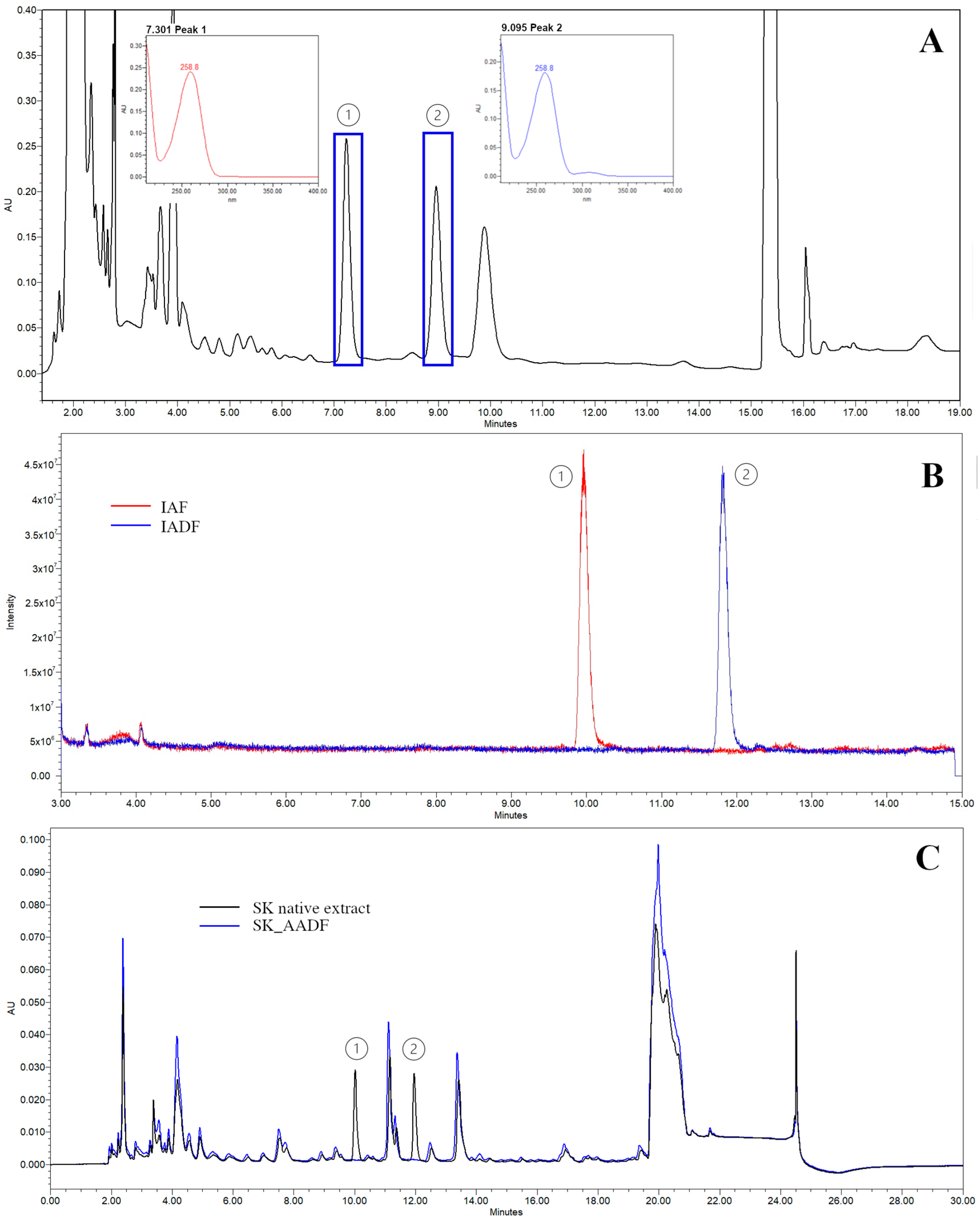
Acid removal from the mobile phase used for the isolation of adenosine and its derivative (Figure 2A) affected the elution order of the compounds in the extract (Figure 1). Based on these changes, time-based isolation windows were defined for the target peaks (1 = adenosine, 2 = adenosine derivative), allowing collection of each compound with minimal co-elution of additional compounds.
The isolated fractions displayed a single sharp peak each, as can be observed in the total ion chromatogram (Figure 2B). Overall, this approach minimized the risk of co-eluting compounds being inadvertently enriched, which was further supported by the high relative purity calculated for the isolated adenosine (96.26%) and adenosine derivative (94.32%) fractions. To further verify the selectivity of the depletion procedure, SK_AF and SK_AADF fractions were re-analyzed using both the quantification and isolation methods. In these fractions, adenosine and its derivative peaks were no longer detectable, with signal intensities falling below the defined LOD (0.165 µg/mL), confirming their complete removal. It can be seen in Figure 2C, in which an overlay of the native extract and the stepwise depleted fractions is depicted, that the characteristic peaks of adenosine and its derivative were no longer detectable, with signal intensities falling below the defined LOD (0.165 µg/mL). Additionally, the figure shows that all other matrix peaks were preserved, confirming that the depletion procedure was selective and did not alter the overall chemical profile.

3.1.3. Chemical Characterization of the Isolated Fraction

The UPLC-PDA-HRMS analysis of SK native extract revealed two distinct peaks with closely related spectra properties, as mentioned in Section 3.1.1 and Section 3.1.2. Adenosine was detected at retention time 15.43 min, by comparison with a reference standard based on matching retention time, UV absorbance maxima (256.7 nm), and MS fragmentation profile. A second peak at 17.05 min displayed a UV spectrum nearly superimposable with that of adenosine (Figure 2A) and a similar MS/MS fragmentation pattern (see Supplementary Materials, Figure S1). The molecular formula was established as C15H21N5O8 as indicated by the [M + H]+ ion at m/z of 400.1474, consistent with the calculated value for C15H22N5O8 (m/z 400.1468, Δ = +0.6 mDa). Fragment interpretation confirmed characteristic ions at m/z 268.1056 (adenosine core) and m/z 136.0626 (adenine moiety), both exhibiting minimal mass errors (≤1.0 mDa). HRMS analysis was also performed in negative ion mode where additional peaks were observed at m/z 434.1086 and 444.1370, which were assigned as [M + Cl] and [M + HCOOH] adducts, respectively, commonly observed under electrospray conditions.
The 1H-NMR analysis of the isolated fraction containing the adenosine derivative (IADF), redissolved in D2O, revealed aromatic signals at δ 8.31 and 8.12 ppm, consistent with H-8 and H-2 of the adenine ring. A doublet at δ 6.00 ppm was observed, corresponding to the anomeric proton of a β-ribofuranosyl unit. Multiplets in the range δ 4.67–4.07 ppm were consistent with the remaining ribose protons (H-2′ to H-5′), supporting the presence of an intact adenosine moiety. Additional aliphatic resonances were detected in 1H NMR spectra, but due to signal overlapping and limited resolution, the structural features beyond the adenosine core could not be conclusively interpreted (see Supplementary Materials, Figure S2). Combined spectroscopic data confirm that the IADF fraction contains an adenosine derivative, in which the purine and ribose core structures are preserved. However, the identity, composition, and point of attachment of the additional moiety remain undetermined mainly due to the presence of minor impurities, as shown in Figure 2B.

3.2. Bioactivity Assays

3.2.1. Direct Agonist Activity and Adenosine Role

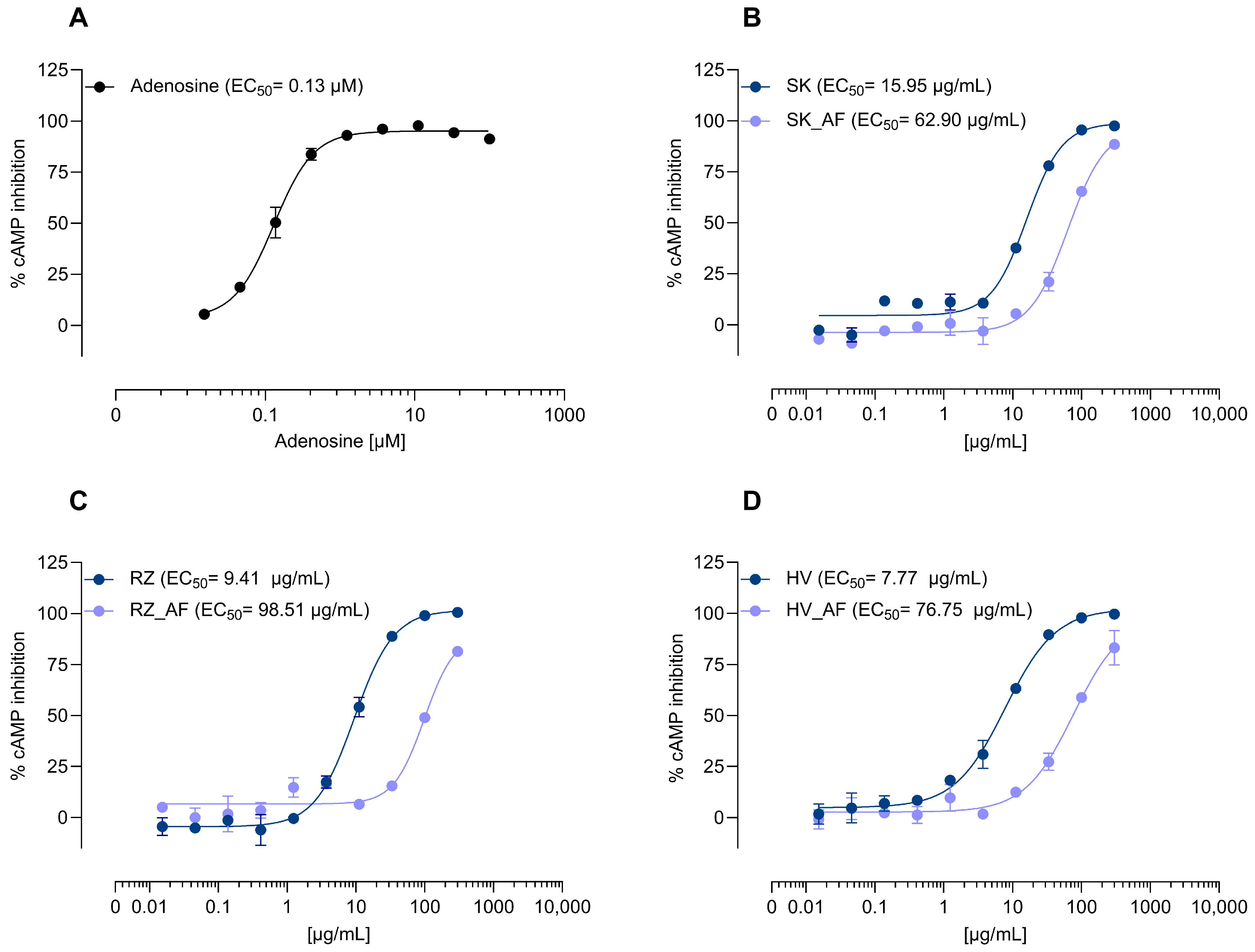
To investigate the contribution of adenosine to A1AR activation, three C. pepo extracts were selected based on their adenosine content: SK (lowest, 0.60 ± 0.01 mg/g dw), RZ (intermediate, 0.83 ± 0.01 mg/g dw), and HV (highest, 1.18 ± 0.01 mg/g dw). The samples were evaluated in a cAMP inhibition assay using CHO-K1 cells overexpressing the human A1AR (Figure 3). All extracts induced a dose-dependent decrease in intracellular cAMP concentrations, indicating a receptor activation via Gi-coupled signaling, with adenosine contributing significantly. Receptor specificity was supported by a preliminary analysis in which the A1AR antagonist DPCPX was applied simultaneously with C. pepo extracts prepared using the same protocol, but from different seed batches. While the extracts alone resulted in a dose-dependent response, the cAMP concentration remained stable when 1 µM DPCPX was added to the treatments (see Supplementary Materials, Figure S3). Based on the quantified adenosine content and corresponding EC50 values, the calculated concentrations of adenosine at the active doses were 35.81 nM for SK, 29.23 nM for RZ, and 34.31 nM for HV. These values were substantially below the EC50 determined for pure adenosine (130 nM) (Figure 3A), suggesting the presence of additional constituents, acting independently or synergistically in enhancing the A1AR activation.
Corresponding adenosine-depleted versions of the selected extracts (SK_AF, RZ_AF, HV_AF) were prepared to determine the possible contribution of other constituents to A1AR activation. Adenosine removal resulted in a 3.9-fold increase in EC50 for SK extract (15.95 to 62.90 µg/mL) compared to a 10.5-fold increase for RZ (9.41 to 98.51 µg/mL) and a 9.9-fold increase for HV (7.7 to 76.75 µg/mL). These findings support that the A1AR activity of C. pepo seed extracts is primarily driven by their adenosine content. However, the smaller shift indicated that SK extract was less affected by adenosine removal and retained a larger proportion of its original bioactivity. Interestingly, despite having the lowest adenosine content, SK maintained a larger proportion of activity post-depletion suggesting the involvement of other synergistic or structurally related components capable of modulating A1AR signaling (Figure 3B–D). Given these observations, the SK sample was further fractionated to remove both adenosine and its derivative in order to clarify the contribution of individual purine components to A1AR activation (Figure 4A).
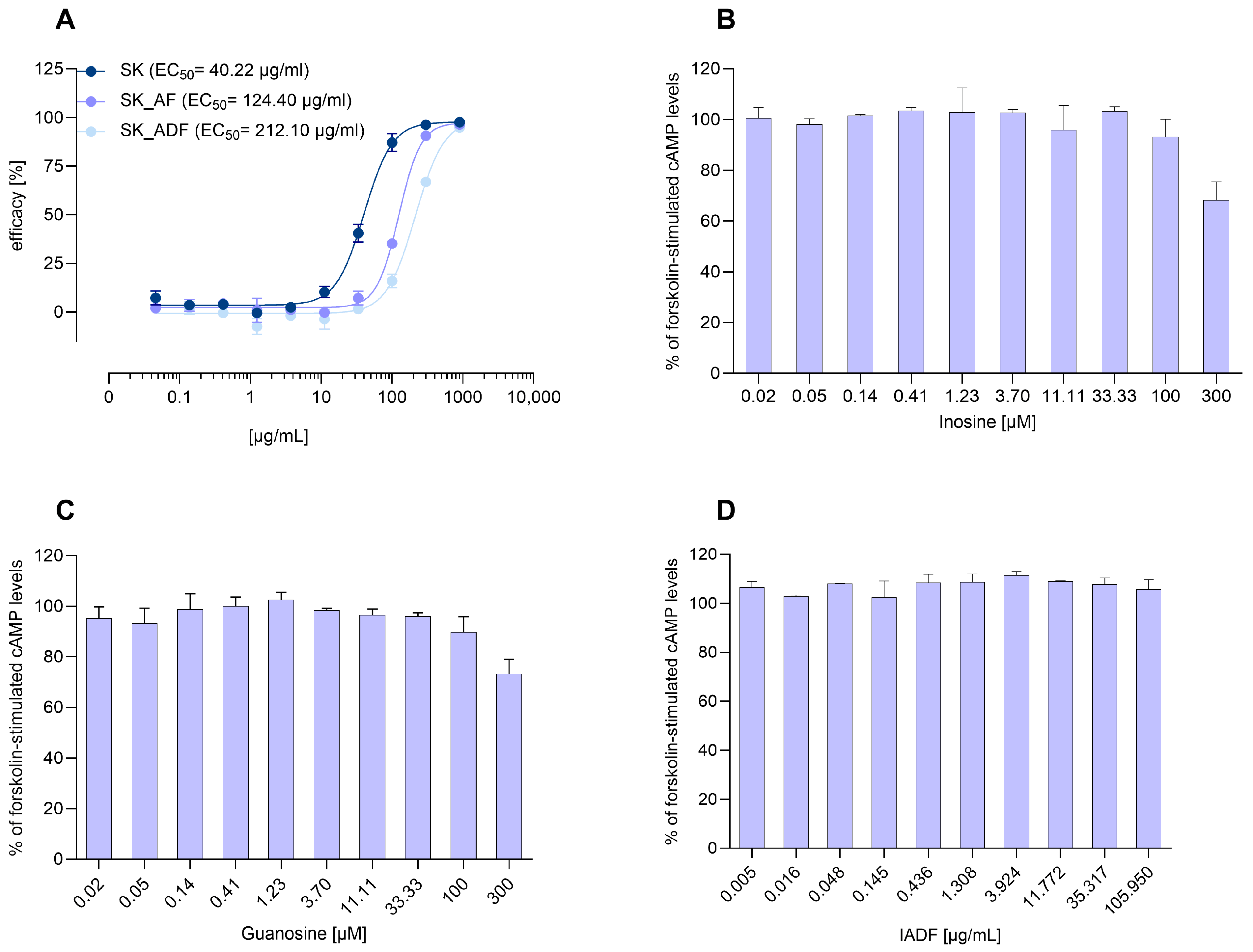
In this experiment, the top tested concentrations of the extracts were increased (900 µg/mL) to improve dose–response curve resolution, allowing for a more accurate EC50 determination in both native and depleted variants. The native SK extract inhibited forskolin-induced cAMP accumulation in a dose-dependent manner (EC50 = 40.22 µg/mL). Adenosine removal showed again a rightward shift in the dose–response curve, increasing the EC50 to 124.40 µg/mL. Further elimination of the adenosine derivative (SK_ADF) led to an additional rightward shift with an EC50 of 212.10 µg/mL. This progressive change supports adenosine as the principal active compound, while the derivative may exert a minor or indirect modulatory effect.
Individual contributions of structurally related purines, inosine and guanosine, were assessed at concentrations up to 300 µM (Figure 4B–D). While inosine and guanosine induced minimal or rather unspecific inhibition at the highest concentration, the derivative showed no detectable A1AR-mediated response. The weak responses of inosine and guanosine at high concentrations, along with the absence of measurable activity for the adenosine derivative, suggest that these structurally related purines do not act as direct A1AR agonists under the tested conditions.

3.2.2. Allosteric Modulation of A1AR

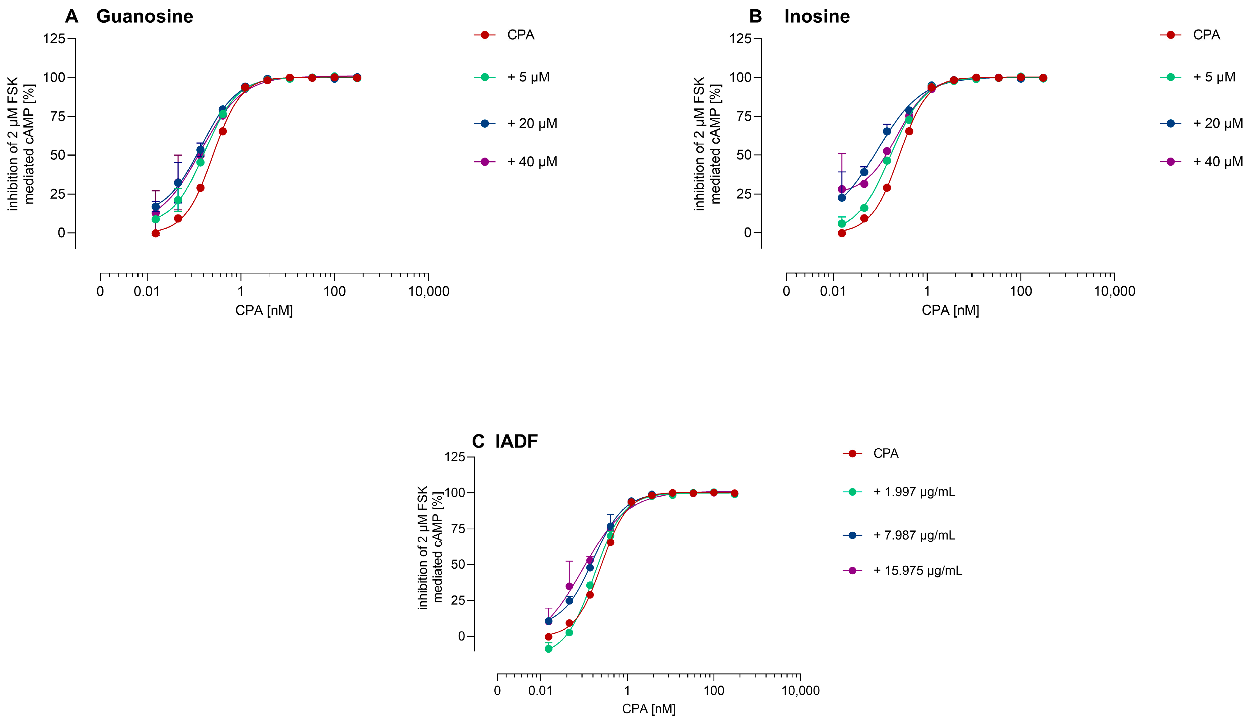
To determine potential PAM activity at A1AR, CPA-induced inhibition of forskolin-stimulated cAMP production was assessed in the presence of guanosine and inosine at concentrations of 5, 20, and 40 µM (Figure 5). Both guanosine and inosine produced slight leftward shifts in the CPA dose–response curves at 20 and 40 µM. These effects were rather modest and did not significantly enhance the maximal inhibitory response. In contrast, co-administration of IADF did not affect the efficacy of CPA at any tested concentration, indicating no observable PAM effect under the experimental conditions.

3.3. Total Phenolic Content and Antioxidant Activity

The highest TPC, according to Table 2, was recorded in RZ sample (4.24 ± 0.17 mg GAE/g extract), followed by SS and SW (3.56 and 3.13 mg GAE/g extract, respectively), while the lowest contents were determined in SK and GA samples (1.18 ± 0.16 and 1.25 ± 0.38 mg GAE/g extract, respectively).
The antioxidant activity of the extracts, assessed by their DPPH radical scavenging capacity, increased in a concentration-dependent manner across the tested range (0.5–4 mg/mL) (Figure 6). At the highest concentration, inhibition values ranged from 72.83% (SKR) to 91.99% (SW). IC50 values ranged from 1.02 mg/mL (SS) to 4.19 mg/mL (GA), with most extracts showing values below 2 mg/mL (Table 2 and Figure 6A). A statistically significant negative correlation (Spearman ρ = 0.83, p = 0.0047) was observed between TPC and IC50 values, indicating that higher phenolic content was generally associated with stronger antioxidant activity (Figure 6B).

4. Discussion

Adenosine is an ubiquitous endogenous purine nucleoside in the human body generated intracellularly via the hydrolysis of S-adenosyl-L-homocysteine and both intra- and extracellularly through the enzymatic degradation of ATP, ADP, AMP, or cAMP [53,54]. Directly, adenosine activates four G protein-coupled receptors, named A1AR, A2AAR, A2BAR, and A3AR, which comprise the P1 receptor family. Indirectly, via presynaptic A1 heteroreceptors, adenosine inhibits the release of neurotransmitters including ACh, glutamate, and gamma-aminobutyric acid (GABA) [55,56]. A1AR and A3AR are coupled to the Gi/o proteins leading to the inhibition of adenylate cyclase (AC), whereas A2AAR and A2BAR are associated with Gs proteins, which stimulate AC and increase the intracellular cAMP production. All four receptors have distinct affinity for adenosine [53], the A1AR and A2AAR have a high affinity for adenosine (10–100 nM), while A2BAR and A3AR have a lower affinity [57].
Adenosine receptors are widely distributed throughout the cardiovascular, respiratory, and central nervous systems [58]. Specifically, A1AR have also been detected in the lumbosacral spinal cord [59], testis, liver, and kidney; A2AR in the spleen and kidney; A2BR in the large intestine and bladder; and A3AR in the liver and testis [55]. In the human bladder, all four receptor subtypes were identified and are expressed in both uroepithelium and detrusor smooth muscle [60,61]. A1ARs are localized at the apical membrane of umbrella cells and near cholinergic nerve terminals in the detrusor, where they inhibit ACh release and suppress nerve-mediated contractions [39,60,61]. A2AAR and A2BAR are localized on the basolateral membrane of umbrella cells and within the detrusor, where they modulate smooth muscle tone and cytokine signaling [60]. A3AR expression was observed in the basolateral plasma membrane of umbrella cells and the plasma membranes of the underlying epithelial cell layers [60].
Earlier reports have documented the presence of adenosine in C. pepo seed extracts [25,33]. In the present study, adenosine was confirmed as the primary compound from C. pepo hydrophilic extracts capable of activating A1AR. Differences in activity loss following adenosine depletion were not always proportional to its initial content. For example, SK extract retained notable receptor activity after adenosine removal, with an EC50 shift from 15.95 to 62.90 µg/mL. As effects occurring below 100 µg/mL are generally considered pharmacologically relevant for complex plant extracts [62], this residual activity remained noteworthy, justifying a second depletion step to remove the identified adenosine derivative. In the follow-up experiment, the SK extract and its stepwise depleted variants (SK_AF and SK_ADF) were re-evaluated using an extended concentration range up to 900 µg/mL to improve dose–response resolution. Under these optimized conditions, a more pronounced loss of activity was observed in the depleted variants. This contrasts with the initial assay, where limited data points above the EC50 values have masked the true extent of potency. The improved curve fitting confirmed adenosine as the principal active compound suggesting that the adenosine derivative contributed minimally, through indirect or matrix-dependent mechanisms to A1AR activation. A comparable phenomenon was reported for Valeriana officinalis extract, which exhibited stronger A1AR-mediated cAMP inhibition than pure adenosine, due to synergistic contributions from other extract components [50]. Likewise, the SK_AF extract, although devoid of adenosine, retained measurable activity, suggesting that minor constituents or matrix interactions may facilitate receptor activation without directly acting as agonists. Typically, chromatographic depletion inherently carries the risk of altering the extract matrix through the loss of synergistic compounds or enrichment of co-eluting constituents. Precautions were taken to minimize this, yet residual activity in adenosine-depleted fractions may still partly reflect such matrix effects. Although this study focused on A1AR-mediated responses, the possibility of cross-reactivity with other adenosine receptor subtypes (A2AAR, A2BAR, A3AR) cannot be excluded [63]. Broader receptor profiling is warranted to rule out non-specific or off-target effects.
To clarify whether other purine-related components might account for the A1AR activity, guanosine and inosine were also tested as pure standards. Guanosine was included based on its confirmed presence in the extract, while inosine was considered as a known degradation product of adenosine [64]. Both compounds were evaluated for direct agonistic effects as well as potential positive allosteric modulation at the A1AR. However, neither exhibited measurable activity in either mode, suggesting that they do not contribute directly to the receptor-mediated effects of the extracts. In evaluating purine solubility and extract behavior, it was noteworthy that pure guanosine exhibited limited solubility in aqueous solutions, the guanosine present in the C. pepo extract was fully soluble in 8% ethanol. This suggests that the extract matrix can enhance the solubility of certain compounds, influencing their bioavailability in functional assays [65].
Naturally occurring adenosine derivatives, particularly N6-substituted analogs have been identified in several biological sources and are increasingly recognized for their pharmacological relevance. An example is N6-(4-hydroxybenzyl)adenosine isolated from Gastrodia elata, a traditional medicinal plant from the Orchidaceae family, commonly used in East Asian phytotherapy for the treatment of neurological disorders [66]. This compound exhibited marked neuroprotective effects in serum-deprived PC12 cells and showed moderate affinity for the A2AR, while displaying no significant interaction with A1AR. Another N6-modified derivative, N6-isopentenyladenosine, was isolated from a marine sponge of the genus Oceanapia and demonstrated cytotoxic activity, while its phosphorylated analog was inactive [67]. While structurally distinct, both compounds demonstrated the biological potential of natural adenosine derivatives. In our study, a fraction containing an adenosine derivative was isolated using analytical-scale chromatographic separation. Spectroscopic data confirmed the preservation of the purine and ribose core, indicating that the molecule shares key structural features with adenosine. However, the structure and attachment site of the additional moiety could not be resolved due to limited spectral clarity and the presence of impurities. Functionally, the isolated fraction exhibited no detectable activity at the A1AR, either in direct agonist mode or via PAM. This observation may reflect altered receptor interactions due to structural modification and supports further pharmacological profiling of the fraction at other adenosine receptor subtypes, particularly A2A, where modified nucleosides have previously shown subtype-selective activity [66]. Moreover, studies in rats with bladder outlet obstruction models demonstrated that intravesical administration of the selective A1AR agonist 2-chloro-N6-cyclopentyladenosine failed to improve bladder dysfunction, whereas pharmacological modulation of A2AR significantly influenced non-voiding contractions [68]. This suggests that adenosine receptor contributions to bladder control are variable across physiological and pathological conditions and are not restricted to A1AR, further underscoring the need to investigate possible involvement of A2AR and other subtypes in the activity of C. pepo extracts.
While the study findings underscore the dominant role of adenosine in vitro and raise the possibility of minor contributions from nucleoside derivatives, it remains unlikely that adenosine itself mediates clinical effects in vivo due to its rapid metabolism and poor oral bioavailability [69]. Thus, the A1AR activation observed here should be regarded as an in vitro phenomenon and not directly extrapolated to oral ingestion. This limitation highlights the importance of exploring other components of the extract such as constituents with antioxidant potential, which may provide more physiologically relevant contributions.
In the present study, TPC of C. pepo seed extracts ranged from 1.18 ± 0.16 to 4.24 ± 0.17 mg GAE/g extract, similar to the 2.34 ± 0.06 mg GAE/g extract reported for hydrophilic extracts [70], aligning closely with values of free phenols in roasted seeds (2.44–3.82 mg GAE/g seed flour) [71]. The observed DPPH IC50 values 1.02–4.19 mg/mL suggest an extremely low antioxidant activity when measured against rigorous standards [62]. As these IC50 values exceed the 100 µg/mL limit for extracts by a wide margin, the antioxidant potential appears modest at best. However, the measured activities are within the range of some literature values, such as 2.53 ± 0.40 mg/mL for raw hydrophilic extracts, 1.74 ± 0.24 mg/mL for roasted extracts [72], and 784 µg/mL for an ethanolic extract of C. maxima seeds [73]. These findings do not exclude potential antioxidant effects in biological systems. The DPPH assay primarily measures chemical radical scavenging [74], which may not reflect antioxidant activity in a cellular context. Given the physiological relevance of oxidative stress in urological conditions, further investigations using biologically relevant models, such as intracellular ROS quantification assays [75] is warranted to better assess the extract’s antioxidant potential.
Several limitations should be considered when interpreting the results of this study. The adenosine derivative could not be fully structurally characterized due to overlapping NMR signals and minor impurities in the isolated fraction. As IADF contained minor impurities, the absence of A1AR activity may reflect interference from other matrix components or insufficient concentrations of active species. Without a defined structure, however, its pharmacological profile remains uncertain as it may act on other adenosine receptor subtypes or even as an antagonist. Indeed, subtle modifications of nucleosides are known to alter receptor selectivity and can switch ligands from agonists to antagonists, as shown recently for A3AR ligands [76]. Further structural and pharmacological studies are needed to clarify its functional role. In addition, dose–response experiments were conducted in duplicate rather than in triplicate due to the limited availability of adenosine-free fractions, which required a time-consuming isolation process to ensure complete agonist removal while preserving the native profile. Although this reduced the number of replicates, the use of ten sample concentrations per curve and the consistency of duplicate determinations support the interpretability of the observed trends. Moreover, receptor profiling was limited to A1AR, and antioxidant activity was assessed using only a chemical assay. Although adenosine was identified as the principal in vitro driver of A1AR activation, its pharmacokinetic instability limits in vivo relevance. Therefore, clinical effects of C. pepo are more likely mediated by alternative pathways (e.g., NO-mediated detrusor relaxation) [36]. This underscores the need for future studies focused on comprehensive receptor profiling and cellular assays.

5. Conclusions

This study demonstrates that naturally occurring adenosine is the predominant component responsible for A1AR activation in hydrophilic C. pepo seed extracts, as confirmed through a stepwise depletion strategy and receptor-based assays. However, due to adenosine’s poor oral bioavailability and rapid metabolism, this in vitro activity is unlikely to account for the clinical efficacy of pumpkin seed preparations. Notably, the adenosine derivative and purine-related compounds such as guanosine and inosine exhibited no direct agonistic or modulatory activity at A1AR, suggesting that their role in receptor activation is minimal under the tested conditions. Instead, alternative mechanisms such as antioxidant contributions may be more relevant in vivo. Importantly, our findings illustrate the need to identify agonists within complex plant extracts to avoid overestimation of pharmacological effects, providing a methodological framework for future phytomedicine quality control and receptor-focused research. Such rigor is essential to advance the scientific understanding and therapeutic development of phytopharmaceuticals targeting purinergic signaling pathways.

Supplementary Materials

The following supporting information can be downloaded at: https://www.mdpi.com/article/10.3390/scipharm93040048/s1, Figure S1: HRMS spectra of (A) adenosine derivative in negative ionization mode, (B) adenosine derivative in positive ionization mode, (C) adenosine in positive ionization mode; Figure S2: 1H NMR spectra of adenosine derivative isolated fraction (IADF); Figure S3: Modulation of adenosine A1 receptor (A1AR) activity by Cucurbita pepo extracts and DPCPX.

Author Contributions

Conceptualization, V.B., C.H., G.Z., A.S. and A.M.; methodology, V.B., A.-E.G., R.S. and G.Z.; formal analysis C.-I.C. and I.I.M.; investigation, A.-E.G., R.S. and G.Z.; resources, V.B., C.H., A.S. and I.I.M.; visualization, A.-E.G., R.S. and C.-I.C.; writing—original draft, A.-E.G.; writing—review and editing: V.B., A.S., C.H., R.S., G.Z. and A.M.; supervision, V.B., C.H., A.M. and A.S. All authors have read and agreed to the published version of the manuscript.

Funding

This research received no external funding.

Institutional Review Board Statement

Not applicable.

Informed Consent Statement

Not applicable.

Data Availability Statement

The data that support the findings of this study are available from the corresponding authors upon reasonable request.

Acknowledgments

Max Zeller Söhne AG is gratefully acknowledged for supporting this work. We thank Karl Egil Malterud from the University of Oslo, Norway and Satyajit D. Sarker from Liverpool John Moores University, UK for their valuable support. We gratefully acknowledge the Institute of Interdisciplinary Research—CERNESIM Centre, Alexandru Ioan Cuza University of Iasi, Romania, for the support provided in conducting the NMR analysis.

Conflicts of Interest

V.B., C.H., R.S., A.S. and G.Z. are employed at Max Zeller Söhne AG, CH-8590 Romanshorn, Switzerland. The remaining authors declare that the research was conducted in the absence of any commercial or financial relationships that could be construed as a potential conflict of interest.

References

  1. White, N.; Iglesia, C.B. Overactive bladder. Obstet. Gynecol. Clin. 2016, 43, 59–68. [Google Scholar] [CrossRef]
  2. Singam, P.; Hong, G.E.; Ho, C.; Hee, T.G.; Jasman, H.; Inn, F.X.; Bahadzor, B.; Tamil, A.; Zainuddin, Z. Nocturia in patients with benign prostatic hyperplasia: Evaluating the significance of ageing, co-morbid illnesses, lifestyle and medical therapy in treatment outcome in real life practice. Aging Male 2015, 18, 112–117. [Google Scholar] [CrossRef] [PubMed]
  3. Zhang, L.; Cai, N.; Mo, L.; Tian, X.; Liu, H.; Yu, B. Global Prevalence of Overactive Bladder: A Systematic Review and Meta-analysis. Int. Urogynecol. J. 2025; ahead of print. [Google Scholar] [CrossRef]
  4. Roehrborn, C.G. Male lower urinary tract symptoms (LUTS) and benign prostatic hyperplasia (BPH). Med. Clin. 2011, 95, 87–100. [Google Scholar] [CrossRef]
  5. Sandhu, J.S.; Bixler, B.R.; Dahm, P.; Goueli, R.; Kirkby, E.; Stoffel, J.T.; Wilt, T.J. Management of lower urinary tract symptoms attributed to benign prostatic hyperplasia (BPH): AUA guideline amendment 2023. J. Urol. 2024, 211, 11–19. [Google Scholar] [CrossRef]
  6. Qin, C.; Wang, Y.; Gao, Y. Overactive bladder symptoms within nervous system: A focus on etiology. Front. Physiol. 2021, 12, 747144. [Google Scholar] [CrossRef] [PubMed]
  7. Traish, A.M.; Melcangi, R.C.; Bortolato, M.; Garcia-Segura, L.M.; Zitzmann, M. Adverse effects of 5α-reductase inhibitors: What do we know, don’t know, and need to know? Rev. Endocr. Metab. Disord. 2015, 16, 177–198. [Google Scholar] [CrossRef]
  8. Liu, L.; Zhao, S.; Li, F.; Li, E.; Kang, R.; Luo, L.; Luo, J.; Wan, S.; Zhao, Z. Effect of 5α-reductase inhibitors on sexual function: A meta-analysis and systematic review of randomized controlled trials. J. Sex. Med. 2016, 13, 1297–1310. [Google Scholar] [CrossRef]
  9. Chapple, C.; Khullar, V.; Gabriel, Z.; Dooley, J.A. The effects of antimuscarinic treatments in overactive bladder: A systematic review and meta-analysis. Eur. Urol. 2005, 48, 5–26. [Google Scholar] [CrossRef] [PubMed]
  10. Kim, A.; LEE, K.S.; Jung, R.; Na, S.; KIM, J.c.; Kim, H.G.; Choo, M.S. Health related quality of life in patients with side-effects after antimuscarinic treatment for overactive bladder. Low. Urin. Tract. Symptoms 2017, 9, 171–175. [Google Scholar] [CrossRef]
  11. Kelleher, C.; Hakimi, Z.; Zur, R.; Siddiqui, E.; Maman, K.; Aballéa, S.; Nazir, J.; Chapple, C. Efficacy and tolerability of mirabegron compared with antimuscarinic monotherapy or combination therapies for overactive bladder: A systematic review and network meta-analysis. Eur. Urol. 2018, 74, 324–333. [Google Scholar] [CrossRef]
  12. Farag, F.; Sakalis, V.I.; Arteaga, S.M.; Sihra, N.; Karavitakis, M.; Arlandis, S.; Bø, K.; Cobussen-Boekhorst, H.; Costantini, E.; de Heide, M. What are the short-term benefits and potential harms of therapeutic modalities for the management of overactive bladder syndrome in women? A review of evidence under the auspices of the European Association of Urology, female non-neurogenic lower urinary tract symptoms guidelines panel. Eur. Urol. 2023, 84, 302–312. [Google Scholar]
  13. Yoshida, M.; Kakizaki, H.; Takahashi, S.; Nagai, S.; Kurose, T. Long-term safety and efficacy of the novel β3-adrenoreceptor agonist vibegron in Japanese patients with overactive bladder: A phase III prospective study. Int. J. Urol. 2018, 25, 668–675. [Google Scholar] [CrossRef]
  14. Cindolo, L.; Pirozzi, L.; Fanizza, C.; Romero, M.; Tubaro, A.; Autorino, R.; De Nunzio, C.; Schips, L. Drug adherence and clinical outcomes for patients under pharmacological therapy for lower urinary tract symptoms related to benign prostatic hyperplasia: Population-based cohort study. Eur. Urol. 2015, 68, 418–425. [Google Scholar] [CrossRef] [PubMed]
  15. Chen, J.; Carmel, M.E. Quality, Value, and Efficacy of Alternative Medicine in the Treatment of Overactive Bladder. Curr. Bladder Dysfunct. Rep. 2023, 18, 139–146. [Google Scholar] [CrossRef]
  16. Damiano, R.; Cai, T.; Fornara, P.; Franzese, C.A.; Leonardi, R.; Mirone, V. The role of Cucurbita pepo in the management of patients affected by lower urinary tract symptoms due to benign prostatic hyperplasia: A narrative review. Arch. Ital. Urol. Androl. 2016, 88, 136–143. [Google Scholar] [CrossRef]
  17. Salehi, B.; Capanoglu, E.; Adrar, N.; Catalkaya, G.; Shaheen, S.; Jaffer, M.; Giri, L.; Suyal, R.; Jugran, A.K.; Calina, D. Cucurbits plants: A key emphasis to its pharmacological potential. Molecules 2019, 24, 1854. [Google Scholar] [CrossRef]
  18. Wal, A.; Singh, M.R.; Gupta, A.; Rathore, S.; Rout, R.R.; Wal, P. Pumpkin seeds (Cucurbita spp.) as a nutraceutical used in various lifestyle disorders. Nat. Prod. J. 2024, 14, 118–137. [Google Scholar] [CrossRef]
  19. Widy-Tyszkiewicz, E.; Matlawska, I.; Bylka, W. Assessment Report on Cucurbita pepo L., Semen; EMA/HMPC/136022/2010; Committee on Herbal Medicinal Products (HMPC): Amsterdam, The Netherlands, 2012; Volume 20. [Google Scholar]
  20. Gažová, A.; Valášková, S.; Žufková, V.; Castejon, A.M.; Kyselovič, J. Clinical study of effectiveness and safety of CELcomplex® containing Cucurbita pepo seed extract and Flax and Casuarina on stress urinary incontinence in women. J. Trad. Complent. Med. 2019, 9, 138–142. [Google Scholar] [CrossRef]
  21. Zerafatjou, N.; Amirzargar, M.; Biglarkhani, M.; Shobeirian, F.; Zoghi, G. Pumpkin seed oil (Cucurbita pepo) versus tamsulosin for benign prostatic hyperplasia symptom relief: A single-blind randomized clinical trial. BMC Urol. 2021, 21, 147. [Google Scholar] [CrossRef] [PubMed]
  22. Theil, G.; Richter, M.; Schulze, M.; Köttig, T.; Patz, B.; Heim, S.; Krauß, Y.; Markov, M.; Fornara, P. Extract from Cucurbita pepo improves BPH symptoms without affecting sexual function: A 24-month noninterventional study. World J. Urol. 2022, 40, 1769–1775. [Google Scholar] [CrossRef]
  23. Yanagisawa, E.; Satoh, I. Clinical study of mixed processed foods containing of pumpkin seed extract and soybean germ extract on stress urinary incontinence (SUI) in women. Jpn. J. Med. Pharm. Sci. 2003, 50, 313–322. [Google Scholar]
  24. Shim, B.; Jeong, H.; Lee, S.; Hwang, S.; Moon, B.; Storni, C. A randomized double-blind placebo-controlled clinical trial of a product containing pumpkin seed extract and soy germ extract to improve overactive bladder-related voiding dysfunction and quality of life. J. Funct. Foods 2014, 8, 111–117. [Google Scholar] [CrossRef]
  25. Leibbrand, M.; Siefer, S.; Schön, C.; Perrinjaquet-Moccetti, T.; Kompek, A.; Csernich, A.; Bucar, F.; Kreuter, M.H. Effects of an oil-free hydroethanolic pumpkin seed extract on symptom frequency and severity in men with benign prostatic hyperplasia: A pilot study in humans. J. Med. Food 2019, 22, 551–559. [Google Scholar] [CrossRef] [PubMed]
  26. Perez Gutierrez, R.M. Review of Cucurbita pepo (pumpkin) its phytochemistry and pharmacology. Med. Chem. 2016, 6, 12–21. [Google Scholar] [CrossRef]
  27. Adnan, M.; Gul, S.; Batool, S.; Fatima, B.; Rehman, A.; Yaqoob, S.; Shabir, H.; Yousaf, T.; Mussarat, S.; Ali, N. A review on the ethnobotany, phytochemistry, pharmacology and nutritional composition of Cucurbita pepo L. J. Phytopharmacol. 2017, 6, 133–139. [Google Scholar] [CrossRef]
  28. Ramak, P.; Mahboubi, M. The beneficial effects of pumpkin (Cucurbita pepo L.) seed oil for health condition of men. Food Rev. Int. 2019, 35, 166–176. [Google Scholar] [CrossRef]
  29. Nourmohammadi, E.; SadeghiMahoonak, A.; Alami, M.; Ghorbani, M. Amino acid composition and antioxidative properties of hydrolysed pumpkin (Cucurbita pepo L.) oil cake protein. Int. J. Food Prop. 2017, 20, 3244–3255. [Google Scholar] [CrossRef]
  30. El-Adawy, T.A.; Taha, K.M. Characteristics and composition of watermelon, pumpkin, and paprika seed oils and flours. J. Agric. Food Chem. 2001, 49, 1253–1259. [Google Scholar] [CrossRef] [PubMed]
  31. Grzybek, M.; Kukula-Koch, W.; Strachecka, A.; Jaworska, A.; Phiri, A.M.; Paleolog, J.; Tomczuk, K. Evaluation of anthelmintic activity and composition of pumpkin (Cucurbita pepo L.) seed extracts—In vitro and in vivo studies. Int. J. Mol. Sci. 2016, 17, 1456. [Google Scholar] [CrossRef]
  32. Iswaldi, I.; Gómez-Caravaca, A.M.; Lozano-Sánchez, J.; Arráez-Román, D.; Segura-Carretero, A.; Fernández-Gutiérrez, A. Profiling of phenolic and other polar compounds in zucchini (Cucurbita pepo L.) by reverse-phase high-performance liquid chromatography coupled to quadrupole time-of-flight mass spectrometry. Food Res. Int. 2013, 50, 77–84. [Google Scholar] [CrossRef]
  33. Grasu, A.-E.; Senn, R.; Halbsguth, C.; Schenk, A.; Butterweck, V.; Miron, A. Profiling Hydrophilic Cucurbita pepo Seed Extracts: A Study of European Cultivar Variability. Plants 2025, 14, 2308. [Google Scholar] [CrossRef] [PubMed]
  34. Illes, P.; Xu, G.-Y.; Tang, Y. Purinergic signaling in the central nervous system in health and disease. Neurosci. Bull. 2020, 36, 1239–1241. [Google Scholar] [CrossRef] [PubMed]
  35. Hata, K.; Tanahashi, S.; Wakida, Y.; Tatsuzaki, M.; Koide, A. Effects of pumpkin seed extract on urinary bladder function in anesthesized rats. Med. Sci. Pharm. Sci. 2005, 54, 1–10. [Google Scholar]
  36. Persson, K.; Igawa, Y.; Mattiasson, A.; Andersson, K.E. Effects of inhibition of the L-arginine/nitric oxide pathway in the rat lower urinary tract in vivo and in vitro. Br. J. Pharmacol. 1992, 107, 178–184. [Google Scholar] [CrossRef] [PubMed]
  37. Kwon, J.; Kim, D.Y.; Cho, K.J.; Hashimoto, M.; Matsuoka, K.; Kamijo, T.; Wang, Z.; Karnup, S.; Robertson, A.M.; Tyagi, P. Pathophysiology of overactive bladder and pharmacologic treatments including β3-adrenoceptor agonists-basic research perspectives. Int. Neurourol. J. 2024, 28, S2. [Google Scholar] [CrossRef]
  38. Searl, T.J.; Dynda, D.I.; Alanee, S.R.; El-Zawahry, A.M.; McVary, K.T.; Silinsky, E.M. A1 adenosine receptor–mediated inhibition of parasympathetic neuromuscular transmission in human and murine urinary bladder. J. Pharmacol. Exp. Ther. 2016, 356, 116–122. [Google Scholar] [CrossRef]
  39. Silva, I.; Costa, A.F.; Moreira, S.; Ferreirinha, F.; Magalhães-Cardoso, M.T.; Calejo, I.; Silva-Ramos, M.; Correia-de-Sá, P. Inhibition of cholinergic neurotransmission by β3-adrenoceptors depends on adenosine release and A1-receptor activation in human and rat urinary bladders. Am. J. Physiol. Ren. Physiol. 2017, 313, F388–F403. [Google Scholar] [CrossRef]
  40. Silva-Ramos, M.; Silva, I.; Faria, M.; Magalhães-Cardoso, M.; Correia, J.; Ferreirinha, F.; Correia-de-Sá, P. Impairment of ATP hydrolysis decreases adenosine A 1 receptor tonus favoring cholinergic nerve hyperactivity in the obstructed human urinary bladder. Purinergic Signal. 2015, 11, 595–606. [Google Scholar] [CrossRef]
  41. Prakasam, H.S.; Herrington, H.; Roppolo, J.R.; Jackson, E.K.; Apodaca, G. Modulation of bladder function by luminal adenosine turnover and A1 receptor activation. Am. J. Physiol. Ren. Physiol. 2012, 303, F279–F292. [Google Scholar] [CrossRef]
  42. Kitta, T.; Chancellor, M.B.; de Groat, W.C.; Kuno, S.; Nonomura, K.; Yoshimura, N. Roles of adenosine A1 and A2A receptors in the control of micturition in rats. Neurourol. Urodyn. 2014, 33, 1259–1265. [Google Scholar] [CrossRef] [PubMed]
  43. Šamec, D.; Loizzo, M.R.; Gortzi, O.; Çankaya, İ.T.; Tundis, R.; Suntar, İ.; Shirooie, S.; Zengin, G.; Devkota, H.P.; Reboredo-Rodríguez, P. The potential of pumpkin seed oil as a functional food—A comprehensive review of chemical composition, health benefits, and safety. Compr. Rev. Food Sci. Food Saf. 2022, 21, 4422–4446. [Google Scholar] [CrossRef]
  44. Hussain, A.; Kausar, T.; Din, A.; Murtaza, A.; Jamil, M.A.; Noreen, S.; Iqbal, M.A. Antioxidant and antimicrobial properties of pumpkin (Cucurbita maxima) peel, flesh and seeds powders. J. Biol. Agric. Healthc. 2021, 11, 42–51. [Google Scholar]
  45. Kaltsas, A.; Giannakas, T.; Stavropoulos, M.; Kratiras, Z.; Chrisofos, M. Oxidative stress in benign prostatic hyperplasia: Mechanisms, clinical relevance and therapeutic perspectives. Diseases 2025, 13, 53. [Google Scholar] [CrossRef]
  46. Khosla, L.; Gong, S.; Weiss, J.P.; Birder, L.A. Oxidative stress biomarkers in age-related lower urinary tract disorders: A systematic review. Int. Neuroul. J. 2022, 26, 3–19. [Google Scholar] [CrossRef]
  47. Minciullo, P.L.; Inferrera, A.; Navarra, M.; Calapai, G.; Magno, C.; Gangemi, S. Oxidative stress in benign prostatic hyperplasia: A systematic review. Urol. Int. 2015, 94, 249–254. [Google Scholar] [CrossRef] [PubMed]
  48. Wu, Y.-H.; Chueh, K.-S.; Chuang, S.-M.; Long, C.-Y.; Lu, J.-H.; Juan, Y.-S. Bladder hyperactivity induced by oxidative stress and bladder ischemia: A review of treatment strategies with antioxidants. Int. J. Mol. Sci. 2021, 22, 6014. [Google Scholar] [CrossRef] [PubMed]
  49. Haslauer, K.E.; Hemmler, D.; Schmitt-Kopplin, P.; Heinzmann, S.S. Guidelines for the use of deuterium oxide (D2O) in 1H NMR metabolomics. Anal. Chem. 2019, 91, 11063–11069. [Google Scholar] [CrossRef]
  50. Senn, R.; Schertler, L.; Bussmann, H.; Drewe, J.; Boonen, G.; Butterweck, V. Valerenic Acid and Pinoresinol as Positive Allosteric Modulators: Unlocking the Sleep-Promoting Potential of Valerian Extract Ze 911. Molecules 2025, 30, 2344. [Google Scholar] [CrossRef]
  51. Trifan, A.; Zengin, G.; Sinan, K.I.; Esslinger, N.; Grubelnik, A.; Wolfram, E.; Skalicka-Woźniak, K.; Minceva, M.; Luca, S.V. Influence of the post-harvest storage time on the multi-biological potential, phenolic and pyrrolizidine alkaloid content of comfrey (Symphytum officinale L.) roots collected from different European regions. Plants 2021, 10, 1825. [Google Scholar] [CrossRef]
  52. Fuso, A.; Dejonghe, W.; Cauwenberghs, L.; Rosso, G.; Rosso, F.; Manera, I.; Caligiani, A. DPPH radical scavenging activity of xylo-oligosaccharides mixtures of controlled composition: A step forward in understanding structure–activity relationship. J. Funct. Foods 2023, 101, 105417. [Google Scholar] [CrossRef]
  53. Fredholm, B.B.; IJzerman, A.P.; Jacobson, K.A.; Linden, J.; Müller, C.E. International Union of Basic and Clinical Pharmacology. LXXXI. Nomenclature and classification of adenosine receptors—An update. Pharmacol. Rev. 2011, 63, 1–34. [Google Scholar] [CrossRef] [PubMed]
  54. Jackson, E.K.; Raghvendra, D.K. The extracellular cyclic AMP-adenosine pathway in renal physiology. Annu. Rev. Physiol. 2004, 66, 571–599. [Google Scholar] [CrossRef] [PubMed]
  55. Effendi, W.I.; Nagano, T.; Kobayashi, K.; Nishimura, Y. Focusing on adenosine receptors as a potential targeted therapy in human diseases. Cells 2020, 9, 785. [Google Scholar] [CrossRef]
  56. Sichardt, K.; Nieber, K. Adenosine A 1 receptor: Functional receptor-receptor interactions in the brain. Purinergic Signal. 2007, 3, 285–298. [Google Scholar] [CrossRef] [PubMed]
  57. Stockwell, J.; Jakova, E.; Cayabyab, F.S. Adenosine A1 and A2A receptors in the brain: Current research and their role in neurodegeneration. Molecules 2017, 22, 676. [Google Scholar] [CrossRef]
  58. Sheth, S.; Brito, R.; Mukherjea, D.; Rybak, L.P.; Ramkumar, V. Adenosine receptors: Expression, function and regulation. Int. J. Mol. Sci. 2014, 15, 2024–2052. [Google Scholar] [CrossRef]
  59. Zhang, L.; Chen, X.; Li, M.; Lv, R.; Gu, B.; Chen, Z. Activation of the adenosine A1 receptor in the lumbosacral spinal cord improves bladder overactivity in rats with cystitis induced by cyclophosphamide. Int. Urol. Nephrol. 2023, 55, 2183–2191. [Google Scholar] [CrossRef]
  60. Yu, W.; Zacharia, L.C.; Jackson, E.K.; Apodaca, G. Adenosine receptor expression and function in bladder uroepithelium. Am. J. Physiol. Cell Physiol. 2006, 291, C254–C265. [Google Scholar] [CrossRef]
  61. Pakzad, M.; Ikeda, Y.; McCarthy, C.; Kitney, D.G.; Jabr, R.I.; Fry, C.H. Contractile effects and receptor analysis of adenosine-receptors in human detrusor muscle from stable and neuropathic bladders. Naunyn-Schmiedeberg’s Arch. Pharmacol. 2016, 389, 921–929. [Google Scholar] [CrossRef] [PubMed]
  62. Butterweck, V.; Nahrstedt, A. What is the best strategy for preclinical testing of botanicals? A critical perspective. Planta Med. 2012, 78, 747–754. [Google Scholar] [CrossRef]
  63. Verzijl, D.; IJzerman, A.P. Functional selectivity of adenosine receptor ligands. Purinergic Signal. 2011, 7, 171–192. [Google Scholar] [CrossRef]
  64. Haskó, G.; Sitkovsky, M.V.; Szabó, C. Immunomodulatory and neuroprotective effects of inosine. Trends Pharmacol. Sci. 2004, 25, 152–157. [Google Scholar] [CrossRef]
  65. Domínguez Moré, G.P.; Cardona, M.I.; Sepúlveda, P.M.; Echeverry, S.M.; Oliveira Simões, C.M.; Aragón, D.M. Matrix effects of the hydroethanolic extract of Calyces of Physalis peruviana L. on rutin pharmacokinetics in Wistar rats using population modeling. Pharmaceutics 2021, 13, 535. [Google Scholar] [CrossRef]
  66. Huang, N.-K.; Chern, Y.; Fang, J.-M.; Lin, C.-I.; Chen, W.-P.; Lin, Y.-L. Neuroprotective principles from Gastrodia elata. J. Nat. Prod. 2007, 70, 571–574. [Google Scholar] [CrossRef]
  67. Nakamukai, S.; Ise, Y.; Ohtsuka, S.; Okada, S.; Matsunaga, S. Isolation and identification of N6-isopentenyladenosine as the cytotoxic constituent of a marine sponge Oceanapia sp. Biosci. Biotechnol. Biochem. 2019, 83, 1985–1988. [Google Scholar] [CrossRef]
  68. Takaoka, E.i.; Kurobe, M.; Matsuoka, K.; Kamijo, T.; Kimura, S.; Watton, P.N.; Robertson, A.M.; Yoshimura, N. Adenosine receptor mechanisms underlying bladder dysfunction in male rats with bladder outlet obstruction. Neurourol. Urodyn. 2025, 44, 1370–1377. [Google Scholar] [CrossRef]
  69. Kutryb-Zając, B.; Kawecka, A.; Nasadiuk, K.; Braczko, A.; Stawarska, K.; Caiazzo, E.; Koszałka, P.; Cicala, C. Drugs targeting adenosine signaling pathways: A current view. Biomed. Pharmacother. 2023, 165, 115184. [Google Scholar] [CrossRef] [PubMed]
  70. Saavedra, M.; Aires, A.; Dias, C.; Almeida, J.; De Vasconcelos, M.; Santos, P.; Rosa, E. Evaluation of the potential of squash pumpkin by-products (seeds and shell) as sources of antioxidant and bioactive compounds. J. Food Sci. Technol. 2015, 52, 1008–1015. [Google Scholar] [CrossRef] [PubMed]
  71. Peng, M.; Lu, D.; Liu, J.; Jiang, B.; Chen, J. Effect of Roasting on the Antioxidant Activity, Phenolic Composition, and Nutritional Quality of Pumpkin (Cucurbita pepo L.) Seeds. Front. Nutr. 2021, 8, 647354. [Google Scholar] [CrossRef] [PubMed]
  72. Akomolafe, S.F.; Olasehinde, T.A.; Aluko, B.T. Diets supplemented with raw and roasted pumpkin (Cucurbita pepo L.) seeds improved some biochemical parameters associated with erectile function in rats. J. Food Biochem. 2021, 45, e13629. [Google Scholar] [CrossRef] [PubMed]
  73. Monica, S.J.; John, S.; Madhanagopal, R.; Sivaraj, C.; Khusro, A.; Arumugam, P.; Gajdacs, M.; Lydia, D.E.; Sahibzada, M.U.K.; Alghamdi, S. Chemical composition of pumpkin (Cucurbita maxima) seeds and its supplemental effect on Indian women with metabolic syndrome. Arab. J. Chem. 2022, 15, 103985. [Google Scholar] [CrossRef]
  74. Gulcin, İ.; Alwasel, S.H. DPPH radical scavenging assay. Processes 2023, 11, 2248. [Google Scholar] [CrossRef]
  75. Nascimento da Silva, L.C.; Bezerra Filho, C.M.; Paula, R.A.d.; Silva e Silva, C.S.; Oliveira de Souza, L.I.; Silva, M.V.d.; Correia, M.T.d.S.; Figueiredo, R.C.B.Q.d. In vitro cell-based assays for evaluation of antioxidant potential of plant-derived products. Free Radic. Res. 2016, 50, 801–812. [Google Scholar] [CrossRef] [PubMed]
  76. Lee, Y.; Hou, X.; Lee, J.H.; Nayak, A.; Alexander, V.; Sharma, P.K.; Chang, H.; Phan, K.; Gao, Z.-G.; Jacobson, K.A. Subtle chemical changes cross the boundary between agonist and antagonist: New A3 adenosine receptor homology models and structural network analysis can predict this boundary. J. Med. Chem. 2021, 64, 12525–12536. [Google Scholar] [CrossRef] [PubMed]
Figure 1. Chromatogram of native Cucurbita pepo extract (black; SK = C. pepo convar. citrullina var. styriaca) overlayed with a mix of reference standards (blue) composed of adenosine (1) and guanosine (2), confirming peak identities. Additionally peak number 3 was identified as the adenosine derivative.
Scipharm 93 00048 g001
Figure 2. (A) Acid-free isolation method with time-based windows selected for peak 1 (adenosine) and peak 2 (adenosine derivative) and insets proving their identical UV spectra. (B) Purity check of isolated fractions: single sharp peaks for the isolated adenosine fraction (1 = IAF) and isolated adenosine derivative fraction (2 = IADF), confirmed by MS analysis. (C) Overlay of SK (C. pepo convar. citrullina var. styriaca) native extract and adenosine/adenosine derivative–depleted fraction (SK_AADF), demonstrating complete removal of target compounds while preserving the overall matrix profile (1 = adenosine peak, 2 = adenosine derivative peak).
Scipharm 93 00048 g002
Figure 3. Effect of selected Cucurbita pepo extracts and their adenosine-free analogs on cAMP levels in CHO-K1-hA1R cells. CHO-K1 cells expressing the human A1 adenosine receptor (A1AR) were co-stimulated with 1 μM forskolin and increasing concentrations of C. pepo seed extracts or adenosine-free variants (top tested concentration of 300 µg/mL for each extract). Intracellular cAMP levels were measured to assess A1AR activation. (A) Concentration–response curve for pure adenosine in CHO-K1-hA1R cells (EC50 = 0.13 µM). (BD) native extracts (dark blue: SK, RZ, HV) exhibited a concentration-dependent inhibition of cAMP formation with EC50 values below 20 μg/mL. Their corresponding adenosine-free analogs (light purple: SK-AF, RZ-AF, HV-AF) exhibited reduced inhibitory activity. Each analysis was performed in duplicate; error bars represent standard deviation (SD), though in some cases they are not visible due to low variability. Abbreviations: SK = C. pepo convar. citrullina var. styriaca; RZ = C. pepo var. giromontia “Radu” cultivar; HV = C. pepo “Hungarian” cultivar; AF = adenosine-free extract.
Scipharm 93 00048 g003
Figure 4. Effect of Cucurbita pepo var. styriaca extract variants and selected nucleosides on cAMP levels in CHO-K1-hA1R cells. CHO-K1 cells expressing the human A1 adenosine receptor (A1AR) were co-stimulated with 1 μM forskolin and increasing concentrations C. pepo seed extracts and modified variants (A), or selected nucleosides (BD). (A) Dose–response curves of native extract (SK- dark blue), its adenosine-free variant (SK_AF, light purple), and both adenosine and derivative-free fraction (SK_ADF—light blue). All extracts were tested up to 900 µg/mL, and % efficacy was defined as % inhibition of forskolin-stimulated cAMP. (BD). Guanosine and inosine were tested individually at concentrations up to 300 µM, while the isolated adenosine derivative fraction (IADF) was tested starting with concentrations of 105 µg/mL. Inosine and guanosine induced a weak activation of A1AR only at 300 µM, while the derivative was inactive. Data points represent the mean ± SD of an experiment conducted in duplicate. These results further indicate that adenosine is the primary active compound in SK extract-mediated A1AR activation. Abbreviations: SK = C. pepo convar. citrullina var. styriaca; AF = adenosine-free fraction; ADF = adenosine and derivative-free fraction; IADF = isolated adenosine derivative fraction.
Scipharm 93 00048 g004
Figure 5. Assessment of potential positive allosteric modulation (PAM) of CPA-induced A1 adenosine receptor (A1AR) activation by guanosine, inosine, and the adenosine derivative. CHO-K1 cells expressing the human A1AR were co-stimulated with 1 µM forskolin and increasing concentrations of CPA, either alone (red) or in the presence of guanosine (A), inosine (B), or the isolated adenosine derivative fraction (IADF) (C). Guanosine and inosine were evaluated at 5, 20, and 40 µM, while IADF was tested starting with concentrations of 1.997, 7.987 and 15.975 µg/mL, to assess their potential to enhance CPA-mediated inhibition of cAMP accumulation. None of the compounds produced a clear leftward shift or enhancement of CPA efficacy, suggesting a lack of positive allosteric modulation under the tested conditions. Data points represent the mean ± SD of an experiment conducted in duplicate.
Scipharm 93 00048 g005
Figure 6. (A) Antioxidant activity (DPPH assay) of the studied Cucurbita pepo (CP) extracts tested at different concentrations (0.5–4 mg/mL). Bars represent the mean % of DPPH inhibition ± SD (n = 3). (B) Dot plot revealing the Spearman correlation between the total phenolic content (TPC) and DPPH IC50 values of the extracts. Sample codifications are as follows: SK (CP convar. citrullina var. styriaca), SKR (CP var. styriaca cultivar Gleisdorfer Rustikal), LN (CP—Lady Nail), SW (CP—Snow White), RZ (CP var. giromontia—Radu), SS (CP—Shine Skin), GA (CP—Greek Cultivar), GV (CP—Gray Volga), BBZ (CP var. cylindrica—Black Beauty), HV (CP—Hungarian Cultivar); SD = standard deviation.
Scipharm 93 00048 g006
Table 1. Adenosine, guanosine, and adenosine derivative contents in hydrophilic extracts of different Cucurbita pepo (CP) cultivars. Results are expressed as mean ± SD in mg/g dry weight based on n = 3 technical replicates. The adenosine derivative content is expressed as adenosine equivalents.
Nr.SampleAdenosine
[mg/g] ± SD
Guanosine
[mg/g] ± SD
Adenosine Derivative
[mg/g Adenosine Equivalents] ± SD
1.SK0.60 ± 0.010.84 ± 0.000.58 ± 0.06
2.SKR0.99 ± 0.000.52 ± 0.000.50 ± 0.05
3.LN0.86 ± 0.011.03 ± 0.040.68 ± 0.07
4.SW1.02 ± 0.000.33 ± 0.021.58 ± 0.16
5.RZ0.83 ± 0.010.71 ± 0.040.48 ± 0.05
6.SS0.92 ± 0.010.91 ± 0.060.40 ± 0.04
7.GA0.67 ± 0.001.23 ± 0.040.67 ± 0.07
8.GV1.05 ± 0.000.74 ± 0.010.14 ± 0.01
9.BBZ0.76 ± 0.001.05 ± 0.010.29 ± 0.03
10.HV1.17 ± 0.010.90 ± 0.050.47 ± 0.05
Sample codifications: SK (CP convar. citrullina var. styriaca), SKR (CP var. styriaca cultivar Gleisdorfer Rustikal), LN (CP—Lady Nail), SW (CP—Snow White), RZ (CP var. giromontia—Radu), SS (CP—Shine Skin), GA (CP—Greek Cultivar), GV (CP—Gray Volga), BBZ (CP var. cylindrica—Black Beauty), HV (CP—Hungarian Cultivar); SD = standard deviation.
Table 2. Total phenolic contents (TPC) expressed as mean (mg GAE/g extract) ± SD (n = 3) and antioxidant activity in the studied hydrophilic extracts of Cucurbita pepo (CP) seeds.
Nr.Sample TPC
[mg GAE/g Extract]
DPPH IC50 [mg/mL]95% CI [mg/mL]
1.SK1.18 ± 0.162.81 ± 0.262.27–5.40
2.SKR2.19 ± 0.261.34 ± 0.160.99–1.60
3.LN2.91 ± 0.042.26 ± 0.041.16–1.36
4.SW3.13 ± 0.161.49 ± 0.161.35–1.66
5.RZ4.24 ± 0.171.14 ± 0.171.04–1.23
6.SS3.56 ± 0.311.02 ± 0.310.59–1.19
7.GA1.25 ± 0.384.19 ± 0.383.35–6.45
8. GV2.01 ± 0.083.92 ± 0.083.25–5.58
9.BBZ1.91 ± 0.391.90 ± 0.391.56–2.13
10.HV2.99 ± 0.221.50 ±0.221.37–1.65
Sample codifications are as follows: SK (CP convar. citrullina var. styriaca), SKR (CP var. styriaca cultivar Gleisdorfer Rustikal), LN (CP—Lady Nail), SW (CP—Snow White), RZ (CP var. giromontia—Radu), SS (CP—Shine Skin), GA (CP—Greek Cultivar), GV (CP—Gray Volga), BBZ (CP var. cylindrica—Black Beauty), HV (CP—Hungarian Cultivar); SD= standard deviation.
Disclaimer/Publisher’s Note: The statements, opinions and data contained in all publications are solely those of the individual author(s) and contributor(s) and not of MDPI and/or the editor(s). MDPI and/or the editor(s) disclaim responsibility for any injury to people or property resulting from any ideas, methods, instructions or products referred to in the content.