Article

An Integrated In Silico and In Vitro Assays of Dipeptidyl Peptidase-4 and α-Glucosidase Inhibition by Stellasterol from Ganoderma australe

by
Krisyanti Budipramana
1,2,*,
Junaidin Junaidin
3,
Komar Ruslan Wirasutisna
1,
Yanatra Budi Pramana
4 and
Sukrasno Sukrasno
1,5
1
Pharmaceutical Biology Researched Group, School of Pharmacy, Bandung Institute of Technology, Bandung 40132, Indonesia
2
Department of Pharmaceutical Biology, Faculty of Pharmacy, University of Surabaya, Surabaya 60293, Indonesia
3
Muhammadiyah Tangerang College of Pharmacy, Tangerang 15118, Indonesia
4
Faculty of Industrial Technology, Universitas PGRI Adi Buana, Surabaya 60234, Indonesia
5
Department of Pharmacy, Sumatera Institute of Technology, South Lampung 35365, Indonesia
*
Author to whom correspondence should be addressed.
Sci. Pharm. 2019, 87(3), 21; https://doi.org/10.3390/scipharm87030021
Submission received: 10 July 2019 / Revised: 3 August 2019 / Accepted: 7 August 2019 / Published: 14 August 2019

Abstract

:
Background: Ganoderma fungus is rich in terpenoids. These compounds are known for their anti-hyperglycemic activities. However, the study of terpenoids as the secondary metabolite from Ganoderma as a dipeptidyl peptidase-4 (DPP-4) inhibitor remains unexplored. In addition, we examined the α-glucosidase inhibition activity. Objective: This study aimed to isolate the major terpenoid from non-laccate Ganoderma and examined its inhibitor activity on DPP-4 and α-glucosidase enzymes, and its interaction. Methods: The compound was isolated using column chromatography from Ganoderma australe. The structure of the isolated compound was confirmed by 1H and 13C nuclear magnetic resonance spectroscopy, while the inhibitory activity was evaluated using an enzymatic assay. The interaction of the isolated compound with DPP-4 and α-glucosidase enzymes was investigated using an in silico study. Results: The isolated compound was identified as stellasterol; IC50 values for DPP-4 and α-glucosidase inhibitor were 427.39 µM and 314.54 µM, respectively. This study revealed that the inhibitory effect of stellasterol on DPP-4 enzyme is through hydrophobic interaction, while the α-glucosidase enzyme is due to the interaction with six amino acids of the enzyme. Conclusion: Stellasterol is the major component of the steroid from G. australe. Enzyme inhibitory assay and in silico study suggest that stellasterol may contribute antidiabetic activity with a mechanism closer to acarbose rather than to sitagliptin.

1. Introduction

Ganoderma is a cosmopolitan woody polypore fungus with a broad distribution in tropical regions, including Indonesia. Ganoderma comprises two subgenera based on the pileus surface, the shiny or laccate subgenus (G. lucidum complex~Ganoderma), and the non-shiny or non-laccate subgenus (G. applanatum complex~Elfvingia) [1]. The fruit bodies of Ganoderma lucidum have been used for diabetes mellitus treatment as food and folk medicine [2,3,4]. Moreover, Ganoderma is classified as a nutraceutical mushroom similar to cordyceps, shiitake, maitake, chaga, and others for herbal remedies [5]. In contrast, the non-laccate subgenus of Ganoderma has received less attention for medical purposes [6].
Antidiabetic drugs that have been released on the market have action mechanisms such as stimulating insulin secretion, delaying digestion and absorption, improving peripheral glucose uptake, suppressing hepatic glucose production, and increasing the sensitivity of insulin receptors [7,8]. The drawbacks of these antidiabetic drugs are hypoglycemia and weight gain [9]. A novel therapeutic approach for diabetes mellitus treatment with weight neutrality uses incretin hormones [8].
Incretin hormones are secreted from the gut and can be classified into two predominant hormones, i.e., glucagon-like peptide (GLP-1) and glucose-dependent insulinotropic polypeptide (GIP). Food ingestion stimulates the secretion of incretin hormones, then these hormones induce insulin release from pancreatic β-cells, which further reduce the blood glucose [10]. In addition, GLP-1 has some advantages, such as pancreatic β-cell protection, reducing appetite in the brain, and slowing gastric emptying [10,11]. However, the half-life of GLP-1 is short at approximately 2 min, due to the rapid metabolism by the enzyme DPP-4 [12]. According to their structures, DPP-4 inhibitors fall into two categories: peptidomimetic and non-peptidomimetic. Sitagliptin was the first peptidomimetic DPP-4 inhibitor launched, followed by tenegliptin, gemigliptin, anagliptin, and evogliptin during 2012 to 2015. Retagliptin, denagliptin, and gosogliptin are peptidomimetic DPP-4 inhibitors under clinical trials. The peptidomimetic DPP-4 inhibitors cover inhibitors structurally similar to peptide or amide bonds, while non-peptidomimetic inhibitors do not require peptide scaffolds. Since the peptidomimetic DPP-4 inhibitors were related to enzymatic cleavage, the non-peptidomimetic inhibitors are now being explored. Linagliptin, anagliptin, omarigliptin, and trelagliptin are non-peptidomimetic DPP-4 inhibitors that have been launched, while carmegliptin and imigliptin are under clinical trials [13,14,15,16,17].
The α-glucosidase inhibitors show minimal effects in inducing hypoglycemia and in weight loss. The main downside of α-glucosidase inhibitors is abdominal discomfort due to its mechanism in preventing the degradation of polysaccharides into monosaccharides. The undigested polysaccaharides are digested by bacteria in the colon, thus inducing bloating and diarrhea [18].
This study was aimed to isolate major terpenoid compounds from non-laccate Ganoderma. In vitro and in silico assays were conducted to understand the inhibition profile of this compound against DPP-4 and α-glucosidase enzyme.

2. Materials and Methods

2.1. Materials

Fresh Ganoderma australe was harvested in September 2016 in Bogor Botanical Gardens. The voucher specimens were deposited in Bogor Botanical Gardens (BO22952) and DNA identification was done with GenBank code MK131240. Silica gel 60 (0.063–0.200 mm) for column chromatography and all organic solvents were purchased from Merck. Human DPP-4 enzyme (Product number: D4943), glycine-proline-p-nitroaniline (CAS 103213-34-9) as the chromogenic substrate DPP-4, and Trizma® HCl as the DPP-4 buffer were purchased from Sigma. The sodium dihydrogen phosphate monohydrate (NaH2PO4.H2O) and disodium hydrogen phosphate (Na2HPO4) as α-glucosidase buffer were purchased from Merck. Para-nitrophenyl α-D-glucopyranoside (EC. 223-189-3 and CAS 3767-28-0) and α-glucosidase from Saccharomyces cerevisiae (EC. 232-604-7 and CAS 9001-42-7) were obtained from Sigma. The absorbance was measured with an Infinite® M200 Pro microplate reader.

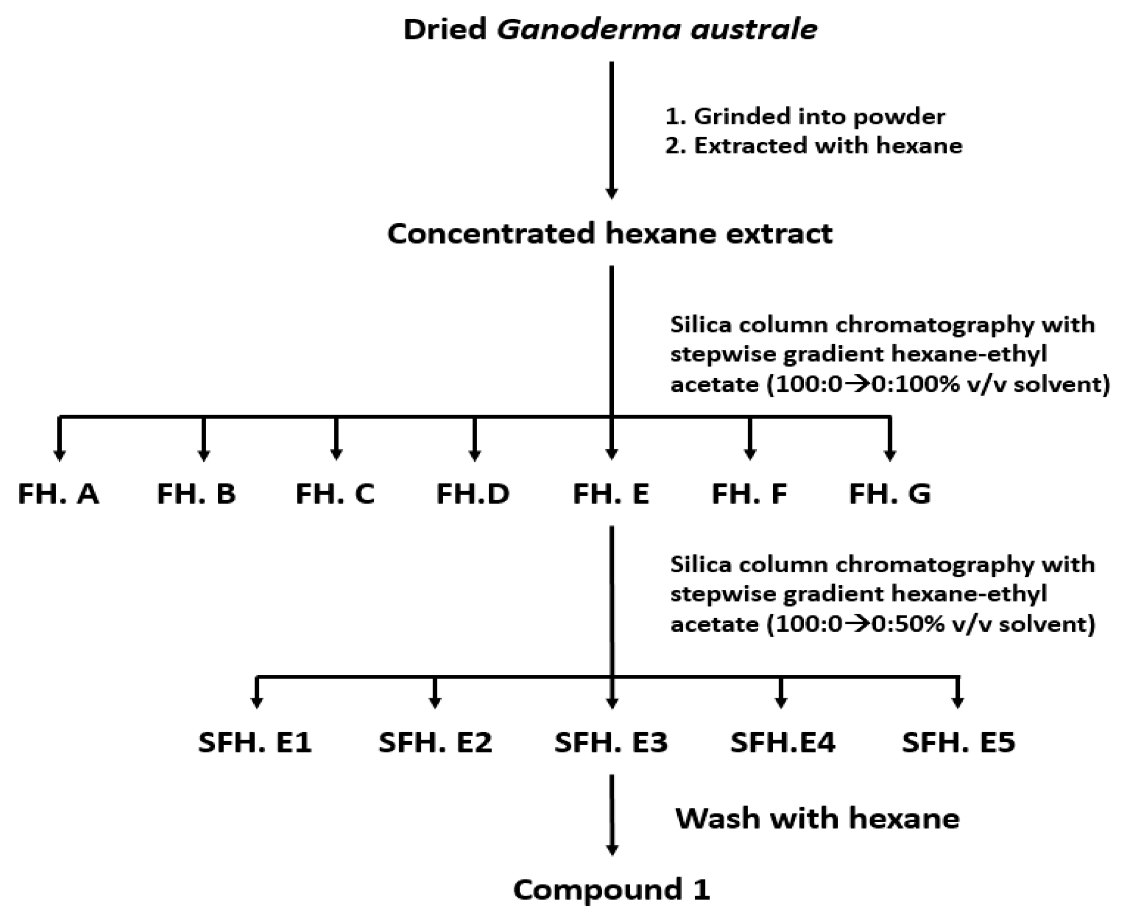
2.2. Extraction and Isolation

The dried and pulverized fruit bodies of Ganoderma australe (3 kg) were extracted exhaustively with hexane using a Soxhlet apparatus. The evaporated hexane extract was subjected to column chromatography to yield seven fractions (A–G) with a stepwise gradient elution of hexane-ethyl acetate (100:0→0:100 v/v) solvent. Fraction E (1.5 g) was further purified using silica gel column chromatography eluted with hexane-ethyl acetate (100:0 → 0:50 v/v) to yield five subfractions (E1-E5). Subfraction E3 was further purified by washing with hexane to obtain the isolated compound (Figure 1).

2.3. Instrumentation

The spectra of 1H and 13C-NMR of the isolated compound were recorded on an Agilent spectrometer (13C:125 MHz; 1H:500) with tetramethylsilane (TMS) as the internal standard. The spectrum was measured using deuterated chloroform (CDCl3) as a solvent.

2.4. In Vitro Study of DPP-4 Inhibition

Ten milligrams of the isolated compound were diluted in 10 mL Trizma® HCl buffer at pH 7.5 (50 mM). Various concentrations of samples at 60, 125, 250, and 500 µM and sitagliptin as positive control at 0.5, 1, 2, 4, and 8 µM were used. The assay was performed according to Chakrabarti et al. [19] and Al-Masri et al. [20]. Each well contained a mixture of 15 µL of DPP-4 enzyme (0.05 U/mL) and 35 µL of standard or sample. The mixture was incubated at 37 °C for 10 min as the first incubation. Subsequently, 50 µL of substrate 0.2 mM were added to this mixture, followed by the second incubation at 37 °C for 30 min. Finally, to stop the reaction, 25 µL acetic acid 25% were added. The absorbance was measured at λ 405 nm and the inhibition activity was calculated by the following formula:
% Inhibition = [(Abs control − Abs standard or sample)/Abs control] × 100

2.5. In Vitro Study of α-Glucosidase Inhibition

Ten milligrams of the isolated compound were diluted in 10 mL phosphate buffer at pH 6.9. Various concentrations of the isolated compound and 60, 125, 250, and 500 µM acarbose were used. The assay was performed according to Munim et al. [21] with slight modifications. The α-glucosidase enzyme was dissolved in 100 mL phosphate buffer at pH 6.9. Phosphate buffer was prepared by dissolving 1.42 g Na2HPO4 and 1.38 g NaHPO4.H2O in 100 mL containing 200 mg bovine serum albumin. Each well contained a mixture of 10 µL of standard or sample and 25 µL of substrate 0.625 mM, followed by first incubation at 37 °C for 10 min. Subsequently, 25 µL of an enzyme (0.05 U/mL) were added and incubated for 15 min at 37 °C. One hundred milliliters of sodium carbonate 0.2 mM were used to stop the enzymatic reaction. The absorbance was measured at λ 405 nm and the inhibition activity was calculated with the same formula used to measure DPP-4 inhibition.

2.6. In Silico Study

Crystallography of human dipeptidyl peptidase-4 (PDB: 1X70) complex with sitagliptin and α-glucosidase from Saccharomyces cerevisiae (PDB: 3W37) complex with acarbose were obtained from the Protein Data Bank. The structure of the isolated compound was downloaded from the PubChem database. Preparation of the ligand and the protein was performed using the AutoDock Tools 1.5.6 program (ADT). The interaction pattern between the ligand and the receptor was studied using the Lamarckian genetic algorithm (LGA). Docking studies were carried out using AutoDock Tools v.1.5.6 embedded in MGL Tools [22]. The grid box of sitagliptin was adjusted at x 41.376; y 51.2; z 36.2 and the grid box of acarbose was adjusted at x 0.699; y −1.87; z −23.212.

2.7. Statistical Analysis

All inhibitory data are expressed as mean ± SD (n = 3). The IC50 data were calculated using GraphPad Prism 8.0.1 software free trial with non-linear regression sigmoidal dose-response.

3. Results and Discussion

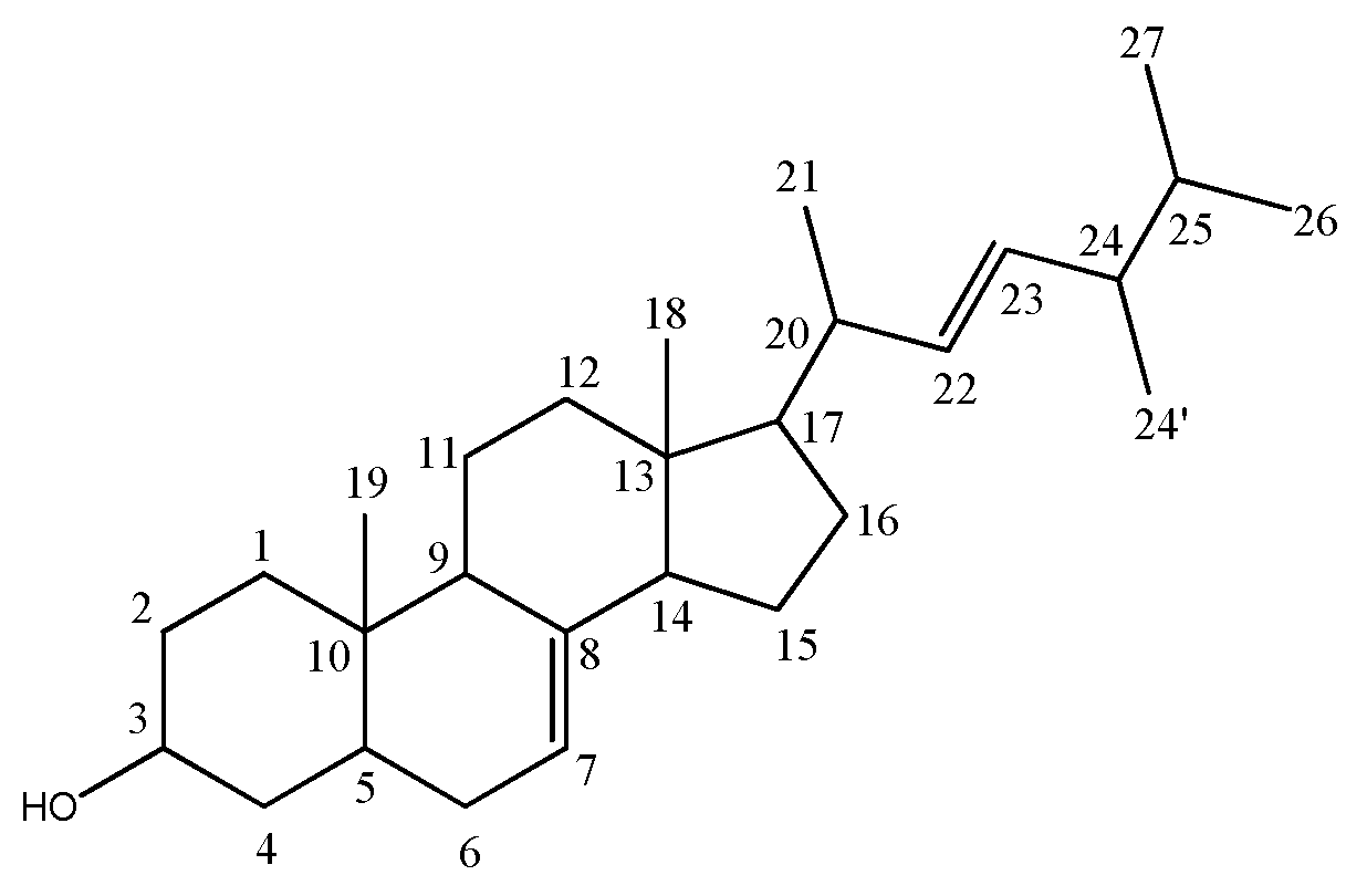
3.1. Structure Elucidation

The 1H NMR spectrum of the isolated compound clearly showed six methyl signals at δ 0.54 (3H, s, H-18); 0.79 (3H, s, H-19); 1.01 (3H, d/s, J = 5.5 Hz, H-21); 0.91 (3H, d, J = 5 Hz, H-24”); 0.81 (3H, d, J = 15 Hz, H-26); 0.81 (3H, s, H-27) ppm. The 13C-NMR revealed the presence of 28 carbon signals ascribed to six methyls at δ 12.1 (C-18); 13.1 (C-19); 21.2 (C-21); 17.7 (C-24”); 19.8 (C-26); 20.1 (C-27) ppm, eight methylenes at δ 37.3 (C-1); 31.6 (C-2); 38.1 (C-4); 29.8 (C-6); 21.7 (C-11); 39.6 (C-12); 23.0 (C-15); 28.2 (C-16) ppm, and eleven methines at δ 71.2 (C-3); 40.6 (C-5); 117.1 (C-7); 49.6 (C-9); 55.3 (C-14); 56.1 (C-17); 40.4 (C-20); 135.9 (C-22); 132.1 (C-23); 43.0 (C-24); 33.2 (C-25) ppm. It also showed the presence of two double bonds signals at δ 117.6 (C-7); 132.1 (C-23); 135.9 (C-22); and 139.7 (C-8) ppm (Figure 2). By comparison with published NMR data, the isolated compound was identified as stellasterol [23,24].

3.2. Assay of DPP-4 and α-Glucosidase Inhibition

Our previous study showed that hexane extract had the highest activity to inhibit α-glucosidase enzyme followed by ethyl acetate and methanol extract (data not shown). These results suggest that there are responsible compounds as α-glucosidase inhibitor present in hexane extract. Based on extraction-guided activity, we decided to isolate the hexane extract then examined two different inhibition enzyme mechanisms. Our isolation revealed that stellasterol was the major compound from the hexane extract.
Stellasterol showed DPP-4 inhibition activity with IC50 at 427.39 µM, while sitagliptin as the positive control had IC50 0.73 µM. Sitagliptin showed much stronger activity against DPP-4 enzyme than stellasterol. As the α-glucosidase inhibitors, stellasterol and acarbose were 314.54 µM and 207.87 µM, respectively. By comparing the standards, it seems that the activity of stellasterol was more similar to acarbose rather than sitagliptin as a hypoglycemic agent. These results were studied using in silico interaction.

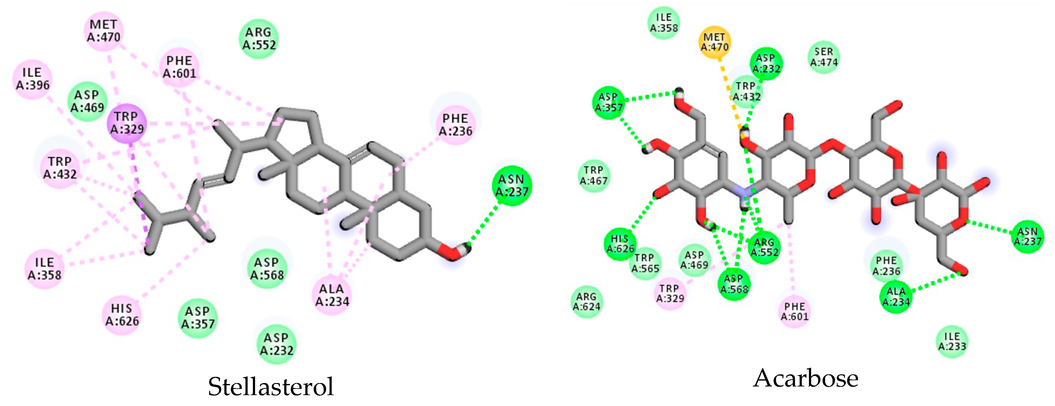
3.3. Stellasterol In Silico Study for DPP-4 Enzyme

The in silico approach was performed to predict the protein–ligand binding pose between stellasterol, either with DPP-4 or α-glucosidase enzyme. The root mean square (RMSD) of 0.50 Ǻ was gained for stellasterol with the native ligand extracted from the DPP-4 enzyme, while RMSD score for stellasterol in comparison to the origin of the α-glucosidase enzyme was 1.18 Ǻ. The most negative docking score was chosen to be used in analyzing pose interaction between ligand and receptor [25].
Docking simulation of stellasterol into the DPP-4 cavity revealed that the main interaction was hydrophobic. Stellasterol filled the hydrophobic cavity involving Trp659, Tyr547, Tyr631, Tyr662, Tyr666, Phe357, Val656, and Val711 (Table 1). Marvanita and Patel [26] suggested that these amino acid residues were the natural hydrophobic cavity of human DPP-4 (Table 2). Furthermore, linagliptin had π–π interactions in addition to Tyr547 and Trp629 residues compared to alogliptin. These additional interactions are believed to be the leading factor to elevate the potential for linagliptin compared to alogliptin [27]. Arulmozhiraja et al. [28] proposed that the interaction with Glu206 and Glu205 through hydrogen bonds in sitagliptin, alogliptin, linagliptin, and teneligliptin inhibitors are the first specifications for DPP-4 inhibitor beside Tyr547, Trp629, Tyr666, and Phe357. In addition, the interaction with S2’ is important and interaction with the S2 extensive can elevate not only the potency but also the selectivity. Furthermore, larger inhibitors that fulfill the hydrophobic cavity could elevate the inhibitory potential and stabilize the ligand–receptor complex [28,29]. Stellasterol only interacts with Tyr547 in π–alkyl interaction and other interactions mostly through hydrophobic bonds, suggesting its lower capacity to bind DPP-4 enzyme.
Kalhotra et al. [30,31] examined the profile for the addition of hydroxyl groups to inhibit the DPP-4 enzyme. Their results showed that galangin (a flavonoid with two hydroxyl groups) gives higher inhibition than chrysin (a flavonoid with two hydroxyl groups). Saleem et al. [32] also revealed that quinovic acid has an IC50 value of 30.7 µM, whereas the addition of two glucose moieties in quinovic acid elevate the activity (IC50 23.5 µM).
Stellasterol has only one free hydroxyl group at C-3; perhaps this is caused by the lower inhibition of the DPP-4 enzyme in comparison to the previously structure. Stellasterol, a steroid derivative, has a non-polar structure in nature exposing domination of alkyl and proton aliphatic groups. Conversely, sitagliptin as the positive control contains fluoro and amine capable of forming hydrogen bonds with some amino acids in the DPP-4 cavity. A hydroxyl group of stellasterol interacts through hydrogen bonding with amino acid Tyr585 in the DPP-4 pocket. This amino acid Tyr585 is a different type to the amino acids that bind with sitagliptin, Glu205, Glu206, Arg125, Arg358, and Asn710. The hexane extract showed higher inhibition of DPP-4 enzyme than stellasterol, indicating the activity against DPP-4 enzyme may not be contributed by stellasterol as the major isolate. According to Arulmozhiraja et al. [28], stellasterol fulfills the second and third specifications for DPP-4 inhibitor.

3.4. Stellasterol In Silico Study for α-Glucosidase Enzyme

The docking result revealed that acarbose, an oligosaccharide, is surrounded by many hydroxyl groups that are able to form a hydrogen bond with amino acid residues, such as Arg552, Asp568, Asp357, His626, Asp232, Asn237, and Ala234 (Figure 3).
In the α-glucosidase cavity, a hydroxyl group position at C-3 of stellasterol interacted with Asn237 through a hydrogen bond similar to acarbose as a positive control. Tagami et al. [33] proposed that Asn237 was one of the residues that play a role in the long-chain specificity inside the N-loop. This interaction possibly has a role in increasing the activity of stellasterol against α-glucosidase rather than DPP-4 enzyme. In addition, stellasterol binds with the same two amino acids Trp329 and Phe601, similar to acarbose in the hydrophobic pocket. Stellasterol also binds with His626, Ala234, and Met470 in π–alkyl interaction (Table 3).
Several terpenoid compounds have been tested as α-glucosidase inhibitors, including ursolic acid, 3β-hydroxy-11-ursen-28,13-olide, and ursonic acid. These compounds demonstrated IC50 8.38, 203.76, and 100.99 µM. Those compounds showed higher inhibitory activity compared to stellasterol. These might be due to the presence of carbonyl or the additional hydroxyl groups, or the carboxylic acid group, which leads to the stronger interaction with α-glucosidase enzyme [34].
Four triterpenoids from Psidium guajava leaves, namely trihydroxyopsidiumlanostenoic acid, 12β-hydroxypsidiumlanostenoic acid, psidiumlanostenoic acid glucoside, and psidiumlanostenoic acid, as shown in Figure 4, have been tested in vivo for antidiabetic activity. The administration of 50 mg/kg of these isolate compounds for 1 week in diabetic rats model showed blood glucose levels of 142.50, 154.50, 165.67, and 172.17 mg/dL, respectively. However, the normal control rat and the diabetic control rat showed 87.66 and 327.33 mg/dL blood glucose levels after 1 week [35].
Although in vitro study showed that the IC50 as an α-glucosidase inhibitor is relatively high, another study reported that similar compounds have the capacity to reduce the blood glucose level. Therefore, with stellasterol it may contribute to the anti-diabetic activity in G. australe.

4. Conclusions

The in silico approach helps one to understand and support the in vitro result. According to in vitro and in silico results, stellasterol was more likely well-fitted as an α-glucosidase inhibitor rather than a DPP-4 inhibitor.

Author Contributions

S.S. designed the experiments and reviewed the manuscript. K.B., Y.B.P., and K.R.W. collected the mushroom, performed extraction and isolation, performed the experiments, and analyzed the NMR results. K.B. and J.J. performed the in silico experiment. K.B. wrote the manuscript.

Funding

The authors are sincerely grateful to the Indonesia Endowment Fund for Education (LPDP) for sponsoring this project.

Acknowledgments

The authors wish to thank Kimia Farma and Dexa Medica for kindly providing the sitagliptin and acarbose. The authors are also thankful to Dewi Susan from Research Center for Biology, Cibinong, Indonesia Institute of Sciences (LIPI), for helping to isolate and interpret DNA results of Ganoderma species. The authors are grateful to Frangky Sanghande from Pharmacy Prisma University for great discussion and Syaikhul Aziz from Pharmacy Sumatera Institute of Technology for helping interpret NMR results.

Conflicts of Interest

The authors declare no conflict of interest.

References

  1. Gottlieb, A.M.; Ferrer, E.; Wright, J.E. rDNA analyses as an aid to the taxonomy of species of Ganoderma. Mycol. Res. 2000, 104, 1033–1045. [Google Scholar] [CrossRef]
  2. Li, F.; Zhang, Y.; Zhong, Z. Antihyperglycemic effect of Ganoderma lucidum polysaccharides on streptozotocin-induced diabetic mice. Int. J. Mol. Sci. 2011, 12, 6135–6145. [Google Scholar] [CrossRef] [PubMed]
  3. Fatmawati, S.; Shimizu, K.; Kondo, R. Ganoderol B: A potent α-glucosidase inhibitor isolated from the fruiting body of Ganoderma lucidum. Phytomedicine 2011, 18, 1053–1055. [Google Scholar] [CrossRef] [PubMed]
  4. Wang, K.; Bao, L.; Xiong, W.; Ma, K.; Han, J.; Wang, W.; Yin, W.; Liu, H. Lanostane triterpenes from the Tibetan medicinal mushroom Ganoderma leucocontextum and their inhibitory effects on HMG-CoA reductase and α-glucosidase. J. Nat. Pro. 2015, 78, 77–89. [Google Scholar] [CrossRef] [PubMed]
  5. Bishop, K.S.; Kao, C.H.; Xu, Y.; Glucina, M.P.; Paterson, R.R.; Ferguson, L.R. From 2000 years of Ganoderma lucidum to recent developments in nutraceuticals. Phytochemistry 2015, 114, 56–65. [Google Scholar] [CrossRef]
  6. Richter, C.; Wittsein, K.; Kirk, P.M.; Stadler, M. An assessment of the taxonomy and chemotaxonomy of Ganoderma. Fungal Divers. 2015, 71, 1–15. [Google Scholar] [CrossRef]
  7. Bailey, C.J. Treating insulin resistance in type 2 diabetes with metformin and thiazolidinediones. Diabetes Obes. Metab. 2005, 7, 675–691. [Google Scholar] [CrossRef]
  8. Pathak, R.; Bridgeman, M.B. Dipeptidyl peptidase-4 (DPP-4) inhibitors in the management of diabetes. Pharm. Ther. 2010, 35, 509–513. [Google Scholar]
  9. Verspohl, E.J. Novel therapeutics for type 2 diabetes: Incretin hormone mimetics (glucagon-like peptide-1 receptor agonists) and dipeptidyl peptidase-4 inhibitors. Pharmacol. Ther. 2009, 124, 113–138. [Google Scholar] [CrossRef]
  10. Tian, L.; Jin, T. The incretin hormone GLP-1 and mechanisms underlying its secretion. J. Diabetes 2016, 8, 753–765. [Google Scholar] [CrossRef] [Green Version]
  11. Lee, Y.S.; Jun, H.S. Anti-diabetic actions of glucagon-like peptide-1 on pancreatic beta-cells. Metabolism 2014, 63, 9–19. [Google Scholar] [CrossRef]
  12. Ahren, B. Dipeptidyl peptidase-4 inhibitors: Clinical data and clinical implications. Diabetes Care. 2007, 30, 1344–1350. [Google Scholar] [CrossRef]
  13. Liu, Y.; Hu, Y.; Liu, T. Recent advances in non-peptidomimetic dipeptidyl peptidase 4 inhibitors: Medicinal chemistry and preclinical aspects. Curr. Med. Chem. 2012, 19, 3982–3999. [Google Scholar] [CrossRef]
  14. Kushwaha, R.N.; Haq, W. Sixteen-years of clinically relevant dipeptidyl peptidase-IV (DPP-IV) inhibitors for treatment of type-2 diabetes: A perspective. Curr. Med. Chem. 2014, 21, 1–33. [Google Scholar] [CrossRef]
  15. McKeage, K. Trelagliptin: First global approval. Drugs 2015, 75, 1161–1164. [Google Scholar] [CrossRef]
  16. Galloway, I.; Mckay, G.; Fisher, M. Omarigliptin. Pract. Diabetes 2017, 34, 70–71. [Google Scholar] [CrossRef]
  17. Ramanathan, B. DPP-4 inhibitors in the management of type 2 diabetes mellitus. In Progress in Medicine 2017; The Asscociation of Physicians of India: Mumbai, India, 2017; Volume 1, pp. 231–237. [Google Scholar]
  18. Rosa, M.M.; Dias, T. Commonly used endocrine drugs. In Handbook of Clinical Neurology, 3rd ed.; Biller, J., Ferro, J.M., Eds.; Elsevier: Amsterdam, The Netherlands, 2014; Volume 120, pp. 809–824. [Google Scholar]
  19. Chakrabarti, R.; Bhavtaran, S.; Narendra, P.; Varghese, N.; Vanchhawng, L.; Hyder, M.S.; Shihabudeen, H.; Thirumurugan, K. Dipeptidyl peptidase-IV inhibitory activity of Berberis aristata. J. Nat. Prod. 2011, 4, 158–163. [Google Scholar]
  20. Al-Masri, I.M.; Mohammad, M.K.; Tahaa, M.O. Inhibition of dipeptidyl peptidase IV (DPP IV) is one of the mechanisms explaining the hypoglycemic effect of berberine. J. Enzyme Inhib. Med. Chem. 2009, 24, 1061–1066. [Google Scholar] [CrossRef]
  21. Munim, A.; Katrin; Azizahwati; Andriani, A.; Mahmudah, K.F.; Mashita, M. Screening of alpha glucosidase inhibitory activity of some Indonesian medicinal plants. Int. J. Med. Arom. Plants. 2013, 3, 144–150. [Google Scholar]
  22. Morris, G.M.; Huey, R.; Lindstrom, W.; Sanner, M.F.; Belew, R.K.; Goodsell, D.S.; Olson, A.J. AutoDock4 and AutoDockTools4: Automated docking with selective receptor flexibility. J. Comput. Chem. 2009, 30, 2785–2791. [Google Scholar] [CrossRef] [Green Version]
  23. Borlagdan, M.; de Castro, M.E.; van Altena, I.; Ragasa, C. Sterols from Trametes versicolor. Res. J. Pharm. Biol. Chem. Sci. 2017, 8, 740–744. [Google Scholar]
  24. Seo, H.W.; Hung, T.M.; Na, M.; Junga, H.J.; Kim, J.C.; Choi, J.S.; Kim, J.H.; Lee, H.K.; Lee, I.; Bae, K.; et al. Steroids and triterpenes from the fruit bodies of Ganoderma lucidum and their anti-complement activity. Arch. Pharm. Res. 2009, 32, 1573–1579. [Google Scholar] [CrossRef]
  25. Levita, J.; Rositama, M.R.; Alias, N.; Khalida, N.; Saptarini, N.M.; Megantara, S. Discovering COX-2 inhibitors from flavonoids and diterpenoids. J. Appl. Pharm. Sci. 2017, 7, 103–110. [Google Scholar]
  26. Marvaniya, H.M.; Patel, H.U. Role of dipeptidyl peptidase-IV (DPP-4) inhibitor in the management of type 2 diabetes. World J. Pharm. Pharmaceut. Sci. 2017, 6, 551–566. [Google Scholar] [CrossRef]
  27. Lai, Z.W.; Li, C.; Liu, J.; Kong, L.; Wen, X.; Sun, H. Discovery of highly potent DPP-4 inhibitors by hybrid compound design based on linagliptin and alogliptin. Eur. J. Med. Chem. 2014, 83, 547–560. [Google Scholar] [CrossRef]
  28. Arulmozhiraja, S.; Matsuo, N.; Ishitsubo, E.; Okazaki, S.; Shimano, H.; Tokiwa, H. Comparative binding analysis of dipeptidyl peptidase IV (DPP-4) with antidiabetic drugs—An Ab Initio fragment molecular orbital study. PLoS ONE 2016, 11, e0166275. [Google Scholar] [CrossRef]
  29. Kishimoto, M. Teneligliptin: A DPP-4 inhibitor for the treatment of type 2 diabetes. Diabetes Metab. Syndr. Obes. 2013, 6, 187–195. [Google Scholar] [CrossRef]
  30. Kalhotra, P.; Chittepu, V.C.S.R.; Osorio-Revilla, G.; Gallardo-Velázquez, T. Structure—activity relationship and molecular docking of natural product library reveal Chrysin as a novel dipeptidyl peptidase-4 (DPP-4) inhibitor. Molecules 2018, 23, 1368. [Google Scholar] [CrossRef]
  31. Kalhotra, P.; Chittepu, V.C.S.R.; Osorio-Revilla, G.; Gallardo-Velázquez, T. Discovery of galangin as a potential DPP-4 inhibitor that improves insulin-stimulated skeletal muscle glucose uptake: A combinational therapy for diabetes. Int. J. Mol. Sci. 2019, 20, 1228. [Google Scholar] [CrossRef]
  32. Saleem, S.; Jafri, L.; Haq, I.U.; Chang, L.C.; Calderwood, D.; Green, B.D.; Mirza, B. Plants Fagonia cretica L. and Hedera nepalensis K. Koch contain natural compounds with potent dipeptidyl peptidase-4 (DPP-4) inhibitory activity. J. Ethnopharmacol. 2014, 156, 26–32. [Google Scholar] [CrossRef]
  33. Tagami, T.; Yamashita, K.; Okuyama, M.; Mori, H.; Yao, M.; Kimura, A. Molecular basis for the recognition of long-chain substrates by plant α-glucosidases. J. Biol. Chem. 2013, 288, 19296–19303. [Google Scholar] [CrossRef]
  34. Thanh Tam, N.; Thiện, Đ.; Van Sung, T.; Nguyen, T.H.A.; Thi Thuy, T.; Huu Trung, K.; Dang Xuan, T.; Khanh, T. Evaluation of ursolic acid as the main component isolated from Catharanthus roseus against hyperglycemia. Int. Lett. Nat. Sci. 2016, 50, 7–17. [Google Scholar] [CrossRef]
  35. Bagri, P.; Ali, M.; Aeri, V.; Bhowmik, M. Isolation and antidiabetic activity of new lanostenoids from the leaves of Psidium guajava L. Int. J. Pharm. Pharm. Sci. 2016, 8, 14–18. [Google Scholar] [CrossRef]
Figure 1. Isolation scheme of stellasterol from G. australe.
Scipharm 87 00021 g001
Figure 2. Stellasterol isolated from Ganoderma australe.
Scipharm 87 00021 g002
Figure 3. The binding mode 2D view of the α-glucosidase enzyme with stellasterol and acarbose.
Scipharm 87 00021 g003
Figure 4. Lanosterol derivates as α-glucosidase inhibitor [35].
Scipharm 87 00021 g004
Table 1. Interaction of stellasterol and sitagliptin with the binding pocket of the human DPP-4 enzyme.
LigandHydrogen BondHydrophobic Bond
Glu206Glu205Arg358Arg125Asn710Tyr585Tyr631Tyr547Tyr666Tyr662Val711Val656Trp659Phe357
Stellasterol-----+π–alkylπ–alkylπ–alkylπ–alkylAlkyl–alkylAlkyl–alkylπ–alkylπ–alkyl
Sitagliptin+++++---π-ππ-π---π–π
Table 2. Amino acid residues in the active site of the DPP-4 enzyme [28].
Amino Acid ResiduesPocket
S2’S1’S1S2S2 Ext
Trp629Tyr547Tyr666Arg125Phe357
His740Tyr631Ser630Arg669Arg358
Ser630Phe357Val656Glu205Ser209
Tyr547Pro550Trp659Glu206Val207
Tyr666Tyr662Phe357
Val711Arg358
Asn710
Table 3. Interaction if stellasterol and acarbose with the binding pocket of the α-glucosidase enzyme.
LigandHydrogen Bond Hydrophobic Bond
Arg552Asp568Asp367His626Asp232Asn237Ala234Ile358Ile233Ile396Trp467Trp329Trp432Trp329Phe601Phe236Ph3476Met470
Stellasterol---π–alkyl-+π–alkyl+-+-++-++-π–alkyl
Acarbose+++++++----+--+--+

