Article

The Anti-Inflammatory Activity of Nigella sativa Balm Sticks

Department of Pharmacy and Science, Universitas Muhammadiyah Prof. Dr HAMKA (UHAMKA), Jakarta 13460, Indonesia
*
Author to whom correspondence should be addressed.
Sci. Pharm. 2019, 87(1), 3; https://doi.org/10.3390/scipharm87010003
Submission received: 28 December 2018 / Revised: 9 January 2019 / Accepted: 9 January 2019 / Published: 14 January 2019

Abstract

:
Nigella sativa oil has been known to have potent anti-inflammatory activity. This research aimed to determine the anti-inflammation activity of Nigella sativa oil in a simple balm stick by topical application. The activity was checked using two methods: carrageenan-induced paw oedema and granuloma pouch on rats. The results showed that balm sticks which contained 10% Nigella sativa could overcome both acute and sub-acute inflammation showing by high oedema inhibition (60.64%), low leucocytes count (43.55% lower than control) as well as a notable TNF-α concentration (50% lower than control) on the inflamed area. In conclusion, topical application of a Nigella sativa balm stick was effective for both acute and sub-acute forms of inflammation.

1. Introduction

Nigella sativa (Ranunculaceae family) or commonly known as black cumin has long been used as a phytomedicine for antidiarrhea, appetite enhancer, diuretic, antibacterial, analgesic, anthelmintic, various skin diseases, anti-inflammatory, back pain, hemiplegia (paralysis of the hands or feet), and rheumatism [1,2,3]. The bioactive content of Nigella sativa included p-cymene, α-thujene, longifolene, β-pinene, α-pinene, carvacrol and the main compound is thymoquinone [4,5,6].
Both Nigella sativa oil and thymoquinone are potent anti-inflammatory agents that have been demonstrated in various disease models such as encephalomyelitis, colitis, peritonitis, oedema and arthritis through suppression of inflammatory mediators such as prostaglandins and leukotrienes [7]. Orally gavage of 4 mL/kg/day Nigella sativa oil for 31 days showed a reduction of IL-4 and NO production in rats [8]. Thymoquinone at a dose of 10 mg/kg body weight of rats showed anti-inflammatory activity through inhibition of cyclooxygenase (COX) and 5-lipooxygenase (5-LPO) pathways [9,10] and at a dose of 5 mg/kg body weight of rats could lower TNF-α and IL-1β levels in arthritis [11]. Anti-inflammatory activity of Nigella sativa also has been shown in humans, for example the use of the oil for geriatric patients with osteoarthritis [12].
However, there are only a few studies showing the use of Nigella sativa oil topically. For instance, topical Nigella sativa could be effective treatment for psoriasis [13], application of 50% Nigella sativa oil in cream could heal burn wounds in rats [14], while Nigella sativa essential oil nanoparticles have also been shown to have anti-inflammatory properties in combination with indomethacin [15]. The topical use of Nigella sativa oil needs to be developed in the form of a practical dosage form such as a balm stick. In this study, a Nigella sativa balm stick was tested on acute and subacute inflammatory models.

2. Materials and Methods

2.1. Materials

Nigella sativa seed oil (PT. Saraswanti Indo Genetech, Bogor, Indonesia) was produced by cold press method without chemicals. According to the COA, the Nigella sativa seed fixed oil contain vitamin E, Calcium, Fe, Omega 3, 6 and 9. Gas Chromatography Mass Spectrometry (GCMS) analysis showed that the Nigella sativa seed oil contained 23.75% Thymoquinone. The other materials were cera alba, adeps lanae, vaselin alba, lanolin hydrate, cetyl alcohol, butyl hidroxitoluen (BHT) (PT. Brataco, Jakarta, Indonesia), virgin coconut oil (VCO) and oleum sesame (Herba Bagoes, Jakarta, Indonesia), hydrocortisone cream 2.5% (Kalbe Farma, Jakarta, Indonesia) Turk solution (PT. Gresik Sarana Tirta, Jakarta, Indonesia), carrageenan (Sigma, St. Louis, MO, USA), and TNF-α Kit (Sigma).

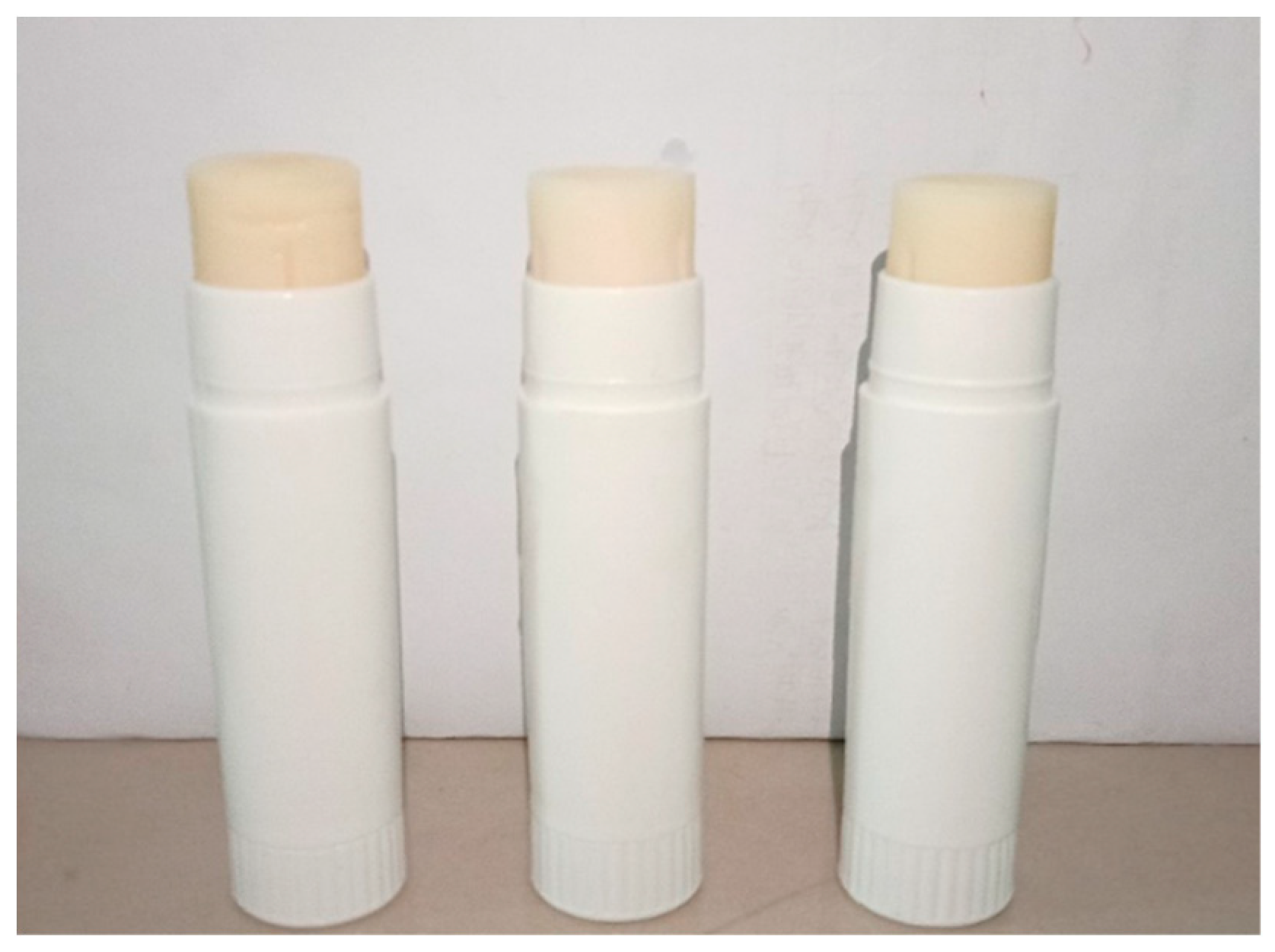
2.2. Balm Stick Preparation

Balm sticks were made with three formulas with variations of Nigella sativa oil content (Table 1). BHT (butyl Hydroxytoluene) was dissolved in VCO (mass 1). Cera alba was melted on water bath at 65 °C and stirred (mass 2). Setil alcohol was added into mass 2, followed by adeps lanae and melted into a homogeneous mixture. The mixture was removed from the water bath, then added with mass 1 and stirred well. Finally, Nigella sativa oil was added and stirred to the homogeneous mixture, then formed into sticks (Figure 1).

2.3. Animal

Wistar rats (Rattus novergicus L.) aged 2–3 months with weights of ±200 g were used in this study, 50 rats in total. The study protocol was approved by the Research Ethics Commission, Faculty of Medicine, University of Indonesia by applying the principles of replacement, reduction and refinement (the 3Rs) with approval under ethics number 516/UN2.F1/ETIK/2018 and 517/UN2.F1/ETIK/2018. All animals were acclimatized in the cage with a temperature of ±23 °C for one week.

2.4. Acute Anti-Inflammatory Test (Carrageenan-Induced Paw Oedema)

The procedure of carrageenan-induced paw oedema in accordance with Pise and Padwal (2017) [16] with modification. The right foot of the rat was measured using a plethysmometer, then recorded as the initial volume (Vo). The feet were then smeared with the test substances topically in accordance by the group. The negative control group was smeared with control balm, positive control was smeared with 2.5% hydrocortisone cream, the NB (Nigella sativa balm stick) groups was smeared with the balm containing 5%, 7.5% and 10% Nigella sativa oil respectively. All test substances were applied topically 50 times on the right foot of each rat. Then immediately, the feet were injected intraplantar with 0.1 ml of 1% w/v carrageenan solution (in oleum sesame) [17]. The oedema volume was measured using plethysmometer at minutes 30, 90, 150, 210, and 270 after carrageenan injection, and recorded as Vt. The oedema volume data of each time measured were then used to calculate the inhibition percentage.
inhibition   % = mean   increment   volume   of   control mean   increment   volume   of   test   substance mean   increment   volume   of   control × 100 %

2.5. Sub-Acute Anti-Inflammatory Test (Granuloma Pouch Method)

The back of rats was shaved, then the rats were anaesthetized with ketamine (i.m, 40 mg/kg BW). The back of each rat was then injected with 20 mL sterile air subcutaneously to form a pouch. 24 h later, the pouch was injected with 3 mL of 2% (w/v) carrageenan solution (in oleum sesame). The inflamed areas were then smeared with the test substances twice a day. The second day, the pouch was rinsed with 2 mL phosphate buffered saline (PBS) (0.01 M, pH 7.4), then total exudate was taken. The exudate was then used to determine the number of leukocyte cells and TNF-α levels of each group [18].

2.5.1. Leucocyte Count

The exudate of each rat was diluted with Turk solution (1:20) and mixed homogeneously. The solution was then dripped to the hemocytometer and left to be distributed throughout the area of the count room. Preparations were examined under a microscope with 40× magnification. The number of leukocytes was calculated by the following formula [19]:
Leucocytes   count   ( mm 3 ) = N × Dilution   factor Σ   C h a m b e r   V o l u m e

2.5.2. Determination of TNF-α

The exudate was centrifuged at 5400 g for 10 min at 4 °C to obtained supernatant aliquoted and then stored at −8 °C [19]. TNF-α was determined using an ELISA kit (Sigma, St. Louis, MO, USA), according to the manufacturer’s instructions.

2.6. Data Analysis

Data were analyzed using One-Way ANOVA followed by Tukey and Duncan tests.

3. Results

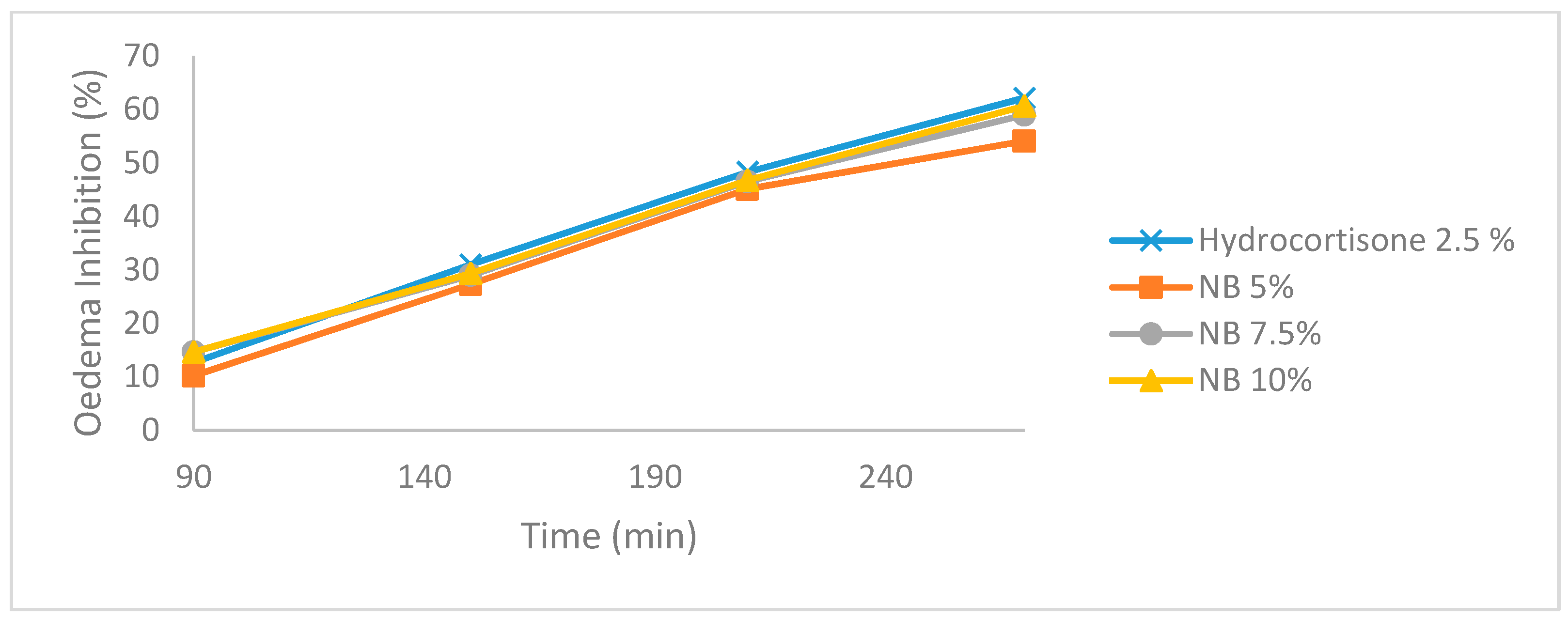
3.1. Carrageenan-Induced Paw Edema

The study showed that the injection of 0.1 ml of 1% carrageenan via intraplantar resulted in oedema, which was observed to show signs of inflammation starting from minute 30. Afterwards, the volume of oedema continued to increase. Hydrocortisone was used as the reference drug [20]. Data in Table 1 demonstrate that there is a difference in the percentage of oedema that occurs due to changes in time in all groups. The Nigella sativa Balm (NB) group showed an anti-inflammatory activity as indicated by the decrease in oedema volume at minute 150. However, in the control group, the volume of oedema continued to increase. The results of statistical tests showed significant differences in the paw oedema of NB group against control starting at minute 90 (Table 2). The oedema inhibition in the NB 10% group showed the highest percentage compared to 5% and 7.5% NB. The three groups showed comparable results with hydrocortisone 2.5% (Figure 2).

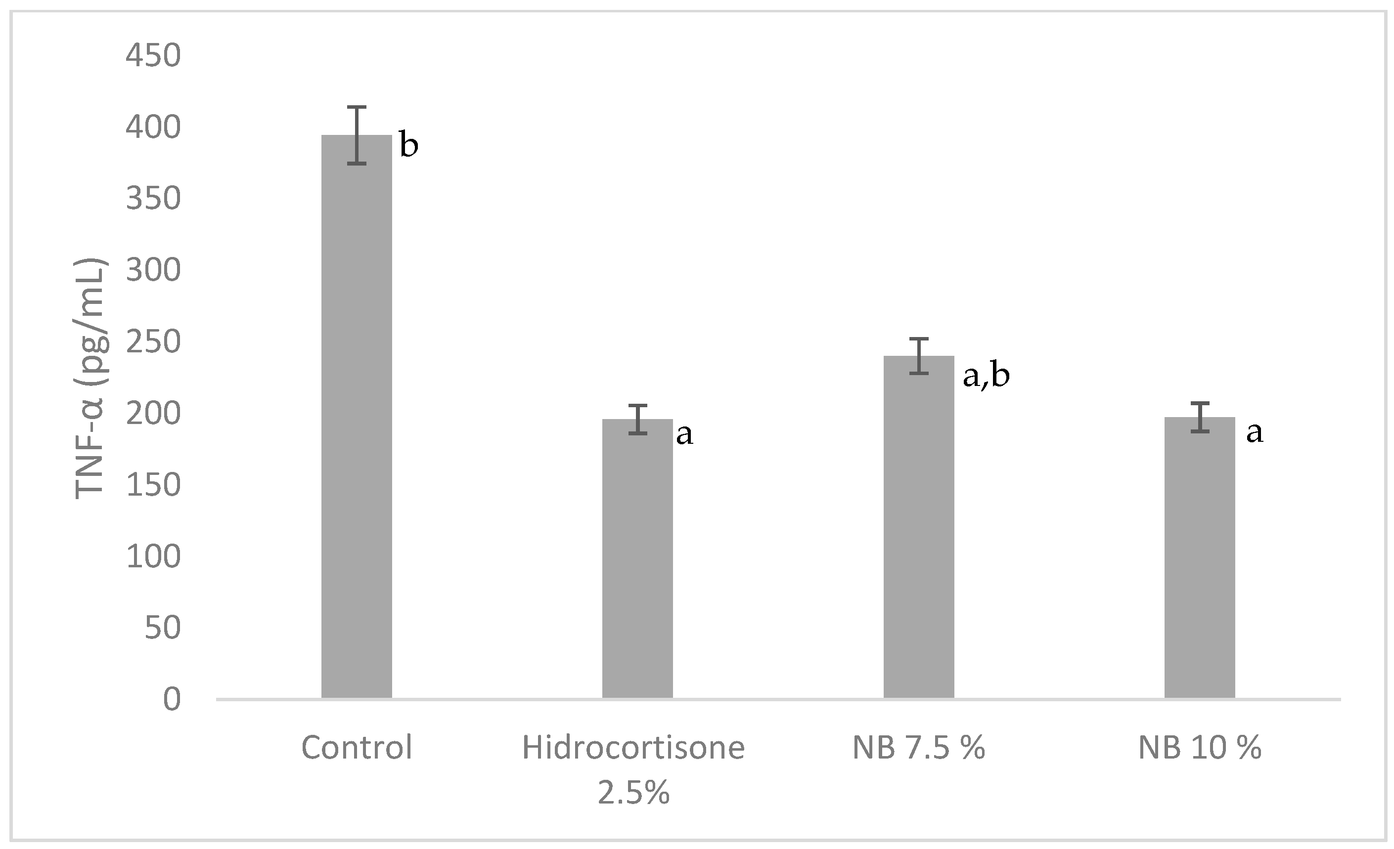
3.2. Sub-Scute Inflammation (Granuloma Pouch)

The results of the exudates volume measurements show that NB 7.5% and 10% groups comparable to hydrocortisone 2.5%, while the number of leukocytes in exudates showed a decrease of 22.02%; 32.14% and 43.55% in the NB 5%, 7.5%, and 10% groups respectively (Table 3). Statistically, only NB 10% group showed a decrease of leucocyte percentage comparable to hydrocortisone 2.5% (47.64%).
Granuloma pouch tissue of rats could also be used as an assessment of inflammation. Biochemistry observation of exudates was carried out on TNF-α levels. Test results showed 7.5% and 10 % NB groups could significantly reduce TNF-α levels (p < 0.05) (Figure 3), with reductions of around 39% (NB 7.5%) and 50% (NB 10%) compared to control. The NB 10% group was able to lower TNF-α levels comparable to 2.5% hydrocortisone.

4. Discussion

Acute inflammation is an initial inflammatory response that begins with vasodilation and increased capillary permeability, involving a process of fluid exudation (oedema) and migration of polymorphonuclear cells from blood to damaged tissue, especially neutrophils. This response is relatively short, lasting for only a few hours or days [21].
In this study, the acute anti-inflammatory activity test was carried out using the carrageenan-induced paw oedema method. Carraageenan caused inflammation by a mechanism involving phospholipase A2 and production of the inflammatory mediator such as cytokines, serotonin, histamine, prostaglandins and leukotrienes, which facilitate the migration of leukocytes to inflammatory sites [17,22]. Oedema occurs due to increased capillary permeability so that the flow of fluid rises continuously to the inflammatory tissue. At 0–1 h after injection of carrageenan, basophils and thrombocytes release of histamine and serotonin from the site of inflammation, therefore oedema was formed. The process of acute inflammation reaches the peak within 2–3 h after injection of carrageenan [23]. In this research, a Nigella contained balm stick has been proven to reduce oedema in carrageenan-induced paw. Table 2 showed a decrease in the percentage of oedema in all groups except control. Significant oedema inhibition of all NB groups starts at 150 min, which was around 28% and the total inhibition of oedema in the NB 10% group at 270 min was 60.64%. As seen in the result, the percentage of oedema in the NB group was 5%, 7.5%, and 10%, continued to decrease to 270 min drastically, and the oedema inhibition had the same trend as hydrocortisone (Figure 2).
Observation on rat’s granuloma pouch also showed the anti-inflammatory activity of NB. In sub-acute inflammation, leukocyte and phagocytic infiltrates to the site of injury. Carrageenan caused an increase in the number of leukocytes in inflammatory tissue due to vascular permeability in the inflammatory area, which increases fluid flow resulting in the migration of inflammatory cells [10]. This mediator activates cells from the tissue that are followed by the release of metalloproteinase which causes pain or inflammation. Tumor necrosis factor (TNF) α is considered to be the main inflammatory mediator [24]. The number of leukocytes in granuloma pouch (Table 3) showed that the lowest leukocyte count was obtained in the hydrocortisone 2.5% group followed by NB 10%, 7.5% and 5% respectively. This therefore showed NB anti-inflammatory activity in inhibiting leukocyte migration to the site of inflammation. The best activity was seen in NB 10% with a leukocyte number that was 43.55% lower than control. This result was in line with Table 3 as well as in Figure 3 where the amount of pouch exudate and TNF-α in this group was also the lowest among the three NB groups. These results indicate the ability of NB 10% to overcome sub-acute inflammation equivalent to hydrocortisone 2.5%.
Topical Nigella sativa balm activity shows comparable results to Pise and Padwal (2017) research [16], where Nigella sativa oil with a dose of 10 ml/kg rats BW p.o showed a 39.64% oedema inhibition. These results indicate that the anti-inflammatory activity of Nigella sativa is clear whether it is given topically or orally. Haj-allahyari et al. (2018) also reported that Nigella sativa alcohol extract could decrease COX-2 expression by 36.64% and TNF-α by 34.02%, while in this research NB 10% showed higher (50%) TNF-α inhibition. In conclusion, despite many advance formulations on Nigella sativa oil, a simple formula like a balm stick could also result in similar anti-inflammatory activity.

Author Contributions

Conceptualization was done by L.P.D. and K.Y.; Methodology and experimental works were done by L.P.D. and K.Y.; Data Analysis was done by S.N.G.; writing, review and editing the paper were done by L.P.D. and K.Y.

Funding

This research was funded by Kemenristek Dikti 2018, contract number 025/KM/PNT/2018.

Conflicts of Interest

The authors declare no conflict of interest.

References

  1. Gilani, A.H.; Jabeen, Q.; Khan, M.A.U. A review of medicinal uses and pharmacological activities of Nigella sativa. Pak. J. Biol. Sci. 2004, 4, 441–451. [Google Scholar]
  2. Duncker, S.C.; Philippe, D.; Martin-Paschoud, C.; Moser, M.; Mercenier, A.; Nutten, S. Nigella sativa (Black cumin) seed extract alleviates symptoms of allergic diarrhea in mice, involving opioid receptors. PLoS ONE 2012, 7, e39841. [Google Scholar] [CrossRef] [PubMed]
  3. Ahmad, S.; Beg, Z.H. Mitigating role of thymoquinone rich fractions from Nigella sativa oil and its constituents, thymoquinone and limonene on lipidemic-oxidative injury in rats. SpringerPlus 2014, 3, 316. [Google Scholar] [CrossRef] [PubMed]
  4. Ali, K.; Ghazali, H.M.; Yassoralipour, A.; Ramakrishnan, Y.; Ganjloo, A. Physicochemical characteristics of Nigella seed (Nigella sativa L.) oil as affected by different extraxtion methods. J. Am. Oil Chem. Soc. 2011, 88, 533–540. [Google Scholar] [CrossRef]
  5. Hassanien, M.F.R.; Assiri, A.M.A.; Alzohairy, A.M.; Oraby, H.F. Health-promoting value and food applications of black cumin essential oil: An overview. J. Food Sci. Technol. 2015, 52, 6136–6142. [Google Scholar] [CrossRef] [PubMed]
  6. Islam, R.; Nazmul, H.; Siddiqui, S.A.; Rashid, M.A.; Mahmud, A.Z.; Rahman, M.S.; Rahman, A. The black seed Nigella sativa linnaeus: A study of the antioxidant activity of the essential oil and extacts. J. Nat. Sci. Sustain. Technol. 2010, 7, 37–41. [Google Scholar]
  7. Salem, M.L. Immunomodulatory and therapeutic properties of the Nigella sativa L. seed. Int. Immunopharmacol. 2005, 5, 1749–1770. [Google Scholar] [CrossRef]
  8. Khaldi, T.; Chekchaki, N.; Boumendjel, M.; Taibi, F.; Abdellaoui, M.; Messarah, M.; Boumendjel, A. Ameliorating effects of Nigella sativa oil on aggravation of inflammation, oxidative stress and cytotoxicity induced by smokeless tobacco extract in an allergic asthma model in Wistar rats. Allergol. Immunopathol. (Madrid) 2018, 46, 472–481. [Google Scholar] [CrossRef]
  9. Alam, M.; Galav, V. Anti-inflammatory effect and toxicological evaluation of thymoquinone (volatile oil of black seed) on adjuvant-induced arthritis in Wistar rat. Indian J. Life Sci. 2013, 2, 17–22. [Google Scholar]
  10. Amin, B.; Hosseinzadeh, H. Black cumin (Nigella sativa) and its active constituent Thymoquinone: An overview on the black cumin (Nigella sativa) and its active constituent, thymoquinone: An overview on the analgesic and anti-inflammatory effects. Planta Med. Sci. 2016, 82, 8–16. [Google Scholar] [CrossRef]
  11. Tekeoglu, I.; Dogan, A.; Demiralp, L. Retracted: Effects of thymoquinone (volatile oil of black cumin) on rheumatoid arthritis in rat models. Phyther. Res. 2006, 20, 869–871. [Google Scholar] [CrossRef] [PubMed]
  12. Tuna, H.I.; Babadag, B.; Ozkaraman, A.; Balci Alparslan, G. Investigation of the effect of black cumin oil on pain in osteoarthritis geriatric individuals. Complement. Ther. Clin. Pract. 2018, 31, 290–294. [Google Scholar] [CrossRef] [PubMed]
  13. Ahmed, J. The effectiveness of Nigella sativa, methotrexate and their combination in the treatment of moderate to severe psoriasis. J. Clin. Exp. Investig. 2014, 5, 521–528. [Google Scholar] [CrossRef]
  14. Yaman, I.; Durmus, A.S.; Ceribasi, S.; Yaman, M. Effects of Nigella sativa and silver sulfadiazine on burn wound healing in rats. Int. J. Vet. Biomed. Sci. 2010, 55, 619–624. [Google Scholar] [CrossRef]
  15. Badri, W.; El Asbahani, A.; Miladi, K.; Baraket, A.; Agusti, G.; Nazari, Q.A.; Errachid, A.; Fessi, H.; Elaissari, A. Poly (ε-caprolactone) nanoparticles loaded with indomethacin and Nigella sativa L. essential oil for the topical treatment of inflammation. J. Drug Deliv. Sci. Technol. 2018, 46, 234–242. [Google Scholar] [CrossRef]
  16. Pise, H.N.; Padwal, S.L. Evaluation of anti-inflammatory activity of Nigella sativa: An experimental study. Natl. J. Physiol. Pharm. Pharmacol. 2017, 7, 707–711. [Google Scholar] [CrossRef]
  17. Necas, J.; Bartosikova, L. Carrageenan: A review. Vet. Med. (Praha) 2013, 58, 187–205. [Google Scholar] [CrossRef]
  18. Alabi, A.; Ajayi, A.M.; Olooto, W.E.; Emegoakor, C.; Oladunjoye, O.; Obikoya, Y. Antinociceptive and anti-inflammatory properties of a polyherbal extract of Plumbago zeylinica and Capsicum frutescens in rodents. Afr. J. Biomed. Res. 2017, 20, 277–285. [Google Scholar]
  19. Ajayi, A.M.; Ologe, M.O.; Ben-Azu, B.; Okhale, S.E.; Adzu, B.; Ademowo, O.G. Ocimum gratissimum Linn. Leaf extract inhibits free radical generation and suppressed inflammation in carrageenan-induced inflammation models in rats. J. Basic Clin. Physiol. Pharmacol. 2017, 28, 531–541. [Google Scholar] [CrossRef]
  20. Salvemini, D.; Wang, Z.Q.; Wyatt, P.S.; Bourdon, D.M.; Marino, M.H.; Manning, P.T.; Currie, M.G. Nitric oxide: A key mediator in the early and late phase of carrageenan-induced rat paw inflammation. Br. J. Pharmacol. 1996, 118, 829–838. [Google Scholar] [CrossRef]
  21. Cavaillon, J.-M.; Singer, M. Inflammation: From Molecular and Cellular Mechanisms to the Clinic; Cavaillon, J.-M., Singer, M., Eds.; Wiley-VCH: Weinheim, Germany, 2018; Volume 1,2, ISBN 9783527338993. [Google Scholar]
  22. Vogel, H.G. Drug Discovery and Evaluation Pharmacological Assays; Springer: Berlin, Germany, 2002; ISBN 3540423966. [Google Scholar]
  23. Menezes, L.D.; Gomes, G.O.; Antônio, J.; Neto, S.; De Mesquita, L.; Ferreira, V.M.; Valero, M. Evaluation of anti-inflammatory and antinociceptive activities of the Austroplenckia populnea extract in topical formulations. Afr. J. Pharm. 2014, 8, 1180–1185. [Google Scholar] [CrossRef]
  24. Haj-allahyari, S. Anti-inflammatory effect of alcoholic extract of Nigella sativa L. on bovine fibroblast-like synoviocyte and THP-1. Int. J. Contemp. Res. Rev. 2018, 9, 20181–20191. [Google Scholar] [CrossRef]
Figure 1. Nigella sativa Balm Stick.
Scipharm 87 00003 g001
Figure 2. Oedema inhibition percentage of paw edema.
Scipharm 87 00003 g002
Figure 3. TNF-α concentration of the exudate from granuloma pouch. a The mean difference is significant (p < 0.001) compared to Control; b the mean difference is significant (p < 0.05) compared to Hydrocortisone 2.5%.
Scipharm 87 00003 g003
Table 1. Nigella sativa balm stick formula.
No.MaterialsNB 5% (%)NB 7.5% (%)NB 10% (%)Control (%)
1Nigella sativa oil57.510-
2Cera alba30303030
3Adeps lanae10101010
4Setil alcohol10101010
5Butyl Hydroxytoluene0.10.10.10.1
6VCOad 100ad 100ad 100ad 100
Table 2. Oedema percentage of Carrageenan-induced paw oedema.
GroupsEdema percentage (%)
At 30 minAt 90 minAt 150 minAt 210 minAt 270 min
Control110 ± 13.69135 ± 13.69170 ± 27.39205 ± 13.69210 ± 11.18
Hydrocortisone 2.5%96 ± 8.9496 ± 8.94 a77 ± 14.40 a58 ± 16.05 a10 ± 13.69 a
NB 5%92 ± 10.95 b92 ± 10.95 a,b74 ± 16.36 a,b51 ± 14.32 a,b28 ± 12.55 a
NB 7.5%100 ± 0100 ± 0 a,b85 ± 13.69 a,b65 ± 13.69 a,b25 ± 0 a,b
NB 10%96 ± 8.94 b96 ± 8.94 a,b82 ± 17.54 a,b58 ± 16.05 a,b15 ± 13.69 a,b
a The mean difference is significant at p < 0.05 compared to Control; b The mean is comparable with hydrocortisone 2.5% (p > 0.05).
Table 3. Granuloma pouch exudate examination.
GroupsExudate Volume (mL)Leucocytes Count (mm3)
Control4.3 ± 0.83 b52,525 ± 9787.87 b
Hydrocortisone 2.5%0.62 ± 0.26 a27500 ± 1388.64 a
NB 5%3.8 ± 0.40 a,b41816.67 ± 483.39 a,b
NB 7.5%1.4 ± 0.68 a35641.66 ± 638.29 a,b
NB 10%0.9 ± 0.49 a29650 ± 463.68 a
a The mean difference is significant at p < 0.05 compared to Control; b The mean is significant at p < 0.05 compared to Hydrocortisone 2.5%.

