Communication

NO-Donor Nitrosyl Iron Complex with 2-Aminophenolyl Ligand Induces Apoptosis and Inhibits NF-κB Function in HeLa Cells

by
Tatiana Stupina
1,
Anastasia Balakina
1,
Tatiana Kondrat’eva
1,
Galina Kozub
1,
Natalia Sanina
1,2,3 and
Alexei Terent’ev
1,2,3,*
1
Institute of Problems of Chemical Physics RAS, 142432 Chernogolovka, Russia
2
Faculty of Fundamental Physical and Chemical Engineering, M.V. Lomonosov Moscow State University, 119991 Moscow, Russia
3
Medicinal Chemistry Research and Education Center, Moscow Region State University, 141014 Mytishchi, Russia
*
Author to whom correspondence should be addressed.
Sci. Pharm. 2018, 86(4), 46; https://doi.org/10.3390/scipharm86040046
Submission received: 5 September 2018 / Revised: 8 October 2018 / Accepted: 9 October 2018 / Published: 11 October 2018

Abstract

:
NO donating iron nitrosyl complex with 2-aminothiophenyl ligand (2-AmPh complex) was studied for its ability to cause cell death and affect nuclear factor kappa B (NF-κB) signaling. The complex inhibited viability of HeLa cells and induced cell death that was accompanied by loss of mitochondrial membrane potential and characteristic for apoptosis phosphatidylserine externalization. At IC50, 2-AmPh caused decrease in nuclear content of NF-κB p65 polypeptide and mRNA expression of NF-κB target genes encoding interleukin-8 and anti-apoptotic protein BIRC3. mRNA levels of interleukin-6 and anti-apoptotic protein BIRC2 encoding genes were not affected. Our data demonstrate that NO donating iron nitrosyl complex 2-AmPh can inhibit tumor cell viability and induce apoptosis that is preceded by impairment of NF-κB function and suppression of a subset of NF-κB target genes.

Graphical Abstract

1. Introduction

Since the discovery of important roles of nitric oxide (NO) in biology [1,2,3], there has been an exponential growth of interest in its biochemistry and in studying nitrosyl transition metal complexes, particularly, biomimetic complexes of iron and copper [4,5,6]. During the last decade, mono- and binuclear nitrosyl complexes of tetrahedral iron with functional sulfur-containing ligands, being mimetics of active centers of nitrosyl non-heme [nFe-mS] proteins, have been of interest for researchers as the basis for developing new medicines [7,8,9,10,11,12,13,14]. Nitrosyl iron complexes (NICs) of the general structure [Fe2(SR)2(NO)4] with R ligand being aliphatic or aromatic thioamines have been shown to have a potential as therapeutic agents [15]. For example, cardioprotective effect of NIC with penicillamine ligand was demonstrated on the model of heart injury caused by ischemia/reperfusion [16]. NIC with cysteamine ligand suppressed the formation of bacterial biofilms with efficiency comparable to that of the antibiotic ciprofloxacin [17]. Antitumor potential of NICs with thioamine ligands has been also demonstrated [18,19,20].
The thioamine ligands of these NICs can inhibit DNA synthesis [21] and cell growth [22,23], while the NO moieties confer additional activities related to biological effects of nitric oxide. NO has been found to take part in the regulation of both cell survival and cell death. Multiple mechanisms have been suggested for the regulation of apoptosis by NO that involve the direct effects of NO on heme-containing enzymes (e.g., soluble guanylate cyclase), generation of reactive nitrogen species, and post-translational modifications [24,25,26,27,28,29]. All proposed mechanisms can underlie both pro- and anti-apoptotic effects of NO, and the resulting effects on the cell proved to be dependent on both NO concentration (and/or generation rate) and the cell type [24,25,28,29].
A number of transcription factors have been shown to be responsible for the effects of NO on cell survival [27,30,31,32]. Among others, the NF-κB signaling pathway has emerged during the last few decades as a target for pharmacological modulation for cancer therapy, since constitutive activation of NF-κB proved to be a common feature of most major human cancers [33,34]. The cell survival promoted by NF-κB involves activation of anti-apoptotic genes, e.g., anti-apoptotic Bcl-2 family members, the inhibitors of apoptosis proteins (IAPs) family, tumor necrosis factor receptor associated factor (TRAF), and others, which in turn confers resistance of cells to apoptosis triggered via receptor- or mitochondria-mediated pathways [27,33,35]. Thus, the paradigm of NF-κB pathway targeting in cancer therapy implies the inhibition of anti-apoptotic activity of NF-κB [36].
Inhibition of NF-κB by NO can occur via several mechanisms. NO donors impair nuclear localization of NF-κB [37,38,39,40,41] through stabilization of the inhibitor protein IκBα [41,42,43,44] that is a consequence of suppression of the inhibitory κB kinase (IKK) [43,44]. On the other hand, NO donors inhibit DNA binding of NF-κB [45,46,47,48]. Though the observed DNA binding suppression can be related to the impairment of nuclear translocation under certain experimental conditions, NO donors were shown to cause S-nitrosylation of NF-κB that accounts for the loss of DNA binding [46]. S-nitrosylation can be responsible also for other effects exerted by NO on NF-κB [49,50]. Yet another NO-induced posttranslational modification, the tyrosine nitration, has been demonstrated to lead to cytoplasmic retention of p65 subunit of NF-κB [51].
Inhibition of NF-κB functions by NO leads to suppression of its anti-apoptotic target genes, which in turn augments apoptotic response in cancer cells [27,31,32]. Thus, nitric oxide exhibits versatile effects on NF-κB, and both NO and NF-κB are considered as important modulators of cellular functions relevant to the cancer therapy. Recently, NO-donor NIC bearing thioamine ligand with the amino group in the ortho-position of the phenyl ring, the 2-AmPh complex, has been synthesized and found to donate NO in a pH dependent manner and to be toxic towards a number of cancer cell lines of different origin [19].
Here we present data demonstrating that 2-AmPh induces apoptotic cell death in HeLa cells that is preceded by inhibition of NF-κB function as evidenced by the decrease in the nuclear content of p65 subunit of NF-κB and inhibition of some NF-κB target genes.

2. Materials and Methods

2.1. Synthesis of 2-AmPh

2-AmPh complex (Figure 1) was synthesized according to the method designated “method 1” in [19]. In brief, an aqueous mixture containing 0.3 g of KOH and 0.62 g of 2-aminothiophenol in 20 mL was added to 20 mL of aqueous solution of 0.496 g of Na2S2O3∙5H2O and 0.57 g of Na2[Fe2(S2O3)2(NO)4]∙4H2O under an argon atmosphere. The complex was obtained as a precipitate that was collected by filtration. After desiccation, the complex was recrystallized from acetonitrile.

2.2. Cell Culture

The experiments were carried out on HeLa cell culture, M subclone (purchased from the Russian Collection of Cell Cultures of Vertebrates, Institute of Cytology RAS, St. Petersburg, Russia). The cells were cultured at 37 °C in an atmosphere of 5% CO2 in the Eagle’s minimum essential medium (EMEM) supplemented with 10% fetal calf serum and antibiotics (100 U/mL penicillin and 100 µg/mL streptomycin).

2.3. Cytotoxicity Studies

Cytotoxicity was studied using MTT assay. Cells were plated in 96-well plates (5 × 10³ cells per well) 24 h before experiments in the standard incubation medium. The complex 2-AmPh was dissolved in DMSO immediately before use and added into the incubation medium. The final DMSO concentration in all samples was 0.1%. After 24 h of incubation in the presence of 2-AmPh, cells were stained with 3-(4,5-dimethylthiazol-2-yl)-2,5-diphenyltetrazolium bromide (MTT) at the concentration of 0.45 mg/mL for 4 h, then the medium was aspirated and MTT-formazan was dissolved in 100 μL of DMSO. The staining intensity was measured at 570 nm and background absorption was determined at 620 nm. MTT staining of cells treated with DMSO was taken as 100%. The IC50 values were calculated using the median effect analysis [52].

2.4. Flow Cytometry

Cells were seeded to 10 cm cell culture dishes (106 cells per dish). 24 h after seeding, cells were exposed to 2-AmPh at the IC50 for 24 h. Control cells were exposed to 0.1% DMSO. After exposure to test compounds, cells were collected by trypsinization and washed thrice with PBS (pH 7.4).
To study the cell cycle profile, cells were fixed and permeabilized by a dropwise addition of 70% ethanol pre-chilled to −20 °C followed by holding at 4 °C for at least 12 h. Ethanol from the fixed samples was removed by triple washing with 1% BSA in PBS, then cells were resuspended in staining solution containing 0.1% Triton X-100, 0.01 mg/mL propidium iodide (PI) and 0.1 mg/mL Rnase A in PBS, and incubated at room temperature for 30 min [53].
To study the externalization of phosphatidylserine (PS), cells were resuspended in 100 µL of annexin staining solution containing 5 mM HEPES, pH 7.4, 140 mM NaCl, 2.5 mM CaCl2, 1 µL Annexin V-FITC solution (Sigma-Aldrich, St. Louis, MO, USA), 1 µg/µL 7-Aminoactinomycin D (7-AAD) and incubated for 30 min at room temperature. The samples were subjected to flow cytometry on a Guava easyCyte System (Millipore, Billerica, MA, USA) with Guava® Cell Cycle Assay software (guavaSoft™ 3.1.1, Millipore). Fluorescence was detected with a 488 nm excitation laser and a 695 nm emission filter for propidium iodide or 7-AAD, and 525 nm emission filter for annexin V-FITC.

2.5. Evaluation of Mitochondrial Membrane Potential

Cells were plated in 96-well plate in the standard incubation medium (8 × 10⁴ cells per well). 24 h after plating, 2-AmPh was added into the incubation medium at IC50. DMSO concentration in all samples was 0.1%. After 24 h exposure, the cells were washed once with PBS (pH 7.4) and stained using Mitochondrial membrane potential kit (Sigma-Aldrich) according to the manufacturer’s protocol. For positive control of the mitochondrial membrane potential loss, cells were exposed to an uncoupler of oxidative phosphorylation carbonyl cyanide m-chlorophenylhydrazone (CCCP) [54] at the concentration of 10 µM for 30 min. Fluorescence intensity was measured using plate reader Spark 10M (Tecan, Männedorf, Switzerland) with excitation/emission filter pairs 490/525 nm and 540/590 nm for monomeric and aggregated form of JC-10 dye, respectively. The mitochondrial membrane potential was estimated from ratios of emission intensities at 590/525. The 590/525 ratio in DMSO treated cells was taken as 100%.

2.6. Preparation of Nuclear Extracts and Immunoblotting

Nuclear extracts were obtained according to the described method [55] with slight modifications. Cells were plated and treated as described above in the “Flow Cytometry” section. In some experiments NICs containing cysteamine and phenylthiyl ligands, CysAm and Ph complexes, respectively, were used at IC50 doses that are 3.5 µM for CysAm [56] and 25 µM for Ph [57]. After exposure to the studied compounds for 6 h, cells were washed with PBS and lysed on ice for 15 min in the buffer containing 10 mM HEPES (pH 7.9), 1.5 mM MgCl2, 10 mM KCl, 0.1% NP-40, 1 mM DTT, 1 mM PMSF, and protease inhibitor cocktail (Sigma-Aldrich, St. Louis, MO, USA). After centrifugation/washing with the same buffer, pelleted nuclei were resuspended in the buffer containing 20 mM HEPES (pH 7.9), 25% glycerol, 420 mM NaCl, 1.5 mM MgCl2, 0.2 mM EDTA, 1 mM DTT, 1 mM PMSF, and protease inhibitor cocktail. After 45 min stirring at 4 °С, the nuclear extracts were separated from residual nuclei by centrifugation for 15 min at 13,000× g. Protein content in lysates was measured by the bicinchoninic acid method [58].
The proteins of nuclear extracts were separated in 10% PAGE, transferred onto Hybond-C Extra membranes (Amersham Biosciences, Little Chalfont, UK) and then blocked in TBST buffer (100 mM Tris-HCl, pH 7.5, 150 mM NaCl, 0.1% Tween-20) supplied with 5% BSA and 0.02% NaN3. After blocking, the membranes were incubated with NF-κB p65 primary antibody (Thermo Scientific, Waltham, MA, USA) followed by incubation with HRP-conjugated anti-mouse secondary antibody (R&D Systems, Minneapolis, MN, USA). The chemiluminescence reaction was carried out using the mixture containing 0.68 µM p-coumaric acid, 100 mM Tris-HCl, pH 8.5, 1.25 mM luminol, and 0.01% H2O2. The membranes were exposed to X-ray film and then stripped and reprobed with actin primary antibody (Sigma-Aldrich) and HRP-conjugated anti-rabbit secondary antibody (R&D Systems) to ensure equal loading.

2.7. Quantitative Real-Time Polymerase Chain Reaction (qRT-PCR)

Cells plated in 6-well plates were treated with 2-AmPh at IC50 or DMSO at the concentration of 0.1% for 6 h. Total RNA was isolated and purified using a GeneJET™ RNA Purification Kit (Thermo Scientific) according to the manufacturer’s protocol. The reverse transcription of the purified RNA was performed using MMLV RT kit (Evrogen, Moscow, Russia) and oligo-T-primer (Evrogen) according to the manufacturer’s instructions. qRT-PCR was performed using qPCRmix-HS SYBR reaction mixture (Evrogen). The primers used for gene expression analysis were as follows: IL6 sense 5′-TCCTGCAGAAAAAGGCAAAGAAT-3′, reverse 5′-AGCTGCGCAGAATGAGATGAG-3′; IL8 sense 5′-ACCGGAAGGAACCATCTCAC-3′, reverse 5′-GGCAAAACTGCACCTTCACAC-3′; BIRC2 sense 5′-AGCGGGCCGTATCTCCTT-3′, reverse 5′-CTTCAGGGTTGTAAATCGCAGT-3′; BIRC3 sense 5′-GGGCAGCAGGTTTACAAAGG -3′, reverse 5′-AACTACCTCCCGAGATTAGACT-3′; and β-actin (ACTB) sense 5′-AGCGGGAAATCGTGCGTGAC-3′, reverse 5′-AGCAGCCGTGGCCATCTCTT-3′. As a reference for normalization of the expression results, ACTB gene was used. Relative gene expression levels were computed using the REST 2009 software (QIAGEN, Hilden, Germany) from the results of three independent experiments.

3. Results

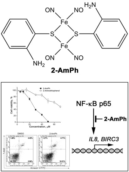
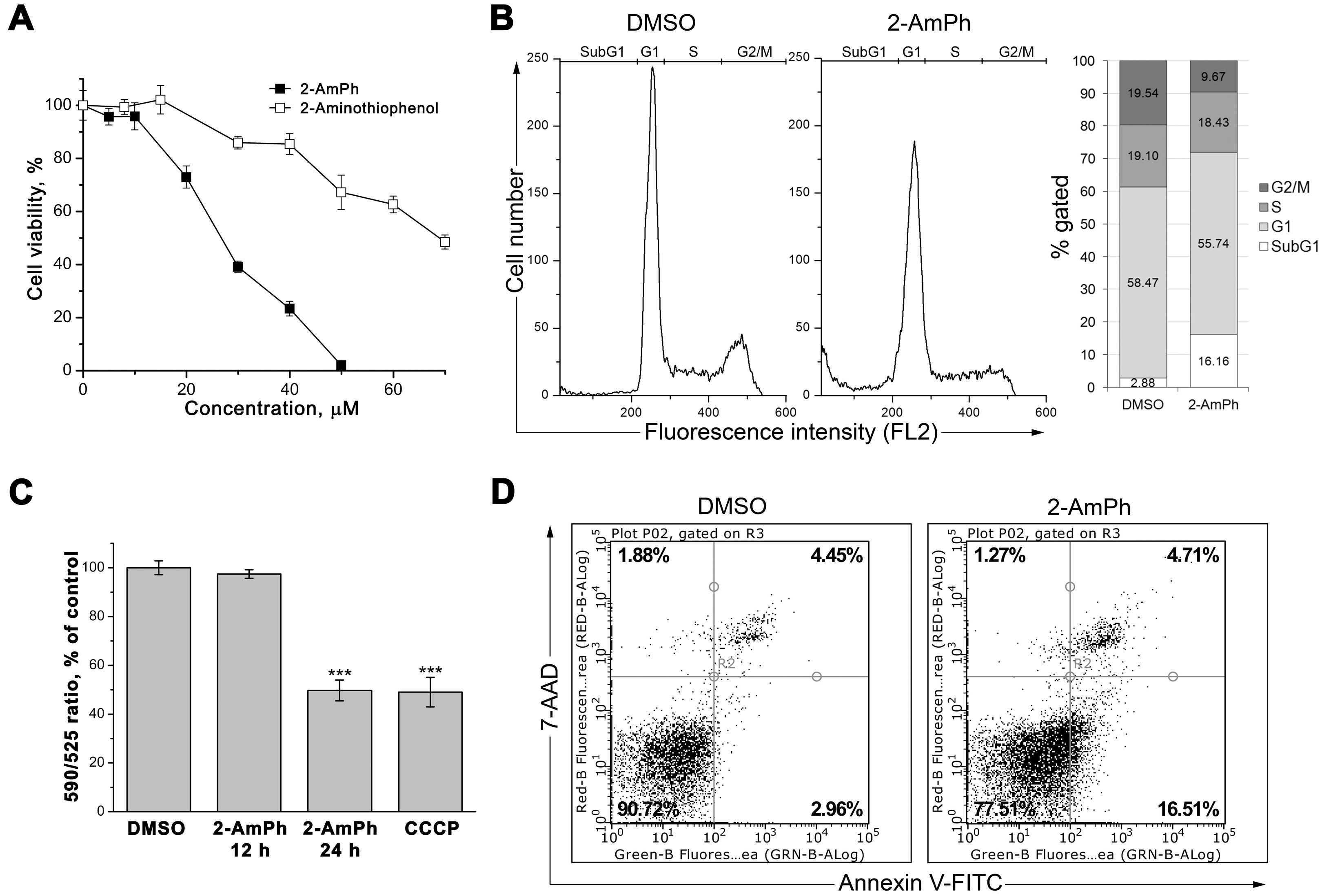
3.1. 2-AmPh Induces Apoptosis in HeLa Cells

In MTT staining experiments, 2-AmPh was studied in comparison with 2-aminothiophenol that was used for the complex synthesis. Both compounds decreased the viability in HeLa cells, but the cytotoxicity of 2-AmPh was appr. 2.5-fold higher compared to 2-aminothiophenol (Figure 2A). The IC50 value for the complex calculated from data of three independent experiments amounted to 29.7 ± 0.9 μM (expressed as mean ± standard deviation). Thus, for the further experiments, the complex was used at the found IC50 dose of 29.7 μM.
After 24 h exposure, 2-AmPh caused cell death as it was demonstrated by an increase in SubG1 population of cells revealed by flow cytometry of PI stained cells (Figure 2B). The increase in SubG1 population takes place at the expense of G2/M with no apparent changes in G1 or S fractions of cells. These results suggest that 2-AmPh causes cell death after either impairment of G2/M checkpoint or cell cycle arrest at phases preceding G2/M phases of the cell cycle.
The cell death caused by 2-AmPh was further characterized. The mitochondrial membrane potential loss is a well characterized sign of cell death [59]. 2-AmPh induces a decrease in the mitochondrial membrane potential as revealed by JC-10 staining (Figure 2C). The complex also caused an increase in the number of cells stained by FITC-conjugated annexin V as it can be seen from Figure 2D. Annexin V binds PS with high affinity, ant its binding to cells that are PI-negative (the lower right quadrant) is a well-defined marker of apoptosis-specific externalization of PS [60]. Thus, at IC50 2-AmPh induces apoptotic cell death in HeLa cells.
As it can be seen from Figure 2, the cell death did not account entirely for the decrease in cell viability. At the IC50, appr. 15% of cells are detected in the SubG1 population (Figure 2B), and no accumulation of late apoptotic or necrotic cells (Figure 2D, the upper right quadrant) was observed compared to the vehicle treated cells. These results show that cells with degraded DNA (SubG1 population) still retained their plasma membrane integrity since PI-positive populations of cells (Figure 2D, upper quadrants) did not increase. On the other hand, cell viability decreases by 50% after exposure to a test compound at IC50 dose. This discrepancy can be explained by the fact that MTT staining of cells depends rather on the mitochondrial electron transport chain functionality than the number of cells with intact DNA. Thus, the 50% decrease in cell viability measured by MTT does not have necessarily to be accompanied by an equal increase in population of cells with degraded DNA.
Our data show that 2-AmPh induces apoptosis in HeLa cells. Earlier, we have found that NICs with cysteamine or phenylthiyl ligands disturbed NF-κB functions [56]. This finding was consistent with other works demonstrating that NO-donor compounds affect NF-κB signaling [39,45,46,47,48,61,62,63,64]. Thus, further we studied the influence exerted by 2-AmPh on NF-κB.

3.2. Effects of 2-AmPh on NF-κB Functions

The cell response to apoptotic stimuli initiates very early, and the alterations of NF-κB activity after NO donor exposure can be observed within several hours, long before the period of apparent cytotoxic effects. For example, the NO donor S-nitroso-N-acetyl-d,l-penicillamine (SNAP) decreased the expression of c-Rel subunit of NF-κB in nuclei of murine leukemia L-1210 cells within 2 h, while the decrease in cell viability was observed after 24 h exposure [38]. The pattern of gene expression in response to SNAP has been demonstrated to change drastically, with main evetns taking place between 4 and 12 h [65]. Thus, to study the effects of 2-AmPh on NF-κB functions, we used a 6 h exposure time point.
NF-κB transcription factor is a dimeric protein comprising of two subunits. The most common composition of NF-κB is a heterodimer of p50 and p65 (RelA) polypeptides. The latter contains transcription activation domain, thus conferring transactivation function to the whole dimer [66]. With use of p65 specific antibody we have found that 2-AmPh affected the nuclear content of p65. After exposure to 2-AmPh the nuclear p65 level decreased substantially compared to the vehicle control (Figure 3A). The effect of 2-AmPh was compared to that exerted by NICs containing cysteamine and phenylthiyl ligands (CysAm [56] and Ph [57] complexes, respectively). All complexes were used at corresponding IC50 doses. Ph decreased p65 nuclear content to the extent similar to that observed under 2-AmPh exposure, while CysAm had a weaker effect on nuclear p65. Ph and CysAm were shown earlier to cause cell death and affect nuclear levels of p50 subunit of NF-κB [56,57]. It is interesting that CysAm exerted weaker effect on nuclear p50 compared to Ph. Thus, at equitoxic doses, NICs containing the phenyl ring (Ph and 2-AmPh) have more pronounced effect on the nuclear content of NF-κB subunits.
Since p65 subunit is important for NF-κB transactivation function, to find out if the decrease in nuclear p65 level can evoke functional consequences, we studied the expression of several NF-κB target genes representing two different functional groups, cytokine encoding genes IL6 and IL8, and genes encoding IAPs family proteins, BIRC2 (cIAP1) and BIRC3 (cIAP2). Under experimental conditions used, one of two cytokine genes, as well as one of two IAPs family genes tested, IL8 and BIRC3, were found to be inhibited by 2-AmPh, whereas IL6 and BIRC2 expression levels were not affected (Figure 3B).
Thus, apoptosis induction by 2-AmPh is preceded by the decrease in nuclear levels of transactivation domain containing NF-κB subunit p65 and inhibition of a subset of NF-κB target genes. Our data suggest that NF-κB signaling pathway is involved into the cell response to 2-AmPh.

4. Discussion

NF-κB signaling is well recognized as an important molecular crossway that contributes to various diseases, including cancer [67,68,69,70,71,72,73]. Chemical compounds that inhibit NF-κB, either directly or via its upstream regulators, cause cell death and suppress tumor growth [74,75,76,77,78,79,80,81,82,83] or sensitize cancer cells to radiation or chemotherapeutic agents [84,85,86].
Nitric oxide, either produced by endogenous sources under conditions of hyperthermia [87], shear stress [88] and exposure to cytotoxic chemicals [89] or generated by exogenous NO donors [39,45], inhibited NF-κB signaling and caused loss of cell viability.
Previously, NO-donor complex 2-AmPh has been shown to be toxic to ovarian carcinoma (SKOV3), large intestine cancer (LS174T), mammary gland carcinoma (MCF7), and non-small cell lung carcinoma (A549) cells with IC50 values ranging from 25 to 74 μM [19]. So far, the mechanism(s) underlying the toxicity of 2-AmPh towards cancer cells has not been studied. Earlier, we have found that NF-κB is one of regulatory factors that are affected by NICs in HeLa cells [56]. Thus, we studied the effects of 2-AmPh on HeLa cells viability and NF-κB function.
The complex inhibited viability of HeLa cells with IC50 corresponding to the IC50 range found earlier for other cell lines. The IC50 of 2-AmPh is similar to that for HeLa cells of the structurally similar Ph complex [57]. Based on the results of MTT assay for 2-AmPh and 2-aminothiophenol (Figure 2A), it can be suggested that the toxicity of 2-AmPh might be partially accounted for by the presence of 2-aminothiophenyl moieties. On the other hand, the mechanism of decomposition of the complex in the course of NO generation is not known, and the resulting products that could be formed during the decomposition are not defined. Thus, determination of contribution of NO and other decomposition products to the overall toxicity of the complex would require more comprehensive studies.
At the IC50 dose, 2-AmPh caused cell death that was defined as apoptotic by annexin V staining. The apoptotic response of HeLa cells to 2-AmPh appears to be not completed by 24 h since the plasma membrane integrity was retained, and relatively small part of the cell population exhibited DNA degradation and PS externalization (Figure 2B,D). The mitochondrial membrane potential was not changed within the first 12 h of exposure and decreased only by 24 h (Figure 2C). These data demonstrate that the irreversible stage of apoptosis characterized by the mitochondrial membrane potential loss in the course of the apoptosis [90] was initiated after 12 h, hence supporting the finding that the execution of the apoptosis program was still ongoing by 24 h.
Cell death caused by 2-AmPh is preceded by inhibition of NF-κB function. 2-AmPh exposure caused the decrease in p65 levels in the nucleus (Figure 3A). Earlier, we have found that NICs with cysteamine or phenylthiyl ligands affected nuclear content of p50 subunit of NF-κB [56]. Thus, both present and previous data demonstrate that NO-donor NICs affect functions of NF-κB through modulation of its nuclear content. While our data are not sufficient to make a conclusion regarding if the NF-κB cytoplasmic-nuclear shuttling is affected by NICs, it is possible that NICs can affect the nuclear import of NF-κB as it was shown in many studies of NO donors [37,38,39,40,41,42,43,44].
The impairment of nuclear expression of p65 subunit is accompanied by the inhibition of a subset of NF-κB target genes. Since both NO and NF-κB play different (and often opposite) roles in the functions of the cell, we studied representative genes belonging to two groups with very different functions: Unrelated to apoptosis cytokine genes IL6 and IL8, and closely related to apoptosis IAPs family genes BIRC2 and BIRC3. Like many other NF-κB target genes, IL6, IL8, BIRC2 and BIRC3 are related to tumor progression and treatment: cytokines are involved into regulation of tumor microenvironment [91,92,93], whereas IAPs contribute to tumor cell survival [94,95,96,97,98]. The observed 2-AmPh-induced suppression of IL8 gene expression demonstrates that NO delivery to tumor cells might be beneficial for interfering with tumor promoting microenvironment conditions, e.g., angiogenesis [91]. The inhibitory effect of 2-AmPh on BIRC3 gene is consistent with the data on apoptosis induction by 2-AmPh. It must be noted that NO affects multiple signaling mechanisms in the cell [24,25,26,27], and its pro-apoptotic activity is not based solely on NF-κB regulation. Transcription factors p53, YY1 and FOXP3 have been shown to be modulated by NO [27,57] and can be involved in NO-induced apoptosis. The observed inhibition of IAPs family gene BIRC3 can lead to decrease in anti-apoptotic capacity of cells and thus contribute to the overall apoptotic response.
Other studied NF-κB target genes, IL6 and BIRC2, were found to be not affected by 2-AmPh under our experimental conditions. The observed variations in the response of genes controlled by NF-κB on the action of 2-AmPh can be related to either different mechanisms of their regulation (e.g., requirement of specific accessory factors) or different kinetics of their response. Determination of mechanisms underlying this variability would require more detailed research.

5. Conclusions

NF-κB, an important factor regulating versatile processes in both healthy and tumor tissues, is considered as a possible molecular target whose inhibition might be beneficial in cancer treatment [33]. We demonstrate that NO-donor NIC with 2-aminothiophenyl ligand causes apoptotic cell death and suppress NF-κB target genes involved in cell survival and tumor progression.

Author Contributions

Funding Acquisition, Project Administration, N.S.; Conceptualization, Supervision, N.S. and A.T.; Formal Analysis, Investigation, Methodology, T.S., A.B., T.K., and G.K.; Writing-Original Draft Preparation, A.T., N.S., T.S., and A.B.; Writing-Review & Editing, A.T. and N.S.

Funding

This research was funded by Federal Agency of Scientific Organizations: State registration N 0089-2014-0037.

Acknowledgments

The work has been performed with financial support of the Federal Agency of Scientific Organizations (State registration N 0089-2014-0037). We acknowledge Raisa Papina and Galina Maximova (Institute of Problems of Chemical Physics RAS) for provision of resources.

Conflicts of Interest

The authors declare no conflicts of interest.

References

  1. Ignarro, L.J. Nitric Oxide: Biology and Pathobiology, 1st ed.; Academic Press: San Diego, CA, USA, 2000; ISBN 9780080525037. [Google Scholar]
  2. Batler, A.; Nicholson, R. Life, Death and Nitric Oxide; The Royal Society of Chemistry: Cambridge, UK, 2003; ISBN 978-1-84755-123-8. [Google Scholar]
  3. McCleverty, J.A. Chemistry of nitric oxide relevant to biology. Chem. Rev. 2004, 104, 403–418. [Google Scholar] [CrossRef] [PubMed]
  4. Dabrowiak, J.C. Metals in Medicine; John Wiley & Sons, Ltd.: Chichester, UK, 2009; ISBN 978-0470681961. [Google Scholar]
  5. Mingos, D.; Michael, P. (Eds.) Nitrosyl Complexes in Inorganic Chemistry, Biochemistry and Medicine II; Springer: Berlin/Heidelberg, Germany, 2014; ISBN 978-3-642-41160-1. [Google Scholar]
  6. Lewandowska, H.; Kalinowska, M.; Brzoska, K.; Wojciuk, K.; Wojciuk, G.; Kruszewski, M. Nitrosyl iron complexes—Synthesis, structure and biology. Dalton Trans. 2011, 40, 8273–8289. [Google Scholar] [CrossRef] [PubMed]
  7. Vanin, A.F. Dinitrosyl iron complexes with thiolate ligands: Physico-chemistry, biochemistry and physiology. Nitric Oxide 2009, 1, 1–13. [Google Scholar] [CrossRef] [PubMed]
  8. Tseng, Y.T.; Chen, C.H.; Lin, J.Y.; Li, B.H.; Lu, Y.H.; Lin, C.H.; Chen, H.T.; Weng, T.C.; Sokaras, D.; Chen, H.Y.; et al. To transfer or not to transfer? Development of a dinitrosyl iron complex as a nitroxyl donor for the nitroxylation of an Fe(III)-porphyrin center. Chemistry 2015, 49, 17570–17573. [Google Scholar] [CrossRef] [PubMed]
  9. Lu, T.T.; Weng, T.C.; Liaw, W.F. X-ray emission spectroscopy: A spectroscopic measure for the determination of NO oxidation states in Fe-NO complexes. Angew. Chem. Int. Ed. Engl. 2014, 43, 11562–11566. [Google Scholar] [CrossRef]
  10. Tsou, C.C.; Lin, Z.S.; Lu, T.T.; Liaw, W.F. Transformation of dinitrosyl iron complexes [(NO)2Fe(SR)2]- (R = Et, Ph) into [4Fe-4S] Clusters [Fe4S4(SPh)4]2-: Relevance to the repair of the nitric oxide-modified ferredoxin [4Fe-4S] clusters. J. Am. Chem. Soc. 2008, 50, 17154–17160. [Google Scholar] [CrossRef] [PubMed]
  11. Tsou, C.C.; Lu, T.T.; Liaw, W.F. EPR, UV-Vis, IR, and X-ray demonstration of the anionic dimeric dinitrosyl iron complex [(NO)2Fe(μ-StBu)2Fe(NO)2]-: Relevance to the products of nitrosylation of cytosolic and mitochondrial aconitases, and high-potential iron proteins. J. Am. Chem. Soc. 2007, 42, 12626–12627. [Google Scholar] [CrossRef] [PubMed]
  12. Fitzpatrick, J.; Kim, E. Synthetic modeling chemistry of iron-sulfur clusters in nitric oxide signaling. Acc. Chem. Res. 2015, 8, 2453–2461. [Google Scholar] [CrossRef] [PubMed]
  13. Burks, P.T.; Garcia, J.V.; GonzalezIrias, R.; Tillman, J.T.; Niu, M.; Mikhailovsky, AA.; Zhang, J.; Zhang, F.; Ford, P.C. Nitric oxide releasing materials triggered by near-infrared excitation through tissue filters. J. Am. Chem. Soc. 2013, 48, 18145–18152. [Google Scholar] [CrossRef]
  14. Pereira, J.C.; Iretskii, A.V.; Han, R.M.; Ford, P.C. Dinitrosyl iron complexes with cysteine. Kinetics studies of the formation and reactions of DNICs in aqueous solution. J. Am. Chem. Soc. 2015, 1, 328–336. [Google Scholar] [CrossRef] [PubMed]
  15. Sanina, N.A.; Aldoshin, S.M. Structure and properties of iron nitrosyl complexes with functionalized sulfur-containing ligands. Russ. Chem. Bull. 2011, 60, 1223–1251. [Google Scholar] [CrossRef]
  16. Sanina, N.A.; Serebryakova, L.I.; Shul’zhenko, V.S.; Pisarenko, O.I.; Roudneva, T.N.; Аldoshin, S.М. The use of sulfur-binuclear nitrosyl cation type iron complex as a vasodilator drug. RU. Patent 2460531, 10 September 2012. [Google Scholar]
  17. Vasil’eva, S.V.; Streltsova, D.A.; Starostina, I.A.; Sanina, N.A. Nitrogen oxide is involved in the regulation of the Fe–S cluster assembly in proteins and the formation of biofilms by Escherichia coli cells. Biol. Bull. 2013, 40, 351–357. [Google Scholar] [CrossRef]
  18. Sanina, N.A.; Lysenko, K.A.; Zhukova, O.S.; Roudneva, T.N.; Emel’yanova, N.S.; Aldoshin, S.M. Water-soluble binuclear nitrosyl iron complexes with natural aliphatic thiolyls possessing cytotoxic, apoptotic and NO donor activity. US Patent 8,067,628 B2, 29 November 2011. [Google Scholar]
  19. Sanina, N.A.; Kozub, G.I.; Zhukova, O.S.; Emel’yanova, N.S.; Kondrat’eva, T.A.; Korchagin, D.V.; Shilov, G.V.; Ovanesyan, N.S.; Aldoshin, S.M. Synthesis, structure, NO donor activity of iron–sulfur nitrosyl complex with 2-aminophenol-2-yl and its antiproliferative activity against human cancer cells. J. Coord. Chem. 2013, 66, 3602–3618. [Google Scholar] [CrossRef]
  20. Zhukova, O.S.; Smirnova, Z.S.; Chikileva, I.O.; Kiselevskii, M.V. Antiproliferative activity of a new nitrosyl iron complex with cysteamine in human tumor cells in vitro. Bull. Exp. Biol. Med. 2017, 162, 583–588. [Google Scholar] [CrossRef] [PubMed]
  21. LaSalle, M.; Billen, D. Inhibition of DNA synthesis in murine bone-marrow cells by AET and cysteamine. Ann. N. Y. Acad. Sci. 1964, 114, 622–629. [Google Scholar] [CrossRef] [PubMed]
  22. Sigdestad, C.P.; Guilford, W.; Perrin, J.; Grdina, D.J. Cell cycle redistribution of cultured cells after treatment with chemical radiation protectors. Cell Tissue Kinet. 1988, 21, 193–200. [Google Scholar] [CrossRef] [PubMed]
  23. Jaffray, P.; Ronot, X.; Adolphe, M.; Fontagne, J.; Lechat, P. Effects of D-penicillamine on growth and cell cycle kinetics of cultured rabbit articular chondrocytes. Ann. Rheum. Dis. 1984, 43, 333–338. [Google Scholar] [CrossRef] [PubMed]
  24. Brüne, B. The intimate relation between nitric oxide and superoxide in apoptosis and cell survival. Antioxid. Redox Signal. 2005, 7, 497–507. [Google Scholar] [CrossRef] [PubMed]
  25. Li, C.Q.; Wogan, G.N. Nitric oxide as a modulator of apoptosis. Cancer Lett. 2005, 226, 1–15. [Google Scholar] [CrossRef] [PubMed]
  26. Tarr, J.M.; Eggleton, P.; Winyard, P.G. Nitric oxide and the regulation of apoptosis in tumour cells. Curr. Pharm. Des. 2006, 12, 4445–4468. [Google Scholar] [CrossRef] [PubMed]
  27. Olson, S.Y.; Garbán, H.J. Regulation of apoptosis-related genes by nitric oxide in cancer. Nitric Oxide 2008, 19, 170–176. [Google Scholar] [CrossRef] [PubMed] [Green Version]
  28. Leon, L.; Jeannin, J.F.; Bettaieb, A. Post-translational modifications induced by nitric oxide (NO): Implication in cancer cells apoptosis. Nitric Oxide 2008, 19, 77–83. [Google Scholar] [CrossRef] [PubMed]
  29. Thomas, D.D.; Ridnour, L.A.; Isenberg, J.S.; Flores-Santana, W.; Switzer, C.H.; Donzelli, S.; Hussain, P.; Vecoli, C.; Paolocci, N.; Ambs, S.; et al. The chemical biology of nitric oxide: Implications in cellular signaling. Free Radic. Biol. Med. 2008, 45, 18–31. [Google Scholar] [CrossRef] [PubMed] [Green Version]
  30. Weigert, A.; Brüne, B. Nitric oxide, apoptosis and macrophage polarization during tumor progression. Nitric Oxide 2008, 19, 95–102. [Google Scholar] [CrossRef] [PubMed]
  31. Bonavida, B.; Garban, H. Nitric oxide-mediated sensitization of resistant tumor cells to apoptosis by chemo-immunotherapeutics. Redox Biol. 2015, 6, 486–494. [Google Scholar] [CrossRef] [PubMed]
  32. Muntané, J.; De la Rosa, A.J.; Marín, L.M.; Padillo, F.J. Nitric oxide and cell death in liver cancer cells. Mitochondrion 2013, 13, 257–262. [Google Scholar] [CrossRef] [PubMed]
  33. Li, F.; Zhang, J.; Arfuso, F.; Chinnathambi, A.; Zayed, M.E.; Alharbi, S.A.; Kumar, A.P.; Ahn, K.S.; Sethi, G. NF-κB in cancer therapy. Arch. Toxicol. 2015, 89, 711–731. [Google Scholar] [CrossRef] [PubMed]
  34. Kaltschmidt, B.; Greiner, J.F.W.; Kadhim, H.M.; Kaltschmidt, C. Subunit-Specific Role of NF-κB in Cancer. Biomedicines 2018, 6, 44. [Google Scholar] [CrossRef] [PubMed]
  35. Mohamed, M.S.; Bishr, M.K.; Almutairi, F.M.; Ali, A.G. Inhibitors of apoptosis: Clinical implications in cancer. Apoptosis 2017, 22, 1487–1509. [Google Scholar] [CrossRef] [PubMed]
  36. Baldwin, A.S. Regulation of cell death and autophagy by IKK and NF-κB: Critical mechanisms in immune function and cancer. Immunol. Rev. 2012, 246, 327–345. [Google Scholar] [CrossRef] [PubMed]
  37. Okamoto, T.; Valacchi, G.; Gohil, K.; Akaike, T.; van der Vliet, A. S-nitrosothiols inhibit cytokine-mediated induction of matrix metalloproteinase-9 in airway epithelial cells. Am. J. Respir. Cell Mol. Biol. 2002, 27, 463–473. [Google Scholar] [CrossRef] [PubMed]
  38. Santos-Silva, M.C.; Sampaio de Freitas, M.; Assreuy, J. Killing of lymphoblastic leukemia cells by nitric oxide and taxol: Involvement of NF-κB activity. Cancer Lett. 2001, 173, 53–61. [Google Scholar] [CrossRef]
  39. Rishi, L.; Dhiman, R.; Raje, M.; Majumdar, S. Nitric oxide induces apoptosis in cutaneous T cell lymphoma (HuT-78) by downregulating constitutive NF-κB. Biochim. Biophys. Acta 2007, 8, 1230–1239. [Google Scholar] [CrossRef] [PubMed]
  40. Jiang, M.Z.; Tsukahara, H.; Hayakawa, K.; Todoroki, Y.; Tamura, S.; Ohshima, Y.; Hiraoka, M.; Mayumi, M. Effects of antioxidants and NO on TNF-alpha-induced adhesion molecule expression in human pulmonary microvascular endothelial cells. Respir. Med. 2005, 99, 580–591. [Google Scholar] [CrossRef] [PubMed]
  41. Katsuyama, K.; Shichiri, M.; Marumo, F.; Hirata, Y. NO inhibits cytokine-induced iNOS expression and NF-κB activation by interfering with phosphorylation and degradation of IκB-alpha. Arterioscler. Thromb. Vasc. Biol. 1998, 18, 1796–1802. [Google Scholar] [CrossRef] [PubMed]
  42. Ohkita, M.; Takaoka, M.; Shiota, Y.; Nojiri, R.; Matsumura, Y. Nitric oxide inhibits endothelin-1 production through the suppression of nuclear factor κB. Clin. Sci. (Lond.) 2002, 103 (Suppl. 48), 68S–71S. [Google Scholar] [CrossRef] [PubMed]
  43. Chawla-Sarkar, M.; Bauer, J.A.; Lupica, J.A.; Morrison, B.H.; Tang, Z.; Oates, R.K.; Almasan, A.; DiDonato, J.A.; Borden, E.C.; Lindner, D.J. Suppression of NF-κB survival signaling by nitrosylcobalamin sensitizes neoplasms to the anti-tumor effects of Apo2L/TRAIL. J. Biol. Chem. 2003, 278, 39461–39469. [Google Scholar] [CrossRef] [PubMed]
  44. Reynaert, N.L.; Ckless, K.; Korn, S.H.; Vos, N.; Guala, A.S.; Wouters, E.F.; van der Vliet, A.; Janssen-Heininger, Y.M. Nitric oxide represses inhibitory κB kinase through S-nitrosylation. Proc. Natl. Acad. Sci. USA 2004, 101, 8945–8950. [Google Scholar] [CrossRef] [PubMed] [Green Version]
  45. Ibe, W.; Bartels, W.; Lindemann, S.; Grosser, T.; Buerke, M.; Boissel, J.P.; Meyer, J.; Darius, H. Involvement of PKC and NF-κB in nitric oxide induced apoptosis in human coronary artery smooth muscle cells. Cell. Physiol. Biochem. 2001, 5, 231–240. [Google Scholar] [CrossRef] [PubMed]
  46. Matthews, J.R.; Botting, C.H.; Panico, M.; Morris, H.R.; Hay, R.T. Inhibition of NF-κB DNA binding by nitric oxide. Nucleic Acids Res. 1996, 12, 2236–2242. [Google Scholar] [CrossRef]
  47. Lee, S.K.; Kim, J.H.; Yang, W.S.; Kim, S.B.; Park, S.K.; Park, J.S. Exogenous nitric oxide inhibits VCAM-1 expression in human peritoneal mesothelial cells. Role of cyclic GMP and NF-κB. Nephron 2002, 4, 447–454. [Google Scholar] [CrossRef] [PubMed]
  48. Paradkar, P.N.; Roth, J.A. Nitric oxide transcriptionally down-regulates specific isoforms of divalent metal transporter (DMT1) via NF-κB. J. Neurochem. 2006, 96, 1768–1777. [Google Scholar] [CrossRef] [PubMed]
  49. Marshall, H.E.; Hess, D.T.; Stamler, J.S. S-nitrosylation: Physiological regulation of NF-κB. Proc. Natl. Acad. Sci. USA 2004, 101, 8841–8842. [Google Scholar] [CrossRef] [PubMed] [Green Version]
  50. Chattopadhyay, M.; Goswami, S.; Rodes, D.B.; Kodela, R.; Velazquez, C.A.; Boring, D.; Crowell, J.A.; Kashfi, K. NO-releasing NSAIDs suppress NF-κB signaling in vitro and in vivo through S-nitrosylation. Cancer Lett. 2010, 298, 204–211. [Google Scholar] [CrossRef] [PubMed]
  51. Park, S.W.; Huq, M.D.; Hu, X.; Wei, L.N. Tyrosine nitration on p65: A novel mechanism to rapidly inactivate nuclear factor-κB. Mol. Cell. Proteomics 2005, 4, 300–309. [Google Scholar] [CrossRef] [PubMed]
  52. Chou, T.C.; Talalay, P. Quantitative analysis of dose-effect relationships: The combined effects of multiple drugs or enzyme inhibitors. Adv. Enzym. Regul. 1984, 22, 27–55. [Google Scholar] [CrossRef]
  53. Pozarowski, P.; Darzynkiewicz, Z. Analysis of cell cycle by flow cytometry. Methods Mol. Biol. 2004, 281, 301–311. [Google Scholar] [CrossRef] [PubMed]
  54. Kane, M.S.; Paris, A.; Codron, P.; Cassereau, J.; Procaccio, V.; Lenaers, G.; Reynier, P.; Chevrollier, A. Current mechanistic insights into the CCCP-induced cell survival response. Biochem. Pharmacol. 2018, 148, 100–110. [Google Scholar] [CrossRef] [PubMed]
  55. Osborn, L.; Kunkel, S.; Nabel, G.J. Tumor necrosis factor alpha and interleukin 1 stimulate the human immunodeficiency virus enhancer by activation of the nuclear factor κB. Proc. Natl. Acad. Sci. USA 1989, 86, 2336–2340. [Google Scholar] [CrossRef] [PubMed]
  56. Stupina, T.S.; Terent’ev, A.A.; Antonova, N.O.; Balalaeva, I.V.; Sanina, N.A.; Aldoshin, S.M. Influence of sulfur-nitrosyl iron complexes of “µ-S” structural type on NF-κB nuclear factor. Int. Sci. J. Med. Biol. Sci. 2014, 1, 23–29. [Google Scholar]
  57. Stupina, T.S.; Parkhomenko, I.I.; Balalaeva, I.V.; Kostyuk, G.V.; Sanina, N.A.; Terent’ev, A.A. Cytotoxic properties of the nitrosyl iron complex with phenylthiyl. Russ. Chem. Bull. 2011, 60, 1488–1494. [Google Scholar] [CrossRef]
  58. Smith, P.K.; Krohn, R.I.; Hermanson, G.T.; Mallia, A.K.; Gartner, F.H.; Provenzano, M.D.; Fujimoto, E.K.; Goeke, N.M.; Olson, B.J.; Klenk, D.C. Measurement of protein using bicinchoninic acid. Anal. Biochem. 1985, 150, 76–85. [Google Scholar] [CrossRef]
  59. Zamzami, N.; Marchetti, P.; Castedo, M.; Decaudin, D.; Macho, A.; Hirsch, T.; Susin, S.A.; Petit, P.X.; Mignotte, B.; Kroemer, G. Sequential reduction of mitochondrial transmembrane potential and generation of reactive oxygen species in early programmed cell death. J. Exp. Med. 1995, 182, 367–377. [Google Scholar] [CrossRef] [PubMed] [Green Version]
  60. Vermes, I.; Haanen, C.; Steffens-Nakken, H.; Reutelingsperger, C. A novel assay for apoptosis. Flow cytometric detection of phosphatidylserine expression on early apoptotic cells using fluorescein labelled Annexin V. J. Immunol. Methods 1995, 184, 39–51. [Google Scholar] [CrossRef]
  61. Jiang, M.Z.; Tsukahara, H.; Ohshima, Y.; Todoroki, Y.; Hiraoka, M.; Maeda, M.; Mayumi, M. Effects of antioxidants and nitric oxide on TNF-alpha-induced adhesion molecule expression and NF-κB activation in human dermal microvascular endothelial cells. Life Sci. 2004, 10, 1159–1170. [Google Scholar] [CrossRef] [PubMed]
  62. Mendes, A.F.; Carvalho, A.P.; Caramona, M.M.; Lopes, M.C. Role of nitric oxide in the activation of NF-κB, AP-1 and NOS II expression in articular chondrocytes. Inflamm. Res. 2002, 7, 369–375. [Google Scholar] [CrossRef]
  63. Amoruso, A.; Bardelli, C.; Fresu, L.G.; Poletti, E.; Palma, A.; Federici Canova, D.; Zeng, H.W.; Ongini, E.; Brunelleschi, S. The nitric oxide-donating pravastatin, NCX 6550, inhibits cytokine release and NF-κB activation while enhancing PPARγ expression in human monocyte/macrophages. Pharmacol. Res. 2010, 5, 391–399. [Google Scholar] [CrossRef] [PubMed]
  64. Bonavida, B.; Baritaki, S. Dual role of NO donors in the reversal of tumor cell resistance and EMT: downregulation of the NF-κB/Snail/YY1/RKIP circuitry. Nitric Oxide 2011, 1, 1–7. [Google Scholar] [CrossRef] [PubMed]
  65. Hemish, J.; Nakaya, N.; Mittal, V.; Enikolopov, G. Nitric oxide activates diverse signaling pathways to regulate gene expression. J. Biol. Chem. 2003, 278, 42321–42329. [Google Scholar] [CrossRef] [PubMed]
  66. Pahl, H.L. Activators and target genes of Rel/NF-κB transcription factors. Oncogene 1999, 18, 6853–6866. [Google Scholar] [CrossRef] [PubMed] [Green Version]
  67. Xia, L.; Tan, S.; Zhou, Y.; Lin, J.; Wang, H.; Oyang, L.; Tian, Y.; Liu, L.; Su, M.; Wang, H.; et al. Role of the NFκB-signaling pathway in cancer. Onco Targets Ther. 2018, 11, 2063–2073. [Google Scholar] [CrossRef] [PubMed] [Green Version]
  68. Begalli, F.; Bennett, J.; Capece, D.; Verzella, D.; D’Andrea, D.; Tornatore, L.; Franzoso, G. Unlocking the NF-κB conundrum: Embracing complexity to achieve specificity. Biomedicines 2017, 5, 50. [Google Scholar] [CrossRef] [PubMed]
  69. D’Ignazio, L.; Batie, M.; Rocha, S. Hypoxia and inflammation in cancer, focus on HIF and NF-κB. Biomedicines 2017, 5, 21. [Google Scholar] [CrossRef] [PubMed]
  70. Park, M.H.; Hong, J.T. Roles of NF-κB in cancer and inflammatory diseases and their therapeutic approaches. Cells 2016, 5, 15. [Google Scholar] [CrossRef] [PubMed]
  71. Zeligs, K.P.; Neuman, M.K.; Annunziata, C.M. Molecular pathways: The balance between cancer and the immune system challenges the therapeutic specificity of targeting nuclear factor-κB signaling for cancer treatment. Clin. Cancer Res. 2016, 22, 4302–4308. [Google Scholar] [CrossRef] [PubMed]
  72. Aggarwal, B.B.; Sung, B. NF-κB in cancer: A matter of life and death. Cancer Discov. 2011, 6, 469–471. [Google Scholar] [CrossRef] [PubMed]
  73. Gupta, S.C.; Sundaram, C.; Reuter, S.; Aggarwal, B.B. Inhibiting NF-κB activation by small molecules as a therapeutic strategy. Biochim. Biophys. Acta 2010, 1799, 775–787. [Google Scholar] [CrossRef] [PubMed] [Green Version]
  74. Nirvanappa, A.C.; Mohan, C.D.; Rangappa, S.; Ananda, H.; Sukhorukov, A.Y.; Shanmugam, M.K.; Sundaram, M.S.; Nayaka, S.C.; Girish, K.S.; Chinnathambi, A.; et al. Novel synthetic oxazines target NF-κB in colon cancer in vitro and inflammatory bowel disease in vivo. PLoS ONE 2016, 11, e0163209. [Google Scholar] [CrossRef] [PubMed]
  75. Ali, A.A.; Lee, Y.R.; Chenm, T.C.; Chen, C.L.; Lee, C.C.; Shiau, C.Y.; Chiang, C.H.; Huang, H.S. Novel anthra[1,2-c][1,2,5]thiadiazole-6,11-diones as promising anticancer lead compounds: Biological evaluation, characterization & molecular targets determination. PLoS ONE 2016, 11, e0154278. [Google Scholar] [CrossRef]
  76. Guzmán, E.; Maher, M.; Temkin, A.; Pitts, T.; Wright, A. Spongiatriol inhibits nuclear factor κB activation and induces apoptosis in pancreatic cancer cells. Mar. Drugs 2013, 11, 1140–1151. [Google Scholar] [CrossRef] [PubMed]
  77. Patil, K.R.; Mohapatra, P.; Patel, H.M.; Goyal, S.N.; Ojha, S.; Kundu, C.N.; Patil, C.R. Pentacyclic triterpenoids inhibit IKKβ mediated activation of NF-κB pathway: In silico and in vitro evidences. PLoS ONE 2015, 10, e0125709. [Google Scholar] [CrossRef]
  78. Guzmán, E.A.; Maers, K.; Roberts, J.; Kemami-Wangun, H.V.; Harmody, D.; Wright, A.E. The marine natural product microsclerodermin A is a novel inhibitor of the nuclear factor κB and induces apoptosis in pancreatic cancer cells. Investig. New Drugs 2015, 1, 86–94. [Google Scholar] [CrossRef] [PubMed]
  79. Mohan, C.D.; Anilkumar, N.C.; Rangappa, S.; Shanmugam, M.K.; Mishra, S.; Chinnathambi, A.; Alharbi, S.A.; Bhattacharjee, A.; Sethi, G.; Kumar, A.P.; et al. Novel 1,3,4-oxadiazole induces anticancer activity by targeting NF-κB in hepatocellular carcinoma cells. Front. Oncol. 2018, 8, 42. [Google Scholar] [CrossRef] [PubMed]
  80. Wang, Z.; Zhao, S.; Song, L.; Pu, Y.; Wang, Q.; Zeng, G.; Liu, X.; Bai, M.; Li, S.; Gao, F.; et al. Natural cyclopeptide RA-V inhibits the NF-κB signaling pathway by targeting TAK1. Cell Death Dis. 2018, 7, 715. [Google Scholar] [CrossRef] [PubMed]
  81. Saud, S.M.; Li, W.; Gray, Z.; Matter, M.S.; Colburn, N.H.; Young, M.R.; Kim, Y.S. Diallyl disulfide (DADS), a constituent of garlic, inactivates NF-κB and prevents colitis-induced colorectal cancer by inhibiting GSK-3β. Cancer Prev. Res. (Phila.) 2016, 7, 607–615. [Google Scholar] [CrossRef] [PubMed]
  82. Lee, S.T.; Wong, P.F.; He, H.; Hooper, J.D.; Mustafa, M.R. Alpha-tomatine attenuation of in vivo growth of subcutaneous and orthotopic xenograft tumors of human prostate carcinoma PC-3 cells is accompanied by inactivation of nuclear factor-κB signaling. PLoS ONE 2013, 2, e57708. [Google Scholar] [CrossRef] [PubMed]
  83. Gupta, S.C.; Prasad, S.; Sethumadhavan, D.R.; Nair, M.S.; Mo, Y.Y.; Aggarwal, B.B. Nimbolide, a limonoid triterpene, inhibits growth of human colorectal cancer xenografts by suppressing the proinflammatory microenvironment. Clin. Cancer Res. 2013, 16, 4465–4476. [Google Scholar] [CrossRef] [PubMed]
  84. Gupta, S.C.; Kannappan, R.; Kim, J.; Rahman, G.M.; Francis, S.K.; Raveendran, R.; Nair, M.S.; Das, J.; Aggarwal, B.B. Bharangin, a diterpenoid quinonemethide, abolishes constitutive and inducible nuclear factor-κB (NF-κB) activation by modifying p65 on cysteine 38 residue and reducing inhibitor of nuclear factor-κB α kinase activation, leading to suppression of NF-κB-regulated gene expression and sensitization of tumor cells to chemotherapeutic agents. Mol. Pharmacol. 2011, 5, 769–781. [Google Scholar] [CrossRef]
  85. Tsolou, A.; Liousia, M.; Kalamida, D.; Pouliliou, S.; Giatromanolaki, A.; Koukourakis, M. Inhibition of IKK-NFκB pathway sensitizes lung cancer cell lines to radiation. Cancer Biol. Med. 2017, 3, 293–2301. [Google Scholar] [CrossRef] [PubMed]
  86. Gupta, S.C.; Prasad, S.; Reuter, S.; Kannappan, R.; Yadav, V.R.; Ravindran, J.; Hema, P.S.; Chaturvedi, M.M.; Nair, M.; Aggarwal, B.B. Modification of cysteine 179 of IkBα kinase by nimbolide leads to down-regulation of NF-κB-regulated cell survival and proliferative proteins and sensitization of tumor cells to chemotherapeutic agents. J. Biol. Chem. 2010, 46, 35406–35417. [Google Scholar] [CrossRef] [PubMed]
  87. Aravindan, N.; Mohan, S.; Herman, T.S.; Natarajan, M. Nitric oxide-mediated inhibition of NF-κB regulates hyperthermia-induced apoptosis. J. Cell Biochem. 2009, 6, 999–1009. [Google Scholar] [CrossRef] [PubMed]
  88. Yurdagul, A., Jr.; Chen, J.; Funk, S.D.; Albert, P.; Kevil, C.G.; Orr, A.W. Altered nitric oxide production mediates matrix-specific PAK2 and NF-κB activation by flow. Mol. Biol. Cell 2013, 3, 398–408. [Google Scholar] [CrossRef] [PubMed]
  89. Hsieh, T.J.; Liu, T.Z.; Lu, F.J.; Hsieh, P.Y.; Chen, C.H. Actinodaphnine induces apoptosis through increased nitric oxide, reactive oxygen species and down-regulation of NF-κB signaling in human hepatoma Mahlavu cells. Food Chem. Toxicol. 2006, 3, 344–354. [Google Scholar] [CrossRef] [PubMed]
  90. Kushnareva, Y.; Newmeyer, D.D. Bioenergetics and cell death. Ann. N. Y. Acad. Sci. 2010, 1201, 50–57. [Google Scholar] [CrossRef] [PubMed] [Green Version]
  91. Brat, D.J.; Bellail, A.C.; Van Meir, E.G. The role of interleukin-8 and its receptors in gliomagenesis and tumoral angiogenesis. Neuro Oncol. 2005, 2, 122–133. [Google Scholar] [CrossRef] [PubMed]
  92. Luppi, F.; Longo, A.M.; de Boer, W.I.; Rabe, K.F.; Hiemstra, P.S. Interleukin-8 stimulates cell proliferation in non-small cell lung cancer through epidermal growth factor receptor transactivation. Lung Cancer 2007, 1, 25–33. [Google Scholar] [CrossRef] [PubMed]
  93. Anestakis, D.; Petanidis, S.; Kalyvas, S.; Nday, C.M.; Tsave, O.; Kioseoglou, E.; Salifoglou, A. Mechanisms and applications of interleukins in cancer immunotherapy. Int. J. Mol. Sci. 2015, 16, 1691–1710. [Google Scholar] [CrossRef] [PubMed]
  94. Ndubaku, C.; Cohen, F.; Varfolomeev, E.; Vucic, D. Targeting inhibitor of apoptosis proteins for therapeutic intervention. Future Med. Chem. 2009, 8, 1509–1525. [Google Scholar] [CrossRef] [PubMed]
  95. Varfolomeev, E.; Vucic, D. Inhibitor of apoptosis proteins: Fascinating biology leads to attractive tumor therapeutic targets. Future Oncol. 2011, 5, 633–648. [Google Scholar] [CrossRef] [PubMed]
  96. Gressot, L.V.; Doucette, T.; Yang, Y.; Fuller, G.N.; Manyam, G.; Rao, A.; Latha, K.; Rao, G. Analysis of the inhibitors of apoptosis identifies BIRC3 as a facilitator of malignant progression in glioma. Oncotarget 2017, 8, 12695–12704. [Google Scholar] [CrossRef] [PubMed]
  97. Fulda, S.; Vucic, D. Targeting IAP proteins for therapeutic intervention in cancer. Nat. Rev. Drug Discov. 2012, 11, 109–124. [Google Scholar] [CrossRef] [PubMed]
  98. Silke, J.; Vucic, D. IAP family of cell death and signaling regulators. Methods Enzymol. 2014, 545, 35–65. [Google Scholar] [CrossRef] [PubMed]
Figure 1. Structure of 2-AmPh.
Scipharm 86 00046 g001
Figure 2. Cytotoxic properties of 2-AmPh for HeLa cells. (A) The “dose-effect” curve of MTT staining of cells. (B) The cell cycle profiles after PI staining of DNA. (C). Decrease in the mitochondrial membrane potential, as revealed from aggregated/monomeric JC-10 staining, *** p < 0.001. CCCP was used as a positive control for the mitochondrial membrane potential loss. (D) Flow cytometry analysis of cells stained with Annexin V-FITC conjugate and 7-AAD.
Scipharm 86 00046 g002
Figure 3. Disturbance of NF-κB function by 2-AmPh. (A) Decrease in the nuclear levels of p65 subunit of NF-κB revealed by the immunoblotting with anti-p65 antibody of nuclear extracts of cells treated with the vehicle, 2-AmPh and NICs bearing cysteamine (CysAm) and phenylthiyl (Ph) ligands. (B) Expression of NF-κB target genes after 6 h exposure to 2-AmPh relative to the control (0.1% DMSO) levels, * p < 0.05.
Scipharm 86 00046 g003

