Article

Potential Gastroprotective Activity of Rice Bran (Oryza sativa L.) Extracted by Ionic Liquid-Microwave-Assisted Extraction against Ethanol-Induced Acute Gastric Ulcers in Rat Model

by
Elsa Trinovita
1,
Fadlina Chany Saputri
2,* and
Abdul Mun’im
3
1
Faculty of Medicine, Universitas Palangka Raya, Palangka Raya 73111, Indonesia
2
Laboratory of Pharmacology, Faculty of Pharmacy, Universitas Indonesia, Depok 16424, Indonesia
3
Laboratory of Pharmacognosy-Phytochemistry, Faculty of Pharmacy, Universitas Indonesia, Depok 16424, Indonesia
*
Author to whom correspondence should be addressed.
Sci. Pharm. 2018, 86(3), 35; https://doi.org/10.3390/scipharm86030035
Submission received: 5 July 2018 / Revised: 28 August 2018 / Accepted: 29 August 2018 / Published: 7 September 2018

Abstract

:
The presence of gamma-oryzanol in rice bran oil can be 10–20-fold higher than tocopherol and tocotrienol. Gamma-oryzanol has various pharmacological properties. The objective of this study was to evaluate the effectiveness of rice bran extract as a gastroprotective in reducing lesions in ethanol-induced acute gastric ulcer models in rat, using the ionic liquid-microwave-assisted extraction (IL-MAE) method. Rice bran extract was obtained using the IL-MAE method with ionic liquid (IL), 1-butyl-3-methylimidazolium tetrafluoroborate [BMIM]BF4 (concentration 0.7 M), and a ratio of solid/liquid of 15 g/mL, 15 min extraction time, and 10% microwave power. The rats were pretreated with rice bran extract at different doses (100, 200, and 400 mg/kg body weight; BW) for seven days and subsequently exposed to acute gastric lesions induced by 80% ethanol. Omeprazole (36 mg/kg BW) was used as a standard anti-ulcer drug. The ulcer index, gastric juice acidity, and mucus levels were measured to assess the degree of gastroprotection. The results showed that the oral administration of rice bran extract at a dose of 400 mg/kg BW significantly inhibited the development of ulcer formation by 66.75% and reduced gastric acid levels. Moreover, gamma oryzanol and omeprazole protected the gastric mucosa from ethanol-induced gastric lesions by increasing the level of gastric mucus. Rice bran extract is effective as a gastroprotective therapy sourced from natural ingredients in treating the incidence of gastric ulcers. Most likely, this is related to gamma oryzanol as a bioactive compound contained in rice bran (Oryza sativa L.).

Graphical Abstract

1. Introduction

Indonesia is a tropical country that has considerable potential in agriculture, especially in rice production. Every year, rice production increases because rice is the staple food of Indonesians. However, this also causes an increase in the output of rice milling byproducts. Rice bran is a waste product of milling that has not been widely used [1].
One of the compounds of rice bran oil is gamma-oryzanol, with concentrations ranging from 1.5 to 3% [2]. Gamma-oryzanol is a combination of at least 10 components of ferulic acid ester and triterpene alcohol. Gamma-oryzanol has four main components: cycloartenol ferulate, 2,4-methylene cycloartenol ferulate, β-sitosteryl ferulate, and campesteryl ferulate [3]. The content of gamma-oryzanol in rice bran oil can be 10–20-fold higher than tocopherol and tocotrienol [4]. The use of an ionic liquid solvent as the application of green chemistry principles can increase the active compounds from natural materials [5]. The IL-MAE method was more efficient for the extraction of gamma-oryzanol compounds from rice bran [6].
Gamma-oryzanol has several biological properties including anti-inflammatory, antineoplastic, hypoallergic, antidiabetic, antiulcerogenic, and hypolipidemic properties [7]. The mechanism of action of gamma-oryzanol as a gastroprotective can be attributed to its antioxidant and anti-inflammatory activities. Previous research has shown that gamma-oryzanol can prevent oxidative damage [8]. An increase in the formation of free radicals can cause oxidative damage, and this plays an important role in the emergence of gastric lesions [9]. Also, gamma-oryzanol may provide anti-inflammatory activity by inhibiting reactive oxygen species (ROS) formation and consequently inhibiting the nuclear factor-kappa B (NF-κB) pathway [10].
Ethanol, as one of the causes of gastric ulcers, can damage the gastric mucosa by altering the permeability of the epithelial barrier—a process that allows tissue damage, especially of blood vessels [11]. Consumption of ethanol is one of the factors that can cause gastrointestinal disorders. Several classes of drugs, such as histamine receptor antagonists (H2), proton pump inhibitors, and anticholinergics, are used for peptic ulcer treatment [12].
Based on the above scientific information, we were interested in researching ionic liquid-microwave-assisted extraction (IL-MAE) extraction for increasing the gamma-oryzanol content of rice bran (Oryza sativa L.). Furthermore, the effectiveness of rice bran extract obtained from IL-MAE extraction as a gastroprotective was tested in the ethanol-induced animal model.

2. Material and Methods

2.1. Materials

Standard gamma-oryzanol and Alcian blue solution was purchased from Sigma-Aldrich Chemical Co. (St. Louis, MO, USA). 1-Butyl-3-methylimidazolium tetrafluoroborate [BMIM]BF4 (>99%) was purchased from Cheng Jie Chemical Co., Ltd., (Shanghai, China). Sodium hydroxide, sodium acetate, carboxymethyl cellulose (CMC), and high performance liquid chromatography (HPLC) grade solvents (methanol, acetonitrile, and isopropanol) were obtained from Merck (Darmstadt, Germany). Sucrose was purchased from DifcoTM, Livonia, MI, USA. Sodium chloride solution (0.9%) was purchased from PT. Otsuka Indonesia, Lawang, East Java, Indonesia. Omeprazole (OMZ) was obtained from Novell Pharmaceutical Laboratories, Jakarta, Indonesia. Ketamine hydrochloride and xylazine were purchased from Alfasan Co. (Woerden, The Netherlands).
The fresh rice bran variety IR 64 (Oryza sativa L.) was obtained from Bogor, West Java, Indonesia. The fresh rice bran samples in a drying tray were inserted into an oven (Memmert, Germany), which was set at 110 °C for 15 min. After that, the rice bran sample was cooled in a container for 30 min to reach room temperature. The stabilized rice bran was then put in clear plastic and stored at room temperature.

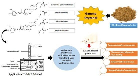
2.2. Extraction with the Ionic Liquid-Microwaved Assisted Extraction (IL-MAE) Method

Pilot studies were carried out on a small scale for optimization using the IL[BMIM]BF4-MAE method using response surface methodology (RSM) version 10. Optimum conditions were determeind to be a 0.7 M concentration, ratio of solid/liquid of 15 g/mL, extraction time 15 min, and 10% microwave power. The filtrate was then mixed in a separating funnel with N-hexane: NaNO3 solution (1:1 v/v). These conditions were subsequently adopted for large-scale extraction.

2.3. High-Performance Liquid Chromatography (HPLC) Analysis

The extract (20 µL) was injected into the HPLC system (Shimadzu Corp., Kyoto, Japan), equipped with Zorbax Eclipse Plus C-18 Analytical 4.6 × 150 mm, 5 μm (Agilent Technologies, Santa Clara, CA, USA). A mixture of methanol:acetonitrile:isopropanol (50%:40%:10%) was used as the mobile phase under isocratic conditions. The ultraviolet (UV) detector wavelength was set to 327 nm, and the flow rate was set to 1 mL/min. Each sample was measured with three repetitions.

2.4. Pharmacological Study

Healthy Sprague-Dawley male rats aged about 2–3 months old and weighing 200–250 g were obtained from the National Agency of Drug and Food Control Agency Republic of Indonesia (Jakarta, Indonesia). The treatment groups were administered orally (p.o.) by gavage with CMC as a vehicle. The extract was resuspended according to the calculation of the dose, with the use of 0.5% carboxymethyl cellulose (CMC) as a vehicle in the manufacture of the suspension. One hundred milligrams of CMC powder was weighed and then sown on mortar containing 5 mL warm aquadest (70 °C). CMC expands when left for about 10 min, ad it was then crushed slowly with dosage extract and dissolved in 20 mL aquadest. All animals were used according to the guidelines of the Health Research Ethics Committee-Universitas Indonesia and Cipto Mangunkusumo Hospital (HREC-FMUI/CMH). This study was approved by the Institutional Animal Care Committee, Faculty of Medicine, Universitas Indonesia (Ethic No. 694/UN2.F1/ETIK/2016, 15 August 2016).
The animals were acclimatized for seven days inside a cage to adapt to the new environment and fed with standard freely available animal feed and water. The 36 animals were divided into groups of six animals, each using a complete randomized design method (RAL). The rice bran extract doses used were adapted from a previous study: 30–300 mg/kg doses of the extract of were used as a gastroprotective [13]. Six groups of rats were orally pre-treated with vehicle CMC as normal groups. Control groups were treated with vehicle CMC (5 mL/kg body weight; BW) as a negative control. The dose of omeprazole (OMZ) in human beings is 20–40 mg and was converted to a dose for rats (36 mg/kg BW), and this group was used as a positive control. Rice bran extract groups were treated at different doses (100, 200, and 400 mg/kg BW in the vehicle). The normal, negative control (CMC), positive control (OMZ), and rice bran extract groups were administered orally for 7 days. Then, 24 h after the last treatment, all groups except the normal group received 80% ethanol (2.5 mL/kg BW). Three hours later, all animals were euthanized using ketamine and xylazine (i.p.), and their stomachs were immediately removed, opened along the greater curvature, washed with 0.9% NaCl, and analyzed [14].

2.4.1. Gastroprotective Assessment

Ulcer diameter (mm) was determined using calipers and scored to be analyzed statistically as follows: 1 ≤ 1.00 mm; 2 = 1.00–2.00 mm; 3 = 2.01–3.00 mm; 4 = 3.01–4.00 mm; 5 = 4.01–5.00 mm; 10 ≥ 5.00 mm; 25 = perforation [15].
The percentage inhibition of ulceration was calculated using the following formula [16].
Inhibition   of   ulceration   ( % )   =   [ ( UI   negative   control UI   treated ) ( UI   negative   control ) ] × 100 %
where the ulcer index (UI) control is the negative control group (CMC); UI treated is the omeprazole group, and rice bran extract group at different doses (100, 200, and 400 mg/kg BW in the vehicle).

2.4.2. Measurement of Gastric Juice Acidity

The stomach was removed and opened along the greater curvature. The gastric content was collected, rinsed using 3 mL of distilled water, and centrifuged at 2000 rpm for 10 min. The forming supernatant was dissolved in distilled water until the volume reached the glass neck of the potentiometer (Potentiometer–Dosismat 702–SM Titrino Metrohm Horison, Herisau, Switzerland). The supernatant was titrated using acid-base titration using 0.01 M NaOH solution to the endpoint. Total gastric acidity was expressed in μEq/200 g BW [16].

2.4.3. Determination of Mucus

Measurements of mucus were quantitatively performed according to the method described by Klein-Junior et al. with a few modifications. The stomach was soaked for 2 h in a 10 mL solution containing 0.02% Alcian blue in 0.16 M sucrose and 0.05 M sodium acetate at pH 5.8 and incubated (MMM MedcenterTM Incubator Friocell, Munchen, Germany) for 24 h at 20 °C. The Alcian blue dye bonding extract was centrifuged (Hettich® Universal 320 centrifuge, Buckinghamshire, UK) at 4500 rpm for 10 min, and the supernatant was obtained. The absorbance of the supernatant was then measured by using a spectrophotometer (UV-Visible Spectrophotometer Evolution 201, Thermo ScientificTM, Waltham, MA, USA) at a wavelength of 615 nm [17].

2.5. Histopathological Evaluations

Transversal gastric tissue cuts were made from the fundus to pylorus and then paraffin blocks were made. Paraffin blocks were made of 3-μm-thick slices and glued to the object glass for hematoxylin and eosin (H&E) staining for light microscope examination (magnification 400×) by a double-blind pathologist.
Histopathological analysis of gastric mucosal abnormalities, including epithelial cell erosion, congestion, and infiltration of inflammatory cells characterized by the presence of neutrophils, was performed by microscopy so that inflammation can be seen.
Degrees of severity of epithelial erosion on mucosal layers were classified as:
(1)
Mild (+): a layer of thick keratin coating the surface of the mucosa of the stomach.
(2)
Moderate (++): the accumulation of edema fluid under the keratin layer, hemorrhage, edema of the submucosa accompanied by infiltration of inflammatory cells.
(3)
Heavy (+++): discontinuity layer of keratin, atrophy of the coats of epithelium, congestion, hemorrhage, edema of the submucosa and infiltration of inflammatory cells, dominated by neutrophils [18].
The degree of congestion severity was classified as:
(1)
Mild (+): 25% of the blood vessels experiencing congestion.
(2)
Moderate (++): 50% of blood vessels experiencing congestion.
(3)
Heavy (+++): 75% or more of blood vessels experiencing congestion [18].

2.6. Statistical Analysis

The data are presented as the mean ± standard deviation (SD). The statistical analysis used in this study is a One-Way Analysis of Variance (ANOVA) using software SPSS 23 (IBM Corporation, Armonk, NY, USA). The normality distribution of the data was analyzed using the Shapiro–Wilk normality test, then tested for homogeneity. Data that had a normal and homogeneous distribution underwent a diversity analysis test using One-Way ANOVA with p < 0.05 and post hoc Tukey honestly significant difference (HSD) analysis. Distribution of normal and non-homogeneous data underwent One-Way ANOVA Welch testing with a value of p < 0.05. Furthermore, post hoc Howell–Games analysis with p < 0.05 was performed to know the significant differences between groups.

3. Results and Discussion

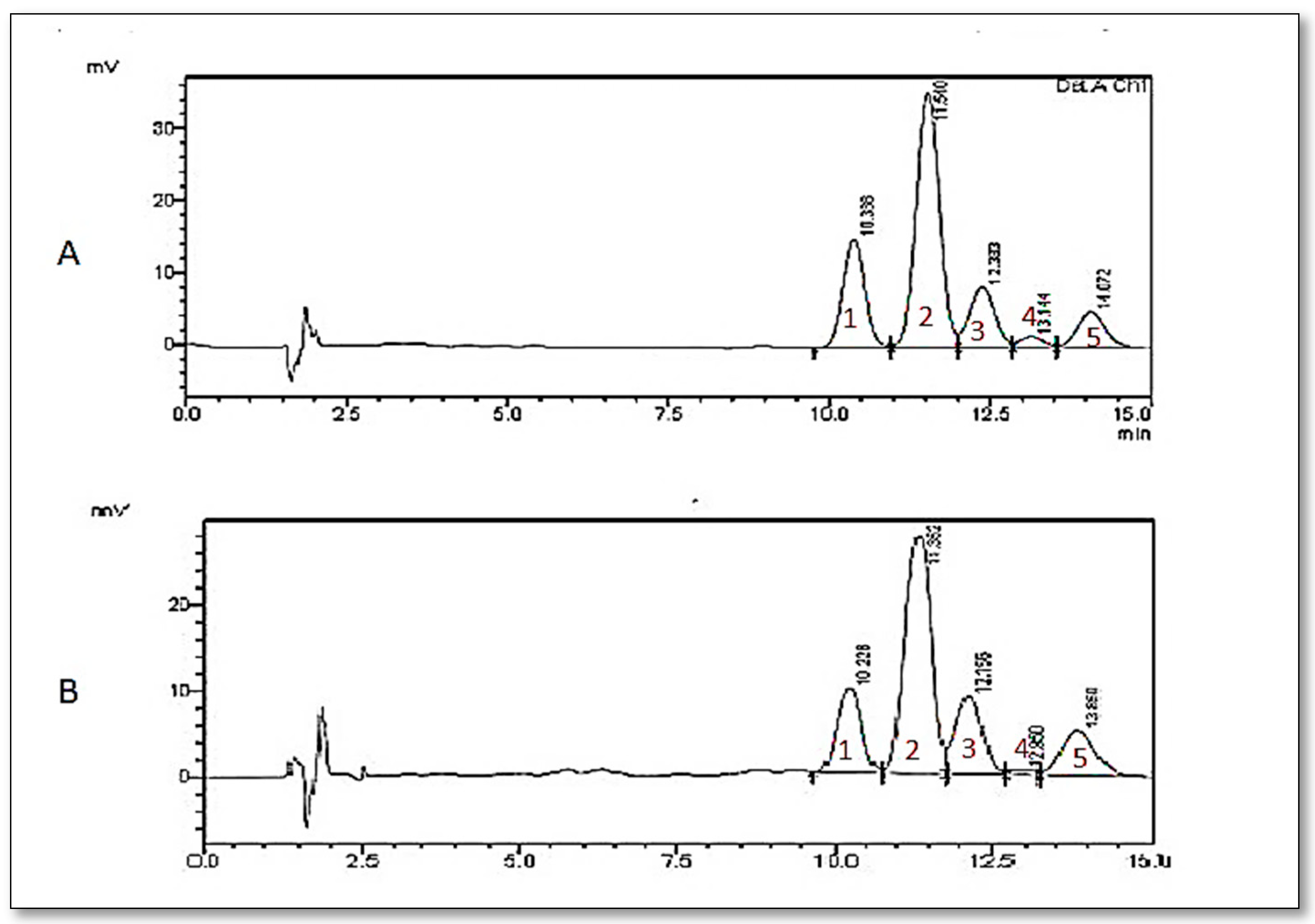
3.1. Analysis of Gamma-Oryzanol in the Extract

The HPLC chromatogram in Figure 1A (standard gamma-oryzanol) and Figure 1B (gamma oryzanol IL-MAE) was obtained at a 327 nm wavelength and retention time of about 10 min. The results of the optimization levels of gamma-oryzanol with IL-MAE were obtained at 4.1%. Based on the results of the HPLC chromatogram, the standard gamma-oryzanol (Figure 1A) showed five peaks for the compound gamma-oryzanol include cycloartenyl ferulate (21.68%), 24-methylenecycloartenyl ferulate (54.53%), campesteryl ferulate (13.29%), camphestenyl ferulate (2.45%), and β-sitosteryl ferulate (8.05%).
The components of the gamma-oryzanol in Figure 1B were identified as cycloartenyl ferulate (20.67%), 24-methylenecycloartenyl ferulate (53.28%), campesteryl ferulate (15.28%), Δ7-cytostenyl ferulate (1.74%), and β-sitosteryl ferulate (9.02%). The component 24-methylenecycloartenyl ferulate had the highest peak (Figure 1A,B).
The ionic liquid solvents are safe, eco-friendly, non-toxic, have pressure negligible vapor, good thermal stability, and flammability [19]. Therefore, in this study, the solvent used was ionic liquid [BMIM]BF4 for extracting rice bran with the MAE method. MAE methods, which in their extraction processes use microwaves, can result in a significant reduction in extraction time. The required organic solvent volume in MAE methods is less than required by the conventional method [20]. IL-MAE is an alternative method of extraction in the separation process of a bioactive compound from the plant. An increased level of gamma oryzanol was demonstrated utilizing IL-MAE compared to the conventional method of extraction [6].
In this research, HPLC analysis at each of the levels of the main components in gamma oryzanol and the results obtained using the approach of the previous study resulted in: 24-methylenecycloartenyl ferulates (34–44%), cycloartenyl ferulate (19–26%), campesteryl ferulate (15–23%), and β-sitosteryl ferulate (7–17%) [21]. Cycloartenyl ferulate, 24-methylenecycloartenyl ferulate, campesteryl ferulate, and β-sitosteryl ferulate were identified as the main contents of gamma-oryzanol [22]. Gamma-oryzanol contains ferulic acid, which is an antioxidant of phenolic acid. Oxidative and inflammatory damage are key to the occurrence of gastric ulcers due to induction of ethanol [23]. Oxidative damage plays an important role in the pathogenesis of various diseases including peptic ulcers; antioxidant activity reportedly plays an important role in the protection of gastric mucosa against various necrotic agents [24].

3.2. In Vivo Studies

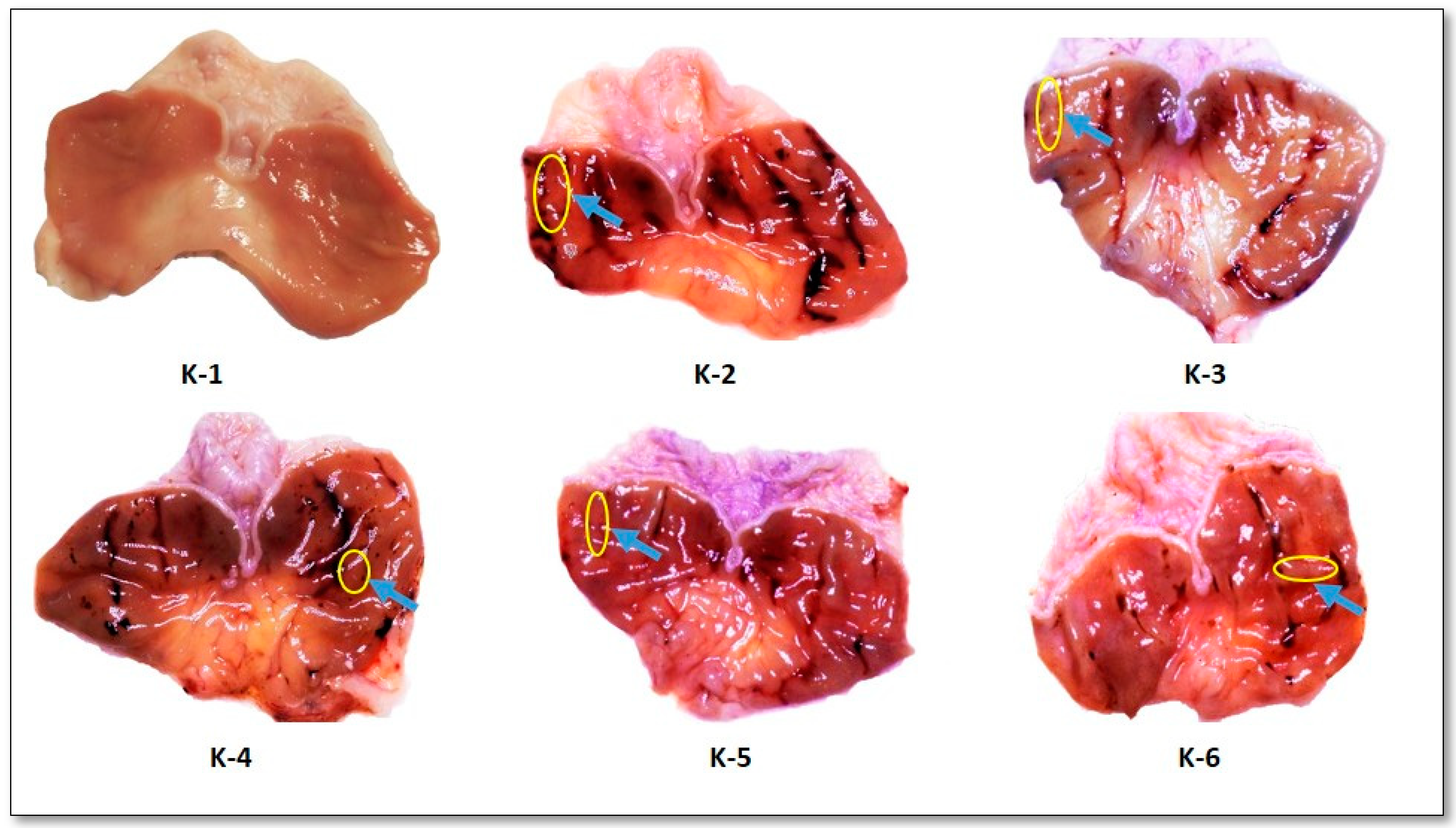
3.2.1. Effect of Rice Bran Extract on Gastric Lesions Induced by Ethanol

Figure 2 macroscopically shows no lesions in the normal group. This is in contrast to the macroscopic picture in the negative group, indicating serious gastric damage followed by bleeding. Table 1 shows that groups of rice bran extract (100, 200, and 400 mg/kg BW) were statistically significant (p < 0.05) when compared with the negative control group. Increasing doses of rice bran extract resulted in decreased formation of gastric lesions. The rice bran extract 400 mg/kg BW group produced the highest decrease in gastric lesion formation compared to the other dose groups of rice bran extract, and showed the highest percentage inhibition of gastric lesion by 66.75%. The results of percentage inhibition of gastric lesions in the OMZ group (positive control) produced the highest inhibition results of 83.25%.
The gamma-oryzanol component that had the highest antioxidant activity was 24-methylenecycloartenyl ferulate. This imbues gamma-oryzanol with antioxidant activity, which can also protect the stomach from the formation of ulcers [25]. The ulcers caused by ethanol induction can directly damage gastric mucosal cells, stimulate free radical formation and lipid peroxidation, which suggests that antioxidant compounds may decrease ulcer activity in experiment conditions [26].
Ethanol can have a destructive effect on gastric mucosal cells, so upon macroscopic observation, can cause gastric erosion [27]. Also, the negative group had the highest ulcer index, with a value of 4 (Table 1). Lesions caused by induction of ethanol can directly damage gastric mucosal cells, stimulating the formation of free radicals and lipid peroxidation [28]. Oxidative stress results in a deleterious process that culminates in damage to the cell structure. The oxidative imbalance is responsible for producing several reactive molecules that are scavenged by the possible mechanisms of gamma oryzanol [10]. Rice bran oil (RBO) has also been reported to inhibit key proinflammatory enzymes, such as cyclooxygenase-1 (COX 1) and cyclooxygenase-2 (COX-2) [29].

3.2.2. Effect of Rice Bran Extract on Gastric Acidity

Table 2 shows that groups of rice bran extract (100, 200, and 400 mg/kg BW) were statistically significant (p < 0.05) when compared with the negative control group. This means that rice bran extract obtained from IL-MAE extract (100, 200, and 400 mg/kg BW) decreased gastric acidity. OMZ, as a positive control, produced significant differences compared with the negative control group. Also, this study also showed that the administration of rice bran extract at various doses decreased the acidity of the stomach to values near the normal group. The results also showed that groups of rice bran extract (100, 200, and 400 mg/kg BW) are comparable to omeprazole in reducing gastric acidity.
Ethanol can increase aggressive factors (include gastric acid, pepsin, bile salts, Helicobacter pylori and nonsteroidal anti-inflammatory drugs (NSAIDs)) by increasing acid secretion, causing decreased gastric acid pH and increased volume of gastric secretion, resulting in gastric ulcers [24]. The thickness of the gastric mucus layer is a form of defense in protecting the gastric wall from pepsin, mechanical and chemical trauma cells, which are both exogenous and endogenous irritants [30].

3.2.3. Effect of Rice Bran Extract on Mucus in Gastric Content

Based on the statistical analysis of gastric mucus formation shown in Table 3, in the rice bran extract 400 mg/kg BW group, there was a significant difference (p < 0.05) in the thickness of mucus formed on the gastric mucosal wall compared with the negative control group. OMZ, as a positive control, showed a statistically significant difference (p < 0.05) compared with the negative group. The higher the dose of rice bran extract, the thicker the gastric mucus. Rice bran extract at a dose of 400 mg/kg BW showed the thickest gastric mucus formation compared to other dose groups of rice bran extracts. This indicated that the ability to form gastric mucus was close to the normal group without any treatment of induction.
Weak mucosal defenses can also cause gastric ulcers, highlighting that mucus plays an important role in protecting the stomach wall from aggressive factors (include gastric acid, pepsin, bile salts, Helicobacter pylori and NSAIDs). Mucus is a protective layer of gastric mucosa that resembles a gel. Mucus consists of mucin, which is a type of glycoprotein produced by cells in the gastric wall and is detected from the amount of Alcian blue bonding. Increased mucus secretion is probably responsible for gastric cytoprotection by acting as an effective barrier in the diffusion of back hydrogen ions, as well as reducing friction in the gastric wall during peristaltic movement and gastric contraction [31]. Prostaglandins, especially PGE2, have a strong cytoprotective effect on the gastric mucosa with various indirect mechanisms, including increased mucus production in epithelial cells and bicarbonate secretion, gastric inhibition of motility, inhibition of acid secretion, improvement of mucosal blood flow, free-radical inhibition, release of neutrophils, and vascular, luminal, and/or extrinsic and intrinsic nerve mechanisms in PGE2. The roles of prostaglandin receptors have been pharmacologically divided into four major subtypes: prostaglandin E receptor 1 (EP1), prostaglandin E receptor 2 (EP2), prostaglandin E receptor 3 (EP3), and prostaglandin E receptor 4 (EP4). EP receptors are induced via indirect mechanisms of gastric mucosal protection by inhibition of acidic motility and bicarbonate stimulation by PGE2, mediated by EP1 receptors. This stimulates mucus production mediated by EP4 receptors, enhancing EP3-mediated mucosal blood flow, and inhibiting mediated acid secretion by EP2/EP3 [32].

3.2.4. Histopathological Evaluations of Gastric Lesions

Administration of ethanol can cause pathological effects on the layers of the stomach of rats with the degrees of severity varying in each layer of the stomach. The histopathological examination was performed qualitatively against the region of the stomach with staining of hematoxylin-eosin (H&E), as shown in Figure 3 and Figure 4. Several epithelial cell erosions, infiltration of inflammation cells, characterized by severe neutrophils, and congestion were shown in the treatment groups-induced ethanol (Figure 3 and Figure 4). The induced ethanol can directly irritate the gastric mucosa, influencing the occurrence of erosion on the surface layer of the gastric mucosa. The results of the examination of the qualitative examination were grouped into three degrees of severity: mild (+), moderate (++), and heavy (+++).
Figure 3 shows the erosion of the surface layer of the mucosa. If the mucosal damage that occurred had a depth of less than 5 mm, this was mild. If the mucosal damage occurred in the form of a discontinuity or tear of the mucosa with a diameter of 5 mm or more to reach the submucosa, accompanied by necrosis of the so-called ulcer, this was considered moderate. Prostaglandins have a positive effect on the mucosa of the gastrointestinal tract, so that inhibition of prostaglandin synthesis lowers the resistance of the mucosa and damages the gastric mucosa. The use of rats with gastric damage induced by ethanol demonstrate mucosal damage accompanied by reduced glycoprotein in the mucus and gastric hypersecretion. The occurrence of hypersecretion of the gastric lymph and pepsin, or decreasing microcirculation of the stomach, are the main causes of the onset of mucosal lesions [18].
The K-1 group did not show erosion of the mucosa, with an intact and continuous layer of keratin, whereas the rice bran extract (dose 400 mg/kg BW) group showed mild epithelial erosion on the mucosal layer (Table 4). Histopathologic observations suggested that ethanol can lead to severe lesions and severe damage to the gastric mucosa. K-2 (the negative control group) showed severe erosion of epithelial cells in the gastric mucosal layer compared to other groups (Figure 3).
Figure 4 shows the infiltration of inflammation in the mucosal muscular layer of the sub-mucosal layer, and the occurrence of congestion with a severe degree of blood vessels in the sub-mucosal lining. The negative control group with omeprazole (K-3) did not show any congestion but the submucosal layer was infiltrated and inflammation of cells was moderate.
Based on the data in Table 5, there is no congestion in the normal group. The negative group showed the heavy occurrence of congestion (Table 5). Histopathological observation in the positive group and groups of rice bran extract found congestion with mild severity. Congestion in blood vessels in the submucosa was microscopically observed with the dilatation of blood-filled blood vessels.
Congestion is a condition where there is an accumulation of blood grains inside blood vessels in certain areas. The accumulation of blood can damage the blood vessels. Excessive congestion can cause hemorrhage, so that the fluid will mix with red blood cells [18]. Ethanol administration caused congestion in the treatment group. Congestion that appeared in the negative group was thought to be preceded by vasodilation of blood vessels and slowing down of the blood flow. Dilated blood vessels, accompanied by a slowing of blood flow due to the release of histamine, can increase accumulation of deep blood grains in the blood vessels, resulting in congestion (Figure 4).
OMZ is a class of proton pump inhibitors that exhibit anti-secretory activity and gastric protection effects. It is effective at treating gastroesophageal reflux disease with both short- and long-term use [33]. OMZ, in a positive control group, reduced gastric acid levels because omeprazole is a proton pump inhibitor (PPI) that reduces gastric acid secretion by inhibiting the activity of H+ and K+ adenosinetriphosphatase (ATPase) enzymes acting selectively on parietal cells. The proton pump enzyme works to break down the enzymes H+ and K+ ATPase, which then generate the energy used to remove acids from the parietal cell canal into the lumen of the stomach [34]. Ethanol can damage the gastric mucosa and allow the occurrence of H+ diffusion into the gastric mucosa. Hypersecretion of gastric sap and pepsin or reduced microcirculation of the stomach are major causes of mucosal lesions [35]. The decrease in infiltration in neutrophil cells can be a parameter in the healing action of peptic ulcers in rat stomach organs [23].
Previous studies reported that γ-oryzanol has broad spectrum biological activity. γ-Oryzanol has antioxidant activity that can prevent oxidative damage due to the presence of hydroxy and phenoxy groups in the ferulic acid structure, so that it can neutralize ROS by donating electrons [8]. Radical formation and free ROS play an important role in the onset of gastric lesions. ROS are produced gradually in normal physiological conditions, and excessive production of ROS can inhibit the defense mechanisms of antioxidants. In pathology, ROS are produced excessively, resulting in oxidative damage, which causes lipid peroxidation. The imbalance between ROS and the antioxidant defense mechanism leads to oxidative modification in the membrane cellular or intracellular molecules [9]. Gastric lesions are caused by an imbalance between oxidants and antioxidant cellular processes. One of the harmful effects of ethanol on gastric mucosa is the increase in lipid peroxidation, and gamma oryzanol compounds contained in rice bran have been reported to suppress lipid peroxidation in mice [10]. Gamma oryzanol has anti-inflammatory activity by inhibiting the formation of ROS and consequently inhibiting the pathway of nuclear factor kappa B (NF-κB) [10]. Also, previous research reported the mechanism of gamma oryzanol by inhibiting the activation of the NF-κB pathway of ulcerative colitis due to the induction of dextran sulphate sodium (DSS) [35]. Activation of NF-κB can lead to damage of the gastric mucosa [36]. According to previous studies, the components of gamma oryzanol, such as cycloartenil ferulic, could reduce ROS and inhibit the activity of the NF-κB pathway in macrophages [37]. Therefore, the role of gamma oryzanol as an antioxidant can reduce gastric mucosal damage due to ethanol.

4. Conclusions

Rice bran extract at a dose of 400 mg/kg BW decreased ulcer formation by 66.75%, reduced gastric acid levels, and increased gastric mucus production. The results showed that rice bran extract is effective as a gastroprotective therapy sourced from natural ingredients in treating the incidence of gastric ulcers. Most likely, this is related to gamma oryzanol as a bioactive compound contained in rice bran (Oryza sativa L.).

Author Contributions

F.C.S. and A.M. conceived and designed the experiments; E.T., F.C.S. and A.M. performed the experiments; E.T., F.C.S. and A.M. analyzed the data; and E.T. and A.M. wrote the paper.

Acknowledgments

This research was supported by the Ministry of Research and Technology, The Republic of Indonesia and the Directorate of Research and Community Service, Universitas Indonesia through PITTA 2017.

Conflicts of Interest

The authors declare no conflict of interest.

References

  1. Zullaikah, S.; Melwita, E.; Jui, Y.H. Isolation of oryzanol from crude rice bran oil. Bioresour. Technol. 2009, 100, 299–302. [Google Scholar] [CrossRef] [PubMed]
  2. Patel, M.; Naik, S.N. Gamma Oryzanol from Rice Bran Oil-A Review. J. Sci. Ind. Res. 2004, 63, 569–578. [Google Scholar]
  3. Makynen, K.; Chitchumroonchokchai, C.; Adisakwattana, S.; Faillla, M.L.; Tipayanate, A. Effect of gamma-oryzanol on the bioaccessibility and synthesis of cholesterol. Eur. Rev. Med. Pharmacol. Sci. 2012, 16, 49–56. [Google Scholar] [PubMed]
  4. Bergman, C.J.; Xu, Z. Genotype and Environment Effects on Tocopherol, Tocotrienol, and gamma oryzanol Contents of Southern U.S. Cereal Chem. 2003, 80, 446–449. [Google Scholar] [CrossRef]
  5. Espino, M.; Fernandez, M.; Gomez, F.J.V.; Silva, M.F. Natural designer solvents for greening analytical chemistry. Trends Anal. Chem. 2016, 76, 126–136. [Google Scholar] [CrossRef]
  6. Trinovita, E.; Sigalingging, E.; Saputri, F.C.; Mun’im, A. Optimization of ionic liquid 1-Butyl-3-Methylimidazolium Hexafluorophosphate ([Bmim]PF6)-based microwave assisted extraction method for gamma oryzanol from rice bran (Oryza sativa L.). J. Appl. Pharm. Sci. 2017, 7. [Google Scholar] [CrossRef]
  7. Islam, S.; Nagasaka, R.; Ohara, K.; Hosoya, T.; Ozaki, H.; Ushio, H.; Hori, M. Biological Abilities of Rice Bran-Derived Antioxidant Phytochemicals for Medical Therapy. Curr. Top. Med. Chem. 2011, 11, 1847–1853. [Google Scholar] [CrossRef] [PubMed]
  8. Juliano, C.; Cossu, M.; Alamanni, M.C.; Piu, L. Antioxidant Activity of Gamma-Oryzanol: Mechanism of action dan its effect on oxidative stability of pharmaceutical oils. Int. J. Pharm. 2005, 299, 146–154. [Google Scholar] [CrossRef] [PubMed]
  9. Kanter, M.; Demir, H.; Karakaya, C.; Ozbek, H. Gastroprotective activity of Nigella sativa L. oil and its constituent, thymoquinone against acute alcohol-induced gastric mucosal injury in rats. World J. Gastroenterol. 2005, 11, 6662–6666. [Google Scholar] [CrossRef] [PubMed]
  10. Minatel, I.O.; Francisqueti, F.V.; Correa, C.R.; Pace, G.; Lima, P. Antioxidant Activity of γ-Oryzanol: A Complex Network of Interactions. Int. J. Mol. Sci. 2016, 17, 1107. [Google Scholar] [CrossRef] [PubMed]
  11. Price, S.A.; Wilson, L.M. Patofisiologi: Konsep Klinis Proses-Proses Penyakit; Terj dari Pathophysiology: Clinical Consepts of Disease Processes, oleh Pendit Bu, Hartanto, Ed 6. 1.; EGC: Jakarta, Indonesia, 2005; pp. 377–383. [Google Scholar]
  12. Sowndhararajan, K.; Paul, S.; Kwon, G.S.; Hwang, C.W.; Kang, S.C. Protective effect of polyamine extract of salt-stressed and sprouted soybean seeds against ethanol-induced gastric ulcer in rats. Biotechnology 2014, 23, 711–716. [Google Scholar] [CrossRef]
  13. Song, D.U.; Jang, M.S.; Kim, H.W.; Yoon, H.J.; Chay, K.O.; Joo, Y.E. Gastroprotective Effects of Glutinous Rice Extract against Ethanol, Indomethacin and Stress-induced Ulcers in Rats. Chonnam Med. J. 2014, 50, 6–14. [Google Scholar] [CrossRef] [PubMed]
  14. Saputri, F.C.; Mun’im, A.; Sari, S.P. Gastroprotective Activity of Combination of Turmeric (Curcuma domestica Linn.) Rhizome Extracts and Neem (Azadirachta indica A. Juss) Bark. Jurnal Bahan Alam Indonesia 2009, 7, 44–46. [Google Scholar]
  15. Suzuki, Y.; Hayashi, M.; Ito, M.; Yamagami, I. Antiulcer effects of 40-(2-carboxyethyl) phenyl trans-4-aminomethyl cyclohexane carboxylate hydrochloride (Cetraxate) on various experimental gastric ulcers in rats. Jpn. J. Pharmacol. 1976, 26, 471–480. [Google Scholar] [CrossRef] [PubMed]
  16. Raji, Y.; Oguwande, I.A.; Osadebe, C.A.; John, G. Effect of Azadirachta indica Extract on Gastric Ulceration and Acid Secretion in Rats. J. Ethnopharmacol. 2004, 90, 167–170. [Google Scholar] [CrossRef] [PubMed]
  17. Klein-Junior, L.C.; Mota da Silva, L.; Boeing, T.; Bordignon, S.L.; Beber, A.P.; Rocha, A.R.J.; Henriques, T.A.; Andrade, F.S.; Filho, C.V. The Protective Potential of Phyllanthus niruri and Corilagin on Gastric Lesions Induced in Rodents by Different Harmful Agent. Planta Med. 2017, 83, 30–39. [Google Scholar] [CrossRef] [PubMed]
  18. Spechler, S.J. Peptic Ulcer Disease and Its Complication. Sleisenger and Fordtran’s Gastrointestinal and Liver Disease. In Pathophysiology/Diagnosis/Management, 7th ed.; Feldman, M., Friedman, L.S., Sleisenger, M.H., Eds.; Saunders: Philadelphia, PA, USA, 2002; p. 715. [Google Scholar]
  19. Xu, J.; Wang, W.; Liang, H.; Zhang, Q.; Li, Q. Optimization of ionic liquid based ultrasonic assisted extraction of antioxidant compounds from Curcuma longa L. using response surface methodology. Ind. Crops Prod. 2015, 276, 487–493. [Google Scholar] [CrossRef]
  20. Huie, C.W. A review of modern sample-preparation techniques for the extraction dan analysis of medicinal plants. Anal. Bioanal. Chem. 2002, 373, 23–30. [Google Scholar] [CrossRef] [PubMed]
  21. Goufo, P.; Trindade, H. Rice antioxidants: Phenolic acids, flavonoids, anthocyanins, proanthocyanidins, tocopherols, tocotrienols, γ-oryzanol, and phytic acid. Food Sci. Nutr. 2014, 2, 75–104. [Google Scholar] [CrossRef] [PubMed]
  22. Garcia, M.J.L.; Martínez, J.M.H.; Alfonso, E.F.S.; Mendonca, C.R.B.; Ramos, G.R. Composition, Industrial processing, and Applications of Rice Bran γ-Oryzanol. Food Chem. 2009, 115, 389–404. [Google Scholar] [CrossRef]
  23. Katary, M.A.; Salahuddin, A. Gastroprotective Effect of Punicalagin against Ethanol-Induced Gastric Ulcer:The Possible Underlying Mechanisms. Biomarkers 2017, 3, 1–8. [Google Scholar]
  24. Abdulla, M.A.; Ahmed, K.A.; Al-bayaty, F.H.; Masood, Y. Gastroprotective effect of Phyllanthus niruri leaf extract against ethanol-induced gastric mucosal injury in rats. Afr. J. Pharm. Pharmacol. 2010, 4, 226–230. [Google Scholar]
  25. Xu, Z.; Hua, N.; Godber, J.S. Antioxidant Activity of Tocopherols, Tocotrienols, and Gamma Oryzanol Components from Rice Bran Against Cholesterol Oxidation Accelerated by 2,2’-Azobis (2-methylpropionamidine) dihydrochloride. J. Agric. Food Chem. 2001, 49, 2077–2081. [Google Scholar] [CrossRef] [PubMed]
  26. Gonzalez, F.G.; Portela, T.Y.; Di Stasi, L.C. Antiulcerogenic and analgesic effects of Maytenus aquifolium, Sorocea bomplandii and Zolernia ilicifolia. J. Ethnopharmacol. 2001, 77, 41–47. [Google Scholar] [CrossRef]
  27. Yang, Y.; Yin, B.; Lv, L.; Wang, Z.; He, J.; Chen, Z.; Wen, X.; Zhang, Y.; Sun, W.; Li, Y.; et al. Gastroprotective effect of aucubin against ethanol-induced gastric mucosal injury in mice. Life Sci. 2017, 189, 44–51. [Google Scholar] [CrossRef] [PubMed]
  28. Boligon, A.A.; Freitas, R.B.D.; Bruma, T.F.D.; Waczukc, E.F.; Klimaczewskic, C.V.; Avilac, D.S.D.; Athaydea, M.L.; Bauermann, L.D.F. Antiulcerogenic activity of Scutia buxifolia on gastric ulcers induced by ethanol in rats. Acta Pharm. Sin. B 2014, 4, 358–367. [Google Scholar] [CrossRef] [PubMed]
  29. Roschek, B.; Fink, R.C.; Li, D.; McMichael, M.; Tower, C.M.; Smith, R.D.; Alberte, R.S. Pro-Inflammatory Enzymes, Cyclooxygenase 1, Cyclooxygenase 2, and 5-Lipooxygenase, Inhibited by Stabilized Rice Bran Extracts. J. Med. Food 2009, 12, 615–623. [Google Scholar] [CrossRef] [PubMed]
  30. Zakaria, Z.A.; Balan, T.; Azemi, A.K.; Omar, M.H.; Mohtarrudin, N.; Ahmad, Z.; Abdullah, M.N.H.; Nasir, M.; Desa, M.; Teh, L.K.; et al. Mechanism(s) of action underlying the gastroprotective effect of ethyl acetate fraction obtained from the crude methanolic leaves extract of Muntingia calabura. BMC Complement. Altern. Med. 2016, 16, 78. [Google Scholar] [CrossRef] [PubMed]
  31. Hoshino, T.; Tsutsumi, S.; Tomisato, W.; Hwang, H.J.; Tsuchiya, T.; Mizushima, T. Prostaglandin E2 Protects Gastric Mucosal Cells from Apoptosis via EP2 and EP4 Receptor Activation. J. Biol. Chem. 2003, 278, 12752–12758. [Google Scholar] [CrossRef] [PubMed]
  32. Sowndhararajan, K.; Chin, N.L. Antioxidant and Anti-ulcer Effects of Ethyl Acetate Fraction of Merremia Tridentata (L.) Hallier F. Root. Agric. Agric. Sci. Procedia 2014, 2, 406–414. [Google Scholar] [CrossRef]
  33. Vakil, N.; Fennerty, M.B. Systematic review: Direct comparative trials of the efficacy of proton pump inhibitors in the management of gastro-oesophageal reflux disease and peptic ulcer disease. Aliment. Pharmacol. Ther. 2003, 18, 559–568. [Google Scholar] [CrossRef] [PubMed]
  34. Srikanta, B.M.; Siddaraju, M.N.; Dharmesh, S.M. A novel phenol-bound pectic polysaccharide from Decalepis hamiltonii with multi-step ulcer preventive activity. World J. Gastroenterol. 2007, 13, 5196–5207. [Google Scholar] [CrossRef] [PubMed]
  35. Islam, M.S.; Murata, T.; Fujisawa, M.; Nagasaka, R.; Ushio, H.; Bari, A.M.; Hori, M.; Ozaki, H. Anti-inflammatory effects of phytosteryl ferulates in colitis induced by dextran sulphate sodium in mice. Br. J. Pharmacol. 2008, 154, 812–824. [Google Scholar] [CrossRef] [PubMed]
  36. Takeuchi, T.; Miura, S.; Uehara, K.; Mizumori, M.; Kishikawa, H.; Hokari, R.; Higuchi, H.; Adachi, M.; Nakamizo, H.; Ishii, H. Nuclear Factor-κB and TNF-α Mediate Gastric Ulceration Induced by Phorbol Myristate Acetate. Dig. Dis. Sci. 2002, 47, 2070–2078. [Google Scholar] [CrossRef] [PubMed]
  37. Nagasaka, R.; Chotimarkorn, C.; Shafiqul, I.; Hori, M.; Ozaki, H.; Ushio, H. Anti-inflammatory effects of hydroxycinnamic acid derivatives. Biochem. Biophys. Res. Commun. 2007, 358, 615–619. [Google Scholar] [CrossRef] [PubMed]
Figure 1. Results of high performance liquid chromatography (HPLC): (A) standard gamma oryzanol, and (B) gamma oryzanol from Ionic Liquid (IL)-1-Butyl-3-Methylimidazolium Tetrafluoroborate [BMIM]BF4-Microwave Assisted Extraction (MAE). Gamma oryzanol compounds include cycloartenyl ferulate (1), 24-methylenecycloartenyl ferulate (2), campesteryl ferulate (3), Δ7-cytostenyl ferulate (4) and β-sitosteryl ferulate (5).
Scipharm 86 00035 g001
Figure 2. The gastric surface of organs in rat models: K-1, Normal Group; K-2, Negative Control Group (CMC); K-3, Positive Control Group (OMZ); K-4, Rice Bran Extract Group 100 mg/kg BW; K-5, Rice Bran Extract Group 200 mg/kg BW; and K-6, Rice Bran Extract Group 400 mg/kg BW. Blue arrows indicate ulcers.
Scipharm 86 00035 g002
Figure 3. Microscopic result of epithelial erosion on mucosal layers of treatment group: K-1, Normal group; K-2, Negative control group (CMC); K-3, Positive control group (OMZ); K-4, Rice Bran Extract group 100 mg/kg BW; K-5, Rice Bran Extract group 200 mg/kg BW; and K-6, Rice Bran Extract group 400 mg/kg BW) (magnification 400×). Scipharm 86 00035 i001 
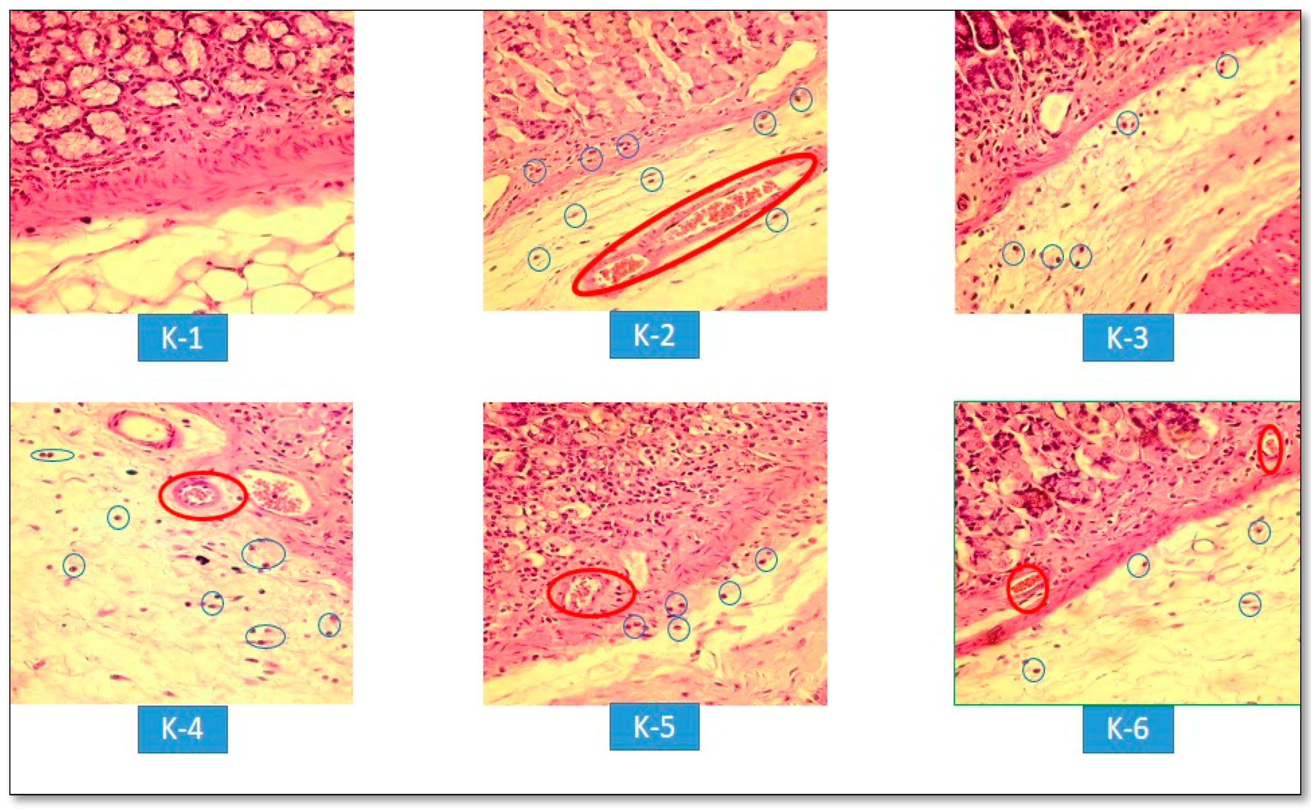
= Epithelial erosion of mucosal layers.
Scipharm 86 00035 g003
Figure 4. Microscopic result infiltration of inflammation cells and congestion: K-1, Normal group; K-2, Negative control group (CMC); K-3, Positive control group (OMZ); K-4, Rice Bran Extract group 100 mg/kg BW; K-5, Rice Bran Extract group 200 mg/kg BW; and K-6, Rice Bran Extract group 400 mg/kg BW) (magnification 400×). Scipharm 86 00035 i002 
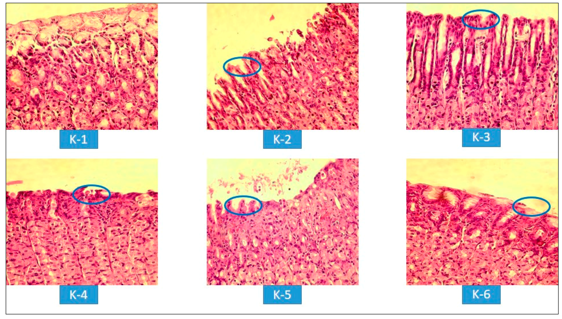
Infiltration of inflammatory cells (neutrophils); Scipharm 86 00035 i003 = Congestion.
Scipharm 86 00035 g004
Table 1. The effect of rice bran extract on gastric lesions induced by ethanol.
Treatment GroupDosageDiameter of Ulcer (mm)Ulcer IndexInhibition of Ulceration (%)
Normal (pre-treated)--0100
Negative control (CMC)5 mL/kg BW3.45 ± 0.704-
Positive control (OMZ)36 mg/kg BW0.47 ± 0.45 *0.6783.25
Rice Bran Extract100 mg/kg BW1.81 ± 0.55 *2.6733.25
200 mg/kg BW1.47 ± 0.39 *250
400 mg/kg BW0.83 ± 0.17 *1.3366.75
Results are the mean ± SD for six rats/group. Statistical comparison was performed using One-Way ANOVA followed by Tukey test: * p < 0.05 compared with the negative control group.
Table 2. The effect of rice bran extract on gastric acidity induced by ethanol.
Treatment GroupDosageGastric Acidity (µek/200 g)
Normal (pre-treated)-1.19 ± 0.57 *
Negative control (CMC)5 ml/kg BW3.94 ± 0.10 #
Positive control (OMZ)36 mg/kg BW1.21 ± 0.19 *
Rice Bran Extract100 mg/kg BW1.58 ± 0.38 *
200 mg/kg BW1.45 ± 0.33 *
400 mg/kg BW1.34 ± 0.04 *
Results as the mean ± SD for six rats/group. Statistical comparison was performed using One-Way ANOVA followed by Tukey test: * p < 0.05 compared with the negative control group; # p < 0.05 compared with the normal group.
Table 3. The effect of rice bran extract on mucus in gastric content-induced by ethanol.
Treatment GroupDosageDetermination of Mucus (Absorbance/g)
Normal (pre-treated)-0.44 ± 0.04 *
Negative control (CMC)5 mL/kg BW0.20 ± 0.01 #
Positive control (OMZ)36 mg/kg BW0.40 ± 0.07 *
Rice Bran Extract100 mg/kg BW0.22 ± 0.03 #
200 mg/kg BW0.28 ± 0.02 #
400 mg/kg BW0.36 ± 0.02 *
Results as the mean ± SD for six rats/group. Statistical comparison was performed using One-Way ANOVA followed by Tukey test: * p < 0.05 compared with the negative control group; # p < 0.05 compared with the normal group.
Table 4. An overview of the histopathology of epithelial cells on the surface of the gastric mucosa of rats in the treatment group induced by ethanol.
GroupDosageDegree of Epithelial Cell Erosion Severity on the Mucosal Surface of the Stomach
Normal (pre-treated)--
Negative control (CMC)5 mL/kg BW+++
Positive control (OMZ)36 mg/kg BW++
Rice Bran Extract100 mg/kg BW++
200 mg/kg BW+
400 mg/kg BW+
Degrees of severity: mild (+), moderate (++), and heavy (+++).
Table 5. An overview of the histopathology of congestion in the ethanol-induced treatment group.
GroupDosageSeverity of Congestion
Normal (pre-treated)--
Negative control (CMC)5 mL/kg BW+++
Positive control (OMZ)36 mg/kg BW+
Rice Bran Extract100 mg/kg BW+
200 mg/kg BW+
400 mg/kg BW+
Degrees of severity: mild (+) and heavy (+++).

