Article

Combined Effects of Cytokinin and UV-C Light on Phenolic Pattern in Ceratonia siliqua Shoot Cultures

by
Antonio Costa-Pérez
,
María A. Ferrer
* and
Antonio A. Calderón
Department of Agronomic Engineering, Universidad Politécnica de Cartagena, Paseo Alfonso XIII 48, 30203 Cartagena, Spain
*
Author to whom correspondence should be addressed.
Agronomy 2023, 13(3), 621; https://doi.org/10.3390/agronomy13030621
Submission received: 19 January 2023 / Revised: 18 February 2023 / Accepted: 20 February 2023 / Published: 22 February 2023

Abstract

:
Carob (Ceratonia siliqua L.) is an underutilized traditional crop in the Mediterranean regions that has gained much interest due to its high nutritional traits and resilience to drought and salinity. However, conventional methods of carob propagation are not enough to meet its increasing market demands. The present study analyzes to what extent benzyl adenine (BA) treatments (0.1, 0.5 and 1.0 mg L−1) alone or in combination with UV-C irradiation (3.34 and 10.01 kJ m−2) affect the antioxidant capacity and the levels of phenolic compounds in in vitro carob shoot cultures as well as their performance in terms of the content of photosynthetic pigments and sugars. Results showed that the combination of both treatments resulted in an increased content of chlorophylls, carotenoids, and sugars, particularly at 0.5 mg L−1 BA and the highest UV-C dose. Antioxidant capacity, assessed by the DPPH method, and the levels of soluble flavonoids, flavones and flavonols, and hydroxycinnamic acids were highly influenced by the interaction between BA and UV-C in the combined treatments. This indicates a clear dependence on BA concentration in the response of carob in vitro shoots to UV-C. This could be the basis for the implementation of more efficient carob micropropagation processes.

1. Introduction

The global climate crisis we are facing today is forcing farmers to change their production strategies. This includes changes in management practices, especially those related to the reduction in the use of water and other production inputs, but also the substitution of current crops for others better adapted to the environmental conditions foreseen for the near future [1].
The Mediterranean basin is characterized by irregular rainfall that determines the prevalence of semi-arid conditions in many parts of it. However, the overexploitation of groundwater and the construction of important hydraulic works to transfer water from rivers to other basins made possible the development of a flourishing, export-oriented irrigated agriculture in many areas [2]. However, reducing rainfall in ceding river basins due to climate change threatens the promise of abundant cheap water for agriculture and makes farmers look back to the nowadays almost abandoned traditional rainfed crops, which are well adapted to harsher environmental conditions [3].
The carob tree (Ceratonia siliqua L.) is one of those neglected Mediterranean crops [4]. Vast areas in the Mediterranean regions were covered with these trees before being replaced by more profitable irrigated crops. Apart from a high resilience with minimal care to harsh conditions, some research works on this species have shown the presence of numerous beneficial compounds in various organs of the plant [4,5]. Carob pods have traditionally been used as food and, above all, as feed. In addition, the pods and other parts of the plant have been used in folk and modern medicine for different purposes; the seeds can be processed to obtain additives for the food and cosmetic industries, and the wood is appreciated in marquetry and furniture manufacturing [4]. Therefore, the carob tree is a multipurpose crop that provides products with a high added value and, hence, there is a growing interest in its cultivation [5,6].
However, conventional methods of propagation are not enough to fulfill the foreseen market demand for carob plants. In fact, seed germination does not seem the most appropriate method due to seedling heterozygosity and rooting of carob cuttings is difficult to achieve at convenient rates [7]. Other methods, such as grafting and air layering, are giving good results but are labor intensive and therefore increase production costs [6]. In this context, the propagation of carob by in vitro culture techniques arises as an alternative to conventional methods. These techniques offer the possibility to clonally multiplying elite genotypes under aseptic conditions and at the desired rate, since growth environment is accurately regulated [8].
In vitro carob regeneration and micropropagation have been previously reported from young and mature tissues [9,10]. Efforts are now focused on optimizing media composition to achieve suitable multiplication rates and shoot quality, the latter being crucial for successful acclimatization to ex vitro conditions [11]. One of the main factors affecting both parameters is the presence of cytokinin in the culture medium. In fact, commercial micropropagation by shoot cultures is almost inconceivable in the absence of cytokinins during the multiplication stage, due to the promotion of cell proliferation and differentiation and the breaking of apical dominance (among other effects) mediated by these compounds [8]. Different cytokinins have been used in carob micropropagation. Comparative studies showed that the adenine-based compounds benzyl adenine (BA) and meta-topolin gave the best results in terms of multiplication rate [6], although interactions among carob cultivar and type and concentration of the applied cytokinin must be taken into account to optimize the micropropagation process [9].
Cytokinins also affect shoot quality through their effects on photosynthetic performance and antioxidant defense systems, among others [12]. The protection of chlorophylls from degradation and the induction of chlorophyll biosynthesis and chloroplasts development have been frequently described in the literature as beneficial effects of cytokinins in plants under both in vitro and field conditions (revised by [12]). In addition, it has been shown that cytokinins promote the production of secondary metabolites in plant materials grown in vitro, especially compounds of a phenolic nature which contribute significantly to an increase in the antioxidant capacity of such materials [13,14,15]. Despite what may be thought, the in vitro environment constitutes a hostile medium for plant materials due to the relatively high levels of available water, nutrients, growth regulators, as well as air composition and physical conditions. Additionally, tissue injury and transfer to ex vitro conditions also cause stress conditions that usually result in an oxidative stress situation [16]. Therefore, it seems plausible that plant materials with fortified antioxidant systems grown in vitro will be more suitable to successfully completing a micropropagation process.
The levels of antioxidant compounds in plants can also be increased by elicitation with physical, chemical, and biological agents [17,18]. Among them, physical agents, such as UV radiation, have the advantage of providing not only a “clean process”, free from the presence of foreign factors in the culture system, but also an efficient and cost-effective way to increase the levels of antioxidant compounds in cell, tissue, and organ in vitro cultures [18,19,20].
The information available on the effects of cytokinins and/or UV-C radiation on secondary metabolites in carob grown in vitro is scarce or null. For this, considering the present (and, possibly, future) relevance of this crop, this study aimed to assess the influence of BA and non-lethal doses of UV-C radiation, alone or in combination, on the antioxidant capacity and the levels of phenolic compounds in carob shoot cultures. Our working hypothesis is that the combination of BA and UV-C treatment can further strengthen the carob antioxidative system by promoting the accumulation of secondary metabolites. This elicitation strategy, hardly used in shoot multiplication protocols, seeks to achieve the aforementioned increase in shoot resilience against oxidative stress and, therefore, improve the process of carob micropropagation.

2. Materials and Methods

2.1. Plant Explant Material, Culture Medium and Growth Conditions

Ceratonia siliqua cv. Mollar seeds were provided by a local farmer in the southeast of Spain. The carob seeds were dipped in 70% ethanol for 30 s and then, were surface sterilized for 30 min in 20% (v/v) Domestos® (4.3% sodium hypochlorite, Unilever, London, UK), were rinsed 3 times with autoclaved distilled water and were left for imbibition in the dark at room temperature for 14 days [7]. Imbibed seeds were placed on Murashige and Skoog (MS) basal medium [21] with macronutrients at half-strength supplemented with casein hydrolysate (250 mg L−1), sucrose (3%, w/v), MS vitamins, and 0.8% Difco Bacto agar (Pronadisa, Madrid, Spain). The pH of the media was set to 5.8 with 100 mM KOH prior to autoclaving at 108 kPa and 121 °C for 20 min. Shoot tips of carob seedlings were used for shoot culture initiation. Shoot tips were placed on solid ½ MS medium in tubes (25 × 90 mm) and kept at 18/25 °C day/night temperature with a 16 h light/8 h dark cycle, and 100 μmol m−2 s−1 photon flux density (Sanyo, versatile environmental test chamber, MLR-351H, Osaka, Japan). After 5 weeks, the newly developed shoots were cut into nodal segments containing one axillary bud and utilized as explants. Five explants were placed on solid ½ MS medium (50 mL) in 0.5 l-glass vessels (diameter 70; height 115 mm). Shoots were sub-cultured regularly to fresh ½ MS medium at an interval of 6 weeks (Supplementary Materials, Figure S1). All cultures were grown under the same temperature and light conditions detailed previously.

2.2. Benzyl Adenine and UV Treatments

Six-week-old plantlets were excised, transferred to ½ MS medium supplemented with different concentrations of benzyl adenine (BA) (0, 0.1, 0.5 and 1.0 mg L−1), and maintained under the abovementioned growth conditions. After 5 weeks of growth, plantlets were divided into three groups. Each group was aseptically exposed to UV-C irradiation in a laminar flow cabinet for either 0, 20, or 60 min. The UV-C intensity (0.278 mW cm−2) was measured with a VLX-254 radiometer (Vilbert Lourmat, Marne la Vallée, France). Thus, the doses of UV-C treatments were 0.00, 3.34, and 10.01 kJ m−2, respectively. After the UV-C treatments, the plantlets were maintained in the same growth conditions as before for a week. The percentage of shoot induction, the number of shoots per explant, shoot length, and the presence and size of basal callus were recorded only in untreated UV explants. Then, shoots were immediately flash frozen in liquid nitrogen and pulverized using a liquid nitrogen-cooled analytical mill. Samples were kept at −80 °C until they were analyzed. Three individual plantlets were considered as one replicate; each treatment had two replicates (24 vessels) and was repeated twice.

2.3. Extraction and Quantification of Photosynthetic Pigments

For photosynthetic pigments analysis, shoot samples (~100 mg) were extracted with ice-cold 96% ethanol, sonicated (37 kHz) at 40 °C for 30 min, and were centrifuged (15,000× g at 4 °C for 15 min) [22]. The supernatants were used for the analysis of chlorophyll a (chl a) and b (chl b) and total carotenoids x + c (xanthophylls and carotenes) contents using the extinction coefficients and the equations reported by [23].

2.4. Extraction and Quantification of Sugars, DPPH Radical Scavenging Activity and Phenolic Compounds

Around 100 mg of liquid nitrogen-powdered samples were extracted with ice-cold 70% ethanol using sonication as described above. After centrifugation, the resulting supernatants were used for the spectrophotometric determination of sugars, DPPH radical scavenging activity, and phenolic compounds. The total sugar content was determined using the anthrone method and glucose as a standard [22].
The DPPH (1,1-diphenyl-2-picrylhydrazyl radical) antioxidant assay was basically carried out according to [24]. In short, an aliquot (25 μL) of ethanolic extracts was added to 0.1 mM methanolic DPPH solution and the absorbance was read at 517 nm after a 2.30-h incubation period at room temperature in the dark. Gallic acid, in 7 different concentrations in the range 5–250 µM, was used as a standard and ran in parallel with experimental samples.
The total soluble phenol content (TPC), flavonoids, flavanols, and hydroxycinnamic acids (HCAs) were determined according to [22]. Shortly, TPC was assessed by the Folin–Ciocalteu procedure and the results were expressed as gallic acid equivalents (GAE) per gram of fresh weight (FW). The total soluble flavonoid content of the extracts was assayed by the aluminum chloride method and the results were expressed as rutin equivalents (RE) per gram FW. The total soluble flavanols were determined using the DMAC reagent and their contents were expressed as catechin equivalents (CE) per gram FW. The content of HCAs was determined using Arnow’s reagent and HCA contents were calculated as caffeic acid equivalents (CAE) per gram FW. The joint determination of flavones and flavonols was carried out as described by [25]. Quercetin, in 7 different concentrations in the range 25–1000 µM, was used as standard for the calculation of flavones and flavonols content, and the results were expressed as quercetin equivalents (QE) per gram FW.

2.5. Statistical Analysis

Data are presented as means ± standard error (SE) from two independent experiments. Statistical analyses were carried out using the SPSS software (version 26.0; SPSS Inc., Chicago, IL, USA). A one-way analysis of variance (ANOVA) was conducted to evaluate the existence of significant differences (p ≤ 0.05) in shoot multiplication parameters. Biochemical data were analyzed using a two-way ANOVA with BA doses and UV treatments as fixed factors. When necessary, a Tukey honestly significant difference (HSD) post hoc test was used to evaluate the existence of significant differences (p ≤ 0.05) among the samples. A principal component analysis (PCA) was also performed using the CANOCO software (version 4.5, Microcomputer Power, Ithaca, NY, USA) to further analyze similarities and differences among treatments.

3. Results

3.1. Effect of BA Concentrations on Carob Shoot Multiplication

No emergency of new shoots was observed in those explants grown in the absence of BA (Table 1). The number of branches per explant increased with increasing BA concentration in the MS/2 medium. This branching increase was associated with a concomitant decrease in the shoot internode length (>50% in explants treated with the highest BA dose). Moreover, BA exposure led to further induction of basal callus whose size augments in a BA dose-dependent manner.

3.2. Effect of BA Concentrations and UV-C Treatments on the Physiological Performance of Carob Shoots

The impact of both BA and UV-C treatments on the physiological performance of carob plantlets in terms of concentration of photosynthetic pigments and soluble sugar contents were evaluated. In general, the presence of BA on the basal media induced a slight reduction in the content of both chlorophyll pigments (Figure 1), although the chl a/chl b ratio showed no statistically significant changes (Supplementary Materials, Figure S2). In contrast, UV-C treatments led to a dose-dependent increase in the content of chl a, chl b, and total chlorophyll (about 1.4-fold in 60′-UV-C-treated plantlets over the basal values). The levels of carotenoids remained almost unchanged, although a slight increase was observed in 60′-UV-C-treated plantlets fortified with BA at 0.1 and 0.5 mg L−1 (Figure 1d).
Similarly, the levels of soluble sugars were enhanced by UV-C exposure in a dose-dependent manner (>2-fold in 60′-UV-C-treated plantlets over the basal values). BA treatments also caused an increase in the sugar contents, although this tendency varied depending on the UV-C irradiation dose applied (Figure 1e). In fact, the two-way ANOVA results revealed that the interaction between BA and UV-C treatments significantly impacted the concentration of sugars (p ≤ 0.001).

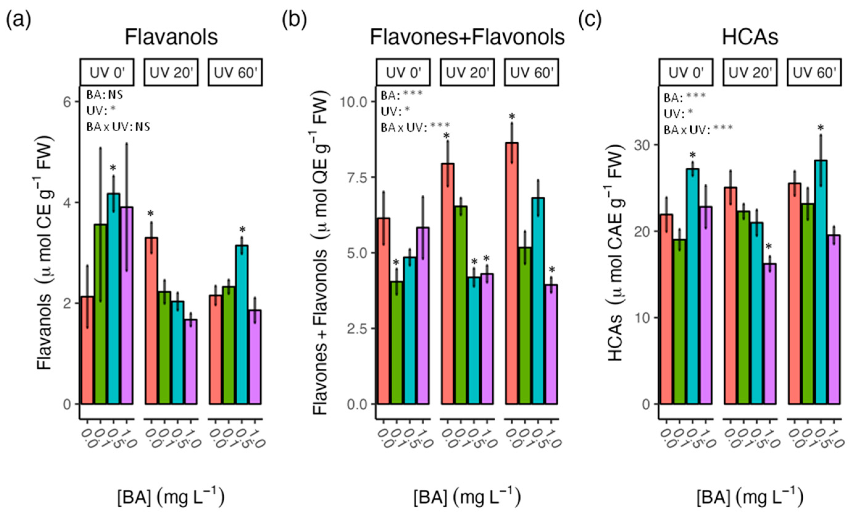
3.3. Effect of BA Concentrations and UV-C Treatments on Antioxidant Activity and Phenolic Content in Carob Shoots

The analysis of total antioxidant activity, measured with the DPPH test, showed that BA treatments caused an increase in the DPPH radical scavenging activity, although this was not statistically significant (Figure 2a). Moreover, a marked increase in the antioxidant activity in all the BA–UV-C treatments was noticed. The highest antioxidant activity (>2.7-fold over untreated controls) was found in 60′-UV-C-treated plantlets fortified with BA at 0.1 mg L−1.
The antioxidant activity is known to be correlated with phenolic concentrations in plant extracts [26], thus, the content of total soluble phenols was analyzed using the Folin–Ciocalteu assay. As shown in Figure 2b, the presence of BA on the basal media caused an increase in the content of soluble phenols, particularly in those explants treated with 0.5 mg L−1 of BA (>1.8-fold over untreated controls). However, UV-C treatments did not cause statistically significant alteration in TPC.
Earlier findings have shown that flavonoids and HCAs constitute important phenolic groups identified in carob [27], so their levels were also studied. The accumulation of flavonoids in UV-C untreated explants (Figure 2c) displayed a trend similar to that observed for TPC, being the highest values found in those explants fortified with BA 0.5 mg L−1 (>1.5-fold over untreated controls). In contrast, the UV-C treatments caused an increase in the levels of flavonoids as well as an alteration in the BA-induced flavonoid accumulation.
Next, the content of the main subclasses of flavonoids, flavanols, and flavones and flavonols were also determined. As shown in Figure 3a, the highest flavanols content was found in explants treated with BA 0.5 mg L−1 (~2-fold in comparison to untreated controls). Low UV-C treatment also led to an increase in the flavones and flavonols content (>1.5-fold over untreated controls), but their levels were in the range of the untreated controls at the highest UV-C dose. In contrast, the concentration of flavones and flavonols increased upon UV-C treatment in a dose-dependent manner, reaching their highest values in the 60′-UV-C-treated plantlets (>1.4-fold over untreated controls) (Figure 3b). In the absence of UV-C irradiation, the flavone and flavonol contents decreased at the lowest BA dose (0.1 mg L−1), but their levels remained similar to controls at higher BA doses, although this pattern changed upon UV-C exposure.
As far as the content of HCAs is concerned, the highest HCA levels were found in explants fortified with BA 0.5 mg L−1, particularly at the highest UV-C dose (~1.3-fold over untreated controls) (Figure 3c). The two-way analysis showed a significant interaction effect between BA and UV-C treatment on the content of flavones and flavonols as well as on the levels of HCAs (p ≤ 0.001).

3.4. Principal Component Analysis

A principal component analysis (PCA) was also performed to summarize and visualize general tendencies among all the biochemical parameters analyzed. As presented in Figure 4, the first two PCA components (PC1 and PC2) account for 64% of the variance in the overall data sets. PC1 was associated positively with the content of chlorophylls, HCAs, flavonoids, flavones and flavonols, carotenoids, and sugars. PC2, which accounted for 17.2% of the total variance, was best explained by flavanols on the positive side of the Y-axis and by the total phenols and DPPH activity on the negative Y-axis.
Although neither the PC1 nor the PC2 made a clear separation among the samples, it can be observed that PC1 tended to separate the untreated UV-C samples, which were mainly positioned in the upper left quadrant of the plot (unfilled symbols), from the untreated BA samples, which were mostly located in the upper right quadrant (circle symbols). Moreover, the PC2 separated the untreated samples, located in the upper part of the PCA plot (unfilled and circle symbols), from the BA- and UV-C-treated samples, placed in the lower part, indicating a different response between individual BA and UV-C treatments and BA and UV-C combined treatments.

4. Discussion

An efficient plant micropropagation process is a trade-off between the multiplication rate and the quality. Factors that tend to increase the rate of propagation in vitro often also weaken propagated materials and make them less apt to survive and thrive under ex vitro conditions. Cytokinins are essential to achieve high and economically profitable multiplication rates in commercial micropropagation based on shoot cultures [8]. However, these growth regulators can cause morphological and physiological alterations when the type and/or doses of cytokinin are not appropriate. These alterations are often associated with relatively high levels of ROS in the tissues and, therefore, the deployment of a robust antioxidant defense system is believed to be crucial for preserving the quality of micropropagated materials [28].

4.1. Increasing BA Doses Favored Carob Shoot Multiplication but also Increase the Development of Basal Callus

In the present study, carob nodal segments were cultivated for 6 weeks on ½ MS solid medium in the presence of increasing doses of BA (0.1, 0.5, and 1.0 mg L−1) to evaluate their effectiveness in the induction of lateral shoot buds. Moreover, we also analyze to what extent BA treatment alone or in combination with UV-C irradiation affects shoot vigor and quality. Our data revealed that BA is effective in increasing the rate of shoot proliferation; although, it also led to the development of basal callus in a BA dose-dependent manner (Table 1). These results are in accordance with previous studies reporting that the adenine-based cytokinins are effective in the activation of axillary buds in carob explants [6,9,10]. In our study, the mean number of shoots obtained per explant was 2.93 ± 0.12 at the BA dose of 1 mg L−1 (Table 1). This value is higher than those reported in other studies using semi-solid media containing the same cytokinin and doses [9,29], but can be enhanced by using other basal media, medium physical state, cytokinin type, or additional growth regulators [6,29,30]. The carob cultivar used in each case could also explain these differences in the multiplication rate, as previously pointed out [9].
A side effect of cytokinin supplementation in culture media is the formation of basal callus due to an alteration in the auxins/cytokinins ratio [8]. Here, in the absence of BA, more than 33% of carob explants presented basal callus, and in the presence of the highest BA dose (1 mg L−1) the percentage was 100% (Table 1). The induction of basal callus by the exogenous application of cytokinins is particularly high in carob species [9,10]; although, the molecular mechanisms underpinning this response remain unclear [31]. Basal callus formation can be an undesirable response in the later stages of micropropagation because it may hinder the root formation process [8]. Romano et al. [9] described the formation of adventitious shoots from carob basal calli, which opens the possibility of obtaining regenerants more prone to somaclonal variation. However, these authors also reported that all the adventitious shoots formed failed to further develop. Therefore, it can be concluded that callus formation at the base of the explant is not a major concern during the multiplication stage of carob micropropagation, as long as adventitious shoots are discarded for a true-to-type propagation.

4.2. Sugar Contents Were Significantly Influenced by the Interaction between BA and UV-C Treatments

Oxidative stress and low photosynthetic capacity in in vitro cultures are considered as key factors affecting transplant vulnerability [32]. Here, the content of chlorophylls and sugars increased in a UV-C dose-dependent manner (Figure 1). Stimulation of chlorophyll content by low levels of UV irradiation has already been described in other plant species, such as rice [33], mung bean, and groundnut [34]. Moreover, low UV exposure has also been reported to have enhancing effects on the photosynthesis rate and sugar accumulation in several crop species [35,36,37]. In fact, UV-C treatment has been reported to regulate the activity of amylolytic enzymes leading to higher levels of starch and total soluble sugars in lily bulbs [38], which was consistent with our findings.
On the other hand, our data also revealed that BA supplementation tended to decrease the levels of chlorophylls and this response was independent of UV-C radiation with regard to the results of the two-way ANOVA. Unfortunately, it was not possible to compare these apparently controversial results with others in the carob micropropagation literature. However, studies on the effects of cytokinins on the levels of photosynthetic pigments have shown both inhibitory and stimulatory effects [39], even though these growth regulators are generally associated with increased synthesis and delayed degradation of chlorophylls. In any case, the chl a/chl b ratio did not show significant changes regardless of the treatment applied, which reflects a similar functionality of the photosynthetic apparatus [39].

4.3. Combined BA and UV-C Treatments Further Enhanced the Antioxidant Capacity of Carob Shoots in Comparison with the Individual Treatments

Growing evidence suggests that exposure to low UV radiation increases plant tolerance to stress by boosting plant defense antioxidant systems [35,40] and the accumulation of antioxidants including UV-shielding compounds such as polyphenols, flavonoids, and HCAs [41,42,43,44]. In addition, it is well known that cytokinins can induce the synthesis and accumulation of antioxidant phenolics [12].
In this study, the total antioxidant capacity was assessed by the DPPH method, as it provides an integrated parameter to estimate a wide range of low molecular weight antioxidants, including ascorbate, carotenoids, and phenolic compounds, among others [45]. Our data revealed that the combination of BA and UV-C treatments noticeably increased the DPPH antiradical activity of carob shoots (Figure 2). These results are in accordance with those recently reported in in vitro shoots of Scutellaria baicalensis treated with BA and UV-B [18] and in tomato plants treated with kinetin, by foliar spray, and exposed to UV-C [46] or UV-B irradiation [47]. Taken together, these results suggest that in UV-stressed plants, cytokinin treatments can boost the antioxidant capacities to increase the tolerance to UV-C stress. Beyond their essential role in plant growth and development, cytokinins have also been proven to function in plant stress abiotic signaling [48,49], although the detailed mechanisms at the molecular level are still unknown.
Moreover, the DPPH activity followed a similar trend to that of the soluble phenol content, suggesting that the total antioxidant capacity in carob explants comes mainly from phenolic compounds, which is consistent with previous studies [24,50,51]. Surprisingly, low to moderate correlations were found between DPPH and the content of the soluble phenol groups analyzed, and even some of them, commonly recognized as having remarkable antioxidant properties, showed negative correlations (flavonoids: r = 0.42 **; flavanols: −0.54 **; flavones and flavonols: −0.2; HCAs: −0.1; Supplementary Materials, Figure S3), as illustrated by the PCA results.

4.4. Phenol Accumulation in Response to UV-C Treatments Was Influenced by BA Concentrations in the Culture Media

The effects of the BA and UV treatments applied separately on the contents of the different groups of phenols were roughly similar, except for the levels of flavones and flavonols (Figure 3). These compounds have been considered effective UV filters, protecting nucleic acids and other essential components from excessive energy absorption [52]. This would explain the increase observed after the UV-C treatment (Figure 3b). On the other hand, it is known that cytokinins differentially induce the synthesis of phenolics in materials grown in vitro depending on the type and dose applied [53,54]. Taken together, these facts would determine different phenol patterns in carob tissues after BA or UV-C treatments. However, in our study, the levels of all the phenol families analyzed were highly influenced by the interaction between BA and UV-C in the combined treatments, except for the flavanols contents in which this interaction was not statistically significant (at p ≤ 0.05). This indicates a clear dependence on the BA concentration of the response of carob shoots to UV-C radiation, which is in line with what was described by [18] in Scutellaria baicalensis shoot cultures treated with BA and UV-B radiation.
The lack of positive correlation between the antioxidant capacity and contents of the phenolic groups analyzed suggests that the main determinant thereof is another type (or other types) of phenolics. To the best of our knowledge, there is no information on phenolic compounds present in carob shoot cultures, but the analysis of leaves from the field-grown plants showed that some of the most abundant components belonged to the family of hydroxybenzoic acids, gallic acid being the main phenolic in those organs [55]. Gallic acid is also present in leaves in the form of tannins (gallotannins or hydrolyzable tannins) and flavanolgalloyl esters [27]. All these forms are characterized by showing outstanding antioxidant properties and would greatly contribute to the antioxidant capacity found in carob shoots in this study.

5. Conclusions

BA and UV-C radiation can separately increase the antioxidant capacity of carob shoots grown in vitro but with a different quantitative phenolic pattern. However, when both effectors are applied jointly, the interaction between them causes a change in these patterns, resulting in increased levels of antioxidant capacity and the content of some phenol groups. This opens a double opportunity in relation to the applicability of carob micropropagation. On the one hand, higher levels of antioxidants can increase the in vitro and ex vitro performance of micropropagated carob. On the other hand, the combined treatments could lay the foundations for the exploitation of carob shoot cultures as biofactories to produce bioactive compounds with applications in the pharmaceutical, cosmetic, or agri-food industries.
Although the application of elicitors to stimulate the production and accumulation of bioactive compounds in unorganized plant cell cultures (i.e., callus and suspension cell cultures) is widely used [56], their application in shoot in vitro cultures intended for micropropagation is scarce. The results of this work highlight the possibility of using physical elicitors to enhance the antioxidant defenses of micropropagated plants, making them more tolerant to stress.
Even though some open questions remain, such as the persistence of treatment effects or the impact of these treatments on subsequent ex vitro plant performance, this simple and low-cost approach could be applied to the micropropagation of other crops, especially to those difficult species, such as woody materials, whose yields are greatly affected by low in vitro performance.

Supplementary Materials

The following supporting information can be downloaded at: https://www.mdpi.com/article/10.3390/agronomy13030621/s1, Figure S1: Representative photos of the different steps carried out during the carob micropropagation process; Figure S2: Effect of different concentrations of BA and UV-C treatments on the chlorophyll a/b ratio in 6-week-old carob shoots; Figure S3: Pearson correlation coefficients and hierarchical clustering using the Euclidean distance between the different biochemical parameters measured in this study.

Author Contributions

Conceptualization, A.A.C.; methodology, M.A.F. and A.A.C.; formal analysis and investigation, A.C.-P.; resources, A.C.-P., M.A.F. and A.A.C.; writing—original draft preparation, M.A.F.; writing—review and editing, M.A.F. and A.A.C.; visualization, M.A.F.; supervision, A.A.C.; project administration, A.A.C. All authors have read and agreed to the published version of the manuscript.

Funding

This research received no external funding.

Institutional Review Board Statement

Not applicable.

Informed Consent Statement

Not applicable.

Data Availability Statement

The data presented in this study are available on request from the corresponding author.

Acknowledgments

This work was partially carried out at the Instituto de Biotecnología Vegetal (Universidad Politécnica de Cartagena).

Conflicts of Interest

The authors declare no conflict of interest.

References

  1. Alrteimei, H.A.; Ash’aari, Z.H.; Muharram, F.M. Last Decade Assessment of the Impacts of Regional Climate Change on Crop Yield Variations in the Mediterranean Region. Agriculture 2022, 12, 1787. [Google Scholar] [CrossRef]
  2. Harmanny, K.S.; Malek, Ž. Adaptations in Irrigated Agriculture in the Mediterranean Region: An Overview and Spatial Analysis of Implemented Strategies. Reg. Environ. Chang. 2019, 19, 1401–1416. [Google Scholar] [CrossRef] [Green Version]
  3. Vidal-Macua, J.J.; Ninyerola, M.; Zabala, A.; Domingo-Marimon, C.; Gonzalez-Guerrero, O.; Pons, X. Environmental and Socioeconomic Factors of Abandonment of Rainfed and Irrigated Crops in Northeast Spain. Appl. Geogr. 2018, 90, 155–174. [Google Scholar] [CrossRef]
  4. Batlle, I.; Tous, J. Carob Tree: Ceratonia siliqua L. Promoting the Conservation and Use of Underutilized and Neglected Crops; Institute of Plant Genetics and Crop Plant, Gatersleben/International Plant Genetic Resources Institute: Rome, Italy, 1997; Volume 17, ISBN 92-9043-328-X. [Google Scholar]
  5. Gioxari, A.; Amerikanou, C.; Nestoridi, I.; Gourgari, E.; Pratsinis, H.; Kalogeropoulos, N.; Andrikopoulos, N.K.; Kaliora, A.C. Carob: A Sustainable Opportunity for Metabolic Health. Foods 2022, 11, 2154. [Google Scholar] [CrossRef]
  6. Shahzad, A.; Akhtar, R.; Bukhari, N.A.; Perveen, K. High Incidence Regeneration System in Ceratonia siliqua L. Articulated with SEM and Biochemical Analysis during Developmental Stages. Trees 2017, 31, 1149–1163. [Google Scholar] [CrossRef]
  7. Pérez-García, F. Germination Characteristics and Intrapopulation Variation in Carob (Ceratonia siliqua L.) Seeds. Span. J. Agric. Res. 2009, 7, 398. [Google Scholar] [CrossRef] [Green Version]
  8. Altman, A.; Loberant, B. Micropropagation of Plants, Principles and Practices. In Encyclopedia of Cell Technology; John Wiley & Sons, Inc.: Hoboken, NJ, USA, 2003. [Google Scholar]
  9. Romano, A.; Barros, S.; Martins-Loução, M.A. Micropropagation of the Mediterranean Tree Ceratonia siliqua. Plant Cell Tissue Organ Cult. 2002, 68, 35–41. [Google Scholar] [CrossRef] [Green Version]
  10. Saïdi, R.; Rahmouni, S.; el Ansari, Z.N.; Maouni, A.; Badoc, A.; Lamarti, A. Effect of Cytokinins on the Micropropagation of Carob (Ceratonia siliqua L.) through Shoot Tip Culture. Am. J. Plant Sci. 2019, 10, 1469–1481. [Google Scholar] [CrossRef] [Green Version]
  11. Gonçalves, S.; Correia, P.J.; Martins-Loução, M.A.; Romano, A. A New Medium Formulation for in vitro Rooting of Carob Tree Based on Leaf Macronutrients Concentrations. Biol. Plant. 2005, 49, 277–280. [Google Scholar] [CrossRef]
  12. Hönig, M.; Plíhalová, L.; Husičková, A.; Nisler, J.; Doležal, K. Role of Cytokinins in Senescence, Antioxidant Defence and Photosynthesis. Int. J. Mol. Sci. 2018, 19, 4045. [Google Scholar] [CrossRef] [Green Version]
  13. Vasconcelos, J.N.C.; Brito, A.L.; Pinheiro, A.L.; Pinto, D.I.J.G.E.C.; Almeida, J.R.G.D.S.; Soares, T.L.; de Santana, J.R.F. Stimulation of 6-Benzylaminopurine and Meta-Topolin-Induced in vitro Shoot Organogenesis and Production of Flavonoids of Amburana cearensis (Allemão) A.C. Smith. Biocatal. Agric. Biotechnol. 2019, 22, 101408. [Google Scholar] [CrossRef]
  14. Hlophe, N.P.; Aremu, A.O.; Gruz, J.; van Staden, J.; Finnie, J.F. Influence of Different Cytokinins on the Phenolic Acids and Antioxidant Activity of Two Brachystelma Species. Plant Cell Tissue Organ Cult. 2021, 145, 689–699. [Google Scholar] [CrossRef]
  15. Grzegorczyk-Karolak, I.; Hnatuszko-Konka, K.; Zarzycka, M.; Kuźma, Ł. The Stimulatory Effect of Purine-Type Cytokinins on Proliferation and Polyphenolic Compound Accumulation in Shoot Culture of Salvia viridis. Biomolecules 2020, 10, 178. [Google Scholar] [CrossRef] [Green Version]
  16. Benson, E. Do Free Radicals Have a Role in Plant Tissue Culture Recalcitrance? Vitr. Plant Cell. Dev. Biol. 2000, 36, 163–170. [Google Scholar] [CrossRef]
  17. Vasconsuelo, A.; Boland, R. Molecular Aspects of the Early Stages of Elicitation of Secondary Metabolites in Plants. Plant Sci. 2007, 172, 861–875. [Google Scholar] [CrossRef]
  18. Yun, C.; Zhao, Z.; Ri, I.; Gao, Y.; Shi, Y.; Miao, N.; Gu, L.; Wang, W.; Wang, H. How Does UV-B Stress Affect Secondary Metabolites of Scutellaria baicalensis in vitro Shoots Grown at Different 6-benzyl Aminopurine Concentrations? Physiol. Plant. 2022, 174, e13778. [Google Scholar] [CrossRef]
  19. López-Orenes, A.; Ferrer, M.A.; Calderón, A.A. Microwave Radiation as an Inducer of Secondary Metabolite Production in Drosera rotundifolia in vitro Plantlets. J. Nat. Prod. 2022, 85, 2104–2109. [Google Scholar] [CrossRef] [PubMed]
  20. Alsoufi, A.S.M.; Pączkowski, C.; Długosz, M.; Szakiel, A. Influence of Selected Abiotic Factors on Triterpenoid Biosynthesis and Saponin Secretion in Marigold (Calendula officinalis L.) in vitro Hairy Root Cultures. Molecules 2019, 24, 2907. [Google Scholar] [CrossRef] [Green Version]
  21. Murashige, T.; Skoog, F. A Revised Medium for Rapid Growth and Bio Assays with Tobacco Tissue Cultures. Physiol. Plant. 1962, 15, 473–497. [Google Scholar] [CrossRef]
  22. López-Orenes, A.; Bueso, M.C.; Conesa, H.; Calderón, A.A.; Ferrer, M.A. Seasonal Ionomic and Metabolic Changes in Aleppo Pines Growing on Mine Tailings under Mediterranean Semi-Arid Climate. Sci. Total Environ. 2018, 637–638, 625–635. [Google Scholar] [CrossRef]
  23. Lichtenthaler, H.K.; Wellburn, A.R. Determinations of Total Carotenoids and Chlorophylls a and b of Leaf Extracts in Different Solvents. Biochem. Soc. Trans. 1983, 11, 591–592. [Google Scholar] [CrossRef] [Green Version]
  24. Pérez-Tortosa, V.; López-Orenes, A.; Martínez-Pérez, A.; Ferrer, M.A.; Calderón, A.A. Antioxidant Activity and Rosmarinic Acid Changes in Salicylic Acid-Treated Thymus membranaceus Shoots. Food Chem. 2012, 130, 362–369. [Google Scholar] [CrossRef]
  25. Kosalec, I.; Bakmaz, M.; Pepeljnjak, S.; Vladimir-Knezević, S. Quantitative Analysis of the Flavonoids in Raw Propolis from Northern Croatia. Acta Pharm. 2004, 1, 65–72. [Google Scholar]
  26. Zheng, W.; Wang, S.Y. Antioxidant Activity and Phenolic Compounds in Selected Herbs. J. Agric. Food Chem. 2001, 49, 5165–5170. [Google Scholar] [CrossRef]
  27. Stavrou, I.J.; Christou, A.; Kapnissi-Christodoulou, C.P. Polyphenols in Carobs: A Review on Their Composition, Antioxidant Capacity and Cytotoxic Effects, and Health Impact. Food Chem. 2018, 269, 355–374. [Google Scholar] [CrossRef]
  28. Baťková, P.; Pospíšilová, J.; Synková, H. Production of Reactive Oxygen Species and Development of Antioxidative Systems during in vitro Growth and ex vitro Transfer. Biol. Plant. 2008, 52, 413–422. [Google Scholar] [CrossRef]
  29. Lozzi, A.; Abdelwahd, R.; Mentag, R.; Abousalim, A. Development of a New Culture Medium and Efficient Protocol for in vitro Micropropagation of Ceratonia siliqua L. Vitr. Cell. Dev. Biol.-Plant 2019, 55, 615–624. [Google Scholar] [CrossRef]
  30. Ahmed, M.; El-Fadl, R.; Hegazi, G.; Elaziem, T. Improving Micropropagation Protocol for Carob (Ceratonia siliqua). Plant Cell Biotechnol. Mol. Biol. 2021, 22, 84–94. [Google Scholar]
  31. Ikeuchi, M.; Sugimoto, K.; Iwase, A. Plant Callus: Mechanisms of Induction and Repression. Plant Cell 2013, 25, 3159–3173. [Google Scholar] [CrossRef] [Green Version]
  32. Cassells, A.C.; Curry, R.F. Oxidative Stress and Physiological, Epigenetic and Genetic Variability in Plant Tissue Culture: Implications for Micropropagators and Genetic Engineers. Plant Cell Tissue Organ Cult. 2001, 64, 145–157. [Google Scholar] [CrossRef]
  33. Xu, K.; Qiu, B.S. Responses of Superhigh-Yield Hybrid Rice Liangyoupeijiu to Enhancement of Ultraviolet-B Radiation. Plant Sci. 2007, 172, 139–149. [Google Scholar] [CrossRef]
  34. Siddiqui, A.; Dawar, S.; Javed Zaki, M.; Hamid, N. Role of Ultraviolet (UV-C) Radiation in the Control of Root Infecting Fungi on Groundnut and Mung Bean. Pak. J. Bot. 2011, 43, 2221–2224. [Google Scholar]
  35. Thomas, D.T.T.; Puthur, J.T. UV Radiation Priming: A Means of Amplifying the Inherent Potential for Abiotic Stress Tolerance in Crop Plants. Environ. Exp. Bot. 2017, 138, 57–66. [Google Scholar] [CrossRef]
  36. Bornman, J.F.; Evert, R.F.; Mierzwa, R.J. The Effect of UV-B and UV-C Radiation on Sugar Beet Leaves. Protoplasma 1983, 117, 7–16. [Google Scholar] [CrossRef]
  37. Lin, Q.; Xie, Y.; Liu, W.; Zhang, J.; Cheng, S.; Xie, X.; Guan, W.; Wang, Z. UV-C Treatment on Physiological Response of Potato (Solanum tuberosum L.) during Low Temperature Storage. J. Food Sci. Technol. 2017, 54, 55–61. [Google Scholar] [CrossRef] [Green Version]
  38. Huang, H.; Ge, Z.; Limwachiranon, J.; Li, L.; Li, W.; Luo, Z. UV-C Treatment Affects Browning and Starch Metabolism of Minimally Processed Lily Bulb. Postharvest Biol. Technol. 2017, 128, 105–111. [Google Scholar] [CrossRef]
  39. Aremu, A.O.; Bairu, M.W.; Szüčová, L.; Finnie, J.F.; van Staden, J. The Role of Meta-Topolins on the Photosynthetic Pigment Profiles and Foliar Structures of Micropropagated ‘Williams’ Bananas. J. Plant Physiol. 2012, 169, 1530–1541. [Google Scholar] [CrossRef]
  40. Urban, L.; Chabane Sari, D.; Orsal, B.; Lopes, M.; Miranda, R.; Aarrouf, J. UV-C Light and Pulsed Light as Alternatives to Chemical and Biological Elicitors for Stimulating Plant Natural Defenses against Fungal Diseases. Sci. Hortic. 2018, 235, 452–459. [Google Scholar] [CrossRef]
  41. Agati, G.; Brunetti, C.; di Ferdinando, M.; Ferrini, F.; Pollastri, S.; Tattini, M. Functional Roles of Flavonoids in Photoprotection: New Evidence, Lessons from the Past. Plant Physiol. Biochem. 2013, 72, 35–45. [Google Scholar] [CrossRef]
  42. Fabón, G.; Martínez-Abaigar, J.; Tomás, R.; Núñez-Olivera, E. Effects of Enhanced UV-B Radiation on Hydroxycinnamic Acid Derivatives Extracted from Different Cell Compartments in the Aquatic Liverwort Jungermannia exsertifolia subsp. cordifolia. Physiol. Plant. 2010, 140, 269–279. [Google Scholar] [CrossRef]
  43. Morales, L.O.; Tegelberg, R.; Brosche, M.; Keinanen, M.; Lindfors, A.; Aphalo, P.J. Effects of Solar UV-A and UV-B Radiation on Gene Expression and Phenolic Accumulation in Betula pendula Leaves. Tree Physiol. 2010, 30, 923–934. [Google Scholar] [CrossRef] [Green Version]
  44. Urban, L.; Charles, F.; de Miranda, M.R.A.; Aarrouf, J. Understanding the Physiological Effects of UV-C Light and Exploiting Its Agronomic Potential before and after Harvest. Plant Physiol. Biochem. 2016, 105, 1–11. [Google Scholar] [CrossRef]
  45. López-Orenes, A.; Ros-Marín, A.F.; Ferrer, M.A.; Calderón, A.A. Antioxidant Capacity as a Marker for Assessing the in vitro Performance of the Endangered Cistus heterophyllus. Sci. World J. 2013, 2013, 176295. [Google Scholar] [CrossRef] [Green Version]
  46. Dawood, M.F.A.; Abu-Elsaoud, A.M.; Sofy, M.R.; Mohamed, H.I.; Soliman, M.H. Appraisal of Kinetin Spraying Strategy to Alleviate the Harmful Effects of UVC Stress on Tomato Plants. Environ. Sci. Pollut. Res. 2022, 29, 52378–52398. [Google Scholar] [CrossRef]
  47. Singh, M.; Bashri, G.; Prasad, S.M.; Singh, V.P. Kinetin Alleviates UV-B-Induced Damage in Solanum lycopersicum: Implications of Phenolics and Antioxidants. J. Plant Growth Regul. 2019, 38, 831–841. [Google Scholar] [CrossRef]
  48. Zwack, P.J.; Rashotte, A.M. Interactions between Cytokinin Signalling and Abiotic Stress Responses. J. Exp. Bot. 2015, 66, 4863–4871. [Google Scholar] [CrossRef] [Green Version]
  49. Li, S.-M.; Zheng, H.-X.; Zhang, X.-S.; Sui, N. Cytokinins as Central Regulators during Plant Growth and Stress Response. Plant Cell Rep. 2021, 40, 271–282. [Google Scholar] [CrossRef]
  50. Dudonné, S.; Vitrac, X.; Coutière, P.; Woillez, M.; Mérillon, J.-M. Comparative Study of Antioxidant Properties and Total Phenolic Content of 30 Plant Extracts of Industrial Interest Using DPPH, ABTS, FRAP, SOD, and ORAC Assays. J. Agric. Food Chem. 2009, 57, 1768–1774. [Google Scholar] [CrossRef]
  51. López-Orenes, A.; Martínez-Moreno, J.M.; Calderón, A.A.; Ferrer, M.A. Changes in Phenolic Metabolism in Salicylic Acid-Treated Shoots of Cistus heterophyllus. Plant Cell Tissue Organ Cult. 2013, 113, 417–427. [Google Scholar] [CrossRef]
  52. Ferreyra, M.L.F.; Serra, P.; Casati, P. Recent Advances on the Roles of Flavonoids as Plant Protective Molecules after UV and High Light Exposure. Physiol. Plant. 2021, 173, 736–749. [Google Scholar] [CrossRef]
  53. Aremu, A.O.; Gruz, J.; Šubrtová, M.; Szüčová, L.; Doležal, K.; Bairu, M.W.; Finnie, J.F.; van Staden, J. Antioxidant and Phenolic Acid Profiles of Tissue Cultured and Acclimatized Merwilla plumbea Plantlets in Relation to the Applied Cytokinins. J. Plant Physiol. 2013, 170, 1303–1308. [Google Scholar] [CrossRef]
  54. Moyo, M.; Amoo, S.O.; Aremu, A.O.; Gruz, J.; Šubrtová, M.; Doležal, K.; van Staden, J. Plant Regeneration and Biochemical Accumulation of Hydroxybenzoic and Hydroxycinnamic Acid Derivatives in Hypoxis hemerocallidea Organ and Callus Cultures. Plant Sci. 2014, 227, 157–164. [Google Scholar] [CrossRef]
  55. de Falco, B.; Grauso, L.; Fiore, A.; Bonanomi, G.; Lanzotti, V. Metabolomics and Chemometrics of Seven Aromatic Plants: Carob, Eucalyptus, Laurel, Mint, Myrtle, Rosemary and Strawberry Tree. Phytochem. Anal. 2022, 33, 696–709. [Google Scholar] [CrossRef]
  56. Ramirez-Estrada, K.; Vidal-Limon, H.; Hidalgo, D.; Moyano, E.; Golenioswki, M.; Cusidó, R.; Palazon, J. Elicitation, an Effective Strategy for the Biotechnological Production of Bioactive High-Added Value Compounds in Plant Cell Factories. Molecules 2016, 21, 182. [Google Scholar] [CrossRef] [PubMed] [Green Version]
Figure 1. Effect of different concentrations of BA and UV-C treatments on the content of photosynthetic pigments and sugars in 6-week-old carob shoots. (a) chl a; (b) chl b; (c) total chlorophyll; (d) total carotenoids; (e) total soluble sugar (TSS) contents. Each value represents the mean ± SE. Significant differences from the untreated controls by Tukey’s HSD test at p ≤ 0.05 are indicated with asterisks over the bars. p-values of the two-way ANOVA for the effects of benzyl adenine dose (BA), UV-C treatment (UV) and their interaction (BA × UV) are indicated by asterisks: *, p ≤ 0.05; **, p ≤ 0.01; ***, p ≤ 0.001; NS, not significant.
Agronomy 13 00621 g001
Figure 2. Effect of different concentrations of BA and UV-C treatments on the antioxidant activity and total phenols and flavonoid contents in 6-week-old carob shoots. (a) DPPH (1,1-diphenyl-2-picrylhydrazyl radical) activity; (b) total soluble phenol content (TPC); (c) total soluble flavonoids. Each value represents the mean ± SE. Significant differences from the untreated controls by Tukey’s HSD test at p ≤ 0.05 are indicated with asterisks over the bars. p-values of the two-way ANOVA for the effects of BA dose (BA), UV-C treatment (UV) and their interaction (BA × UV) are indicated by asterisks: *, p ≤ 0.05; **, p ≤ 0.01; ***, p ≤ 0.001; NS, not significant.
Agronomy 13 00621 g002
Figure 3. Effect of different concentrations of BA and UV-C treatments on the content of flavanols, flavones and flavonols, and hydroxycinnamic acids (HCAs) in 6-week-old carob shoots. (a) Total flavanols; (b) total flavones and flavonols; (c) HCAs. Each value represents the mean ± SE. Significant differences from the untreated controls by Tukey’s HSD test at p ≤ 0.05 are indicated with asterisks over the bars. p-values of the two-way ANOVA for the effects of BA dose (BA), UV-C treatment (UV) and their interaction (BA × UV) are indicated by asterisks: *, p ≤ 0.05; ***, p ≤ 0.001; NS, not significant.
Agronomy 13 00621 g003
Figure 4. Principal component analysis based on the correlation matrix of the studied biochemical parameters. The untreated UV-C data are represented by unfilled symbols. Flavon represents the flavones and flavonols contents. The BA concentrations in the figure legend are expressed in mg L−1. PC1, principal component 1; PC2, principal component 2.
Agronomy 13 00621 g004
Table 1. Effect of different concentrations of benzyl adenine (BA) on carob shoot multiplication after 6 weeks. Each value represents the mean ± SE. Different letters in the same column indicate significant difference in mean values (Tukey’s HSD test) at p ≤ 0.05.
BA Concentration
(mg L−1)
Number of Branches
per Explant
Shoot Length
(cm)
Presence of Basal
Callus (%)
Size of Basal Callus
(Diameter, mm)
0.0No ramification2.1 ± 0.2 a33.3 d1.0 ± 0.1 d
0.11.53 ± 0.13 c1.8 ± 0.2 a66.7 c3.0 ± 0.1 c
0.52.52 ± 0.13 b1.3 ± 0.1 b88.9 b3.5 ± 0.2 b
1.02.93 ± 0.12 a1.0 ± 0.0 c100 a4.6 ± 0.2 a
Disclaimer/Publisher’s Note: The statements, opinions and data contained in all publications are solely those of the individual author(s) and contributor(s) and not of MDPI and/or the editor(s). MDPI and/or the editor(s) disclaim responsibility for any injury to people or property resulting from any ideas, methods, instructions or products referred to in the content.

