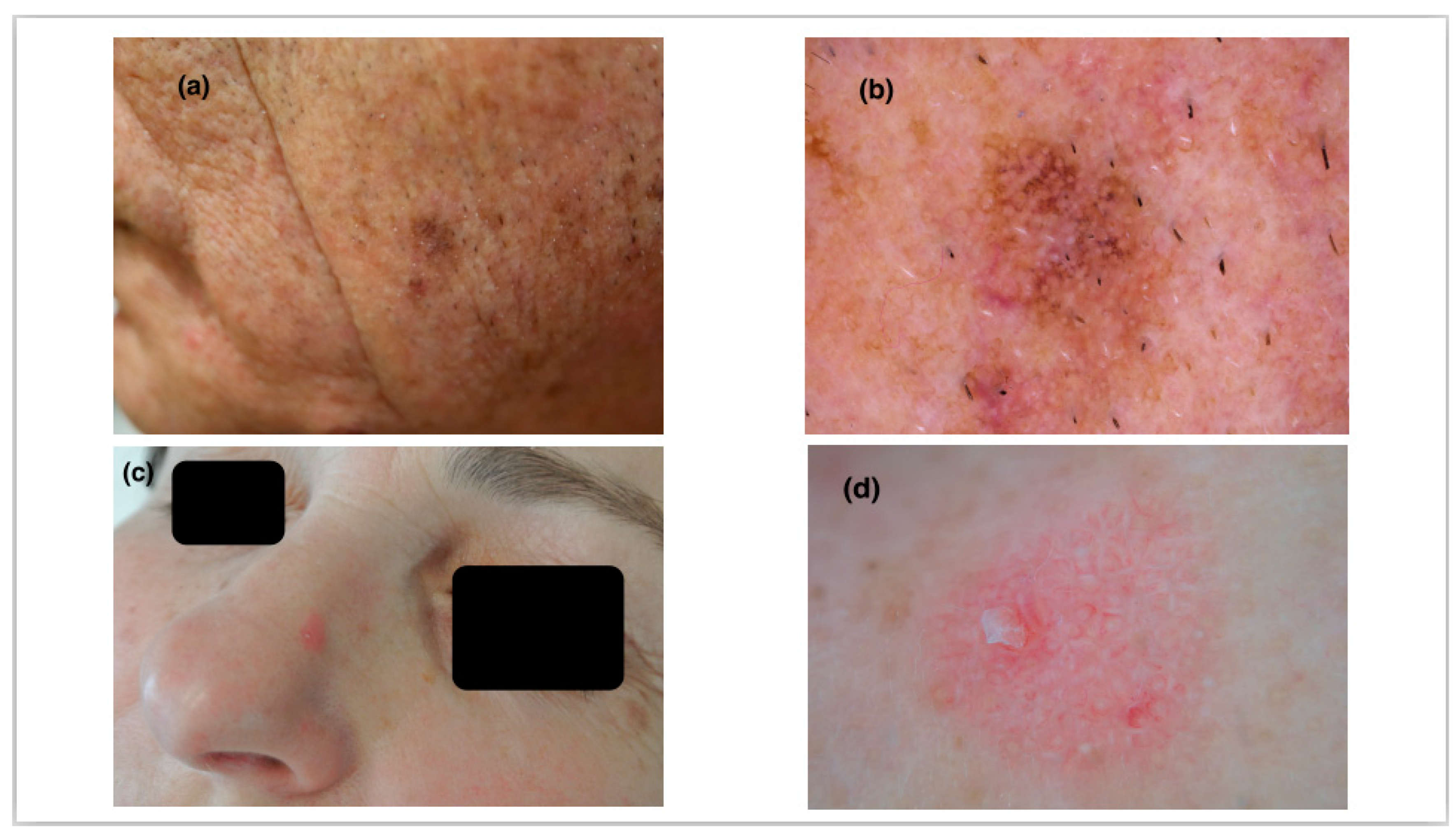
Dermoscopy of Actinic Keratosis: Is There a True Differentiation between Non-Pigmented and Pigmented Lesions?
Abstract
:1. Introduction
2. Methods
2.1. Study Population
2.2. Clinical and Dermoscopic Examinations
2.3. Statistical Analysis
3. Results
3.1. Demographic and Clinical Features of the Study’s Population
3.2. Dermoscopic Features of NPAK and PAK Lesions
3.3. Diagnostic Significance of Dermoscopic Structures
4. Discussion
5. Conclusions
Author Contributions
Funding
Institutional Review Board Statement
Informed Consent Statement
Conflicts of Interest
References
- Reinehr, C.P.H.; Bakos, R.M. Actinic keratoses: Review of clinical, dermoscopic, and therapeutic aspects. An. Bras. Dermatol. 2019, 94, 637–657. [Google Scholar] [CrossRef] [PubMed]
- Casari, A.; Chester, J.; Pellacani, G. Actinic Keratosis and Non-Invasive Diagnostic Techniques: An Update. Biomedicines 2018, 6, 8. [Google Scholar] [CrossRef] [Green Version]
- Glogau, R.G. The risk of progression to invasive disease. J. Am. Acad. Dermatol. 2000, 42, 23–24. [Google Scholar] [CrossRef] [PubMed]
- Marks, R.; Rennie, G.; Selwood, T.S. Malignant transformation of solar keratoses to squamous cell carcinoma. Lancet 1988, 1, 795–797. [Google Scholar] [CrossRef]
- Sgouros, D.; Milia-Argyti, A.; Arvanitis, D.K.; Polychronaki, E.; Kousta, F.; Panagiotopoulos, A.; Theotokoglou, S.; Syrmali, A.; Theodoropoulos, K.; Stratigos, A.; et al. Actinic Keratoses (AK): An Exploratory Questionnaire-Based Study of Patients’ Illness Perceptions. Curr. Oncol. 2022, 29, 5150–5163. [Google Scholar] [CrossRef] [PubMed]
- Sgouros, D.; Theofili, M.; Damaskou, V.; Theotokoglou, S.; Theodoropoulos, K.; Stratigos, A.; Theofilis, P.; Panayiotides, I.; Rigopoulos, D.; Katoulis, A. Dermoscopy as a Tool in Differentiating Cutaneous Squamous Cell Carcinoma from Its Variants. Dermatol. Pract. Concept. 2021, 11, e2021050. [Google Scholar] [CrossRef] [PubMed]
- Russo, T.; Piccolo, V.; Lallas, A.; Argenziano, G. Recent advances in dermoscopy. F1000Research 2016, 5, 184. [Google Scholar] [CrossRef] [PubMed]
- Lallas, A.; Argenziano, G.; Zendri, E.; Moscarella, E.; Longo, C.; Grenzi, L.; Pellacani, G.; Zalaudek, I. Update on non-melanoma skin cancer and the value of dermoscopy in its diagnosis and treatment monitoring. Expert Rev. Anticancer. Ther. 2013, 13, 541–558. [Google Scholar] [CrossRef] [PubMed]
- Zalaudek, I.; Giacomel, J.; Argenziano, G.; Hofmann-Wellenhof, R.; Micantonio, T.; Di Stefani, A.; Oliviero, M.; Rabinovitz, H.; Soyer, H.P.; Peris, K. Dermoscopy of facial nonpigmented actinic keratosis. Br. J. Dermatol. 2006, 155, 951–956. [Google Scholar] [CrossRef] [PubMed]
- Zalaudek, I.; Argenziano, G. Dermoscopy of actinic keratosis, intraepidermal carcinoma and squamous cell carcinoma. Curr. Probl. Dermatol. 2015, 46, 70–76. [Google Scholar] [CrossRef] [PubMed]
- Zalaudek, I.; Ferrara, G.; Leinweber, B.; Mercogliano, A.; D’Ambrosio, A.; Argenziano, G. Pitfalls in the clinical and dermoscopic diagnosis of pigmented actinic keratosis. J. Am. Acad. Dermatol. 2005, 53, 1071–1074. [Google Scholar] [CrossRef] [PubMed]
- Akay, B.N.; Kocyigit, P.; Heper, A.O.; Erdem, C. Dermatoscopy of flat pigmented facial lesions: Diagnostic challenge between pigmented actinic keratosis and lentigo maligna. Br. J. Dermatol. 2010, 163, 1212–1217. [Google Scholar] [CrossRef] [PubMed]
- Ertop Dogan, P.; Akay, B.N.; Okcu Heper, A.; Rosendahl, C.; Erdem, C. Dermatoscopic findings and dermatopathological correlates in clinical variants of actinic keratosis, Bowen’s disease, keratoacanthoma, and squamous cell carcinoma. Dermatol. Ther. 2021, 34, e14877. [Google Scholar] [CrossRef] [PubMed]
- Balagula, Y.; Braun, R.P.; Rabinovitz, H.S.; Dusza, S.W.; Scope, A.; Liebman, T.N.; Mordente, I.; Siamas, K.; Marghoob, A.A. The significance of crystalline/chrysalis structures in the diagnosis of melanocytic and nonmelanocytic lesions. J. Am. Acad. Dermatol. 2012, 67, 194.E1–194.E8. [Google Scholar] [CrossRef] [PubMed] [Green Version]
- Zalaudek, I.; Giacomel, J.; Schmid, K.; Bondino, S.; Rosendahl, C.; Cavicchini, S.; Tourlaki, A.; Gasparini, S.; Bourne, P.; Keir, J.; et al. Dermatoscopy of facial actinic keratosis, intraepidermal carcinoma, and invasive squamous cell carcinoma: A progression model. J. Am. Acad. Dermatol. 2012, 66, 589–597. [Google Scholar] [CrossRef] [PubMed]
- Olsen, E.A.; Abernethy, M.L.; Kulp-Shorten, C.; Callen, J.P.; Glazer, S.D.; Huntley, A.; McCray, M.; Monroe, A.B.; Tschen, E.; Wolf, J.E., Jr. A double-blind, vehicle-controlled study evaluating masoprocol cream in the treatment of actinic keratoses on the head and neck. J. Am. Acad. Dermatol. 1991, 24, 738–743. [Google Scholar] [CrossRef] [PubMed]
- Green, A.C. Epidemiology of actinic keratoses. Curr. Probl. Dermatol. 2015, 46, 1–7. [Google Scholar] [CrossRef] [PubMed]
- Chung, H.J.; McGuigan, K.L.; Osley, K.L.; Zendell, K.; Lee, J.B. Pigmented solar (actinic) keratosis: An underrecognized collision lesion. J. Am. Acad. Dermatol. 2013, 68, 647–653. [Google Scholar] [CrossRef] [PubMed]
- Kelati, A.; Baybay, H.; Moscarella, E.; Argenziano, G.; Gallouj, S.; Mernissi, F.Z. Dermoscopy of Pigmented Actinic Keratosis of the Face: A Study of 232 Cases. Actas Dermosifiliogr. 2017, 108, 844–851. [Google Scholar] [CrossRef] [PubMed]
- Labadie, J.G.; Compres, E.; Sunshine, J.C.; Alam, M.; Gerami, P.; Harikumar, V.; Poon, E.; Arndt, K.A.; Dover, J.S. Actinic Keratosis Color and Its Associations: A Retrospective Photographic, Dermoscopic, and Histologic Evaluation. Dermatol. Surg. 2022, 48, 57–60. [Google Scholar] [CrossRef] [PubMed]

| NPAK 45 (54.2%) | PAK 38 (45.8%) | p | |
|---|---|---|---|
| Age (mean) ± SD, y. | 78.7 ± 11.5 | 80.6 ± 7.9 | 0.41 |
| Sex (male) | 38 (84.4) | 31 (81.6) | 0.73 |
| Fitzpatrick phototype | |||
| I | 8 (17.8) | 2 (5.3) | 0.46 |
| II | 17 (37.8) | 15 (39.5) | |
| III | 13 (28.9) | 16 (42.1) | |
| IV | 7(15.5) | 5(13.1) | |
| Lesion location | |||
| Scalp | 18 (40) | 10 (26.3) | 0.19 |
| Forehead | 8 (17.8) | 2 (5.3) | |
| Temples | 7 (15.6) | 10 (26.3) | |
| Lateral face | 9 (20) | 11 (28.9) | |
| Nose | 3 (6.6) | 4 (10.6) | |
| Ear | 1 (2.6) | ||
| Olsen’s clinical stage | |||
| I | 16 (35.6) | 13 (34.2) | 0.74 |
| II | 18 (40) | 18 (47.4) | |
| III | 11 (24.4) | 7 (18.4) |
| NPAK 45 (54.2%) | PAK 38 (45.8%) | p | |
|---|---|---|---|
| Color | |||
| Red pseudo-network (%) | 38 (84.4) | 23 (60.5) | 0.01 |
| Pigmented pseudo-network (%) | 10 (22.2) | 34 (89.5) | <0.001 |
| Pigment intensity <10% | 9 (20) | 10 (26.3) | 0.001 |
| Pigment intensity 10–50% | 5 (11.1) | 19 (50) | <0.001 |
| Pigment intensity >50% | 0 (0) | 7 (18.4) | N/A |
| Scale | 38 (84.4) | 32 (84.2) | 0.98 |
| Follicular openings | |||
| Widened follicular openings | 35 (77.9) | 29 (76.3) | 0.87 |
| Yellowish dots | 28 (62.2) | 24 (63.2) | 0.93 |
| White circles | 28 (62.2) | 24 (63.2) | 0.93 |
| Rosettes | 14 (31.1) | 16 (42.1) | 0.3 |
| Linear wavy vessels | 30 (66.7) | 22 (57.9) | 0.41 |
| Shiny streaks | 17 (37.8%) | 11 (29%) | 0.4 |
| Red starburst | 4 (8.9%) | 8 (21%) | 0.13 |
| Se | Sp | PPV | NPV | |
|---|---|---|---|---|
| NPAK | ||||
| Red pseudo-network | 84% | 39% | 62% | 68% |
| PAK | ||||
| Pigmented pseudo-network | 89% | 77% | 77% | 89% |
| Pigment intensity <10% | 83% | 77% | 52% | 93% |
| Pigment intensity 10–50% | 90% | 86% | 79% | 93% |
| OR | 95% Cl | p | |
|---|---|---|---|
| NPAK vs. PAK | |||
| Red pseudo-network | 0.28 | 0.1–0.8 | 0.02 |
| Pigmentedpseudo-network | 29.75 | 8.51–104.03 | <0.001 |
| Pigment intensity vs. none | |||
| <10% | 17.22 | 3.18–93.32 | 0.001 |
| 10–50% | 58.9 | 10.37–334.37 | <0.001 |
| >50% | N/A |
Disclaimer/Publisher’s Note: The statements, opinions and data contained in all publications are solely those of the individual author(s) and contributor(s) and not of MDPI and/or the editor(s). MDPI and/or the editor(s) disclaim responsibility for any injury to people or property resulting from any ideas, methods, instructions or products referred to in the content. |
© 2023 by the authors. Licensee MDPI, Basel, Switzerland. This article is an open access article distributed under the terms and conditions of the Creative Commons Attribution (CC BY) license (https://creativecommons.org/licenses/by/4.0/).
Share and Cite
Sgouros, D.; Theofili, M.; Zafeiropoulou, T.; Lallas, A.; Apalla, Z.; Zaras, A.; Liopyris, K.; Pappa, G.; Polychronaki, E.; Kousta, F.; et al. Dermoscopy of Actinic Keratosis: Is There a True Differentiation between Non-Pigmented and Pigmented Lesions? J. Clin. Med. 2023, 12, 1063. https://doi.org/10.3390/jcm12031063
Sgouros D, Theofili M, Zafeiropoulou T, Lallas A, Apalla Z, Zaras A, Liopyris K, Pappa G, Polychronaki E, Kousta F, et al. Dermoscopy of Actinic Keratosis: Is There a True Differentiation between Non-Pigmented and Pigmented Lesions? Journal of Clinical Medicine. 2023; 12(3):1063. https://doi.org/10.3390/jcm12031063
Chicago/Turabian StyleSgouros, Dimitrios, Melpomeni Theofili, Theodora Zafeiropoulou, Aimilios Lallas, Zoe Apalla, Alexios Zaras, Konstantinos Liopyris, Georgia Pappa, Eleni Polychronaki, Fiori Kousta, and et al. 2023. "Dermoscopy of Actinic Keratosis: Is There a True Differentiation between Non-Pigmented and Pigmented Lesions?" Journal of Clinical Medicine 12, no. 3: 1063. https://doi.org/10.3390/jcm12031063
APA StyleSgouros, D., Theofili, M., Zafeiropoulou, T., Lallas, A., Apalla, Z., Zaras, A., Liopyris, K., Pappa, G., Polychronaki, E., Kousta, F., Panagiotopoulos, A., Stratigos, A., Rigopoulos, D., & Katoulis, A. C. (2023). Dermoscopy of Actinic Keratosis: Is There a True Differentiation between Non-Pigmented and Pigmented Lesions? Journal of Clinical Medicine, 12(3), 1063. https://doi.org/10.3390/jcm12031063


