Preoperative and Intraoperative Localization of Small Pulmonary Nodules for Sublobar Resection: Practical Insights into Percutaneous, Bronchoscopic/Robotic, RFID (SuReFInD), and Hybrid-OR CT Workflows

 , , , , ,
Simple Summary
Abstract
1. Introduction
2. Materials and Methods
2.1. Information Sources and Search Strategy
2.2. Eligibility Criteria
2.3. Study Selection and Prioritization
2.4. Data Extraction, Operational Definitions, and Synthesis
2.5. Evidence Limitations
2.6. Historical Evolution and Comparative Taxonomy
3. Preoperative Localization Techniques
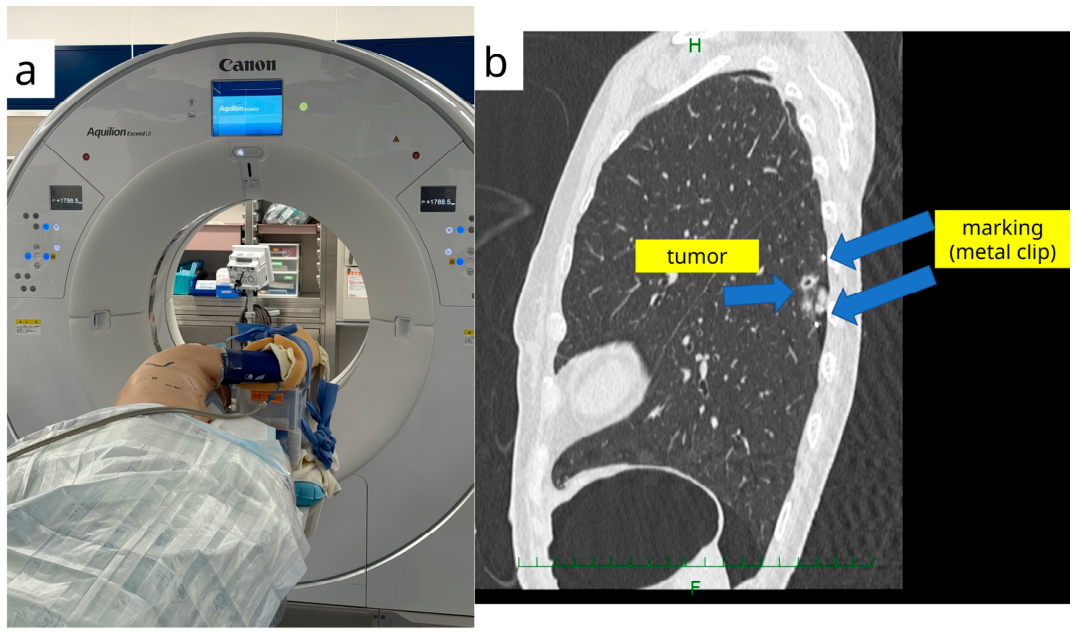
3.1. CT-Guided Percutaneous Marking (Hook-Wire and Related Methods)
3.2. Bronchoscopic Dye Marking and Virtual-Assisted Lung Mapping
3.3. Robotic-Assisted Bronchoscopic Localization
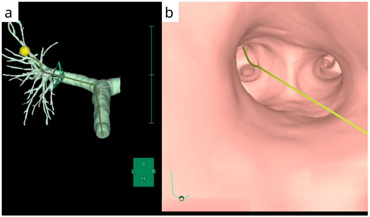
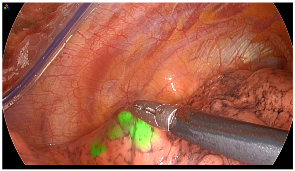
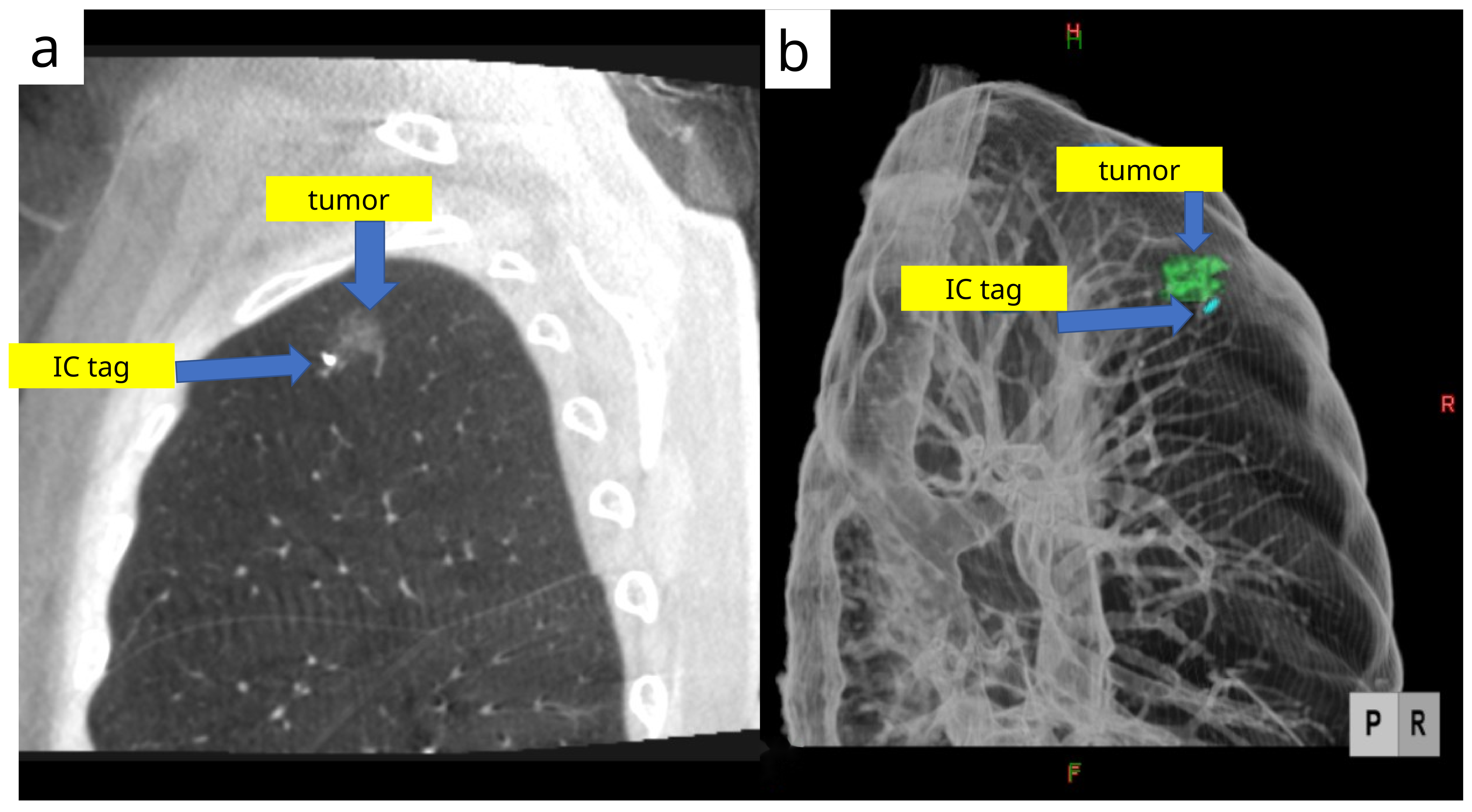
3.4. Bronchoscopic Depth-Aware RFID Tag Localization
4. Intraoperative Localization in Hybrid Operating Rooms
5. Comparative Synthesis and Practical Guidance
5.1. Reported Outcomes and Complications
5.2. Indications: When to Consider Each Technique
5.3. Pitfalls and Troubleshooting
5.4. Cost Structure, Reimbursement, and Operational Burden
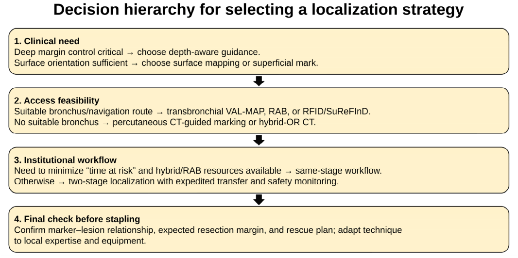
5.5. Proposed Decision Algorithm
6. Future Directions
6.1. Standardized Reporting and Comparative Evidence
6.2. Standardization of Current Robotic Bronchoscopic and Image-Guided Workflows
6.3. Multimodal and Depth-Aware Localization for Margin-Directed Surgery
6.4. Radiation Stewardship and Safety Engineering
6.5. Implementation Science: Manpower, Training, and Cost-Effectiveness
6.6. Toward Personalized Decision Support
7. Conclusions
Author Contributions
Funding
Data Availability Statement
Acknowledgments
Conflicts of Interest
Abbreviations
| CBCT | cone-beam computed tomography |
| CT | computed tomography |
| GA | general anesthesia |
| GGO | ground-glass opacity |
| HRCT | high-resolution computed tomography |
| ICG | indocyanine green |
| IOCT | intraoperative computed tomography |
| IR | interventional radiology/radiologist |
| IQR | interquartile range |
| MDCT | multidetector computed tomography |
| NIR | near-infrared |
| NSCLC | non-small cell lung cancer |
| OR | operating room |
| POCT | preoperative computed tomography |
| RAB | robotic-assisted bronchoscopy |
| RATS | robotic-assisted thoracoscopic surgery |
| RFID | radiofrequency identification |
| SuReFInD | Surgical Real-Time FInger Navigation and Detection |
| VAL-MAP | virtual-assisted lung mapping |
| VATS | video-assisted thoracoscopic surgery |
| VBN | virtual bronchoscopic navigation |
References
- Fischbach, F.; Knollmann, F.; Griesshaber, V.; Freund, T.; Akkol, E.; Felix, R. Detection of pulmonary nodules by multislice computed tomography: Improved detection rate with reduced slice thickness. Eur. Radiol. 2003, 13, 2378–2383. [Google Scholar] [CrossRef] [Scilit]
- Ginsberg, R.J.; Rubinstein, L.V. Randomized trial of lobectomy versus limited resection for T1 N0 non-small cell lung cancer. Ann. Thorac. Surg. 1995, 60, 615–622. [Google Scholar] [CrossRef] [Scilit]
- Suzuki, K.; Watanabe, S.I.; Wakabayashi, M.; Saji, H.; Aokage, K.; Moriya, Y.; Yoshino, I.; Tsuboi, M.; Nakamura, S.; Nakamura, K.; et al. A single-arm study of sublobar resection for ground-glass opacity dominant peripheral lung cancer. J. Thorac. Cardiovasc. Surg. 2022, 163, 289–301. [Google Scholar] [CrossRef] [Scilit]
- Aokage, K.; Suzuki, K.; Saji, H.; Wakabayashi, M.; Kataoka, T.; Sekino, Y.; Fukuda, H.; Endo, M.; Hattori, A.; Mimae, T.; et al. Segmentectomy for ground-glass-dominant lung cancer with a tumour diameter of 3 cm or less including ground-glass opacity (JCOG1211). Lancet Respir. Med. 2023, 11, 540–549. [Google Scholar] [CrossRef] [Scilit] [PubMed]
- Suzuki, K.; Nagai, K.; Yoshida, J.; Ohmatsu, H.; Takahashi, K.; Nishimura, M.; Nishiwaki, Y. Video-assisted thoracoscopic surgery for small indeterminate pulmonary nodules; Indications for preoperative marking. Chest 1999, 115, 563–568. [Google Scholar] [CrossRef] [Scilit] [PubMed]
- Vanstraelen, S.; Rocco, G.; Park, B.J.; Jones, D.R. The necessity of preoperative planning and nodule localization for small pulmonary nodules. JTCVS Open 2024, 18, 347. [Google Scholar] [CrossRef] [Scilit] [PubMed]
- Frank, H.A.; Hall, F.M.; Steer, M.L. Preoperative localization of nonpalpable breast lesions demonstrated by mammography. N. Engl. J. Med. 1976, 295, 259–260. [Google Scholar] [CrossRef] [Scilit]
- Lee, J.W.; Park, C.H.; Lee, S.M.; Jeong, M.; Hur, J. Planting seeds into the lung: Image-guided percutaneous localization to guide minimally invasive thoracic surgery. Korean J. Radiol. 2019, 20, 1498–1514. [Google Scholar] [CrossRef] [Scilit]
- Yi, J.H.; Choi, P.J.; Bang, J.H.; Jeong, S.S.; Cho, J.H. Systemic air embolism after computed tomography-guided hook wire localization: Two case reports and literature review. J. Thorac. Dis. 2018, 10, E59–E64. [Google Scholar] [CrossRef] [Scilit]
- Iguchi, T.; Yoshioka, T.; Muro, M.; Miyasho, K.; Inoue, D.; Hiraki, T.; Kanazawa, S. Systemic air embolism during preoperative pulmonary marking with a short hook wire and suture system under CT fluoroscopy guidance. Jpn. J. Radiol. 2009, 27, 385–388. [Google Scholar] [CrossRef] [Scilit]
- Ichinose, J.; Kohno, T.; Fujimori, S.; Harano, T.; Suzuki, S. Efficacy and complications of computed tomography-guided hook wire localization. Ann. Thorac. Surg. 2013, 96, 1203–1208. [Google Scholar] [CrossRef] [Scilit]
- Iguchi, T.; Hiraki, T.; Gobara, H.; Fujiwara, H.; Matsui, Y.; Miyoshi, S.; Kanazawa, S. CT fluoroscopy-guided preoperative short hook wire placement for small pulmonary lesions. Eur. Radiol. 2016, 26, 114–121. [Google Scholar] [CrossRef] [Scilit]
- Lee, J.H.; Yoon, S.H.; Hong, H.; Rho, J.Y.; Goo, J.M. Incidence, risk factors, and prognostic indicators of symptomatic air embolism after percutaneous transthoracic lung biopsy: A systematic review and pooled analysis. Eur. Radiol. 2021, 31, 2022–2033. [Google Scholar] [CrossRef] [Scilit] [PubMed]
- Fan, H.; Guo, C.; Zou, B. A hook wire dislodged into the subglottic area as a rare complication following computed tomography-guided hook wire localization: A case report. BMC Pulm. Med. 2022, 22, 317. [Google Scholar] [CrossRef] [Scilit] [PubMed]
- Sakiyama, S.; Kondo, K.; Matsuoka, H.; Yoshida, M.; Miyoshi, T.; Yoshida, S.; Monden, Y. Fatal air embolism during computed tomography-guided pulmonary marking with a hook-type marker. J. Thorac. Cardiovasc. Surg. 2003, 126, 1207–1209. [Google Scholar] [CrossRef] [Scilit]
- Glodny, B.; Schönherr, E.; Freund, M.C.; Haslauer, M.; Petersen, J.; Loizides, A.; Grams, A.E.; Augustin, F.; Wiedermann, F.J.; Rehwald, R. Measures to prevent air embolism in transthoracic biopsy of the lung. AJR Am. J. Roentgenol. 2017, 208, W184–W191. [Google Scholar] [CrossRef] [Scilit] [PubMed]
- Hare, S.S.; Gupta, A.; Goncalves, A.T.C.; Souza, C.A.; Matzinger, F.; Seely, J.M. Systemic arterial air embolism after percutaneous lung biopsy. Clin. Radiol. 2011, 66, 589–596. [Google Scholar] [CrossRef] [Scilit]
- Pietersen, P.I.; Kristjansdottir, B.; Laursen, C.B.; Jørgensen, G.M.; Graumann, O. Systemic air embolism following computed tomography-guided transthoracic needle biopsy of lung lesion—A systematic search of case reports and case series. Acta Radiol. Open 2022, 11, 20584601221096680. [Google Scholar] [CrossRef] [Scilit]
- Ramaswamy, R.; Narsinh, K.H.; Tuan, A.; Kinney, T.B. Systemic air embolism following percutaneous lung biopsy. Semin. Interv. Radiol. 2014, 31, 375–377. [Google Scholar] [CrossRef] [Scilit]
- Park, J.B.; Lee, S.A.; Lee, W.S.; Kim, Y.H.; Song, I.; Lee, J.G.; Hwang, J.J. Computed tomography-guided percutaneous hook wire localization of pulmonary nodular lesions before video-assisted thoracoscopic surgery: Highlighting technical aspects. Ann. Thorac. Med. 2019, 14, 205–212. [Google Scholar] [CrossRef] [Scilit]
- Park, C.H.; Han, K.; Hur, J.; Lee, S.M.; Lee, J.W.; Hwang, J.; Seo, J.S.; Lee, K.H.; Kwon, W.; Kim, T.H.; et al. Comparative effectiveness and safety of preoperative lung localization for pulmonary nodules: A systematic review and meta-analysis. Chest 2017, 151, 316–328. [Google Scholar] [CrossRef] [Scilit]
- Krimsky, W.S.; Minnich, D.J.; Cattaneo, S.M.; Sarkar, S.A.; Harley, D.P.; Finley, D.J.; Browning, R.F.; Parrish, S.C. Thoracoscopic detection of occult indeterminate pulmonary nodules using bronchoscopic pleural dye marking. J. Community Hosp. Intern. Med. Perspect. 2014, 4, 23084. [Google Scholar] [CrossRef] [Scilit]
- Kobayashi, T.; Kaneko, M.; Kondo, H.; Nakayama, H.; Asamura, H.; Tsuchiya, R.; Naruke, T.; Kakizoe, T. CT-guided bronchoscopic barium marking for resection of a fluoroscopically invisible peripheral pulmonary lesion. Jpn. J. Clin. Oncol. 1997, 27, 204–205. [Google Scholar] [CrossRef] [Scilit] [PubMed]
- Lee, N.-H.; Chung, H.-S.; Cho, J.-S.; Kim, Y.-D.; Eom, J.-S.; Ahn, H.-Y. Localization technique using mixture of indigo carmine and lipiodol of pulmonary nodule via bronchoscopic navigation. Medicina 2022, 58, 1235. [Google Scholar] [CrossRef] [Scilit]
- Toba, H.; Kondo, K.; Miyoshi, T.; Kajiura, K.; Yoshida, M.; Kawakami, Y.; Takizawa, H.; Kenzaki, K.; Sakiyama, S.; Tangoku, A. Fluoroscopy-assisted thoracoscopic resection after CT-guided bronchoscopic metallic coil marking. Eur. J. Cardiothorac. Surg. 2013, 44, e126–e132. [Google Scholar] [CrossRef] [Scilit]
- Asano, F.; Shindoh, J.; Shigemitsu, K.; Miya, K.; Abe, T.; Horiba, M.; Ishihara, Y. Ultrathin bronchoscopic barium marking with virtual bronchoscopic navigation for fluoroscopy-assisted thoracoscopic surgery. Chest 2004, 126, 1687–1693. [Google Scholar] [CrossRef] [Scilit] [PubMed]
- Sato, M.; Yamada, T.; Menju, T.; Aoyama, A.; Sato, T.; Chen, F.; Sonobe, M.; Omasa, M.; Date, H. Virtual-assisted lung mapping: Outcome of 100 consecutive cases. Eur. J. Cardiothorac. Surg. 2015, 47, e131–e139. [Google Scholar] [CrossRef] [Scilit] [PubMed]
- Nagano, M.; Sato, M. Ten-year outcome and development of virtual-assisted lung mapping in thoracic surgery. Cancers 2023, 15, 1971. [Google Scholar] [CrossRef] [Scilit]
- Kuwata, T.; Shinohara, S.; Matsumiya, H.; Takenaka, M.; Oka, S.; Chikaishi, Y.; Hirai, A.; Imanishi, N.; Kuroda, K.; Tanaka, F. Virtual-assisted lung mapping (VAL-MAP) shortened surgical time of wedge resection for small pulmonary nodules. J. Thorac. Dis. 2018, 10, 1842–1849. [Google Scholar] [CrossRef] [Scilit]
- Chen-Yoshikawa, T.F.; Nakamura, S.; Ueno, H.; Kadomatsu, Y.; Kato, T.; Mizuno, T. Current status and future perspectives of preoperative and intraoperative marking in thoracic surgery. Cancers 2024, 16, 3284. [Google Scholar] [CrossRef] [Scilit]
- Yanagiya, M.; Sato, M.; Ijiri, N.; Kobayashi, K.; Nagano, M.; Konoeda, C.; Kitano, K.; Nakajima, J. Virtual-assisted lung mapping using dual staining with indocyanine green and indigo carmine enhanced marking detectability. J. Thorac. Dis. 2022, 14, 1061–1069. [Google Scholar] [CrossRef] [Scilit]
- Sato, M.; Kuwata, T.; Yamanashi, K.; Kitamura, A.; Misawa, K.; Imashimizu, K.; Kobayashi, M.; Ikeda, M.; Koike, T.; Kosaka, S.; et al. Safety and reproducibility of virtual-assisted lung mapping: A multicentre study in Japan. Eur. J. Cardiothorac. Surg. 2017, 51, 861–868. [Google Scholar] [CrossRef] [Scilit]
- Kojima, F.; Sato, T.; Takahata, H.; Okada, M.; Sugiura, T.; Oshiro, O.; Date, H.; Nakamura, T. A novel surgical marking system for small peripheral lung nodules based on radiofrequency identification technology: Feasibility study in a canine model. J. Thorac. Cardiovasc. Surg. 2014, 147, 1384–1389. [Google Scholar] [CrossRef] [Scilit] [PubMed]
- Sato, T.; Yutaka, Y.; Nakamura, T.; Date, H. First clinical application of radiofrequency identification (RFID) marking system—Precise localization of a small lung nodule. JTCVS Tech. 2020, 4, 301–304. [Google Scholar] [CrossRef] [Scilit]
- Miyahara, S.; Waseda, R.; Ueda, Y.; Yutaka, Y.; Date, H.; Suzuki, J.; Oizumi, H.; Goto, M.; Nakagawa, T.; Kojima, F.; et al. Evaluation of the radiofrequency identification lung marking system: A multicenter study in Japan. Surg. Endosc. 2023, 37, 3619–3626. [Google Scholar] [CrossRef] [Scilit]
- Fang, H.Y.; Chang, K.W.; Chao, Y.K. Hybrid operating room for the intraoperative CT-guided localization of pulmonary nodules. Ann. Transl. Med. 2019, 7, 34. [Google Scholar] [CrossRef] [Scilit]
- Chao, Y.-K.; Pan, K.-T.; Wen, C.-T.; Fang, H.-Y.; Hsieh, M.-J. A comparison of efficacy and safety of preoperative versus intraoperative computed tomography-guided thoracoscopic lung resection. J. Thorac. Cardiovasc. Surg. 2018, 156, 1974–1983.e1. [Google Scholar] [CrossRef] [Scilit] [PubMed]
- Simoff, M.J.; Pritchett, M.A.; Reisenauer, J.S.; Ost, D.E.; Majid, A.; Keyes, C.; Casal, R.F.; Parikh, M.S.; Diaz-Mendoza, J.; Fernandez-Bussy, S.; et al. Shape-sensing robotic-assisted bronchoscopy for pulmonary nodules: Initial multicenter experience using the Ion™ Endoluminal System. BMC Pulm. Med. 2021, 21, 322. [Google Scholar] [CrossRef] [Scilit]
- Chan, J.W.Y.; Chang, A.T.C.; Yu, P.S.Y.; Lau, R.W.H.; Ng, C.S.H. Robotic assisted-bronchoscopy with cone-beam CT ICG dye marking for lung nodule localization: Experience beyond USA. Front. Surg. 2022, 9, 943531. [Google Scholar] [CrossRef] [Scilit]
- Yu, Q.; Yang, H.; Chen, J.; Kong, F.; Zhang, J.; Tian, Z.; Zhang, Z.; Liang, C. Robotic-assisted bronchoscopic localization for small pulmonary nodules: A novel approach to minimally invasive surgery. Front. Surg. 2025, 12, 1641868. [Google Scholar] [CrossRef] [Scilit] [PubMed]
- Tamura, M.; Nakano, S.; Sugahara, T. Reimbursement pricing for new medical devices in Japan: Is the evaluation of innovation appropriate? Int. J. Health Plan. Manag. 2019, 34, 583–593. [Google Scholar] [CrossRef] [Scilit] [PubMed]
- Ministry of Health, Labour and Welfare (MHLW). Guidebook for Insurance Coverage of Medical Devices. 2024. Available online: https://www.mhlw.go.jp/content/12400000/001314594.pdf (accessed on 18 February 2026).
- Health and Global Policy Institute (HGPI). Japan Health Policy Now; HGPI: Tokyo, Japan, 2019; Available online: https://japanhpn.org/wp-content/uploads/2023/06/JHPN_ENG.pdf (accessed on 18 February 2026).
- Yutaka, Y.; Sato, T.; Matsushita, K.; Aiba, H.; Muranishi, Y.; Sakaguchi, Y.; Sugiura, T.; Okada, M.; Nakamura, T.; Date, H. Three-dimensional navigation for thoracoscopic sublobar resection with RFID marking. J. Thorac. Cardiovasc. Surg. 2018, 30, 230–237. [Google Scholar]
- Yutaka, Y.; Sato, T.; Tanaka, S.; Miyahara, S.; Yoshizawa, A.; Morita, S.; Date, H. Feasibility study of a novel wireless localization technique using RFID markers for small and deeply located lung lesions. JTCVS Tech. 2022, 12, 185–195. [Google Scholar] [CrossRef] [Scilit] [PubMed]
- Komatsu, M.; Miura, K.; Yamanaka, M.; Suzuki, Y.; Araki, T.; Goto, N.; Akahane, J.; Sonehara, K.; Matsuoka, S.; Eguchi, T.; et al. Evaluation of radiofrequency identification tag accuracy using bronchoscopy with fluoroscopy and virtual navigation guidance before segmentectomy. Surg. Endosc. 2024, 38, 5438–5445. [Google Scholar] [CrossRef] [Scilit] [PubMed Central]
- Yoshimatsu, K.; Takenaka, M.; Hashimoto, T.; Tanaka, K.; Mori, M.; Kanayama, M.; Taira, A.; Kuroda, K.; Tanaka, F. Efficacy of RFID lung marking system in revision surgery: A case report. Video-Assist. Thorac. Surg. 2024, 9, 11. [Google Scholar] [CrossRef] [Scilit]
- Saul, B.; Ketelaar, E.; Yaish, H.; Wagner, M.; Comrie, R.; Brannan, G. Assessing Root Causes of First Case On-time Start (FCOTS) Delay in the Orthopedic Department at a Busy Level II Community Teaching Hospital. Spartan Med. Res. J. 2022, 7, 36719. [Google Scholar] [CrossRef] [Scilit]
- Patel, S.; Lindenberg, M.; Rovers, M.M.; van Harten, W.H.; Ruers, T.J.; Poot, L.; Retel, V.P.; Grutters, J.P. Understanding the costs of surgery: A bottom-up cost analysis of both a hybrid operating room and conventional operating room. Int. J. Health Policy Manag. 2022, 11, 299. [Google Scholar] [CrossRef] [Scilit] [PubMed]
- Clark, M.; Horton, J. Hybrid Operating Room Suites: CADTH Horizon Scan; Report No.: EH0121; Canadian Agency for Drugs and Technologies in Health: Ottawa, ON, Canada, 2023; Available online: https://www.ncbi.nlm.nih.gov/books/NBK595334/ (accessed on 18 February 2026).



| Aspect | Percutaneous CT-Guided (Hook-Wire/Microcoil/Dye) | Bronchoscopic Surface Mapping (VAL-MAP/ICG-VAL-MAP/RAB Dye) | Bronchoscopic Depth-Aware Tagging (RFID/SuReFInD/Fiducial) | Single-Stage Intraoperative CT (Hybrid OR/CBCT) |
|---|---|---|---|---|
| Access/workflow | Transthoracic; usually 2-stage; TAR driven by CT-to-OR transfer/scheduling | Transbronchial; usually 2-stage + post-map CT; RAB/hybrid may be same-anesthetic | Transbronchial implanted marker; 2-stage or hybrid; TAR depends on confirmation | Intraoperative image-guided; same-stage; no separate preop marking |
| Main guidance signal | Surface point/trajectory; indirect depth from needle/wire path | Multiple pleural marks; guides cut line and surface orientation | Real-time 3D proximity/depth signal to tag; supports margin control | Immediate marker-lesion imaging confirmation |
| Anesthesia/team | Local marking; IR, CT tech, nurse, transfer; GA for resection | Sedation/local bronchoscopy; bronchoscopist + navigation/CT reconstruction; GA for resection | Sedation or GA/hybrid; bronchoscopist + navigation/fluoro + RFID/probe; OR team | GA throughout; hybrid-OR imaging, anesthesia, surgeons, radiation safety |
| Strengths | Simple, widely available, low consumable cost; useful if no bronchus | Avoids pleural puncture/SAE; multiple marks aid margins | Depth-aware; less dependent on pleural color; useful for deep GGO/adhesions/revision | No transfer; immediate confirmation; low TAR |
| Limitations | PTX/hemorrhage, rare SAE, dislodgement, radiation/transport burden | Surface-only; invisible marks in anthracosis/emphysema/central spray; post-map CT needed | Cost/training, retained tag risk, confirmation imaging, Japan-heavy evidence | Capital cost, longer GA/OR time, radiation management, limited availability |
| Best fit | Peripheral lesions; reliable same-day CT-to-OR workflow; limited endoscopic/hybrid resources | Subpleural/surface-oriented lesions; complex wedge/segmentectomy needing surface landmarks | Deep or GGO-dominant lesions needing margin control; revision/intermediate hilar-zone cases | No suitable bronchus; hybrid OR available; minimizing TAR prioritized |
| Pitfalls/rescue | Wire migration, PTX, transfer delay, SAE; expedite transfer, consider coil/dye | Invisible/misplaced mark; use ICG/post-map CT, avoid reliance on one mark | Tag >10 mm/wrong bronchus/dislodgement; confirm by CT, plan retrieval/redeployment | Positioning/ventilation shift, repeated scans/delays; confirm before stapling |
| Technique/Evidence Base | Targets + Endpoint | Outcome Range/Key Data | Complications/Time/Caveats |
|---|---|---|---|
| Percutaneous CT-guided hook-wire/microcoil/dye/ICG [8,9,10,11,12,13,14,15,16,17,18,19,20,21] Multi-region single-center series + reviews; most accessible | Small/deep peripheral nodules. Park 2019: Korea, single center; 113 lesions; 10.8 ± 6.1 mm; depth 20.2 ± 12.4 mm. Endpoint: localization/VATS completion or marker stability; marker-centroid distance inconsistently reported [20]. | Reviews: high VATS/localization success; hook-wire often mid-90%. Park: 96.5% success [20,21]. | PTX/hemorrhage variable; dislodgement uncommon; SAE rare/severe. Park: PTX 23.0%, hemorrhage 7.1%, dislodgement 3.5%, SAE 0.8%; localization 23.7 ± 6.3 min; CT-to-surgery 34.6 ± 19.9 min [20]. |
| VAL-MAP/ICG-VAL-MAP [27,28,29,30,31,32] Mainly Japanese single-/multicenter evidence; ICG improves visibility | Subpleural/surface-oriented lesions; cut-line design. Kuwata 2018: Japan, single center; median tumor 8.0 mm; depth 5.5 mm. Endpoint: mapping success, visible marks, post-map CT confirmation, planned resection/margin [29]. | Kuwata: 90.7% mapping success [29]. Long-term: ~10% dye marks invisible; ICG dual staining improves detectability [28,30,31]. | Severe events uncommon; minor PTX, pneumomediastinum/alveolar hemorrhage, invisible marks. Bronchoscopy example: 20 min (9–90); safety supported by Japanese multicenter data [29,32]. |
| Robotic-assisted bronchoscopic dye/fiducial [38,39,40] Current clinical platform; early platform-specific localization data | Peripheral small/deep/subsolid lesions reachable bronchoscopically. Chan: Hong Kong, 5 nodules; Yu 2025: China, 10 patients. Endpoint: navigation success, dye/fluorescence visibility, needle-lesion distance, specimen confirmation [39,40]. | Chan: 100% navigation, 80% ICG localization [39]. Yu: 10/10 localized; mean 16.9 min; central placement in specimens [40]. | No significant marking-related complications in small series. Limits: cost, CT-to-body divergence, need for confirmation imaging, limited comparative data [38,39,40]. |
| RFID/SuReFInD depth-aware tagging [33,34,35,44,45,46,47] Mainly Japanese feasibility, VBN/fluoro, and multicenter data | Deep or GGO-dominant lesions requiring margin control. Yutaka 2022: 11 lesions/12 markers; Komatsu 2024: 31 patients; Miyahara 2023: 182 patients. Endpoint: marker-target distance, within 10 mm placement, margin-negative/planned resection, surgeon utility [35,45,46]. | Within 10 mm: 58.3% (early hybrid feasibility) to 83.9% (fluoro+VBN). Cited series achieved negative/sufficient margins; multicenter data support safety and utility [35,45,46]. | No PTX/bleeding in cited RFID series; dislodgement 3.2% in Komatsu. Caveats: cost/training, retained tag management, outside-Japan generalizability [46]. |
| Intraoperative CT/hybrid OR [36,37] Single-stage image-guided workflow; comparative timing/resource data | Small/deep solitary nodules. Chao: conventional 2-stage POCT vs. IOCT-guided hybrid OR. Endpoint: immediate marker-lesion confirmation, planned resection, localization-to-incision TAR [37]. | No significant outcome differences; IOCT reduced TAR: 13.06 vs. 215.83 min [37]. | Transfer-related risk reduced, but GA time longer (163.1 vs. 120.61 min) and OR utilization higher (227.41 vs. 168.68 min); throughput/hybrid-OR availability decisive [37]. |
| Dimension | Two-Stage Percutaneous CT | Two-Stage Bronchoscopic Mapping/Tagging | Same-Anesthetic RAB | Single-Stage IOCT/Hybrid OR |
|---|---|---|---|---|
| Time at risk | Highest with CT-to-OR transfer/scheduling delay; expedite same-day pathway | Bronchoscopy-to-incision + post-map CT/3D reconstruction interval | Reduced if marking + resection in one GA; confirmation imaging adds setup | Lowest reported TAR: 13.06 vs. 215.83 min [37] |
| Anesthesia/OR | Marking under local; OR mainly resection | Sedation/local bronchoscopy; CT/reconstruction outside OR | Usually GA; robotic platform/team setup | Longer GA and OR utilization: 227.41 vs. 168.68 min [37] |
| Access-route risk | Pleural puncture: PTX, hemorrhage, rare SAE, dislodgement | Transbronchial: no pleural puncture; limited by airway reach/visibility | Transbronchial with better stability; CT-to-body divergence and cost remain | Intraoperative imaging: radiation/repeat scans, marker shift after lung handling |
| Cost drivers | CT suite + IR staff; transfer delays; PTX/SAE management | Bronchoscopy team; navigation; post-map CT/3D reconstruction; NIR if ICG | Robotic capital/maintenance; disposables; CBCT/fluoro; OR/GA time | Hybrid suite construction/depreciation; imaging technologist; radiation safety; GA/OR time |
| Implementation fit | Scalable where CT localization is routine; rapid transfer/emergency protocol needed | Needs bronchoscopic planning expertise; strongest VAL-MAP/RFID evidence in Japan | Best where RAB already established; localization evidence needs validation | Best with high hybrid-OR utilization and standardized team workflow |
Disclaimer/Publisher’s Note: The statements, opinions and data contained in all publications are solely those of the individual author(s) and contributor(s) and not of MDPI and/or the editor(s). MDPI and/or the editor(s) disclaim responsibility for any injury to people or property resulting from any ideas, methods, instructions or products referred to in the content. |
© 2026 by the authors. Licensee MDPI, Basel, Switzerland. This article is an open access article distributed under the terms and conditions of the Creative Commons Attribution (CC BY) license.
Share and Cite
Tanaka, K.; Takenaka, M.; Meguro, D.; Take, N.; Hashimoto, T.; Fujita, Y.; Manabe, T.; Yoshimatsu, K.; Matsumiya, H.; Mori, M.; et al. Preoperative and Intraoperative Localization of Small Pulmonary Nodules for Sublobar Resection: Practical Insights into Percutaneous, Bronchoscopic/Robotic, RFID (SuReFInD), and Hybrid-OR CT Workflows. Diseases 2026, 14, 195. https://doi.org/10.3390/diseases14060195
Tanaka K, Takenaka M, Meguro D, Take N, Hashimoto T, Fujita Y, Manabe T, Yoshimatsu K, Matsumiya H, Mori M, et al. Preoperative and Intraoperative Localization of Small Pulmonary Nodules for Sublobar Resection: Practical Insights into Percutaneous, Bronchoscopic/Robotic, RFID (SuReFInD), and Hybrid-OR CT Workflows. Diseases. 2026; 14(6):195. https://doi.org/10.3390/diseases14060195
Chicago/Turabian StyleTanaka, Kanji, Masaru Takenaka, Daikichi Meguro, Nobuyuki Take, Teppei Hashimoto, Yasuhiro Fujita, Takehiko Manabe, Katsuma Yoshimatsu, Hiroki Matsumiya, Masataka Mori, and et al. 2026. "Preoperative and Intraoperative Localization of Small Pulmonary Nodules for Sublobar Resection: Practical Insights into Percutaneous, Bronchoscopic/Robotic, RFID (SuReFInD), and Hybrid-OR CT Workflows" Diseases 14, no. 6: 195. https://doi.org/10.3390/diseases14060195
APA StyleTanaka, K., Takenaka, M., Meguro, D., Take, N., Hashimoto, T., Fujita, Y., Manabe, T., Yoshimatsu, K., Matsumiya, H., Mori, M., Nagata, A., & Uramoto, H. (2026). Preoperative and Intraoperative Localization of Small Pulmonary Nodules for Sublobar Resection: Practical Insights into Percutaneous, Bronchoscopic/Robotic, RFID (SuReFInD), and Hybrid-OR CT Workflows. Diseases, 14(6), 195. https://doi.org/10.3390/diseases14060195