Communication

Frequency of Non-Type I/II CALR Mutations in Patients Undergoing Molecular Diagnostics for Myeloproliferative Neoplasms


1
National Medical Research Center for Hematology, 125167 Moscow, Russia
2
Moscow Multidisciplinary Clinical Center “Kommunarka”, 108814 Moscow, Russia
*
Author to whom correspondence should be addressed.
Diseases 2026, 14(6), 190; https://doi.org/10.3390/diseases14060190
Submission received: 10 March 2026 / Revised: 20 May 2026 / Accepted: 27 May 2026 / Published: 28 May 2026

Abstract

Background/Objectives: Somatic CALR gene insertions/deletions in exon 9, causing frameshift, are a diagnostic sign of myeloproliferative neoplasms (MPNs). Besides the most common somatic mutations of type I (52 bp deletion) and type II (5 bp insertion), there are rare ones whose significance is not always clear. This study evaluates the frequency of rare mutations and demonstrates a germline rather than somatic nature for some of them. Methods: A retrospective analysis of 8417 blood samples subjected to molecular diagnosis of myeloproliferative neoplasm (MPN) was performed. Cases suspected as germline variants were sequenced, and paired samples (when available) of buccal epithelium were analyzed. Results: We have identified 632 CALR gene mutation-positive cases. Most of the cases were typical insertions/deletions (5 bp/52 bp). Non-type I/II frameshift or nonframeshift mutations were observed in 68 cases (11%). The buccal swab samples obtained from 4 patients confirmed the germline nature of these variants. It is worth noting that the MPN diagnosis for three of these patients was made considering the presence of the JAK2 V617F mutation (two cases) or BCR::ABL1 translocation (one case). In one case, the diagnosis of MPN was reclassified to CML. Conclusions: Non-type I/II CALR mutations, according to our data, could be found in 0.8% of MPN-suspected cases, and may not be associated with the diagnosis. The detection of a non-standard CALR mutation with an allelic frequency close to 50% should raise suspicion of the possibility of a germline CALR variant, and such cases should be investigated further.

1. Introduction

Calreticulin is a calcium-binding chaperone that performs several functions in the immune response. In the endoplasmic reticulum, calreticulin promotes the folding of molecules of the main histocompatibility complex class I and their assembly factor tapazine, thereby affecting the presentation of antigens to cytotoxic T cells [1,2]. In 2013, the discovery of somatic mutations in the CALR gene, encoding calreticulin, in 70–84% of JAK2 gene mutation-negative patients with essential thrombocythemia (ET) and primary myelofibrosis (PMF) significantly improved the molecular diagnosis of these diseases [3,4]. More than 50 somatic CALR mutations are known, which are insertions/deletions in exon 9 causing frameshift, which leads to the loss of the KDEL terminal signal [5]. The two most common mutations are type I, a 52 bp deletion (p. L367fs*46), and type II, a 5 bp TTGTC insertion (p. K385fs*47). The number of negatively charged amino acids in the C-terminal region of mutant proteins differs from that in wild-type calreticulin, depending on the mutation type. With a 5 bp insertion (type II), roughly half of the negatively charged amino acids are preserved, whereas 52 bp deletions (type I) eliminate nearly all such amino acids [3]. When mutant CALR interacts with the thrombopoietin MPL receptor, uncontrolled activation of the underlying JAK-STAT pathway and cellular transformation occur [6,7]. The value of CALR gene mutations as ET/PMF diagnostic criteria is defined by the WHO classification of myeloid neoplasms and acute leukemia in 2016 and 2022 [8,9,10]. Type I mutations are more commonly found in PMF. Type II is more commonly associated with ET. Type I mutations are associated with a more favorable prognosis, longer overall survival, and a lower risk of leukemic transformation compared to type II or JAK2-mutated PMF [11,12,13].
CALR gene mutations can be identified using several methods: capillary electrophoresis (also known as fragment analysis), real-time PCR, and next-generation sequencing (NGS). Both fragment analysis and NGS have uncovered numerous insertions and deletions in the CALR gene that went undetected with real-time PCR [14]. Earlier studies reported a low frequency of in-frame deletions and insertions in the CALR gene, which are assumed to preserve the terminal KDEL region of the CALR protein [15,16]. These in-frame variants often have an allelic frequency close to 50%, which may indicate a germline polymorphism. The importance of such variants for diagnosing MPNs is not yet clearly established.
A considerable number of publications discuss the investigation of CALR mutations in patient groups with a negative JAK2 V617F status and suspected MPNs. In this study, most Phi-negative patients suspected of having MPN were tested for JAK2 V617F, CALR, and MPL W515L/K mutations simultaneously. Furthermore, subsequently, we were able to find low BCR::ABL p210 expression in two patients with non-type I/II CALR gene deletion. Here, we outline the incidence of non-type I/II mutations in the CALR gene identified during diagnostic examinations of cases suspected to have classical MPN.

2. Materials and Methods

Somatic CALR gene mutations were evaluated for 8417 cases subjected to molecular diagnosis of MPN at the National Medical Research Center for Hematology (Moscow, Russia) from 2016 to 2026 and in a control group of 366 individuals without hematological diagnosis. All included cases were also tested for JAK2 V616F, MPL W515L/K and BCR::ABL1 chimeric transcript, essentially as described previously [17]. DNA and RNA were isolated from 5 to 10 mL of blood and bone marrow using standard salt extraction [18] and using a set of Ribosol-D reagents (Interlabservice, Moscow, Russia). DNA from buccal epithelium was isolated by direct cell lysis with proteinase K, followed by purification with magnetic particles (Pure blood DNA, Syntol, Moscow, Russia) in accordance with the manufacturer’s protocol. The mutation status of exon 9 of the CALR gene was evaluated using fragment length analysis after PCR with fluorescent primers in accordance with the method described by Klampfl et al. [3]. For the amplification of the target CALR gene sequence, primers CALR_AF1 5′-CTGAGGTGTGTGCTCTGCC-3′ and CALR_R FAM 5′-CAGAGACATTATTTGGCGCGG-3′ were used. DNA (200–600 ng) was added to 25 μL of reaction mixture (Syntol, Moscow, Russia), containing: 10 pmol of primers, H2O 14.5 μL, MgCl2 25 mM–1.5 μL, dNTP Mix, concentration of each nucleotide 25 mM–2.5 μL, PCR Buffer-B for Taq DNA polymerase–2.5 μL, Taq DNA polymerase 0.25 μL per reaction. Amplification was performed in a C1000 thermocycler (Bio-Rad, Hercules, CA, USA): denaturation at 95 °C for 10 min, 35 cycles (95 °C for 20 s, 60 °C for 40 s, 72 °C for 60 s), 72 °C for 10 min. After amplification, the PCR mixture was diluted 100 times, denatured at 98 °C for 3 min, and cooled at 4 °C for 5 min. Subsequently, fragment analysis was performed using a Nanofor-05 genetic analyzer (Institute of Analytical Instrumentation of the Russian Academy of Sciences, St. Petersburg, Russia). The profile of the obtained amplicons (length distribution) and the degree of fluorescence were analyzed using GeneMapper v.4 software (Applied Biosystems, Waltham, MA, USA). The allele load (VAF%) was calculated based on the height of the mutant and wild-type peaks, determining the proportion of mutant alleles relative to the total number of alleles (mutant + wild-type) in the sample. Precise evaluation of the non-type I/II CALR gene insertions/deletions was done by targeted NGS. For the amplification of the target CALR gene sequence, the same primers and PCR conditions as for fragment analysis were used. NGS libraries were prepared using the “ShotGun Plus Kit ‘SG GM Plus’” (Sesana, Moscow, Russia). Sequencing was performed on a MiSeq genetic analyzer (Illumina, San Diego, CA, USA), bioinformatic analysis was performed using the open source software Trimmomatic (Version: 0.39), BWA (Version: 0.7.17-r1188), SAMtools (Version: 1.10 using htslib 1.20-30-g61b922b4), Vardict (Version: 1.8.2) and Annovar (Version: Date: 2020-06-07) [19,20,21,22,23]. The potential pathogenicity of the obtained variants was analyzed using the online databases Franklin by Genoox and SESHAT [24,25]. The JAK2 V617F mutation was quantified by real-time allele-specific PCR using the “Kit of reagents for detecting the V617F G/T mutation of the JAK2 gene” (Syntol, Russia) in accordance with the manufacturer’s instructions, with 0.2% detection sensitivity declared. Additionally, DNA from the UKE1 cell line was used as a positive control. MPL W515L/K mutations were evaluated by qualitative allele-specific real-time PCR using a “Set of reagents for determining the W515L/K mutation of the MPL gene” (Syntol, Moscow, Russia).

3. Results

A total of 8417 patients suspected of having MPN and 366 patients unrelated to MPN (control group) were tested for the presence of CALR gene variants. Demographic parameters of the patient sample are presented in Table 1.
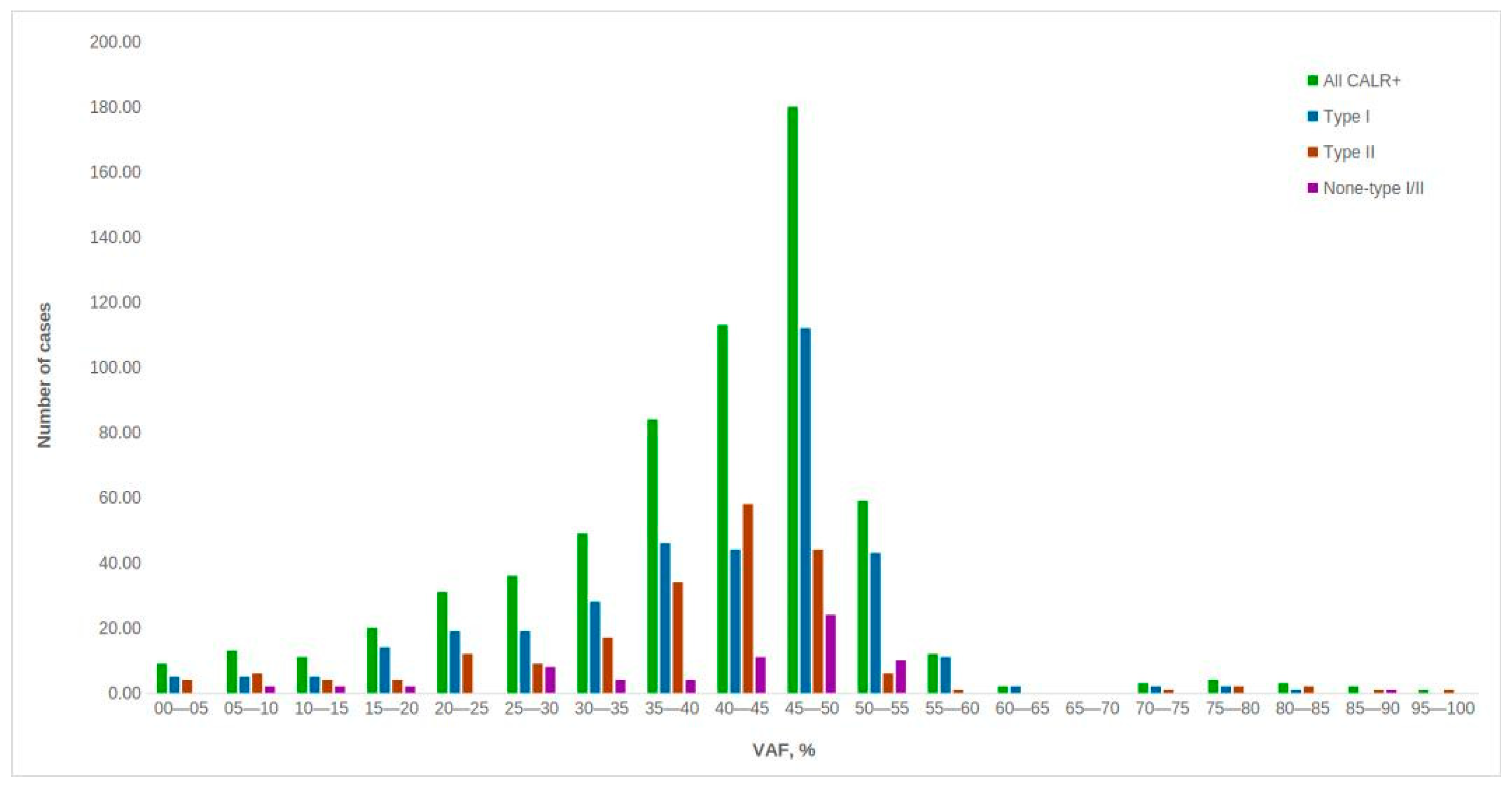
As a control group, we analyzed 366 samples of non-hematological patients tested for thrombophilia markers (described previously elsewhere [26]). No CALR gene variants (including germline) were found in this group. The distribution of CALR mutations by type and VAF for 632 positive patients is shown in Figure 1. Mutations of type I accounted for 57% (360), type II—33% (209), and non-type I/II ones—10% (63). It should be noted that most of the mutations show an allelic load of 40–55%. Our findings are roughly consistent with those reported previously [9,27].
The frequency of CALR type I mutations was higher than that of CALR type II mutations, which aligns with the previously published data [13]. However, the number of detected non-type I/II CALR gene mutations in our patient sample was higher than what had been previously reported [5].
Nine cases with non-type I/II CALR gene mutations of exon 9 were further analyzed. Five were in-frame deletions. In four cases, frameshift mutations, including deletions of 13 bp and 31 bp, were observed. Because the study was retrospective, we were restricted in our ability to collect samples of non-hematopoietic tissue for cases involving CALR gene mutations that might be considered germline. Only for four of these cases were we able to obtain buccal swab samples. In all these cases, the same variants were found in the cells of the oral mucosa and peripheral blood with the same VAFs, which confirms the germline nature of these lesions. Such cases can certainly be considered as “triple-negative” and subjected to additional JAK2 exon 12 sequencing. Clinical characteristics of patients with non-type I/II CALR mutations are presented in Table 2. The median VAF was 50% (range 45–52%). In six cases, an additional JAK2 V617F mutation was found, and two cases were BCR::ABL1 (p210) positive. It should be noted that in all these cases, the pathogenesis is linked specifically to additional mutations rather than CALR gene mutations. In the last two cases, the diagnosis of MPN was reclassified to chronic myeloid leukemia (CML).
All mutations were searched for in the Catalog of Somatic Mutations in Cancer (COSMIC) database and are labeled with the appropriate identity number if it existed at the time of search. The exact nomenclature for non-standard CALR mutations found in our patient cohort is presented in Table 3. Six mutations that were not found in the COSMIC database, to our knowledge, are newly identified mutations and therefore being reported for the first time. All four cases with buccal swab material available for evaluation appeared to be in-frame deletions.
As an example, we would like to give a description of a clinical case (patient No. 6 in Table 2 and Table 3). A 77-year-old patient with a diagnosis of MPN/MDS refractory anemia with excess blasts (RAEB-2) with pleural involvement was observed at the Moscow Multidisciplinary Clinical Center “Kommunarka”. Immunophenotype of blast cells: CD13, CD34, CD117, CD33, CD4, CD38, HLA-DR. Karyotype: 46,XY, del(11)(q14~22)[5]/46, idem, del(7)(q21). An immunohistochemical study revealed that granulocytes express MPO and CD15, megakaryocytes express CD31, and myeloblasts express CD117. Mutation V617F of the JAK2 gene and deletion of three nucleotides in the ninth exon of the CALR gene were detected in the DNA of peripheral blood cells (p.K368del c.1102_1104del). The patient received therapy with hydroxycarbamide, azacitidine (75 mg/m2 × 7 days), and three courses of small doses of cytarabine. Taking into account the JAK2-positivity, an attempt was made to treat with ruxolitinib (15 mg per day), but no significant effect was obtained. Taking into account grade 4 anemia, grade 4 thrombocytopenia, grade 4 neutropenia, an increase in blastosis (28%) and the lack of alternative antitumor therapy, the patient was assigned palliative status. The patient’s clinical data is presented in Table 4. According to NGS data, the allelic load of the JAK2 V617F mutation was 57%, and the CALR p.K368del mutation was 51%. A molecular genetic study of buccal epithelium and hair follicle samples from this patient showed that, unlike the JAK2 V617F mutation, the CALR p.K368del mutation is germline (Table 5). In the described case, ruxolitinib therapy was ineffective, which might indirectly suggest the importance of the CALR p.K368del mutation in the pathogenesis of MPN.

4. Discussion

The gold standard for routine determination of insertions/deletions in the CALR gene is fragment analysis. Most of the CALR tests required to confirm the diagnosis of MPN in the clinic are performed using this method. This method is not always precise enough to reliably distinguish frameshift insertion of 5 bp (Type II mutation) from, say, 3 or 6. It should also be noted that the allelic load of somatic mutations in the CALR gene can be close to 50% according to both our and the literature data. In our diagnostic practice, we have encountered cases in which germline polymorphisms (mostly in-frame indels) that may not directly relate to the diagnosis were misinterpreted as somatic mutations, thereby making a correct diagnosis challenging. The main purpose of this work was to assess the expected frequency of such cases in order to increase the alertness of doctors and diagnosticians and thus reduce the frequency of possible errors.
There are not enough data on non-type I/II mutations of the CALR gene and their significance for the development of MPN in the literature. A 9 bp deletion in the CALR gene in a patient with MPN has been reported, although its non-type I/II nature was not confirmed [9]. It can be assumed that non-type I/II mutations of the CALR gene can influence the acquisition of somatic mutations and/or interact with classical MPN driver mutations. In the study of Szuber N et al., certain CALR gene mutations have been confirmed as non-type I/II in a heterogeneous population of individuals with and without MPN. The authors concluded that germline CARL gene variants are not a diagnostic feature of MPN, unlike somatic ones, which are well described in the literature. Predisposition to MPN, related to germline CALR gene variants, can also not be concluded [15].
At the National Medical Research Center for Hematology, we have a long history of molecular genetic testing for MPNs. We have accumulated over 8417 records of CALR gene mutation tests. This amount of data allowed us to register rare atypical events. Non-type I/II frameshift or nonframeshift mutations were observed in 68 cases. We have found combined mutations in 8 patients (6 patients with CALR + JAK2 V617F mutations and 2 patients with CALR + BCR::ABL p210 mutations). No second MPN-associated mutation was detected in one patient. It should be noted that the diagnosis of MDS was established in this case. It can be assumed that a germline CALR variant without a frameshift may not have pathogenic significance. In other words, only if additional so-called “driver” mutations are present, the development of myeloproliferative neoplasms might be expected.

5. Conclusions

Thus, non-type I/II mutations in the CALR gene are rare and differ from the classical somatic mutations that determine MPN. The detection of CALR mutations of non-I or II types, with VAF close to 50% in MPN, is the basis for suspecting the germline variant and conducting a confirmatory test on a non-hematopoietic tissue sample. The potential role of germline CALR variants in the etiology of MPN requires further investigation. The detection of germline CALR gene variants should be interpreted with caution to avoid false-positive MPN diagnoses. Perhaps, cases with a non-type I/II CALR mutation of a VAF close to 50% found should be suggested for the search for other MPN molecular markers.

Author Contributions

Conceptualization, T.M. and A.S.; methodology, B.B.; investigation, T.M., E.N., S.T. and E.S.; resources, A.K.; data curation, T.M. and A.S.; resources, N.C.; writing—original draft preparation, T.M.; writing—review and editing, A.S.; supervision, A.K.; project administration, A.S. All authors have read and agreed to the published version of the manuscript.

Funding

This research received no external funding.

Institutional Review Board Statement

The study was conducted in accordance with the Declaration of Helsinki, and the protocol was approved by the Ethics Committee of the National Research Center for Hematology (Protocol # 192; date of approval 29 January 2026).

Informed Consent Statement

Informed consent was obtained from all subjects involved in the study.

Data Availability Statement

All the data provided within the article.

Acknowledgments

We appreciate helpful discussion and assistance from Irina Fevraleva.

Conflicts of Interest

The authors declare no conflicts of interest.

Abbreviations

The following abbreviations are used in this manuscript:
CALRCalreticulin gene
MPNsMyeloproliferative neoplasms
CMLChronic myeloid leukemia
ETEssential thrombocythemia
PMFPrimary myelofibrosis
VAFVariant allele frequency
JAK 2Janus kinase 2 gene
WHOWorld Health Organization
MPLMyeloproliferative leukemia virus oncogene
BCR::ABL1 BCR (Breakpoint Cluster Region) + ABL (Abelson murine leukemia viral oncogene homolog)
DNADeoxyribonucleic acid
PCRPolymerase chain reactions
COSMICCatalog of Somatic Mutations in Cancer

References

  1. Raghavan, M.; Wijeyesakere, S.J.; Peters, L.R.; Del Cid, N. Calreticulin in the immune system: Ins and outs. Trends Immunol. 2013, 34, 13–21. [Google Scholar] [CrossRef] [PubMed] [PubMed Central]
  2. Marty, C.; Pecquet, C.; Nivarthi, H.; El-Khoury, M.; Chachoua, I.; Tulliez, M.; Villeval, J.-L.; Raslova, H.; Kralovics, R.; Constantinescu, S.N.; et al. Calreticulin mutants in mice induce an MPL-dependent thrombocytosis with frequent progression to myelofibrosis. Blood 2016, 127, 1317–1324. [Google Scholar] [CrossRef]
  3. Klampfl, T.; Gisslinger, H.; Harutyunyan, A.S.; Nivarthi, H.; Rumi, E.; Milosevic, J.D.; Them, N.C.C.; Berg, T.; Gisslinger, B.; Pietra, D.; et al. Somatic mutations of calreticulin in myeloproliferative neoplasms. N. Engl. J. Med. 2013, 369, 2379–2390. [Google Scholar] [CrossRef]
  4. Nangalia, J.; Massie, C.E.; Baxter, E.J.; Nice, F.L.; Gundem, G.; Wedge, D.C.; Avezov, E.; Li, J.; Kollmann, K.; Kent, D.G.; et al. Somatic CALR mutations in myeloproliferative neoplasms with nonmutated JAK2. N. Engl. J. Med. 2013, 369, 2391–2405. [Google Scholar] [CrossRef]
  5. Belcic Mikic, T.; Pajic, T.; Sever, M. CALR mutations in a cohort of JAK2 V617F negative patients with suspected myeloproliferative neoplasms. Sci. Rep. 2019, 9, 19838. [Google Scholar] [CrossRef] [PubMed] [PubMed Central]
  6. Chachoua, I.; Pecquet, C.; El-Khoury, M.; Nivarthi, H.; Albu, R.-I.; Marty, C.; Gryshkova, V.; Defour, J.-P.; Vertenoeil, G.; Ngo, A.; et al. Thrombopoietin receptor activation by myeloproliferative neoplasm associated calreticulin mutants. Blood 2016, 127, 1325–1335. [Google Scholar] [CrossRef] [PubMed]
  7. Araki, M.; Yang, Y.; Masubuchi, N.; Hironaka, Y.; Takei, H.; Morishita, S.; Mizukami, Y.; Kan, S.; Shirane, S.; Edahiro, Y.; et al. Activation of the thrombopoietin receptor by mutant calreticulin in CALR-mutant myeloproliferative neoplasms. Blood 2016, 127, 1307–1316. [Google Scholar] [CrossRef]
  8. Arber, D.A.; Orazi, A.; Hasserjian, R.; Thiele, J.; Borowitz, M.J.; Le Beau, M.M.; Bloomfield, C.D.; Cazzola, M.; Vardiman, J.W. The 2016 revision to the World Health Organization classification of myeloid neoplasms and acute leukemia. Blood 2016, 127, 2391–2405. [Google Scholar] [CrossRef] [PubMed]
  9. Murugesan, G.; Guenther-Johnson, J.; Mularo, F.; Cook, J.R.; Daly, T.M. Validation of a molecular diagnostic assay for CALR exon 9 indels in myeloproliferative neoplasms: Identification of coexisting JAK2 and CALR mutations and a novel 9 bp deletion in CALR. Int. J. Lab Hematol. 2016, 38, 284–297. [Google Scholar] [CrossRef] [PubMed]
  10. Khoury, J.D.; Solary, E.; Abla, O.; Akkari, Y.; Alaggio, R.; Apperley, J.F.; Bejar, R.; Berti, E.; Busque, L.; Chan, J.K.C.; et al. The 5th edition of the World Health Organization Classification of Haematolymphoid Tumours: Myeloid and Histiocytic/Dendritic Neoplasms. Leukemia 2022, 36, 1703–1719. [Google Scholar] [CrossRef]
  11. Tefferi, A.; Lasho, T.L.; Tischer, A.; Wassie, E.A.; Finke, C.M.; Belachew, A.A.; Ketterling, R.P.; Hanson, C.A.; Pardanani, A.D. The prognostic advantage of calreticulin mutations in myelofibrosis might be confined to type 1 or type 1-like CALR variants. Blood 2014, 124, 2465–2466. [Google Scholar] [CrossRef] [PubMed] [PubMed Central]
  12. Guglielmelli, P.; Rotunno, G.; Fanelli, T.; Pacilli, A.; Brogi, G.; Calabresi, L.; Pancrazzi, A.; Vannucchi, A.M. Validation of the differential prognostic impact of type 1/type 1-like versus type 2/type 2-like CALR mutations in myelofibrosis. Blood Cancer J. 2015, 5, e360. [Google Scholar] [CrossRef] [PubMed] [PubMed Central]
  13. Pan, Y.; Wang, X.; Wen, S.; Liu, X.; Yang, L.; Luo, J. The different variant allele frequencies of type I/type II mutations and the distinct molecular landscapes in CALR-mutant essential thrombocythaemia and primary myelofibrosis. Hematology 2022, 27, 902–908. [Google Scholar] [CrossRef] [PubMed]
  14. Jeromin, S.; Kohlmann, A.; Meggendorfer, M.; Schindela, S.; Perglerová, K.; Nadarajah, N.; Kern, W.; Haferlach, C.; Haferlach, T.; Schnittger, S. Next-generation deep-sequencing detects multiple clones of CALR mutations in patients with BCR-ABL1 negative MPN. Leukemia 2016, 30, 973–976. [Google Scholar] [CrossRef]
  15. Szuber, N.; Lamontagne, B.; Busque, L. Novel germline mutations in the calreticulin gene: Implications for the diagnosis of myeloproliferative neoplasms. J. Clin. Pathol. 2016, 69, 1033–1036. [Google Scholar] [CrossRef]
  16. Verger, E.; Maslah, N.; Schlageter, M.; Chomienne, C.; Kiladjian, J.; Giraudier, S.; Cassinat, B. Pitfalls in CALR exon 9 mutation detection: A single-center experience in 571 positive patients. Int. J. Lab Hematol. 2020, 42, 827–832. [Google Scholar] [CrossRef] [PubMed]
  17. Makarik, T.V.; Abdullaev, A.O.; Nikulina, E.E.; Treglazova, S.A.; Stepanova, E.E.; Subortseva, I.N.; Kovrigina, A.M.; Melikyan, A.L.; Kulikov, S.M.; Sudarikov, A.B. Low JAK2 V617F Allele Burden in Ph-Negative Chronic Myeloproliferative Neoplasms Is Associated with Additional CALR or MPL Gene Mutations. Genes 2021, 12, 559. [Google Scholar] [CrossRef] [PubMed] [PubMed Central]
  18. Barton, D.E. DNA Prep for Eukaryotic Cells. Available online: http://www.bio.net/bionet/mm/methods-and-reagents/1995-July/031231.html (accessed on 10 March 2026).
  19. Bolger, A.M.; Lohse, M.; Usadel, B. Trimmomatic: A flexible trimmer for Illumina sequence data. Bioinformatics 2014, 30, 2114. [Google Scholar] [CrossRef] [PubMed]
  20. Li, H.; Durbin, R. Fast and accurate long-read alignment with Burrows–Wheeler transform. Bioinformatics 2010, 26, 589–595. [Google Scholar] [CrossRef]
  21. Li, H.; Handsaker, B.; Wysoker, A.; Fennell, T.; Ruan, J.; Homer, N. The Sequence Alignment/Map format and SAMtools. Bioinformatics 2009, 25, 2078–2079. [Google Scholar] [CrossRef] [PubMed]
  22. Lai, Z.; Markovets, A.; Ahdesmaki, M.; Chapman, B.; Hofmann, O.; McEwen, R.; Johnson, J.; Dougherty, B.; Barrett, J.C.; Dry, J.R. VarDict: A novel and versatile vari- ant caller for next-generation sequencing in cancer research. Nucleic Acids Res. 2016, 44, e108. [Google Scholar] [CrossRef] [PubMed]
  23. Wang, K.; Li, M.; Hakonarson, H. ANNOVAR: Functional annotation of genetic variants from high-throughput sequencing data. Nucleic Acids Res. 2010, 38, e164. [Google Scholar] [CrossRef] [PubMed]
  24. Expanding Genomic Clinical Knowledge—Together! Franklin. Available online: https://franklin.genoox.com/clinical-db/home (accessed on 10 March 2026).
  25. SESHAT. Available online: http://vps338341.ovh.net/ (accessed on 10 March 2026).
  26. Fevraleva, I.; Mamchich, D.; Vinogradov, D.; Chabaeva, Y.; Kulikov, S.; Makarik, T.; Margaryan, V.; Manasyan, G.; Novikova, V.; Rachina, S.; et al. Role of Genetic Thrombophilia Markers in Thrombosis Events in Elderly Patients with COVID-19. Genes 2023, 14, 644. [Google Scholar] [CrossRef]
  27. Guglielmelli, P.; Szuber, N.; Gangat, N.; Capecchi, G.; Maccari, C.; Harnois, M.; Karrar, O.; Abdelmagid, M.; Balliu, M.; Nacca, E.; et al. CALR mutation burden in essential thrombocythemia and disease outcome. Blood 2024, 143, 1310–1314. [Google Scholar] [CrossRef] [PubMed]
Figure 1. VAF for different types of CALR gene mutations.
Diseases 14 00190 g001
Table 1. Demographic parameters of patients suspected of MPN (N = 8417).
Age at Onset, Years, (Median)Sex (Male/Female)Number of PatientsCALR Mutation Status
513558/42277785CALR
52272/360632CALR+
n/a *n/a366Control group
* not available.
Table 2. Clinical parameters of patients with non-type I/II mutations in the CALR gene.
Pt #Age (Years)GenderWBC (109/L)Hb (g/L)PLT (109/L)VAF (CALR), %Additional MutationsVAF, %Diagnosis (According to WHO 2022)Germline
172F5.286146050NoneNoneMDSYes
258M6.3510712045BCR::ABL p2100.55CML + PMFN/A
358M7.9114945452JAK2 V617F12.7MPNN/A
485F3.469166152JAK2 V617F1ETN/A
591M4.047939252JAK2 V617F100MPNYes
677M262664451JAK2 V617F57PMFYes
785M12.2912747145JAK2 V617F1ETN/A
862M2.47925750JAK2 V617F, ASXL1 (38%), U2AF1 (49%), TET2 (89%)2CMMLN/A
958M1.45884150BCR::ABL 21071.2CML + MPNYes
# patient number; M: male; F: female.
Table 3. Exact description of non-type I/II CALR mutations identified.
Pt #CALR Genetic VariantCOSMIC IDVAF, %Deletion
1CALR:NM_004343:exon9:c.1120_1122del:p.K375delCOSV5713546844inframe
2CALR:NM_004343:exon9:c.1102_1135del:p.K368Rfs*51COSV5711705830frameshift
3CALR:NM_004343:exon9:c.1132_1134del:p.E381delNone45inframe
4CALR:NM_004343:exon9:c.1146_1154delinsTTGTC:p.E383Cfs*46None33frameshift
5CALR:NM_004343:exon9:c.1132_1134del:p.E381delNone46inframe
6CALR:NM_004343:exon9:c.1102_1104del:p.K368delCOSV5712836751inframe
7CALR:NM_004343:exon9:c.1120_1132del:p.K374Rfs*52None39frameshift
8CALR:NM_004343:exon9:c.1132_1134del:p.E381delNone46frameshift
9CALR:NM_004343:exon9:c.1213_1215del:p.E407delNone44inframe
# patient number.
Table 4. Clinical laboratory data of patient No. 6.
ResultReference Value
Hb (g/L)66130–160
RBC (1012/L)2.24.50–5.90
PLT (109/L)44180.0–320.0
WBC (109/L)2624.23–9.00
Blast cells in peripheral blood (%)280.0–0.0
Myelocytes (%)60.0–0.0
Metamyelocytes (%)20.0–0.0
LYM (%)719.0–37.0
Table 5. Allelic load of JAK2 V617F and CALR p.K368del mutations in patient No. 6.
MutationBloodBone MarrowBuccal Epithelium
JAK2 V617F57%57%None
CALR p.K368del51%55%55%
Disclaimer/Publisher’s Note: The statements, opinions and data contained in all publications are solely those of the individual author(s) and contributor(s) and not of MDPI and/or the editor(s). MDPI and/or the editor(s) disclaim responsibility for any injury to people or property resulting from any ideas, methods, instructions or products referred to in the content.