Article

Empowering Kiwifruit Cultivation with AI: Leaf Disease Recognition Using AgriVision-Kiwi Open Dataset

by
Theofanis Kalampokas
1,
Eleni Vrochidou
1,
Efthimia Mavridou
1,
Lazaros Iliadis
2,
Dionisis Voglitsis
3,
Maria Michalopoulou
4,
George Broufas
5 and
George A. Papakostas
1,*
1
MLV Research Group, Department of Informatics, Democritus University of Thrace, 65404 Kavala, Greece
2
Laboratory of Mathematics and Informatics (ISCE), Department of Civil Engineering, Democritus University of Thrace, 67100 Xanthi, Greece
3
Department of Electrical and Computer Engineering, Democritus University of Thrace, 67100 Xanthi, Greece
4
Department of Physical Education and Sports Science, Democritus University of Thrace, 69100 Komotini, Greece
5
Department of Agricultural Development, Democritus University of Thrace, 68200 Orestiada, Greece
*
Author to whom correspondence should be addressed.
Electronics 2025, 14(9), 1705; https://doi.org/10.3390/electronics14091705
Submission received: 15 March 2025 / Revised: 18 April 2025 / Accepted: 19 April 2025 / Published: 22 April 2025

Abstract

:
Kiwifruits are highly valued for their nutritional and health-related benefits as well as for their economic importance, since they significantly contribute to the economy of many countries that cultivate them. However, kiwifruits are very sensitive to diseases that may substantially impact their final quantity and quality. Computer vision (CV) has been extensively employed for disease recognition in the agricultural sector within the last decade; yet there are limited works dealing with kiwifruit disease recognition, and there is an obvious lack of open datasets to promote relevant research, especially when compared to research on other cultivations, e.g., grapes. To this end, this study introduces the first-reported open dataset for kiwifruit leaf disease recognition, including Alternaria, Nematodes and Phytophthora, while image datasets of Nematodes have not been previously reported. The proposed dataset, named AgriVision-Kiwi Dataset, has been used first for leaf detection with You Only Look Once version 11 (YOLOv11), reporting a bounding box loss of 0.053, and then to train various deep learning models for kiwifruit diseases recognition, reporting accuracies of 98.80% ± 0.5, e.g., 98.30% to 99.30%, after 10-fold cross-validation. The introduced dataset aims to encourage the development of CV applications towards the timely prevention of diseases’ spreading.

1. Introduction

Kiwifruit originates from China, and it has been cultivated all over the globe since the 20th century and beyond. It is a nutrient-dense fruit renowned for its high vitamin C content, dietary fiber, and antioxidants [1]. Its regular consumption is associated with several benefits such as immune support and digestive, heart and skin health [2]. However, kiwifruit cultivation is susceptible to several diseases that can severely affect the quality and quantity of yields. In this work, the focus is on Alternaria, Nematodes and Phytophthora diseases. Alternaria, or, Alternata is a common fungal pathogen affecting kiwifruit by impacting photosynthetic ability, weakening the plant health and leading to pre- and post-harvest yield loss [3]. Phytophthora is an oomycete pathogen and is known to infect kiwifruit plants, causing root and collar rot, leading to poor nutrient and plant death if untreated [4]. Nematodes or Meloidogyne species are soil-borne pathogens that infect kiwifruit roots, causing the formation of galls that disrupt water and nutrient uptake. Affected plants have low growth and become vulnerable to additional diseases due to their immune system being weakened [5]. The common denominator of all the above diseases in kiwifruit is that, preventively, it can be recognized in the leaves of the plant that show certain characteristic symptoms, distinct for each disease.
Leaves are commonly utilized by computer vision (CV) algorithms in agriculture for the automated detection and diagnosis of diseases in plants [6]. By analyzing visual data from plant leaf images, CV systems can facilitate the detection of early disease symptoms before the disease is spread to the fruits, minimizing yield loss and enabling timely interventions leading to more cost-effective management by reducing the need for extensive treatments later on as well as the reliance on exhausting manual human inspections [7]. CV technology can, therefore, improve both crop management and yield outcomes [8]. Despite the progress that has been reported regarding disease recognition in agriculture, including kiwifruits, there is a profound lack of open datasets for kiwifruit leaf diseases; researchers need to spend significant time and resources to create their own datasets, leading to possible redundancies and a lack of focus on solving actual challenges, such as diseases symptoms similarities and the recognition of multi-diseases symptoms. In the context of disease recognition in agriculture, a lack of open datasets leads to fewer robust CV models tailored to specific crops, resulting in the lower and non-comparable performance of standardized methods that can be used as commercial tools for farmers. Moreover, due to the lack of open datasets, it is not feasible to implement benchmarks across a variety of deep learning (DL) methods for kiwi disease recognition [9].
An additional research gap is identified for the recognition of Nematodes from plant leaves images. Nematodes pose a significant threat to the growth of healthy kiwifruit plants, leading to severe production losses, and they are easily detected in the kiwifruit root system. In [10], the authors mention a series of nematode infections in kiwifruit plants, in France and Italy, that caused the total destruction of the fields. Similarly, in [11] it is reported that kiwifruit orchards in China reached a 40% yield loss from Nematodes. In [12], samples collected from 25 kiwi orchards across a large region in Turkey revealed that 92% of orchards were infected by Nematodes, while research conducted in an orchard in Portugal [13] indicated that 35 out of 40 kiwifruit plants were infected by Nematodes. One of the main challenges with this specific disease is that it is difficult to detect in its early stages. Current detection methods typically require chemical analysis of soil samples, which is both time-consuming and inefficient. Therefore, it is evident that Nematodes pose a serious threat to kiwifruit plants across various countries worldwide, while CV could contribute towards its efficient early detection.
Based on the above, aiming to address the aforementioned challenges and research gaps, this work presents a novel public dataset of kiwifruit leaves for the purpose of disease recognition. The proposed dataset consists of two parts: first, the annotations for kiwifruit leaf detection and the distinction of detected leaves in four classes, Alternaria, Phytophthora, Nematodes and Healthy plants. For the leaf detection, a You Only Look Once version 11 (YOLOv11) model [14] was trained. Then, the detected leaves were classified into one of the four defined classes by six different DL models. The contribution of this work can be summarized in the following distinct points:
  • A novel open image dataset for kiwifruit disease recognition and kiwifruit leaf detection is introduced. With the contribution of two different annotations, the proposed dataset can be used for a variety of applications, beyond disease recognition.
  • The dataset contains leaf images for the detection of Nematodes disease, which have not been previously available, as far as the authors’ knowledge.
  • The identification of Nematodes disease in kiwifruit plants is achieved using leaf images for the first time. By utilizing non-invasive techniques, Nematodes disease has been studied through laboratory images, e.g., hyperspectral or microscopic images of roots, and not directly on the kiwifruit leaves. The proposed approach aims to investigate whether there are early visual symptoms of Nematodes on the leaves, as well as to identify, among several DL models, the one that could better detect it.
  • The proposed dataset is evaluated by using six deep classification models of varying architectures.
The rest of this paper is organized as follows. In Section 2, related works are presented, highlighting performances, proposed methodologies and underlined complexities along with the lack of an open dataset for the Nematodes disease. In Section 3, materials and methods are presented, where the proposed dataset is introduced, and the experimental setup is analyzed. In Section 4, experimental results are presented, while in Section 5, a discussion is presented about the results and the overall study. Finally, Section 6 concludes the paper.

2. Related Works

This section presents a literature review of the 10 most cited and recent studies on CV-based kiwifruit disease recognition, after searching in the Scopus database. The referenced works are analyzed, highlighting the lack of open dataset availability, as well as the omission of Nematodes diseases in their approaches.
Yao et al. [7] proposed a two-stage detection method for kiwi disease identification. First, YOLOX is applied to detect kiwi leaves, discarding the complex background. Second, a DeepLabV3+ with an axial-attention mechanism is introduced for the semantic segmentation of diseased symptoms on the leaf area. With this approach, the model achieved a 96.60% accuracy in identifying and segmenting kiwi diseases. Banerjee et al. [15] proposed a CNN model combined with an SVM to classify kiwifruit plant leaves into five classes, Healthy, Anthracnose, Brown Spot, Bacterial Canker and Mosaic. The CNN was used for feature extraction from kiwifruit plant leaf images, and the extracted features were used to train the SVM model, achieving an 84.92% precision. Bansal et al. [16] proposed an approach consisting of two models, a CNN for the binary classification (healthy or infected) and an LSTM for the multi-classification of severity levels of Powdery Mildew disease in kiwifruits. The binary classification achieved a 92.14% accuracy, while the multi-classification accuracy reached 95.91%. However, the proposed method was applied directly to kiwifruits, not to the leaves, and the dataset was not made publicly available or expanded to include other diseases.
Mir et al. [17] proposed a similar approach to that in [15], where a CNN was used for feature extraction and an SVM for classifying the features into six classes (Rust, Anthracnose, Botrytis, Phomopsis, Pseudomonas syringae pv. actinidiae (Psa) and Healthy). Liu et al. [18] proposed a custom CNN architecture named Kiwi-ConvNet, inspired by AlexNet and GoogleNet, for classifying three diseases, Brown Spot, Mosaic and Anthracnose. The approach achieved a 98.54% accuracy in validation. Banerjee et al., in [19], followed a similar approach to that in [15,17], by using a random forest (RF) classifier instead of an SVM. The proposed method classified kiwifruit leaf images into 10 classes, including Armillaria Root Rot, Bacterial Blight, Bleeding Canker, Botrytis Fruit Rot, Phytophthora Root, Water Staining, Juice Blotch, Sooty Mold of Fruit, Collar Rot and Crown Rot. This study included the largest diversity of kiwi leaf diseases, achieving an 85.64% accuracy. Li et al. [20] applied a transformer model to identify six kiwifruit diseases based on leaves, achieving a 98.78% identification accuracy. Their work used two well-known public image datasets for plant disease identification, AI_Challenger_2018 [21] and PlantVillage [22] which include a variety of plants and diseases. Additionally, they gathered images of kiwifruit leaf diseases to complete their private dataset and used transfer learning for the transformer model.
Saleem et al. [23] proposed a DL method for disease identification in various plants, such as kiwi, apples and avocados, focusing on both their leaves and fruits. The model achieved a 93.08% precision for multiple disease identification. Senanu Ametefe et al. [24] explored various CNN architectures with the addition of transfer learning for disease identification on kiwifruit leaves, considering Anthracnose, Brown Spot, Leaf Ulcer and Mosaic, reporting a 96.73% accuracy. Wu et al. [25] proposed a method combining CNN and SVM, similar to [15,17,19] to detect Sunscald disease in kiwifruit leaves using hyperspectral images. The method could detect the disease in various stages, achieving a 98.37% F1 score in validation. Table 1 summarizes the mentioned related works along with their main characteristics, focusing on the kiwi diseases studied and whether their datasets were made publicly available, highlighting the main contributions of the current work.
The results and models included in Table 1 refer to the best reported performances and the best performing models, in cases where more than one model was tested. From Table 1, it is evident that Nematodes disease has not been previously addressed, not only in the selected referenced literature but in general, as far as the authors’ knowledge, while Alternaria diseases, such as Brown Spot, have received substantial attention from the research community. Additionally, the lack of publicly available datasets for kiwifruit diseases is evident, hindering related research and development in several ways. Moreover, the proposed methodology provides solid results for both leaf detection and disease classification, in contrast to the referenced literature.
Conversely, Nematodes disease remains unexplored within the CV domain. It should be noted though, that Nematodes has been extensively studied through chemical and biological analyses of soil, trunk samples, and kiwi plant juice [26,27,28], yet not by using leaf color image data. Compared to previous related works, the proposed work aims to identify, among others, the Nematodes disease visually, offering a faster, non-invasive and less complex approach. In the following section, the proposed dataset is presented, accompanied by a detailed description of the experimental designs implemented.

3. Materials and Methods

In this section, the proposed dataset is presented and analyzed, focusing on the visual characteristics of each disease, accompanied by detailed information about the symptoms and their impact on kiwifruit plants. Additionally, the experimental design is outlined, covering key aspects such as data preprocessing, model selection, training and evaluation of the examined approaches. In Figure 1, the overall proposed methodology is presented.
In the first step of the methodology, the proposed dataset is introduced, covering the data acquisition process, where captured raw images are presented along with an analysis of the visual characteristics of each disease. The data preprocessing methods, along with the process of data annotation for both leaf detection and disease recognition, are also described.
In the second step of the methodology, leaf detection is implemented by the YOLOv11 model. Detected leaves are used for data labeling towards the construction of the final annotated AgriVision-Kiwi Dataset.
In the third and final step of the proposed methodology, six deep learning (DL) models are evaluated for the task of kiwifruit leaf disease classification. The performance of these models is assessed using the proposed dataset across a range of evaluation metrics, employing a 10-fold cross-validation approach. The 10-fold cross-validation approach aims to evaluate how well models will generalize to unseen data. Thus, by dividing the dataset into 10 equally sized folds, training the models by using the nine folds and testing with the remaining one, and repeating the same process 10 times, reduces the risks of overfitting, especially when working with limited data, such as in our case.
It should be noted that before continuing into steps 2 and 3 of the proposed methodology, an alternative option was first examined, that of using YOLOv11 for both tasks. Yet, the reported results confirmed that YOLOv11’s architecture is not tailored for the fine-grained classification task examined in this work.

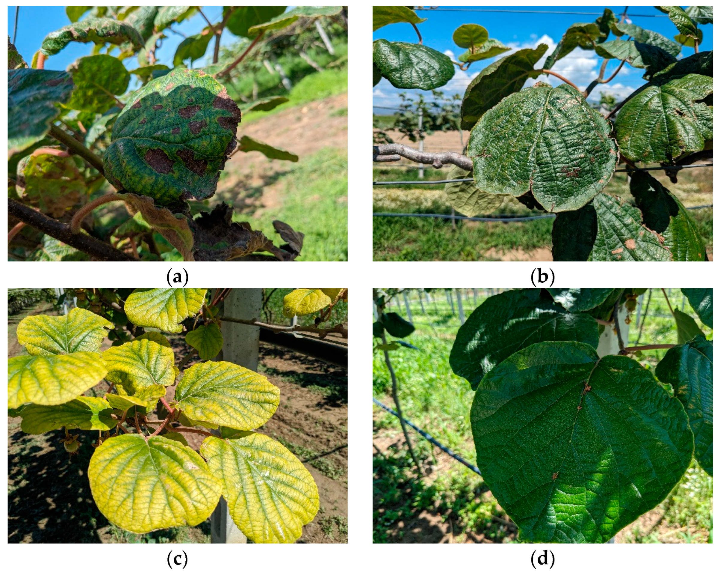
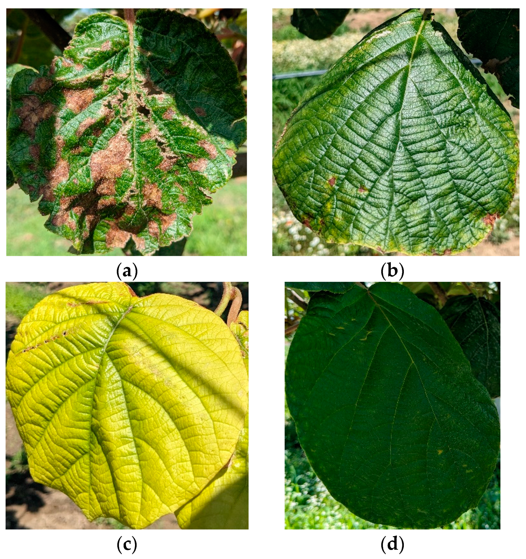
3.1. Dataset Presentation

The proposed image dataset was gathered from kiwifruit fields in Chrysoupoli, in Kavala, Greece, which is one of the three most productive regions for kiwifruits in the country. The raw images were taken using a smartphone camera with a resolution of 1920 × 1024 pixels, leveraging the high-quality sensors found in modern smartphones. Choosing a smartphone camera for data collection offers an accessible and cost-effective solution. This makes it easier for farmers to capture images of kiwifruit leaves and use them with DL models for disease recognition. By requiring only a smartphone, this approach ensures that the dataset is practical and suitable for developing user-friendly commercial applications targeted at farmers and other individuals having limited technical expertise.
The images were captured during various hours of the day in summer from multiple kiwifruit plants infected with one of the three diseases under study: Nematodes, Alternaria and Phytophthora. To ensure comprehensive coverage, images were taken from different distances to capture as many infected leaves as possible. In addition to infected leaves, images of healthy leaves were also captured daily. Creating a “Healthy” class is crucial, as it helps to distinguish infected leaves based on their strong visual differences. A total of 152 images were collected: 28 for Alternaria, 43 for Nematodes, 57 for Phytophthora and 43 for Healthy leaves, forming the so-called raw dataset. Figure 1. provides sample images from each class of the raw dataset to illustrate the image conditions and content.
Figure 2a shows the Alternaria disease, which is caused by a genus of Deuteromycetes fungi. Its symptoms include brown or black spots on the leaves, as shown in the image. Often, these spots lead to defoliation, dieback of shoots and fruit decay. From an impact perspective, the disease reduces the plant’s photosynthesis ability, weakens overall plant health and causes yield loss in both pre- and post-harvest stages. Figure 2b shows the Nematodes disease, caused by roundworms that infect the plant’s roots. Its symptoms include the characteristic curling of kiwi leaves. This curling occurs because nematodes create galls in the roots, disrupting water and nutrition uptake. From an impact perspective, the disease weakens the root system, reduces nutrient absorption and stunts plant growth, ultimately lowering yield. Figure 2c shows the Phytophthora disease, which originates from a genus of plant-damaging oomycetes. Phytophthora causes root and collar rot, leading to symptoms such as yellowing or wilting leaves, reduced shoot growth, and rapid or gradual vine decline. From an impact perspective, it severely affects root health, leading to poor nutrient and water uptake, plant decline and eventual plant death if untreated. In Figure 2d, healthy leaves are shown. Healthy leaves have a much greener color, and a better shape compared to the infected ones, highlighting the plant’s proper hydration and healthy growth.
The 152 images of the raw dataset were annotated using AnyLabeling software [29] with bounding-box shapes to train a YOLOv11 [14] object detection model for detecting both diseased and healthy kiwi leaves. As a pre-processing step, the images were augmented for object detection and image classification, as presented in Table 2 and Table 3, respectively. Heavy augmentation was applied to increase the diversity of the dataset, utilizing 19 operations across geometric transformations, flipping, blur, resizing, arithmetic adjustments and changes in color and contrast.
Raw images were, therefore, augmented, resulting in a total of 5832 images, as seen in Table 2, used for training the YOLOv11 model for leaf object detection.
Then, YOLOv11 was used for the isolation of kiwifruit leaves of the raw dataset into separate images, i.e., one leaf per image. By following this approach, the number of data samples for disease recognition increased, as each original image contained multiple leaves. This process led to the AgriVision-Kiwi Dataset, as presented in Table 3. It should be noted that the isolation of each separate leaf into a single image allows the disease classification model to focus more effectively on disease patterns by removing unnecessary background information. The pipeline for the AgriVision-Kiwi Dataset creation is illustrated in Figure 3.
In Figure 4, indicative sample images from the AgriVision-Kiwi Dataset for each class are presented to provide a visual representation of the final dataset used for disease recognition.
It is clear from the sample images of Figure 4 that each class has distinct visual characteristics that could allow for effective discrimination. The proposed kiwifruit leaf disease dataset is provided through a public repository (see Data Availability Statement).
However, an imbalance in the number of leaf images for each disease category of the AgriVision-Kiwi Dataset was noticed. To address the latter issue, the 19 augmentation operations were split and applied differently across each category to balance the final number of samples, resulting in the augmented dataset presented in Table 3, which was used in the disease recognition task. The formulated augmented images (Table 3) are subsequently used to train six DL models for disease classification towards validating the presence of distinctive characteristic patterns of each disease in color images, as well as towards identifying the best performing model for the problem under study.

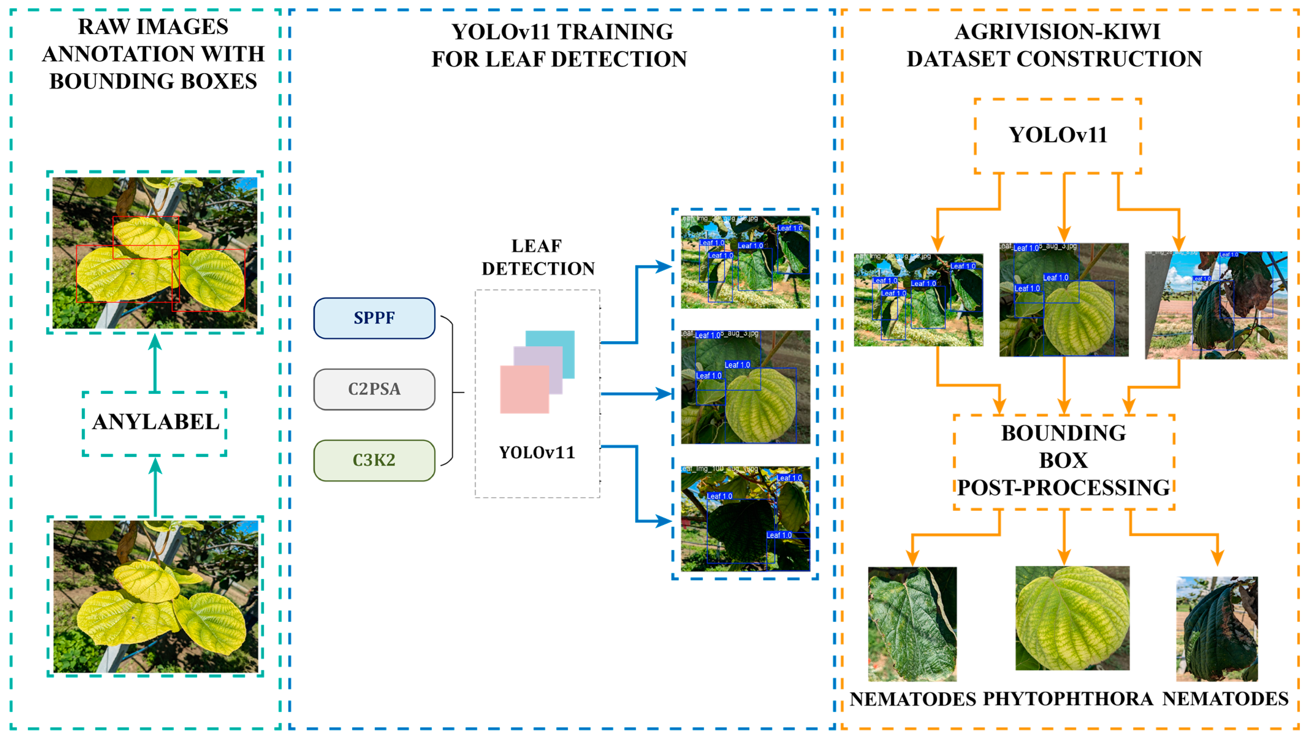
3.2. Leaf Detection and Disease Recognition Using YOLOv11

The YOLOv11 model could be trained to detect leaves and recognize diseases simultaneously. Yet, this would increase the likelihood of false negative predictions and reduce the robustness of disease recognition. The latter is due to the fact that some leaves may be overlapped, flipped or affected by shadows, which would negatively impact disease recognition. Moreover, while YOLOv11 excels in the identification and localization of objects within an image, its backbone network, attention mechanism and loss function are not the optimal solution for fine-grained tasks [30], i.e., for distinguishing between subtle differences within a category, such as imperceptible different defects on leaves.
In order to confirm our concerns, first, the performance of YOLOv11 was experimentally evaluated for both leaf detection and disease classification, as presented in the following. The leaf detection dataset was annotated, with each bounding box labeled according to the corresponding disease. As a result, the 5832 augmented images (5190 training and 642 testing samples) used for leaf detection are adapted into a four-class classification problem. YOLOv11 was then trained for 100 epochs, keeping the number of epochs consistent throughout all experiments for disease recognition presented in this work. Table 4 includes training and validation loss, accuracy, precision and recall of YOLOv11 classification. In Figure 5, the confusion matrix and F1 curve of YOLOv11 validation are presented.
The confusion matrix from the validation of YOLOv11 for leaf detection and disease classification presented in Figure 5a reveals that the model had difficulty identifying healthy and nematode-affected leaves. In many cases, the model predicted both classes as background. Additionally, a significant number of nematode-infected leaf images were misclassified as healthy, indicating the fine-grained nature of the problem under study, such as that of accurately identifying mild leaf diseases like Nematodes.
The same conclusion can be confirmed from Figure 5b, showing that the Healthy and Nematodes classes performed poorly, whereas the other two classes achieved higher accuracy. Figure 6 presents indicative results from the testing sample.
Due to the above results, we concluded that separating the proposed leaf disease recognition methodology into two distinct subsequent tasks, those of leaf detection and disease recognition, would ensure that the disease recognition step would process only the most well-visualized leaves with the richest and most informative features.
Therefore, YOLO v11 was used for leaf detection, while for the disease recognition step of the proposed algorithm, six different DL models for image recognition were employed, aiming to capture the fine-grained details and complex patterns in kiwifruit leaves.

3.3. Leaf Detection

The leaf object detection augmented dataset presented in Table 2 was used for leaf detection using the YOLOv11 model, and more specifically, its yolo11-xlarge variant.
By training the YOLOv11 model, a customized implementation was developed to isolate regions of interest (ROIs) of kiwifruit leaves. This process was employed for two reasons: (1) to significantly increase the number of data samples for the consequent kiwifruit disease recognition, as shown in Table 3, compared to the raw image set of the 152 images, and (2) to better support the overall disease recognition methodology by providing well-visualized leaves, as explained in the following subsection.
As a post-processing step, to filter single kiwifruit leaves using the YOLOv11 model, two basic rules were applied:
  • The kiwi leaf must appear fully within the ROI. Kiwifruit leaves have a characteristic shape that can be affected by certain diseases, like Nematodes. This rule ensures that the selected samples are appropriate for distinguishing the diseases.
  • The kiwifruit leaf must be large enough to provide sufficient information about the disease. The term “large enough” is defined based on the contour area of the largest leaf in the image.
This post-processing step on YOLOv11 predictions is strictly applied only to the 152 raw images towards constructing the AgriVision-Kiwi Dataset.

3.4. Disease Recognition

For the task of disease recognition, the final dataset of augmented single-leaf images of AgriVision-Kiwi Dataset was used (Table 3). This approach aims to select only well-visualized leaves out of the captured images for the construction of the used disease recognition dataset. The purpose of this approach is not only for constructing a bigger dataset but also to support the overall disease recognition methodology; by selecting only the well-visualized leaves, it is ensured that the DL models can better extract detailed and representative features for each disease, thereby possibly achieving higher performances.
For the disease recognition step of the proposed algorithm, six different DL models for image recognition were employed. The complete experimental pipeline of the disease recognition task is illustrated in Figure 7.
The selected six models are well-known and commonly used models in the CV domain:
  • Alexnet was proposed by Alex Krizhevsky, et al. in 2012 [31]. It is one of the most influential DL models in the CV domain. It was the first deep CNN model that demonstrated the power of DL. This model’s performance could provide a strong verification that the targeted classes are distinguishable from the image samples.
  • Densenet-121 was proposed by Gao Huang et al. in 2017 [32] and it is known for its dense connectivity pattern, where each layer is directly connected to every other subsequent layer. This design helps in feature reuse, alleviating the vanishing gradient problem and reducing the number of parameters in comparison to other similar deep models. This model achieved top results in the ImageNet competition.
  • Efficientnet-B3 was proposed by Mingxing Tan and Quoc Le in 2019 [33] and it is one of the Efficientnet family models. Its novelty is a compound scaling method that helps the model to be considered state-of-the-art with an outstanding balance between performance and efficiency. This model focuses on the trade-off between efficiency and performance, making it a good candidate for edge device integration, i.e. for smartphones.
  • MobileNet-V3 was proposed by Andrew Howard et al. in 2019 [34]. Its strong characteristic is its trade-off between performance and efficiency for resource-constrained devices, achieving real-time performance. This model is designed for edge devices like smartphones, which makes it suitable for the proposed application.
  • Resnet-50 was proposed by Kaiming He et al. in 2016 [35] and is a part of the groundbreaking Resnet family models. Its novelty is in the introduced residual connections to tackle the gradient vanishing problem. This model was selected since it is considered the ground base of DL models for CV applications. Additionally, it is one of the most reusable models in the CV domain due to its robustness and versatility.
  • VGG-16 was proposed by Karen Simonyan and Andrew Zisserman in 2015 [36]. Its simplified architecture significantly improves its performance compared to earlier models like AlexNet. It is considered a benchmark architecture for image classification and is widely adopted in industry and research.
Table 5 summarizes some of the main characteristics and relevant information about the architecture of the selected models.
These six models were selected because they were considered state-of-the-art models and remain among the most influential DL architectures in the CV community. Among these models, MobileNet-V3, AlexNet and EfficientNet-B3 are considered lightweight, while the rest are heavier architectures. This distinction provides a more comprehensive view of the proposed dataset’s complexity, as reflected by each model’s performance.
Furthermore, the lightweight models were chosen because they are well-suited for a commercial application in kiwifruit disease recognition. This means that lightweight models were selected for being practical and efficient in designing a technological product. Lightweight models typically have lower computational costs, meaning they require less processing power and memory. This makes them faster and more cost-effective to deploy, especially in a commercial setting where resources might be limited, as in an in-field application of real-time disease detection. Suh models can still provide accurate results, which is crucial for timely and effective disease recognition in kiwifruit. Essentially, this variation in models’ architecture is about exploring the balance between efficiency and accuracy, also considering practical, real-world applications.

3.5. Experimental Setup and Evaluation Metrics

Table 6 provides an overview of the libraries and frameworks used in this work for the implementation and execution of the experiments for each task.
Each model was trained on 90% of the data and tested on the remaining 10%, approximately, by employing 10-fold cross-validation. The learning algorithm was Adam [37]. The training was for 100 epochs, with batch size 10 and a learning rate of 1 × 10⁻⁴. The training configuration was the same for each fold. The selected loss used for minimization was cross-entropy, which is mathematically defined as follows:
L = y l o g ( y ^ )
where y refers to the ground truth values of each data sample that is given as input to each DL model, and y ^ is the predicted value that the model estimates for the input data. The usage of the logarithm (log) is to amplify the loss when the model makes wrong predictions with high probability. In addition, when the model makes correct predictions with high probability (close to 1) the logarithm gives a result near to zero, which is a small contribution.
For performance estimation during the validation phase in each fold, three metrics were used: precision, recall, F1 score and accuracy. The latter metrics are defined as follows:
P r e c i s i o n = T r u e   P o s i t i v e s ( T P ) T r u e   P o s i t i v e s ( T P ) + F a l s e   P o s i t i v e s ( F P )
R e c a l l = T r u e   P o s i t i v e s ( T P ) T r u e   P o s i t i v e s ( T P ) + F a l s e   N e g a t i v e s ( F N )
A c c u r a c y = T r u e   P o s i t i v e s T P + T r u e   N e g a t i v e ( T N ) T r u e   I n s t a n c e s ( T P + T N + F P + F N )
F 1 s c o r e = P r e c i s i o n     R e c a l l P r e c i s i o n   +   R e c a l l
C o n f i d e n c e   S c o r e   =   P r o b a b i l i t y ( O b j e c t )     I o U ( p r e d , g t )
I o U = A r e a   o f   I n t e r s e c t i o n A r e a   o f   U n i o n
where True Positive (TP) represents cases where the model correctly predicts the positive class, False Positive (FP) represents cases where the model incorrectly predicts the positive class, False Negative (FN) represents cases where the model fails to predict the positive class and True Negative (TN) represents the cases where the model correctly predicts the negative class. Area of Intersection is the common area shared by the predicted bounding box and the ground truth. Area of Union is the total area that covered by the two boxes.
The precision score measures the fraction of correctly predicted positive instances out of all positive predictions. A higher precision score means fewer false positives. Recall measures the fraction of actual positive instances that the model identified. High recall means lower false negatives. F1 score is the harmonic mean of precision and recall. It represents precision and recall in one metric. A high F1 score means perfect precision and recall. The confidence score measures the detection performance of the YOLOv11 model. It is calculated by multiplying the probability of a bounding box containing an object of interest and the IoU between the predicted bounding box and the ground truth. IoU represents the measurement of how well a predicted bounding box aligns with the ground truth box. A higher IoU value means better bounding box detection according to the ground truth. Finally, accuracy measures the proportion of correct instances out of the total instances. In the following section, the experimental results are presented.

4. Results

The experimental results are presented separately for each task. In the first subsection, the experimental results of leaf detection are presented and analyzed, while the second subsection deals with the results of leaf disease recognition.

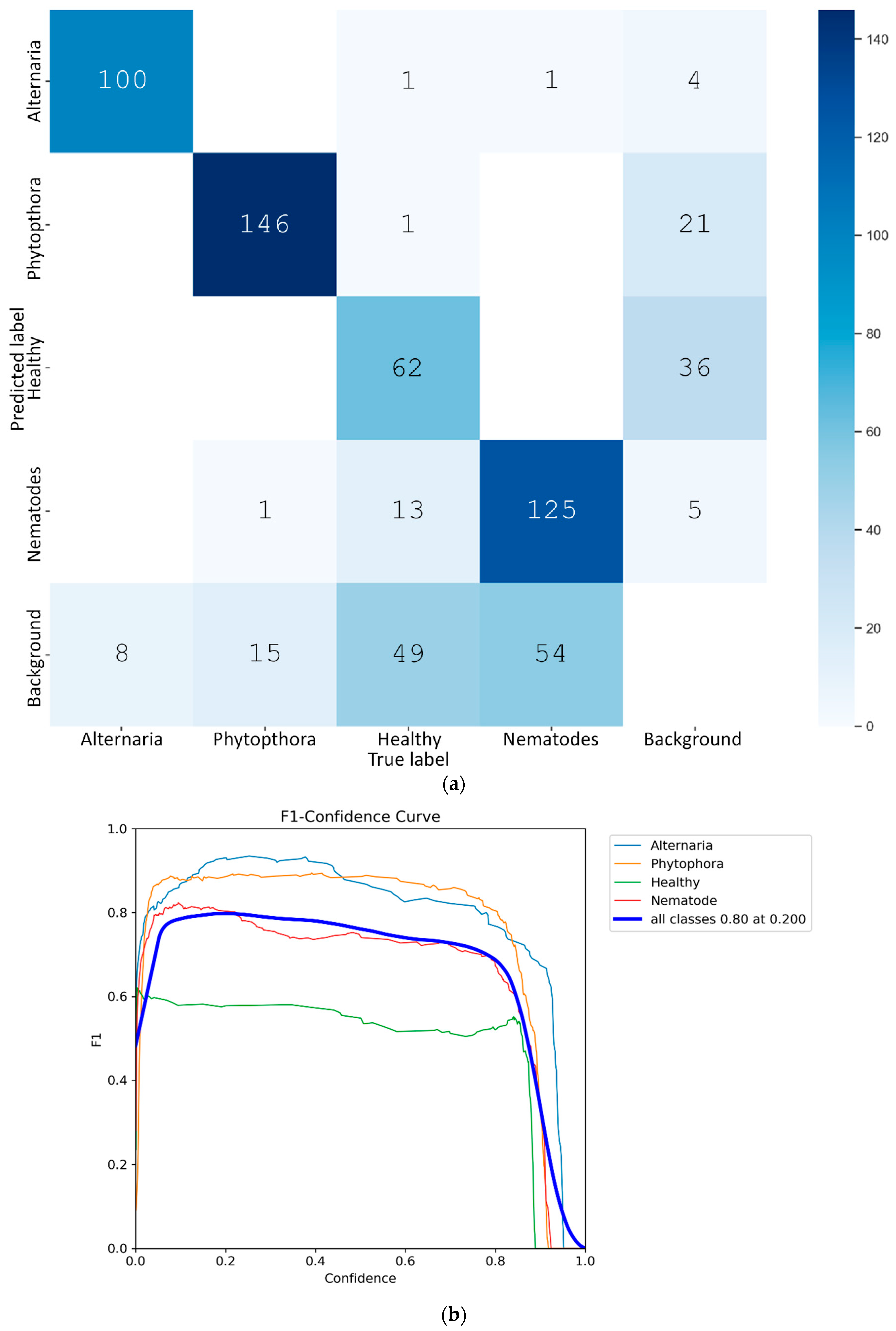
4.1. Leaf Detection Results

For leaf detection, the YOLOv11 model was trained on the augmented leaf detection dataset of Table 2. The model was trained for 100 epochs. Figure 8 illustrates the training and validation results.
From Figure 6, it is clear that YOLOv11 achieved efficient results across all metrics in detecting kiwifruit leaves in the given images.
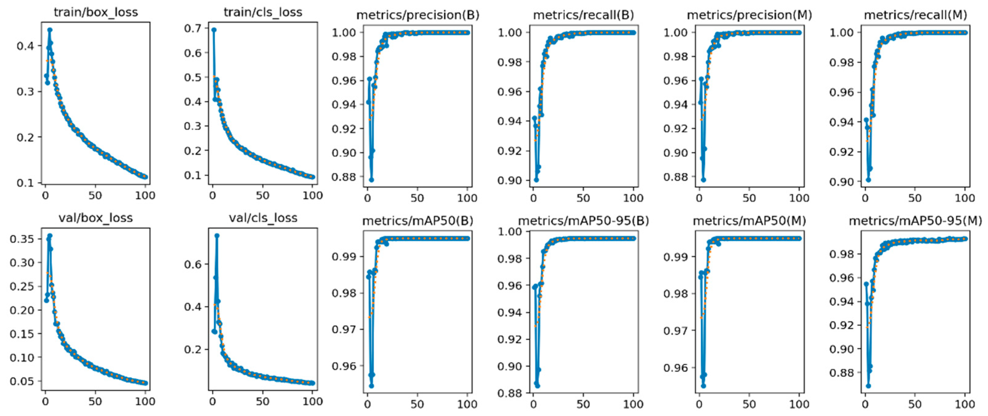
  • The box loss (first column subfigures of Figure 6) indicates the performance of the model localizing the kiwi leaves with strict bounding boxes that contain the most significant features. In our case, the box loss during training continually decreases throughout the epochs, reaching 0.053 at the final epoch.
  • The cls loss (second column subfigures of Figure 6) indicates the performance of the model in classifying the detected bounding boxes into target classes, of which, in this specific application, there are two: leaf and no leaf. In our case, the cls loss during training also decreases throughout the epochs, reaching 0.060 at the final epoch.
  • Regarding the performance metrics of precision and recall, the reported results are high, reaching 0.995 and 0.990, respectively, at the final epoch (3–6 column subfigures of Figure 6). The symbol (B) in the subfigures (columns 3 and 4) of Figure 6 refers to the best performance by the model, while (M) (columns 5 and 6) represents the average macro, which calculates the average performance across all classes.
Based on this, it is clear that the model’s performance for leaf detection is efficient since all four metrics during training are near 1. Moreover, the validation plots verify the model performance; the box loss is 0.039 in the final epoch and the cls loss is 0.043. The performance metrics during validation are also very high, which indicates that the model easily captured the leaf semantic features.
In Figure 9, the F1 curve during training of YOLOv11 is presented. In Figure 9, the relationship between the F1 score and the model confidence for each bounding box is presented. This plot shows that the model’s bounding box predictions and box confidence were in line with the F1 score. As a result, most of the model’s predictions were either true positives or true negatives, with only a few false positives and false negatives. The latter is attributed to the relatively small dataset, allowing differences between classes in feature space to be easily distinguished. The number of images contained in the dataset is the maximum that could be collected from a large region of kiwi fields in one season. Some leaf diseases were more common, such as Phytophthora, and some were rarer, such as Alternaria and Nematodes, while healthy leaves were abundant. The number of images in each class, however, was ultimately chosen to be such that each class was represented in the same proportion, giving a final balanced dataset.
It should be noted that the compiled dataset is currently the first openly available and the only one that contains images of Nematodes disease. Extending the dataset is intended, yet it requires careful planning, as described below.
Leaf diseases are seasonal and depend on environmental/climate conditions, such as temperature and humidity. This seasonality means that although there may be an intention to collect additional data, this is questionable, as it is not known in advance whether and which diseases will occur. Moreover, accessing remote or large agricultural fields can be time consuming and resource intensive, or even infeasible, since kiwifruits are mainly cultivated in northern Greece. Since the images were captured in Chrysoupoli, in northern Greece, which is one of the three most productive regions for kiwifruits in the country, it is our goal to grow the dataset, making it the largest and richest open dataset available.
In Table 7, the YOLOv11 training and validation results are presented in numbers, while the indicative detection results are illustrated in Figure 10.
The images presented in Figure 10 are from the subset of 20 testing images that were not included in the training set and were separated specifically for evaluation purposes. Based on the previously presented results, it is evident that YOLOv11 achieved high performance in detecting kiwifruit leaves in the testing images. In conclusion, the YOLOv11 model’s performance is noteworthy, and the results emphasize its reliability in detecting kiwifruit leaves.

4.2. Leaf Disease Recognition Results

Table 8 includes the training results of cross-entropy loss at each fold, out of the 10-fold cross-validation process, and for each of the six models separately.
The reported results of Table 8 indicate that DensNet-121 and ResNet-50 are the two top-performing models, with cross-entropy losses of 0.064 and 0.063, respectively. This can be attributed to their deep architectures comprising 121 and 50 layers, respectively, making them the two heaviest models among those selected. VGG-16 holds the third position with a cross-entropy loss of 0.069.
In the lightweight model category, MobileNet-V3 achieved a lower loss compared to AlexNet and Efficient-Net-B3. The superior performance of MobileNet-V3 stems from its modern architecture, which incorporates Squeeze-and-Excitation (SE) blocks and mobile inverted bottleneck convolutions. These features enable MobileNet-V3 to optimize the learning process efficiently, focusing on relevant features and suppressing irrelevant ones, even within fewer epochs. In contrast, AlexNet and Efficient-Net-B3 rely on large convolutions and classical activation functions. Table 8 includes the accuracy results of each model validation for all 10 folds.
In Table 9, the same ranking as previously is observed for the two best total reported accuracies.
DenseNet-121 and ResNet-50 are the best performing models, with DenseNet-121 showing a clear advantage over the other models. Among the remaining models, MobileNet-V3 and AlexNet perform better than EfficientNet-B3 and VGG-16, though the differences are small. Overall, all models achieved high accuracy with small variations, indicating that the image features in the presented dataset effectively discriminate the targeted classes. It should be noted here that when models report similar accuracies, of 98% to 99%, such as in our case, small variations might not be apparent without repeating testing; 10-fold cross validation aims to provide a more comprehensive view by calculating the average accuracy of all models across folds, identifying inconsistencies and, thus, providing more reliable results. Moreover, when a small dataset, such as in our case, is split into training and testing, it might result in poor representation of the data distribution in a single testing. By providing 10 tests, it was ensured that each data sample was used for both training and testing; therefore, possible bias from random single splitting is reduced, while the utility of limited data is maximized.
The precision and recall results during each fold are included in Table 10 and Table 11, respectively.
In Table 10, almost all models achieved similar performances, with DenseNet-121, Efficient-Net-B3, ResNet-50 and VGG-16 being in first place with a precision of 0.987. However, a more detailed analysis shows that DenseNet-121 stands out slightly, achieving a precision of 0.988 in three out of 10 folds. ResNet-50 and AlexNet demonstrate the most stable results across all folds compared to the other models. These metrics indicate that the models produced very few false positives and that most predictions were correct across the test data. The results verify that the image features are highly representative for each class and that the dataset’s maximum representation capacity is reached.
Similar to Table 10, in Table 11, DenseNet-121 achieves a slightly better performance in recall metrics, taking the lead in two out of the 10 folds. This suggests its ability to correctly identify true positives more effectively than the other models, particularly in scenarios where small variations in recall can be significant. All models demonstrate a consistently high recall, with differences limited to the third decimal. This indicates that the dataset is structured in a way that allows all models to successfully identify the majority of true positives across validation folds. MobileNet-V3 achieves a slightly lower recall compared to the other models. This could be attributed to the trade-off between capacity and efficiency. While still highly effective, it may not extract as detailed features as the larger models. These results indicate that the models achieved a low false negative prediction, which is critical for applications like disease recognition.
In the context of disease recognition, achieving a low false negative rate is crucial for early disease detection, since if it is not detected (a false negative), it can spread and threaten the yield. Moreover, low false negatives mean that the models are identifying infected kiwifruits accurately, which is critical for developing trustful systems, especially when designated for commercial use.
In Figure 11, the 10-fold results are visually illustrated for all performance metrics and models.
From Figure 11a, it is evident that DenseNet-121 and ResNet-50 achieve the lowest loss compared to the other models. In the performance metric plots, all models achieve high and similar results. In a more detailed analysis, all models except MobileNet-V3 and AlexNet maintain a steady performance across each fold. Additionally, the use of YOLOv11 as a middle task to extract leaves helped the DL models to ensure a high performance above 98% in each metric.
Figure 12 illustrates the confusion matrices for all tested models, aiming to further analyze the behavior of the examined models. All confusion matrices include testing of the same sample images, specifically, for 645 sample images of the augmented AgriVision-Kiwi Dataset (153 Alternaria, 163 Nematodes, 165 Phytophtora and 164 Healthy). As it can be observed, a common mistake across most of the models is that samples belonging to Nematodes are attributed to the Healthy class or vice versa. Recall that Nematodes symptoms include the characteristic curling of kiwi leaves, without affecting seriously their color, such as in the case of Phytophthora, while no defected spots appear on the leaf, such as in the case of Alternaria (Figure 4). However, Alternaria, along with the defected brown spots on the leaves, also exhibits the curling effect, which is a characteristic of Nematodes (Figure 4). As a result, the models, as can be seen from the confusion matrices of Figure 12, also confuse these two classes, especially in cases where probably no intense spots appear on the Alternaria-infected leaves.
Therefore, it is proven that Nematodes is the most challenging disease to detect from leaf images, since its symptoms are milder, and models can easily be confused. Models need to be trained to recognize fine-grained features such as the specific curling patterns of Nematodes, which require advanced algorithms and high-resolution image datasets. Indeed, the absence of distinct spots or discoloration increases the risk of confusing Nematodes-infected leaves with healthy leaves or those with other mild diseases. Data scarcity and environmental factors such as external lighting, shadowing and overlapping leaves can further obstruct the detection of subtle curling of leaves, complicating the detection models. The incorporation of domain-specific knowledge, such as the typical progression of the symptoms of Nematodes, could further improve the models’ accuracy.
The performance of these models demonstrates that the proposed dataset is well-structured, with discriminative features for each class without noise. The small variations observed in each fold indicate that the dataset is uniformly distributed, with no major differences in class distribution or complexity. Even with heavy augmentation, as described previously (e.g., noise, color transformations and scale transformations), the selected models successfully discriminate kiwifruit leaves among the three disease classes and the healthy class.
In general, the introduced dataset is descriptive and well-formed, which explains why the lightweight models perform comparably to the more complex architectures like DenseNet-121 and ResNet-50. These results highlight that the dataset is well structured, straightforward, and free from noise or class overlaps.
Regarding the inference time of each DL model, Table 12 includes the processing time of disease detection on the same image for each model.
AlexNet, as expected, requires less processing time; however, all models’ inference is in milliseconds. Yet, DenseNet-121 reports higher performance metrics; similarly, all models provide equally accurate results, all reporting accuracies of 98–99%. This means that the selection of the appropriate model, ultimately, depends on the specific requirements of the application, since real-time applications require fast inference, while other applications can afford to prioritize accuracy over speed.

5. Discussion

This study provides valuable resources for kiwifruit disease recognition and lays the groundwork for similar datasets in other crops. Note that early disease recognition is the cornerstone of sustainable agricultural management, allowing for timely interventions and, thus, reduced use of chemicals and crop losses, while providing better-quality products of higher standards. Therefore, robust and reliable methods are required to support farmers in early detecting plant diseases, especially in crops like kiwifruit that are vulnerable to a wide range of diseases that can spread rapidly and devastate the entire orchard if not detected timely.
Based on the experimental results, although the proposed dataset is relatively small, it was proven well-structured to support further research on kiwifruit disease recognition. The proposed dataset is stored in a structured format, in a public repository, with clear metadata and documentation.
The metadata and documentation ensure the easy access and reproducibility of the research, as well as the development of accurate and reliable models for kiwifruit disease recognition. This contribution can enhance kiwifruit disease recognition efforts, particularly in addressing Nematodes diseases from leaf images, which does not exist in the known CV literature. Furthermore, to develop more sophisticated and generalized methods, a large amount of data is needed to capture all aspects of diseases, including ways of infection, environmental conditions and disease symptoms. To support this goal, more open datasets are needed for assisting the research community to create benchmarks and provide a consistent basis for comparison. In recent years, transfer learning and the fine-tuning of various CNN architectures have become common practices. The availability of open datasets can further enhance and improve the performance and robustness of new models, methodologies and training strategies for disease recognition applications. Therefore, the proposed dataset aims to provide strong practicability for the research community, as well as to provide an image leaf benchmark for Nematodes towards guiding the research progress to its early detection from leaf images.
Additionally, the proposed dataset was proven suitable for the development of commercial applications that integrate DL models to provide real-time feedback and timely support to farmers regarding the health status of their cultivation. More specifically, the efficient results that were obtained from the selected lightweight models revealed that the proposed disease recognition method could be integrated into mobile applications for commercial purposes. Lightweight models have lower computational costs, meaning they require less processing power and memory, making them faster and more cost-effective to deploy, especially in agricultural settings where resources might be limited.
The efficiency of such disease classification models could be further enhanced if combined with a leaf detection model that can discard redundant background information from images and provide suitable content in the DL models, such as in our case. The latter scheme could be very advantageous for commercial applications where the diversity and volume of input data can scale significantly. Therefore, the proposed strategy can provide information of better quality regarding the natural scene and, thus, promote the disease identification task.
Based on the presented literature, most works focus only on kiwifruit disease recognition by using the entire image, without first localizing the specific region of interest. While this approach is simpler, since the pipeline is more straightforward and needs less inference time, it introduces noise from background information and dynamic environmental conditions. Such noise might not be presented in a controlled dataset captured in the laboratory but can severely affect the models’ performance in real-world applications. Considering the above, the presented study, by separating the disease detection pipeline into two distinctive tasks, leaf detection and disease recognition, provides an efficient and practically applicable methodology that could be integrated with edge devices, such as smartphones. This can also be verified numerically, by the results provided in Table 1 from the related works referenced from the literature; the proposed work reported higher performance metrics compared to similar studies that address kiwifruit disease classification.
One possible limitation of the introduced dataset is that, due to its small size, it may limit the generalization ability of the trained models to more diverse environmental or geographical conditions. In future work, efforts will be made to expand the dataset with different conditions, such as varying seasons and lighting conditions. Additionally, expanding the dataset to cover various stages of each disease would enhance its utility and support the development of proactive mechanisms to support farmers more efficiently in their daily orchard management tasks. Furthermore, future research could also investigate the creation of synthetic images, especially on the various stages of each disease. This approach would provide deeper insights into how each disease’s visual characteristics evolve over time on the object of interest. Additionally, a robust methodology for generating synthetic images would reduce the need to locate diseased kiwifruit plants in their early stages and beyond.

6. Conclusions

This study proposed and evaluated a novel open dataset for kiwifruit disease recognition, presenting its suitability for training high-performance deep learning models. The dataset covers three classes of infections on leaves, caused by Alternaria, Nematodes and Phytophthora diseases, and a class of healthy leaves; it should be noted that Nematodes disease has not been detected from leaf images, nor has a relevant image dataset or recognition study has been reported in the academic literature before.
The conducted evaluation included two main tasks: First, kiwifruit leaf detection was conducted using the well-known YOLOv11 model, reporting a minimum bounding box loss of 0.053 in leaf detection; second, disease recognition was implemented by employing six state-of-the-art deep learning models, reporting all models’ classification performance metrics of precision, recall and accuracy above 0.98 in all cases after 10-fold cross-validation. These findings suggest that the proposed dataset is well balanced and suitable for training deep learning models. By making such a novel dataset publicly available, the scientific community can work more effectively towards developing accurate, efficient and scalable solutions for kiwifruit-related agricultural challenges.

Author Contributions

Conceptualization, G.A.P. and T.K.; software, T.K.; validation, T.K., E.V., L.I., D.V., L.I. and G.A.P.; investigation, T.K.; resources, T.K.; data curation, T.K. and E.M.; writing—original draft preparation, T.K. and E.V.; writing—review and editing, E.V., L.I., D.V., M.M., G.B. and G.A.P.; visualization, G.A.P. and E.V.; supervision, G.A.P., L.I., D.V., M.M. and G.B. All authors have read and agreed to the published version of the manuscript.

Funding

This research received no external funding.

Data Availability Statement

The original data of AgriVision-Kiwi Dataset presented in the study are openly available in GitHub Free repository at [https://github.com/MachineLearningVisionRG/AgriVision-Kiwi, accessed on 14 March 2025].

Acknowledgments

This research has been conducted in the context of the research program “DigiAgriFood” DIGITAL TRANSFORMATION AND GREEN TRANSITION OF THE AGRIFOOD VALUE CHAIN IN CENTRAL AND NORTHERN GREECE, under MIS 6001525, funded by the ‘Digital Europe’ and national resources through the Public Investment Program. The authors extend their sincere gratitude to agronomist Apostolos Kitsos for his invaluable guidance and support throughout the duration of this project.

Conflicts of Interest

The authors declare no conflicts of interest.

References

  1. Ciacci, C.; Russo, I.; Bucci, C.; Iovino, P.; Pellegrini, L.; Giangrieco, I.; Tamburrini, M.; Ciardiello, M.A. The Kiwi Fruit Peptide Kissper Displays Anti-Inflammatory and Anti-Oxidant Effects in in-Vitro and Ex-Vivo Human Intestinal Models. Clin. Exp. Immunol. 2014, 175, 476–484. [Google Scholar] [CrossRef]
  2. Richardson, D.P.; Ansell, J.; Drummond, L.N. The Nutritional and Health Attributes of Kiwifruit: A Review. Eur. J. Nutr. 2018, 57, 2659–2676. [Google Scholar] [CrossRef] [PubMed]
  3. Huang, K.; Tang, J.; Zou, Y.; Sun, X.; Lan, J.; Wang, W.; Xu, P.; Wu, X.; Ma, R.; Wang, Q.; et al. Whole Genome Sequence of Alternaria Alternata, the Causal Agent of Black Spot of Kiwifruit. Front. Microbiol. 2021, 12, 713462. [Google Scholar] [CrossRef] [PubMed]
  4. Bi, X.; Hieno, A.; Otsubo, K.; Kageyama, K.; Liu, G.; Li, M. A Multiplex PCR Assay for Three Pathogenic Phytophthora Species Related to Kiwifruit Diseases in China. J. Gen. Plant Pathol. 2019, 85, 12–22. [Google Scholar] [CrossRef]
  5. Banihashemian, S.N.; Jamali, S.; Golmohammadi, M.; Ghasemnezhad, M. Isolation and Identification of Endophytic Bacteria Associated with Kiwifruit and Their Biocontrol Potential against Meloidogyne Incognita. Egypt. J. Biol. Pest Control 2022, 32, 111. [Google Scholar] [CrossRef]
  6. Hosny, K.M.; El-Hady, W.M.; Samy, F.M.; Vrochidou, E.; Papakostas, G.A. Multi-Class Classification of Plant Leaf Diseases Using Feature Fusion of Deep Convolutional Neural Network and Local Binary Pattern. IEEE Access 2023, 11, 62307–62317. [Google Scholar] [CrossRef]
  7. Yao, J.; Wang, Y.; Xiang, Y.; Yang, J.; Zhu, Y.; Li, X.; Li, S.; Zhang, J.; Gong, G. Two-Stage Detection Algorithm for Kiwifruit Leaf Diseases Based on Deep Learning. Plants 2022, 11, 768. [Google Scholar] [CrossRef]
  8. Liu, J.; Wang, X. Plant Diseases and Pests Detection Based on Deep Learning: A Review. Plant Methods 2021, 17, 22. [Google Scholar] [CrossRef]
  9. Kalampokas, T.; Vrochidou, E.; Tzampazaki, M.; Papakostas, G.A. Unraveling the Potential of AI Towards Digital and Green Transformation in Kiwifruit Farming. In Proceedings of the 2024 International Conference on Software, Telecommunications and Computer Networks (SoftCOM), Split, Croatia, 26 September 2024; pp. 1–6. [Google Scholar]
  10. El-Borai, F.E.; Duncan, L.W. Nematode Parasites of Subtropical and Tropical Fruit Tree Crops. In Plant Parasitic Nematodes in Subtropical and Tropical Agriculture; CABI Publishing: Wallingford, UK, 2005; pp. 467–492. [Google Scholar]
  11. Tao, Y.; Xu, C.; Yuan, C.; Wang, H.; Lin, B.; Zhuo, K.; Liao, J. Meloidogyne Aberrans Sp. Nov. (Nematoda: Meloidogynidae), a New Root-Knot Nematode Parasitizing Kiwifruit in China. PLoS ONE 2017, 12, e0182627. [Google Scholar] [CrossRef]
  12. AYDINLI, G.; MENNAN, S. Samsun İli (Türkiye) Kivi Bahçelerindeki Kök-Ur Nematodlarının Yaygınlığı ve Meyve Verimine Etkileri. Turk. J. Entomol. 2022, 46, 187–197. [Google Scholar] [CrossRef]
  13. Gonçalves, A.R.; Conceição, I.L.; Carvalho, R.P.; Costa, S.R. Meloidogyne Hapla Dominates Plant-parasitic Nematode Communities Associated with Kiwifruit Orchards in Portugal. Plant Pathol. 2025, 74, 171–179. [Google Scholar] [CrossRef]
  14. Khanam, R.; Hussain, M. YOLOv11: An Overview of the Key Architectural Enhancements. arXiv 2024, arXiv:2410.17725. [Google Scholar]
  15. Banerjee, D.; Kukreja, V.; Hariharan, S.; Sharma, V. Fast and Accurate Multi-Classification of Kiwi Fruit Disease in Leaves Using Deep Learning Approach. In Proceedings of the 2023 International Conference on Innovative Data Communication Technologies and Application (ICIDCA), Uttarakhand, India, 14 March 2023; pp. 131–137. [Google Scholar]
  16. Bansal, A.; Sharma, R.; Sharma, V.; Jain, A.K.; Kukreja, V. An Automated Approach for Accurate Detection and Classification of Kiwi Powdery Mildew Disease. In Proceedings of the 2023 IEEE 8th International Conference for Convergence in Technology (I2CT), Lonavla, India, 7 April 2023; pp. 1–4. [Google Scholar]
  17. Mir, T.A.; Gupta, S.; Aggarwal, P.; Pokhariya, H.S.; Banerjee, D.; Lande, A. Multiclass Kiwi Leaf Disease Detection Using CNN-SVM Fusion. In Proceedings of the 2024 IEEE International Conference on Information Technology, Electronics and Intelligent Communication Systems (ICITEICS), Bangalore, India, 28 June 2024; pp. 1–6. [Google Scholar]
  18. Liu, B.; Ding, Z.; Zhang, Y.; He, D.; He, J. Kiwifruit Leaf Disease Identification Using Improved Deep Convolutional Neural Networks. In Proceedings of the 2020 IEEE 44th Annual Computers, Software, and Applications Conference (COMPSAC), Madrid, Spain, 13–17 July 2020; pp. 1267–1272. [Google Scholar]
  19. Banerjee, D.; Kukreja, V.; Chamoli, S.; Thapliyal, S. Optimizing Kiwi Fruit Plant Leaves Disease Recognition: A CNN-Random Forest Integration. In Proceedings of the 2023 3rd International Conference on Smart Generation Computing, Communication and Networking (SMART GENCON), Bangalore, India, 29–31 December 2023; pp. 1–6. [Google Scholar]
  20. Li, X.; Chen, X.; Yang, J.; Li, S. Transformer Helps Identify Kiwifruit Diseases in Complex Natural Environments. Comput. Electron. Agric. 2022, 200, 107258. [Google Scholar] [CrossRef]
  21. Wu, J.; Zheng, H.; Zhao, B.; Li, Y.; Yan, B.; Liang, R.; Wang, W.; Zhou, S.; Lin, G.; Fu, Y.; et al. AI Challenger: A Large-Scale Dataset for Going Deeper in Image Understanding. arXiv 2017, arXiv:1711.06475. [Google Scholar] [CrossRef]
  22. Hughes, D.P.; Salathe, M. An Open Access Repository of Images on Plant Health to Enable the Development of Mobile Disease Diagnostics. arXiv 2015, arXiv:1511.08060. [Google Scholar]
  23. Saleem, M.H.; Potgieter, J.; Arif, K.M. A Performance-Optimized Deep Learning-Based Plant Disease Detection Approach for Horticultural Crops of New Zealand. IEEE Access 2022, 10, 89798–89822. [Google Scholar] [CrossRef]
  24. Senanu Ametefe, D.; Seroja Sarnin, S.; Mohd Ali, D.; Caliskan, A.; Tatar Caliskan, I.; Adozuka Aliu, A.; John, D. Enhancing Leaf Disease Detection Accuracy through Synergistic Integration of Deep Transfer Learning and Multimodal Techniques. Inf. Process. Agric. 2024, in press. [CrossRef]
  25. Wu, K.; Jia, Z.; Duan, Q. The Detection of Kiwifruit Sunscald Using Spectral Reflectance Data Combined with Machine Learning and CNNs. Agronomy 2023, 13, 2137. [Google Scholar] [CrossRef]
  26. Sekhukhune, M.K.; Shokoohi, E.; Maila, M.Y.; Mashela, P.W. Free-Living Nematodes Associated with Kiwifruit and Effect of Soil Chemical Properties on Their Diversity. Biologia 2022, 77, 435–444. [Google Scholar] [CrossRef]
  27. Kumar, S.; Rawat, S.; Joshi, V.; Kundu, A.; Somvanshi, V.S. Northern Root-Knot Nematode Meloidogyne Hapla Chitwood, 1949 Infecting Kiwi Fruit (Actinidia Chinensis) in Bageshwar District of Uttarakhand State. Indian Phytopathol. 2023, 76, 665–667. [Google Scholar] [CrossRef]
  28. Kang, H.; Je, H.; Choi, I. Occurrence and Distribution of Root-Knot Nematodes in Kiwifruit Orchard. Res. Plant Dis. 2023, 29, 45–51. [Google Scholar] [CrossRef]
  29. Neural Research Lab. AnyLabeling. Available online: https://anylabeling.nrl.ai/ (accessed on 27 January 2024).
  30. Liu, Z.; Wei, L.; Song, T. Optimized YOLOv11 Model for Lung Nodule Detection. Biomed. Signal Process. Control 2025, 107, 107830. [Google Scholar] [CrossRef]
  31. Krizhevsky, A.; Sutskever, I.; Hinton, G.E. ImageNet Classification with Deep Convolutional Neural Networks. Commun. ACM 2017, 60, 84–90. [Google Scholar] [CrossRef]
  32. Huang, G.; Liu, Z.; van der Maaten, L.; Weinberger, K.Q. Densely Connected Convolutional Networks. arXiv 2016, arXiv:1608.06993. [Google Scholar]
  33. Tan, M.; Le, Q.V. EfficientNet: Rethinking Model Scaling for Convolutional Neural Networks. In Proceedings of the 36th International Conference on Machine Learning, ICML 2019, Long Beach, CA, USA, 9–15 June 2019. [Google Scholar]
  34. Howard, A.; Sandler, M.; Chen, B.; Wang, W.; Chen, L.-C.; Tan, M.; Chu, G.; Vasudevan, V.; Zhu, Y.; Pang, R.; et al. Searching for MobileNetV3. In Proceedings of the 2019 IEEE/CVF International Conference on Computer Vision (ICCV), Paris, France, 6 October 2019; pp. 1314–1324. [Google Scholar]
  35. He, K.; Zhang, X.; Ren, S.; Sun, J. Deep Residual Learning for Image Recognition. In Proceedings of the 2016 IEEE Conference on Computer Vision and Pattern Recognition (CVPR), Las Vegas, NV, USA, 27–30 June 2016; pp. 770–778. [Google Scholar]
  36. Simonyan, K.; Zisserman, A. Very Deep Convolutional Networks for Large-Scale Image Recognition. In Proceedings of the 3rd International Conference on Learning Representations, ICLR 2015—Conference Track Proceedings, San Diego, CA, USA, 7–9 May 2015. [Google Scholar]
  37. Kingma, D.P.; Ba, J. Adam: A Method for Stochastic Optimization. In Proceedings of the 3rd International Conference on Learning Representations, ICLR 2015—Conference Track Proceedings, San Diego, CA, USA, 7–9 May 2015. [Google Scholar]
Figure 1. Proposed methodology.
Electronics 14 01705 g001
Figure 2. Leaf samples from the raw dataset captured for each class: (a) Alternaria disease; (b) Nematodes disease; (c) Phytophthora disease; (d) Healthy leaves.
Electronics 14 01705 g002
Figure 3. AgriVision-Kiwi Dataset creation pipeline.
Electronics 14 01705 g003
Figure 4. Image samples for disease recognition from the AgriVision-Kiwi Dataset for each class: (a) Alternaria disease; (b) Nematodes disease; (c) Phytophthora disease; (d) Healthy leaf.
Electronics 14 01705 g004
Figure 5. (a) Confusion matrix and (b) F1 confidence curve for YOLOv11 classification task.
Electronics 14 01705 g005
Figure 6. Indicative testing image results of YOLOv11 for both leaf detection and disease classification tasks: (a) correct detection and classification of Nematodes; (b) correct classification and failure to detect one leaf; (c) correct detection and misclassification of healthy leaf as Alternaria; (d) correct detection and misclassification of healthy leaf as Nematodes.
Electronics 14 01705 g006
Figure 7. Leaves disease recognition methodology.
Electronics 14 01705 g007
Figure 8. YOLOv11 training and validation results for leaf detection.
Electronics 14 01705 g008
Figure 9. F1 curve during training of YOLOv11.
Electronics 14 01705 g009
Figure 10. Indicative results of leaf detection with YOLOv11.
Electronics 14 01705 g010
Figure 11. Representative confusion matrices for (a) losses, (b) precision, (c) recall and (d) accuracy.
Electronics 14 01705 g011
Figure 12. Confusion matrices of the models, referring to one fold of 645 testing sample images (153 Alternaria, 163 Nematodes, 165 Phytophtora and 164 Healthy) of the augmented AgriVision-Kiwi Dataset: (a) AlexNet; (b) DenseNet-121; (c) EfficientNet-B3; (d) MobileNet-V3; (e) ResNet-50; (f) VGG-16.
Electronics 14 01705 g012
Table 1. Characteristics of related works. Check marks indicate “yes” and dash “no”.
CharacteristicsRelated Works
[7][15][16][17][18][19][20][23][24][25]
DiseaseBrown Spot-----
Bacterial Canker -------
Anthracnose ------
Mosaic------
Powdery Mildew---------
Rust ---------
Botrytis--------
Phomopsis ---------
PSA---------
Armillaria Root Rot ---------
Bacterial Bligh---------
Bleeding Canker ---------
Phytophthora Root ---------
Water Staining ---------
Juice Blotch ---------
Sooty Mold of Fruit ---------
Collar Rot---------
Crown Rot---------
Black Spot---------
Yellow Leaf---------
Ulcer--------
Sunscald---------
TaskLeaf
detection
ModelYOLOX------RFCN--
Results<na>------0.938 prec.--
Disease recognitionModelDeepLabV3+CNN + SVMCNN + LSTMCNN + SVMCNNCNN + RFConvViT-BRFCNDensenet-201MS-CNN
Results0.966 acc.0.8348 acc.0.9570 prec.0.9568 acc.0.9854 acc.0.8564 acc.0.9986 acc.0.9380 prec.0.9989 acc0.99 acc.
DataPublic datasetNoNoNoNoNoNoNoNoNoNo
Table 2. Leaf object detection dataset.
TaskRaw DatasetAugmented Raw DatasetTargets
Leaf Object Detection152 58322 Classes (Leaf, Background)
Table 3. Leaf disease recognition dataset.
TaskRaw DatasetAgriVision-Kiwi Dataset Augmented AgriVision-Kiwi DatasetTargets
Leaf Disease
Recognition
57 (Phytophthora),
24 (Healthy),
43 (Nematodes),
28 (Alternaria)
108 (Phytophthora),
51 (Healthy),
70 (Nematodes),
33 (Alternaria)
1680 (Phytophthora),
1588 (Healthy),
1588 (Nematodes),
1591 (Alternaria)
4 Classes
(Phytophthora, Healthy, Nematodes, Alternaria)
Table 4. Performance metrics of YOLOv11 classification.
Training
Loss
Validation
Loss
AccuracyPrecision Recall
0.11230.1340.670.830.86
Table 5. Selected DL models’ characteristics.
Model NameYearLayersLayers
AlexNet20128First deep CNN to win ImageNet
DenseNet-1212017121Efficient depth with fewer parameters
EfficientNet-B3201932Best trade-off between accuracy and efficiency
MobileNet-V3201916Suitable architecture for mobile and edge devices
ResNet-50201550Enabled very deep networks, first for dealing vanishing gradient problem
VGG-16201416Simplicity of design and improved accuracy by increasing depth
Table 6. Used libraries and frameworks.
TaskLibrary—FrameworkDetails
Image ProcessingOpenCV, PillowData handling, image pre-processing
Image AugmentationImgaugImage augmentation
Leaves Object DetectionUltralyticsYOLOv11 training and inference
Leaves Disease RecognitionPytorchDeep learning models training and inference for image classification
Table 7. YOLOv11 training and validation results.
MetricYOLOv11
Train-box-loss0.053
Train-cls-loss0.060
Val-box-loss0.043
Val-cls-loss0.041
Precision(B)0.99
Recall(B)0.99
Precision(M)0.99
Recall(M)0.99
F1 score0.99
Confidence score0.99
Table 8. Cross-entropy loss of each model training at each fold. The two best total results are marked in bold.
ModelAlexNetDenseNet-121EfficientNet-B3MobileNet-V3ResNet-50VGG-16
Fold 10.0690.0630.0880.0750.0640.073
Fold 20.0720.0650.0860.0700.0620.060
Fold 30.0780.0600.0870.0690.0630.058
Fold 40.0720.0630.0860.0760.0640.075
Fold 50.0720.0650.0860.0710.0640.075
Fold 60.0740.0610.0870.0660.0620.055
Fold 70.0700.0640.0850.0740.0610.070
Fold 80.0720.0680.0880.0700.0640.081
Fold 90.0720.0690.0830.0700.0610.070
Fold 100.0750.0640.0850.0690.0620.071
Mean0.0730.0640.0860.0710.0630.069
Table 9. Accuracy of each model validation at each fold. The two best total results are marked in bold.
ModelAlexNetDenseNet-121EfficientNet-B3MobileNet-V3ResNet-50VGG-16
Fold 10.9890.9960.9810.9820.9890.984
Fold 20.9890.9950.9870.9870.9920.990
Fold 30.9900.9890.9900.9840.9860.984
Fold 40.9820.9950.9760.9890.9930.995
Fold 50.9820.9930.9810.9810.9810.986
Fold 60.9930.9950.9820.9930.9870.986
Fold 70.9840.9920.9810.9810.9900.987
Fold 80.9820.9900.9840.9870.9870.978
Fold 90.9900.9930.9860.9890.9900.982
Fold 100.9790.9950.9790.9920.9890.973
Mean0.9860.9930.9830.9860.9890.984
Table 10. Precision of each model validation at each fold. The best total results are marked in bold.
ModelAlexNetDenseNet-121EfficientNet-B3MobileNet-V3ResNet-50VGG-16
Fold 10.9860.9880.9880.9890.9870.986
Fold 20.9860.9870.9880.9890.9870.987
Fold 30.9860.9870.9870.9850.9870.987
Fold 40.9860.9870.9870.9850.9870.987
Fold 50.9860.9870.9870.9850.9870.987
Fold 60.9860.9870.9870.9850.9870.987
Fold 70.9860.9870.9870.9850.9870.987
Fold 80.9860.9870.9860.9860.9870.987
Fold 90.9860.9880.9850.9850.9870.987
Fold 100.9860.9880.9860.9860.9870.987
Mean0.9860.9870.9870.9860.9870.987
Table 11. Recall of each model validation at each fold. The best total results are marked in bold.
ModelAlexNetDenseNet-121EfficientNet-B3MobileNet-V3ResNet-50VGG-16
Fold 10.9860.9880.9880.9840.9880.987
Fold 20.9860.9870.9870.9840.9870.987
Fold 30.9860.9870.9870.9850.9870.987
Fold 40.9860.9870.9870.9850.9870.987
Fold 50.9860.9870.9870.9850.9870.987
Fold 60.9860.9870.9870.9850.9870.987
Fold 70.9860.9870.9870.9860.9870.987
Fold 80.9860.9870.9870.9850.9870.987
Fold 90.9860.9870.9870.9850.9870.987
Fold 100.9860.9880.9870.9850.9870.987
Mean0.9860.9870.9870.9850.9870.987
Table 12. Inference time of models for disease recognition.
ModelAlexNetDenseNet-121EfficientNet-B3MobileNet-V3ResNet-50VGG-16
Time192 ms676 ms602 ms496 ms600 ms579 ms
Disclaimer/Publisher’s Note: The statements, opinions and data contained in all publications are solely those of the individual author(s) and contributor(s) and not of MDPI and/or the editor(s). MDPI and/or the editor(s) disclaim responsibility for any injury to people or property resulting from any ideas, methods, instructions or products referred to in the content.

