Article

Design and Evaluation of a Hip-Only Actuated Lower Limb Exoskeleton for Lightweight Gait Assistance

1
Institute of Intelligent Rehabilitation Engineering, University of Shanghai for Science and Technology, Shanghai 200093, China
2
Shanghai Engineering Research Center of Assistive Devices, Shanghai 200093, China
3
Key Laboratory of Neural-Functional Information and Rehabilitation Engineering of the Ministry of Civil Affairs, Shanghai 200093, China
4
Department of Biomedical Engineering, The Chinese University of Hong Kong, Hong Kong SAR 999077, China
*
Author to whom correspondence should be addressed.
Electronics 2025, 14(19), 3853; https://doi.org/10.3390/electronics14193853
Submission received: 3 September 2025 / Revised: 19 September 2025 / Accepted: 24 September 2025 / Published: 29 September 2025

Abstract

This paper presents the design and evaluation of a lightweight, minimally actuated lower limb exoskeleton that emphasizes hip–knee coordination for natural and efficient gait assistance. The system adopts a hip-only motorized actuation strategy in combination with an electromagnetically controlled knee locking mechanism, ensuring rigid stability during stance while providing compliant assistance during swing. To support sit-to-stand transitions, a gas spring–ratchet mechanism is integrated, which remains disengaged in the seated position, delivers assistive torque during rising, and provides cushioning during the descent to enhance safety and comfort. The control framework fuses foot pressure and thigh-mounted IMU signals for finite state machine (FSM)-based gait phase detection and employs a fuzzy PID controller to achieve adaptive hip torque regulation with coordinated hip–knee control. Preliminary human-subject experiments demonstrate that the proposed design enhances lower-limb coordination, reduces muscle activation, and improves gait smoothness. By integrating a minimal-actuation architecture, a practical sit-to-stand assist module, and an intelligent control strategy, this exoskeleton strikes an effective balance between mechanical simplicity, functional support, and gait naturalness, offering a promising solution for everyday mobility assistance in elderly or mobility-impaired users.

1. Introduction

With the increasing prevalence of age-related mobility impairments and neuro-muscular disorders [1,2], wearable exoskeletons have emerged as promising solutions for re-storing or enhancing lower limb function. For elderly users or individuals with mild motor deficits, maintaining stable standing and executing smooth walking motions remains a critical challenge, particularly when muscular strength or balance is compromised [3,4,5,6]. To address this, lower limb exoskeletons must provide not only assistive torque during locomotion but also structural support during stance, all while minimizing device weight and control complexity [7,8].
Most current exoskeleton systems adopt multi-joint actuation strategies that involve independently powered hip and knee joints [9,10]. While these approaches can generate sufficient assistance, they often result in excessive mechanical complexity, increased power consumption, and reduced portability—limiting their usability in real-world daily scenarios. Moreover, many existing systems focus solely on actuation strength while over-looking the biomechanical coordination between joints, leading to unnatural gait patterns and poor user comfort [11,12].
For patients with paraplegia or severe muscle loss, rehabilitation and medically assistive exoskeletons must prioritize safety and stability, which typically results in relatively high device weights. At present, representative systems include REX (~38 kg), ReWalk (~24 kg), Indego (~18 kg), HAL (~23 kg), and the WalkON Suit (~50 kg) [13,14,15,16,17,18]. Recent research has highlighted the importance of hip-knee synergy in generating natural gait kinematics. Specifically, controlled hip flexion combined with compliant or passive knee behavior can closely approximate physiological motion, especially when the knee is selectively locked during stance and released during swing [19,20,21,22]. Building upon this principle, minimally actuated exoskeletons that rely on a single actuation source at the hip, combined with passive or semi-active knee mechanisms, have gained attention as a lightweight and practical alternative. However, challenges remain in ensuring sufficient support during stance and enabling smooth transitions across gait phases, particularly in elderly populations.
This study introduces a hip-driven lower limb exoskeleton that incorporates an electromagnetically controlled locking mechanism at the knee. The system provides active torque at the hip joint while ensuring rigid knee support during the stance phase and passive compliance during swing. This configuration eliminates the need for active knee actuation while maintaining biomechanical coordination. In addition, a gas spring-assisted mechanism with a ratchet locking structure is embedded to support sit-to-stand transitions, ensuring that the spring remains disengaged during seated posture and activated only during ascent.
The control architecture leverages foot pressure sensors and thigh-mounted IMUs to detect gait phases in real time. A fuzzy PID controller dynamically adjusts the hip assistance profile based on user motion, aiming to improve gait naturalness and reduce muscular effort. Mechanical simulations and preliminary user trials were conducted to assess joint kinematics, assistive behavior, and muscle activation.
This work aims to bridge the gap between biomechanical fidelity, lightweight structure, and functional support in lower limb exoskeletons. By leveraging hip-knee coupling principles and semi-passive knee mechanics, the proposed system offers a practical solution for daily mobility enhancement in aging or mobility-limited populations.

2. Materials and Methods

To develop a lightweight lower limb exoskeleton capable of providing rigid support during the stance phase and compliant assistance during the swing phase, we designed and implemented a complete system architecture that spans mechanical design, sensing integration, and control strategy. The prototype is shown in Figure 1, where Figure 1a illustrates the design concept rendering, Figure 1b shows the side view of the physical prototype, Figure 1c presents the rear view, and Figure 1d displays the system worn by a human subject.
The exoskeleton features a lightweight, modular, and wearable design suitable for daily use. Based on the lower limb anthropometric dimensions listed in Table 1 and Table 2, the system accommodates users between 160 and 180 cm in height. The total system weight is approximately 14.8 kg, including battery and control electronics. All signal processing, control logic, and motor commands are handled by an embedded STM32F407 microcontroller platform (STMicroelectronics N.V., Geneva, Switzerland).

2.1. System Overview

The proposed exoskeleton adopts a hip-only motorized actuation architecture, paired with a non-actuated knee joint that integrates an electromagnetically controlled locking mechanism. This configuration delivers active assistive torque at the hip joint while allowing the knee to perform natural, passive flexion–extension, synchronized with the user’s gait cycle. The system is designed for overground walking and sit-to-stand transitions, particularly in elderly users or individuals with limited mobility.
Unlike conventional passive locking designs, the knee joint in this system requires active unlocking at the onset of the swing phase. A finite state machine (FSM) implemented on the STM32F407 platform classifies gait phases in real time, based on signals from foot pressure sensors and thigh-mounted inertial measurement units (IMUs). When a transition from stance to swing is detected, the controller activates the hip motor to initiate flexion and energizes the knee electromagnet to release the latch. At the end of swing, once the knee is fully extended, the latch re-engages passively, ensuring stable knee support during the following stance phase. The knee electromagnet operates at low voltage (12 V) and low duty cycle (only active during swing phase), and the electromagnetic field generated is well within ICNIRP and IEEE SAR safety guidelines. Since the actuator is mechanically isolated from the skin and has negligible thermal emission, we expect minimal thermal or RF tissue interaction. Key hardware components include:
  • A rigid structural frame made of aluminum alloy;
  • A brushless DC motor that actuates the bilateral hip joints via a belt-driven transmission;
  • A passive knee joint with an electromagnetically controlled mechanical latch;
  • A gas spring and ratchet mechanism for sit-to-stand assistance (inactive during walking).

2.2. Mechanical Structure

The mechanical structure of the proposed exoskeleton is constructed using a rigid linkage frame, designed to support the hip and knee joints and distribute mechanical loads through the shoulder harness, pelvic belt, and ultimately to the plantar interface (feet). Segment lengths for the hip, thigh, shank, and foot are adjustable according to the anthropometric references in Table 1 and Table 2, with specific adjustment ranges summarized in Figure 2.
To align the exoskeleton design with natural human gait, normative kinematic data (CGA) were consulted, indicating that lower-limb motion during level walking predominantly occurs in the sagittal plane. Accordingly, the required ranges of motion were defined as shown in Figure 2: hip flexion/extension from −20° to 120°, hip internal/external rotation from −30 to 30, knee flexion/extension from 0° to 120°, and ankle dorsiflexion/plantarflexion from −15° to 30°. In addition, a temporal analysis of the normal gait cycle was performed to determine the specific phases demanding hip actuation and knee stabilization. These findings served as the biomechanical foundation for implementing coordinated hip–knee control in the exoskeleton [23,24,25].
The system’s mass is primarily concentrated in the hip and thigh modules, with the load transferred to the ground through rigid linkages. To improve mass distribution, we adopted a rear-mounted motor design and an aluminum alloy frame. Additionally, flexible and adjustable hook-and-loop straps are used to comfortably and quickly accommodate varying thigh widths among users, ensuring a secure and stable fit.
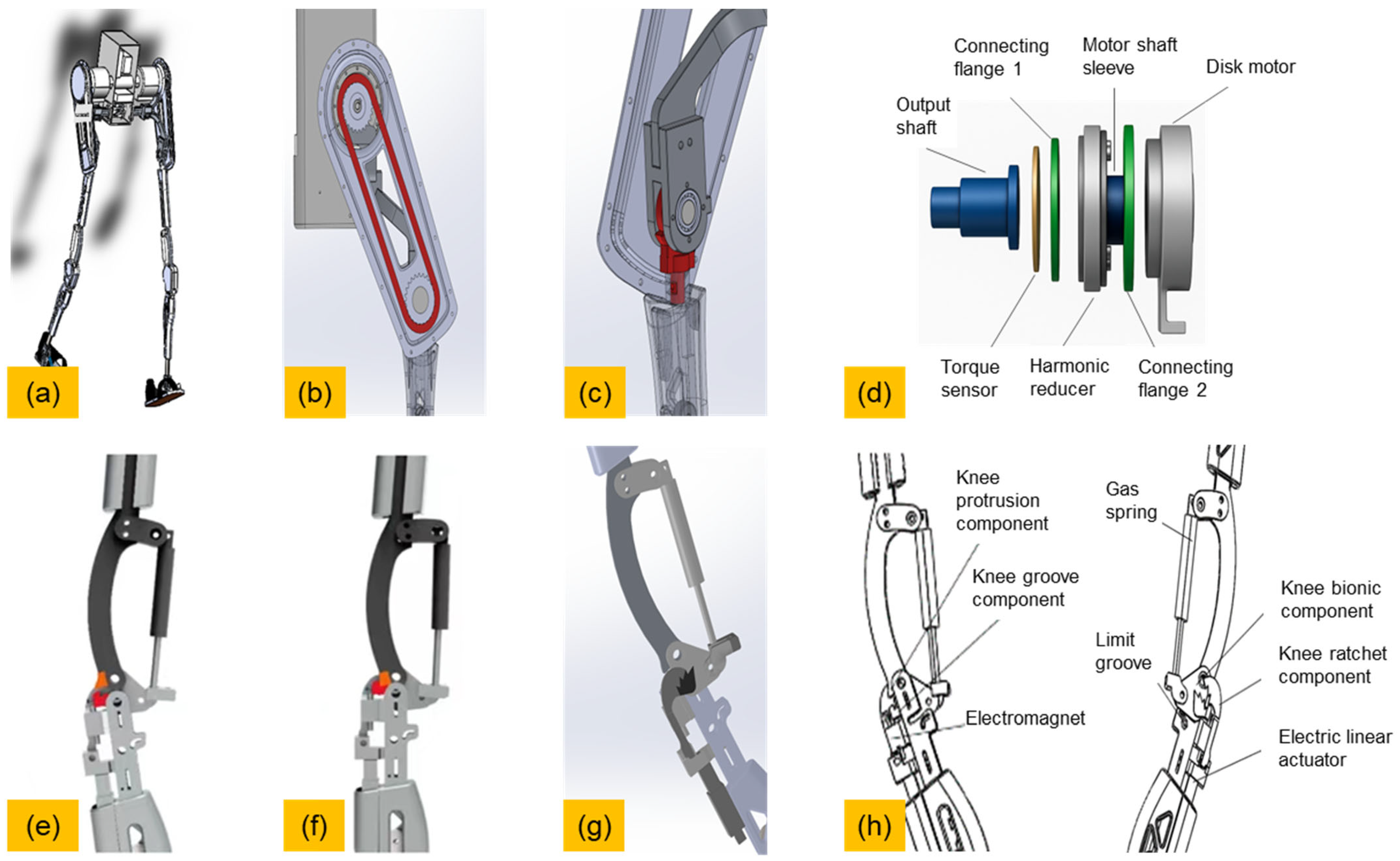
An overview of the exoskeleton’s structural design is shown in Figure 3a. The hip joint is actively actuated using a brushless DC motor (Maxon EC-i 40, 100 W, Rated voltage 24 V, Rated current 6.06 A, Stall current 70 A, Rated speed 2590 rpm, Rated torque 0.444 Nm, Stall torque 4.940 Nm; Maxon Motor AG, Sachseln, Switzerland). As illustrated in Figure 3d, the motor is coupled with a harmonic reducer and torque sensor, enabling controlled torque output. It is mounted on a posterior waist plate, and torque is transmitted to the thigh linkage through a high-efficiency belt and pulley system, as shown in Figure 3b,c. This setup allows smooth and controllable hip flexion–extension during walking.
The knee joint is non-actuated but includes an electromagnetically controlled mechanical latch for lock–unlock control. Under normal conditions, the latch remains engaged to keep the knee fully extended during stance (Figure 3f). When the control system detects the initiation of swing, the electromagnet is activated to disengage the latch (Figure 3e), allowing the knee to flex passively due to inertial forces. No motor is required at the knee. At the end of swing, as the leg returns to full extension, mechanical interference between the latch and its housing automatically re-engages the lock, ensuring rigid knee support for the next stance phase. We performed software-based modeling and simulations to analyze the relationship between knee unlocking and hip–knee coupling.
To facilitate sit-to-stand transitions, a gas spring module is installed between the thigh and shank segments, equipped with a ratchet mechanism controlled by a micro linear actuator (Figure 3g). During walking or seated postures, the ratchet remains disengaged and the spring inactive. Upon initiating a standing motion, the actuator locks the ratchet, allowing the gas spring to release stored energy and assist in hip and knee extension. During the descent phase of sitting, the ratchet absorbs the compressive energy, enabling a smooth and controlled sitting motion.
A complete mechanical layout of the knee joint assembly is provided in Figure 3h. All structural components are made from aluminum alloy to optimize strength-to-weight ratio. High-precision bearings are employed at each joint to reduce friction and ensure smooth, anatomically consistent motion. The design supports full sagittal-plane mobility of the lower limbs while preventing mechanical interference in the seated position [26,27].

2.3. Sensing and Gait Phase Recognition

The exoskeleton system adopts a compact and low-latency sensor configuration, with all sensors fully integrated into the exoskeleton structure, requiring no additional wearable modules for the user. This configuration enables real-time gait phase detection and seamless control synchronization during locomotion. The sensor layout is illustrated in Figure 4a.
Four types of sensors are integrated into the exoskeleton system to enable real-time gait detection and control coordination:
  • Inertial Measurement Units (IMUs)
  • Torque Sensors
  • Joint Angle Encoders
  • Foot Pressure Sensors
The torque and angle sensors serve primarily for safety monitoring and emergency braking. A high-sensitivity torque sensor (model M2210E;) is mounted at the hip joint to detect abrupt torque changes that may indicate unsafe conditions.
A total of seven IMUs (MPU6050) are used: one on the back, and three on each leg—placed on the thigh, shank, and foot sole. These IMUs measure the angular velocity and joint orientation of each segment, providing kinematic data for gait analysis and control.
The foot pressure sensing system uses 402-type resistive thin-film pressure sensors, selected for their flexibility and thin form factor. Due to the complex structure of the human foot and its pressure redistribution during a full gait cycle, pressure tends to concentrate at specific regions. Based on plantar pressure analysis, four key zones were identified for sensor placement. As shown in Figure 4c, these sensors monitor plantar pressure variation during gait and rehabilitation training.
To enhance usability and avoid discomfort, the sensors are not directly mounted on the skin but are instead embedded into custom insole positions that correspond to the four pressure regions. This design eliminates slippage during walking and enhances data consistency. Signal wires are routed to the acquisition module and processed via an STM32F103C8T6 microcontroller. The analog resistance signals are converted to voltage through signal conditioning circuits and then transmitted to a host computer for synchronized data acquisition and visualization. An example of the in-shoe pressure sensor installation is shown in Figure 4c.
All sensor signals are sampled at 100 Hz and processed on the embedded STM32F407 controller. A finite state machine (FSM) is implemented to classify the gait cycle into four discrete phases: 1. Heel Strike (HS); 2. Foot Flat (FF); 3. Heel Off (HO); 4. Swing, as shown in Figure 4b.
To classify gait phases based on plantar pressure, a threshold-based ratio analysis is applied using the signals from the four foot-mounted FSR sensors. Let p1, p2, p3 and p4 represent the normalized pressure contributions of sensors FSRA, FSRB, FSRC, and FSRD, respectively. These are defined as the proportion of each sensor’s signal relative to the total pressure sum at each sampling instance.
p i = F S R i 4 j = 1 F S R j , i = 1 , 2 , 3 , 4
FSR1, FSR2, FSR3, and FSR4 represent the pressure values at the first metatarsal head, second metatarsal head, third to fifth metatarsal heads, and heel, respectively, while P1, P2, P3, and P4 denote the corresponding weights of FSR1–FSR4.
Gait phase segmentation is determined by identifying the dominant pressure region across different phases, as shown in Figure 4d. For example:
  • During heel strike (HS), p4 (heel) dominates.
  • During foot flat (FF), all pi values are near equilibrium.
  • During swing, all pi values are zero.
  • During heel off (HO), p1 and p2 (forefoot) increases significantly.
When the FSM detects the Heel Off phase, it triggers two parallel actions: The hip motor is commanded to output flexion torque to assist leg swing; The knee electromagnet is energized to unlock the latch, allowing passive knee flexion.
After the leg swings forward and the knee joint reaches full extension, the FSM logic automatically cuts off power to the electromagnet. The mechanical latch re-engages passively, ensuring stability at the next stance.
The combination of plantar pressure and thigh IMU enables reliable, low-computation gait recognition without requiring vision or machine learning algorithms [28,29,30,31,32,33]. The overall detection-to-actuation delay is below 30 ms, ensuring effective synchronization between sensor input and actuator response.

2.4. Control Architecture

The control architecture of the exoskeleton is designed to deliver adaptive hip assistance and timely knee unlocking by synchronizing actuation with gait phase transitions. As shown in Figure 5a, the overall strategy integrates sensor feedback, phase detection, torque generation, and knee lock control into a real-time control loop.
A fuzzy-tuned PID controller regulates the brushless DC motor at the hip joint. This controller dynamically adjusts its proportional (Kp), integral (Ki), and derivative (Kd) gains based on: the error between desired and measured hip angle and the angular velocity of the hip joint [34,35,36,37]. The fuzzy inference system (FIS) employs a rule base and two-dimensional membership functions to adapt gain values according to error magnitude and rate of change. This allows compliant torque output during both stance and swing phases, accommodating variations in gait speed and user posture without manual tuning.
Theoretical joint angle curves (hip and knee) when using the exoskeleton are illustrated in Figure 5b, reflecting the coordinated motion expected from the fuzzy-assisted system. Knee unlocking is governed by the finite state machine (FSM), which classifies the gait cycle using foot pressure and thigh IMU data. Upon detecting the Heel Off phase, the FSM triggers two parallel actions: commands the hip motor to generate flexion torque; activates the knee electromagnet to disengage the mechanical latch and allow passive knee flexion.
After the leg swings forward and reaches full extension, the FSM automatically deactivates the electromagnet, and the latch passively re-engages—ensuring rigid support in the next stance phase. The knee unlocking timing is shown in Figure 5d.
The full FSM structure and task switching logic are presented in Figure 5c. The control program is implemented in embedded C and runs under FreeRTOS (v10.5.1), which enables the parallel execution of sensor acquisition, gait phase detection, fuzzy PID updates, motor control, and knee electromagnet switching.
Communication among sensors (IMUs, FSRs), actuators, and control units is handled via the CANopen protocol, ensuring low-latency and fault-tolerant data exchange. The sensing-to-actuation loop completes in under 30–40 ms, supporting reliable and responsive assistance during overground walking.

3. Results

3.1. Human Subject Evaluation

Before conducting human trials, a series of preliminary tests were carried out using a self-developed intelligent humanoid testing platform, as shown in Figure 6a. To evaluate system safety, we simulated spastic movements by programming the humanoid to apply low torques at the joints, and verified the system’s response under mild perturbations, as shown in Figure 6b. Subsequently, treadmill-based trials were performed to assess hip torque output and validate simulated gait patterns. Finally, human wearability experiments were conducted, as illustrated in Figure 6d–f, to evaluate user comfort and gait smoothness under assistive conditions. Users subjectively reported acceptable comfort and secure attachment during short trials.
Following these validations, a pilot study was conducted to preliminarily evaluate the proposed exoskeleton system on human subjects. Seven healthy adult participants (six males and one female, aged 22–30 years, BMI: 19.5–24.8) were recruited. All participants had no history of musculoskeletal disorders and provided written informed consent prior to testing. All procedures involving human participants were approved by the Medical Ethics Committee of Xinhua Hospital, Shanghai Jiao Tong University School of Medicine (XHEC-C-2024-202-1), and conducted in accordance with institutional ethical guidelines.
To evaluate the effectiveness of sit-to-stand assistance, we first conducted a dedicated experiment. Using the Noraxon motion capture and EMG analysis system, we collected kinematic and muscular data during the sit-to-stand transitions performed by healthy subjects, as shown in Figure 7a–c. Based on the recorded data, a musculoskeletal model was constructed in OpenSim 4.4, as illustrated in Figure 7d, to analyze joint motion and muscle activation patterns.
Simulation results revealed the timing and magnitude of assistance required during the transition, along with the hip–knee coupling characteristics needed for coordinated motion, as depicted in Figure 7h. These findings were used to inform the design of the control strategy for the exoskeleton.
Subsequently, we performed real-world wearability tests under sit-to-stand conditions, as shown in Figure 7e–g, to verify the system’s assistive performance and user comfort.
Subsequently, human walking experiments were conducted with participants wearing the exoskeleton to assess its assistive performance. During overground walking trials, surface electromyography (sEMG) and joint kinematic data were simultaneously collected using the Noraxon Trigno™ wireless system (Noraxon USA Inc., Scottsdale, AZ, USA). The system offers a sampling rate of 1000 Hz, a bandwidth of 20–450 Hz, and a transmission baud rate of 115,200 bps, ensuring high-fidelity signal acquisition with minimal noise or delay.
As illustrated in Figure 8a, the sEMG signals were processed through a standard pipeline that included bandpass filtering, full-wave rectification, and normalization to each subject’s maximum voluntary contraction (MVC). This yielded normalized muscle activation levels, which served as a quantitative metric for evaluating the exoskeleton’s assistance effectiveness.
Sensor placement on the body is shown in Figure 8b,c. Surface electromyography (sEMG) was recorded from five major lower-limb muscles on each participant, including the rectus femoris (RF), vastus medialis (VM), vastus lateralis (VL), semitendinosus (ST), and biceps femoris (BF).
Electrode placements followed SENIAM (Surface Electromyography for the Non-Invasive Assessment of Muscles) guidelines, and specific locations are illustrated in Figure 8d,e.
This setup enabled a comprehensive comparison of muscle activation patterns between assisted and unassisted walking conditions, facilitating objective assessment of muscle load reduction and gait efficiency improvements.

3.2. Exoskeleton Output Torque

As shown in Figure 9a, the measured hip joint torque generated by the exoskeleton reaches a peak value of 72 Nm, which closely approximates the typical torque required during normal human walking. The slight deviation from the theoretical target of 80 Nm can be attributed to factors such as joint friction, assembly tolerances, and elasticity in the belt transmission. Figure 9b shows the knee joint assistance torque provided by the gas spring during the sit-to-stand transition, achieving a peak value of 28 Nm, which is sufficient to support the user during ascent.

3.3. Gait Compliance

Figure 9c,d illustrates the kinematic trajectories of the hip and knee joints during assisted walking. The recorded trajectories closely match the reference values for human gait. Minor delays may be due to system communication latency. While the knee joint shows reduced range of motion during the locked stance phase, it demonstrates compliant motion during the unlocked swing phase. Some premature activation may result from the inertial effects of the exoskeleton mass. Figure 9e plots the torque-angle relationship of the hip joint, which aligns well with reference curves, verifying the effectiveness of the proposed fuzzy PID control strategy.

3.4. Evaluation of Assistive Effect

As shown in Figure 9f, a comparison of the normalized RMS surface EMG of the rectus femoris between baseline and assisted conditions revealed an average reduction of 5.3% when wearing the exoskeleton. Figure 9g further compares the normalized EMG profiles across the gait cycle. The EMG amplitude during the swing phase was consistently lower and delayed under the assisted condition, suggesting effective torque offloading by the exoskeleton. The relatively modest improvement may be attributed to participants’ unfamiliarity with the device and the fact that all subjects were healthy individuals without impairments.

4. Discussion

This study demonstrates that a hip-only actuated exoskeleton with passively locking knees can effectively reduce muscle activation levels and improve both gait stability and compliance, without the need for additional joint actuators. The proposed fuzzy PID controller exhibited strong adaptability across different individuals, enabling smooth assistive transitions without compromising user comfort [38,39].
The passive knee locking mechanism proved stable and reliable under all tested conditions. Unlike conventional actively powered knee joints, this design provides rigid support during the stance phase and compliant release during the swing phase—achieving low energy consumption and reduced mechanical complexity. In addition, the integrated gas spring–ratchet module further extends system functionality by assisting sit-to-stand transitions without interfering with normal gait dynamics [40].
These experimental findings highlight the feasibility and practical value of a “minimal actuation + mechanical intelligence” strategy for daily-use wearable robotic systems that support functional lower-limb mobility.
Although the total system weight (14.8 kg) offers advantages compared to similar exoskeletons, further weight reduction remains a priority. Future work will focus on replacing structural components with carbon fiber–aluminum composites, using more compact actuator modules, and optimizing joint modules for reduced mass. Additionally, we plan to include objective usability assessments such as NASA-TLX or the System Usability Scale (SUS) in upcoming clinical trials.
To strengthen safety validation, the next prototype iteration will integrate temperature sensors and apply Specific Absorption Rate (SAR) modeling to evaluate long-term electromagnetic exposure. For more comprehensive analysis of muscle behavior, future trials will use AI-assisted sEMG analysis (e.g., CNN-based fatigue classification and RNN-based gait prediction) [41,42,43]. We also acknowledge the limitations of FSR + IMU-based gait recognition and will compare it against state-of-the-art AI-driven models [44,45].
Uneven terrain introduces further control challenges. Future iterations will incorporate terrain-adaptive strategies through environmental sensing (e.g., terrain IMUs or depth cameras) and adaptive control logic. To better evaluate sit-to-stand performance, we will integrate non-invasive monitoring tools for both the user’s biomechanical response and structural state of the exoskeleton.

5. Conclusions

This paper presents and validates a lower limb exoskeleton system featuring single-hip actuation combined with an electromagnetically controlled passive knee joint. The design emphasizes lightweight structural integration, natural gait coordination, and effective sit-to-stand assistance for elderly users or individuals with limited mobility.
The system incorporates several core technical features:
  • Active hip actuation is achieved via a brushless DC motor that delivers assistive torque for hip flexion and extension;
  • The knee joint adopts a structurally passive design, integrated with an electromagnetically actuated locking mechanism. This allows active unlocking at the beginning of the swing phase for compliant motion, and automatic locking during stance to ensure joint stability;
  • The control system fuses plantar pressure data and inertial measurements to enable real-time gait phase recognition and synchronized fuzzy PID torque control;
  • An auxiliary gas spring–ratchet module is included to facilitate sit-to-stand transitions, without interfering with normal walking dynamics.
Preliminary experimental results confirm the following performance outcomes:
  • Significant reduction in lower-limb muscle loading, as evidenced by decreased surface electromyography (sEMG) activation levels;
  • Smooth and symmetric hip joint trajectories under assistive control;
  • Rapid control responsiveness with sensor-to-actuator delays consistently under 40 ms, enabling accurate synchronization with gait events;
  • Reliable passive locking behavior of the knee joint during dynamic overground walking.
By eliminating the need for active knee actuators and leveraging minimal actuation with intelligent mechanical design, the proposed exoskeleton strikes a balance between structural simplicity, energy efficiency, and user-friendliness. Future work will focus on extending trials to elderly populations, conducting long-duration wear tests, and developing adaptive control strategies for outdoor and uneven terrain locomotion.

Author Contributions

Conceptualization, M.L.; methodology, D.X.; software, M.L.; validation, M.L. and H.L.; data curation, Y.S.; writing—original draft preparation, M.L.; visualization, M.L.; supervision, R.K.Y.T.; project administration, H.Y. All authors have read and agreed to the published version of the manuscript.

Funding

This research was funded by the National Natural Science Foundation of China (62473263).

Data Availability Statement

The data presented in this study are available on request from the corresponding author. The data are not publicly available due to restrictions (they contain information that could compromise the privacy of research participants).

Acknowledgments

The authors have reviewed and edited the output and take full responsibility for the content of this publication.

Conflicts of Interest

The authors declare no conflicts of interest.

Abbreviations

The following abbreviations are used in this manuscript:
PIDProportional Integral Derivative
FSMFinite State Machine
DCDirect current
IMUInertial Measurement Unit
FSRForce Sensing Resistor
CGAClinical Gait Analysis
STM32STMicroelectronics 32-bit Series Microcontroller Chip
sEMGSurface Electromyography
RMSRoot Mean Square
MTHMetatarsal Head

References

  1. Yu, X.; Cai, Y.; Yang, R.; Ma, F.; Kim, W. Revisiting Sensor-Based Intelligent Fall Risk Assessment for Older People: A Systematic Review. Eng. Appl. Artif. Intell. 2025, 144, 110176. [Google Scholar] [CrossRef]
  2. Moon, S.W. Gait and Cognitive Function in the Elderly with AD Dementia, MCI and Healthy. Alzheimer’s Dement. 2024, 20, e083970. [Google Scholar] [CrossRef]
  3. Lai, X.; Bo, L.; Zhu, H.; Chen, B.; Wu, Z.; Du, H.; Huo, X. Effects of Lower Limb Resistance Exercise on Muscle Strength, Physical Fitness, and Metabolism in Pre-Frail Elderly Patients: A Randomized Controlled Trial. BMC Geriatr. 2021, 21, 447. [Google Scholar] [CrossRef] [PubMed]
  4. Parraca, J.A.; Adsuar, J.C.; Domínguez-Muñoz, F.J.; Barrios-Fernandez, S.; Tomas-Carus, P. Test-Retest Reliability of Isokinetic Strength Measurements in Lower Limbs in Elderly. Biology 2022, 11, 802. [Google Scholar] [CrossRef] [PubMed]
  5. Sitthiracha, P.; Eungpinichpong, W.; Chatchawan, U. Effect of Progressive Step Marching Exercise on Balance Ability in the Elderly: A Cluster Randomized Clinical Trial. Int. J. Environ. Res. Public Health 2021, 18, 3146. [Google Scholar] [CrossRef] [PubMed]
  6. Nishimura, T.; Imai, A.; Fujimoto, M.; Kurihara, T.; Kagawa, K.; Nagata, T.; Sanada, K. Adverse Effects of the Coexistence of Locomotive Syndrome and Sarcopenia on the Walking Ability and Performance of Activities of Daily Living in Japanese Elderly Females: A Cross-Sectional Study. J. Phys. Ther. Sci. 2020, 32, 227–232. [Google Scholar] [CrossRef]
  7. Romanato, M.; Spolaor, F.; Beretta, C.; Fichera, F.; Bertoldo, A.; Volpe, D.; Sawacha, Z. Quantitative Assessment of Training Effects Using EksoGT® Exoskeleton in Parkinson’s Disease Patients: A Randomized Single Blind Clinical Trial. Contemp. Clin. Trials Commun. 2022, 28, 100926. [Google Scholar] [CrossRef]
  8. Wiśniowska-Szurlej, A.; Wołoszyn, N.; Brożonowicz, J.; Ciąpała, G.; Pietryka, K.; Grzegorczyk, J.; Leszczak, J.; Ćwirlej-Sozańska, A.; Sozański, B.; Korczowski, B. Enhanced Rehabilitation Outcomes of Robotic-Assisted Gait Training with EksoNR Lower Extremity Exoskeleton in 19 Stroke Patients. Med. Sci. Monit. 2023, 29, e940511-1–e940511-10. [Google Scholar] [CrossRef]
  9. Tefertiller, C.; Hays, K.; Jones, J.; Jayaraman, A.; Hartigan, C.; Bushnik, T.; Forrest, G.F. Initial Outcomes from a Multicenter Study Utilizing the Indego Powered Exoskeleton in Spinal Cord Injury. Top. Spinal Cord. Inj. Rehabil. 2018, 24, 78–85. [Google Scholar] [CrossRef]
  10. Awad, L.N.; Esquenazi, A.; Francisco, G.E.; Nolan, K.J.; Jayaraman, A. The ReWalk ReStoreTM Soft Robotic Exosuit: A Multi-Site Clinical Trial of the Safety, Reliability, and Feasibility of Exosuit-Augmented Post-Stroke Gait Rehabilitation. J. Neuroeng. Rehabil. 2020, 17, 80. [Google Scholar] [CrossRef]
  11. Wang, S.; Zhang, X.; Zheng, J.; Chen, G.; Jiao, G.; Peng, S. Integration of Spinal Musculoskeletal System Parameters for Predicting OVCF in the Elderly: A Comprehensive Predictive Model. Glob. Spine J. 2024, 15, 1966–1975. [Google Scholar] [CrossRef] [PubMed]
  12. Nasr, A.; Zhu, K.; McPhee, J. Using Musculoskeletal Models to Generate Physically-Consistent Data for 3D Human Pose, Kinematic, Dynamic, and Muscle Estimation. Multibody Syst. Dyn. 2024, 64, 737–770. [Google Scholar] [CrossRef]
  13. Chiu, K.I.A.; Taylor, C.; Saha, P.; Geddes, J.; Bishop, T.; Bernard, J.; Lui, D. Actively Controlled Exoskeletons Show Improved Function and Neuroplasticity Compared to Passive Control: A Systematic Review. Glob. Spine J. 2025, 21925682251343529. [Google Scholar] [CrossRef]
  14. Zhang, Y.; Zhao, W.; Wan, C.; Wu, X.; Huang, J.; Wang, X.; Huang, G.; Ding, W.; Chen, Y.; Yang, J.; et al. Exoskeleton Rehabilitation Robot Training for Balance and Lower Limb Function in Sub-Acute Stroke Patients: A Pilot, Randomized Controlled Trial. J. Neuroeng. Rehabil. 2024, 21, 98. [Google Scholar] [CrossRef]
  15. Ma, Y.; Liu, D.; Yan, Z.; Yu, L.; Gui, L.; Yang, C.; Yang, W. Optimizing Exoskeleton Assistance: Muscle Synergy-Based Actuation for Personalized Hip Exoskeleton Control. Actuators 2024, 13, 54. [Google Scholar] [CrossRef]
  16. Kim, Y.-S. Effects of a Walking Program Using the “WalkON” Mobile App among College Students. Digit. Health 2024, 10, 20552076241269463. [Google Scholar] [CrossRef]
  17. Grimmer, M.; Zhao, G. Hip Exoskeleton for Cycling Assistance. Bioengineering 2024, 11, 683. [Google Scholar] [CrossRef]
  18. Shushtari, M.; Arami, A. Human–Exoskeleton Interaction Force Estimation in Indego Exoskeleton. Robotics 2023, 12, 66. [Google Scholar] [CrossRef]
  19. Fang, Q.; Xu, T.; Zheng, T.; Cai, H.; Zhao, J.; Zhu, Y. A Rehabilitation Training Interactive Method for Lower Limb Exoskeleton Robot. Math. Probl. Eng. 2022, 2022, 2429832. [Google Scholar] [CrossRef]
  20. Changcheng, C.; Li, Y.-R.; Chen, C.-T. Assistive Mobility Control of a Robotic Hip-Knee Exoskeleton for Gait Training. Sensors 2022, 22, 5045. [Google Scholar] [CrossRef]
  21. Haufe, F.L.; Kober, A.M.; Wolf, P.; Riener, R.; Xiloyannis, M. Learning to Walk with a Wearable Robot in 880 Simple Steps: A Pilot Study on Motor Adaptation. J. Neuroeng. Rehabil. 2021, 18, 157. [Google Scholar] [CrossRef]
  22. Kunyou, H.; Lumin, C. Research of Fuzzy PID Control for Lower Limb Wearable Exoskeleton Robot. In Proceedings of the 2021 4th International Conference on Intelligent Autonomous Systems (ICoIAS), Wuhan, China, 14–16 May 2021; IEEE: Piscataway, NJ, USA; pp. 385–389. [Google Scholar]
  23. Li, C.; He, Y.; Chen, T.; Chen, X.; Tian, S. Real-Time Gait Event Detection for a Lower Extremity Exoskeleton Robot by Infrared Distance Sensors. IEEE Sens. J. 2021, 21, 27116–27123. [Google Scholar] [CrossRef]
  24. Zhang, Y.-P.; Cao, G.-Z.; Li, L.-L.; Diao, D.-F. Interactive Control of Lower Limb Exoskeleton Robots: A Review. IEEE Sens. J. 2024, 24, 5759–5784. [Google Scholar] [CrossRef]
  25. Pérez-Ibarra, J.C.; Siqueira, A.A.G.; Krebs, H.I. Identification of Gait Events in Healthy Subjects and With Parkinson’s Disease Using Inertial Sensors: An Adaptive Unsupervised Learning Approach. IEEE Trans. Neural Syst. Rehabil. Eng. 2020, 28, 2933–2943. [Google Scholar] [CrossRef]
  26. Moeller, T.; Beyerlein, M.; Herzog, M.; Barisch-Fritz, B.; Marquardt, C.; Dežman, M.; Mombaur, K.; Asfour, T.; Woll, A.; Stein, T.; et al. Human Motor Performance Assessment with Lower Limb Exoskeletons as a Potential Strategy to Support Healthy Aging—A Perspective Article. Prog. Biomed. Eng. 2025, 7, 013001. [Google Scholar] [CrossRef]
  27. Awad, L.N.; Bae, J.; O’Donnell, K.; De Rossi, S.M.M.; Hendron, K.; Sloot, L.H.; Kudzia, P.; Allen, S.; Holt, K.G.; Ellis, T.D.; et al. A Soft Robotic Exosuit Improves Walking in Patients after Stroke. Sci. Transl. Med. 2017, 9, eaai9084. [Google Scholar] [CrossRef]
  28. Delfi, G.; Al Bochi, A.; Dutta, T. A Scoping Review on Minimum Foot Clearance Measurement: Sensing Modalities. Int. J. Environ. Res. Public Health 2021, 18, 10848. [Google Scholar] [CrossRef]
  29. Shahini, N.; Bahrami, Z.; Sheykhivand, S.; Marandi, S.; Danishvar, M.; Danishvar, S.; Roosta, Y. Automatically Identified EEG Signals of Movement Intention Based on CNN Network (End-To-End). Electronics 2022, 11, 3297. [Google Scholar] [CrossRef]
  30. Chaisaen, R.; Autthasan, P.; Mingchinda, N.; Leelaarporn, P.; Kunaseth, N.; Tammajarung, S.; Manoonpong, P.; Mukhopadhyay, S.C.; Wilaiprasitporn, T. Decoding EEG Rhythms During Action Observation, Motor Imagery, and Execution for Standing and Sitting. IEEE Sens. J. 2020, 20, 13776–13786. [Google Scholar] [CrossRef]
  31. Gurve, D.; Delisle-Rodriguez, D.; Romero-Laiseca, M.; Cardoso, V.; Loterio, F.; Bastos, T.; Krishnan, S. Subject-Specific EEG Channel Selection Using Non-Negative Matrix Factorization for Lower-Limb Motor Imagery Recognition. J. Neural Eng. 2020, 17, 026029. [Google Scholar] [CrossRef] [PubMed]
  32. Sun, Z.; Zhang, X.; Liu, K.; Shi, T.; Wang, J. A Multi-Joint Continuous Motion Estimation Method of Lower Limb Using Least Squares Support Vector Machine and Zeroing Neural Network Based on sEMG Signals. Neural Process Lett. 2023, 55, 2867–2884. [Google Scholar] [CrossRef]
  33. Sun, T.; Hu, Q.; Gulati, P.; Atashzar, S.F. Temporal Dilation of Deep LSTM for Agile Decoding of sEMG: Application in Prediction of Upper-Limb Motor Intention in NeuroRobotics. IEEE Robot. Autom. Lett. 2021, 6, 6212–6219. [Google Scholar] [CrossRef]
  34. Cao, W.; Ma, Y.; Chen, C.; Zhang, J.; Wu, X. Hardware Circuits Design and Performance Evaluation of a Soft Lower Limb Exoskeleton. IEEE Trans. Biomed. Circuits Syst. 2022, 16, 384–394. [Google Scholar] [CrossRef]
  35. Bajpai, A.; Carrasquillo, C.; Carlson, J.; Park, J.; Iyengar, D.; Herrin, K.; Young, A.J.; Mazumdar, A. Design and Validation of a Versatile High Torque Quasidirect Drive Hip Exoskeleton. IEEE/ASME Trans. Mechatron. 2024, 29, 789–797. [Google Scholar] [CrossRef]
  36. Wu, B.; Lv, J.; Chen, X.; Fang, S.; Pan, W.; Qiang, L.; Song, D. Design and Performance Evaluation Method of Wearable Hip Joint Power-Assisted Exoskeleton. Biomed. Signal Process. Control 2025, 101, 107229. [Google Scholar] [CrossRef]
  37. Cao, W.; Zhang, Z.; Chen, C.; He, Y.; Wang, D.; Wu, X. Biomechanical and Physiological Evaluation of a Multi-Joint Exoskeleton with Active-Passive Assistance for Walking. Biosensors 2021, 11, 393. [Google Scholar] [CrossRef] [PubMed]
  38. Zhong, B.; Shen, M.; Liu, H.; Zhao, Y.; Qian, Q.; Wang, W.; Yu, H.; Zhang, M. A Cable-Driven Exoskeleton with Personalized Assistance Improves the Gait Metrics of People in Subacute Stroke. IEEE Trans. Neural Syst. Rehabil. Eng. 2023, 31, 2560–2569. [Google Scholar] [CrossRef]
  39. Jeong, Y.J.; Kazerooni, H. Design of Low Profile Actuators for Medical Exoskeletons. In Proceedings of the ASME 2015 International Mechanical Engineering Congress and Exposition, Houston, TX, USA, 13–19 November 2015; American Society of Mechanical Engineers: New York, NY, USA, 2015; p. V003T03A094. [Google Scholar]
  40. Tiwari, R.S.; Das, T.K.; Tripathy, A.K.; Li, K.-C. Gait Identification Based on Deepwalk Features Using CNN and LSTM: An Advanced Biometric Approach. Telecommun. Syst. 2025, 88, 83. [Google Scholar] [CrossRef]
  41. Vidyarani, K.R.; Talasila, V.; Umar, R.; Prem, V.; Jagannath, R.P.K.; Prashanth, G.R. Inertial Sensor-Based Heel Strike and Energy Expenditure Prediction Using a Hybrid Machine Learning Approach. Digit. Health 2025, 11, 20552076251333375. [Google Scholar] [CrossRef]
  42. Luo, M.; Yin, M.; Li, J.; Li, Y.; Kobsiriphat, W.; Yu, H.; Xu, T.; Wu, X.; Cao, W. Lateral Walking Gait Recognition and Hip Angle Prediction Using a Dual-Task Learning Framework. Cyborg Bionic Syst. 2025, 6, 0250. [Google Scholar] [CrossRef]
  43. Laganà, F.; Pellicanò, D.; Arruzzo, M.; Pratticò, D.; Pullano, S.A.; Fiorillo, A.S. FEM-Based Modelling and AI-Enhanced Monitoring System for Upper Limb Rehabilitation. Electronics 2025, 14, 2268. [Google Scholar] [CrossRef]
  44. Lin, Y.; Zhang, Y.; Zhong, W.; Xiong, W.; Xi, Z.; Chen, Y.-F.; Zhang, M. Leveraging Extended Windows in End-to-End Deep Learning for Improved Continuous Myoelectric Locomotion Prediction. IEEE Trans. Neural Syst. Rehabil. Eng. 2025, 33, 1170–1179. [Google Scholar] [CrossRef] [PubMed]
  45. Zhang, Z.; Li, X.; Ma, W.; Gao, S. Piezoresistive Plantar Pressure Sensors and CNN-Based Body Weight and Load Estimation. Med. Biol. Eng. Comput. 2025; ahead of print. [Google Scholar] [CrossRef]
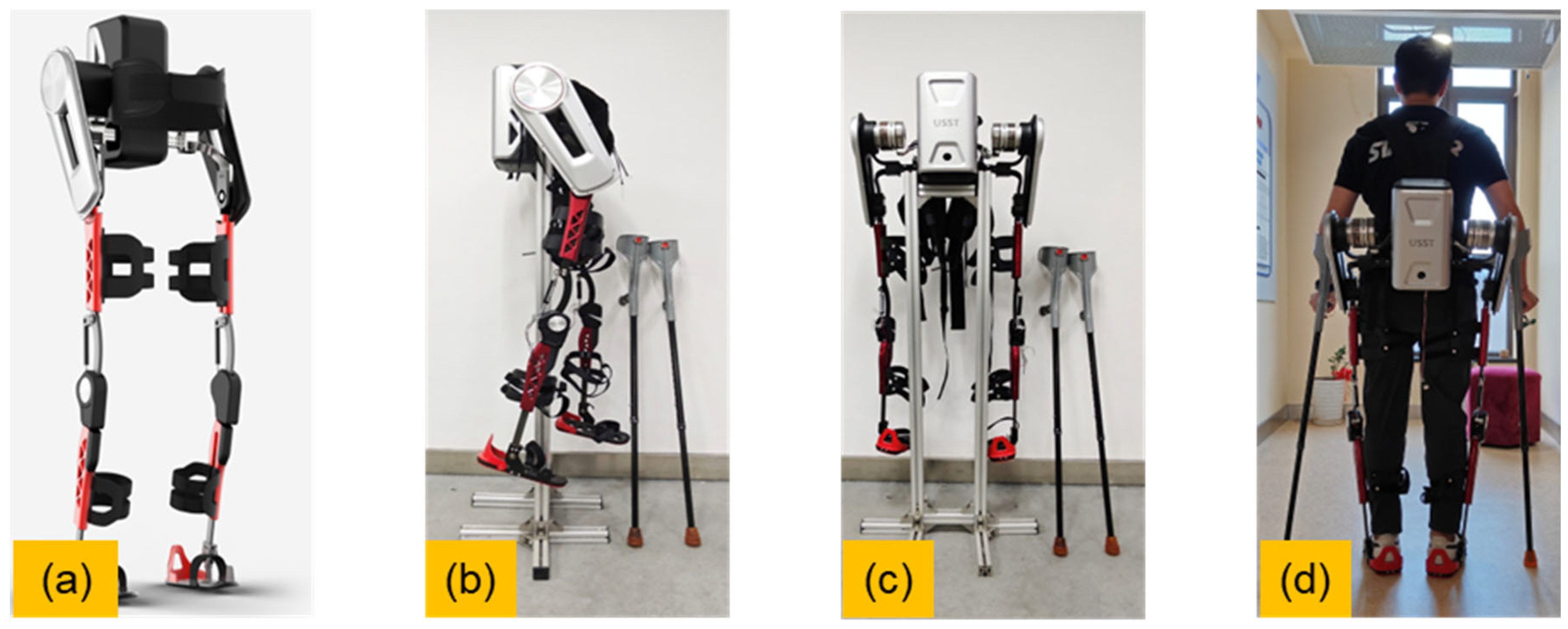
Figure 1. External view of the exoskeleton system. (a) CAD-based design rendering; (b) Side view of the physical prototype; (c) Rear view of the prototype; (d) Standing posture while worn.
Electronics 14 03853 g001
Figure 2. The adjustable modular structure of the exoskeleton. Including hip width adjustment, thigh length adjustment, and calf length adjustment. The degrees of freedom of the hip, knee and ankle.
Electronics 14 03853 g002
Figure 3. Mechanical structure and component layout of the hip–knee coordinated exoskeleton system. (a) Overall model of the exoskeleton; (b) Belt-driven hip joint transmission; (c) Hip joint limiting mechanism; (d) Exploded view of the hip drive assembly: output shaft, torque sensor, harmonic reducer, motor shaft sleeve, disk motor, and connecting flanges; (eg) Knee joint locking/unlocking mechanisms: (e) shows the unlocked state of the knee joint, where the red and orange components are disengaged, allowing free knee flexion; (f) shows the locked state of the knee joint, where the red and orange components are engaged, keeping the knee extended and fixed; (h) Schematic of knee bionic structure.
Electronics 14 03853 g003
Figure 4. Sensor distribution. (a) Arrangement of multiple sensors on the human–exoskeleton system, including the inertial sensor (MPU6050; TDK InvenSense, Inc., San Jose, CA, USA), torque sensor (M2210E; Interface, Inc., Scottsdale, AZ, USA), angle sensor (LF53464TMR; Littelfuse, Inc., Chicago, IL, USA), and foot pressure sensor (FSR402; Interlink Electronics, Inc., Irvine, CA, USA); (b) Gait phase diagram indicating heel strike, foot flat, and heel off stages; (c) Schematic layout of foot pressure sensor locations at the first metatarsal head (1st MTH), second metatarsal head (2nd MTH), third to fifth metatarsal heads (3rd–5th MTH), and heel; (d) Gait phase recognition based on foot pressure ratio algorithm.
Electronics 14 03853 g004
Figure 5. Hip–knee coupled gait control strategy and trajectory planning. (a) Closed-loop control framework combining gait phase recognition, learning-based trajectory generation, gravity/friction compensation, impedance modulation, and guidance; (b) Comparison of hip and knee joint trajectories during CGA-based walking and modified knee trajectory when wearing the exoskeleton; (c) Finite-state machine transitions for gait phase control; (d) Joint angle trajectories of bilateral hips and bilateral knees during two gait cycles of walking when wearing the exoskeleton. Shaded regions indicate the knee unlocking intervals: gold for the left knee and silver for the right knee.
Electronics 14 03853 g005
Figure 6. Safety validation and gait testing of the exoskeleton system. (a) Intelligent dummy combined with a body-weight support system for safety validation; (b) Gait testing experiment using an intelligent dummy on a treadmill platform; (c) Safety performance test simulating spastic movement using the intelligent dummy; (df) Walking trials with a healthy subject using crutches while wearing the exoskeleton.
Electronics 14 03853 g006
Figure 7. Sit-to-stand motion analysis and assistance experiment. (ac) Marker-based motion capture of sit-to-stand task by a healthy subject; (d) Biomechanical simulation of sit-to-stand transition across multiple time frames; (eg) Exoskeleton-assisted sit-to-stand experiment using the proposed hip-knee coordination system; (h) Joint angle variation of hip and knee during the sit-to-stand motion, with phase division and state changes of knee actuator and electromagnet.
Electronics 14 03853 g007
Figure 8. EMG and Kinematic Signal Acquisition. (a) Workflow of the gait data acquisition system using the Noraxon platform to collect surface EMG (red arrows) and joint kinematic data (blue arrows). Signals are amplified, filtered, and rectified before feature extraction and fusion analysis. The system enables comparative evaluation of assistance effects before and after exoskeleton usage; (b,c) Front and rear views of the subject showing placement of surface EMG electrodes and inertial measurement units (IMUs); (d,e) Anatomical mapping of muscles targeted by the electrodes, including the rectus femoris, vastus medialis, vastus lateralis, biceps femoris, and semitendinosus.
Electronics 14 03853 g008
Figure 9. Experimental data of the lower-limb exoskeleton. (a) Measured output torque of the hip motor; (b) Measured torque provided by the knee gas spring; (c) Comparison between the measured hip joint angle and CGA data; (d) Comparison between the measured knee joint angle and CGA data; (e) Relationship between hip joint angle and torque; (f) Comparison of normalized RMS EMG before and after wearing the exoskeleton; (g) Comparison of normalized EMG waveform before and after wearing the exoskeleton.
Electronics 14 03853 g009
Table 1. Lower Limb Anthropometric Dimensions: Males (16–60 years).
Height (mm)Hip Width (mm)Thigh Length (mm)Calf Length (mm)Percentile (%)
15452734143241
15882824273385
160828843634510
168330646637050
175532749539790
177633450540395
181534652142099
Table 2. Lower Limb Anthropometric Dimensions: Females (16–60 years).
Height (mm)Hip Width (mm)Thigh Length (mm)Calf Length (mm)Percentile (%)
14492753873001
14812904023135
150329641031910
147031743834450
164034046737090
165934647637695
169736049439099
Disclaimer/Publisher’s Note: The statements, opinions and data contained in all publications are solely those of the individual author(s) and contributor(s) and not of MDPI and/or the editor(s). MDPI and/or the editor(s) disclaim responsibility for any injury to people or property resulting from any ideas, methods, instructions or products referred to in the content.